Maternal immune activation evoked by polyinosinic:polycytidylic acid does not evoke microglial cell...
14
ORIGINAL RESEARCH published: 05 August 2015 doi: 10.3389/fncel.2015.00301 Edited by: Takahiro A. Kato, Kyushu University, Japan Reviewed by: Andrew MacLean, Tulane University School of Medicine, USA Manabu Makinodan, Nara Medical University, Japan *Correspondence: Bert Brône, BIOMED – Hasselt University, Martelarenlaan 42, 3500 Hasselt, Belgium [email protected] † These authors have contributed equally to this work. Received: 24 March 2015 Accepted: 22 July 2015 Published: 05 August 2015 Citation: Smolders S, Smolders SMT, Swinnen N, Gärtner A, Rigo J-M, Legendre P and Brône B (2015) Maternal immune activation evoked by polyinosinic:polycytidylic acid does not evoke microglial cell activation in the embryo. Front. Cell. Neurosci. 9:301. doi: 10.3389/fncel.2015.00301 Maternal immune activation evoked by polyinosinic:polycytidylic acid does not evoke microglial cell activation in the embryo Silke Smolders 1,2† , Sophie M. T. Smolders 1,3,4,5† , Nina Swinnen 1 , Annette Gärtner 2 , Jean-Michel Rigo 1† , Pascal Legendre 3,4,5† and Bert Brône 1 * † 1 BIOMED – Hasselt University, Hasselt, Belgium, 2 Laboratory of Neuronal Differentiation, VIB Center for the Biology of Disease, Leuven and Center for Human Genetics, KU Leuven, Leuven, Belgium, 3 INSERM, UMR S 1130, Université Pierre et Marie Curie, Paris, France, 4 CNRS, UMR 8246, Université Pierre et Marie Curie, Paris, France, 5 UM 119 NPS, Université Pierre et Marie Curie, Paris, France Several studies have indicated that inflammation during pregnancy increases the risk for the development of neuropsychiatric disorders in the offspring. Morphological brain abnormalities combined with deviations in the inflammatory status of the brain can be observed in patients of both autism and schizophrenia. It was shown that acute infection can induce changes in maternal cytokine levels which in turn are suggested to affect fetal brain development and increase the risk on the development of neuropsychiatric disorders in the offspring. Animal models of maternal immune activation reproduce the etiology of neurodevelopmental disorders such as schizophrenia and autism. In this study the poly (I:C) model was used to mimic viral immune activation in pregnant mice in order to assess the activation status of fetal microglia in these developmental disorders. Because microglia are the resident immune cells of the brain they were expected to be activated due to the inflammatory stimulus. Microglial cell density and activation level in the fetal cortex and hippocampus were determined. Despite the presence of a systemic inflammation in the pregnant mice, there was no significant difference in fetal microglial cell density or immunohistochemically determined activation level between the control and inflammation group. These data indicate that activation of the fetal microglial cells is not likely to be responsible for the inflammation induced deficits in the offspring in this model. Keywords: neuropsychiatric disorders, maternal immune activation, microglia, embryo, cortex Introduction Schizophrenia and autism are neurodevelopmental disorders that can arise early during postnatal life. Although genetic deficits are important risk factors, perturbations of local environment, especially during pregnancy, are suspected to play a central role in the occurrence of these neurodevelopmental disorders. Maternal immune activation (MIA) during pregnancy is considered as a risk factor for schizophrenia and autism in the offspring (Brown, 2012). To study the mechanisms behind this association several animal models were developed in which pregnant rodents were infected with the influenza virus, polyinosinic:polycytidylic acid [poly (I:C)] or Frontiers in Cellular Neuroscience | www.frontiersin.org 1 August 2015 | Volume 9 | Article 301
Transcript of Maternal immune activation evoked by polyinosinic:polycytidylic acid does not evoke microglial cell...

ORIGINAL RESEARCHpublished: 05 August 2015
doi: 10.3389/fncel.2015.00301
Edited by:Takahiro A. Kato,
Kyushu University, Japan
Reviewed by:Andrew MacLean,
Tulane University School of Medicine,USA
Manabu Makinodan,Nara Medical University, Japan
*Correspondence:Bert Brône,
BIOMED – Hasselt University,Martelarenlaan 42, 3500 Hasselt,
†These authors have contributedequally to this work.
Received: 24 March 2015Accepted: 22 July 2015
Published: 05 August 2015
Citation:Smolders S, Smolders SMT,
Swinnen N, Gärtner A, Rigo J-M,Legendre P and Brône B (2015)
Maternal immune activation evokedby polyinosinic:polycytidylic acid does
not evoke microglial cell activationin the embryo.
Front. Cell. Neurosci. 9:301.doi: 10.3389/fncel.2015.00301
Maternal immune activation evokedby polyinosinic:polycytidylic aciddoes not evoke microglial cellactivation in the embryoSilke Smolders1,2†, Sophie M. T. Smolders1,3,4,5† , Nina Swinnen1, Annette Gärtner2 ,Jean-Michel Rigo1†, Pascal Legendre3,4,5† and Bert Brône1*†
1 BIOMED – Hasselt University, Hasselt, Belgium, 2 Laboratory of Neuronal Differentiation, VIB Center for the Biology ofDisease, Leuven and Center for Human Genetics, KU Leuven, Leuven, Belgium, 3 INSERM, UMR S 1130, Université Pierreet Marie Curie, Paris, France, 4 CNRS, UMR 8246, Université Pierre et Marie Curie, Paris, France, 5 UM 119 NPS, UniversitéPierre et Marie Curie, Paris, France
Several studies have indicated that inflammation during pregnancy increases the riskfor the development of neuropsychiatric disorders in the offspring. Morphological brainabnormalities combined with deviations in the inflammatory status of the brain can beobserved in patients of both autism and schizophrenia. It was shown that acute infectioncan induce changes in maternal cytokine levels which in turn are suggested to affectfetal brain development and increase the risk on the development of neuropsychiatricdisorders in the offspring. Animal models of maternal immune activation reproduce theetiology of neurodevelopmental disorders such as schizophrenia and autism. In thisstudy the poly (I:C) model was used to mimic viral immune activation in pregnant mice inorder to assess the activation status of fetal microglia in these developmental disorders.Because microglia are the resident immune cells of the brain they were expected to beactivated due to the inflammatory stimulus. Microglial cell density and activation level inthe fetal cortex and hippocampus were determined. Despite the presence of a systemicinflammation in the pregnant mice, there was no significant difference in fetal microglialcell density or immunohistochemically determined activation level between the controland inflammation group. These data indicate that activation of the fetal microglial cellsis not likely to be responsible for the inflammation induced deficits in the offspring in thismodel.
Keywords: neuropsychiatric disorders, maternal immune activation, microglia, embryo, cortex
Introduction
Schizophrenia and autism are neurodevelopmental disorders that can arise early during postnatallife. Although genetic deficits are important risk factors, perturbations of local environment,especially during pregnancy, are suspected to play a central role in the occurrence ofthese neurodevelopmental disorders. Maternal immune activation (MIA) during pregnancy isconsidered as a risk factor for schizophrenia and autism in the offspring (Brown, 2012). To studythe mechanisms behind this association several animal models were developed in which pregnantrodents were infected with the influenza virus, polyinosinic:polycytidylic acid [poly (I:C)] or
Frontiers in Cellular Neuroscience | www.frontiersin.org 1 August 2015 | Volume 9 | Article 301
Smolders et al. Immune activation and embryonic microglia
lipopolysaccharide (LPS) (Patterson, 2009). These modelsconfirmed that prenatal infection leading to MIA can lead tobehavioral and neurological disorders in the offspring (Shi et al.,2003; Meyer et al., 2006; Fortier et al., 2007; Lowe et al., 2008;Harvey and Boksa, 2012; Giovanoli et al., 2013; Squarzoni et al.,2014). During MIA evoked by poly (I:C), an elevated maternalserum cytokine, interleukin-6 (IL-6), was found to be critical forthe development of these neurological deficits in the offspring(Samuelsson et al., 2006; Smith et al., 2007). Differences inbehavioral abnormalities observed in the offspring at adult age arecritically dependent on the time of maternal poly (I:C) challenge,being related to differences in cytokine responses in the fetal brainshortly after the induction of MIA (Meyer et al., 2006, 2008).However, the source of the cytokine response in the fetal brainremains a matter of debate as it can originate from maternal,placental and/or embryonic tissue. An endogenous increase infetal brain cytokine production was demonstrated using mRNAanalysis of the cytokine expression level upon maternal poly (I:C)challenge during the late gestation stage in mice (17 embryonicdays, E17; Meyer et al., 2006). This was not observed whenmaternal poly (I:C) challenge was performed at mid gestationstage (E9; Meyer et al., 2006), a developmental age at whichimmature microglia, the resident immune cells of the brain, havenot yet invaded the fetal central nervous system (CNS; Ginhouxet al., 2010; Rigato et al., 2011; Swinnen et al., 2013).
Microglia colonize the brain early during embryonicdevelopment (E11.5 in the mouse embryo; Ginhoux et al., 2010;Rigato et al., 2011; Swinnen et al., 2013) and are known tocontrol several developmental processes in the brain at perinataldevelopmental stages (Cunningham et al., 2013; Squarzoni et al.,2014; Michell-Robinson et al., 2015). First, embryonic microgliahave been shown to be involved in angiogenesis through closecontact with vessel sprouts and endothelial tip cells and thesecretion of soluble factors that stimulate angiogenesis duringdevelopment (Fantin et al., 2010; Rymo et al., 2011). Secondly,during CNS development microglial cells clear cellular debrisand induce programmed cell death in developing neurons viathe production of superoxide ions (Marin-Teva et al., 2004;Wakselman et al., 2008) and tumor necrosis factor (TNF)-α(Sedel et al., 2004). Thirdly, several studies have pointed towardan important role for microglia in synaptic remodeling andsynapse elimination (Tremblay et al., 2010; Paolicelli et al.,2011; Schafer et al., 2012; Zhan et al., 2014). Finally, microglialcells can also influence the development and differentiationof neural cells. Microglia-conditioned media can influenceembryonic precursor migration and differentiation in primarycultures (Aarum et al., 2003; Jonakait et al., 2011). In addition,microglial cells can regulate cortical precursor proliferationand astrogenesis (Nakanishi et al., 2007). Primary cultureexperiments on embryonic precursor cultures showed thatmicroglial cells are important for precursor proliferation andastrogenesis. In microglia-depleted cultures and cultures fromPU.1 knock out embryos proliferation and astrogenesis weredecreased. Addition of microglia to these cultures restoredboth processes and an abnormal increase in microglial cellnumbers resulted in increased astrogenesis (Antony et al.,2011). Deactivation of embryonic microglia with tetracyclines
or elimination via the macrophages suicide technique led to anincrease in neural precursor cells, while microglial activation hadthe opposite effect (Cunningham et al., 2013).
Maternal immune activation induces an imbalance incytokines levels, of which maternal IL-6 has been shown tobe a critical mediator in inducing the effects of MIA onbrain development and behavioral changes (Smith et al., 2007).IL-6 is known to induce activation of adult microglial cells;leading to the production of pro-inflammatory factors, such asnitric oxide, reactive oxygen species, proteolytic enzymes, andTNF-α by microglial cell cultures (Krady et al., 2008), microglialproliferation (in vitro; Streit et al., 2000) and infiltration(in vivo; Lacroix et al., 2002) or the upregulation of microglialCX3CR1, making them more sensitive to fractalkine signaling(Lee et al., 2010). An imbalance in cytokine levels causedby MIA might thus be able to activate embryonic microglia,even at early developmental stages, and alter their normalfunctions. This can trigger a cascade of events that couldlead to developmental defects observed in the offspring ofLPS or poly (I:C) treated pregnant mice. Indeed, MIA evokedby LPS injection evoked microglia activation and enhancedphagocytosis of neural precursors by microglia at prenatalstages in rats (Cunningham et al., 2013). However, the questionremains whether an endogenous increase in fetal brain cytokineproduction in response to maternal poly (I:C) challenge isof microglial origin. Accordingly it remains unclear whetherpoly (I:C)-induced MIA results in the activation of embryonicmicroglia during fetal development.
To determine to what extent MIA evoked by poly (I:C) canalter cortex invasion by microglia and/or change embryonicmicroglial cell activation state, we evoked MIA using a single(at E11.5) or a double injection (at E11.5 and E15.5) of poly(I:C) (Meyer et al., 2006; Shi et al., 2009). This developmentaltime window is an important time point for cortex invasion byimmature microglia as their cell density dramatically increasesduring this period (Swinnen et al., 2013). We show thatpoly (I:C)-induced MIA does not affect microglial density andactivation level during embryonic development suggesting thatpathological activation of embryonic microglial cells at theonset of their colonization processes cannot explain neurologicaldeficits observed at postnatal stages in offspring after poly (I:C)-induced MIA.
Materials and Methods
AnimalsAll experiments were conducted in accordance with theEuropean Community guiding principles on the care and useof animals and with the approval of the Ethical Committee onAnimal Research of Hasselt University. Mice were maintained inthe animal facility of the Hasselt University in accordance withthe guidelines of the Belgian Law and the European CouncilDirective. To visualize microglia in the embryonic cortex thetransgenic CX3CR1-eGFP knock-in mice (Jung et al., 2000) wereused. The heterozygous CX3CR1-eGFP embryos used in thisstudy were obtained by crossing wild type C57BL/6 females with
Frontiers in Cellular Neuroscience | www.frontiersin.org 2 August 2015 | Volume 9 | Article 301
Smolders et al. Immune activation and embryonic microglia
homozygous CX3CR1-eGFP +/+ male mice (obtained from theEuropean Mouse Mutant Archive – EMMA with the approvalof Jung et al., 2000). The day of conception was designated asembryonic day 0.5 (E0.5).
Maternal Immune ActivationAt day E11.5 (single injection) or at E11.5 and E15.5 (doubleinjection) mice received i.p. a dose of poly (I:C) (20 mg/kg;Polyinosinic–polycytidylic acid potassium salt; Sigma–Aldrich,Bornem, Belgium) or vehicle (saline). Five hours after injectionthe maternal blood was collected, the serum was aliquoted andstored at −80◦C until the IL-6 assay was performed (Shi et al.,2003; Smith et al., 2007). The maternal IL-6 concentrationswere determined using the Mouse IL-6 ELISA Kit from ThermoScientific (Rockford, IL, USA), following the manufacturer’sinstructions. The analysis was conducted using a FLUOstarOPTIMA plate reader (BMG Labtech, Ortenberg, Germany).
Fluorescent Immunostaining of EmbryonicBrainsPregnant mice were sacrificed and embryonic tissue processedas described before (Swinnen et al., 2013). The heads of E11.5and E12.5 embryos were fixed in 4% paraformaldehyde for 3 hat 4◦C and 5 h for E17.5 embryos. After fixation, the embryonicheads were cryoprotected overnight in phosphate-buffered saline(PBS) + 30% sucrose, frozen in optimal cutting temperaturecompound (Tissue-Tek) and stored at −80◦C until sectioned.Ten micrometer-thick coronal tissue sections were cut on a LeicaCM1900 uv cryostat, mounted on Superfrost Plus glasses andstored at −20◦C until staining.
To check whether embryonic microglia can be directlyactivated by poly (I:C), IL-6 or LPS, 300-μm thick coronal brainslices (E15.5) were cultured for 24 h with either saline, poly(I:C) (50 μg/ml), IL-6 (10 ng/ml), or LPS (1 μg/ml). To thisend, pregnant mothers were euthanized at E15.5. Embryonicbrains were isolated in ice-cold PBS-glucose (pH 7.4; 25 mM),embedded in 3% low melting agarose (Fisher Scientific) andsliced coronally at a thickness of 300 μm using a MicromHM650V Vibrating Blade Microtome. Slices were mounted onMilliCell organotypic inserts (Millipore) and maintained in semi-hydrous conditions at 37◦C and 5% CO2 for 24 h. The mediaconsisted of Neurobasal medium supplemented with 2 mM L-glutamine, B27 supplement, N2 supplement, and 0.5% penicillin–streptomycin (all from Invitrogen) with either saline, poly (I:C)(50 μg/ml), IL-6 (10 ng/ml) or LPS (1 μg/ml) added. Afterwardslices were fixed for 1 h in 4% PFA and cryoprotected overnightin PBS + 30% sucrose, frozen in optimal cutting temperaturecompound (Tissue-Tek) and stored at −80◦C until sectioned.Ten micrometer-thick coronal tissue sections were cut on a LeicaCM1900 uv cryostat, mounted on Superfrost Plus glasses andstored at −20◦C until staining.
In order to determine the activation state of the microglia,we used antibodies against interleukin (IL)-1β, inducible nitricoxide synthase (iNOS) and Mac-2/Galectin-3 (Rigato et al., 2011;Cunningham et al., 2013). All primary antibodies and workingsolutions are listed in Table 1.
TABLE 1 | Overview of the antibodies used for immunostainings and flowcytometry experiments.
Antibody Company Reference Dilution
Immunohistochemistry
Anti-IL1β (rabbitpolycl.)
Abcam ab9722 1:100
Anti-iNOS (rabbitpolycl.)
Abcam ab15323 1:250
Anti-Mac-2 (ratmonocl.)
American typeculture collection
TIB-166 1:250
Flow cytometry
Anti-IL1β PE (ratmonocl.)
LifeSpanBioSciences
LS-C184791 1:300
Anti-iNOS PE-Cy7(rat monocl.)
eBioscience 25-5920 1:300
Anti-Mac-2 PE (ratmonocl.)
eBioscience 12-5301 1:300
Isolation of Microglia and Flow CytometryExperimentsBrains were isolated from CX3CR1-eGFP E17.5 embryos frommothers subjected to a single saline or poly (I:C) injection onE11.5, or a double poly (I:C) injection on E11.5 and E15.5.All steps occurred at 4◦C or on ice, unless stated otherwise,to avoid microglia activation. Meninges were removed, thecortical area identical to the immunohistochemical analysiswas dissected out and incubated during 30 min at 30◦C inDMEM/F-12(1:1) + GlutaMAX (Life Technologies) containing48 U/ml Papain from papaya latex (Sigma). Papain containingsupernatants was discarded and the tissue was mechanicallydisrupted in medium through fast pipetting using a 1 ml pipet.Afterward, the homogenate was centrifuged at 400g during5 min, resuspended in 40% isotonic Percoll (GE Healthcare)and centrifuged at 700g during 10 min without break. Thepellet was resuspended in PBS and filtered through a 35 μmcell strainer. Cell suspensions were fixed and permeablizedin Cytofix/Cytoperm buffer (BD Cytofix/CytopermTM PlusFixation/Permeabilization Kit, BD Biosciences) during 20 minon ice, washed and incubated on ice for 30 min in Perm/Washbuffer with a mix of fluorochrome-conjugated rat anti-mouseantibodies: iNOS-PE-Cy7 (clone CXNFT, eBioscience), Mac-2-PE (clone eBioM3/38, eBioscience) and, IL1β-PE (clone 11n92,LifeSpan BioSciences) (Table 1). The following isotype controlswere used: Rat IgG2aκ PE-Cy7, Rat IgG2aκ PE and RatIgG2b PE (all from eBioscience). After washes, cells wereresuspended in FACS buffer (PBS, 2% FCS, sodium azide),acquired in a FACS Aria II and analyzed with FACS Diva6.1.3 software (BD Biosciences). Isotype-marker overlay graphswere created in FlowJo 10.0.8 Software. Inside the singletpopulation, the eGFP positive microglia (1000–12000 cells perexperiment) were gated (Figure 5A), and within this population,the percentage of Mac-2, iNOS, and IL1β positive microgliawas analyzed. Isotype controls were used to gate the positivecell population (Figure 5B). Per group, embryos were derivedfrom one to three different mothers (saline, single poly (I:C),double poly(I:C)). BV-2 cells (Supplementary Data) were used
Frontiers in Cellular Neuroscience | www.frontiersin.org 3 August 2015 | Volume 9 | Article 301
Smolders et al. Immune activation and embryonic microglia
as positive controls for the different antibodies (SupplementaryFigure S1).
Analysis and StatisticsQuantitative analysis of microglial cells was performed on imagesof coronal embryonic brain sections. We focused our analysis onthe cerebral cortex area located dorsally to the lateral ganglioniceminences (LGE) and medial ganglionic eminences (MGE),containing the frontal and pariental cortex on E11.5 and E12.5,and the somatosensory and motor cortex at E17.5. This regionof the cortex is well characterized on the functional and cellularlevel and the two GE structures are the major sources of corticalinterneurons during embryonic neurogenesis (Tan et al., 1998;Anderson et al., 2001). For the quantifications of the hippocampalarea at E17.5 only the dorsal hippocampus was included in theanalysis.
Images were taken with a Nikon Eclipse 80i microscopeand a Nikon digital sight camera DS-2MBWc [10x Nikon planobjective (numerical aperture (NA) of 0.25) and a 20x Plan Fluorobjective (NA of 0.5)]. Images (1600 × 1200) were analyzedwith ImageJ 1.45e software (NIH, USA; http://rsb.info.nih.gov/ij/). Only eGFP-positive cell bodies were taken into account forthe measurements. Density analysis was performed by countingthe number of eGFP positive cell bodies per mm2 (Swinnenet al., 2013). For analysis of activation state we calculated thepercentage of the eGFP positive cells that were also showingimmunoreactivity for the activation marker. All values areexpressed as mean ± SEM. The number of sections used isindicated as n, the number of embryos or blood samples asN; # sections/# embryos is thus designated in the text as n/N.Statistical significance was assessed by non-parametric Mann–Whitney test or Kruskal–Wallis test, P-values smaller than 0.05were considered significant.
Results
An increase in IL-6 level in the maternal blood is a crucialfactor in the development of MIA-induced deficits and changesobserved in the offspring (Smith et al., 2007). To control that thepoly (I:C) injection procedure we used evoked an increase in IL-6 level in the maternal blood, we analyzed the IL-6 level in thematernal serum samples 5 hours after injection of either saline orpoly (I:C). We found a significant increase (P < 0.0001; Mann–Whitney test) in the level of IL-6 in the sera of femalemice primedwith poly (I:C) (1876 ± 389.2 pg/ml, N = 22) when comparedto those injected with saline (14.8 ± 3.3 pg/ml, N = 26), thusindicating that themice in the poly (I:C) group effectively sufferedfrom a systemic immune response.
In response to brain injury, microglia proliferate and shiftto beneficial or detrimental activation states depending on thelocal environment. When activated, microglial cells adopt aphagocytic phenotype in order to clear dying cells (Kettenmannet al., 2011). In pathological conditions, such as in the mousemodel of LPS-induced MIA, phagocytosis of neuronal precursorcells by microglia was also increased, which resulted in adecrease in the size of the precursor cell pool in the cerebral
cortex (Cunningham et al., 2013). It must also be noted thatmicroglial disturbances were also observed in patients sufferingfrom autistic or schizophrenic disorders. Microglial activationhas been observed in the brains of autistic (Vargas et al., 2005;Morgan et al., 2010) and schizophrenic patients (Radewiczet al., 2000; Wierzba-Bobrowicz et al., 2005; Monji et al.,2013). Recent studies also indicated that there is an increasein microglial density in different brain regions in the adultpoly (I:C) MIA offspring (Juckel et al., 2011; Ratnayake et al.,2012).
To determine if poly (I:C)-evoked MIA alters the embryonicmicroglial cell colonization process in the fetal brain wecompared cell density after single injection of poly (I:C), doubleinjection of poly (I:C) or saline treatments, in the cortex atE11.5, E12.5 and at E17.5 (single injection) or at E17.5 (doubleinjections) and in the hippocampal area at E17.5 (single anddouble injections). At all ages tested we did not find anysignificant difference in microglia cell density (Mann–Whitneytest; P > 0.05, for detailed P-values see Table 2) in the cortexor in the hippocampus after a single or after double injections(Figure 1; Table 2), thus suggesting that poly (I:C)-evoked MIAdoes not alter early invasion of the cortex and the hippocampusby microglial cells in the embryo.
To determine if MIA induced a change inmicroglial activationlevel after a single poly (I:C) injection (E11.5), we performedan immunostaining for three different activation markers: Mac-2/Galectin-3, iNOS and IL1β at E11.5 and E17.5.Mac-2/Galectin-3 is a marker of microglial phagocytic activation state (Dumicet al., 2006; Rotshenker, 2009) while iNOS and IL1β aremarkers of a cytotoxic activation state (Cunningham et al.,2013). At E11.5 none of the microglia located in the cortex wasimmmunopositive for Mac-2 staining both after saline injection
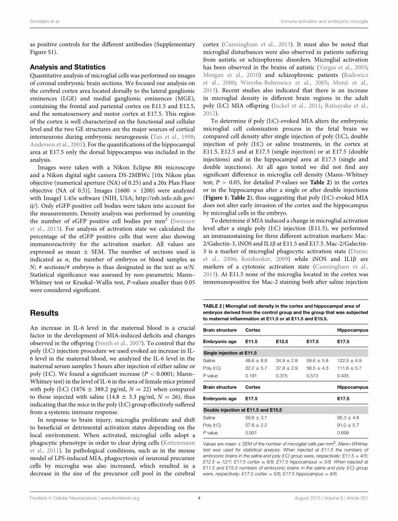
TABLE 2 | Microglial cell density in the cortex and hippocampal area ofembryos derived from the control group and the group that was subjectedto maternal inflammation at E11.5 or at E11.5 and E15.5.
Brain structure Cortex Hippocampus
Embryonic age E11.5 E12.5 E17.5 E17.5
Single injection at E11.5
Saline 48.6 ± 8.8 34.9 ± 2.8 59.6 ± 5.8 122.5 ± 4.9
Poly (I:C) 32.2 ± 5.7 37.8 ± 2.9 56.5 ± 4.3 111.6 ± 5.7
P value 0.191 0.375 0.573 0.435
Brain structure Cortex Hippocampus
Embryonic age E17.5 E17.5
Double injection at E11.5 and E15.5
Saline 59.8 ± 3.1 95.3 ± 4.8
Poly (I:C) 57.8 ± 2.2 91.0 ± 5.7
P value 0.931 0.699
Values are mean ± SEM of the number of microglial cells per mm2, Mann–Whitneytest was used for statistical analysis. When injected at E11.5 the numbers ofembryonic brains in the saline and poly (I:C) group were, respectively: E11.5 = 4/5;E12.5 = 12/7; E17.5 cortex = 6/8; E17.5 hippocampus = 5/8. When injected atE11.5 and E15.5 numbers of embryonic brains in the saline and poly (I:C) groupwere, respectively: E17.5 cortex = 5/6; E17.5 hippocampus = 6/6.
Frontiers in Cellular Neuroscience | www.frontiersin.org 4 August 2015 | Volume 9 | Article 301

Smolders et al. Immune activation and embryonic microglia
FIGURE 1 | Embryonic microglial cell density is not increased aftersingle and double injection of poly (I:C). Microglial cell density in the cortexand hippocampal area was not affected after poly (I:C)-induced MIA. Valuesare mean ± SEM of the number of microglial cells per mm2, Mann–Whitneytest was used for statistical analysis. When injected at E11.5 the numbers ofembryonic brains in the saline and poly (I:C) group were, respectively:E11.5 = 4/5; E12.5 = 12/7; E17.5 cortex = 6/8; E17.5 hippocampus = 5/8.When injected at E11.5 and E15.5 numbers of embryonic brains in the salineand poly (I:C) group were, respectively: E17.5 cortex = 5/6; E17.5hippocampus = 6/6. c, cortex; h, hippocampal area; D, double injection.
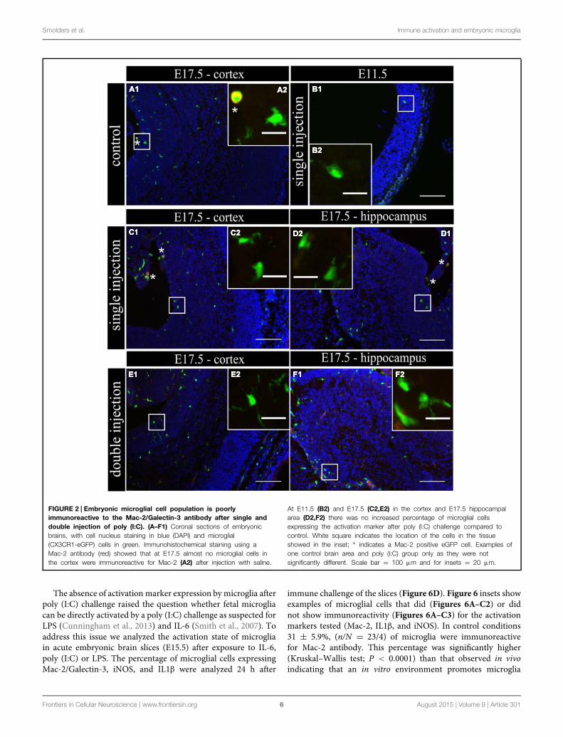
(n/N = 14/3) and after poly (I:C) challenge (n/N = 18/3;Figure 2B1,B2). At E17.5, 2.5 ± 0.5% (n/N = 38/4) of themicroglia in the cortex (Figure 2A1,A2) and 3.2 ± 0.7%(n/N = 27/4) of the microglia in the hippocampal areaexpressed Mac-2 after saline injection. We did not find anysignificant difference (Kruskal–Wallis test; P = 0.448) afterpoly (I:C) challenge. After poly (I:C) challenge, 1.9 ± 0.7%(n/N = 23/4) of the microglia in the cortex and 2.5 ± 1%(n/N = 15/4) of microglia in hippocampal area expressed Mac-2 (Figures 2C1,C2,D1,D2). We next investigated the expressionof IL1β and iNOS (Cunningham et al., 2013) to determineif embryonic microglia can adopt a cytotoxic activation stateafter a single injection of poly (I:C). Induction of MIA by asingle injection of poly (I:C) did not result in a significantincrease in the percentage of microglia expressing IL1β eitherat E11.5 and E17.5 (Kruskal–Wallis test; P = 0.136). In controlconditions, 0 ± 0% (n/N = 6/3) and 2.2 ± 1% (n/N = 15/4)of microglia located in the cortex expressed IL1β at E11.5and E17.5 (Figure 3A1,A2), respectively, while 3.1 ± 1.3%(n/N = 17/4) expressed IL1β in the hippocampal area (E17.5).After poly (I:C) challenge, 3.3 ± 3.3% (n/N = 10/3) and 3.5 ± 1%(n/N = 19/4) of microglia located in the cortex expressedIL1β at E11.5 (Figure 3B1,B2) and at E17.5 (Figure 3C1,C2),respectively, while 7.2 ± 2.6% (n/N = 17/4) expressed IL1βin the hippocampal area (E17.5; Figure 3D1,D2). We foundsimilar results when analyzing iNOS expression at E11.5 andE17.5 in the cortex and in the hippocampal area (E17.5). CorticaliNOS expression in control conditions [E11.5: 8.3 ± 5.7%,n/N = 10/3; E17.5: 2.0 ± 1.1%, n/N = 15/4 (Figure 4A1,A2)]was not significantly different when compared to the poly (I:C)condition [E11.5: 0 ± 0%, n/N = 8/3 (Figure 4B1,B2); E17.5:1.9 ± 1.1%, n/N = 12/4 (Figure 4C1,C2; Kruskal–Wallis test;P = 0.471)]. In the hippocampal area, 1.5 ± 1.0% of microglia(n/N = 14/4) express iNOS in control conditions while 0 ± 0%,
of microglia (n/N = 10/4) express iNOS after poly (I:C) challenge(Figure 4D1,D2, being not significantly different (Kruskal–Wallis test; P = 0.471)).
This lack of change in embryonic microglia activationstate after a single poly (I:C) injection could possibly leadonly to a “primed” microglial state. Indeed, two injections ofLPS were necessary in rat to elicit MIA induced microgliadysfunction during phagocytosis of cortical neural precursorcells (Cunningham et al., 2013), suggesting that the microglialphenotype could become only fully altered after the secondinflammatory challenge. To determine if this is also the casefor poly (I:C) we reanalyzed microglial density and activationlevel after a repeated injection of poly (I:C). Consequently, themothers suffered from a double immune stimulation (on E11.5as well as on E15.5). Despite the presence of a maternal immuneresponse after both injections, there was no significant increasein microglial cell density (Mann–Whitney test; P > 0.05, fordetailed P-values see Table 2) (Figure 1; Table 2). Microglialactivation states were analyzed at E17.5 as described above.We did not find any significant difference (Kruskal–Wallis test;Mac-2, P = 0.139; IL1β, P = 0.945; iNOS, P = 0.093) in thepercentage of microglia expressingMac-2, IL1β, or iNOS betweencontrol conditions and after double injections of poly (I:C).After double injections of poly (I:C) the percentage of microgliaimmunoreactive for Mac-2 antibody was 0 ± 0% (n/N = 29/6) inthe cortex (Figure 2E1,E2) and 2.0 ± 0.7% (n/N = 22/6) in thehippocampal area (Figure 2F1,F2). In the cortex (Figure 3E1,E2)and hippocampal area (Figure 3F1,F2) 1.4 ± 0.7% (n/N = 34/6)and 1.4 ± 1.0% (n/N = 25/6) of the microglial cells showedimmunoreactivity for the IL1β antibody, while 1.8 ± 0.7%(n/N = 34/6) and 0 ± 0% (n/N = 23/6) of the microglia werepositive for iNOS in the cortex (Figure 4E1,E2) and hippocampalarea (Figure 4F1,F2), respectively. These results indicate thateven double injections of poly (I:C) did not evoke microgliaactivation in the embryo.
In addition to the immunohistochemical stainings, thepresence of the activation markers on microglial cells at E17.5was investigated by flow cytometry. The gating strategy andpositive controls are shown in Figures 5A,B and SupplementaryFigure S1. The results of the flow cytometric quantifications weresimilar to those obtained by immunohistochemistry. There wasno significant difference in the proportion of microglial cellsthat were positive for Mac-2 after single poly (I:C) injection(16.8± 0.0%; N= 10) or double poly (I:C) injection (27.0± 4.6%;N = 10) when compared to the control group (15.5 ± 4.3; N = 5;Figure 5C, left; Kruskal–Wallis test, P = 0.161). The proportionof microglial cells that were positive for IL1β in the control group(14.2 ± 3.1%; N = 10) was not significantly different (Figure 5C,middle; Kruskal–Wallis test, P = 0.093) to the percentage ofmicroglia that was positive for IL1β after a single (22.3 ± 3.9%;N = 8) or double poly (I:C) injection (16.0 ± 2.1%; N = 6). Thepercentage of microglial cells positive for iNOS in the controlgroup was 9.1 ± 2.8% (N = 5). There was no significant effect(Figure 5C, right; Kruskal–Wallis test, P = 0.816) of a singlepoly (I:C) (7.1 ± 1.5%; N = 10) or double poly (I:C) challenge(9.9 ± 2.7%; N = 10) on the percentage of microglia expressingthis marker.
Frontiers in Cellular Neuroscience | www.frontiersin.org 5 August 2015 | Volume 9 | Article 301
Smolders et al. Immune activation and embryonic microglia
FIGURE 2 | Embryonic microglial cell population is poorlyimmunoreactive to the Mac-2/Galectin-3 antibody after single anddouble injection of poly (I:C). (A–F1) Coronal sections of embryonicbrains, with cell nucleus staining in blue (DAPI) and microglial(CX3CR1-eGFP) cells in green. Immunohistochemical staining using aMac-2 antibody (red) showed that at E17.5 almost no microglial cells inthe cortex were immunoreactive for Mac-2 (A2) after injection with saline.
At E11.5 (B2) and E17.5 (C2,E2) in the cortex and E17.5 hippocampalarea (D2,F2) there was no increased percentage of microglial cellsexpressing the activation marker after poly (I:C) challenge compared tocontrol. White square indicates the location of the cells in the tissueshowed in the inset; ∗ indicates a Mac-2 positive eGFP cell. Examples ofone control brain area and poly (I:C) group only as they were notsignificantly different. Scale bar = 100 μm and for insets = 20 μm.
The absence of activation marker expression by microglia afterpoly (I:C) challenge raised the question whether fetal microgliacan be directly activated by a poly (I:C) challenge as suspected forLPS (Cunningham et al., 2013) and IL-6 (Smith et al., 2007). Toaddress this issue we analyzed the activation state of microgliain acute embryonic brain slices (E15.5) after exposure to IL-6,poly (I:C) or LPS. The percentage of microglial cells expressingMac-2/Galectin-3, iNOS, and IL1β were analyzed 24 h after
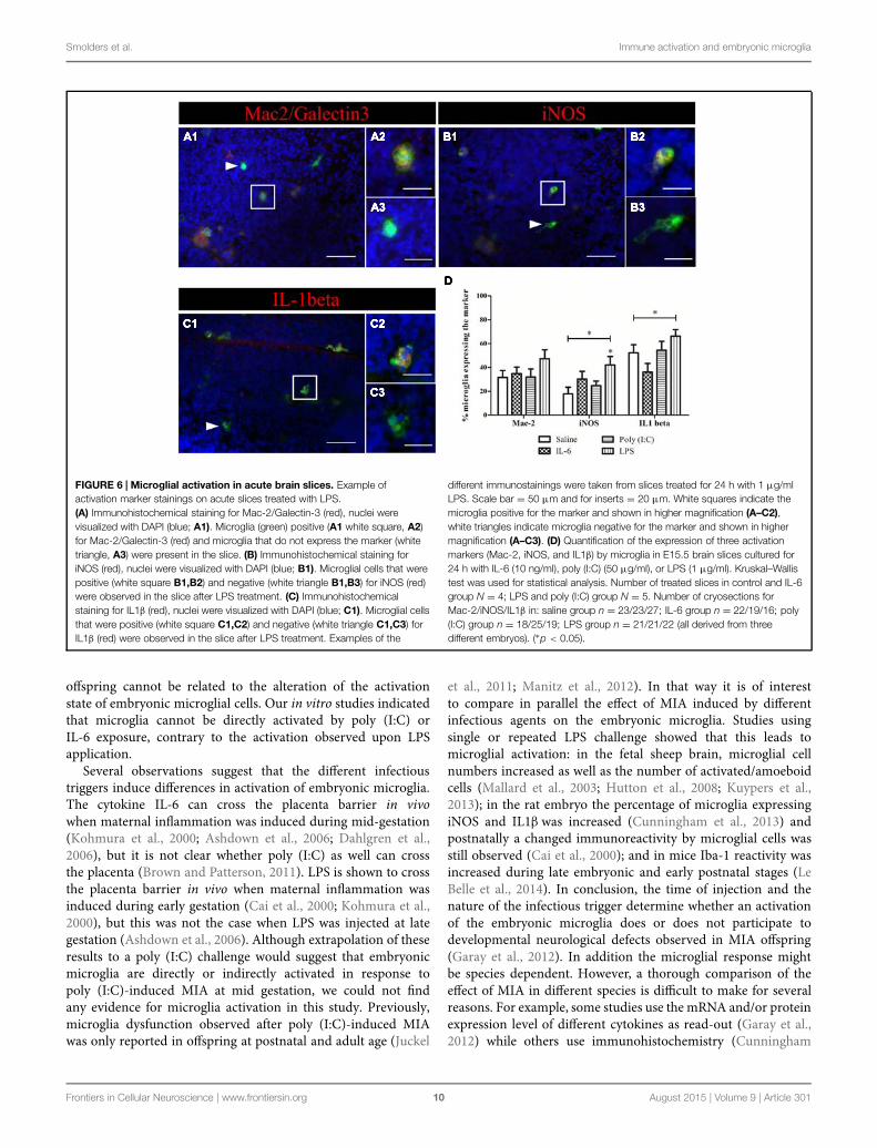
immune challenge of the slices (Figure 6D). Figure 6 insets showexamples of microglial cells that did (Figures 6A–C2) or didnot show immunoreactivity (Figures 6A–C3) for the activationmarkers tested (Mac-2, IL1β, and iNOS). In control conditions31 ± 5.9%, (n/N = 23/4) of microglia were immunoreactivefor Mac-2 antibody. This percentage was significantly higher(Kruskal–Wallis test; P < 0.0001) than that observed in vivoindicating that an in vitro environment promotes microglia
Frontiers in Cellular Neuroscience | www.frontiersin.org 6 August 2015 | Volume 9 | Article 301

Smolders et al. Immune activation and embryonic microglia
FIGURE 3 | Embryonic microglia show no increased expression of IL1β
after single and double injection of poly (I:C). (A–F1) Coronal sections ofembryonic brains, with cell nucleus staining in blue (DAPI) and microglial(CX3CR1-eGFP) cells in green. Immunohistochemical staining using an IL1β
antibody (red) showed that at E17.5 almost no microglial cells in the cortex wereimmunoreactive for IL1β (A2) after injection with saline. At E11.5 (B2) and E17.5
(C2,E2) in the cortex and E17.5 hippocampal area (D2,F2) there was noincreased percentage of microglial cells expressing the activation marker afterpoly (I:C) challenge compared to control. White square indicates the location ofthe cells in the tissue showed in the inset; ∗ indicates an IL1β positive eGFP cell.Examples of one control brain area and poly (I:C) group only as they were notsignificantly different. Scale bar = 100 μm and for insets = 20 μm.
phagocytic activation state. However, there was no significanteffect (Kruskal–Wallis test; P = 0.274) of IL-6, poly (I:C) or LPStreatment on the percentage of microglia being immunoreactiveto Mac-2 antibody (Figure 6D), being 34 ± 5.5% (n/N = 22/4)after IL-6 exposure, 32 ± 6.7%, (n/N = 18/5) after poly (I:C)exposure and 47 ± 7.5% (n/N = 21/5) after LPS exposure(Figure 6D). As observed for Mac-2, the percentage of IL1βimmunoreactive microglia was significantly higher than in in vivoconditions [in control conditions 52 ± 6.8%, (n/N = 27/4;
Kruskal–Wallis test; P < 0.0001)] and for iNOS a trend to ahigher percentage was observed under control conditions [incontrol conditions 18 ± 5.7%, (n/N = 23/4; Kruskal–Wallis test;P = 0.091)]. As shown in Figure 6D treatment with IL-6 or poly(I:C) did not significantly change the percentage of microgliaimmunoreactive for IL1β or iNOS antibodies. When looking atIL1β immunoreactivity, 36 ± 7.2% (n/N = 16/4) of the microgliawas positive after IL-6 exposure and 54 ± 7.5%, (n/N = 19/5)after poly (I:C) exposure (Figure 6D). For iNOS they were
Frontiers in Cellular Neuroscience | www.frontiersin.org 7 August 2015 | Volume 9 | Article 301

Smolders et al. Immune activation and embryonic microglia
FIGURE 4 | Embryonic microglia cell population is poorlyimmunoreactive to the iNOS antibody after single and doubleinjection of poly (I:C). (A–F1) Coronal sections of embryonic brains,with cell nucleus staining in blue (DAPI) and microglial(CX3CR1-eGFP) cells in green. Immunohistochemical staining using aniNOS antibody (red) showed that at E17.5 almost no microglial cellsin the cortex were immunoreactive for iNOS (A2) after injection with
saline. At E11.5 (B2) and E17.5 (C2,E2) in the cortex and E17.5hippocampal area (D2,F2) there was no increased percentage ofmicroglial cells expressing the activation marker after poly (I:C)challenge compared to control. White square indicates the location ofthe cells in the tissue showed in the inset. Examples of one controlbrain area and poly (I:C) group only as they were not significantlydifferent. Scale bar = 100 μm and for insets = 20 μm.
30 ± 6.5% (n/N = 19/4) after IL-6 exposure and 25 ± 3.9%,(n/N = 25/5) after poly (I:C) exposure (Figure 6D). However,we found that LPS, contrary to IL-6 or poly (I:C), can directlyactivate microglia to a detrimental activation state. Indeed LPSexposure significantly increased the percentage of microgliaimmunoreactive for IL1β (Kruskal–Wallis test; P = 0.025) oriNOS antibodies (Kruskal–Wallis test; P= 0.025). In the presenceof LPS 66 ± 5.5 (n/N = 22/5) and 42 ± 7.1% (n/N = 21/5)of microglia were immunoreactive for IL1β antibody or iNOSantibody, respectively.
Discussion
Maternal immune activation-induced behavioral andneurological alterations observed in the offspring at juvenile andadult stages in animals are supposed to be correlated with theetiology of neuropsychiatric disorders in humans. Our studyin mice demonstrates, for the first time, that MIA evoked bysingle or double poly (I:C) injections does not change microgliadensity and their activation state in the embryo in vivo. Thissuggests that the behavioral and neurological alterations in the
Frontiers in Cellular Neuroscience | www.frontiersin.org 8 August 2015 | Volume 9 | Article 301

Smolders et al. Immune activation and embryonic microglia
FIGURE 5 | Flow cytometry reveals that embryonic microglial cellsshow a poor expression of activation markers Mac-2, IL1β andiNOS. (A) Gating strategies for the microglial cells. In the wholeembryonic cortex cell suspension, a gate was created on the non-debrispopulation (left). Inside this population, single cells were selected (middle)and within this population, the microglial cells were gated based onCX3CR1-eGFP intensity (right). SSC, Side scatter; FSC, Forward scatter.(B) Gating strategies for positive Mac-2, iNOS and IL1β populations.Microglial cell count of representative samples is shown for Mac-2 (left),IL1β (middle) and iNOS (right; full lines) for embryos derived from saline,single poly (I:C) and double poly (I:C) injected mothers. Gates forpositive populations were drawn based on the isotype fluorescence
intensity (dotted lines). FI, fluorescence intensity. (C) Left panels: atE17.5 only a small percentage of microglial cells shows reactivity forMac-2. There is no significant effect of poly (I:C) injection on thispercentage. Number of embryos tested: Saline N = 5; single poly (I:C)N = 10 and double poly (I:C) N = 10. Middle panels: in controlconditions, less than 15% of the microglial cells is positive for IL1β.There is no significant effect of poly (I:C) injection on this proportion.Number of embryos tested: Saline N = 10; single poly (I:C) N = 8and double poly (I:C) N = 6. Right panels: at E17.5 less than 10% ofthe microglial cells is positive for iNOS. Poly (I:C) challenge has nosignificant effect on this percentage. Number of embryos tested: salineN = 5; single poly (I:C) N = 10 and double poly (I:C) N = 10.
Frontiers in Cellular Neuroscience | www.frontiersin.org 9 August 2015 | Volume 9 | Article 301
Smolders et al. Immune activation and embryonic microglia
FIGURE 6 | Microglial activation in acute brain slices. Example ofactivation marker stainings on acute slices treated with LPS.(A) Immunohistochemical staining for Mac-2/Galectin-3 (red), nuclei werevisualized with DAPI (blue; A1). Microglia (green) positive (A1 white square, A2)for Mac-2/Galectin-3 (red) and microglia that do not express the marker (whitetriangle, A3) were present in the slice. (B) Immunohistochemical staining foriNOS (red), nuclei were visualized with DAPI (blue; B1). Microglial cells that werepositive (white square B1,B2) and negative (white triangle B1,B3) for iNOS (red)were observed in the slice after LPS treatment. (C) Immunohistochemicalstaining for IL1β (red), nuclei were visualized with DAPI (blue; C1). Microglial cellsthat were positive (white square C1,C2) and negative (white triangle C1,C3) forIL1β (red) were observed in the slice after LPS treatment. Examples of the
different immunostainings were taken from slices treated for 24 h with 1 μg/mlLPS. Scale bar = 50 μm and for inserts = 20 μm. White squares indicate themicroglia positive for the marker and shown in higher magnification (A–C2),white triangles indicate microglia negative for the marker and shown in highermagnification (A–C3). (D) Quantification of the expression of three activationmarkers (Mac-2, iNOS, and IL1β) by microglia in E15.5 brain slices cultured for24 h with IL-6 (10 ng/ml), poly (I:C) (50 μg/ml), or LPS (1 μg/ml). Kruskal–Wallistest was used for statistical analysis. Number of treated slices in control and IL-6group N = 4; LPS and poly (I:C) group N = 5. Number of cryosections forMac-2/iNOS/IL1β in: saline group n = 23/23/27; IL-6 group n = 22/19/16; poly(I:C) group n = 18/25/19; LPS group n = 21/21/22 (all derived from threedifferent embryos). (∗p < 0.05).
offspring cannot be related to the alteration of the activationstate of embryonic microglial cells. Our in vitro studies indicatedthat microglia cannot be directly activated by poly (I:C) orIL-6 exposure, contrary to the activation observed upon LPSapplication.
Several observations suggest that the different infectioustriggers induce differences in activation of embryonic microglia.The cytokine IL-6 can cross the placenta barrier in vivowhen maternal inflammation was induced during mid-gestation(Kohmura et al., 2000; Ashdown et al., 2006; Dahlgren et al.,2006), but it is not clear whether poly (I:C) as well can crossthe placenta (Brown and Patterson, 2011). LPS is shown to crossthe placenta barrier in vivo when maternal inflammation wasinduced during early gestation (Cai et al., 2000; Kohmura et al.,2000), but this was not the case when LPS was injected at lategestation (Ashdown et al., 2006). Although extrapolation of theseresults to a poly (I:C) challenge would suggest that embryonicmicroglia are directly or indirectly activated in response topoly (I:C)-induced MIA at mid gestation, we could not findany evidence for microglia activation in this study. Previously,microglia dysfunction observed after poly (I:C)-induced MIAwas only reported in offspring at postnatal and adult age (Juckel
et al., 2011; Manitz et al., 2012). In that way it is of interestto compare in parallel the effect of MIA induced by differentinfectious agents on the embryonic microglia. Studies usingsingle or repeated LPS challenge showed that this leads tomicroglial activation: in the fetal sheep brain, microglial cellnumbers increased as well as the number of activated/amoeboidcells (Mallard et al., 2003; Hutton et al., 2008; Kuypers et al.,2013); in the rat embryo the percentage of microglia expressingiNOS and IL1β was increased (Cunningham et al., 2013) andpostnatally a changed immunoreactivity by microglial cells wasstill observed (Cai et al., 2000); and in mice Iba-1 reactivity wasincreased during late embryonic and early postnatal stages (LeBelle et al., 2014). In conclusion, the time of injection and thenature of the infectious trigger determine whether an activationof the embryonic microglia does or does not participate todevelopmental neurological defects observed in MIA offspring(Garay et al., 2012). In addition the microglial response mightbe species dependent. However, a thorough comparison of theeffect of MIA in different species is difficult to make for severalreasons. For example, some studies use the mRNA and/or proteinexpression level of different cytokines as read-out (Garay et al.,2012) while others use immunohistochemistry (Cunningham
Frontiers in Cellular Neuroscience | www.frontiersin.org 10 August 2015 | Volume 9 | Article 301
Smolders et al. Immune activation and embryonic microglia
et al., 2013; Giovanoli et al., 2013) or cell number (Hutton et al.,2008; Manitz et al., 2012) to investigate microglial cell activationafter MIA. In addition, the effect of MIA is studied on severaldifferent postnatal and adult time points.
Microglial activation in postnatal to adult brains has beenfound to correlate to neurodevelopmental diseases. An activeneuroinflammatory process, with microglial cell activation, wasdescribed in the brains of autistic patients (Vargas et al., 2005;Morgan et al., 2010) and of schizophrenic patients (Radewiczet al., 2000; Wierzba-Bobrowicz et al., 2005; Monji et al., 2013).However, it remains unclear if microglia activation participatesto neuronal disorders or reflects a normal microglia response toneural dysfunctions. Our results show that poly (I:C)-inducedMIA does not lead to activation of embryonic microglia. Yet,they cannot exclude that the embryonic microglial cells becomeprimed, which could result in a more vigorous response toa subsequent inflammatory stimulation in the adult. In someneurodegenerative disease models in rodents (for exampleAlzheimer’s, Parkinson’s, and prion disease) the injection ofLPS or poly (I:C) leads to a more severe pathology. Thecombined exposure of a prenatal immune challenge [poly (I:C)at E9] and peripubertal stress (from P30 to 40) resulted inthe development of sensorimotor gating deficiencies and led toincreased dopamine levels in the adult hippocampus (Giovanoliet al., 2013). At peripubertal age, the combination of bothstressors resulted in altered neuroimmune responses, presentedas increased microglial cell number and elevated levels of IL1βand TNFα in the hippocampus and prefrontal cortex (Giovanoliet al., 2013). These latter changes were transient, as they werenot longer present in the adult. Finally, low doses of poly(I:C) worsened the deficits in pre-pulse inhibition and latentinhibition in 16 week-old mice with mutations in a schizophreniasusceptibility gene but had no effect in wild-type animals, thusindicating that genetic and environmental factors can interactto worsen the schizophrenia-related behavior (Lipina et al.,2013).
MIA induces not only a cytokine response in the maternalunit but also alters several cytokine levels in the placenta and inthe fetus (Patterson et al., 2008; Pratt et al., 2013). Under normalconditions cytokines are present in the placental unit where theyplay an important role in controlling the tissue homeostasis andbalance of the different T-cell types present in this structure.In addition, toll-like receptors (TLR), such as TLR-2 and 4, areexpressed on human chorionic villi (Jonakait, 2007). Maternalinjection with IL-6 is known lead to endocrine changes in theplacenta (Hsiao and Patterson, 2011) and injection of a high doseof LPS results in placental inflammation (Girard et al., 2010) andinduction of pro-inflammatory cytokines in the amniotic fluid(Gayle et al., 2004). In addition, a direct injection of LPS into theuteroplacental circulation leads to a reaction in the embryonicbrain, suggesting the placental unit can contribute to perinatalbrain damage through the induction of an inflammatory reactionas a response to infection during pregnancy (Hutton et al., 2008).This complicates elucidating the site where the cytokines actupon to potentially alter brain development since they can actdirectly on neural progenitors and neurons (Bauer et al., 2007;Deverman and Patterson, 2009). For example, IL-6 and LIF can
influence the differentiation of neural progenitor cells (Nakanishiet al., 2007).
These data, in combination with the lack of microglialactivation in our MIA study suggests that the acute maternalinflammation induced by poly (I:C) could affect other systemsor cell types during embryonic stages. These MIA-induced earlyabnormalities might result in an altered CNS environmentin the offspring that in turn affects the microglial cells atlater developmental stages. This hypothesis is supported bythe observed changes in neurotransmitter systems in the adultoffspring and not in the pre-pubertal period after challenge withpoly (I:C) (Manitz et al., 2012). GABAergic gene expression,like GABA receptor subunits and vesicular transporters, canbe altered in the adult prefrontal cortex after MIA (Richettoet al., 2013). In addition, serotonin and glutamate signalingwas altered (Holloway et al., 2013). These changes were notpresent at pre-pubertal ages. It is also important to note that,although microglia do not invade the CNS of mouse embryoat E9 (Rigato et al., 2011; Swinnen et al., 2013), poly (I:C)challenge at this gestation stage resulted in the suppressionof spatial exploration in the adult (Meyer et al., 2006). Thisreinforces the idea that embryonic microglia dysfunction, if any,is unlikely to be the main mechanism inducing developmentaldisorders featuring pathological behavior. Accordingly, poly (I:C)challenge at E9 did not evoke any increase in cytokine mRNAlevel in the fetal brain (Meyer et al., 2006). Poly (I:C) mightinduce developmental deficits via direct action on neuronaldevelopment. However, our results cannot exclude that poly(I:C) evokes an embryonic microglia priming resulting in anexaggerated response of microglia to homeostatic disturbancesat postnatal stages and subsequently makes neuronal dysfunctionworse.
Conclusion
Our findings show that a single and double injection of poly(I:C) is not sufficient to induce changes in fetal microgliaactivation phenotype during mid or late embryonic development.In addition they suggest a different response of the embryonicbrain to MIA depending on the challenge procedure used.
Author Contributions
Induction of MIA, IL-6 ELISA assays, immunohistochemicalstainings, and quantifications were done by SS, SMTS, and NS.Guidance of the study, writing, and correction of the manuscriptwas performed by all authors.
Acknowledgments
We want to thank Dorien Deluyker for assistance in bloodsampling and Prof. Niels Hellings, Nele Claes, Tess DHaeze, andMarjan Vanheusden for advise concerning the flow cytometryexperiments. Financial support for this research was granted bythe Impulse financing tUL (transnationale Universiteit Limburg),
Frontiers in Cellular Neuroscience | www.frontiersin.org 11 August 2015 | Volume 9 | Article 301

Smolders et al. Immune activation and embryonic microglia
the UHasselt, the Research Foundation of Flanders (FWOGOA0513), the Association Française contre les myopathies(AFM grant n◦ 18564) and the Interuniversity Attraction PolesProgramme – Belgian State – Belgian Science Policy (IAP-P6/31and P7/10).
Supplementary Material
The Supplementary Material for this article can be foundonline at: http://journal.frontiersin.org/article/10.3389/fncel.2015.00301
References
Aarum, J., Sandberg, K., Haeberlein, S. L., and Persson, M. A. (2003). Migrationand differentiation of neural precursor cells can be directed by microglia.Proc. Natl. Acad. Sci. U.S.A. 100, 15983–15988. doi: 10.1073/pnas.2237050100
Anderson, S. A., Marin, O., Horn, C., Jennings, K., and Rubenstein, J. L. (2001).Distinct cortical migrations from the medial and lateral ganglionic eminences.Development 128, 353–363.
Antony, J. M., Paquin, A., Nutt, S. L., Kaplan, D. R., and Miller, F. D. (2011).Endogenous microglia regulate development of embryonic cortical precursorcells. J. Neurosci. Res. 89, 286–298. doi: 10.1002/jnr.22533
Ashdown, H., Dumont, Y., Ng, M., Poole, S., Boksa, P., and Luheshi, G. N.(2006). The role of cytokines in mediating effects of prenatal infection onthe fetus: implications for schizophrenia. Mol. Psychiatry 11, 47–55. doi:10.1038/sj.mp.4001748
Bauer, S., Kerr, B. J., and Patterson, P. H. (2007). The neuropoietic cytokine familyin development, plasticity, disease and injury. Nat. Rev. Neurosci. 8, 221–232.doi: 10.1038/nrn2054
Brown, A. S. (2012). Epidemiologic studies of exposure to prenatal infectionand risk of schizophrenia and autism. Dev. Neurobiol. 72, 1272–1276. doi:10.1002/dneu.22024
Brown, A. S., and Patterson, P. H. (2011). The Origins of Schizophrenia. Columbia:Columbia University Press.
Cai, Z., Pan, Z. L., Pang, Y., Evans, O. B., and Rhodes, P. G. (2000).Cytokine induction in fetal rat brains and brain injury in neonatal ratsafter maternal lipopolysaccharide administration. Pediatr. Res. 47, 64–72. doi:10.1203/00006450-200001000-00013
Cunningham, C. L., Martinez-Cerdeno, V., and Noctor, S. C. (2013). Microgliaregulate the number of neural precursor cells in the developing cerebralcortex. J. Neurosci. 33, 4216–4233. doi: 10.1523/JNEUROSCI.3441-12.2013
Dahlgren, J., Samuelsson, A. M., Jansson, T., and Holmang, A. (2006). Interleukin-6 in the maternal circulation reaches the rat fetus in mid-gestation. Pediatr. Res.60, 147–151. doi: 10.1203/01.pdr.0000230026.74139.18
Deverman, B. E., and Patterson, P. H. (2009). Cytokines and CNS development.Neuron 64, 61–78. doi: 10.1016/j.neuron.2009.09.002
Dumic, J., Dabelic, S., and Flogel, M. (2006). Galectin-3: an open-endedstory. Biochim. Biophys. Acta 1760, 616–635. doi: 10.1016/j.bbagen.2005.12.020
Fantin, A., Vieira, J. M., Gestri, G., Denti, L., Schwarz, Q., Prykhozhij, S., et al.(2010). Tissue macrophages act as cellular chaperones for vascular anastomosisdownstream of VEGF-mediated endothelial tip cell induction. Blood 116,829–840. doi: 10.1182/blood-2009-12-257832
Fortier, M. E., Luheshi, G. N., and Boksa, P. (2007). Effects of prenatal infectionon prepulse inhibition in the rat depend on the nature of the infectiousagent and the stage of pregnancy. Behav. Brain Res. 181, 270–277. doi:10.1016/j.bbr.2007.04.016
Garay, P. A., Hsiao, E. Y., Patterson, P. H., and Mcallister, A. K. (2012). Maternalimmune activation causes age- and region-specific changes in brain cytokinesin offspring throughout development. Brain Behav. Immun. 31, 54–68. doi:10.1016/j.bbi.2012.07.008
Gayle, D. A., Beloosesky, R., Desai, M., Amidi, F., Nunez, S. E., and Ross,M. G. (2004). Maternal LPS induces cytokines in the amniotic fluid andcorticotropin releasing hormone in the fetal rat brain. Am. J. Physiol.Regul. Integr. Comp. Physiol. 286, R1024–R1029. doi: 10.1152/ajpregu.00664.2003
Ginhoux, F., Greter, M., Leboeuf, M., Nandi, S., See, P., Gokhan, S., et al. (2010).Fate mapping analysis reveals that adult microglia derive from primitivemacrophages. Science 330, 841–845. doi: 10.1126/science.1194637
Giovanoli, S., Engler, H., Engler, A., Richetto, J., Voget, M., Willi, R., et al.(2013). Stress in puberty unmasks latent neuropathological consequencesof prenatal immune activation in mice. Science 339, 1095–1099. doi:10.1126/science.1228261
Girard, S., Tremblay, L., Lepage,M., and Sebire, G. (2010). IL-1 receptor antagonistprotects against placental and neurodevelopmental defects induced bymaternal inflammation. J. Immunol. 184, 3997–4005. doi: 10.4049/jimmunol.0903349
Harvey, L., and Boksa, P. (2012). A stereological comparison of GAD67 and reelinexpression in the hippocampal stratum oriens of offspring from two mousemodels of maternal inflammation during pregnancy. Neuropharmacology 62,1767–1776. doi: 10.1016/j.neuropharm.2011.11.022
Holloway, T., Moreno, J. L., Umali, A., Rayannavar, V., Hodes, G. E., Russo,S. J., et al. (2013). Prenatal stress induces schizophrenia-like alterationsof serotonin 2A and metabotropic glutamate 2 receptors in the adultoffspring: role of maternal immune system. J. Neurosci. 33, 1088–1098. doi:10.1523/JNEUROSCI.2331-12.2013
Hsiao, E. Y., and Patterson, P. H. (2011). Activation of the maternal immune systeminduces endocrine changes in the placenta via IL-6. Brain Behav. Immun. 25,604–615. doi: 10.1016/j.bbi.2010.12.017
Hutton, L. C., Castillo-Melendez, M., Smythe, G. A., and Walker, D. W. (2008).Microglial activation, macrophage infiltration, and evidence of cell deathin the fetal brain after uteroplacental administration of lipopolysaccharidein sheep in late gestation. Am. J. Obstet. Gynecol. 198, 117.e1–e11. doi:10.1016/j.ajog.2007.06.035
Jonakait, G. M. (2007). The effects of maternal inflammation on neuronaldevelopment: possible mechanisms. Int. J. Dev. Neurosci. 25, 415–425. doi:10.1016/j.ijdevneu.2007.08.017
Jonakait, G. M., Pratt, L., Acevedo, G., and Ni, L. (2011). Microglial regulation ofcholinergic differentiation in the basal forebrain. Dev. Neurobiol. 72, 857–864.doi: 10.1002/dneu.20969
Juckel, G., Manitz, M. P., Brune, M., Friebe, A., Heneka, M. T., andWolf, R. J. (2011). Microglial activation in a neuroinflammational animalmodel of schizophrenia–a pilot study. Schizophr. Res. 131, 96–100. doi:10.1016/j.schres.2011.06.018
Jung, S., Aliberti, J., Graemmel, P., Sunshine, M. J., Kreutzberg, G. W., Sher, A.,et al. (2000). Analysis of fractalkine receptor CX(3)CR1 function by targeteddeletion and green fluorescent protein reporter gene insertion. Mol. Cell. Biol.20, 4106–4114. doi: 10.1128/MCB.20.11.4106-4114.2000
Kettenmann, H., Hanisch, U. K., Noda, M., and Verkhratsky, A. (2011).Physiology of microglia. Physiol. Rev. 91, 461–553. doi: 10.1152/physrev.00011.2010
Kohmura, Y., Kirikae, T., Kirikae, F., Nakano, M., and Sato, I. (2000).Lipopolysaccharide (LPS)-induced intra-uterine fetal death (IUFD) in mice isprincipally due to maternal cause but not fetal sensitivity to LPS. Microbiol.Immunol. 44, 897–904. doi: 10.1111/j.1348-0421.2000.tb02581.x
Krady, J. K., Lin, H. W., Liberto, C. M., Basu, A., Kremlev, S. G.,and Levison, S. W. (2008). Ciliary neurotrophic factor and interleukin-6differentially activate microglia. J. Neurosci. Res. 86, 1538–1547. doi: 10.1002/jnr.21620
Kuypers, E., Jellema, R. K., Ophelders, D. R., Dudink, J., Nikiforou, M., Wolfs,T. G., et al. (2013). Effects of intra-amniotic lipopolysaccharide and maternalbetamethasone on brain inflammation in fetal sheep. PLoS ONE 8:e81644. doi:10.1371/journal.pone.0081644
Lacroix, S., Chang, L., Rose-John, S., and Tuszynski, M. H. (2002). Deliveryof hyper-interleukin-6 to the injured spinal cord increases neutrophil andmacrophage infiltration and inhibits axonal growth. J. Comp. Neurol. 454,213–228. doi: 10.1002/cne.10407
Le Belle, J. E., Sperry, J., Ngo, A., Ghochani, Y., Laks, D. R., Lopez-Aranda, M.,et al. (2014). Maternal inflammation contributes to brain overgrowth and
Frontiers in Cellular Neuroscience | www.frontiersin.org 12 August 2015 | Volume 9 | Article 301

Smolders et al. Immune activation and embryonic microglia
autism-associated behaviors through altered redox signaling in stem andprogenitor cells. Stem Cell Rep. 3, 725–734. doi: 10.1016/j.stemcr.2014.09.004
Lee, K. M., Jeon, S. M., and Cho, H. J. (2010). Interleukin-6 inducesmicroglial CX3CR1 expression in the spinal cord after peripheral nerve injurythrough the activation of p38 MAPK. Eur. J. Pain 14, 682. e1–e12. doi:10.1016/j.ejpain.2009.10.017
Lipina, T. V., Zai, C., Hlousek, D., Roder, J. C., and Wong, A. H. (2013). Maternalimmune activation during gestation interacts with Disc1 point mutation toexacerbate schizophrenia-related behaviors in mice. J. Neurosci. 33, 7654–7666.doi: 10.1523/JNEUROSCI.0091-13.2013
Lowe, G. C., Luheshi, G. N., and Williams, S. (2008). Maternal infectionand fever during late gestation are associated with altered synaptictransmission in the hippocampus of juvenile offspring rats. Am. J. Physiol.Regul. Integr. Comp. Physiol. 295, R1563–R1571. doi: 10.1152/ajpregu.90350.2008
Mallard, C., Welin, A. K., Peebles, D., Hagberg, H., and Kjellmer, I.(2003). White matter injury following systemic endotoxemia or asphyxiain the fetal sheep. Neurochem. Res. 28, 215–223. doi: 10.1023/A:1022368915400
Manitz, M. P., Esslinger, M., Wachholz, S., Plumper, J., Friebe, A., Juckel, G.,et al. (2012). The role of microglia during life span in neuropsychiatric disease- an animal study. Schizophr. Res. 143, 221–222. doi: 10.1016/j.schres.2012.10.028
Marin-Teva, J. L., Dusart, I., Colin, C., Gervais, A., Van Rooijen, N., andMallat, M.(2004). Microglia promote the death of developing Purkinje cells. Neuron 41,535–547. doi: 10.1016/S0896-6273(04)00069-8
Meyer, U., Murray, P. J., Urwyler, A., Yee, B. K., Schedlowski, M., andFeldon, J. (2008). Adult behavioral and pharmacological dysfunctions followingdisruption of the fetal brain balance between pro-inflammatory and IL-10-mediated anti-inflammatory signaling. Mol. Psychiatry 13, 208–221. doi:10.1038/sj.mp.4002042
Meyer, U., Nyffeler, M., Engler, A., Urwyler, A., Schedlowski, M.,Knuesel, I., et al. (2006). The time of prenatal immune challengedetermines the specificity of inflammation-mediated brain and behavioralpathology. J. Neurosci. 26, 4752–4762. doi: 10.1523/JNEUROSCI.0099-06.2006
Michell-Robinson, M. A., Touil, H., Healy, L. M., Owen, D. R., Durafourt,B. A., Bar-Or, A., et al. (2015). Roles of microglia in brain development,tissue maintenance and repair. Brain 138, 1138–1159. doi: 10.1093/brain/awv066
Monji, A., Kato, T. A., Mizoguchi, Y., Horikawa, H., Seki, Y., Kasai, M., et al.(2013). Neuroinflammation in schizophrenia especially focused on the roleof microglia. Prog. Neuropsychopharmacol. Biol. Psychiatry 42, 115–121. doi:10.1016/j.pnpbp.2011.12.002
Morgan, J. T., Chana, G., Pardo, C. A., Achim, C., Semendeferi, K., Buckwalter, J.,et al. (2010). Microglial activation and increased microglial density observed inthe dorsolateral prefrontal cortex in autism. Biol. Psychiatry 68, 368–376. doi:10.1016/j.biopsych.2010.05.024
Nakanishi, M., Niidome, T., Matsuda, S., Akaike, A., Kihara, T., andSugimoto, H. (2007). Microglia-derived interleukin-6 and leukaemiainhibitory factor promote astrocytic differentiation of neural stem/progenitorcells. Eur. J. Neurosci. 25, 649–658. doi: 10.1111/j.1460-9568.2007.05309.x
Paolicelli, R. C., Bolasco, G., Pagani, F., Maggi, L., Scianni, M., Panzanelli, P.,et al. (2011). Synaptic pruning by microglia is necessary for normalbrain development. Science 333, 1456–1458. doi: 10.1126/science.1202529
Patterson, P., Xu, W., Smith, S., Devarman, B. (2008). “Maternal immuneactivation, cytokines and autism,” in Autism, ed. A. W. Zimmerman (Totowa,NJ: Humana Press). doi: 10.1007/978-1-60327-489-0_13
Patterson, P. H. (2009). Immune involvement in schizophrenia and autism:etiology, pathology and animal models. Behav. Brain Res. 204, 313–321. doi:10.1016/j.bbr.2008.12.016
Pratt, L., Ni, L., Ponzio, N. M., and Jonakait, G. M. (2013). Maternal inflammationpromotes fetal microglial activation and increased cholinergic expression inthe fetal basal forebrain: role of interleukin-6. Pediatr. Res. 74, 393–401. doi:10.1038/pr.2013.126
Radewicz, K., Garey, L. J., Gentleman, S. M., and Reynolds, R. (2000). Increase inHLA-DR immunoreactive microglia in frontal and temporal cortex of chronicschizophrenics. J. Neuropathol. Exp. Neurol. 59, 137–150.
Ratnayake, U., Quinn, T. A., Castillo-Melendez, M., Dickinson, H., and Walker,D. W. (2012). Behaviour and hippocampus-specific changes in spiny mouseneonates after treatment of the mother with the viral-mimetic Poly I:C at mid-pregnancy. Brain Behav. Immun. 26, 1288–1299. doi: 10.1016/j.bbi.2012.08.011
Richetto, J., Calabrese, F., Riva, M. A., and Meyer, U. (2013). Prenatalimmune activation induces maturation-dependent alterations in theprefrontal GABAergic transcriptome. Schizophr. Bull. 40, 351–361. doi:10.1093/schbul/sbs195
Rigato, C., Buckinx, R., Le-Corronc, H., Rigo, J. M., and Legendre, P. (2011).Pattern of invasion of the embryonic mouse spinal cord by microglial cells atthe time of the onset of functional neuronal networks. Glia 59, 675–695. doi:10.1002/glia.21140
Rotshenker, S. (2009). The role of Galectin-3/MAC-2 in the activation of the innate-immune function of phagocytosis in microglia in injury and disease. J. Mol.Neurosci. 39, 99–103. doi: 10.1007/s12031-009-9186-7
Rymo, S. F., Gerhardt, H., Wolfhagen Sand, F., Lang, R., Uv, A., and Betsholtz, C.(2011). A two-way communication between microglial cells and angiogenicsprouts regulates angiogenesis in aortic ring cultures. PLoS ONE 6:e15846. doi:10.1371/journal.pone.0015846
Samuelsson, A. M., Jennische, E., Hansson, H. A., and Holmang, A. (2006).Prenatal exposure to interleukin-6 results in inflammatory neurodegenerationin hippocampus with NMDA/GABA(A) dysregulation and impaired spatiallearning. Am. J. Physiol. Regul. Integr. Comp. Physiol. 290, R1345–R1356. doi:10.1152/ajpregu.00268.2005
Schafer, D. P., Lehrman, E. K., Kautzman, A. G., Koyama, R., Mardinly, A. R.,Yamasaki, R., et al. (2012). Microglia sculpt postnatal neural circuits inan activity and complement-dependent manner. Neuron 74, 691–705. doi:10.1016/j.neuron.2012.03.026
Sedel, F., Bechade, C., Vyas, S., and Triller, A. (2004). Macrophage-derived tumornecrosis factor alpha, an early developmental signal for motoneuron death.J. Neurosci. 24, 2236–2246. doi: 10.1523/JNEUROSCI.4464-03.2004
Shi, L., Fatemi, S. H., Sidwell, R. W., and Patterson, P. H. (2003). Maternalinfluenza infection causes marked behavioral and pharmacological changes inthe offspring. J. Neurosci. 23, 297–302.
Shi, L., Smith, S. E., Malkova, N., Tse, D., Su, Y., and Patterson, P. H. (2009).Activation of the maternal immune system alters cerebellar developmentin the offspring. Brain Behav. Immun. 23, 116–123. doi: 10.1016/j.bbi.2008.07.012
Smith, S. E., Li, J., Garbett, K., Mirnics, K., and Patterson, P. H. (2007). Maternalimmune activation alters fetal brain development through interleukin-6.J. Neurosci. 27, 10695–10702. doi: 10.1523/JNEUROSCI.2178-07.2007
Squarzoni, P., Oller, G., Hoeffel, G., Pont-Lezica, L., Rostaing, P., Low, D., et al.(2014). Microglia modulate wiring of the embryonic forebrain. Cell Rep. 8,1271–1279. doi: 10.1016/j.celrep.2014.07.042
Streit, W. J., Hurley, S. D., Mcgraw, T. S., and Semple-Rowland, S. L. (2000).Comparative evaluation of cytokine profiles and reactive gliosis supports acritical role for interleukin-6 in neuron-glia signaling during regeneration.J. Neurosci. Res. 61, 10–20. doi: 10.1002/1097-4547(20000701)61:1<10::AID-JNR2>3.0.CO;2-E
Swinnen, N., Smolders, S., Avila, A., Notelaers, K., Paesen, R., Ameloot, M.,et al. (2013). Complex invasion pattern of the cerebral cortex bymicroglialcells during development of the mouse embryo. Glia 61, 150–163. doi:10.1002/glia.22421
Tan, S. S., Kalloniatis, M., Sturm, K., Tam, P. P., Reese, B. E., and Faulkner-Jones, B. (1998). Separate progenitors for radial and tangential cell dispersionduring development of the cerebral neocortex. Neuron 21, 295–304. doi:10.1016/S0896-6273(00)80539-5
Tremblay,M. E., Lowery, R. L., and Majewska, A. K. (2010). Microglial interactionswith synapses are modulated by visual experience. PLoS Biol. 8:e1000527. doi:10.1371/journal.pbio.1000527
Vargas, D. L., Nascimbene, C., Krishnan, C., Zimmerman, A. W., and Pardo, C. A.(2005). Neuroglial activation and neuroinflammation in the brain of patientswith autism. Ann. Neurol. 57, 67–81. doi: 10.1002/ana.20315
Wakselman, S., Bechade, C., Roumier, A., Bernard, D., Triller, A., andBessis, A. (2008). Developmental neuronal death in hippocampus
Frontiers in Cellular Neuroscience | www.frontiersin.org 13 August 2015 | Volume 9 | Article 301
Smolders et al. Immune activation and embryonic microglia
requires the microglial CD11b integrin and DAP12 immuno-receptor. J. Neurosci. 28, 8138–8143. doi: 10.1523/JNEUROSCI.1006-08.2008
Wierzba-Bobrowicz, T., Lewandowska, E., Lechowicz, W., Stepien, T., andPasennik, E. (2005). Quantitative analysis of activated microglia, ramifiedand damage of processes in the frontal and temporal lobes of chronicschizophrenics. Folia Neuropathol. 43, 81–89.
Zhan, Y., Paolicelli, R. C., Sforazzini, F., Weinhard, L., Bolasco, G., Pagani, F., et al.(2014). Deficient neuron-microglia signaling results in impaired functionalbrain connectivity and social behavior. Nat. Neurosci. 17, 400–406. doi:10.1038/nn.3641
Conflict of Interest Statement: The authors declare that the research wasconducted in the absence of any commercial or financial relationships that couldbe construed as a potential conflict of interest.
Copyright © 2015 Smolders, Smolders, Swinnen, Gärtner, Rigo, Legendre and Brône.This is an open-access article distributed under the terms of the Creative CommonsAttribution License (CC BY). The use, distribution or reproduction in other forumsis permitted, provided the original author(s) or licensor are credited and that theoriginal publication in this journal is cited, in accordance with accepted academicpractice. No use, distribution or reproduction is permitted which does not complywith these terms.
Frontiers in Cellular Neuroscience | www.frontiersin.org 14 August 2015 | Volume 9 | Article 301