Lower limb muscle activation during a 40km cycling time trial: Co-activation and pedalling technique
10
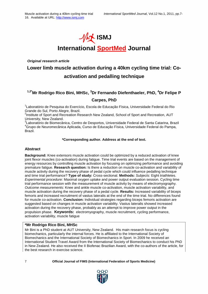
Muscle activation during a 40km cycling time trial International SportMed Journal, Vol.12 No.1, 2011, pp.7- 16. Available at URL: http://www.ismj.com 7 Official Journal of FIMS (International Federation of Sports Medicine) ISMJ International SportMed Journal Original research article Lower limb muscle activation during a 40km cycling time trial: Co- activation and pedalling technique 1,2* Mr Rodrigo Rico Bini, MHSc, 3 Dr Fernando Diefenthaeler, PhD, 4 Dr Felipe P Carpes, PhD 1 Laboratório de Pesquisa do Exercício, Escola de Educação Física, Universidade Federal do Rio Grande do Sul, Porto Alegre, Brazil. 2 Institute of Sport and Recreation Research New Zealand, School of Sport and Recreation, AUT University, New Zealand. 3 Laboratório de Biomecânica, Centro de Desportos, Universidade Federal de Santa Catarina, Brazil 4 Grupo de Neuromecânica Aplicada, Curso de Educação Física, Universidade Federal do Pampa, Brazil. *Corresponding author. Address at the end of text. Abstract Background: Knee extensors muscle activation could be optimized by a reduced activation of knee joint flexor muscles (co-activation) during fatigue. Time trial events are based on the management of energy resources by controlling muscle activation by focusing on optimizing performance and avoiding premature fatigue. Research question: Is there a reduction on muscle co-activation and variability of muscle activity during the recovery phase of pedal cycle which could influence pedalling technique and time trial performance? Type of study: Cross-sectional. Methods: Subjects: Eight triathletes. Experimental procedure: Maximal oxygen uptake and power output evaluation session. Cycling time trial performance session with the measurement of muscle activity by means of electromyography. Outcome measurements: Knee and ankle muscle co-activation, muscle activation variability, and muscle activation during the recovery phase of a pedal cycle. Results: Increased variability of biceps femoris and increased recruitment of vastus lateralis at the end of the time trial. No differences found for muscle co-activation. Conclusion: Individual strategies regarding biceps femoris activation are suggested based on changes in muscle activation variability. Vastus lateralis showed increased activation during the recovery phase, probably as an attempt to improve power output in the propulsion phase. Keywords: electromyography, muscle recruitment, cycling performance, activation variability, muscle fatigue *Mr Rodrigo Rico Bini, MHSc Mr Bini is a PhD student at AUT University, New Zealand. His main research focus is cycling biomechanics, particularly the internal forces. He is affiliated to the International Society of Biomechanics and the International Society of Biomechanics in Sport. In 2009 he received an International Student Travel Award from the International Society of Biomechanics to conduct his PhD in New Zealand. He also received the II Biofenac Brazilian Award, with the co-authors of the article, for the best research in exercise science.
Transcript of Lower limb muscle activation during a 40km cycling time trial: Co-activation and pedalling technique
Muscle activation during a 40km cycling time trial International SportMed Journal, Vol.12 No.1, 2011, pp.7-16. Available at URL: http://www.ismj.com
7 Official Journal of FIMS (International Federation of Sports Medicine)
ISMJ
International SportMed Journal
Original research article
Lower limb muscle activation during a 40km cycling time trial: Co-activation and pedalling technique
1,2*Mr Rodrigo Rico Bini, MHSc, 3Dr Fernando Diefenthaeler, PhD, 4Dr Felipe P
Carpes, PhD
1Laboratório de Pesquisa do Exercício, Escola de Educação Física, Universidade Federal do Rio Grande do Sul, Porto Alegre, Brazil. 2Institute of Sport and Recreation Research New Zealand, School of Sport and Recreation, AUT University, New Zealand. 3Laboratório de Biomecânica, Centro de Desportos, Universidade Federal de Santa Catarina, Brazil 4Grupo de Neuromecânica Aplicada, Curso de Educação Física, Universidade Federal do Pampa, Brazil.
*Corresponding author. Address at the end of text.
Abstract Background: Knee extensors muscle activation could be optimized by a reduced activation of knee joint flexor muscles (co-activation) during fatigue. Time trial events are based on the management of energy resources by controlling muscle activation by focusing on optimizing performance and avoiding premature fatigue. Research question: Is there a reduction on muscle co-activation and variability of muscle activity during the recovery phase of pedal cycle which could influence pedalling technique and time trial performance? Type of study: Cross-sectional. Methods: Subjects: Eight triathletes. Experimental procedure: Maximal oxygen uptake and power output evaluation session. Cycling time trial performance session with the measurement of muscle activity by means of electromyography. Outcome measurements: Knee and ankle muscle co-activation, muscle activation variability, and muscle activation during the recovery phase of a pedal cycle. Results: Increased variability of biceps femoris and increased recruitment of vastus lateralis at the end of the time trial. No differences found for muscle co-activation. Conclusion: Individual strategies regarding biceps femoris activation are suggested based on changes in muscle activation variability. Vastus lateralis showed increased activation during the recovery phase, probably as an attempt to improve power output in the propulsion phase. Keywords: electromyography, muscle recruitment, cycling performance, activation variability, muscle fatigue *Mr Rodrigo Rico Bini, MHSc Mr Bini is a PhD student at AUT University, New Zealand. His main research focus is cycling biomechanics, particularly the internal forces. He is affiliated to the International Society of Biomechanics and the International Society of Biomechanics in Sport. In 2009 he received an International Student Travel Award from the International Society of Biomechanics to conduct his PhD in New Zealand. He also received the II Biofenac Brazilian Award, with the co-authors of the article, for the best research in exercise science.

Muscle activation during a 40km cycling time trial International SportMed Journal, Vol.12 No.1, 2011, pp.7-16. Available at URL: http://www.ismj.com
8 Official Journal of FIMS (International Federation of Sports Medicine)
Dr Fernando Diefenthaeler, PhD Dr Diefenthaeler’s main research focus is cycling biomechanics related to fatigue. He is affiliated to the International Society of Biomechanics. Together with the co-authors of this article, he received the II Biofenac Brazilian Award for the best research in exercise science. Email: [email protected] Dr Felipe Carpes, PhD Dr Carpes’ main research focus is on neuromechanics, particularly related to lower extremity movement production and control. He is affiliated to the International Society. In 2008 he received an International Student Travel Award from the International Society of Biomechanics to conduct research in Canada. He also received the II Biofenac Brazilian Award, with the co-authors of the article, for the best research in exercise science. Email: [email protected]
Introduction Muscle activation measurement has been a source of evidences for motor control 1 and performance during cycling 2. The literature supports an important link between muscle activation and power output during time trial cycling events 2. However, controversial results were provided by different studies addressing muscle activation during time trial events. Duc et al. 3 reported a steady state of muscular activity during a 30min cycling time trial, whereas Bini et al. 4 observed a selective increased activation of vastus lateralis without significant changes for other lower limb’s muscles at the end of a 40km time trial. As reported by Tucker et al. 5 and Carpes et al. 6 respectively, an “end spurt” (increased power output in the final stages of time trial event) hs been observed which can be related to an increased activation of mono-articular knee joint extensor muscles 4. This strategy for time trial events could be based on the management of energy resources by pacing the power output 7. Premature fatigue is probably prevented by a muscular steady-state in an attempt to sustain the performance 3. Nevertheless, Hautier et al. 8 proposed that muscle activation should be optimized by a reduced activation of knee joint flexor muscles during fatigue. A fine-tune between knee extensor and flexor muscles would allow the cyclist to increase knee extensor joint moment and improve performance. Muscle activity during cycling has also been based on the analysis of co-activation between flexor and extensor muscles 9. Candotti et al. 10 observed increased co-activation between flexors and extensors muscles of the knee joint for triathletes, which was related to a poor
pedalling technique compared to cyclists. Chapman et al. 9 also observed an increased co-activation between flexor and extensor muscles of the ankle joint, as well as an increased variability in muscle activation for triathletes and novice cyclists respectively, which was suggested to be related to a reduced pedalling skill. Since time trial events are based on the management of energy resources by controlling muscle activation 3, these authors expected muscle co-activation, and variability would be managed by focussing on optimizing performance and avoiding premature fatigue. Besides the effects of fatigue and pacing strategies during time trial events, pedalling technique is not clearly described during cycling events. Therefore ankle joint kinematics have been described as affecting pedalling technique 11 and muscle efficiency 12. An increased activation of the ankle flexor muscle group (i.e. tibialis anterior), would be associated with improvements in pedalling technique and increased effectiveness at the end of a time trial. Gaps in muscle co-activation, variability of muscle activation and effects of pedalling technique on muscle recruitment during the cycling time trial can be observed in the literature. Increased co-activation during the cycling time trial event could suggest poor muscle coordination and result in premature fatigue. Therefore the aims of the present study were: (1) to analyze the triathletes’ muscle co-activation during a 40km cycling time trial event, and (2) to analyze pedalling technique observing muscle activation variability and muscle activation at the recovery phase of the pedalling cycle. Based on previous evidence 4, the primary hypothesis was that the co-

Muscle activation during a 40km cycling time trial International SportMed Journal, Vol.12 No.1, 2011, pp.7-16. Available at URL: http://www.ismj.com
9 Official Journal of FIMS (International Federation of Sports Medicine)
activation of knee joint muscles would be reduced during the time trial based on the possible optimization of knee joint extensor/flexor activation. A secondary hypothesis was that muscle activation variability would be reduced and the muscle activation at the recovery phase of the pedalling cycle would increase as a result of an improved pedalling technique. Methods Subjects Eight triathletes volunteered for this study. All subjects had competitive experience of 5.18 ± 3.79 years. They signed an Informed Consent document in agreement with the Committee of Ethics in Research with Humans of the Institution where this study was conducted. Volunteers were asked to refrain from high-intensity or exhaustive exercise at least 24 hours prior to the laboratory trials. Mean and standard deviation of age, body mass, maximal oxygen uptake (VO2MAX), peak power output (POPEAK) and power/mass ratio of the subjects were 31.7 ± 5.2 years, 76.9 ± 6.7kg, 61 ± 8.2ml.kg-1.min-1, 414 ± 39W, and 5.51 ± 0.87W.kg-1 respectively. Experimental procedures On the first day, a graded exercise test to determine maximal oxygen uptake (VO2MAX) test was performed on a stationary cycle ergometer CardiO2 (Medical Graphics Corp., St. Louis, USA) adapted with drop handlebars, clipless pedals and a racing saddle. Prior to the VO2MAX test, the O2 and CO2 analyzers were calibrated using calibrated medical grade gases that spanned air in the physiological range. A ramp protocol was started with an initial load of 50W with increments of 25W every minute 13 and pedalling cadence between 70 and 110rpm until the triathletes were no longer able to maintain 70rpm. VO2MAX was defined as the highest VO2 value of the last minute of the test 3. VO2 and carbon dioxide produced were continuously determined breath-by-breath during the test using an open-circuit indirect gas exchange system (MGC CPX/D, Medical Graphics Corp., St Louis, EUA). During the incremental test, the power output 14 was continually recorded to determine POPEAK 3. At least 48 hours following the maximal test the subjects performed a 40km time trial using their own bicycles mounted on a stationary wind-trainer (Cateye CS1000, Cateye Co., Osaka, Japan) in the fastest time possible, using a freely chosen pacing strategy (3, 4, 6).
During the 40km time trial, the subjects prevented alterations in posture by adopting the conventional cycling posture with ~75º trunk inclination and grasping the handlebars with elbows slightly bent to remain in a seated position. Muscle activity was measured at the 3rd, 20th and 38th km. A reed switch attached to the bicycle frame recorded the pedalling cadence by registering each crank revolution as described elsewhere 4. Surface electromyography (EMG) was employed to measure activity from the tibialis anterior (TA), medial head of the gastrocnemius (GM), the long head of the biceps femoris (BF), the rectus femoris 15 and the vastus lateralis (VL) muscles from the left lower limb. Pairs of Ag/AgCl electrodes (bipolar configuration) with a diameter of 22mm were positioned on the skin after carefully shaving and cleaning the area using an abrasive cleaner and alcohol swabs to reduce skin impedance, according to the International Society of Electrophysiology and Kinesiology 16. Electrodes were placed over the belly of the muscles, parallel to the muscle fibres and taped to the skin using micro pore tape (3M Company, USA). Also, a bandage was wrapped around the electrode to minimize sweat interference. A reference electrode was placed over an electrically neutral site (anterior surface of tibia). The electrode’s wires were taped to the skin to reduce movement artifact. A reed switch attached to the bicycle frame recorded the pedalling cadence by registering each crank revolution. The electrical pulse produced by the reed switch was used to identify the beginning and the end of each crank cycle. This information was also used to analyze muscle activity related to crank cycle 4. Data analysis The raw EMG signals were pre-amplified and band-passed filtered at 10-500Hz through a five-order Butterworth digital filter. One minute of EMG signal was collected employing an eight-channel electromyography system (Bortec Eletronics Inc., Calgary, Canada) when subjects achieved the 3rd, 20th and 38th km, at a sample rate of 2000Hz per channel. Offline data analysis was supported by the software WINDAQ® (WINDAQ, DataQ Instruments Inc., USA) and MATLAB® 7.3 (MathWorks Inc., USA). The root mean square (RMS) envelope was averaged in 40ms moving windows 17 for the

Muscle activation during a 40km cycling time trial International SportMed Journal, Vol.12 No.1, 2011, pp.7-16. Available at URL: http://www.ismj.com
10 Official Journal of FIMS (International Federation of Sports Medicine)
entire raw signal, and cut in ten consecutive crank revolutions to determine the average and standard-deviation of each muscle respectively. For each muscle in each of the ten crank revolutions, the mean value of the RMS envelope was calculated and normalized by each subject’s own peak value observed in the 3rd km, which corresponds to the beginning of the test when no effect of fatigue was expected. From RMS envelope, co-activation (the percentage of the pedal cycle which a pair of flexor-extensor muscles were simultaneously activated), muscle activation variability (average coefficient of variance of ten analyzed crank cycles), and the mean value of the RMS envelope during recovery phase (from 180º to 360º of the crank cycle) were calculated. For the computation of co-activation, a threshold value of 10% in the RMS envelope was set as a criterion for onset and offset reference for muscle activation dynamics 18. Co-activation was computed for the selected pairs of flexor-extensor muscles: TA-GM, BF-RF, and BF-VL.
Statistical analysis For data analysis, three of the five instances recorded during the time trial event (3rd, 20th, and 38th km) were selected in attempt to reduce the possibility of Type I error and preserve the statistical power by multiple-related comparisons 19. For these three instances, normality of data distribution and sphericity of co-activation, variability (CV) and RMS of evaluated muscles during the recovery phase were analyzed, for each muscle, by Shapiro-Wilk and Mauckly’s tests, respectively. When data normality was not confirmed, a logarithm transform was applied. Greenhouse-Geisser corrections were employed when data violated sphericity assumption. Mean and standard deviation (SD) for all variables were reported. ANOVA for repeated
measures with full factorial linear and quadratic models was employed for the comparison of CV and RMS at the recovery phase (dependent variables) for the three instances of the time trial test (3rd, 20th, and 38th km). Post-hoc analysis, where the main effects or interactions were significant, was subsequently performed using the Least Significant Difference (LSD) test followed by Holm’s correction 19, 20. Models were an attempted to verify any trends of data distribution by the analysis of probability for predicting effects on the data (p value), partial eta squared (η2) and observed power of the model prediction (1 - β). For the statistical procedures SPSS 12.0 package (SPSS Inc., USA) was used with level of Type I error set at 5% for 75% of observed power. Results Due to the effects of sweat on the function of the electrodes, some missing data can be observed. Co-activation results did not report significant differences for TA-GM (N = 6), F2,10 = 1.632, p = 0.24, η2 = 0.25, 1 - β = 0.27, either for BF-RF (N = 8), F2,14 = 3.481, p = 0.06, η2 = 0.33, 1 - β = 0.55. BF-VL pair (N = 7) was statistically different (F2,12 = 4.518, p = 0.03), but with low partial eta squared and observed power (η2 = 0.43, 1 - β = 0.65), and after Holm’s correction, the probability of Type I error was 6,8%. The linear model indicated trends of linear increase for BF-RF (F1,7 = 8.266, p = 0.02) and BF-VL (F1,6 = 7.435, p = 0.03). However, both models reported low partial eta squared and observed power (η2 = 0.54, 1 - β = 0.70, and η2 = 0.55, 1 - β = 0.70). These results are depicted in Figure 1.

Muscle activation during a 40km cycling time trial International SportMed Journal, Vol.12 No.1, 2011, pp.7-16. Available at URL: http://www.ismj.com
11 Official Journal of FIMS (International Federation of Sports Medicine)
Figure 1: Average co-activation of TA-GM (N = 6), BF-RF (N = 8), and BF-VL (N = 7) for the three evaluated instances during the time trial Trend lines were included based on ANOVA model predictions (see details in the text). &Indicates statistical significance to reject the null hypothesis (Type I error). However, none of the models achieved the statistical level to avoid Type II error. Variability of activation level during the crank cycle was measured by the average coefficient of variance (CV). These results are shown in Figure 2.

Muscle activation during a 40km cycling time trial International SportMed Journal, Vol.12 No.1, 2011, pp.7-16. Available at URL: http://www.ismj.com
12 Official Journal of FIMS (International Federation of Sports Medicine)
Figure 2: Average + SD of variance coefficient (CV) of biceps femoris (N = 7), gastrocnemius medialis (N = 6), rectus femoris (N = 7), tibialis anterior (N = 6), and vastus lateralis (N = 7) for the three evaluated instances of the time trial # Indicates statistical difference in relation to 20 km (p < 0.05 and 1-β > 0.75) Only BF CV reported a statistical increase (F2,14 = 6.017, p = 0.03, η2 = 0.46, 1-β = 0.80) at the 38th km in relation to the 20th km of the time trial. A quadratic model of ANOVA also suited data distribution (F1,7 = 8.177, p = 0.02, η2 = 0.54, 1-β = 0.69) but with lower observed power. A quadratic model fitted GM distribution (F1,7 = 7.388, p = 0.03, η2 = 0.51, 1-β = 0.65), but also with lower observed power. For RF (F2,14 = 0.706, p = 0.51, η2 = 0.09, 1-β = 0.15), TA (F2,12 = 1.807, p = 0.21, η2 = 0.23, 1-β = 0.30), and VL (F2,12 = 1.741, p = 0.22, η2 = 0.22, 1-β = 0.29), no significant differences were observed.
Muscle activation during the recovery phase of the crank cycle was measured by the RMS mean value. These results are depicted in Figure 3.

Muscle activation during a 40km cycling time trial International SportMed Journal, Vol.12 No.1, 2011, pp.7-16. Available at URL: http://www.ismj.com
13 Official Journal of FIMS (International Federation of Sports Medicine)
Figure 3: Average + SD of RMS of biceps femoris, gastrocnemius medialis, rectus femoris, tibialis anterior and vastus lateralis during the recovery phase of crank cycle for the three instances during the time trial *Indicates a statistical difference in relation to the 3 km instance (p < 0.05 and 1-β > 0.75).
For RMS during the recovery phase, differences for BF were observed (F2,14 = 5.254, p = 0.02, η2 = 0.43, 1-β = 0.74) but with lower observed power and higher probability of Type I error after Holm’s correction (6,8%). Otherwise, the linear model suited BF distribution (F1,7 = 6.905, p = 0.03, η2 = 0.50, 1-β = 0.62) but with lower observed power. VL increased during the time trial (F2,12 = 5.977, p = 0.02, η2 = 0.50, 1-β = 0.78) with significant differences between the 3rd and 38th km (p = 0.03) and the linear model suited data distribution (F1,6 = 12.176, p = 0.01, η2 = 0.67, 1-β = 0.83). For GM (F2,14 = 3.515, p = 0.06, η2 = 0.33, 1-β = 0.56), RF (F1.200,8.403 = 2.882, p = 0.12, η2 = 0.29, 1-β = 0.35), and TA (F2.10 = 0.739, p = 0.50, η2 = 0.13, 1-β = 0.14), there were no significant differences.
Discussion Previous research indicated that during cycling performance evaluation, such as time trial events, the increase of power output seemed to be dependent on the selective recruitment of mono-articular knee joint extensors 4. However, co-activation analysis would indicate whether changes in muscle coordination are related to pedalling technique during the trial. Therefore the aims of the present study were to analyze the knee and ankle muscles’ co-
activation and to infer these effects on pedalling technique based on muscle activation variability and muscle activation patterns during the recovery phase of cycling. Two main results emerged from these analyses: (1) no significant changes in muscle co-activation were observed, (2) increased activation variability for the biceps femoris and increased activation for vastus lateralis during the recovery phase of the pedal cycle at the end of the time trial. Both results were the novelties of the present study, because most of the previous research considered only the analysis of average muscle activation for the entire cycle, without looking at the coordinate effects of each cycle and between the muscles 2, 3, 12.
Recent evidence indicated that muscle activation variability can be related to pedalling skill and that triathletes have increased activation variability and co-activation for the ankle joint muscles 9, 21. Candotti et al. 10 also reported that triathletes showed a higher level of co-activation between the knee joint flexors and extensor muscles compared to trained cyclists. Only a few studies attempted to measure muscle activity during a time trial without reporting on muscle co-activation 3, 4. Both studies observed a steady state of muscular activity during the test, mentioning an
Muscle activation during a 40km cycling time trial International SportMed Journal, Vol.12 No.1, 2011, pp.7-16. Available at URL: http://www.ismj.com
14 Official Journal of FIMS (International Federation of Sports Medicine)
attempt to avoid premature fatigue based on muscle activation and energy expenditure management to optimize the “end spurt” at the end of the test. Moreover, these researchers cannot make definitive conclusions about the role of the pedalling technique on the increase of power output observed at the end of the time trial by considering only the results of this present study. It has been reported that pedal force analysis is the best measurement for studying the pedalling technique 15, 22, 23, which is a limitation for this study as was not this measured in this study. The increased variability in the biceps femoris would indicate an attempt by some athletes to improve their pedalling technique. As the variability of activation was analyzed by means of the coefficient of variation, the reduction of the mean value and/or increase in activation variability results in higher CV 14. In this regard, these authors can infer that the biceps femoris activation could be controlled to avoid an increased resistive moment of the knee extensor muscle on the downstroke, as previously observed during cycling to fatigue 8. Nevertheless, the statistical analysis did not indicate significant differences for RMS in the recovery phase for any of flexor muscle groups analyzed. This result could be related to a poor pedalling technique in triathletes, as previously observed by EMG analyses 9, 21, 24. Changes in pedalling technique have been described as dependent on muscle activation patterns 12 and reduced mechanical efficiency 15, 23. Uncertainties regarding the effective contribution of pedalling technique for cycling performance emerged 25, 26, based on recent proposals regarding the lower efficiency of the flexor muscle group (i.e. tibialis anterior and biceps femoris), which would lead to an increased energy cost to recruit these muscles 15, 23. Therefore the increased activation observed for the vastus lateralis during the recovery phase in the final stage of the time trial could be associated with an early recruitment of this muscle for knee joint extension. Due to a delay between muscle activation and force production 27, this early increased activation is observed on the recovery phase of pedalling as an attempt to improve power output 4. Based on the present results, co-activation of the knee and ankle joint muscles did not change during the time trial, but the variability of the biceps femoris and the activation of the vastus lateralis during the recovery phase increased in the last stage of the time trial.
These results support an increase of power output related to higher knee joint extensor muscle activation 4. With regard to the variability of the biceps femoris, two possibilities can be suggested: (1) there would have to have been an attempt to increase biceps femoris activation for some athletes, and (2) there would have to have been an attempt to minimize biceps femoris activation for others. The first is based on the knee joint extensor moment, which was reported to contribute to force transfer from the thigh to the shank by the hamstrings 28. The second possibility is related to previous research that cyclists could manage biceps femoris activation to avoid premature knee extensor muscle fatigue 8. As the pathways of motor control during cycling are complex it is not possible to conclude which of these two possibilities had been selected by each of the subjects in this present study. Future studies should include pedal forces and kinematics measurements to provide more information on the possible function of the hamstrings during a cycling time trial and its importance for pedalling technique training. Because during a time trial the cyclist manages the gear ratio and pedalling cadence to control power output, one may argue that such variables may affect muscle recruitment. However, previous research indicates that pedalling cadence is not affected by pacing strategy during a time trial 4, 29, 30, which supports the hypothesis of power output control based on the management of gear ratio. Conclusion An increased variability of the biceps femoris and the increased recruitment of the vastus lateralis at the end of the time trial were observed. The possibility of individual strategies being used regarding biceps femoris activation was suggested based on the changes in muscle activation variability. The increase in vastus lateralis activation during the recovery phase was probably related to an attempt to improve power output during the propulsion phase.

Muscle activation during a 40km cycling time trial International SportMed Journal, Vol.12 No.1, 2011, pp.7-16. Available at URL: http://www.ismj.com
15 Official Journal of FIMS (International Federation of Sports Medicine)
Address for correspondence: Mr Rodrigo Bini, Sport Performance Research Institute New Zealand, School of Sport and Recreation, Auckland University of Technology, Private Bag 92006, Auckland, New Zealand Tel.: +0064 (9) 477 2056 Fax: +0064 (9) 921 9960 Email: [email protected] References
1. Ryan MM, Gregor RJ. EMG profiles of lower extremity muscles during cycling at constant workload and cadence. Journal of Electromyography and Kinesiology 1992; 2(2):69-80.
2. St Clair Gibson A, Schabort EJ, Noakes TD. Reduced neuromuscular activity and force generation during prolonged cycling. American Journal of Physiology - Regulatory Integrative and Comparative Physiology 2001; 281(1 50-1).
3. Duc S, Betik AC, Grappe F. EMG activity does not change during a time trial in competitive cyclists. International Journal of Sports Medicine 2005; 26(2):145-150.
4. Bini RR, Carpes FP, Diefenthaeler F, et al. Physiological and electromyographic responses during 40-km cycling time trial: Relationship to muscle coordination and performance. Journal of Science and Medicine in Sport 2008; 11(4):363-370.
5. Tucker R, Bester A, Lambert EV, et al. Non-random fluctuations in power output during self-paced exercise. British Journal of Sports Medicine 2006; 40(11):912-917.
6. Carpes FP, Rossato M, Faria IE, et al. Bilateral pedaling asymmetry during a simulated 40-km cycling time-trial. Journal of Sports Medicine and Physical Fitness 2007; 47(1):51-57.
7. De Koning JJ, Bobbert MF, Foster C. Determination of optimal pacing strategy in track cycling with an energy flow model. Journal of Science and Medicine in Sport 1999; 2(3):266-277.
8. Hautier CA, Arsac LM, Deghdegh K, et al. Influence of fatigue on EMG/force ratio and cocontraction in cycling. Medicine and Science in Sports and Exercise 2000; 32(4):839-843.
9. Chapman AR, Vicenzino B, Blanch P, et al. Patterns of leg muscle recruitment vary between novice and highly trained cyclists. Journal of
Electromyography and Kinesiology 2008; 18(3):359-371.
10. Candotti CT, Loss JF, Bagatini D, et al. Cocontraction and economy of triathletes and cyclists at different cadences during cycling motion. Journal of Electromyography and Kinesiology 2009; 19(5):915-921.
11. Black AH, Sanderson DJ, Hennig EM. Kinematic and kinetic changes during an incremental exercise test on a bicycle ergometer. XIVth ISB Congress in Biomechanics 1993: 186-187.
12. Cannon DT, Kolkhorst FW, Cipriani DJ. Effect of pedaling technique on muscle activity and cycling efficiency. European Journal of Applied Physiology 2007; 99(6): 659-664.
13. Lucía A, Hoyos J, Pérez M, et al. Inverse relationship between VO2max and economy/efficiency in world-class cyclists. Medicine and Science in Sports and Exercise 2002; 34(12): 2079-2084.
14. Hug F, Drouet JM, Champoux Y, et al. Interindividual variability of electromyographic patterns and pedal force profiles in trained cyclists. European Journal of Applied Physiology 2008; 104(4): 667-678.
15. Korff T, Romer LM, Mayhew I, et al. Effect of pedaling technique on mechanical effectiveness and efficiency in cyclists. Medicine and Science in Sports and Exercise 2007; 39(6): 991-995.
16. De Luca CJ. The use of surface electromyography in biomechanics. Journal of Applied Biomechanics 1997; 13(2): 135-163.
17. Neptune RR, Kautz SA, Hull ML. The effect of pedaling rate on coordination in cycling. Journal of Biomechanics 1997; 30(10):1051-1058.
18. Baum BS, Li L. Lower extremity muscle activities during cycling are influenced by load and frequency. Journal of Electromyography and Kinesiology 2003; 13(2): 181-190.
19. Atkinson G. Analysis of repeated measurements in physical therapy research: Multiple comparisons amongst level means and multi-factorial designs. Physical Therapy in Sport 2002; 3(4):191-203.
20. Knudson D. Significant and meaningful effects in sports biomechanics research. Sports Biomechanics 2009; 8(1):96-104.

Muscle activation during a 40km cycling time trial International SportMed Journal, Vol.12 No.1, 2011, pp.7-16. Available at URL: http://www.ismj.com
16 Official Journal of FIMS (International Federation of Sports Medicine)
21. Chapman AR, Vicenzino B, Blanch P, et al. Leg muscle recruitment during cycling is less developed in triathletes than cyclists despite matched cycling training loads. Experimental Brain Research 2007; 181(3): 503-518.
22. Rossato M, Bini RR, Carpes FP, et al. Cadence and workload effects on pedaling technique of well-trained cyclists. International Journal of Sports Medicine 2008; 29(9): 746-752.
23. Mornieux G, Stapelfeldt B, Gollhofer A, et al. Effects of pedal type and pull-up action during cycling. International Journal of Sports Medicine 2008; 29(10):817-822.
24. Candotti CT, Loss JF, Bagatini D, et al. Cocontraction and economy of triathletes and cyclists at different cadences during cycling motion. Journal of Electromyography and Kinesiology (In press).
25. Coyle EF, Feltner ME, Kautz SA, et al. Physiological and biomechanical factors associated with elite endurance cycling performance. Medicine and Science in Sports and Exercise 1991; 23(1):93-107.
26. Diefenthaeler F, Bini RR, Carpes FP, et al. Analysis of pedaling technique during a maximal cycling exercise. In:
Menzel H-J, Chagas MH (Eds). 25th International Symposium on Biomechanics in Sports; Ouro Preto, Brazil: International Society of Biomechanics in Sports; 2007. pp. 394-397.
27. Li L, Baum BS. Electromechanical delay estimated by using electromyography during cycling at different pedaling frequencies. Journal of Electromyography and Kinesiology 2004; 14(6): 647-652.
28. Gregor RJ, Cavanagh PR, LaFortune M. Knee flexor moments during propulsion in cycling - a creative solution to Lombard's paradox. Journal of Biomechanics 1985; 18(5): 307-316.
29. Bernard T, Hausswirth C, Le Meur Y, et al. Distribution of power output during the cycling stage of a triathlon world cup. Medicine and Science in Sports and Exercise 2009; 41(6):1296-1302.
30. Perrey S, Grappe F, Girard A, et al. Physiological and metabolic responses of triathletes to a simulated 30-min time-trial in cycling at self-selected intensity. International Journal of Sports Medicine 2003; 24(2): 138-143.