Mapping the MinE Site Involved in Interaction with the MinD Division Site Selection Protein of...
9
10.1128/JB.185.16.4948-4955.2003. 2003, 185(16):4948. DOI: J. Bacteriol. Lu-Yan Ma, Glenn King and Lawrence Rothfield Escherichia coli Selection Protein of Interaction with the MinD Division Site Mapping the MinE Site Involved in http://jb.asm.org/content/185/16/4948 Updated information and services can be found at: These include: REFERENCES http://jb.asm.org/content/185/16/4948#ref-list-1 at: This article cites 21 articles, 13 of which can be accessed free CONTENT ALERTS more» articles cite this article), Receive: RSS Feeds, eTOCs, free email alerts (when new http://journals.asm.org/site/misc/reprints.xhtml Information about commercial reprint orders: http://journals.asm.org/site/subscriptions/ To subscribe to to another ASM Journal go to: on August 26, 2014 by guest http://jb.asm.org/ Downloaded from on August 26, 2014 by guest http://jb.asm.org/ Downloaded from
Transcript of Mapping the MinE Site Involved in Interaction with the MinD Division Site Selection Protein of...
10.1128/JB.185.16.4948-4955.2003.
2003, 185(16):4948. DOI:J. Bacteriol. Lu-Yan Ma, Glenn King and Lawrence Rothfield
Escherichia coliSelection Protein of Interaction with the MinD Division Site Mapping the MinE Site Involved in
http://jb.asm.org/content/185/16/4948Updated information and services can be found at:
These include:
REFERENCEShttp://jb.asm.org/content/185/16/4948#ref-list-1at:
This article cites 21 articles, 13 of which can be accessed free
CONTENT ALERTS more»articles cite this article),
Receive: RSS Feeds, eTOCs, free email alerts (when new
http://journals.asm.org/site/misc/reprints.xhtmlInformation about commercial reprint orders: http://journals.asm.org/site/subscriptions/To subscribe to to another ASM Journal go to:
on August 26, 2014 by guest
http://jb.asm.org/
Dow
nloaded from
on August 26, 2014 by guest
http://jb.asm.org/
Dow
nloaded from
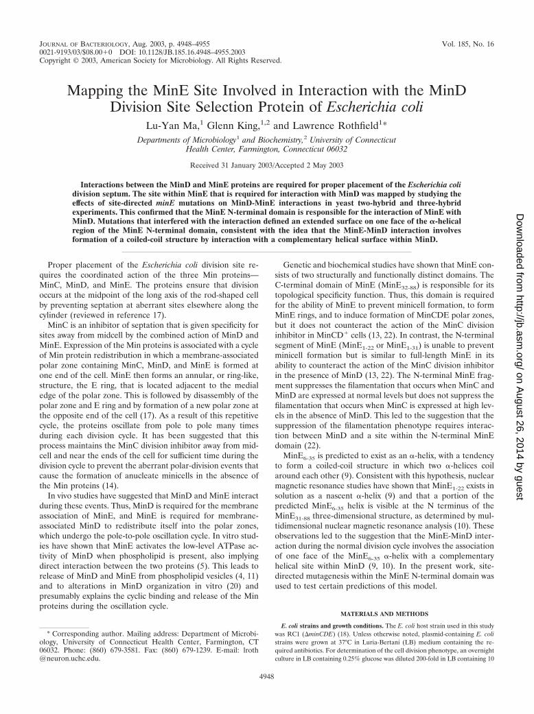
JOURNAL OF BACTERIOLOGY, Aug. 2003, p. 4948–4955 Vol. 185, No. 160021-9193/03/$08.00�0 DOI: 10.1128/JB.185.16.4948–4955.2003Copyright © 2003, American Society for Microbiology. All Rights Reserved.
Mapping the MinE Site Involved in Interaction with the MinDDivision Site Selection Protein of Escherichia coli
Lu-Yan Ma,1 Glenn King,1,2 and Lawrence Rothfield1*Departments of Microbiology1 and Biochemistry,2 University of Connecticut
Health Center, Farmington, Connecticut 06032
Received 31 January 2003/Accepted 2 May 2003
Interactions between the MinD and MinE proteins are required for proper placement of the Escherichia colidivision septum. The site within MinE that is required for interaction with MinD was mapped by studying theeffects of site-directed minE mutations on MinD-MinE interactions in yeast two-hybrid and three-hybridexperiments. This confirmed that the MinE N-terminal domain is responsible for the interaction of MinE withMinD. Mutations that interfered with the interaction defined an extended surface on one face of the �-helicalregion of the MinE N-terminal domain, consistent with the idea that the MinE-MinD interaction involvesformation of a coiled-coil structure by interaction with a complementary helical surface within MinD.
Proper placement of the Escherichia coli division site re-quires the coordinated action of the three Min proteins—MinC, MinD, and MinE. The proteins ensure that divisionoccurs at the midpoint of the long axis of the rod-shaped cellby preventing septation at aberrant sites elsewhere along thecylinder (reviewed in reference 17).
MinC is an inhibitor of septation that is given specificity forsites away from midcell by the combined action of MinD andMinE. Expression of the Min proteins is associated with a cycleof Min protein redistribution in which a membrane-associatedpolar zone containing MinC, MinD, and MinE is formed atone end of the cell. MinE then forms an annular, or ring-like,structure, the E ring, that is located adjacent to the medialedge of the polar zone. This is followed by disassembly of thepolar zone and E ring and by formation of a new polar zone atthe opposite end of the cell (17). As a result of this repetitivecycle, the proteins oscillate from pole to pole many timesduring each division cycle. It has been suggested that thisprocess maintains the MinC division inhibitor away from mid-cell and near the ends of the cell for sufficient time during thedivision cycle to prevent the aberrant polar-division events thatcause the formation of anucleate minicells in the absence ofthe Min proteins (14).
In vivo studies have suggested that MinD and MinE interactduring these events. Thus, MinD is required for the membraneassociation of MinE, and MinE is required for membrane-associated MinD to redistribute itself into the polar zones,which undergo the pole-to-pole oscillation cycle. In vitro stud-ies have shown that MinE activates the low-level ATPase ac-tivity of MinD when phospholipid is present, also implyingdirect interaction between the two proteins (5). This leads torelease of MinD and MinE from phospholipid vesicles (4, 11)and to alterations in MinD organization in vitro (20) andpresumably explains the cyclic binding and release of the Minproteins during the oscillation cycle.
Genetic and biochemical studies have shown that MinE con-sists of two structurally and functionally distinct domains. TheC-terminal domain of MinE (MinE32-88) is responsible for itstopological specificity function. Thus, this domain is requiredfor the ability of MinE to prevent minicell formation, to formMinE rings, and to induce formation of MinCDE polar zones,but it does not counteract the action of the MinC divisioninhibitor in MinCD� cells (13, 22). In contrast, the N-terminalsegment of MinE (MinE1-22 or MinE1-31) is unable to preventminicell formation but is similar to full-length MinE in itsability to counteract the action of the MinC division inhibitorin the presence of MinD (13, 22). The N-terminal MinE frag-ment suppresses the filamentation that occurs when MinC andMinD are expressed at normal levels but does not suppress thefilamentation that occurs when MinC is expressed at high lev-els in the absence of MinD. This led to the suggestion that thesuppression of the filamentation phenotype requires interac-tion between MinD and a site within the N-terminal MinEdomain (22).
MinE6-35 is predicted to exist as an �-helix, with a tendencyto form a coiled-coil structure in which two �-helices coilaround each other (9). Consistent with this hypothesis, nuclearmagnetic resonance studies have shown that MinE1-22 exists insolution as a nascent �-helix (9) and that a portion of thepredicted MinE6-35 helix is visible at the N terminus of theMinE31-88 three-dimensional structure, as determined by mul-tidimensional nuclear magnetic resonance analysis (10). Theseobservations led to the suggestion that the MinE-MinD inter-action during the normal division cycle involves the associationof one face of the MinE6-35 �-helix with a complementaryhelical site within MinD (9, 10). In the present work, site-directed mutagenesis within the MinE N-terminal domain wasused to test certain predictions of this model.
MATERIALS AND METHODS
E. coli strains and growth conditions. The E. coli host strain used in this studywas RC1 (�minCDE) (18). Unless otherwise noted, plasmid-containing E. colistrains were grown at 37°C in Luria-Bertani (LB) medium containing the re-quired antibiotics. For determination of the cell division phenotype, an overnightculture in LB containing 0.25% glucose was diluted 200-fold in LB containing 10
* Corresponding author. Mailing address: Department of Microbi-ology, University of Connecticut Health Center, Farmington, CT06032. Phone: (860) 679-3581. Fax: (860) 679-1239. E-mail: [email protected].
4948
on August 26, 2014 by guest
http://jb.asm.org/
Dow
nloaded from

�M IPTG (isopropyl-�-D-thiogalactopyranoside) and grown to an optical densityat 600 nm (OD600) of �0.9.
Plasmids. Subscripts in the names of genes indicate the protein products (e.g.,minE1-31 indicates a minE fragment coding for amino acids 1 to 31; minEL22R
indicates a mutant allele coding for a mutant protein in which amino acid 22 hasbeen changed from leucine to arginine). Tables 1 and 2 list the plasmid geno-types.
Point mutations within minE were generated by mutagenic PCR of plasmidpSY1083 (Plac-minCDE) (18), as previously described (19), to generate the pEMseries of plasmids (Table 1). For plasmids to be used in yeast two- and three-hybrid studies (Table 2), protein fusions to the Gal4 activation domain (AD) andGal4 binding domain (BD) were obtained by insertion of DNA fragments intopBridge or pGBKT7 (for BD fusions) or into pGAD424 or pGADKT7 (for ADfusions); the plasmid vectors were obtained from Clontech. The DNA fragmentswere obtained by PCR, using pSY1083 as a template for wild-type minC, minD,and minE and the appropriate pEM plasmids (Table 1) as templates to generatemutant minE fragments. The pBridge vector, in addition to coding for thedesired BD protein fusion, contains a second cloning site downstream of PMet.DNA coding for a second protein was inserted at this site, generating in-framefusions in which the protein was downstream of PMet and a nuclear localizationsignal and hemagglutinin epitope (Clontech manual).
pYEC plasmids (Plac-yfp::minD minE::cfp), coding for proteins in which Yfp(yellow fluorescent protein) was fused to the N-terminal end of MinD and Cfp(cyan fluorescent protein) was fused to the C-terminal end of mutant MinE, wereconstructed by three fragment ligations between (i) the XbaI/XmnI fragmentfrom pYLS68 (Plac-yfp::minD minE::cfp) (19), containing a minD fragment; (ii)the XbaI/BamHI fragment from pYLS68 that contains yfp, cfp, and thepMLB1113 vector; and (iii) XmnI/BamHI fragments from pEM plasmids (seeabove) containing mutant minE alleles. pMEC plasmids (Plac-minE::cfp) wereconstructed by ligating the XmnI/HindIII fragment containing minE::cfp frompYLS68 (19), or from pYEC plasmids containing mutant minE::cfp alleles (seeabove), into the SmaI/HindIII site of the pMLB1113 vector (1).
pYLS41G (Plac-minE::gfp) was generated by ligation of the XmnI/HindIIIfragment from pSY1083G (19) that contains minE::gfp into the SmaI/HindIII siteof the pMLB1113 vector. pMEG25 (Plac-minEI25R::gfp) was constructed simi-larly, using the XmnI/HindIII fragment containing minEI25R::gfp from pEG25,which was obtained by religation of BamHI-treated pEM25 to generate anin-frame minEI25R::gfp fusion. pMA101 (Plac-minE1-31::gfp), pMA102 (Plac-minE32-88::gfp), and pMA22 (Plac-minE1-31,L22S::gfp) were constructed by ligationof the large EcoRI/BamHI fragment from pSY1083 that contains gfp and thepMLB1113 vector to fragments containing minE1-31, minE32-88, or minE1-31,L22S,which were obtained by PCR using pSY1083 and pEM22S as templates.
Yeast two- and three-hybrid systems. Plasmids coding for the appropriate BDand AD fusion proteins (Table 2) were transformed into Saccharomyces cerevi-siae SFY526 containing the Gal4-inducible reporter gene lacZ (Clontech). Plas-mid-containing cells were obtained and characterized, and further analysis wasperformed as described in the Clontech manual. Interaction between the BD andAD probes was monitored after 3 days of growth at 30°C by �-galactosidaseactivity, as estimated from colony color on X-Gal (5-bromo-4-chloro-3-indolyl-�-D-galactopyranoside) plates and by liquid assay (Clontech manual, protocolPT3024-1); specific activity is given in Miller units (12) and represents theaverage of three independent measurements. Three-hybrid assays utilized thepBridge vector; the third protein inserted downstream of PMet was induced bygrowth in the absence of methionine.
Fluorescence microscopy. Cells were grown in the presence of 10 �M IPTG,and Yfp-, Cfp-, and Gfp-labeled proteins were detected by fluorescence micros-copy as previously described (19). Oscillation rates were determined as previ-ously described (19) and are given as the average for 12 to 15 cells. The oscil-lation rate of the MinD polar zone in pYLS68 (Plac-yfp::minD minE::cfp) was0.15 � 0.07 cycles/min, similar to values previously described for cells containingdoubly labeled MinD and MinE (3, 19).
Cell fractionation. Cells were suspended in SHD buffer (20% sucrose, 10 mMHEPES, 5 mM dithiothreitol), broken using a French pressure cell, and centri-fuged at 3,000 rpm at 4°C for 10 min in a Beckman Microfuge 18 centrifuge toremove unbroken cells (8). The supernatant fraction was centrifuged for 90 minat 100,000 rpm at 4°C in a Beckman TL100 centrifuge to obtain the crudecytoplasmic (supernatant) and membrane (pellet) fractions for Western blotanalysis. Coomassie blue-stained sodium dodecyl sulfate (SDS) gels confirmedthat the pellet contained all of the detectable major outer membrane proteinbands with negligible amounts of the protein bands that characterized the cyto-plasmic fraction.
Western blot analysis. Western blot analysis was performed as previouslydescribed on SDS extracts of intact cells (21) or of membrane and cytoplasmicfractions. Protein concentrations were determined by bicinchoninic acid assay(Pierce) of the SDS extracts. For quantitation of individual proteins, 10-, 20-, or30-�g samples were subjected to Western blot analysis as previously described,using polyclonal anti-MinD or anti-MinE2-19 antibody (21). The relative inten-sities of the stained bands were determined on digitized images using the Im-ageQuant program (Molecular Dynamics). For assays of hybrid proteins in yeast,cultures were grown in synthetic dropout (minus Leu, minus Trp, minus Met)medium (Clontech), and proteins were extracted by the trichloroacetic acidmethod (Clontech manual, protocol PT3024-1). Protein extracted from 0.5OD600 unit of cells was analyzed using polyclonal antibody (Clontech) directedagainst the hemagglutinin tag that is part of the fusion protein.
RESULTS
Min protein interactions. The yeast two-hybrid system wasused to study interactions between MinD and MinE, using�-galactosidase expression as an indicator of interaction. Inthese experiments, MinD was fused to the Gal4 AD (AD-MinD) and MinE was fused to the Gal4 BD (BD-MinE).Consistent with previous reports (7), full-length MinE (MinE1-
88) interacted very weakly with MinD, as indicated by colordevelopment on X-Gal plates and direct enzyme assay (Table3). We therefore separately examined the abilities of the twofunctional domains of MinE (MinE1-31 and MinE32-88) to in-teract with MinD in the two-hybrid system (Table 3). Thisrevealed a strong interaction of MinD with MinE1-31, the MinEdomain that was predicted from genetic experiments to func-tionally interact with MinD (9, 13, 22). The interaction, as
TABLE 1. Plasmids used for determination of cell divisionphenotype and fluorescence microscopya
Plasmid Relevant genotype Source orreference
pSY1083 Plac-minC minD minE 2pEM4 Plac-minC minD minEL4R This studypEM8 Plac-minC minD minEL8R This studypEM15 Plac-minC minD minEA15R This studypEM18 Plac-minC minD minEA18T This studypEM19 Plac-minC minD minEK19A This studypEM20 Plac-minC minD minEE20A This studypEM22 Plac-minC minD minEL22R This studypEM22S Plac-minC minD minEL22S This studypEM23 Plac-minC minD minEQ23R This studypEM24 Plac-minC minD minEI24R This studypEM25 Plac-minC minD minEI25R This studypEM26 Plac-minC minD minEV26R This studypEM27 Plac-minC minD minEA27R This studypEM28 Plac-minC minD minEE28A This studypEM30 Plac-minC minD minER30A This studypYEC15 Plac-yfp::minD minEA15R::cfp This studypYEC18 Plac-yfp::minD minEA18T::cfp This studypYEC19 Plac-yfp::minD minEK19A::cfp This studypYEC22 Plac-yfp::minD minEL22R::cfp This studypYEC22S Plac-yfp::minD minEL22S::cfp This studypYEC25 Plac-yfp::minD minEI25R::cfp This studypYEC30 Plac-yfp::minD minER30A::cfp This studypMEC1 Plac-minE::cfp This studypMEC22 Plac-minEL22R::cfp This studypMEC22S Plac-minEL22S::cfp This studypMEC25 Plac-minEI25R::cfp This studypYLS41G Plac-minE::gfp 19pMEG25 Plac-minEI25R::gfp This studypMA101 Plac-minE1–31::gfp This studypMA102 Plac-minE32–88::gfp This study
a See Materials and Methods for further details.
VOL. 185, 2003 MinD-MinE INTERACTIONS 4949
on August 26, 2014 by guest
http://jb.asm.org/
Dow
nloaded from

judged by �-galactosidase activity, was �100-fold greater thanthe interaction of full-length MinE with MinD. As expected,the MinE32-88 domain, which appears not to be involved inMinE-MinD interactions in vivo, showed no interaction. Thedemonstration that MinE1-31 interacts strongly with MinD inthe two-hybrid system provided a convenient assay for studyingdeterminants involved in the MinD-MinE interaction.
As previously shown (7), MinD interacted strongly with
MinC in the two-hybrid system (Tables 3 and 4). MinE stronglyinterfered with the MinC-MinD interaction, as shown by a99% decrease in �-galactosidase expression when MinE wasexpressed together with AD-MinD and BD-MinC (Table 4); asimilar result was obtained when MinE was replaced byMinE1-31, as shown by a 94% reduction in �-galactosidaseexpression (Table 4). The ability of MinE and MinE1-31 toblock the MinC-MinD interaction is likely to reflect interactionbetween MinD and MinE, but interaction between MinE andMinC cannot be excluded, despite the fact that MinE-MinC (7)and MinE1-31-MinC (unpublished data) interactions have notbeen observed in two-hybrid experiments. MinE32-88 partiallyinterfered with the MinC-MinD interaction, as indicated by adecrease of �50% in �-galactosidase activity (Table 4). Thismay reflect the presence in MinE32-88 of the C-terminal por-tion of the MinE6-35 �-helix that is believed to be responsiblefor the MinE-MinD interaction (9) (see below). The ability toperturb the MinC-MinD interaction provided a second assayfor mapping the interacting site.
The ability of MinE to interfere with the MinC-MinD inter-action presumably explains in vitro observations that MinE canpromote the release of MinC from a MinC-MinD-phospho-lipid complex without the release of MinD (6, 11).
The full-length MinE protein (MinE1-88) was highly effectivein interfering with the MinC-MinD interaction, although itshowed very little activity when assayed for the ability to inter-act directly with MinD (Table 3). The cause of the difference inbehavior of full-length MinE in the two assays is not known.Further work will be needed to determine whether it reflectsan inhibitory effect of the Gal4 BD, when fused to full-lengthMinE, on the accessibility of the MinD BD or whether thedifference in behavior has implications for the in vivo functionsof the Min proteins in E. coli.
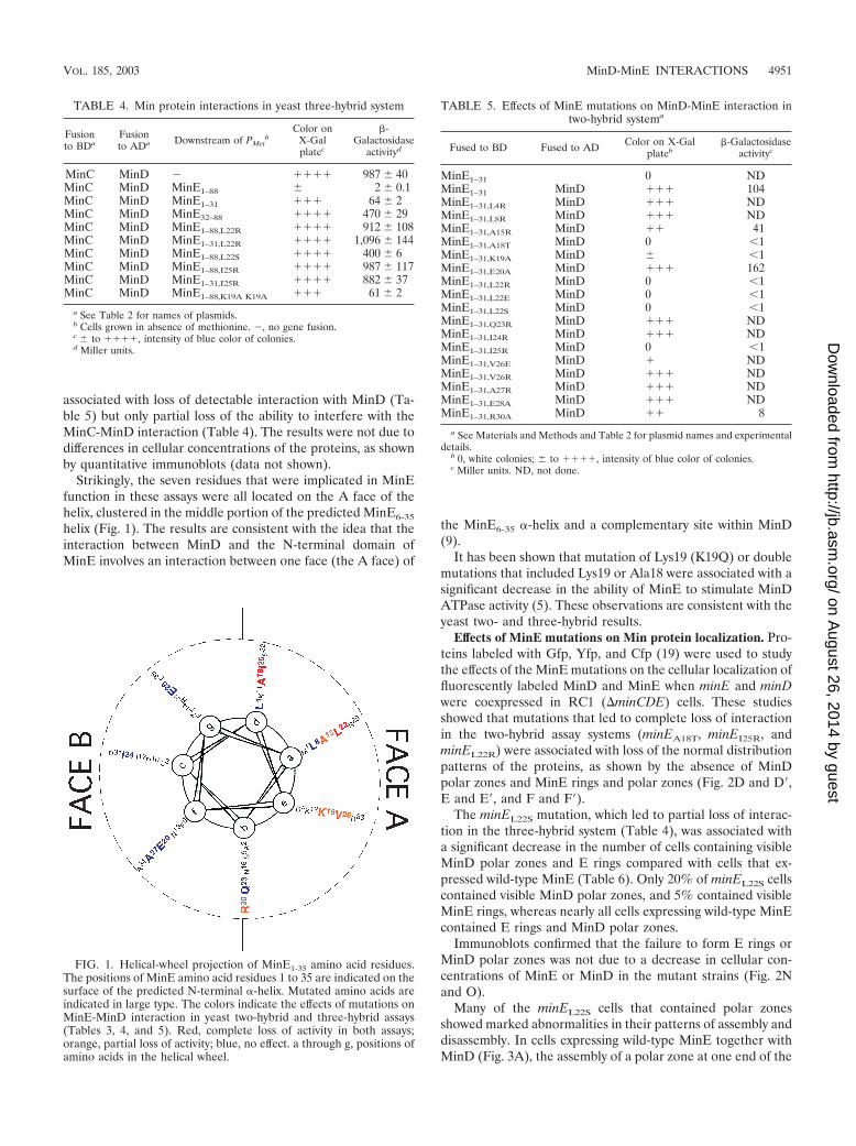
If the region of MinE1-31 that interacts with MinD exists inan �-helical conformation, residues within MinE1-31 that func-tionally interact with MinD would be expected to cluster onone face of the helix. To examine this prediction, we mu-tagenized conserved residues (10) that are distributed aroundthe predicted MinE1-35 helix. In each case, the mutationchanged a nonpolar to a polar amino acid, or vice versa. Themutant proteins were then assayed in the two-hybrid andthree-hybrid assay systems for the ability to interact with MinDor to disrupt the MinC-MinD interaction. For convenience, wedivided the predicted helical surface into two faces, called theA and B faces (Fig. 1).
Mutations in 7 (Ala15, Ala18, Lys19, Leu22, Ile25, Val26,and Arg30) of the 14 mutagenized residues led to a completeor partial loss of MinE function in the two-hybrid and three-hybrid assay systems. This was shown by the failure of themutated proteins to induce �-galactosidase expression in theMinE1-31-MinD system (Table 5) and/or by inability to inter-fere with the increase in �-galactosidase expression that resultsfrom interaction between MinC and MinD (Table 4). Theother mutated proteins did not differ from unmutagenizedMinE in these assays. In the case of Leu22, three substitutionsthat led to conversion of the leucine residue to glutamate(L22E), arginine (L22R), and serine (L22S) were individuallystudied. The L22E and L22R mutations led to complete loss ofinteraction in the two-hybrid and three-hybrid experiments(Tables 4 and 5). The conversion to serine, in MinEL22S, was
TABLE 2. Plasmids used in yeast two- and three-hybrid assaysa
Plasmid Relevant genotype Source
pBridge PADH1-BD; PMet25b Clontech
pGBKT7 PADH1-BD ClontechpGAD424 PADH1-AD ClontechpGADT7 PADH1-AD ClontechpYW9 PADH1-AD::minD in pGAD424 This studypMDB1 PADH1-BD::minD in pGBKT7 This studypMCA1 PADH1-AD::minC in pGADT7 This studypYW10 PADH1-BD::minC; PMet25 in pBridge This studypBE1 PADH1-BD::minE; PMet25 in pBridge This studypBE2 PADH1-BD::minE1–31; PMet25 in pBridge This studypBE3 PADH1-BD::minE32–88; PMet25 in pBridge This studypBEC1 PADH1-BD::minE1–31; PMet25-minC in pBridge This studypBCE1 PADH1-BD::minC; PMet25-minE in pBridge This studypBCE2 PADH1-BD::minC; PMet25-minE1–31 in pBridge This studypBCE3 PADH1-BD::minC; PMet25-minE32–88 in pBridge This studypBCE11 PADH1-BD::minC; PMet25-minEL22R in pBridge This studypBCE12 PADH1-BD::minC; PMet25-minE1–31,L22R in pBridge This studypBCE13 PADH1-BD::minC; PMet25-minE1–31,125R in pBridge This studypBCE15 PADH1-BD::minC; PMet25-minEK19A in pBridge This studypBEM16 PADH1-BD::minC; PMet25-minEL22S in pBridge This studypBEM4 PADH1-BD::minE1–31,L4R; PMet25 in pBridge This studypBEM8 PADH1-BD::minE1–31,L8Rl PMet25 in pBridge This studypBEM15 PADH1-BD::minE1–31,A15R; PMet25 in pBridge This studypBEM18 PADH1-BD::minE1–31,A18T; PMet25 in pBridge This studypBEM19 PADH1-BD::minE1–31,K19A; PMet25 in pBridge This studypBEM20 PADH1-BD::minE1–31,E20A; PMet25 in pBridge This studypBEM22 PADH1-BD::minE1–31,L22R; PMet25 in pBridge This studypBEM22S PADH1-BD::minE1–31,L22S; PMet25 in pBridge This studypBEM23 PADH1-BD::minE1–31,Q23R; PMet25 in pBridge This studypBEM24 PADH1-BD::minE1–31,I24R; PMet25 in pBridge This studypBEM25 PADH1-BD::minE1–31,I25R; PMet25 in pBridge This studypBEM27 PADH1-BD::minE1–31,A27R; PMet25 in pBridge This studypBEM28 PADH1-BD::minE1–31,E28A; PMet25 in pBridge This studypBEM30 PADH1-BD::minE1–31,R30A; PMet25 in pBridge This study
a See Materials and Methods for constructions and further details.b PMet25, methionine promoter, repressible by growth in methionine.
TABLE 3. Min protein interactions in yeast two-hybrid system
Fusion to BDa Fusion to ADa,b Color on X-Galplatec
�-Galactosidaseactivityd
b MinD 0 0.2MinE1–88 0 NDMinE1–88 MinD � 0.9 � 0.09MinE1–31 0 NDMinE1–31 MinD ��� 112 � 5MinE32–88 0 NDMinE32–88 MinD 0 0.2MinE1–31 MinE32–88 0 0.2MinE1–31 MinE1–31 0 0.2MinE32–88 MinE32–88 0 0.2MinD MinD �� 42 � 1MinC MinD ���� 715 � 5MinC MinC � 6 � 1
a See Materials and Methods and Table 1 for names of plasmids.b , no gene fusion.c 0, white colonies; � to ����, intensity of blue color of colonies.d Miller units. ND, not done.
4950 MA ET AL. J. BACTERIOL.
on August 26, 2014 by guest
http://jb.asm.org/
Dow
nloaded from
associated with loss of detectable interaction with MinD (Ta-ble 5) but only partial loss of the ability to interfere with theMinC-MinD interaction (Table 4). The results were not due todifferences in cellular concentrations of the proteins, as shownby quantitative immunoblots (data not shown).
Strikingly, the seven residues that were implicated in MinEfunction in these assays were all located on the A face of thehelix, clustered in the middle portion of the predicted MinE6-35
helix (Fig. 1). The results are consistent with the idea that theinteraction between MinD and the N-terminal domain ofMinE involves an interaction between one face (the A face) of
the MinE6-35 �-helix and a complementary site within MinD(9).
It has been shown that mutation of Lys19 (K19Q) or doublemutations that included Lys19 or Ala18 were associated with asignificant decrease in the ability of MinE to stimulate MinDATPase activity (5). These observations are consistent with theyeast two- and three-hybrid results.
Effects of MinE mutations on Min protein localization. Pro-teins labeled with Gfp, Yfp, and Cfp (19) were used to studythe effects of the MinE mutations on the cellular localization offluorescently labeled MinD and MinE when minE and minDwere coexpressed in RC1 (�minCDE) cells. These studiesshowed that mutations that led to complete loss of interactionin the two-hybrid assay systems (minEA18T, minEI25R, andminEL22R) were associated with loss of the normal distributionpatterns of the proteins, as shown by the absence of MinDpolar zones and MinE rings and polar zones (Fig. 2D and D�,E and E�, and F and F�).
The minEL22S mutation, which led to partial loss of interac-tion in the three-hybrid system (Table 4), was associated witha significant decrease in the number of cells containing visibleMinD polar zones and E rings compared with cells that ex-pressed wild-type MinE (Table 6). Only 20% of minEL22S cellscontained visible MinD polar zones, and 5% contained visibleMinE rings, whereas nearly all cells expressing wild-type MinEcontained E rings and MinD polar zones.
Immunoblots confirmed that the failure to form E rings orMinD polar zones was not due to a decrease in cellular con-centrations of MinE or MinD in the mutant strains (Fig. 2Nand O).
Many of the minEL22S cells that contained polar zonesshowed marked abnormalities in their patterns of assembly anddisassembly. In cells expressing wild-type MinE together withMinD (Fig. 3A), the assembly of a polar zone at one end of the
FIG. 1. Helical-wheel projection of MinE1-35 amino acid residues.The positions of MinE amino acid residues 1 to 35 are indicated on thesurface of the predicted N-terminal �-helix. Mutated amino acids areindicated in large type. The colors indicate the effects of mutations onMinE-MinD interaction in yeast two-hybrid and three-hybrid assays(Tables 3, 4, and 5). Red, complete loss of activity in both assays;orange, partial loss of activity; blue, no effect. a through g, positions ofamino acids in the helical wheel.
TABLE 4. Min protein interactions in yeast three-hybrid system
Fusionto BDa
Fusionto ADa Downstream of PMet
bColor on
X-Galplatec
�-Galactosidase
activityd
MinC MinD ���� 987 � 40MinC MinD MinE1–88 � 2 � 0.1MinC MinD MinE1–31 ��� 64 � 2MinC MinD MinE32–88 ���� 470 � 29MinC MinD MinE1–88,L22R ���� 912 � 108MinC MinD MinE1–31,L22R ���� 1,096 � 144MinC MinD MinE1–88,L22S ���� 400 � 6MinC MinD MinE1–88,I25R ���� 987 � 117MinC MinD MinE1–31,I25R ���� 882 � 37MinC MinD MinE1–88,K19A K19A ��� 61 � 2
a See Table 2 for names of plasmids.b Cells grown in absence of methionine. , no gene fusion.c � to ����, intensity of blue color of colonies.d Miller units.
TABLE 5. Effects of MinE mutations on MinD-MinE interaction intwo-hybrid systema
Fused to BD Fused to AD Color on X-Galplateb
�-Galactosidaseactivityc
MinE1–31 0 NDMinE1–31 MinD ��� 104MinE1–31,L4R MinD ��� NDMinE1–31,L8R MinD ��� NDMinE1–31,A15R MinD �� 41MinE1–31,A18T MinD 0 1MinE1–31,K19A MinD � 1MinE1–31,E20A MinD ��� 162MinE1–31,L22R MinD 0 1MinE1–31,L22E MinD 0 1MinE1–31,L22S MinD 0 1MinE1–31,Q23R MinD ��� NDMinE1–31,I24R MinD ��� NDMinE1–31,I25R MinD 0 1MinE1–31,V26E MinD � NDMinE1–31,V26R MinD ��� NDMinE1–31,A27R MinD ��� NDMinE1–31,E28A MinD ��� NDMinE1–31,R30A MinD �� 8
a See Materials and Methods and Table 2 for plasmid names and experimentaldetails.
b 0, white colonies; � to ����, intensity of blue color of colonies.c Miller units. ND, not done.
VOL. 185, 2003 MinD-MinE INTERACTIONS 4951
on August 26, 2014 by guest
http://jb.asm.org/
Dow
nloaded from

FIG. 2. Fluorescence microscopy of labeled MinD and MinE. Cells were prepared from strain RC1 (�minCDE) containing the followingplasmids: pYLS68 (Plac-yfp::minD minE::cfp) (A and A�); pYEC19 (Plac-yfp::minD minEK19A::cfp) (B and B�); pYLS68 (C and C�); pYEC18(Plac-yfp::minD minEA18T::cfp) (D and D�); pYEC25 (Plac-yfp::minD minEI25R::cfp) (E and E�); pYEC22 (Plac-yfp::minD minEL22R::cfp) (F and F�);pWEC1 (Plac-minE::cfp) (G); pMEG25 (Plac-minEI25R::gfp) (H); pMEC22 (Plac-minEL22R::cfp) (I); pMEC22S (Plac-minEL22S::cfp) (J); pYLS41G(Plac-minE::gfp) (K); pMA101 (Plac-minE1-31::gfp) (L); and pMA102 (Plac-minE32-88::gfp) (M). (A, A�, B, and B�) Fixed cells. (C to M) Unfixed cells.Yfp and Gfp are green, and Cfp is blue. MinE-Cfp images were acquired 10 s after the corresponding Yfp-MinD images. The labels to the leftand right of the fluorescence images indicate the minE alleles of the corresponding images. WT, wild type. (N and O) Western blot analyses ofcells from the experiments described in panels A to F and the corresponding experiments described in the text. (P) Western blot analysis of cellsfrom the experiments described in panels K and L. (Q) Western blot analysis of cell fractions from cells expressing minE::gfp and minE1-31::gfp fromplasmids pYLS41G (Plac-minE::gfp) and pMA101 (Plac-minE1-31::gfp), respectively. p, membrane (pellet fraction); s, cytoplasm (supernatantfraction); t, total extract. The aliquots applied to each lane were derived from 0.04 OD600 unit of cells for lanes 1 to 6 and 0.02 OD600 unit of cellsfor lanes 7 to 12. Anti-MinE2-19 antibody was used for panels N, P, and Q; anti-MinD antibody was used for panel O.
4952 MA ET AL. J. BACTERIOL.
on August 26, 2014 by guest
http://jb.asm.org/
Dow
nloaded from

cell is followed by its shrinkage from midcell toward the pole,terminating in its disappearance from the original cell pole (2),apparently by release into the cytoplasm (16). A new polarzone is then assembled at the opposite end of the cell. Incontrast, in the minEL22S cells (Fig. 3B), the disappearance ofMinD from the polar zone was often not followed by formationof a new polar zone at the opposite cell pole. Instead, after thedisappearance of the polar zone, MinD was present around theentire cell periphery, presumably membrane associated, sug-gesting that the disappearance of the polar zone might beaccomplished by the random redistribution of MinD within thecytoplasmic membrane. This was followed by retraction of themembrane-associated MinD from the original pole, leading toaccumulation of MinD within a “new” polar zone at the op-posite cell pole (Fig. 3B). Thus, in contrast to the normalsituation, the new MinD polar zones in many minEL22S cellsappeared not to be formed de novo but often reflected mem-
brane-associated MinD that was left at one cell pole as a resultof the disappearance or redistribution of membrane-associatedMinD from the other end of the cell. The rate of disassemblyof the membrane-associated MinD was much lower than thatof the disassembly of MinD polar zones in cells expressingwild-type MinE (Fig. 3A) (19). Interestingly, this phenotyperesembled in part the phenotype resulting from mutation atthe MinED45/V49 site within the C-terminal topological-speci-ficity domain of MinE that is required for E-ring formation,where a general peripheral distribution of MinE was oftenfollowed by retraction from one end of the cell (19). Thepossible implications of this similarity remain to be explored.
The minEK19A mutation, which was also associated with apartial loss of interaction with MinD in the two-hybrid andthree-hybrid assay systems (Tables 4 and 5), showed an unusu-ally high percentage of cells that contained MinD polar zonesat both poles (70% of the cells) (Fig. 2B and Table 6). Incomparison, in cells expressing wild-type MinE in parallel cul-tures, only 3% of the cells contained visible polar zones at bothpoles (Table 6). The rate of pole-to-pole oscillation in theminEK19A cells was lowered to �40% of the rate in wild-typecells. A similar observation was made by Hu and Lutkenhaus(5), who observed a decrease in the oscillation rates of �50%in the presence of two other mutations affecting the Lys19residue (minEK19Q and minEK19A,E20A). In the remaining mu-tants, essentially all cells contained polar zones and E rings andthe oscillation rates of the polar zones were not significantlyperturbed.
Effect of MinE mutations on cell division phenotype. Ex-pression of normal levels of MinCD in the absence of MinEleads to a general inhibition of septation, leading to formationof long nonseptate filaments (1). The filamentation can beprevented by expression of either full-length MinE or N-ter-minal MinE fragments (1, 13, 22). To determine the effect ofthe minE mutations on the cell division phenotype, the mutantalleles were substituted for wild-type minE in plasmid pSY1083(Plac-minCDE), and the cell division phenotype was deter-mined in host strain RC1 (�minCDE) after IPTG induction.
FIG. 3. Oscillation of Yfp-MinD in living cells. Time lapse fluorescence micrographs of Yfp-MinD in strain RC1 containing pYLS68 (Plac-yfp::minD minE::cfp) (A) and pYEC22S (Plac-yfp::minD minEL22S::cfp) (B). The time at which the image was acquired (in seconds) is indicated ineach micrograph. The cells were observed at 15-s intervals between micrographs to confirm that no dramatic change occurred during the intervals.In one image in each panel, as indicated, fluorescence from MinE-Cfp was captured. The arrows indicate the MinE ring (R) and the MinD polarzone (PZ).
TABLE 6. Phenotypes of MinE mutants
MinEa MinD-MinEinteractionb
MinDpolar zones E ringse Phenotype
1c 2d
WT � 92 3 95 WTA15R � 96 1 97 WTK19A � 27 70 74 Sep Min
L22S � 20 1 5 Sep
R30A � 95 1 94 WTA18T 0 1 1 1 Sep
L22R 0 1 1 1 Sep
I25R 0 1 1 1 Sep
a The mutated amino acids in mutant MinE proteins are shown; 100 cells ofeach type were analyzed to determine the percentage of cells with polar zones orMinE rings. WT, wild type.
b MinD-MinE interaction as indicated by induction of �-galactosidase in two-hybrid assays (Table 5) and/or by ability to inhibit MinC-MinD interaction inthree-hybrid assays (Table 4). 0, no interaction in either assay; �, partial inter-action compared with wild-type MinE in one or both assay systems; �, normalinteraction compared with wild-type MinE.
c Percentage of cells containing one MinD polar zone.d Percentage of cells containing polar zones at both poles.e Percentage of cells with E rings.
VOL. 185, 2003 MinD-MinE INTERACTIONS 4953
on August 26, 2014 by guest
http://jb.asm.org/
Dow
nloaded from

The ability to counteract the division inhibition is thought torequire interaction between MinD and a site within the N-terminal MinE domain (22). Consistent with this view, MinEmutations that led to complete loss of MinD-MinE interactionin the two-hybrid system (mutations A18T, I25R, L22R, andL22S) were associated with a filamentation phenotype (Fig. 4Bto D and Table 6). Mutation K19A, which led to significant butnot total loss of detectable interaction in the two-hybrid assay(Table 5), showed a mixture of filaments, minicells, and cells ofapproximately normal length. We assume that this mixed phe-notype represented the effects of different levels of expressionof the partially active MinEK19A protein in different cellswithin the culture.
The possibility that the loss of ability to counteract theMinCD division inhibition was due to a decrease in the cellularconcentrations of the mutant proteins was excluded by quan-titative immunoblot analysis (data not shown).
Membrane localization of MinE in the absence of MinD. Ithas been shown that full-length MinE (MinE1-88) fails to lo-calize to the peripheral portion of the cell unless MinD iscoexpressed, indicating that MinD is required for the mem-brane association of MinE (2, 15). These observations wereconfirmed in the present study, where MinE-Cfp and MinE-Gfp, when expressed in the absence of MinD, were diffuselydistributed within the cell (Fig. 2G and K), indicating a cyto-plasmic localization. However, in contrast to full-length MinE,MinE1-31-Gfp showed a peripheral distribution even when ex-pressed in the absence of MinD (Fig. 2L). This unexpectedresult was obtained in several independent experiments andwas seen in living cells as well as fixed cells. This suggested thatthe MinE1-31 protein was able to associate with the cell mem-brane in the absence of MinD. The possibility that the differ-ence between the behaviors of full-length MinE and MinE1-31
was due to differences in cellular concentration was excludedby immunoblot analysis, which showed that the cellular con-centrations of MinE1-88-Gfp and MinE1-31-Gfp were similar(Fig. 2P).
The distributions of MinE1-88-Gfp and MinE1-31-Gfp werealso examined in crude cytoplasmic and membrane fractions ofcells that expressed minE1-88::gfp or minE1-31::gfp in the ab-sence of minD (Fig. 2Q). The results were consistent with thefluorescence localization studies in intact cells (Fig. 2L).Nearly all of the MinE1-88-Gfp was recovered in the cytoplas-mic fraction, whereas �70% of the MinE1-31-Gfp was recov-ered in the crude membrane fraction.
In the case of several of the mutant alleles (minEI25R,minEL22R, and minEL22S), the full-length MinE-Cfp proteinalso showed a peripheral distribution when expressed in theabsence of MinD (Fig. 2H to J). The observations were repro-ducible. These unexpected results, which are surprising in viewof the fact that MinD is required to bring wild-type MinE1-88 tothe membrane, are discussed further below.
DISCUSSION
It is likely that interactions between MinE and MinD play arole in several aspects of Min protein function. These includethe MinD-dependent membrane association of MinE (15), theMinE-dependent formation and oscillation of MinD polarzones (2, 16), the ability of MinE to counteract the MinCDdivision inhibitor (1), and the ability of MinE to stimulateMinD ATPase activity (5) and thereby promote release ofmembrane-associated MinD (4, 11, 20). The present studyprovides direct evidence that the MinE-MinD interaction in-volves the N-terminal MinE1-31 domain and maps the sitewithin the N-terminal MinE domain that is responsible for theinteraction.
Although the mutations that were originally introduced intothe N-terminal MinE domain were designed to be distributedon both faces of the MinE1-31 helical region, it was striking thatthe mutations that affected the MinD-MinE interaction werelocated exclusively on one face of the helix, the A face, wherethey defined an extended contiguous surface domain. Becausethe MinE6-35 helical region is predicted to have a propensity toform coiled-coil structures (9), it is reasonable to speculate thatthe site on the A face interacts with a corresponding surface onan �-helix within MinD to form a supercoiled structure. Therelatively large area of the putative MinD-binding site on the Aface, involving two or three turns of the helix, is consistent withthe idea that it represents a surface that interacts in a side-by-side configuration with a surface-exposed �-helix in MinD. Wepresume that this interaction plays an essential role in theability of MinD to bring MinE to the membrane and possiblyin other MinE/MinD-dependent functions of the Min system.Mutations that interfered with MinD-MinE interactions in thetwo-hybrid system were associated with the complete or partialabsence of MinD polar zones and E rings. This is consistentwith the idea that interaction between MinD and the putativebinding site on the A face of MinE1-31 plays an important rolein the ability of MinE to promote the membrane redistributionevents that lead to formation of the MinD polar zone. Themutations also interfered with the ability of MinE to counter-act the MinCD division inhibitor, consistent with the idea thatthis function requires that MinE be brought to the membranein a reaction that needs interaction with MinD (15).
It is not known whether binding to MinD is the only functionof the N-terminal domain of MinE. Full-length MinE does not
FIG. 4. Effects of minE mutations on cell division patterns. Micro-graphs of Nomarski images of RC1 containing pSY1083 (Plac-minCminD minE) (A), pEM22S (Plac-minC minD minEL22S) (B), pEM25(Plac-minC minD minEI25R) (C), and pEM19 (Plac-minC minDminEK19A) (D) are shown. The cells were prepared and analyzed aspreviously described (1).
4954 MA ET AL. J. BACTERIOL.
on August 26, 2014 by guest
http://jb.asm.org/
Dow
nloaded from
localize to the cytoplasmic membrane of E. coli in the absenceof MinD, as shown by the diffuse distribution of MinE-Gfpwithin the cell. This indicates that MinE requires MinD tomove from cytoplasm to membrane (references 2 and 15 andthis study). In contrast to the behavior of full-length MinE, itwas observed in the present study that MinE1-31-Gfp expressedin the absence of MinD was located around the periphery ofthe cell, in a pattern that is usually interpreted to indicateassociation with the membrane or cell envelope. This interpre-tation was supported by the finding in extracts of these cellsthat MinE1-31 was recovered primarily in the membrane frag-ment whereas full-length MinE was found almost exclusively inthe cytoplasmic fraction. Several of the MinE mutant proteinsshowed similar peripheral cellular localization patterns whenexpressed in the absence of MinD, even when present as full-length proteins. The apparent membrane localization ofMinE1-31 and of the mutant proteins may represent experimen-tal artifacts, in which the mutations or the removal of the MinEC-terminal domain uncovers nonspecific and biologically irrel-evant membrane-binding sites within the N-terminal MinE1-31
domain. This would be consistent with current models, inwhich MinD is the sole membrane anchor for membrane-associated MinE. However, the present observations may in-stead provide a clue that MinE1-31 contains membrane-bindingsites that function after MinE is brought to the membrane byMinD. According to this view, the membrane-binding siteswithin MinE1-31 are normally occluded by the C-terminal do-main and can be exposed as a result of conformational changesduring the MinD-dependent membrane assembly process orexperimentally by removal of the C-terminal domain or bycertain mutations. The putative MinE-membrane interactioncould act to stabilize the membrane association of the E ring orcould play another role in MinE function. Further work will beneeded to test these ideas.
ACKNOWLEDGMENTS
This work was supported by National Institutes of Health grantsGM60632 to L.R. and GM45583 to G.K.
REFERENCES
1. de Boer, P. A. D., R. E. Crossley, and L. I. Rothfield. 1989. A divisioninhibitor and a topological specificity factor coded for by the minicell locusdetermine proper placement of the division septum in E. coli. Cell 56:641–649.
2. Fu, X., Y.-L. Shih, Y. Zhang, and L. I. Rothfield. 2001. The MinE ringrequired for proper placement of the division site is a mobile structure that
changes its cellular location during the Escherichia coli division cycle. Proc.Natl. Acad. Sci. USA 98:980–985.
3. Hale, C., H. Meinhardt, and P. de Boer. 2001. Dynamic localization cycle ofthe cell division regulator MinE in Escherichia coli. EMBO J. 20:1563–1572.
4. Hu, Z., E. Gogol, and J. Lutkenhaus. 2002. Dynamic assembly of MinD onphospholipid vesicles regulated by ATP and MinE. Proc. Natl. Acad. Sci.USA 99:6761–6766.
5. Hu, Z., and J. Lutkenhaus. 2001. Topological regulation of cell division in E.coli: spatiotemporal oscillation of MinD occurs through stimulation of itsATPase activity by MinE and phospholipid. Mol. Cell 7:1337–1343.
6. Hu, Z., C. Saez, and J. Lutkenhaus. 2003. Recruitment of MinC, an inhibitorof Z-ring formation, to the membrane in Escherichia coli: role of MinD andMinE. J. Bacteriol. 185:196–203.
7. Huang, J., C. Cao, and J. Lutkenhaus. 1996. Interaction between FtsZ andinhibitors of cell division. J. Bacteriol. 178:5080–5085.
8. Ishidate, K., E. S. Creeger, J. Zrike, S. Deb, B. Glauner, T. J. MacAlister,and L. I. Rothfield. 1986. Isolation of differentiated membrane domains fromEscherichia coli and Salmonella typhimurium, including a fraction containingattachment sites between the inner and outer membranes and the mureinskeleton of the cell envelope. J. Biol. Chem. 261:428–443.
9. King, G. F., S. L. Rowland, B. Pan, J. Mackay, G. Mullen, and L. I. Rothfield.1999. The dimerization and topological specificity functions of MinE residein a structurally autonomous C-terminal domain. Mol. Microbiol. 31:1161–1169.
10. King, G. F., Y.-L. Shih, M. W. Maciejewski, N. P. S. Bains, B. Pan, S.Rowland, G. P. Mullen, and L. I. Rothfield. 2000. Structural basis for thetopological specificity function of MinE. Nat. Struct. Biol. 7:1013–1017.
11. Lachner, L., D. Raskin, and P. de Boer. 2003. ATP-dependent interactionsbetween Escherichia coli Min proteins and the phospholipid membrane invitro. J. Bacteriol. 185:735–749.
12. Miller, J. H. 1972. Experiments in molecular genetics. Cold Spring HarborLaboratory, Cold Spring Harbor, N.Y.
13. Pichoff, S., B. Vollrath, C. Touriol, and J.-P. Bouche. 1995. Deletion analysisof gene minE which encodes the topological specificity factor of cell divisionin Escherichia coli. Mol. Microbiol. 18:321–329.
14. Raskin, D., and P. de Boer. 1999. MinDE-dependent pole-to-pole oscillationof division inhibitor MinC in Escherichioa coli. J. Bacteriol. 181:6419–6424.
15. Raskin, D., and P. de Boer. 1997. The MinE ring: an FtsZ-independent cellstructure required for selection of the correct division site in E. coli. Cell91:685–694.
16. Raskin, D., and P. de Boer. 1999. Rapid pole-to-pole oscillation of a proteinrequired for directing division to the middle of Escherichia coli. Proc. Natl.Acad. Sci. USA 96:4971–4976.
17. Rothfield, L. I., Y.-L. Shih, and G. F. King. 2001. Polar explorers: membraneproteins that determine division site placement. Cell 106:13–16.
18. Rowland, S. L., X. Fu, M. A. Sayed, Y. Zhang, W. R. Cook, and L. I.Rothfield. 2000. Membrane redistribution of the Escherichia coli MinD pro-tein induced by MinE. J. Bacteriol. 182:613–619.
19. Shih, Y.-L., X. Fu, G. F. King, T. Le, and L. I. Rothfield. 2002. Division siteplacement in E. coli: mutations that prevent formation of the MinE ring leadto loss of the normal midcell arrest of growth of polar MinD membranedomains. EMBO J. 21:3347–3357.
20. Suefuji, K., R. Valluzzi, and D. RayChaudhuri. 2002. Dynamic assembly ofMinD into filament bundles modulated by ATP, phospholipids, and MinE.Proc. Natl. Acad. Sci. USA 99:16776–16781.
21. Zhang, Y., S. Rowland, G. King, and L. Rothfield. 1998. Relation of theoligomeric structure of MinE to its topological specificity function. Mol.Microbiol. 30:265–273.
22. Zhao, C.-R., P. de Boer, and L. Rothfield. 1995. Proper placement of the E.coli division site requires two functions that are associated with differentdomains of the MinE protein. Proc. Natl. Acad. Sci. USA 92:4313–4317.
VOL. 185, 2003 MinD-MinE INTERACTIONS 4955
on August 26, 2014 by guest
http://jb.asm.org/
Dow
nloaded from