
Predicting Genetic Interactions in Caenorhabditis elegans ...
Upload
independentCategory
view
1download
0
RESEARCH ARTICLE Open Access
mab-31 and the TGF-b pathway act in the raylineage to pattern C. elegans male sensory raysYan-Fung Wong, Qing Sheng, Janet WL Chung, Jacky KF Chan, King L Chow*
Abstract
Background: C. elegans TGF-b-like Sma/Mab signaling pathway regulates both body size and sensory raypatterning. Most of the components in this pathway were initially identified by genetic screens based on the smallbody phenotype, and many of these mutants display sensory ray patterning defect. At the cellular level, little isknown about how and where these components work although ray structural cell has been implicated as one ofthe targets. Based on the specific ray patterning abnormality, we aim to identify by RNAi approach additionalcomponents that function specifically in the ray lineage to elucidate the regulatory role of TGF-b signaling in raydifferentiation.
Result: We report here the characterization of a new member of the Sma/Mab pathway, mab-31, recovered from agenome-wide RNAi screen. mab-31 mutants showed ray cell cluster patterning defect and mis-specification of theray identity. mab-31 encodes a nuclear protein expressed in descendants of ray precursor cells impacting on theray cell’s clustering properties, orientation of cell division plane, and fusion of structural cells. Genetic experimentsalso establish its relationship with other Sma/Mab pathway components and transcription factors acting upstreamand downstream of the signaling event.
Conclusion: mab-31 function is indispensable in Sma/Mab signal recipient cells during sensory rays specification.Both mab-31 and sma-6 are required in ray lineage at the late larval stages. They act upstream of C. elegans Pax-6homolog and repress its function. These findings suggested mab-31 is a key factor that can integrate TFG-b signalsin male sensory ray lineage to define organ identity.
BackgroundMembers of the transforming growth factor-beta (TGF-b) family are highly conserved multi-functional cell-cellsignaling molecules found in many organisms [1,2]. Thebasic components of this pathway are secreted ligands,two receptor serine/threonine protein kinases (receptortypes I and II), and the Smad (for C. elegans Small andDrosophila mad) proteins [3]. In worms, conventionalTGF-b signaling pathways have been identified. Theyact downstream of two well characterized ligands DAF-7 and DBL-1 [4-6], which are referred to as the Dauerpathway and the Sma/Mab pathway, respectively.The Sma/Mab pathway regulates body size and devel-
opment of the sensory rays [7]. The secreted ligandencoded by dbl-1 triggers signaling events via sma-6/daf-4 receptors and Smad transducer molecules sma-2,
sma-3 and sma-4 for downstream signaling [8,9].Mutant animals with defective pathway are small inbody size (Small) and mutant males have ray patterningdefects in the tail (Male abnormal), i.e. fusion of raypairs (4-5, 6-7, and 8-9) with different penetrance.While it is well established that the body size is a func-tion of body hypodermis, less is known about the cellu-lar context of the BMP signaling event in male sensoryrays. Nonetheless, the wild-type sensory rays wouldneed to express an array of genes to orchestrate devel-opmental decisions that give rise to cells of distinctmorphogenetic and functional identities, and dictatecellular positioning and assembly of individual organs.Most components of the Sma/Mab pathway were
identified by forward genetic approaches with a Smaphenotype [10]. More recent works using reverse genet-ics, DNA microarray analysis and yeast two-hybridscreens continue to identify modifiers, targets, and com-ponents of the pathways to uncover hitherto unknown
* Correspondence: [email protected] of Biology, The Hong Kong University of Science andTechnology, Clear Water Bay, Kowloon HONG KONG
Wong et al. BMC Developmental Biology 2010, 10:82http://www.biomedcentral.com/1471-213X/10/82
© 2010 Wong et al; licensee BioMed Central Ltd. This is an Open Access article distributed under the terms of the Creative CommonsAttribution License (http://creativecommons.org/licenses/by/2.0), which permits unrestricted use, distribution, and reproduction inany medium, provided the original work is properly cited.

biological roles of existing pathways and novel compo-nents [11,12]. Many of the newly identified smallmutants, however, have no male tail defects at all[13-15]. These results suggest that distinct downstreamsignaling components may be required for male taildevelopment functions to occur. We report here thecharacterization of a gene encoding a new TGF-b path-way component, mab-31, from a genome-wide RNAiscreen. Animals with attenuated mab-31 activity dis-played Mab phenotypes, specifically ray 4,5 fusion and6,7 fusion. Through subsequent characterization of dele-tion mutants and genetic analysis, we demonstrate thatmab-31 acts in Sma/Mab pathway to pattern specificmale sensory rays. Epistatic analysis showed that theSma/Mab regulatory cascade acts in the ray cell groupand more specifically the ray structural cells, to attenu-ate the function of Pax-6 isoform, mab-18, during sen-sory ray development. Finally, we provide evidence thatmab-31 encodes a nuclear protein working in the TGF-b signaling pathway and may represent a novel modula-tory and context-dependent component acting in thissensory organ differentiation process.
ResultMale tail defect in mab-31 mutant resembles phenotypesin Sma/Mab mutantsC. elegans male tail is a fan-shaped structure (Figure 1A)required for copulation. It consists of nine bilateral pairsof peripheral organs known as sensory rays. Each rayconsists primarily of a tube like hypodermal layer sur-rounding two sensory neurons and a glial-like structuralcell [16]. Although sensory rays are similar in structure,each has a unique morphology and identity [17-19].Each structural cell interacts with neurons from thesame lineage and repels the neighboring structural cellsand neurons [20]. Male tail defects in mutants of Sma/Mab pathway have been characterized as a transforma-tion of ray identity [21], resulting in a high percentageof fusion in rays 4-5, rays 6-7, and rays 8-9 (Figure 1B).We isolated Mab mutants based on the specific abnor-mal sensory ray pattern, ray 4,5 and ray 6,7 fusion,through a genome-wide RNAi screen (Yip et al., in pre-paration). The RNAi bacterial clone I-7J10 from theAhringer RNAi library could knock-down a nematode-specific gene, Y54E10A.16, on chromosome I. After theknockdown, male animals displayed a ray fusion pheno-type and hermaphrodites showed a reduced fecundity.The gene harbored in this fragment has no previouslycharacterized function and yet this knockdown gave aspecific ray phenotype in male mutants, resembling thatof the dbl/sma mutants.The Y54E10A.16 gene is embedded in intron 9 of
cogc-1, and the two genes are transcribed in oppositedirections (Additional file 1). Null allele of cogc-1 did
not show any male tail phenotype. The Y54E10A.16(tm2718) mutation kindly provided by NBRP, Japan, hasa 435 bp deletion. This lesion is predicted to result in atruncated product lacking one-third of protein towardsthe C-terminus. Y54E10A.16(tm2718) mutants exhibitray defects, with fusion of rays 4-5 (17%), rays 6-7 (60%)and rays 8-9 (31%) (Figure 1C) (Table 1, line 2). The raypatterning defects of mab-31 are reminiscent of those ofdbl-1, sma-6, daf-4, sma-2, -3, or -4 (existing mutants inthe Sma/Mab pathway) and displayed a penetrancematching the strongest ray phenotype of dbl/sma path-way mutations, e.g., null mutation of sma-6. We showedthat the frequency of the ray fusion defects could not beenhanced further by feeding tm2718 males with I-7J10RNAi bacteria. Such a result suggests that tm2718behaves like a null mutation of Y54E10A.16. This geneis essential for ray patterning and display of the distinctmale tail defect in mutant animals; the gene was desig-nated as mab-31.Each wild-type ray consists of dendritic processes of
two ultra-structurally distinct sensory neurons, RnA andRnB. The dendritic endings of these neurons are held atan opening to the environment by a support cell, i.e. thestructural cell (Rnst) (Figure 1D). Structural examinationof the abnormal ray in a ray-fusion mutant by electronmicroscopy showed the presence of a fused structuralcell and separate neuronal processes [21]. We examinedthe cellular defect of the fused rays in mab-31(tm2718)males using two cell-specific reporters, E1-GFP [22] andppkd-2::gfp2 [23], which mark Rnst and RnB cells,respectively. In both sma-6(wk7) (Figure 1E) and mab-31(tm2718) (Figure 1F), the structural processes werecompletely fused in 70% of defective rays at a frequencyconsistent with the observable defects revealed by DICmicroscopy. The ppkd-2::gfp2 reporter was active inneuronal B processes from ray 1 to ray 9, but not ray 6(Figure 1J). In both sma-6 and mab-31 mutants, theneuronal processes in the fused rays were well-separated(Figure 1K and Figure 1L). These results suggest thatcellular defects in fused rays of both sma-6 and mab-31mutants are similar. Abnormal fusion was observed onlyin extended distal processes of structural cells but notthe neuronal processes. All existing mutants in Sma/Mab pathway with ray phenotype also displayed smallbody phenotype [24]. mab-31(tm2718), however, is notSmall and exhibits only a typical Sma/Mab ray fusionphenotype. The average body length of mab-31(tm2718)is 1.25 ± 0.07 mm (n = 100), in contrast to that of wild-type animals (1.23 ± 0.06 mm, n = 100) and sma-6(wk7)mutant (0.81 ± 0.03 mm, n = 100). mab-31(tm2718)mutation impacted other developmental processes also.For example, in mutant males, backward movement inresponse to an anterior touch is uncoordinated. Theyhave crumpled copulatory spicules, which render the
Wong et al. BMC Developmental Biology 2010, 10:82http://www.biomedcentral.com/1471-213X/10/82
Page 2 of 11
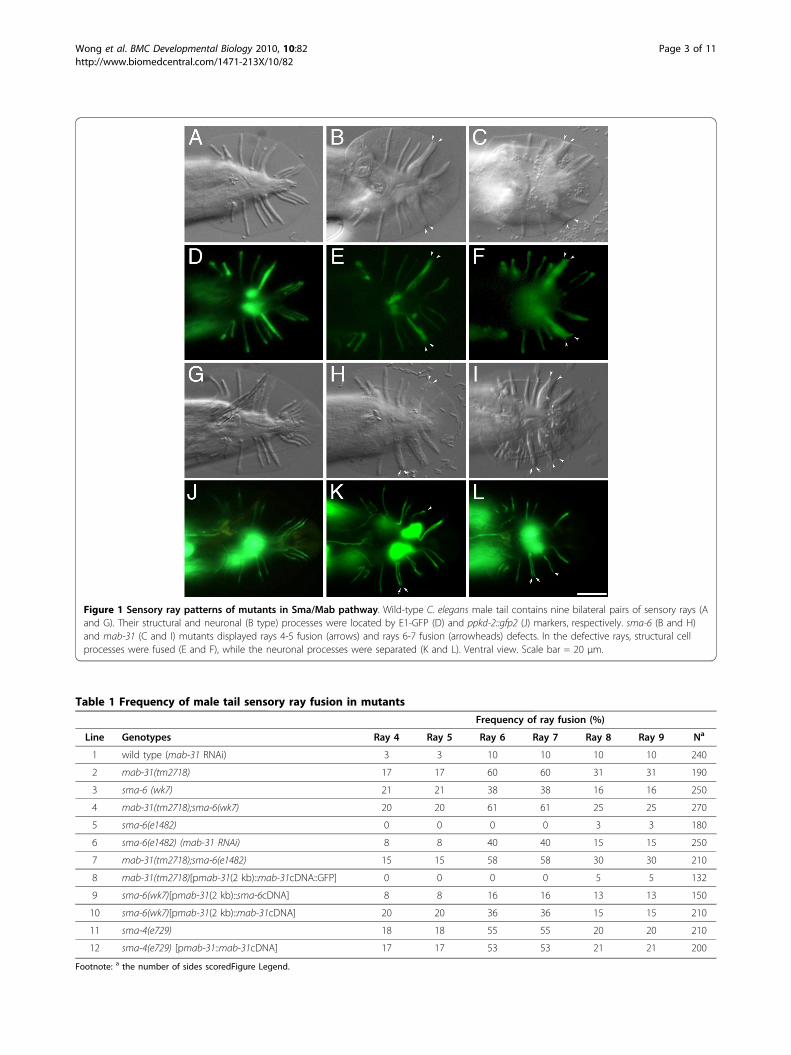
Figure 1 Sensory ray patterns of mutants in Sma/Mab pathway. Wild-type C. elegans male tail contains nine bilateral pairs of sensory rays (Aand G). Their structural and neuronal (B type) processes were located by E1-GFP (D) and ppkd-2::gfp2 (J) markers, respectively. sma-6 (B and H)and mab-31 (C and I) mutants displayed rays 4-5 fusion (arrows) and rays 6-7 fusion (arrowheads) defects. In the defective rays, structural cellprocesses were fused (E and F), while the neuronal processes were separated (K and L). Ventral view. Scale bar = 20 μm.
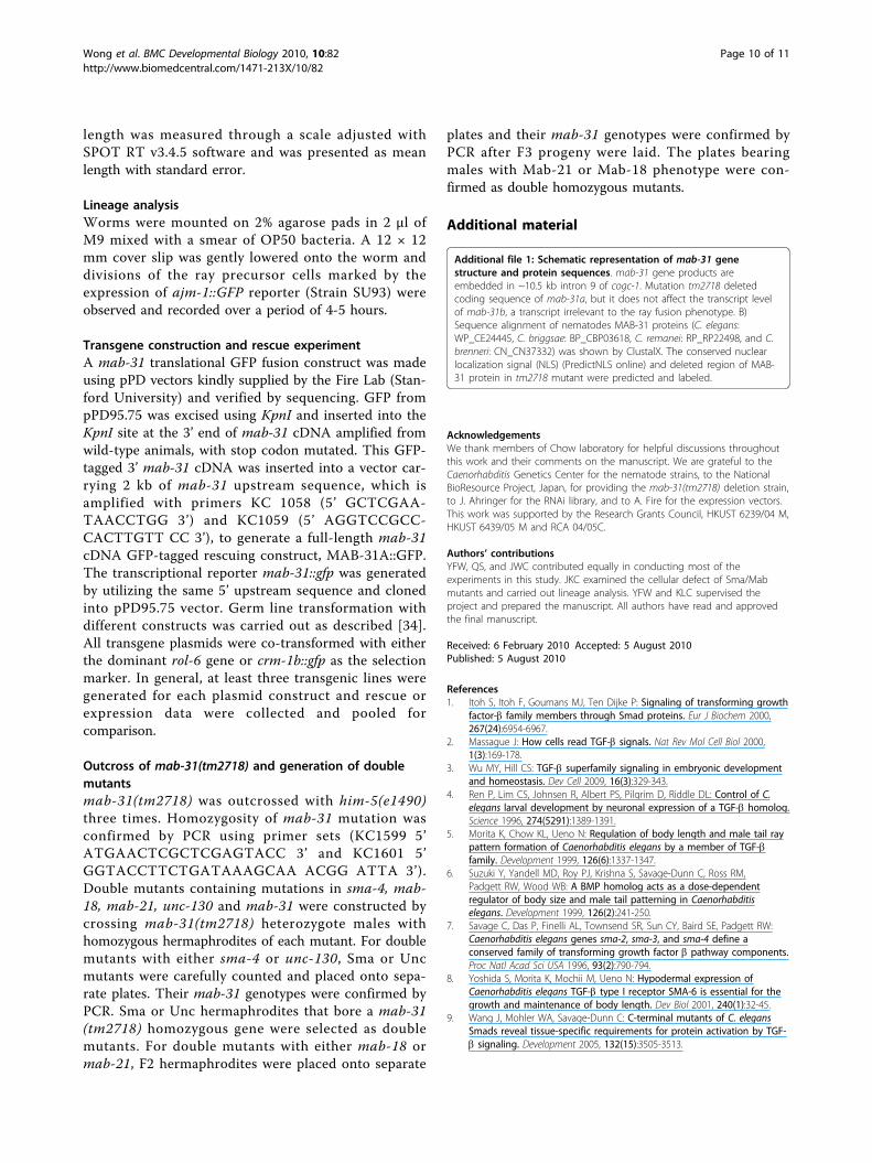
Table 1 Frequency of male tail sensory ray fusion in mutants
Frequency of ray fusion (%)
Line Genotypes Ray 4 Ray 5 Ray 6 Ray 7 Ray 8 Ray 9 Na
1 wild type (mab-31 RNAi) 3 3 10 10 10 10 240
2 mab-31(tm2718) 17 17 60 60 31 31 190
3 sma-6 (wk7) 21 21 38 38 16 16 250
4 mab-31(tm2718);sma-6(wk7) 20 20 61 61 25 25 270
5 sma-6(e1482) 0 0 0 0 3 3 180
6 sma-6(e1482) (mab-31 RNAi) 8 8 40 40 15 15 250
7 mab-31(tm2718);sma-6(e1482) 15 15 58 58 30 30 210
8 mab-31(tm2718)[pmab-31(2 kb)::mab-31cDNA::GFP] 0 0 0 0 5 5 132
9 sma-6(wk7)[pmab-31(2 kb)::sma-6cDNA] 8 8 16 16 13 13 150
10 sma-6(wk7)[pmab-31(2 kb)::mab-31cDNA] 20 20 36 36 15 15 210
11 sma-4(e729) 18 18 55 55 20 20 210
12 sma-4(e729) [pmab-31::mab-31cDNA] 17 17 53 53 21 21 200
Footnote: a the number of sides scoredFigure Legend.
Wong et al. BMC Developmental Biology 2010, 10:82http://www.biomedcentral.com/1471-213X/10/82
Page 3 of 11

males unable to mate. The mutant hermaphrodites hada low brood size of (33.7 ± 15.4) as compared withwild-type (212.3 ± 31.5) [n = 20 for both]. These pheno-types are all specific, as they resemble those displayedby mab-31(RNAi) animals, and all abnormal phenotypescould be rescued by the functional transgene, pmab-31(2 kb)::mab-31cDNA.
Specification of sensory rays requires both mab-31 andsma-6 at the L3/L4 larval stageRay identity determination is a dynamic process of rayprecursor (Rn) cells differentiation in late larval stagesof C. elegans male development. The underlying processof cellular specification of Rn cells is not well under-stood. Therefore, we examined and compared ray cellcluster profiles of wild-type, sma-6(wk7), and mab-31(tm2718) animals. To achieve this, we traced the Rncells lineage with an apical junction marker ajm-1::GFP.We were particularly interested in differentiation of R6and R7, since rays 6-7 fusion was the most obvious phe-notype in both mab-31 and other sma mutants. WhenR(6/7).a and R(6/7).p cells were born, there was nodetectable difference in wild-type (Figure 2A), sma-6(Figure 2E) and mab-31 mutants (Figure 2I). In the
wild-type animal, R7.aa and R7.ap (descendents of R7.a)were produced along the A-P axis and stayed on thedorsal side of R7.p (Figure 2B). However, in sma-6 andmab-31 mutants, these two cells were present on theventral side of R7.p after their birth (Figure 2F and 2J).In these mutants, the abnormal ray cell group derivedfrom R7 sat next to that of R6 (Figure 2G and 2K) andsubsequently, their structural cells were juxtaposed toeach other and were fused together (Figure 2H and 2L).However, no cellular abnormality was observed in differ-entiation of R6a.a, R6a.p. or R6.p cells, in both mutants,as compared with those in wild-type animals. The defectin rays 6-7 fusion involved mis-localization of ray 7 pre-cursors to ray 6 precursors during ray identity determi-nation window at the L4 larval stage. Our observationsuggested that both sma-6 and mab-31 mutants have awild-type ray lineage with ray precursor cells born atthe right time. The defects in these mutants were simplydue to transformation of the ray cell identity, which dic-tates the cell cluster’s positioning with respect to otherray cell groups. Indeed, similar defects were subse-quently observed in other sma mutants (sma-4 and dbl-1, data not shown) also, inferring that the mab-31 geneprobably acts in the same canonical dbl/sma pathway.
Figure 2 Abnormal ray clustering patterns of mutants in Sma/Mab pathway. The formation of daughter cells from ray precursor cells (Rn, n= 1-9) in different developmental stages was examined with apical junction markers ajm-1::gfp. Between mid-L3 and mid-L4 stage, Rn cells weredivided by a stereotyped lineage pattern giving rise to ray cell groups (RCG) and were subsequently assembled (A-D) in wild-type animals. Noabnormality is noted in the division of Rn.a and Rn.p cells in sma-6 (E) and mab-31 (I) mutants. In wild-type male, R7.aa and R7.ap are born atthe dorsal side of R7.p (B). However, in sma-6 (F) and mab-31 (J) mutants, both R7.aa cells and R7.ap cells are skewed towards the ventral side ofR7.p cell. The abnormal R7.aa and R7.ap reside next to the R6.aa and R6.ap cells. RCGs were well-separated in wild-type male tail (C) but not inboth in sma-6 (G) and mab-31 (K) mutants. At a later stage, cellular components of ray 6 and ray 7 are clustered in close proximity and theirstructural cells are fused together (arrows in H and L). Lateral view, left side upwards. Hour (hrs) post-hatching at 20°C. Scale bar = 10 μm.
Wong et al. BMC Developmental Biology 2010, 10:82http://www.biomedcentral.com/1471-213X/10/82
Page 4 of 11

It was shown that the ray pattern in the adult maletail results from targeting of ray structural cell processesto specific sites, called papillae, in the L4 lateral epider-mis [20]. Fused rays arise when multiple papillae arejuxtaposed to each other in a cluster. Some of papillaewere displaced in mab-31(tm2718) (65%) and sma-6(wk7) (45%) mutants (N = 50). For example, the ray 7papillae in mutant animals were not found in theirnative position but often at an ectopic position just nextto the ray 6 papillus. This displacement is also consis-tent with our observation that ray 6 and 7 cell groupswere next to each other during ray development asdescribed above (Figure 2G &2K). The same featureapplies to papillae of ray 4 and 5, and to those of ray 8and 9.We further used a genetic approach to determine the
functional relationship between sma-6 and mab-31 inthe specification of sensory rays. Both mab-31(tm2718)and sma-6(wk7) carry recessive null mutations for raypatterning defects. We compared the frequency of rays6-7 fusion phenotype in each single mutant as well as indouble mutants. Worms doubly mutant for mab-31 andsma-6 do not exhibit an enhanced frequency of fusionfor rays 6-7 (58%) compared to mab-31 single mutants(60%; Table 1). Thus, they are unlikely to be working inparallel for the ray 7 specification. The null allele ofsma-6(wk7) showed both Sma and Mab phenotypes.However, in a weak loss-of-function allele sma-6(e1482),the male tail defect could not be observed. It was sug-gested that the residual activity of sma-6(e1482) is suffi-cient for the male tail development, but its level isbelow the thresholds required for body size regulation[25]. Interestingly, a reduction of sma-6 activity in e1482mutant background could enhance mab-31(RNAi) rays6-7 fusion phenotype by four-fold, as compared with thesame treatment in wild-type animals (Table 1, lines 1 &6). This synergistic effect strongly argues that these twogenes act in the same process and pathway. Further-more, phenotypes of mab-31(tm2718);sma-6(e1482) dou-ble mutants resembled those observed in the mab-31single mutant (Table 1, lines 2 & 7), suggesting thatmab-31 is an indispensable component required forSma/Mab pathway signaling during male taildevelopment.
MAB-31 may cooperate with SMAD inside nucleusIn the Sma/Mab pathway, SMA-6 receptors transducethe extracellular signal to the effectors, SMA-2, SMA3,and SMA-4, which regulate the expression of down-stream targets. Would mab-31 be one of the targets withexpression regulated by the SMA-6 receptor andmediated by SMAD transcription factors in rays? Toaddress this question, we first characterized the expres-sion pattern of mab-31 gene. A 2 kb 5’ upstream
sequence of the mab-31 translational start was utilizedfor construction of a transcriptional mab-31::gfp reporter.The promoter sequence contained regulatory elementsrequired for ray development, as it could be employedfor expressing mab-31 cDNA to rescue both male tail(Table 1, lines 2 & 8) and other phenotypes in corre-sponding mab-31(tm2718) mutants. Indeed, the same 2kb mab-31 5’ upstream sequence could be used to driveexpression of sma-6 cDNA and rescue sma-6(wk7)mutant ray phenotype (from ray 6,7 fusion drops from38% to 16%, Table 1, lines 3 & 9). But the significant res-cue was not observed in body length (0.82 ± 0.04 mm forsma-6(wk7) of the animals and 0.87 ± 0.06 mm for thesame mutant with a transgene). This mab-31 2 Kb 5’franking sequence was active in gut cells of the pretzel-stage embryo and throughout the larval stages. In adultmales and hermaphrodites, gfp reporter signal wasobserved in the pharynx, body hypodermis, and intestine.Interestingly, the type I receptor SMA-6 is expressed inall these tissues overlapping with the mab-31 pattern[25]. Yet, while sma-6 mutant animals have a small bodyphenotype, mab-31 mutants do not. Such a difference isprobably due to the fact that mab-31 is not required norexpressed at a significant level at the early larval stagewhen sma-6 and smad functions are needed [26]. On theother hand, mab-31 expression is also observed in sup-port cells of neuronal sensilla, like amphid socket cells(Figure 3B) and phasmid socket cells (Figure 3D), whereno expression of sma-6 has been documented. In thewild-type male tail, the mab-31 transcriptional reporterexpression was detected in structural cells highlightingthe cell bodies and processes of all sensory rays (Figure3F). Most importantly, the expression level was not chan-ged in either sma-6 or sma-4 mutant backgrounds (datanot shown). Indeed, high copies of mab-31 functionaltransgenes in ray structural cells cannot by-pass therequirements of either sma-6 or sma-4 in rays (Table 1,lines 3 & 10, 11 & 12). These observations suggest thatmab-31 is not acting transcriptionally downstream of theSma/Mab signaling cascade.We explored the molecular function of MAB-31 in the
Sma/Mab pathway by evaluating its cellular localization.MAB-31 is highly conserved across several Caenorhab-ditis species (Additional file 1), but it does not resembleany protein other than its worm counterparts. Based onthe amino acid sequence, a stretch of polypeptide(RKRREK) was predicted as a nuclear localization signal(NLS) (PredictNLS online) implicating it to functioninside the nucleus. To ascertain its in vivo subcellularlocalization, we generated transgenic animals carrying aMAB-31 translational fusion with GFP tagged at its C-terminus. This MAB-31::GFP protein was a functionalequivalent to endogenous MAB-31, since it could fullyrescue rays fusion phenotypes in mab-31(tm2718)
Wong et al. BMC Developmental Biology 2010, 10:82http://www.biomedcentral.com/1471-213X/10/82
Page 5 of 11

(Rescue efficiency = 100%, N = 50) (Table 1, line 8).MAB-31:GFP was found to be localized in nuclei (Figure3H), but not in cytoplasm in all developmental stagesthat we had examined. It was produced at the pretzel-stage and then in all expressing cells implicated by thetranscriptional reporter. Localization of MAB-31::GFPprotein was altered neither in sma-4(e729) nor in sma-6(wk7) mutants (N = 20 in both cases) throughout theray differentiation period and in the adult stage. Thus,MAB-31 is likely acting constitutively in the nuclearcompartment rather than being shuttled into thenucleus, like the Smad proteins.
Sma/Mab signaling is modulated by epistatic interactionsamong mab-18, mab-31, and unc-130 in ray patterningSeveral components in Sma/Mab pathway were shownto negatively regulate the mab-21 gene in ray patterning
[5]. In mab-21 mutants, the ray 6 structural cell appar-ently undergoes a specific transformation into a ray 4morphogenetic identity and ray 6 is fused with ray 4.Double mutant of mab-21 with either sma-6 or sma-4resulted in rays 6-4 fusion phenotype, which mimickedthe Mab-21 phenotype. We could also establish a similarepistatic relationship between mab-31 and the compo-nents in mab-21 pathway. In the double mutant ofmab-31(tm2718); mab-21(bx53), only Mab-21 phenotypecould be observed (Figure 4A). Thus the role of mab-31is similar to Sma/Mab pathway, which negatively regu-lates mab-21 during the specification of ray 6.Since the specification of ray 6 by Sma/Mab pathway
could be used to dissect genetic function for mab-31,we extended our study to look for the epistatic relation-ship of mab-31 with other candidates. Pax-6 is a con-served transcriptional factor that plays a variety of rolesduring metazoan development, primarily in patterningelements of the nervous system [27]. The C. eleganspairbox-less Pax6 isoform, mab-18, acts cell-autono-mously in ray 6 [28,29]. Loss-of-function mab-18 alsoresults in transformation of ray 6 to ray 4 identity (Fig-ure 4C). In double mutants of mab-31 and mab-18, onlyMab-18 ray fusion phenotype was observed (Figure 4Aand 4E). The same phenotype was also observed in dou-ble mutants of mab-18 with either sma-4 or sma-2 (datanot shown, N>100). Thus, Sma/Mab pathway is pro-posed to have acted upstream of mab-18.The fork-head transcription factor unc-130 functions in
both ventral body muscle and male tail developments[30]. unc-130 acts as a negative regulator of unc-129 andworks autonomously to repress unc-129 expression inventral body muscles. In the male tail, unc-130 isrequired for keeping ray 6 identity independent of unc-129. unc-130(ev505) mutant displayed a rays 6-4 fusionphenotype of 82% of the animals. In the double mutantof mab-31(tm2718);unc-130(ev505), only the rays 6-7fusion phenotype was observed (Figure 4A). This findingsuggests that unc-130 works further upstream of Sma/Mab pathway and negatively regulates mab-31 in the raypatterning. Based on all these observations, we propose anovel genetic cascade acting uniquely in the male tailsensory rays specification; mab-18 is the downstream tar-get of mab-31. MAB-31 is localized in the nucleus, andwe predict that it acts downstream of the SMA-6 recep-tor (and also DAF-4 receptor, data not shown), and thisTGF-b signaling cascade is negatively regulated by unc-130 acting as the most upstream regulator in this process.
DiscussionInterpretation of the Sma/Mab signal by the ray lineageduring male developmentIn this paper, we have reported the characterization of anuclear factor encoded by mab-31 in the C. elegans for
Figure 3 Expression pattern of mab-31. The gfp expression frommab-31::gfp transcriptional reporter is found in amphid sockets(arrows) (B) and phasmid sockets (arrowheads) (D) in ahermaphrodite. In the male tail, gfp signal is detected in structuralcell processes of all sensory rays (F). The functional MAB-31::GFPprotein is localized in multiple ray cell nuclei (H). Lateral view, leftside upwards for A-D. Ventral view for E-H. Scale bar = 20 μm.
Wong et al. BMC Developmental Biology 2010, 10:82http://www.biomedcentral.com/1471-213X/10/82
Page 6 of 11

sensory ray patterning. mab-31 mutants present nosmall body phenotype, but exhibit male tail defectsresembling the ray fusion phenotypes in existing Smamutants of Sma/Mab pathway. We have shown thatboth MAB-31 nuclear factor and SMA-6 receptor arerequired to function in the ray lineage and the structuralcells during the ray patterning process. They regulatethe ray patterning event by altering the position of the
R7 derived ray cell cluster and how it associates withthe neighboring cluster at the late L4 males. Finally, weestablish the genetic relationship of conserved transcrip-tional regulators in ray patterning, such as unc-130 andmab-18, with Sma/Mab pathway components throughepistatic analysis.In Sma/Mab pathway, the signaling ligand dbl-1 is
expressed primarily in motor neurons along the ventral
Figure 4 Epistatic analysis of Sma/Mab pathway in ray 6 specification. Frequency of ray fusion phenotypes (rays 6-7 fusion or rays 6-4fusion) is shown in double mutants of mab-31 with those of Sma/Mab pathway (A) and with those showing rays 6-4 fusion phenotypes. Indouble mutants of mab-31 and mab-18 (E), only rays 6-4 fusion phenotype was observed, as compared with wild-type (B), mab-18 (C), and mab-31 (D) male tails. Ventral view for B-E. Scale bar = 20 μm.
Wong et al. BMC Developmental Biology 2010, 10:82http://www.biomedcentral.com/1471-213X/10/82
Page 7 of 11

nerve cord in both hermaphrodites and males [5,6]. Wehave previously showed that ectopic expression of Xeno-pus BMP4 in these neurons could functionally rescuebody size [15] and corrected the ray fusion defect(Wong and Chow, unpublished data) in dbl-1 mutant.Expression of the dbl-1 ligand in neuronal processesalong the body is sufficient for its function in the raypatterning, probably through diffusion of this extracellu-lar ligand. Hence, the specificity of Sma/Mab signalingfor ray patterning is likely to be controlled by receptorsresiding in cellular components of the sensory rays butnot by the source of the signal acting at a distance.However, expression patterns revealed by transcriptionalreporters for TGF-b receptors and Smads did not revealtheir expression in any specific cells within the sensoryrays [8,25]. For example, hypodermal expression of sma-6, which is governed by a GATA sequence in the sma-6promoter, was shown to be necessary for maintenanceof body length [8]. This expression, however, was notsufficient to rescue the male tail mutant defect (Ho andChow, unpublished data). In this report, we have shownfor the first time that the mab-31 promoter with activityin the ray structural cell (Figure 3F) could functionallyexpress sma-6 transgene and successfully rescue the rayfusion defect of sma-6 mutant (Table 1, line 9) evenwhen the structural cell-specific sma-6 regulatory ele-ment remains undefined. This finding infers that bothmab-31 and sma-6 likely have overlapping expressionsin the same cells in the ray sublineage including thestructural cells as part of the executing machinery of thedbl-1 signaling event. It is likely that their expression isrequired even earlier in the structural cell precursorsR7.ap or R7.a. Without a good structural cell-specificpromoter active at the early stage of this cell type or inits precursors, direct experimental verification of thisnotion would not be possible. Neither would we be ableto rule out the possibility that simultaneous expressionof sma-6 nor mab-31 in both hypodermis and structuralcells is necessary for establishing correct ray identity. Inaddition, with the cellular defects revealed in mab-31,sma-6 and other sma mutants which converge on hav-ing an abnormal division plane of the R7.a, the action ofthe dbl-1 pathway receptors and relay molecules mayindeed act even as early as the R7.a cell is born.Examination of morphology of ray structural processes
in fused rays of mab-31, sma-6, sma-4, and dbl-1mutants also showed that fusion of structural cells butnot fusion of neuronal processes within rays is the pri-mary defect. This observation is similar to the results ofthe histological examination of the fused ray in mab-21mutant, which implicates that fused structural cells arethe primary cause of ray fusion after the ray identitywas altered [21]. Collectively, these evidences supportthe notion that single ray formation acts through the
ray lineage and manifested through assumption of aunique identity which can be manifested by the struc-tural cells. The outcome is abnormal ray fusion, whenstructural cells of two adjacent rays lose specificity-determinants required to keep the rays apart as inde-pendent entities [16].
Biological role of Sma/Mab signaling in sensory raypatterningMultiple evidences have shown that Sma/Mab signalingin the structural cells is required in patterning of sen-sory rays. The attachment of the structural cells to spe-cific positions of the epidermis and cuticle at late L4stage dictates where the papillae will be formed. In turn,the ultimate position of a ray is determined [20]. Whenthe attachments are altered in different mutants, such asin sma mutants or many mab mutants, ray patterning ischanged [21,31]. However, the underlying process gov-erned by Sma/Mab signaling on how structural cells arelocalized to distinct sites in the epidermis has not beencharacterized. Ray cell groups tend to reside at the junc-tions of two Rn.p cells. These hypodermal cells are gen-erated at the first ray precursor cell division before thecell cluster patterning is obvious (Figure 2C). Ray 5 and7 cell groups are born above their respective Rn.p cellswhile the others stay beneath the hypodermal daughterat birth. We have analyzed the arrangement and geome-try of developing ray cell groups of the late L3 and L4stages to examine how these sites are altered in mutantsof Sma/Mab pathway. Our results demonstrate thatpositions of R6 derived cells are essentially normal inthese mutants. Sma/Mab signaling is not involved inpre-patterning of ray 6 precursor cells. Similarly, forma-tion of parental cells R7.a and R7.p in either mab-31 orsma-6 mutants did not differ from that in wild-type ani-mals. However, in mutants, R7.a derivatives were abnor-mally positioned beneath the R7.p cells (Figure 2G and2K), which may probably have been caused by the initialskewing of cell divisions leading to the positioning ofR7.aa and R7.ap ventral to R7.p (Figure 2F and 2J). Thisfeature indicates that in wild-type animals, Sma/Mabsignaling event is required in the R7.a for proper forma-tion of its daughter cells, R7.aa and R7.ap, possibly byskewing their division plane towards the dorsal side ofR7.a. Such organization is maintained in wild-type indi-viduals to separate the daughters of R6.a and R7.a overa distance by R5.p and R6.p. We therefore hypothesizethat a novel mechanism of Sma/Mab signaling acts insensory rays patterning. Receptors SMA-6 and DAF-4are located at R7.a and are activated upon binding ofthe DBL-1 ligand, which is secreted from the ventralnerve cord. The signal is then transmitted throughintracellular SMAD molecules and nuclear factor MAB-31, to its targets which are potentially required for
Wong et al. BMC Developmental Biology 2010, 10:82http://www.biomedcentral.com/1471-213X/10/82
Page 8 of 11

establishment and maintenance of cell polarity of R7.aand thus identity of its descendants. Disruption of thissignaling event may change the cellular fate of R7.a andits subsequent plain of division leading to an abnormalposition of the daughter cells with respect to the neigh-boring cells. As a result, fusion of ray structural cells, adescendent of R7.a, is possible because of the loss ofidentity and physical proximity between the fusing part-ners. It ends up with a ray 6-7 fusion defect. Interest-ingly, in the absence of mab-18, this Sma/Mab signal isdispensable and the identity of ray 7 could be restored.Would the maintenance of ray 7 identity involve deregu-lation of mab-18 activity through the Sma/Mab signal-ing? That’s certainly a possibility since MAB-18subcellular localization in the ray 7 cluster is develop-mentally regulated [29]. Nevertheless, the precise mole-cular regulation involving MAB-31 and SMADmolecules demands a more detailed characterization. Itwill be an important prerequisite before the operation ofa complete genetic network during sensory ray pattern-ing can be addressed.
Conclusionmab-31 is identified to encode a nuclear protein thatacts in the Sma/Mab pathway during C. elegans malesensory ray cell development. We have shown by genet-ics that mab-31 acts in Sma/Mab signal receiving cellsin the ray lineage. Furthermore, we have demonstratedby epistatic analysis that the Sma/Mab pathway actsupstream of mab-21 and mab-18 within the ray lineageincluding the structural cells, and the entire pathway isnegatively regulated by unc-130 upstream of the dbl-1signals. These data collectively suggest potential cross-talk between ray cells with patterning signal acting viathe diffusible DBL-1 and the involvement of importantstructural cell-specific molecules to mediate cell fatespecification and subsequent differentiation.
MethodsStrainsStrains were maintained as described by Brenner [10].To facilitate male tail analysis, each strain, including themab-31(tm2718) mutant, was crossed with him-5(e1490)V to generate a double mutant (see below). Strainnames and genotypes of animals used were:FX27180: mab-31(tm2718)I,NF2990: cogc-1(k179)I,CB40880: him-5(e1490)V,KC8250: mab-31(tm2718)I; him5(e1490) V,KC1260: sma-6(wk7)II; him-5(e1490)V,KC5880: sma-6(e1482)II; him-5(e1490)V,KC5470: unc-130(ev505)II; him-5(e1490)V,KC4470: rrf-3(pk1426)II; him-5(e1490)V,EM1280: mab-21(bx53)III; him-5(e1490)V,
KC5360: sma-4(e729)III; him-5(e1490)V,EM660: him-5(e1490) V; mab-18 (bx23) X,KC8720: mab-31(tm2718)I; dpy-17(e164) mab-21
(bx53) III; him-5(e1490)V,KC9250: mab-31(tm2718)I; sma-6(wk7)II; him-5
(e1490)V,KC10700: mab-31(tm2718)I; him-5(e1490)V; mab-18
(bx23)X,KC10710: mab-31(tm2718)I; unc-130(ev505)II; him-5
(e1490)V,KC8410: wxIs29(pRF4+pKS+T24C2sphIGFPNLS-)I;
sma-6(wk7)II; him-5(e1490)V,KC8430: sma-6(wk7)II; su93[jam1::GFP; pRF4; unc-29]
JcIs1IV; him-5(e1490)V,KC8420: sma-6(wk7)II; him-5(e1490) wxIs52
[pB0024.14b::gfp+ppkd-2::gfp2]V,KC8470: mab-31(tm2718) wxIs29(pRF4+pKS+
T24C2sphIGFPNLS-)I;him-5(e1490)V,KC8480: mab-31(tm2718)I;him-5(e1490)V;wxIs52
[pB0024.14b::gfp+ppkd-2::gfp2]V,KC8490: mab-31(tm2718)I; jcIs1[ajm1::gfp; pRF4; unc-
29]IV; him-5(e1490)V,KC8090: him-5(e1490)V; wxEx64[pRF4+pmab-31::gfp
(2.0 k)],KC9640: him-5(e1490)V; wxEx123[pRF4+pmab-31(2
kb)::cDNA::GFP], andKC9650: mab-31(tm2718)I; su93[jam-1::GFP; pRF4;
unc-29]JcIs1IV;him-5(e1490)V; wxEx122[pcrm-1b::gfp+pmab-31(2 kb)::cDNA::GFP]
RNAiRNAi clone I-7J10 was from the Ahringer RNAi library(MRC Geneservice) [32]. The identity of this clone wasdetermined by in house sequencing. RNAi bacteria culti-vation and double-stranded RNA induction were per-formed as described by Kamath et al. [33]. KC447worms were synchronized by hypochlorite treatment,and the resulting L1 animals were placed on bacteriaspotted plates and incubated at 20°C. Male tail pheno-types were assessed in adult worms after 3 days. In allRNAi assays, E. coli HT115(DE3) carrying the emptyRNAi vector L4440 was fed to the same strain as anegative control.
Male tail phenotype characterization and body-lengthmeasurementThe male tails were examined by Nomarski microscopy(N > 100). Of the transgenic strains, only transformedworms carrying the markers were imaged and scored.For the body length measurement, L4 hermaphroditesgrown at 20°C were transferred to fresh NGM plates.Five days later, 100 F1 adult hermaphrodites were ran-domly photographed under 5× or 10× objectives withSPOT RT camera (Diagnostic Instruments). Their body
Wong et al. BMC Developmental Biology 2010, 10:82http://www.biomedcentral.com/1471-213X/10/82
Page 9 of 11
length was measured through a scale adjusted withSPOT RT v3.4.5 software and was presented as meanlength with standard error.
Lineage analysisWorms were mounted on 2% agarose pads in 2 μl ofM9 mixed with a smear of OP50 bacteria. A 12 × 12mm cover slip was gently lowered onto the worm anddivisions of the ray precursor cells marked by theexpression of ajm-1::GFP reporter (Strain SU93) wereobserved and recorded over a period of 4-5 hours.
Transgene construction and rescue experimentA mab-31 translational GFP fusion construct was madeusing pPD vectors kindly supplied by the Fire Lab (Stan-ford University) and verified by sequencing. GFP frompPD95.75 was excised using KpnI and inserted into theKpnI site at the 3’ end of mab-31 cDNA amplified fromwild-type animals, with stop codon mutated. This GFP-tagged 3’ mab-31 cDNA was inserted into a vector car-rying 2 kb of mab-31 upstream sequence, which isamplified with primers KC 1058 (5’ GCTCGAA-TAACCTGG 3’) and KC1059 (5’ AGGTCCGCC-CACTTGTT CC 3’), to generate a full-length mab-31cDNA GFP-tagged rescuing construct, MAB-31A::GFP.The transcriptional reporter mab-31::gfp was generatedby utilizing the same 5’ upstream sequence and clonedinto pPD95.75 vector. Germ line transformation withdifferent constructs was carried out as described [34].All transgene plasmids were co-transformed with eitherthe dominant rol-6 gene or crm-1b::gfp as the selectionmarker. In general, at least three transgenic lines weregenerated for each plasmid construct and rescue orexpression data were collected and pooled forcomparison.
Outcross of mab-31(tm2718) and generation of doublemutantsmab-31(tm2718) was outcrossed with him-5(e1490)three times. Homozygosity of mab-31 mutation wasconfirmed by PCR using primer sets (KC1599 5’ATGAACTCGCTCGAGTACC 3’ and KC1601 5’GGTACCTTCTGATAAAGCAA ACGG ATTA 3’).Double mutants containing mutations in sma-4, mab-18, mab-21, unc-130 and mab-31 were constructed bycrossing mab-31(tm2718) heterozygote males withhomozygous hermaphrodites of each mutant. For doublemutants with either sma-4 or unc-130, Sma or Uncmutants were carefully counted and placed onto sepa-rate plates. Their mab-31 genotypes were confirmed byPCR. Sma or Unc hermaphrodites that bore a mab-31(tm2718) homozygous gene were selected as doublemutants. For double mutants with either mab-18 ormab-21, F2 hermaphrodites were placed onto separate
plates and their mab-31 genotypes were confirmed byPCR after F3 progeny were laid. The plates bearingmales with Mab-21 or Mab-18 phenotype were con-firmed as double homozygous mutants.
Additional material
Additional file 1: Schematic representation of mab-31 genestructure and protein sequences. mab-31 gene products areembedded in ~10.5 kb intron 9 of cogc-1. Mutation tm2718 deletedcoding sequence of mab-31a, but it does not affect the transcript levelof mab-31b, a transcript irrelevant to the ray fusion phenotype. B)Sequence alignment of nematodes MAB-31 proteins (C. elegans:WP_CE24445, C. briggsae: BP_CBP03618, C. remanei: RP_RP22498, and C.brenneri: CN_CN37332) was shown by ClustalX. The conserved nuclearlocalization signal (NLS) (PredictNLS online) and deleted region of MAB-31 protein in tm2718 mutant were predicted and labeled.
AcknowledgementsWe thank members of Chow laboratory for helpful discussions throughoutthis work and their comments on the manuscript. We are grateful to theCaenorhabditis Genetics Center for the nematode strains, to the NationalBioResource Project, Japan, for providing the mab-31(tm2718) deletion strain,to J. Ahringer for the RNAi library, and to A. Fire for the expression vectors.This work was supported by the Research Grants Council, HKUST 6239/04 M,HKUST 6439/05 M and RCA 04/05C.
Authors’ contributionsYFW, QS, and JWC contributed equally in conducting most of theexperiments in this study. JKC examined the cellular defect of Sma/Mabmutants and carried out lineage analysis. YFW and KLC supervised theproject and prepared the manuscript. All authors have read and approvedthe final manuscript.
Received: 6 February 2010 Accepted: 5 August 2010Published: 5 August 2010
References1. Itoh S, Itoh F, Goumans MJ, Ten Dijke P: Signaling of transforming growth
factor-b family members through Smad proteins. Eur J Biochem 2000,267(24):6954-6967.
2. Massague J: How cells read TGF-b signals. Nat Rev Mol Cell Biol 2000,1(3):169-178.
3. Wu MY, Hill CS: TGF-b superfamily signaling in embryonic developmentand homeostasis. Dev Cell 2009, 16(3):329-343.
4. Ren P, Lim CS, Johnsen R, Albert PS, Pilgrim D, Riddle DL: Control of C.elegans larval development by neuronal expression of a TGF-b homolog.Science 1996, 274(5291):1389-1391.
5. Morita K, Chow KL, Ueno N: Regulation of body length and male tail raypattern formation of Caenorhabditis elegans by a member of TGF-bfamily. Development 1999, 126(6):1337-1347.
6. Suzuki Y, Yandell MD, Roy PJ, Krishna S, Savage-Dunn C, Ross RM,Padgett RW, Wood WB: A BMP homolog acts as a dose-dependentregulator of body size and male tail patterning in Caenorhabditiselegans. Development 1999, 126(2):241-250.
7. Savage C, Das P, Finelli AL, Townsend SR, Sun CY, Baird SE, Padgett RW:Caenorhabditis elegans genes sma-2, sma-3, and sma-4 define aconserved family of transforming growth factor b pathway components.Proc Natl Acad Sci USA 1996, 93(2):790-794.
8. Yoshida S, Morita K, Mochii M, Ueno N: Hypodermal expression ofCaenorhabditis elegans TGF-b type I receptor SMA-6 is essential for thegrowth and maintenance of body length. Dev Biol 2001, 240(1):32-45.
9. Wang J, Mohler WA, Savage-Dunn C: C-terminal mutants of C. elegansSmads reveal tissue-specific requirements for protein activation by TGF-b signaling. Development 2005, 132(15):3505-3513.
Wong et al. BMC Developmental Biology 2010, 10:82http://www.biomedcentral.com/1471-213X/10/82
Page 10 of 11

10. Brenner S: The genetics of Caenorhabditis elegans. Genetics 1974,77(1):71-94.
11. Mochii M, Yoshida S, Morita K, Kohara Y, Ueno N: Identification oftransforming growth factor-b- regulated genes in Caenorhabditis elegansby differential hybridization of arrayed cDNAs. Proc Natl Acad Sci USA1999, 96(26):15020-15025.
12. Li S, Armstrong CM, Bertin N, Ge H, Milstein S, Boxem M, Vidalain PO,Han JD, Chesneau A, Hao T, Goldberg DS, Li N, Martinez M, Rual JF,Lamesch P, Xu L, Tewari M, Wong SL, Zhang LV, Berriz GF, Jacotot L,Vaglio P, Reboul J, Hirozane-Kishikawa T, Li Q, Gabel HW, Elewa A,Baumgartner B, Rose DJ, Yu H, Bosak S, Sequerra R, Fraser A, Mango SE,Saxton WM, Strome S, Van Den Heuvel S, Piano F, Vandenhaute J, Sardet C,Gerstein M, Doucette-Stamm L, Gunsalus KC, Harper JW, Cusick ME,Roth FP, Hill DE, Vidal M: A map of the interactome network of themetazoan C. elegans. Science 2004, 303(5657):540-543.
13. Savage-Dunn C, Maduzia LL, Zimmerman CM, Roberts AF, Cohen S,Tokarz R, Padgett RW: Genetic screen for small body size mutants in C.elegans reveals many TGF-b pathway components. Genesis 2003,35(4):239-247.
14. Maduzia LL, Roberts AF, Wang H, Lin X, Chin LJ, Zimmerman CM, Cohen S,Feng XH, Padgett RW: C. elegans serine-threonine kinase KIN-29modulates TGF-b signaling and regulates body size formation. BMC DevBiol 2005, 5:8.
15. Wong YF, Ko FCF, Cheah KSE, Chow KL: crm-1 facilitates BMP signaling tocontrol body size in Caenorhabditis elegans. Dev Biol 2007, 311(1):95-105.
16. Sulston JE, Albertson DG, Thomson JN: The Caenorhabditis elegans male:postembryonic development of non-gonadal structures. Dev Biol 1980,78(2):542-576.
17. Loer CM, Kenyon CJ: Serotonin-deficient mutants and male matingbehavior in the nematode Caenorhabditis elegans. J Neurosci 1993,13(12):5407-5417.
18. Liu KS, Sternberg PW: Sensory regulation of male mating behavior inCaenorhabditis elegans. Neuron 1995, 14(1):79-89.
19. Troemel ER, Chou JH, Dwyer ND, Colbert HA, Bargmann CI: Divergentseven transmembrane receptors are candidate chemosensory receptorsin C. elegans. Cell 1995, 83(2):207-218.
20. Baird SE, Fitch DH, Kassem IA, Emmons SW: Pattern formation in thenematode epidermis: determination of the arrangement of peripheralsense organs in the C. elegans male tail. Development 1991,113(2):515-526.
21. Chow KL, Hall DH, Emmons SW: The mab-21 gene of Caenorhabditiselegans encodes a novel protein required for choice of alternate cellfates. Development 1995, 121(11):3615-3626.
22. Yu RY, Nguyen CQ, Hall DH, Chow KL: Expression of ram-5 in thestructural cell is required for sensory ray morphogenesis inCaenorhabditis elegans male tail. EMBO J 2000, 19(14):3542-3555.
23. Barr MM, Sternberg PW: A polycystic kidney-disease gene homologuerequired for male mating behaviour in C. elegans. Nature 1999,401(6751):386-389.
24. Gumienny TL, Padgett RW: A small issue addressed. Bioessays 2003,25(4):305-308.
25. Krishna S, Maduzia LL, Padgett RW: Specificity of TGF-b signaling isconferred by distinct type I receptors and their associated SMADproteins in Caenorhabditis elegans. Development 1999, 126(2):251-260.
26. Jiang M, Ryu J, Kiraly M, Duke K, Reinke V, Kim SK: Genome-wide analysisof developmental and sex-regulated gene expression profiles inCaenorhabditis elegans. Proc Natl Acad Sci USA 2001, 98:218-223.
27. Callaerts P, Halder G, Gehring WJ: PAX-6 in development and evolution.Annu Rev Neurosci 1997, 20:483-532.
28. Zhang Y, Emmons SW: Specification of sense-organ identity by aCaenorhabditis elegans Pax-6 homologue. Nature 1995, 377(6544):55-59.
29. Zhang Y, Ferreira HB, Greenstein D, Chisholm A, Emmons SW: Regulatednuclear entry of the C. elegans Pax-6 transcription factor. Mech Dev 1998,78(1-2):179-187.
30. Nash B, Colavita A, Zheng H, Roy PJ, Culotti JG: The forkhead transcriptionfactor UNC-130 is required for the graded spatial expression of theUNC-129 TGF-b guidance factor in C. elegans. Genes Dev 2000,14(19):2486-2500.
31. Chow KL, Emmons SW: HOM-C/Hox genes and four interacting locidetermine the morphogenetic properties of single cells in thenematode male tail. Development 1994, 120:2579-2592.
32. Kamath RS, Ahringer J: Genome-wide RNAi screening in Caenorhabditiselegans. Methods 2003, 30(4):313-321.
33. Kamath RS, Martinez-Campos M, Zipperlen P, Fraser AG, Ahringer J:Effectiveness of specific RNA-mediated interference through ingesteddouble-stranded RNA in Caenorhabditis elegans. Genome Biol 2001, 2(1):Research0002.
34. Mello CC, Kramer JM, Stinchcomb D, and Ambros V: Efficient gene transferin C. elegans: extrachromosomal maintenance and integration oftransforming sequences. EMBO J 1991, 10:3959-3970.
doi:10.1186/1471-213X-10-82Cite this article as: Wong et al.: mab-31 and the TGF-b pathway act inthe ray lineage to pattern C. elegans male sensory rays. BMCDevelopmental Biology 2010 10:82.
Submit your next manuscript to BioMed Centraland take full advantage of:
• Convenient online submission
• Thorough peer review
• No space constraints or color figure charges
• Immediate publication on acceptance
• Inclusion in PubMed, CAS, Scopus and Google Scholar
• Research which is freely available for redistribution
Submit your manuscript at www.biomedcentral.com/submit
Wong et al. BMC Developmental Biology 2010, 10:82http://www.biomedcentral.com/1471-213X/10/82
Page 11 of 11
Copyright © 2022 FDOKUMEN


