Axonal transport defects: a common theme in neurodegenerative diseases

Author's personal copy
Integrating the stress response:lessons for neurodegenerativediseases from C. elegansVeena Prahlad and Richard I. Morimoto
Department of Biochemistry, Molecular Biology and Cell Biology, Rice Institute for Biomedical Research, Northwestern University,Evanston, IL 60208, USA
All cells possess surveillance and homeostatic mech-anisms to adjust protein biogenesis to the demands ofgrowth, differentiation, ageing and environmentalstress. However, under certain circumstances, thesemechanisms fail to adequately respond to proteotoxicimbalances and result in the accumulation of mis-folded proteins. In humans, this can lead to neurode-generation and other protein conformational diseases.To protect itself, the cell employs highly conservedstress responses and chaperone networks to maintainprotein-folding homeostasis (proteostasis). Althoughthe regulation of stress responses, such as the heat-shock response, and of proteostasis have been widelyconsidered to be cell autonomous, recent studiesusing Caenorhabditis elegans have shown that theseprocesses are regulated by neuronal signaling andendocrine pathways and integrated into other func-tions of the organism. The hierarchical control of thecellular proteostasis machinery affords insight into theorganization of stress regulatory networks in multi-cellular organisms and offers novel targets for thetreatment of human protein conformational diseases.
Protein misfolding and the stress responseCells respond to rapid changes in their protein biogenesisrequirements or exposure to proteotoxic environmentalconditions, such as heat, oxidative stress or transitionmetals, by inducing a highly conserved program of generegulation (the stress response) [1–4]. Studies on unicel-lular organisms such as bacteria and yeast and on isolatedDrosophila and mammalian cells have shown that thestress response is triggered cell autonomously by changesin the intracellular flux of misfolded proteins that accom-pany these physiological or environmental challenges [1–4]. However, the genetic program implemented by isolatedcultured cells in response to stress can differ from that oforgans or whole organisms exposed to similar stress regi-mens [5,6]. The exposure of an organism to stress con-ditions can result in a selective and asynchronousactivation of the stress response in different tissues[6,7]. Moreover, in human diseases of protein misfolding,such as Parkinson’s disease andHuntington’s disease, onlycertain cells and tissues are at risk. Despite the accumu-lation of damaged proteins, the stress response is not
appreciably induced [3,8–10] in the affected cells, indicat-ing that additional levels of control can exist at the orga-nismal level. Recent advances in the understanding ofstress responses and proteostasis in C. elegans show thatthe cell-autonomous stress responses of individual cells areregulated by neuronal and endocrine signaling to yield anintegrated systemic response [11–13]. C. elegans has pro-ven valuable as a model system to study this issue [4,14–22] (Box 1). Here, we summarize studies on the complexinterplay between cell-autonomous and organismal regu-lation of the stress response and proteostasis in C. elegans.
The role of heat-shock proteins in cytoprotectionThe stress response and the molecular machinery for itsimplementation are conserved from archaebacteria tomammals. The upregulation of heat-shock proteins (HSPs)is central to the stress response. This is concomitant withthe downregulation of genes for normal cellular function[1,2,4]. Elevated expression of HSPs is sufficient to protectcells from a wide range of cytotoxic conditions [1,2,4].HSPs, as molecular chaperones, typically bind to non-native conformations of proteins that persist upon cellstress and these interactions protect against misfolding,aggregation or premature clearance and enable refoldingand the restoration of native conformations [23–25](Figure 1a). Thus, the interaction of chaperones withdiverse substrates in stressed cells or upon increasedprotein biogenesis enhances the stability of the proteomeand restores the activities of signaling and growth regu-latory molecules re-establishing cellular homeostasis[1,2,23,25].
The key regulator of HSP transcription in eukaryotes isheat-shock factor 1 (HSF1), which is highly conserved andubiquitously expressed [24,26]. In the absence of stress, theDNA-binding and transcriptional activities of HSF1 areinhibited by HSPs, which associate weakly to maintain arepressed state (Figure 1a). An increase in the level ofintracellular misfolded proteins thought to trigger thestress response [2,25,27,28] is proposed to titrate HSPsaway from their association with HSF1, enabling HSF1 totrimerize and translocate into the nucleus and activateHSP gene transcription (Figure 1a). Thus, it seems that thecell has evolved an elegant and efficient mechanism toautonomously deploy resources proportional to proteinbiogenesis needs, or in response to damage incurred bythe environmental insult. The basal levels of HSPs set the
Review
Corresponding author: Morimoto, R.I. ([email protected]).
52 0962-8924/$ – see front matter ! 2008 Elsevier Ltd. All rights reserved. doi:10.1016/j.tcb.2008.11.002 Available online 26 December 2008
Author's personal copy
threshold of the stress response, whereas the autoregula-tion of HSF1-dependent HSP transcription ensures there-establishment and maintenance of proteostasis [1–3,23,24].
Whereas the stress response describes the molecularevents associated with damaged proteins in the cytoplasmand nucleus, the unfolded protein response (UPR) providesthe same functionalities for protein misfolding in theendoplasmic reticulum and the mitochondria [29–32](Box 2). Currently, there is limited understanding ofwhy protein misfolding and aggregation cause cellulartoxicity. Although cellular dysfunction caused by aggrega-tion can, in part, be attributed to the loss of function ofthese proteins, protein misfolding seems to also have moregeneral pleiotropic effects on cellular function by limitingessential factors for folding, transport and secretion[3,10,23]. In many cases, these deleterious effects seemto be a result of the ability of misfolded and aggregatedproteins to engage and perturb the proteostasis machineryof the cell [3,33].
Neuroendocrine pathways regulate growth,development and stress tolerance in C. elegans.Although HSP expression is required to re-establish pro-teostasis after stress exposure, the prolonged overexpres-sion of HSPs is detrimental to cell growth and division[2,34,35]. Because the autonomous triggering of the stressresponse within individual cells inmulticellular organismscould disrupt the coordinated functions and interactions oftheir cells and tissue, metazoans must have evolved mech-anisms to integrate their cellular stress response withother organismal processes.
The autoregulatory mechanism of HSP induction andthe steps involved in HSF1 activation provide multipleopportunities for additional regulatory input (Figure 1b,c).HSP genes have cis-regulatory elements that can bindtranscription factors other than HSF1. In C. elegans,two other stress-regulated transcription factors, the FOXOhomologue DAF-16 [36,37] and the nrf-1 homologue SKN-1[38] can also transcribe hsp genes. The upregulation ofHSPs by these alternative pathways would increase the
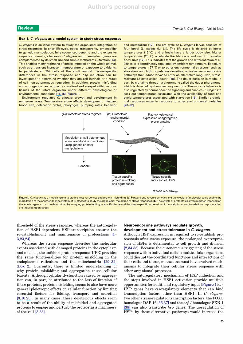
Box 1. C. elegans as a model system to study stress responses
C. elegans is an ideal system to study the organismal integration ofstress responses. Its short life cycle, optical transparency, amenabilityto genetic manipulation, fully sequenced genome and the extensivesequence homology between C. elegans and mammalian genes arecomplemented by its small size and simple method of cultivation [14].This enables many regimens of stress imposed on the whole animal,such as a transient increase in temperature or exposure to oxidants,to penetrate all 959 cells of the adult animal. Tissue-specificdifferences in the stress response and hsp induction can beinvestigated to determine whether they are cell intrinsic or a resultof cell non-autonomous regulation. In addition, protein misfoldingand aggregation can be directly visualized and assayed within varioustissues of the intact organism under different physiological orenvironmental conditions [15,16] (Figure I).
Environment regulates C. elegans growth and development innumerous ways. Temperature alone affects development, lifespan,brood size, defecation cycles, pharyngeal pumping rates, behavior
and metabolism [17]. The life cycle of C. elegans larvae consists offour larval (L) stages (L1–L4). The life cycle is delayed at lowertemperatures (15 8C) and animals have a larger body size; highertemperatures (25 8C) accelerate the life cycle and result in smallerbody sizes [17]. This indicates that the growth and differentiation of all959 cells is coordinately regulated by ambient temperature. Exposureto temperatures >27 8C or to other environmental stressors, such asstarvation and high population densities, activates neuroendocrinepathways that induce larvae to enter an alternative long-lived, stress-resistant L3 state called ‘dauer’ [18]. The dauer decision is made, inpart, by signaling through a pheromone called the dauer pheromone,which is detected by chemosensory neurons. Thermotaxis behavior isalso regulated by neuroendocrine signaling and enables C. elegans toseek out temperatures associated with the availability of food andavoid temperatures associated with starvation [19]. Similar organis-mal responses occur in response to other environmental variables[20–22].
Figure I. C. elegans as a model system to study stress responses and protein misfolding. (a) Forward and reverse genetics and the wealth of molecular tools enable themodulation of the neuroendocrine system of C. elegans to study the organismal regulation of stress responses. (b) The effects of proteotoxic stress regimen imposed onthe whole organism can be determined by assaying protein folding in specific tissue and the tissue-specific expression of transcriptional and translational reporters thatare induced upon stress.
Review Trends in Cell Biology Vol.19 No.2
53
Author's personal copy
tolerance of the cells for misfolded proteins and reset ahigher threshold for HSF1 activation upon stress. Inaddition, HSP induction by HSF1 is itself a multi-stepprocess (Figure 1b,c): activation of HSF1 from an inertmonomer to trimer and binding to DNA are insufficient forHSP transcription and at least one other signal is required[24,26]. HSF1 also undergoes numerous post-translationalmodifications (Figure 1c) including phosphorylation bygrowth-related kinases that affect DNA binding, trimer-ization and transcriptional activity, thereby providingpossibilities for the regulation of the cellular stressresponse by signals other than the increase in misfoldedprotein species [24,26].
Every aspect of C. elegans biology is affected by environ-mental conditions [17,39] (Box 1). Conversely, the overallability of the organism to withstand exposure to adverseenvironmental conditions is modulated by its developmen-tal, physiological, metabolic and nutritional state[11,40,41]. Two kinds of stress response have been widelystudied in C. elegans. The first is the switch in the devel-opmental program of young (L1–L2; Box 1) larvae fromcontinuous reproductive development to developmentallyarrested, stress-resistant dauers [40] (Box 1). The entryinto dauer is regulated by neuroendocrine signaling [40].Thus, the ability of neuroendocrine pathways to regulatestress at the organismal level in C. elegans has been
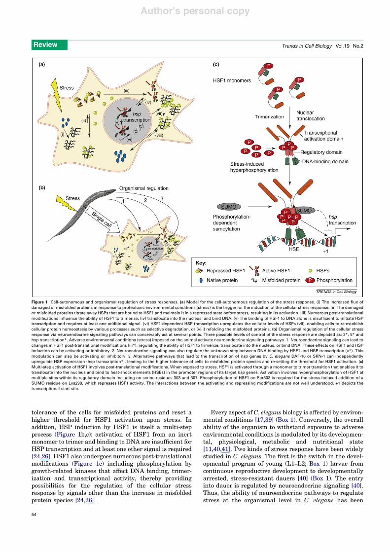
Figure 1. Cell-autonomous and organismal regulation of stress responses. (a) Model for the cell-autonomous regulation of the stress response. (i) The increased flux ofdamaged or misfolded proteins in response to proteotoxic environmental conditions (stress) is the trigger for the induction of the cellular stress response. (ii) The damagedor misfolded proteins titrate away HSPs that are bound to HSF1 and maintain it in a repressed state before stress, resulting in its activation. (iii) Numerous post-translationalmodifications influence the ability of HSF1 to trimerize, (iv) translocate into the nucleus, and bind DNA. (v) The binding of HSF1 to DNA alone is insufficient to initiate HSPtranscription and requires at least one additional signal. (vi) HSF1-dependent HSP transcription upregulates the cellular levels of HSPs (vii), enabling cells to re-establishcellular protein homeostasis by various processes such as selective degradation, or (viii) refolding the misfolded proteins. (b) Organismal regulation of the cellular stressresponse via neuroendocrine signaling pathways can conceivably act at several points. Three possible levels of control of the stress response are depicted as: 3*, 5* andhsp transcription*. Adverse environmental conditions (stress) imposed on the animal activate neuroendocrine signaling pathways. 1. Neuroendocrine signaling can lead tochanges in HSF1 post-translational modifications (iii*), regulating the ability of HSF1 to trimerize, translocate into the nucleus, or bind DNA. These effects on HSF1 and HSPinduction can be activating or inhibitory. 2. Neuroendocrine signaling can also regulate the unknown step between DNA binding by HSF1 and HSP transcription (v*). Thismodulation can also be activating or inhibitory. 3. Alternative pathways that lead to the transcription of hsp genes by C. elegans DAF-16 or SKN-1 can independentlyupregulate HSP expression (hsp transcription*), leading to the higher tolerance of cells to misfolded protein species and re-setting the threshold for HSF1 activation. (c)Multi-step activation of HSF1 involves post-translational modifications. When exposed to stress, HSF1 is activated through a monomer to trimer transition that enables it totranslocate into the nucleus and bind to heat-shock elements (HSEs) in the promoter regions of its target hsp genes. Activation involves hyperphosphorylation of HSF1 atmultiple sites within its regulatory domain including on serine residues 303 and 307. Phosphorylation of HSF1 on Ser303 is required for the stress-induced addition of aSUMO residue on Lys298, which represses HSF1 activity. The interactions between the activating and repressing modifications are not well understood. +1 depicts thetranscriptional start site.
Review Trends in Cell Biology Vol.19 No.2
54

Author's personal copy
predominantly examined with respect to their ability toregulate dauer-specific gene expression. The second type ofresponse, active in both larvae and adults, is the upregula-tion of hsps and other stress-inducible genes by transcrip-tion factors such as HSF1, DAF-16 and SKN-1 [13,42].These responses are not necessarily distinct [43]: neuro-endocrine pathways that modulate dauer developmentalso regulate the transcription factors involved in stress-induced gene expression [38,44]. Likewise, DAF-16 andHSF1 are also involved in the regulation of the dauerprogram [44,45].
Three neuroendocrine signaling pathways modulatestress tolerance in C. elegans [11,46–48] (Figure 2). Thesepathways, insulin-like growth factor (IGF)/insulin-like sig-naling (ILS) pathway, the transforming growth factor-b(TGF-b) pathway and the nuclear hormone receptor (NR)pathway also regulate development, growth, body size,reproduction, fecundity, metabolism and behavior. Themajority of studies have focused on DAF-16 as the down-stream effector of neuroendocrine regulation [36,44,48–51]. SKN-1 is also regulated by some of the same neuro-endocrine pathways [38]. DAF-16 transcribes specific hspsand genes that regulate growth and metabolism and nor-mal and dauer development. Similarly, SKN-1 transcribeshsps and other genes induced by oxidative stress, inaddition to genes required for the development of theintestine and mesendoderm [52]. HSF1, besides beingthe master regulator of stress-dependent hsp expression,also has a role in C. elegans growth and development: hsf1deletion renders the organism inviable [45]. The regula-tion of cell growth, differentiation and stress tolerance bythe same neuroendocrine signaling pathways and tran-scription factors indicates that these functions need to becoordinately regulated to yield a unified response at theorganismal level. In yeast, cell growth induces HSF1phosphorylation at specific sites repressing its activation[53]. An examination of the relationships between growth,
development and the stress response within the differentcells and tissues of C. elegans, and their regulation androles in systemic functionwill provide anunderstanding ofhow stress responses and proteostasis are integratedwithin multicellular organisms.
Insulin-like signaling (ILS) pathwayThe fundamental principles of IGF/IL signaling seem to bethe same in C. elegans and higher organisms [11,46,48,54].The binding of insulin ligands to the insulin receptorsignals the presence of abundant food and optimal growthconditions. Under these conditions, animals are less stresstolerant. Inhibition of IL signaling indicates stressful con-ditions and results in an increase in organismal stresstolerance. The C. elegans genome encodes one insulin re-ceptor, DAF-2, and 38 insulin-like ligands, most of whichremain to be characterized. Ligand binding to DAF-2initiates a phosphorylation cascade (Figure 2a) wherebyDAF-2 phosphorylation activates a P13 kinase (AGE-1 inC. elegans), which results in the phosphorylation of severalACG kinases (AKT-1 and AKT-2, homologous to humanserine–threonine [Akt]/protein kinase B [PKB] kinase, andserum and glucocorticoid inducible kinase 1 [SGK-1], hom-ologous to serum and corticoid-responsive kinase [SGK] inhumans) and ultimately affects the phosphorylation statusand localization of DAF-16. Under optimal growth con-ditions, DAF-16 is phosphorylated, inactivated andretained in the cytoplasm. High temperatures, the absenceof food, oxidative stress or other suboptimal conditionsdisrupt the DAF-2 phosphorylation cascade: DAF-16 isnot phosphorylated, translocates to the nucleus andinduces the transcription of hsps and other target genes[11,46,48,54].More recently, SKN-1 has also been shown tobe regulated in a similar manner whereby the nuclearlocalization of SKN-1 and the transcription of its targetgenes upon oxidative stress depend on DAF-2 and ILS [38](Figure 2a). ILS also modulates HSF1 function [55].
Box 2. Unfolded protein response
ER stress response (UPRer)The endoplasmic reticulum (ER) is the cellular compartment withinwhich proteins destined for insertion into the plasma membrane orsecretion are folded and post-translationally modified. An increase inthe ER protein-folding demands, such as increased secretory functionof the cell or exposure to environmental toxins that disrupt ERfunction, result in an accumulation of unprocessed ER client proteins.This triggers an ER stress response called the unfolded proteinresponse (UPRer) [29,30]. As with cytoplasmic stress response, theUPRer is thought to be cell autonomously triggered by the increase inunfolded proteins within the ER. UPRer induction results in thetranscriptional upregulation of ER-specific HSPs and proteins in-volved in processing, trafficking and degradation of the unprocessedER client proteins and the re-establishment of ER folding home-ostasis. The UPRer uses transcription factors and signaling moleculesdistinct from those that induce the cytoplasmic stress response. Inyeast, the transcription factor responsible for the UPRer is Hac1. Hac1upregulation requires the activity of a transmembrane protein kinaseand endonuclease, Ire1, which modulates the post-transcriptionalprocessing of the Hac1 mRNA to enable its accumulation and thetranscription of its Hac1 genes. In addition to IRE1 homologues and aHAC1-like transcription factor, XBP-1, metazoans also possess twonew pathways for UPRer induction. The pancreatic-enriched ER kinase(PERK) phosphorylates eukaryotic translation initiation factor 2 to
attenuate protein synthesis during the UPRer, decreasing the proteinfolding load on the ER during stress. PERK also activates theexpression of UPRer target genes [29,30]. The transcription factorATF6 also directly activates UPRer target genes. Inactivation of UPRsignaling impairs cell survival and the accumulation of misfoldedproteins in the ER have an important role in human diseases [1–3].
Mitochondrial stress response (UPRmt)The mitochondria consist of numerous multimeric protein com-plexes, which require the synthesis and assembly of subunitstranscribed by both the nuclear and mitochondrial genomes. Disrup-tion of the protein-folding environment in the mitochondria, eitherowing to the malfunction of transport into the mitochondria, a globalcompromise in expression of the mitochondrial genome (the rho!
state), or to the expression of a aggregation-prone mitochondrialproteins, leads to accumulation of unassembled subunits in themitochondria and elicits the mitochondrial unfolded protein response(UPRmt) [31]. The UPRmt upregulates mitochondrial chaperones andother factors that assist in the re-folding and degradation of theunassembled mitochondrial subunits and remodel the mitochondrial-folding environment [31]. The components of the UPRmt are only nowbeing elucidated. Recent data from C. elegans reveal similaritiesbetween the machinery that activates the UPRmt and components ofthe bacterial stress response [31,32].
Review Trends in Cell Biology Vol.19 No.2
55
Author's personal copy
However, the molecular and cellular details of this regu-lation have yet to be worked out.
Many of the insulin ligands are expressed in the nervoussystem and intestine of C. elegans, indicating that thesetissues could be the primary initiators of ILS and that theILS-dependent regulation of stress tolerance is cell non-autonomous [54,56,57]. Indeed, neuronal pathways thatregulate sensory perception affect longevity through ILS[58], which, in C. elegans, is inextricably linked to stress
tolerance [59]. Changing DAF-16 levels in the intestineand neuronal cells changes lifespan [56,57,60] and stresstolerance. Overexpression of HSF1 or expression of adominant-negative HSF1 in neuronal or muscle cells alsoincreases or decreases longevity (and perhaps stress tol-erance), respectively [55]. Both these effects depend onDAF-2/ILS. Similarly, whereas exposure to stressincreases life span, the tissue in which the stress responseis activated and the concomitant growth and metabolic
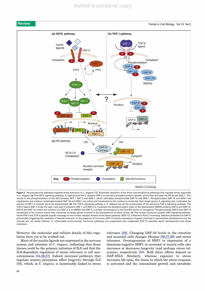
Figure 2. Neuroendocrine pathways regulate stress tolerance in C. elegans [13]. Schematic depiction of the three neuroendocrine pathways that regulate stress responsesin C. elegans. (a) The IGF/IL signaling pathway. IL ligands bind the IL receptor DAF-2 to activate a phosphorylation cascade, which then activates the PI3 kinase AGE-1. Thisresults in the phosphorylation of the ACG kinases, AKT-1 AKT-2 and SGK-1, which ultimately phosphorylate DAF-16 and SKN-1. Phosphorylated DAF-16 and SKN-1 arecytoplasmic and inactive. Unphosphorylated DAF-16 and SKN-1 are active and translocate to the nucleus to transcribe their target genes. IL signaling also modulates theactivity of HSF1 in manner yet to be characterized. (b) The TGF-b signaling pathway in C. elegans has all the components of the canonical TGF-b signaling pathway. TheTGF-b ligand DAF-7 binds the type I and type II receptors DAF-1 and DAF-4 to modulate the phosphorylation state of the downstream SMAD proteins DAF-8 and DAF-14.DAF-8 and DAF-14 inhibit the function of a DAF-3, a co-SMAD and DAF-5, a protein homologous to the Sno/Ski family of oncogenes. Phosphorylated DAF-8 and DAF-14translocate to the nucleus where they transcribe its target genes involved in the development of dauer larvae. (c) The nuclear hormone receptor (NR) signaling pathway.Insulin/IGF-I and TGF-b peptide signals converge on the nuclear receptor branch of the dauer pathway. NCR-1/2, a Niemann-Pick C1 homolog, delivers cholesterol to DAF-9,presumably triggering the synthesis of steroid hormone. In the presence of hormone, DAF-12 directs expression of genes involved in reproductive development and theanimals are not stress tolerant. In unfavorable environments, hormonal pathways are suppressed and unliganded DAF-12 specifies dauer development and stressresistance.
Review Trends in Cell Biology Vol.19 No.2
56

Author's personal copy
changes within the organism can be decoupled dependingon the cell type within which the IL signal is modulated[56,61,62]. Thus, although downregulation of DAF-2 sig-naling has similar effects on stress resistance, its down-regulation during early development induces larvae todevelop into stress-resistant dauers (Box 1), whereas itsdownregulation at late developmental stages or in adultsincreases longevity [11,46]. Caloric restriction via ILSsignaling activates SKN-1 within specific neurons,whereas the resultant increase in metabolic activity occursin peripheral tissues [63]. A systematic understanding ofthe response of the different tissues to ILS and the result-ing changes in their gene expression profiles upon stresswill enable us to better comprehend the cell non-autonom-ous regulation of organismal stress responses.
Transforming growth factor b (TGF-b) signaling pathwayThe TGF-b signaling pathway consists of a family ofsecreted peptides that include the activins and bonemorphogenetic proteins (BMP) in vertebrates. Five TGF-b-related ligands, numerous receptors and SMAD homol-ogues have been identified in C. elegans based on sequencehomology [64] (Figure 2b). TGF-b signaling affects stresstolerance by inducing the dauer program in C. elegans[40,65], and DAF-7 is a prominent ligand regulating thisprocess [40]. Under favorable conditions, DAF-7 isexpressed in a pair of sensory neurons called ASI andpromotes non-dauer development. The repression ofDAF-7 under stressful environmental conditions leads tothe development of dauer larvae [40,65]. The transcriptionfactors that are downstream of DAF-7 signaling and imple-ment daf-7-dependent dauer formation are distinct fromthe three stress-activated transcription factors. However,the nuclear localization of DAF-16 has been shown to bemodulated by DAF-7 at the time of commitment betweendauer and reproductive development [40,65]. In addition,the TGF-b signaling pathway interacts with the ILS path-way to regulate longevity. Because the dauer and/or long-evity programs must be implemented by all cells, theeffects of TGF-b signaling on organismal stress resistancemust be cell non-autonomous. The stress resistance ofdauer larvae is associated with elevated expression ofcertain hsp genes [66]. However, analyses of the tissueswithin which hsp expression is necessary and sufficient toconfer the stress-resistant properties of dauer larvae havenot been conducted.
Nuclear hormone signaling pathwayThe nuclear hormone receptor (NR) pathway in metazoansconsists of a family of transcription factors regulated bysmall lipophilic molecules including steroids, retinoids andbile and fatty acids, which mediate endocrine control [67].The C. elegans genome encodes 284 NR receptors, com-pared to 48 for humans and 21 for flies [68]. Many of thesereceptors have homologs in other species and function inmetabolism, nervous system development, sex determi-nation, developmental timing, molting and entry intothe stress-resistant dauer pathway. Two components ofthe NR pathway, DAF-12 and DAF-9, have been studied inC. elegans with respect to their involvement in organismalstress tolerance. DAF-12, the steroid hormone receptor, is
thought to bind to hormones that are cholesterol deriva-tives (D4-dafachronic acid and D7-dafachronic acid [69])[69–71] (Figure 2c). DAF-9, a P450 cytochrome related tofatty acid and steroidogenic hydroxylases is involved in thebiogenesis of these hormones. Genetic studies place DAF-12 downstream of both the ILS and TGF- b pathways and itis, therefore, thought to have a crucial role in integratingneuroendocrine signaling to regulate the dauer program.DAF-12 is widely expressed throughout development andadulthood [71,72]. DAF-9 expression, however, is spatiallyrestricted to a pair of anterior ganglion nuclei, hypodermisand spermatheca and depends on the developmental stageof the animal [70]. Although a strong loss-of-functionmutation of DAF-9 also results in stress resistance andthe arrest of larvae in dauer, expression in the hypodermisalone is sufficient to rescue this dauer phenotype [70].Thus, dauer-specific gene expression is cell non-autonom-ously regulated by the steroid hormone signaling pathway.Cell non-autonomous regulation of the stress response bysteroid hormone signaling is also evident in experiments inwhich removal of the germline by microsurgery increasesthe resistance of the animal to oxidative and heat stressand promotes the nuclear localization of DAF-16 in theintestine [60,73,74]. The effects of germline ablation arethought to be caused by the regulation of daf-9 expression[70,71] and seem to be conserved in other organisms suchas Drosophila and the mouse [75].
In all the cases described here, it is unclear whether hspexpression within specific cells or tissues can suffice toprotect the entire organism from stress-induced cellularprotein damage. Neuroendocrine regulation of stress couldindicate that proteotoxic damagewithin different cell typeshave different outcomes with regard to organismal health.It would be interesting to understand whether organismscan tolerate different levels of misfolded and damagedproteins under certain growth or metabolic conditions,even at the cost of cytotoxic damage to certain cells, andwhether neuroendocrine signals can override cellular auto-nomy in the response of cells to environmental stress.
Neuronal signaling overrides cell-autonomous HSPinduction and response to stressThe neuroendocrine pathways described earlier couldregulate organismal stress tolerance by changing the basallevels of expressed HSPs and decreasing the overall cel-lular proteotoxicity that results from exposure to stress.However, there is also evidence that neuronal signaling isrequired for HSP induction upon the administration oftemperature stress (heat shock), despite the probableincrease in cellular misfolded and damaged proteins[13]. InC. elegans, two neurons called AFDs detect ambienttemperature and regulate thermotaxis behavior [19].Mutations affecting only these neurons inhibited heat-shock-dependent HSP induction throughout C. elegans.This included tissues such as the intestine and sper-matheca, which were not directly innervated by theseneurons, indicating that AFD regulation occurred throughneuroendocrine pathways. The AFD-dependent upregula-tion of HSPs after heat shock depended on the metabolicstatus of the animal. Thus, neuronal regulation seemed tointegrate the stress response with other organismal func-
Review Trends in Cell Biology Vol.19 No.2
57

Author's personal copy
tions. A model proposed to explain these findings postu-lates that the cellular proteostasis machinery is negativelyregulated by (at least) twomutually inhibitory pathways: atemperature-sensing pathway and a growth-regulatedpathway. Disruption of either pathway results in the netinhibition of heat-shock-dependent HSP transcription,whereas the presence or absence of both pathways enablescells to express HSPs upon heat stress. The downstreamtarget of the AFDs seems to be HSF1. Data from otherstudies on nutrient-dependent signaling in C. elegansindicate that the growth-related signal could act throughDAF-16 [58]. Thus, as in mammalian tissue culture cells oryeast [24,26], organismal growth and HSF1-dependentHSP expression within C. elegans could also be mutuallyantagonistic.
C. elegans as a model system for neurodegenerativeand other protein misfolding diseasesNeurodegenerative diseasemodels inC. elegans created byexpressing human disease-related proteins inC. elegans insome cases show cell-autonomous induction of HSPs. How-ever, the ILS pathway also modulates the protein misfold-ing and aggregation of these disease-related proteins,indicating that hierarchical interactions exist betweencell-autonomous and cell non-autonomous controls onthe proteostasis machinery.
Huntington’s disease (HD)PolyQ-containing proteins are implicated in HD [76–80](Box 3) and other human age-related neurodegenerativediseases [81]. Expression of aggregation-prone polyQ-con-taining proteins within C. elegans muscle and neuronaltissues [82–84] recapitulates aspects of HD, includingformation of Q-length-dependent intracellular aggregatesthat cause toxicity [81]. The aggregation-dependent
toxicity phenotypes in C. elegans reflect the cell type inwhich the polyQ protein is expressed; inmuscle cells, polyQproteins cause muscle-cell dysfunction, whereas expres-sion in neuronal cells causes neuronal dysfunction [82,83].The toxicity associated with polyQ proteins seems to becaused by the global disruption of the cellular proteostasismachinery [77]. This disruption is cell autonomous: whentemperature-sensitive metastable proteins were used assensors of cellular protein folding capacity, polyQ expres-sion in muscle cells destabilized temperature-sensitiveproteins in muscle but not in neurons; likewise, polyQaggregates in neurons destabilized temperature-sensitiveproteins expressed only in neurons [77].
However, although some features of the response of C.elegans to polyQ misfolding are decidedly cell autonomous,the onset of aggregation and toxicity in the animal, as inthe human disease, is age-dependent and regulated by theILS pathway [55,85]. Downregulation of ILS by mutationsin phosphoinsitide-3-kinase, AGE-1 or the insulin receptorDAF-2 causes the constitutive activation of DAF-16(Figure 2) and this delays and/or suppresses polyQ aggre-gation and toxicity. ILS modulation of polyQ aggregationrequires HSF1 [55,85] (Figure 2). The effects of ILS arethought to be caused by the cellular upregulation of cha-perones and other transcriptional targets of DAF-16 andHSF1 [3].
PolyQ aggregation within C. elegansmuscle cells is alsoregulated cell non-autonomously by neuronal gamma ami-nobutyric acid (GABA)-ergic and/or cholinergic signalingpathways [12]. Mutations in these pathways and themodulation of neuronal signaling by small moleculesaltered polyQ aggregation and increasing amounts ofGABA even rescued polyQ aggregation and toxicity.GABA-ergic signaling also modulated the misfolding andaccumulation of endogenous temperature-sensitive
Box 3. Neurodegenerative diseases of protein misfolding and aggregation
Huntington’s diseaseHuntington’s disease (HD) is a genetic neurological disorder thataffects up to seven in 100 000 people. It is characterized byuncoordinated body movements (chorea), a decline in cognitiveabilities and, ultimately, a severe reduction in life expectancy. HD isone of many trinucleotide repeat diseases caused by an increase inthe length of a repetitive DNA sequence, CAG, within the Huntintingene (Htt) and is inherited in an autosomal dominant manner. CAGencodes the amino acid glutamine (Q). The number of Q repeatscorrelates with disease severity, age-dependent onset and the rate ofprogression of neurological symptoms. In the general population, thenumber of Q repeats within Htt rarely exceeds 27; sequences of 36 ormore glutamines result in the selective neurodegeneration of striatalprojection neurons and cortical pyramidal neurons in various regionsof the brain [76].
The function of Htt and the reason why an increase in its Q lengthcauses neurodegeneration is unclear. Numerous potential mechan-isms for neurotoxicity have been proposed, including dysregulationof transcriptional pathways, disruption of mitochondrial transport,excitotoxicity, caspase activation and disruption of proteostasis[76,77]. The expression of the aggregation-prone polyQ stretch alonewithin animal models is sufficient to mimic toxic aspects of HD,indicating that its pathology is, at least in part, a consequence ofmisfolding and aggregation of mutant Htt in neurons [76,77].Treatment for Huntington’s disease is at an early stage and currentlythe symptoms can be alleviated through the use of medicationincluding antidepressants and antipsychotics, rehabilitation methodsand nutrition management [76–78].
Alzheimer’s diseaseAlzheimer’s disease (AD) is the most common form of dementiaestimated to affect >30 million people worldwide. This neurodegen-erative disease is terminal, age-related and typically diagnosed inpeople >65 years of age. The earliest observable symptoms includememory loss with subsequent symptoms including confusion,irritability, language breakdown and general social withdrawal. ADis associated with the appearance of plaques and tangles in affectedbrains. However, the cause of these deposits and their correlationwith the progression of AD is poorly understood [79].There are currently several hypotheses put forth to explain the
cause of AD [79]. Currently available drug therapies are mainly basedon the cholinergic hypothesis, which proposes that AD is caused byreduced synthesis of the neurotransmitter acetylcholine. Anothertheory is the amyloid hypothesis, which postulates that amyloid b(Ab) deposits are the fundamental cause of AD. In support of this,people with an extra copy of the amyloid b precursor (APP) proteinexhibit an earlier onset of AD symptoms, and a major genetic riskfactor for AD is apolipoprotein E (APOE4), which causes excessiveamyloid buildup. Transgenic mice that express a mutant form of thehuman APP gene develop fibrillar amyloid plaques and Alzheimer’s-like brain pathology and neurological symptoms. The appearance ofdamaged proteins is furthered by the tau hypothesis, which indicatesthat hyperphosphorylation and aggregation of the microtubule-associated protein tau causes AD. Both the amyloid and the tauhypotheses have in common the cellular misfolding and aggregationof proteins, indicating that the general dysfunction in proteostasiscould have a central role in AD pathogenesis [79,80].
Review Trends in Cell Biology Vol.19 No.2
58

Author's personal copy
proteins in C. elegans muscle. Although the effects ofGABA-ergic signaling are thought to be caused byincreased excitotoxicity in muscle cells, in C. elegans,GABA-ergic neurons also regulate numerous motor func-tions [12] and are themselves targets of stress-inducedsignaling pathways [86]. This indicates that the observedcell non-autonomous regulation of polyQ proteins by theseneurons could be part of a larger neuroendocrine networkthat modulates organismal function.
Alzheimer’s disease (AD)TheA(1–42 peptide, a prevalent component of plaques in thebrains of patients with AD (Box 3), also forms aggregateswithinC. elegansmuscle cells [16]. These aggregates inter-act with and induce specific HSPs that are downstreamtargets of both HSF1 and DAF-16 in the tissue in whichthey are expressed, again revealing the cell-autonomousnature of the regulation of proteostasis [87,88]. In addition,as in the polyQ models, Ab1–42 aggregation-toxicity is alsoregulated by the ILS pathway [89] through DAF-16 andHSF1. Downregulation of DAF-16 and HSF1, althoughhaving similar effects on Ab1–42-dependent toxicity, hasopposing effects on intracellular Ab1–42 accumulation andmisfolding. Ab1–42 aggregation and misassembly withincells is increased upon lowering HSF1 levels but isdecreased upon lowering DAF-16 [89].
Thus, both the polyQ and Ab1–42 models of proteinmisfolding are regulated by neuroendocrine signalingand specifically the ILS pathway, through DAF-16 andHSF1. Whereas the neuroendocrine pathway that affectsproteostasis is common to both models, the effects at thecellular level, implemented through the two stress-regulated transcription factors, differ depending on themisfolded or disease-causing protein species. One perplex-ing feature of protein misfolding in C. elegans, as has beennoted for various human protein conformational diseases,is that the intracellular accumulation of misfolded proteinssuch as polyQ only sporadically activates HSP expression[84] and requires the downregulation of ILS signaling forits clearance and modulation [55,89]. This indicates thatthe cell-autonomous response to protein misfolding can beoverridden by organismal regulation.
Concluding remarksThe cell non-autonomous regulation of stress responsesand proteostasis by neurohormonal signaling in C. elegansindicates that similar forms of control are likely to occur inother eukaryotes. Although a systematic study of thehierarchical layers of organismal stress regulation hasnot been conducted in other animal models, ILS signalingis known to control lifespan in Drosophila melanogasterand mice [50,90–92]. Furthermore, the machinery for cellnon-autonomous neurohormonal control of HSF1 andHSPs has been observed in specific tissues of mammals:restraint stress [7,93] results in the upregulation of specificHSPs in the adrenal cortex and the endothelial cells of thethoracic aorta of rats. This requires the hypothalamic-pituitary-adrenal (HPA) axis and is adrenocorticotropichormone-dependent. Likewise, the cell non-autonomouscontrol of HSPs by HSF1 is dependent on organismalphysiology and declines with age [7]. The ability of
multi-cellular organisms to regulate cellular HSP expres-sion, the stress response and proteostasis machinery byneurohormonal signaling could be influenced further bythe ecology and life history of the organism [94].
Neurohormonal cell non-autonomous regulation mightaccount, in part, for the pathophysiological situations thataccompany protein misfolding diseases whereby the intra-cellular accumulation of misfolded and aggregated proteinfails to adequately upregulate protective HSPs [3,8–10].Similarly, diseases such as Hashimoto’s thyroiditis,Graves disease, arthritis and cancer [95,96] have increasedlevels of HSP expression within affected cells, indicatingthat these cells might have lost their responsiveness to theneurohormonal regulation imposed by the organism. Anunderstanding of the systemic stress regulation in organ-isms would enable an examination of whether neurohor-monal perturbations indeed correlate with diseaseconditions. Small molecules that can modulate the com-ponents of ILS or other neuroendocrine pathways to resultin the upregulation of specific cytoprotective HSPs in thetissue harboring disease-related protein aggregates andre-establish cellular proteostasis could provide novel drugtargets for combating diseases of protein conformation.
AcknowledgementsWe thank the members of the Morimoto laboratory for discussions andcomments on the manuscript and Sue Fox and Kai Orton for help with thefigures. These studies were supported by grants to R.I.M from the NIH(NIGMS and NIA), the Huntington’s Disease Society of America Coalitionfor the Cure and the Daniel F. and Ada L. Rice Foundation.
References1 Lindquist, S. and Craig, E.A. (1988) The heat-shock proteins. Annu.
Rev. Genet. 22, 631–6772 Morimoto, R.I. (1998) Regulation of the heat shock transcriptional
response: cross talk between a family of heat shock factors,molecular chaperones, and negative regulators. Genes Dev. 12,3788–3796
3 Balch,W.E. et al. (2008) Adapting proteostasis for disease intervention.Science 319, 916–919
4 Morimoto, R.I. (2008) Proteotoxic stress and inducible chaperonenetworks in neurodegenerative disease and aging. Genes Dev. 22,1427–1438
5 Pardue, M.L. (1988) The heat shock response in biology and humandisease: a meeting review. Genes Dev. 2, 783–785
6 Blake, M.J. et al. (1990) Discordant expression of heat shock proteinmRNAs in tissues of heat-stressed rats. J. Biol. Chem. 265, 15275–15279
7 Fawcett, T.W. et al. (1994) Effects of neurohormonal stress and agingon the activation of mammalian heat shock factor 1. J. Biol. Chem. 269,32272–32278
8 Hay, D.G. et al. (2004) Progressive decrease in chaperone protein levelsin a mouse model of Huntington’s disease and induction of stressproteins as a therapeutic approach. Hum. Mol. Genet. 13, 1389–1405
9 Zourlidou, A. et al. (2007) Hsp27 overexpression in the R6/2 mousemodel of Huntington’s disease: chronic neurodegeneration does notinduce Hsp27 activation. Hum. Mol. Genet. 16, 1078–1090
10 Barral, J.M. et al. (2004) Roles of molecular chaperones in proteinmisfolding diseases. Semin. Cell Dev. Biol. 15, 17–29
11 Baumeister, R. et al. (2006) Endocrine signaling in Caenorhabditiselegans controls stress response and longevity. J. Endocrinol. 190, 191–202
12 Garcia, S.M. et al. (2007) Neuronal signaling modulates proteinhomeostasis in Caenorhabditis elegans post-synaptic muscle cells.Genes Dev. 21, 3006–3016
13 Prahlad, V. et al. (2008) Regulation of the cellular heat shock responsein Caenorhabditis elegans by thermosensory neurons. Science 320,811–814
Review Trends in Cell Biology Vol.19 No.2
59
Author's personal copy
14 Brenner, S. (1974) The genetics of Caenorhabditis elegans.Genetics 77,71–94
15 Brignull, H.R. et al. (2006) Modeling polyglutamine pathogenesis in C.elegans. Methods Enzymol. 412, 256–282
16 Link, C.D. et al. (2001) Visualization of fibrillar amyloid deposits inliving, transgenic Caenorhabditis elegans animals using the sensitiveamyloid dye, X-34. Neurobiol. Aging 22, 217–226
17 Devaney, E. (2006) Thermoregulation in the life cycle of nematodes.Int. J. Parasitol. 36, 641–649
18 Hu, P.J. (2007) Dauer. WormBook, ed. The C. elegans ResearchCommunity, WormBook, DOI: 10.1895/wormbook.1.144.1
19 Mori, I. et al. (2007) Worm thermotaxis: a model system for analyzingthermosensation and neural plasticity.Curr. Opin. Neurobiol. 17, 712–719
20 de Bono, M. et al. (2002) Social feeding in Caenorhabditis elegansis induced by neurons that detect aversive stimuli. Nature 419,899–903
21 Gray, J.M. et al. (2004) Oxygen sensation and social feeding mediatedby a C. elegans guanylate cyclase homologue. Nature 430, 317–322
22 Wang, D.Y. and Wang, Y. (2008) Phenotypic and behavioral defectscaused by barium exposure in nematode Caenorhabditis elegans. Arch.Environ. Contam. Toxicol. 54, 447–453
23 Hartl, F.U. et al. (1994)Molecular chaperones in protein folding: the artof avoiding sticky situations. Trends Biochem. Sci. 19, 20–25
24 Morano, K.A. and Thiele, D.J. (1999) Heat shock factor function andregulation in response to cellular stress, growth, and differentiationsignals. Gene Expr. 7, 271–282
25 Bukau, B. et al. (2006) Molecular chaperones and protein qualitycontrol. Cell 125, 443–451
26 Anckar, J. and Sistonen, L. (2007) Heat shock factor 1 as a coordinatorof stress and developmental pathways. Adv. Exp. Med. Biol. 594, 78–88
27 Hightower, L.E. (1991) Heat shock, stress proteins, chaperones, andproteotoxicity. Cell 66, 191–197
28 Goldberg, A.L. (1972) Degradation of abnormal proteins in Escherichiacoli (protein breakdown-protein structure-mistranslation-amino acidanalogs-puromycin). Proc. Natl. Acad. Sci. U. S. A. 69, 422–426
29 Schroder, M. and Kaufman, R.J. (2005) The mammalian unfoldedprotein response. Annu. Rev. Biochem. 74, 739–789
30 Ron, D. and Walter, P. (2007) Signal integration in the endoplasmicreticulum unfolded protein response. Nat. Rev. Mol. Cell Biol. 8, 519–529
31 Broadley, S.A. and Hartl, F.U. (2008) Mitochondrial stress signaling: apathway unfolds. Trends Cell Biol. 18, 1–4
32 Haynes, C.M. et al. (2007) ClpP mediates activation of a mitochondrialunfolded protein response in C. elegans. Dev. Cell 13, 467–480
33 Wang, X. et al. (2006) Hsp90 cochaperone Aha1 downregulationrescues misfolding of CFTR in cystic fibrosis. Cell 127, 803–815
34 Abravaya, K. et al. (1991) Attenuation of the heat shock response inHeLa cells is mediated by the release of bound heat shock transcriptionfactor and is modulated by changes in growth and in heat shocktemperatures. Genes Dev. 5, 2117–2127
35 Feder, J.H. et al. (1992) The consequences of expressing hsp70 inDrosophila cells at normal temperatures. Genes Dev. 6, 1402–1413
36 Huang, H. and Tindall, D.J. (2007) Dynamic FoxO transcriptionfactors. J. Cell Sci. 120, 2479–2487
37 Murphy, C.T. (2006) The search for DAF-16/FOXO transcriptionaltargets: approaches and discoveries. Exp. Gerontol. 41, 910–921
38 Tullet, J.M. et al. (2008) Direct inhibition of the longevity-promotingfactor SKN-1 by insulin-like signaling in C. elegans. Cell 132, 1025–1038
39 Gutteling, E.W. et al. (2007) Environmental influence on the geneticcorrelations between life-history traits in Caenorhabditis elegans.Heredity 98, 206–213
40 Fielenbach, N. and Antebi, A. (2008) C. elegans dauer formation andthe molecular basis of plasticity. Genes Dev. 22, 2149–2165
41 Chen, D. et al. (2007) Longevity determined by developmental arrestgenes in Caenorhabditis elegans. Aging Cell 6, 525–533
42 Rea, S.L. et al. (2005) A stress-sensitive reporter predicts longevity inisogenic populations of Caenorhabditis elegans. Nat. Genet. 37, 894–898
43 Harvey, S.C. et al. (2008) Quantitative genetic analysis of life historytraits of Caenorhabditis elegans in stressful environments. BMC Evol.Biol. 8, 15
44 Henderson, S.T. and Johnson, T.E. (2001) daf-16 integratesdevelopmental and environmental inputs to mediate aging in thenematode Caenorhabditis elegans. Curr. Biol. 11, 1975–1980
45 Walker, G.A. et al. (2003) Heat shock factor functions at theconvergence of the stress response and developmental pathways inCaenorhabditis elegans. FASEB J. 17, 1960–1962
46 Tatar, M. et al. (2003) The endocrine regulation of aging by insulin-likesignals. Science 299, 1346–1351
47 Antebi, A. (2006) Nuclear hormone receptors in C. elegans. WormBook1–13
48 Braeckman, B.P. et al. (2001) Insulin-like signaling, metabolism, stressresistance and aging inCaenorhabditis elegans.Mech. Ageing Dev. 122,673–693
49 Antebi, A. (2004) Tipping the balance toward longevity. Dev. Cell 6,315–316
50 Finch, C.E. and Ruvkun, G. (2001) The genetics of aging. Annu. Rev.Genomics Hum. Genet. 2, 435–462
51 Lee, S.S. et al. (2003) DAF-16 target genes that control C. elegans life-span and metabolism. Science 300, 644–647
52 An, J.H. and Blackwell, T.K. (2003) SKN-1 links C. elegansmesendodermal specification to a conserved oxidative stressresponse. Genes Dev. 17, 1882–1893
53 Lee, P. et al. (2008) Yeast Yak1 kinase, a bridge between PKA andstress-responsive transcription factors, Hsf1 and Msn2/Msn4. Mol.Microbiol. 70, 882–895
54 Samuelson, A.V. et al. (2007) Gene activities thatmediate increased lifespan of C. elegans insulin-like signaling mutants.Genes Dev. 21, 2976–2994
55 Morley, J.F. and Morimoto, R.I. (2004) Regulation of longevity inCaenorhabditis elegans by heat shock factor and molecularchaperones. Mol. Biol. Cell 15, 657–664
56 Wolkow, C.A. et al. (2000) Regulation of C. elegans life-span byinsulinlike signaling in the nervous system. Science 290, 147–150
57 Libina, N. et al. (2003) Tissue-specific activities ofC. elegansDAF-16 inthe regulation of lifespan. Cell 115, 489–502
58 Alcedo, J. and Kenyon, C. (2004) Regulation of C. elegans longevity byspecific gustatory and olfactory neurons. Neuron 41, 45–55
59 Cypser, J. and Johnson, T.E. (2001) Hormesis extends the correlationbetween stress resistance and life span in long-lived mutants ofCaenorhabditis elegans. Hum. Exp. Toxicol. 20, 295–296
60 Lin, K. et al. (2001) Regulation of the Caenorhabditis elegans longevityprotein DAF-16 by insulin/IGF-1 and germline signaling. Nat. Genet.28, 139–145
61 Apfeld, J. and Kenyon, C. (1998) Cell nonautonomy of C. elegansdaf-2 function in the regulation of diapause and life span. Cell 95,199–210
62 Iser, W.B. et al. (2007) Insulin signaling in Caenorhabditis elegansregulates both endocrine-like and cell-autonomous outputs. Dev. Biol.303, 434–447
63 Bishop, N.A. and Guarente, L. (2007) Two neurons mediate diet-restriction-induced longevity in C. elegans. Nature 447, 545–549
64 ten Dijke, P. and Hill, C.S. (2004) New insights into TGF-b-Smadsignalling. Trends Biochem. Sci. 29, 265–273
65 Patterson, G.I. and Padgett, R.W. (2000) TGF b-related pathways.Roles in Caenorhabditis elegans development. Trends Genet. 16, 27–33
66 Wang, J. and Kim, S.K. (2003) Global analysis of dauer gene expressionin Caenorhabditis elegans. Development 130, 1621–1634
67 Mangelsdorf, D.J. et al. (1995) The nuclear receptor superfamily: thesecond decade. Cell 83, 835–839
68 Sluder, A.E. and Maina, C.V. (2001) Nuclear receptors in nematodes:themes and variations. Trends Genet. 17, 206–213
69 Motola, D.L. et al. (2006) Identification of ligands for DAF-12 thatgovern dauer formation and reproduction in C. elegans. Cell 124, 1209–1223
70 Gerisch, B. andAntebi, A. (2004) Hormonal signals produced byDAF-9/cytochrome P450 regulate C. elegans dauer diapause in response toenvironmental cues. Development 131, 1765–1776
71 Gerisch, B. et al. (2001) A hormonal signaling pathway influencing C.elegansmetabolism, reproductive development, and life span.Dev. Cell1, 841–851
72 Antebi, A. et al. (2000) daf-12 encodes a nuclear receptor that regulatesthe dauer diapause and developmental age inC. elegans.Genes Dev. 14,1512–1527
Review Trends in Cell Biology Vol.19 No.2
60
Author's personal copy
73 Arantes-Oliveira, N. et al. (2002) Regulation of life-span by germ-linestem cells in Caenorhabditis elegans. Science 295, 502–505
74 Hsin, H. and Kenyon, C. (1999) Signals from the reproductive systemregulate the lifespan of C. elegans. Nature 399, 362–366
75 Kenyon, C. (2005) The plasticity of aging: insights from long-livedmutants. Cell 120, 449–460
76 Bates, G. et al. (2002) Huntington’s Disease. (3rd edn), OxfordUniversity Press
77 Gidalevitz, T. et al. (2006) Progressive disruption of cellular proteinfolding in models of polyglutamine diseases. Science 311, 1471–1474
78 Roze, E. et al. (2008) Pathophysiology of Huntington’s disease: fromhuntingtin functions to potential treatments. Curr. Opin. Neurol. 21,497–503
79 Selkoe, D.J. (2007) Developing preventive therapies for chronicdiseases: lessons learned from Alzheimer’s disease. Nutr. Rev. 65,S239–S243
80 Skovronsky, D.M. et al. (2006) Neurodegenerative diseases: newconcepts of pathogenesis and their therapeutic implications. Annu.Rev. Pathol. 1, 151–170
81 Soto, C. (2003) Unfolding the role of protein misfolding inneurodegenerative diseases. Nat. Rev. Neurosci. 4, 49–60
82 Brignull, H.R. et al. (2006) Polyglutamine proteins at the pathogenicthreshold display neuron-specific aggregation in a pan-neuronalCaenorhabditis elegans model. J. Neurosci. 26, 7597–7606
83 Morley, J.F. et al. (2002) The threshold for polyglutamine-expansionprotein aggregation and cellular toxicity is dynamic and influenced byaging in Caenorhabditis elegans. Proc. Natl. Acad. Sci. U. S. A. 99,10417–10422
84 Satyal, S.H. et al. (2000) Polyglutamine aggregates alter proteinfolding homeostasis in Caenorhabditis elegans. Proc. Natl. Acad. Sci.U. S. A. 97, 5750–5755
85 Hsu, A.L. et al. (2003) Regulation of aging and age-related disease byDAF-16 and heat-shock factor. Science 300, 1142–1145
86 Jorgensen, E.M. (2005) Gaba. WormBook, ed. The C. elegans ResearchCommunity, Wormbook, DOI: 10.1895/wormbook.1.14.1
87 Fonte, V. et al. (2002) Interaction of intracellular b amyloid peptidewith chaperone proteins. Proc. Natl. Acad. Sci. U. S. A. 99, 9439–9444
88 Fonte, V. et al. (2008) Suppression of in vivo b-amyloid peptide toxicityby overexpression of the HSP-16.2 small chaperone protein. J. Biol.Chem. 283, 784–791
89 Cohen, E. et al. (2006) Opposing activities protect against age-onsetproteotoxicity. Science 313, 1604–1610
90 Holzenberger, M. et al. (2004) IGF-1 signaling and aging. Exp.Gerontol. 39, 1761–1764
91 Gems, D. and Partridge, L. (2001) Insulin/IGF signalling and ageing:seeing the bigger picture. Curr. Opin. Genet. Dev. 11, 287–292
92 Suh, Y. et al. (2008) Functionally significant insulin-like growth factor Ireceptor mutations in centenarians. Proc. Natl. Acad. Sci. U. S. A. 105,3438–3442
93 Blake, M.J. et al. (1991) Stress-induced heat shock protein 70expression in adrenal cortex: an adrenocorticotropic hormone-sensitive, age-dependent response. Proc. Natl. Acad. Sci. U. S. A.88, 9873–9877
94 Feder, M.E. and Hofmann, G.E. (1999) Heat-shock proteins, molecularchaperones, and the stress response: evolutionary and ecologicalphysiology. Annu. Rev. Physiol. 61, 243–282
95 Van Eden, W. et al. (2007) Stress, heat shock proteins, andautoimmunity: how immune responses to heat shock proteins are tobe used for the control of chronic inflammatory diseases. Ann. N. Y.Acad. Sci. 1113, 217–237
96 Calderwood, S.K. et al. (2006) Heat shock proteins in cancer:chaperones of tumorigenesis. Trends Biochem. Sci. 31, 164–172
Review Trends in Cell Biology Vol.19 No.2
61
Copyright © 2022 FDOKUMEN