Synthetic prions and other human neurodegenerative proteinopathies
13
Please cite this article in press as: Le, N.T.T., et al., Synthetic prions and other human neurodegenerative proteinopathies. Virus Res. (2014), http://dx.doi.org/10.1016/j.virusres.2014.10.020 ARTICLE IN PRESS G Model VIRUS-96433; No. of Pages 13 Virus Research xxx (2014) xxx–xxx Contents lists available at ScienceDirect Virus Research j ourna l h o mepa ge: www.elsevier.com/locate/virusres Synthetic prions and other human neurodegenerative proteinopathies Nhat Tran Thanh Le a , Joanna Narkiewicz a , Suzana Auli ´ c a , Giulia Salzano a , Hoa Thanh Tran a , Denis Scaini b , Fabio Moda c , Gabriele Giachin a,∗ , Giuseppe Legname a,d,∗∗ a Laboratory of Prion Biology, Department of Neuroscience, Scuola Internazionale Superiore di Studi Avanzati (SISSA), Trieste, Italy b Life Science Department, University of Trieste, Trieste, Italy c Carlo Besta Neurological Institute, Department of Neuropathology and Neurology 5, Milan, Italy d Elettra-Sincrotrone Trieste S.C.p.A., Area Science Park, Basovizza, Trieste, Italy a r t i c l e i n f o Article history: Available online xxx Keywords: Prions Neurodegenerative disorders Synthetic prions Strains Protein misfolding diseases Structural biology a b s t r a c t Transmissible spongiform encephalopathies (TSE) are a heterogeneous group of neurodegenerative dis- orders. The common feature of these diseases is the pathological conversion of the normal cellular prion protein (PrP C ) into a -structure-rich conformer-termed PrP Sc . The latter can induce a self-perpetuating process leading to amplification and spreading of pathological protein assemblies. Much evidence sug- gests that PrP Sc itself is able to recruit and misfold PrP C into the pathological conformation. Recent data have shown that recombinant PrP C can be misfolded in vitro and the resulting synthetic conformers are able to induce the conversion of PrP C into PrP Sc in vivo. In this review we describe the state-of-the-art of the body of literature in this field. In addition, we describe a cell-based assay to test synthetic pri- ons in cells, providing further evidence that synthetic amyloids are able to template conversion of PrP into prion inclusions. Studying prions might help to understand the pathological mechanisms gover- ning other neurodegenerative diseases. Aggregation and deposition of misfolded proteins is a common feature of several neurodegenerative disorders, such as Alzheimer’s disease, Parkinson’s disease, amyo- trophic lateral sclerosis and other disorders. Although the proteins implicated in each of these diseases differ, they share a common prion mechanism. Recombinant proteins are able to aggregate in vitro into -rich amyloid fibrils, sharing some features of the aggregates found in the brain. Several studies have reported that intracerebral inoculation of synthetic aggregates lead to unique pathology, which spread progressively to distal brain regions and reduced survival time in animals. Here, we review the prion-like features of different proteins involved in neurodegenerative disorders, such as -synuclein, superoxide dismutase-1, amyloid- and tau. © 2014 Published by Elsevier B.V. Abbreviations: A, amyloid- peptide; AD, Alzheimer’s disease; AFM, atomic force microscopy; ALS, amyotrophic lateral sclerosis; dpi, days post injection; HuPrP, human PrP C ; HX-MS, hydrogen/deuterium exchange measured using mass spec- trometry; LBs, Lewy bodies; PD, Parkinson’s disease; PMCA, protein misfolding cyclic amplification; PrP, prion protein; PrP C , physiological, -helical form of the cellu- lar prion protein; PrP Sc , prion, the pathological isoform of PrP C ; PK, proteinase-K; rec, recombinant; SOD1, superoxide dismutase-1; Tg, transgenic; TSE, transmissible spongiform encephalopathies. ∗ Corresponding author at: Scuola Internazionale Superiore di Studi Avanzati (SISSA), Via Bonomea 265, 34136 Trieste, Italy. Tel.: +39 040 3787 760; fax: +39 040 3787 702. ∗∗ Corresponding author at: Scuola Internazionale Superiore di Studi Avanzati (SISSA), Via Bonomea 265, 34136 Trieste, Italy. Tel.: +39 040 3787 715; fax: +39 040 3787 702. E-mail addresses: [email protected] (G. Giachin), [email protected] (G. Legname). 1. Introducing prions and prion diseases The physiological cellular form of the prion protein (PrP C ) is a glycosylphosphatidylinositol (GPI)-anchored glycoprotein local- ized on the outer leaflet of the cellular membrane with highest expression levels at the presynaptic membrane of neurons (Herms et al., 1999; Horiuchi et al., 1995; Prusiner, 1998). The mature human PrP C (HuPrP) is composed of 209 residues including a largely unstructured N-terminal region and a globular -helix rich C-terminal domain (Zahn et al., 2000). Despite being highly con- served among mammals, its physiological function has not been established with certainty. Proposed PrP C functions range from neuronal growth and differentiation (Steele et al., 2006), synap- tic plasticity (Caiati et al., 2013; Maglio et al., 2004), and cell signaling (Mouillet-Richard et al., 2000; Santuccione et al., 2005), to NMDA receptor modulator (Khosravani et al., 2008) and brain http://dx.doi.org/10.1016/j.virusres.2014.10.020 0168-1702/© 2014 Published by Elsevier B.V.
-
Upload
independent -
Category
Documents
-
view
0 -
download
0
Transcript of Synthetic prions and other human neurodegenerative proteinopathies

V
S
NHGa
b
c
d
a
AA
KPNSSPS
fhtalrs
(f
(f
h0
ARTICLE IN PRESSG ModelIRUS-96433; No. of Pages 13
Virus Research xxx (2014) xxx–xxx
Contents lists available at ScienceDirect
Virus Research
j ourna l h o mepa ge: www.elsev ier .com/ locate /v i rusres
ynthetic prions and other human neurodegenerative proteinopathies
hat Tran Thanh Lea, Joanna Narkiewicza, Suzana Aulic a, Giulia Salzanoa,oa Thanh Trana, Denis Scainib, Fabio Modac, Gabriele Giachina,∗,iuseppe Legnamea,d,∗∗
Laboratory of Prion Biology, Department of Neuroscience, Scuola Internazionale Superiore di Studi Avanzati (SISSA), Trieste, ItalyLife Science Department, University of Trieste, Trieste, ItalyCarlo Besta Neurological Institute, Department of Neuropathology and Neurology 5, Milan, ItalyElettra-Sincrotrone Trieste S.C.p.A., Area Science Park, Basovizza, Trieste, Italy
r t i c l e i n f o
rticle history:vailable online xxx
eywords:rionseurodegenerative disordersynthetic prionstrainsrotein misfolding diseasestructural biology
a b s t r a c t
Transmissible spongiform encephalopathies (TSE) are a heterogeneous group of neurodegenerative dis-orders. The common feature of these diseases is the pathological conversion of the normal cellular prionprotein (PrPC) into a �-structure-rich conformer-termed PrPSc. The latter can induce a self-perpetuatingprocess leading to amplification and spreading of pathological protein assemblies. Much evidence sug-gests that PrPSc itself is able to recruit and misfold PrPC into the pathological conformation. Recent datahave shown that recombinant PrPC can be misfolded in vitro and the resulting synthetic conformers areable to induce the conversion of PrPC into PrPSc in vivo. In this review we describe the state-of-the-artof the body of literature in this field. In addition, we describe a cell-based assay to test synthetic pri-ons in cells, providing further evidence that synthetic amyloids are able to template conversion of PrPinto prion inclusions. Studying prions might help to understand the pathological mechanisms gover-ning other neurodegenerative diseases. Aggregation and deposition of misfolded proteins is a commonfeature of several neurodegenerative disorders, such as Alzheimer’s disease, Parkinson’s disease, amyo-trophic lateral sclerosis and other disorders. Although the proteins implicated in each of these diseasesdiffer, they share a common prion mechanism. Recombinant proteins are able to aggregate in vitro into
�-rich amyloid fibrils, sharing some features of the aggregates found in the brain. Several studies havereported that intracerebral inoculation of synthetic aggregates lead to unique pathology, which spreadprogressively to distal brain regions and reduced survival time in animals. Here, we review the prion-likefeatures of different proteins involved in neurodegenerative disorders, such as �-synuclein, superoxidedismutase-1, amyloid-� and tau.© 2014 Published by Elsevier B.V.
Please cite this article in press as: Le, N.T.T., et al., Synthetic prions a(2014), http://dx.doi.org/10.1016/j.virusres.2014.10.020
Abbreviations: A�, amyloid-� peptide; AD, Alzheimer’s disease; AFM, atomicorce microscopy; ALS, amyotrophic lateral sclerosis; dpi, days post injection; HuPrP,uman PrPC; HX-MS, hydrogen/deuterium exchange measured using mass spec-rometry; LBs, Lewy bodies; PD, Parkinson’s disease; PMCA, protein misfolding cyclicmplification; PrP, prion protein; PrPC, physiological, �-helical form of the cellu-ar prion protein; PrPSc, prion, the pathological isoform of PrPC; PK, proteinase-K;ec, recombinant; SOD1, superoxide dismutase-1; Tg, transgenic; TSE, transmissiblepongiform encephalopathies.∗ Corresponding author at: Scuola Internazionale Superiore di Studi Avanzati
SISSA), Via Bonomea 265, 34136 Trieste, Italy. Tel.: +39 040 3787 760;ax: +39 040 3787 702.∗∗ Corresponding author at: Scuola Internazionale Superiore di Studi AvanzatiSISSA), Via Bonomea 265, 34136 Trieste, Italy. Tel.: +39 040 3787 715;ax: +39 040 3787 702.
E-mail addresses: [email protected] (G. Giachin), [email protected] (G. Legname).
ttp://dx.doi.org/10.1016/j.virusres.2014.10.020168-1702/© 2014 Published by Elsevier B.V.
1. Introducing prions and prion diseases
The physiological cellular form of the prion protein (PrPC) isa glycosylphosphatidylinositol (GPI)-anchored glycoprotein local-ized on the outer leaflet of the cellular membrane with highestexpression levels at the presynaptic membrane of neurons (Hermset al., 1999; Horiuchi et al., 1995; Prusiner, 1998). The maturehuman PrPC (HuPrP) is composed of 209 residues including alargely unstructured N-terminal region and a globular �-helix richC-terminal domain (Zahn et al., 2000). Despite being highly con-served among mammals, its physiological function has not beenestablished with certainty. Proposed PrPC functions range from
nd other human neurodegenerative proteinopathies. Virus Res.
neuronal growth and differentiation (Steele et al., 2006), synap-tic plasticity (Caiati et al., 2013; Maglio et al., 2004), and cellsignaling (Mouillet-Richard et al., 2000; Santuccione et al., 2005),to NMDA receptor modulator (Khosravani et al., 2008) and brain
ING ModelV
2 esearc
mrsead
Jv(i(aaaac2
pp1pm(sftoaoTbmPocTctaL
oipcvaap
pea
1
hPfiid
ARTICLEIRUS-96433; No. of Pages 13
N.T.T. Le et al. / Virus R
etal homeostasis (Pushie et al., 2011). Defining PrPC function(s)emains an absolute requirement for understanding transmissiblepongiform encephalopathies (TSE). These neurodegenerative dis-ases are caused by the posttranslational conversion of PrPC into
�-sheet enriched, partially proteinase-K (PK) resistant isoformenoted PrPSc or prion (Prusiner, 1982).
In humans, TSE include idiopathic forms as sporadic Creutzfeldt-akob disease (sCJD), sporadic fatal insomnia (sFI) and theariably proteinase sensitive prionopathies (VPSPr). Familial CJDfCJD), Gerstmann–Sträussler–Scheinker disease (GSS), fatal famil-al insomnia (FFI) and prion protein cerebral amyloid angiopathyPrP-CAA) are genetic forms of human TSE. The acquired formsre transmitted from human to human, as iatrogenic CJD (iCJD)nd Kuru; or from cattle to human, as variant CJD (vCJD) (Headnd Ironside, 2012). In animals, relevant TSE are scrapie in sheepnd goats, bovine spongiform encephalopathy (BSE) in cattle, andhronic wasting disease (CWD) in cervids (Imran and Mahmood,011).
The central molecular event during prion diseases is the self-ropagating conformational conversion of PrPC to PrPSc. Thisostulate is known as the “protein-only” hypothesis (Prusiner,998). Two different mechanisms of prion replication have beenroposed. In the template assistance model PrPSc exists as aonomer that is thermodynamically more stable than PrPC
Prusiner, 1991). In the rare event that a PrPSc molecule is formedpontaneously (or provided exogenously) it can template the mis-olding of PrPC by direct interaction. In this model, the critical step inhe conversion is the formation of a dimer between PrPSc and PrPC,r a partially destabilized folding intermediate of PrPC denoteds PrP*. PrPSc acts as a template able to catalyze the refoldingf PrPC to a thermodynamically more stable PrPSc conformation.he nucleation-polymerization model proposes that the conversionetween PrPC and PrPSc is reversible, but the PrPSc monomer isuch less stable than PrPC (i.e. the equilibrium strongly favors
rPC). Stabilization of PrPSc occurs only upon formation of a stableligomeric nucleus. Once the nucleus has formed, monomeric PrPC
ould efficiently add to it by adopting the conformation of PrPSc.he rate-limiting step in this mechanism is not the conformationalonversion itself but the nucleation step. This step, responsible forhe lag phase in the spontaneous conversion, can be by-passednd accelerated by addition of preformed PrPSc seeds (Jarrett andansbury, 1993).
One of the most important recent advancements in prion biol-gy has been the discovery of the de novo generation of prionnfectivity from recombinant (rec) protein sources. Amyloid fibrilsrepared in vitro from bacterially expressed rec PrPC (recPrP) haveonfirmed that PrPSc is the principal causative agent of TSE, pro-iding the definitive proof for the prion hypothesis. These recPrPmyloid fibrils can be used as a synthetic surrogate of PrPSc to obtain
model for understanding the structural and molecular basis ofrion conversion.
In the next section, we present an overview of considerablerogress that has been made in our understanding of prion dis-ases through the development of several protocols for producingmyloid fibrils made by recPrP.
.1. Discovering the first synthetic prion strains
The earliest efforts to define the process of prion conversionave been described in the 1990s by Caughey and collaborators.urified PrPC was incubated with different PrPSc strains derived
Please cite this article in press as: Le, N.T.T., et al., Synthetic prions a(2014), http://dx.doi.org/10.1016/j.virusres.2014.10.020
rom scrapie-diseased animals. The interaction with PrPSc resultedn the formation of a PK-resistant form (Kocisko et al., 1994). Thencubation of PrPC with two different strains of PrPSc, the hyper androwsy strains of hamster transmissible mink encephalopathy,
PRESSh xxx (2014) xxx–xxx
generated two distinct sets of PK-resistant forms (Bessen et al.,1995). Additionally, the mouse/hamster chimeric PrPC, termedMH2M, extracted from cell culture has been converted into aPK-resistant form after incubation with the Syrian hamster (SHa)263 K scrapie strain. However, no infectivity was detected whenthe converted material was inoculated into wild-type mice (Hillet al., 1999). These pioneering studies recapitulate many featuresassociated with prion transmission in vitro, demonstrating that thedirect interaction between PrPSc and PrPC is one of the key eventsduring the conformational transition.
Another largely explored strategy consisted of using severalphysico-chemical approaches to induce misfolding of the recPrPinto �-strand rich states. Such studies are relevant because theyaddress the question whether PrPC alone is sufficient for the spon-taneous formation of prions without the presence of any exogenousagent. A plethora of studies have attempted to provide an answerto this question, but these experiments have largely failed in pro-ducing infectivity in vivo or the infectivity potential has not beentested in animal model yet [reviewed in Benetti and Legname, 2009;Legname et al., 2012].
In 2004 the production of synthetic prions via the in vitro con-version of recPrP was reported (Legname et al., 2004). In an earlierstudy (Baskakov et al., 2002) the same authors analyzed in detail themisfolding pathways of the truncated fragment of recPrP leadingto �-sheet rich conformers. Depending on the reaction conditions,two misfolded forms were adopted: at acidic pH values and in thepresence of partially denaturing urea a PrPSc-like oligomer wasobserved; whereas under neutral or slightly acidic pH values andat low concentration of urea recPrP aggregated in fibrillar struc-tures which developed into amyloids. Importantly, in this workauthors discovered that the addition of a seed of pre-folded amyloidto the reaction substantially reduced the time of the fibrillization(called lag phase) process, demonstrating that recPrP fibrils can beinduced by seeding. Starting from these findings, Legname and col-laborators addressed the question of whether these synthetic fibrilswere infectious when inoculated into mice. The pre-folded amyloidfibrils (denoted as “unseeded”) and the seeded fibrils composedof recMoPrP(89–230) were intracerebrally injected into transgenic(Tg) mice which overexpress MoPrP(89–230). Seeded amyloid fib-rils exhibited shorter incubation time (382 days) and PK-resistancethan unseeded (473 days and PK-sensitivity). Interestingly, theneuropathological features associated with seeded and unseededamyloids were different in terms of vacuolation, gray matter PrPSc
deposition and conformational stability as measured by the GndHClconcentration required to denature half of the sample (Legnameet al., 2004, 2005). Subsequent serial passages of these strains ledto shortened incubation periods and a decreased conformationalstability of the resulting prions. Combining these data with thoseavailable for naturally occurring prion strains, it was shown thatthe length of the incubation time in mice is directly proportional tothe conformational stability of the prion strain (Colby et al., 2007;Legname et al., 2006).
1.2. New synthetic prions confirm the protein-only hypothesis
As follow-up of the first synthetic prion experiment (Legnameet al., 2004), a series of recPrP amyloid fibers were producedfrom the same laboratory and intracerebrally injected in Tgmice overexpressing full-length PrPC at 4–8 times normal levels(Tg4053). Interestingly, different inocula were able to propagatein vivo and induce the formation of different prion strains (Colbyet al., 2009). In another study from Prusiner’s group, authors
nd other human neurodegenerative proteinopathies. Virus Res.
performed serial inoculation passages of biochemically differentsynthetic prions showing that synthetic prions may reach a com-mon, adapted and convergent state with similar physico-chemicalfeatures (Ghaemmaghami et al., 2013). Additionally, Colby and
ARTICLE IN PRESSG ModelVIRUS-96433; No. of Pages 13
N.T.T. Le et al. / Virus Research xxx (2014) xxx–xxx 3
Table 1Strategies for producing synthetic prions from different recPrP substrates.
Strategy Methods Result (infectivity) Reference
ASA Incubation and shaking in partially denaturingcondition of recMoPrP (89–230)
Positive in Tg9949 and afterpassage in WT FVB mice andTg4053
Colby et al. (2009, 2010),Ghaemmaghami et al. (2013) andLegname et al. (2004)
ASA + Annealing Incubation of recPrP amyloid with normal brainhomogenate at different heating/cooling cycles
Positive in WT Syrian hamsters Makarava et al. (2010)
PMCA Generation of prions starting from normal brainhomogenates in the presence of PrPSc seed by serialPMCA with high dilution factor of initial PrPSc seed
Positive in WT Syrian hamster Castilla et al. (2005)
Generation of prions by modified serial PMCAstarting from normal brain homogenates in theabsence of any PrPSc seed
Positive in WT Syrian hamster Barria et al. (2009)
recPMCA De novo prions by serial PMCA, starting from recMoPrP, POPG and RNA
Positive in WT CD-1 mice Wang et al. (2011, 2010, 2012), andZhang et al. (2013)
cpfitstIpri2
giichiatt(
ua(aiittmtottPwTbrbah2
p
ollaborators reported the generation of PK-sensitive, syntheticrions in vitro during the polymerization of recPrP into amyloidbers. In this study, the PK-sensitive recPrP amyloid preparationsransmitted disease to Tg9949 mice resulting in novel, PK-sensitive,ynthetic prions, which caused severe neuropathology and wereransmissible both in Tg9949 and Tg4053 mice (Colby et al., 2010).nterestingly, these results have shown that PK-sensitive syntheticrions can be both transmissible and pathogenic, and that PK-esistance is not an obligatory PrPSc feature, as has been reportedn some naturally occurring cases of prion diseases (Gambetti et al.,011; Safar et al., 1998).
Additionally, it has been reported that synthetic prions can beenerated when amyloid fibrils from full-length SHa recPrP arentracerebrally injected in hamsters. Authors converted SHa recPrPnto amyloid fibrils and subjected them to “annealing”. This pro-edure consists in the incubation of SHa recPrP with normal brainomogenate, followed by several heating and cooling cycles. Once
njected in animals, no disease was produced in the first passagelthough PrPSc was detected in the brain; however, after serialransmission, a novel TSE disease phenotype appeared with dis-inct neuropathological features and a very long incubation timeBocharova et al., 2006; Makarava et al., 2010).
Another strategy for generating synthetic prions consists in these of sonication. This approach was developed by Soto’s groupnd it is termed protein misfolding cyclic amplification (PMCA)Saborio et al., 2001). Briefly, in this method PrPSc is mixed withn excess of PrPC (provided by healthy brain homogenate). A shortmpulse of ultrasonic waves disaggregates PrPSc fibrils, generat-ng different PrPSc seeds that can recruit and convert normal PrPC
o PrPSc. After an incubation period (of about 30 min), the sonica-ion is repeated and the entire process is performed in a cyclic
anner. After 24–48 h (1 PMCA round), the amplified material ishen diluted in healthy brain homogenate and additional roundsf PMCA are performed. Brain homogenates subjected to morehan 100 PMCA cycles (corresponding to a 10−20 dilution fac-or of the initial PrPSc seed) are able to generate new infectiousrPSc material. This result has been reported in an initial studyhere PMCA-generated PrPSc was inoculated in SHa, leading to
SE with histopathological features similar to prion disease causedy brain derived PrPSc (Castilla et al., 2005). Subsequently, Bar-ia and collaborators have modified the original PMCA conditionsy increasing the number of rounds which led to de novo gener-tion of infectious PrPSc deriving from healthy SHa and Mo brain
Please cite this article in press as: Le, N.T.T., et al., Synthetic prions a(2014), http://dx.doi.org/10.1016/j.virusres.2014.10.020
omogenates not seeded with any PrPSc molecules (Barria et al.,009).
An important step forward in defining the chemical com-osition of mammalian prions derived from studies showing
that polyanions, particularly RNA (Deleault et al., 2007, 2003;Geoghegan et al., 2007), synthetic lipids in combination withpurified RNA (Wang et al., 2007; White et al., 2001) or onlysynthetic lipids (Deleault et al., 2012a,b) can facilitate PrPC con-version in vitro, and thus might promote the de novo generationof prions. On the basis of these findings, Wang et al. appliedPMCA to produce new prion strains starting from full-length MorecPrP (recPMCA) using different combinations of both lipid (POPG)and RNA (isolated from mouse liver) (Wang et al., 2011, 2010).The newly generated PrPSc was found to be infectious, and theinfectivity was maintained after several transmission studies inwild-type CD1 mice. Animals developed classic neuropatholog-ical traits of prion diseases at around 150 days post infection(dpi). The same group has shown that infectious prions can begenerated by replacing the total RNA with synthetic polyriboad-enylic acid (Wang et al., 2012). Recently, Zhang and collaboratorssuccessfully used the same procedure described by Wang et al. tocreate de novo prions (Wang et al., 2010) in a laboratory that hadnever been exposed to any prions. These results clearly showedthat an infectious rec prion can be de novo generated, causing priondisease in wild-type mice with a 100% attack rate (Zhang et al.,2013).
These findings suggest that RNA and lipids are importantcofactors for PrP conversion, and answer the question of whetheraltered recPrP conformations cause prion disease in wild-typemice.
The findings that several synthetic prions produced in vitro bydifferent methods (Table 1) are able to cause TSE both in Tg and inwild-type animals have provided the ultimate evidence in supportof the protein-only hypothesis.
1.3. RT-QuIC and PMCA as emerging techniques for ante-mortemprion detection
The generation of synthetic prions by different approachesrepresents an invaluable tool for a better understanding of thepathological and molecular mechanisms governing prion diseases.Additionally, these techniques display many applications foreffective prion disease diagnosis due to their high sensitivity. Anew emerging diagnostic tool is the real-time quaking-inducedconversion (RT-QuIC) technique (Wilham et al., 2010). This methodcombines several aspects of the amyloid seeding assay (ASA) (Colbyet al., 2007), such as intermittent shaking, recPrP as a substrate, the
nd other human neurodegenerative proteinopathies. Virus Res.
ThT readout, and native conditions of standard quaking-inducedconversion (s-QuIC) (Atarashi et al., 2008) with refolded recPrP innon-denatured buffer to minimize the formation of spontaneousfibrils. RT-QuIC has been increasingly applied in diagnostics

ING ModelV
4 esearc
al(tcwe
gseat2sTtprdtafas
aarTsavaIswtte
1
iiat�Goh
bPad
ofis(bptl
ARTICLEIRUS-96433; No. of Pages 13
N.T.T. Le et al. / Virus R
iming to detect small amount of prions circulating in bio-ogical fluids (e.g. nasal tissues, cerebrospinal fluid and blood)Atarashi et al., 2011, 2008; Wilham et al., 2010). Recently,his technique successfully detected PrPSc aggregates in theerebrospinal fluid (CSF) of patients suffering from sCJDith a sensitivity of 91% and a specificity of 98% (McGuire
t al., 2012).Different reports showed that disease-related protein aggre-
ates are present in the olfactory bulb and primary olfactoryensory cortices of patients with different neurodegenerative dis-ases (Ruan et al., 2012). PrPSc deposition was detected in centralnd peripheral regions of the olfactory sensory pathway, includinghe olfactory neuroepithelium of subjects with sCJD (Zanusso et al.,003). Remarkably, olfactory dysfunction is an early and commonign for Alzheimer’s disease (AD), Parkinson’s disease (PD) and CJD.he cause of this dysfunction is poorly understood, however it ishought to be dependent on the accumulation of disease-relatedroteins. The presence of these aggregates in the olfactory neu-oepithelium is of pivotal importance for an early diagnosis of theseisorders and RT-QuIC has the potential to be optimized for detec-ing misfolded proteins confined in the olfactory tissues. Strikingly,
recent report has shown that olfactory brushing samples collectedrom patients with sCJD have significant levels of seeding activitys detected in RT-QuIC reaction with a 97% sensitivity and 100%pecificity (Orru et al., 2014).
Significant contributions for the ante-mortem prion detectionsrise from the use of PMCA as a method to detect both prionsnd A� oligomers in urine and CSF samples. Moda and collabo-ators were able to detect PrPSc in urine of patients with vCJD.his test was 100% specific and 93% sensitive and indicates thatmall amount of PrPSc (estimated concentration 1 × 10⁻¹⁶ g/mL)re excreted in urine (under the limit of detection with con-entional techniques) thus providing an important substrate for
definitive ante-mortem diagnosis for vCJD (Moda et al., 2014).nterestingly, PMCA has been recently applied also to demon-trate that A� oligomers are present in the CSF of patientsith AD. This test displayed sensitivity, specificity and predic-
ive values higher than currently used ELISA tests measuringhe levels of A�42, tau and phospho-tau in CSF (Salvadorest al., 2014).
.4. Structural biology of synthetic prions
Determining the structure of prions is of utmost importancen prion biology to understand the molecular mechanisms lead-ng to the conversion process whereby �-helical motifs in PrPC
re replaced by �-sheet secondary structures in PrPSc. Early spec-roscopic studies revealed that the amyloid form of PrPSc is-sheet enriched (Caughey et al., 1998,1991; Gasset et al., 1993;oormaghtigh et al., 1990; Pan et al., 1993). The insoluble naturef PrPSc and its propensity to aggregate have hampered the use ofigh-resolution techniques such as NMR or X-ray crystallography.
Several groups have employed low-resolution biophysical andiochemical approaches aimed at obtaining structural insights intorPSc. These studies have provided structural models on PrPSc 3Drchitecture; however, the ultimate PrPSc structure still needs to beetermined (Requena and Wille, 2014).
Cobb and colleagues have proposed a PrPSc structural model –n the basis of constraints obtained by HX-MS studies on recPrPbrils – characterized by a parallel, in-register alignment of �-trands within a core domain comprising of residues 106–220Cobb et al., 2007). An alternative �-helix model was proposed
Please cite this article in press as: Le, N.T.T., et al., Synthetic prions a(2014), http://dx.doi.org/10.1016/j.virusres.2014.10.020
ased on EM data on mouse brain-purified PrPSc. In this model therion N-terminal residues (∼89–175) form left-handed �-heliceshat are horizontally stacked and include a long unstructuredoop encompassing residues 145–163, while �-helices 2 and 3
PRESSh xxx (2014) xxx–xxx
retain their native fold (Govaerts et al., 2004). Earlier moleculardynamic (MD) studies proposed an alternative model, termed “spi-ral”, where the amyloid PrPSc core is composed of three short�-strands (residues 116–119, 129–132 and 160–164) and an iso-lated �-strand (135–140), while the three native �-helices remainunaltered (DeMarco and Daggett, 2004). Studies based on X-rayfiber diffraction on purified samples of PrP27-30 suggest a molec-ular architecture of PrP27-30 containing �-helix or �-solenoidstructure, composed by four �-strands in a cross-� configurationresulting in a repeating unit size of 19.2 A (Wille et al., 2009).Recently, Surewicz and collaborators have proposed a radically dif-ferent PrPSc model, denoted as the extended in-register �-sheetmodel, based on HX-MS studies on PrPSc extracted from Tg miceexpressing unglycosylated PrPC lacking the GPI anchor (�GPI).This model proposes that prion conversion involves refolding ofthe entire C-terminal region (residues ∼80–90) consisting in PrPSc
of a continuum of �-strands and short turns/loops without any�-helical segments present (Smirnovas et al., 2011). The lack of�-helices in PrPSc extracted from Tg �GPI mice has been recentlyconfirmed by Requena’s group where authors have analyzed PrPSc
samples using cryo-EM and cryo-ET and provided the first 3D-reconstruction of individual PrPSc fibrils organized in a continuumof �-strands of PrPSc molecules arranged in a head-to-head manneralong the fibril axis (Vázquez-Fernández et al., 2014).
Important data have derived also from the small angle X-rayscattering (SAXS) which was used to analyze a sample of purifiedSHa PrP 27–30 fibers (Amenitsch et al., 2013). The calculated diam-eter of PrPSc in suspension fibers was 11.0 ± 0.2 nm, correspondingto a maximum diameter of approximately 5.5 nm for each of thetwo intertwined fibers and confirming the previous data basedon negative stain electron microscopy of SHa-based PrP27-30 fib-rils which showed a diameter of 5.7 nm ± 1.1 nm per protofilament(Wille et al., 2009).
Among high-resolution techniques, solution-state and solid-state NMR spectroscopic approaches have provided relevantcontributions for understanding the structural architecture of dif-ferent amyloid fibrils [reviewed in Heise, 2008; Narkiewicz et al.,2014; Tycko and Wickner, 2013].
In the prion field, solid-state NMR experiments on recPrP fib-rils made by the N-terminal PrP domain (residues 23–144) haverevealed the presence of a �-strand-turn-�-strand motif includingthe last ∼30 C-terminal residues (Helmus et al., 2008). A par-allel solid-state NMR study employing selectively 13CO-labeledfull-length recPrP confirmed the presence of an in-register paral-lel arrangement of �-strands, thus corroborating the HX-MS data(Tycko et al., 2010). Recently, solution-state NMR studies on thehuman recPrP (HuPrP) (residues 90–230) have analyzed the struc-tural rearrangements occurring at the HuPrP urea-denatured stateconverted in a fibrillar amyloid state, showing that within the pro-tein sequence only the C-terminal residues ∼145–223 constitutethe rigid fibril core (Kumar et al., 2010).
A powerful tool for unveiling local structural rearrangementswithin intrinsically disordered proteins linked to protein misfol-ding diseases is nanobody-aided crystallography (De Genst et al.,2010; De Meyer et al., 2014). A recent approach of this tech-nique in the prion field was employed for determining the crystalstructure of the full-length HuPrP bound to a nanobody. In thisstudy the sequence including the palindromic motif AGAAAAGA(residues 113–120) arranges in a novel previously unresolved �-strand, which folds into a three-stranded antiparallel �-sheet with�1 and �2, feeding the hypothesis that this motif mediates �-enrichment in the PrPC monomer as one of the early events in prion
nd other human neurodegenerative proteinopathies. Virus Res.
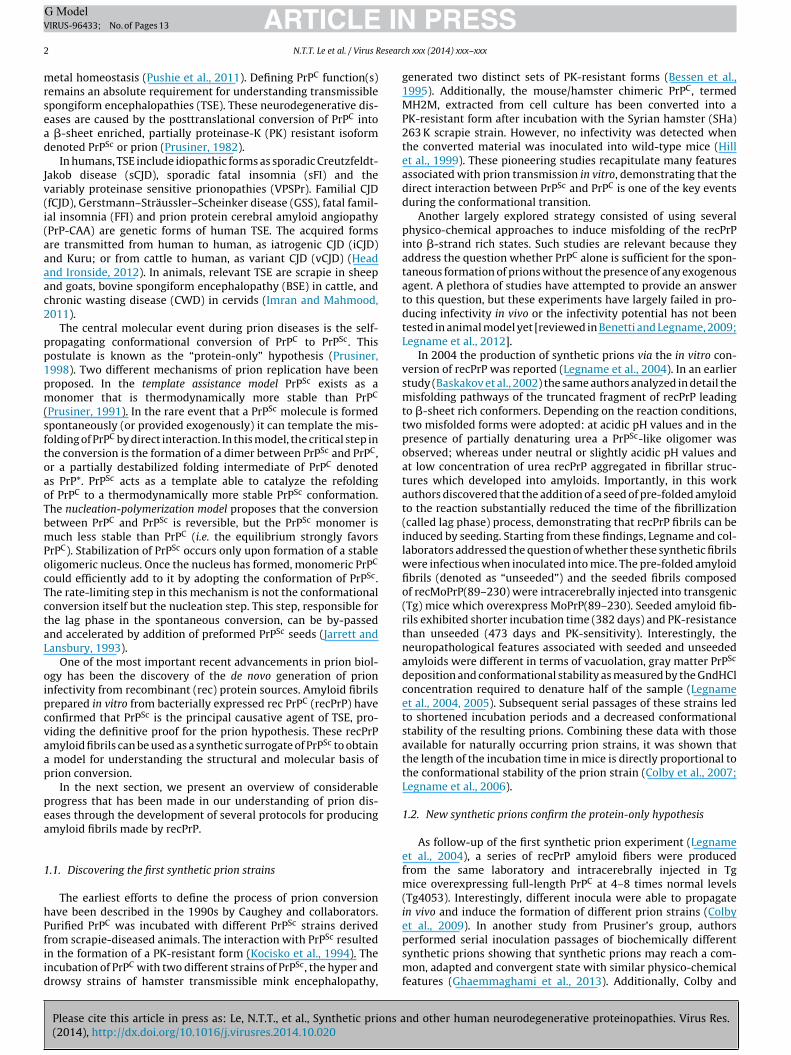
formation (Abskharon et al., 2014).Fig. 1 shows a schematic representation of the fib-
ril core regions proposed in the discussed PrPSc structuralmodels.

ARTICLE IN PRESSG ModelVIRUS-96433; No. of Pages 13
N.T.T. Le et al. / Virus Research xxx (2014) xxx–xxx 5
Fig. 1. Schematic representation of proposed amyloid core regions present in different published PrPSc models. Comparison between the PrPC secondary structure obtainedby NMR (a) (Zahn et al., 2000) and �-sheet rich regions (here represented as anti-parallel �-strands linked by loops) identified in PrPSc or recPrP fibrils using differentexperimental approaches, including cryo-EM in the �-helix model (b) (Govaerts et al., 2004), HX-MS in the continuum of �-strand model (c) (Smirnovas et al., 2011), MDi r et ao tted b( lices,
2
eatfsccen
2
tems�pTstpac
cnatsasntuh
n the spiral model (d) (DeMarco and Daggett, 2004), solution-state NMR (e) (Kumaf HuPrP obtained by nanobody-aided X-ray crystallography with highlighted (doAbskharon et al., 2014). Yellow ribbons, red arrows, and black lines represent �-he
. Prion phenomena in other neurodegenerative diseases
An increasing body of evidence suggests that as with prion dis-ase, most of the neurodegenerative disorders, such as AD, PD,myotrophic lateral sclerosis (ALS) and Huntington’s disease arehought to be caused by the intracerebral accumulation of mis-olded protein aggregates. In each disease, one or more proteinself-associate into toxic oligomers that disrupt cellular function andommunication, and proceed to form insoluble amyloid aggregatesharacterized by fibrillar morphology and cross-� structure. Recentvidence argues that prions feature in the pathogenesis of severaleurodegenerative diseases.
.1. Characterization of prion-like properties of ˛-synuclein
The presence of Lewy bodies (LBs) and Lewy neurites (LNs) inhe brain is a major pathological hallmark of several neurodegen-rative diseases, including PD, dementia with Lewy bodies (DBL),ultiple system atrophy (MSA) and other disorders known as
ynucleinopathies. The major component of LBs is represented by-synuclein (Spillantini et al., 1998), which is a natively unfoldedrotein (140-amino-acid residues) widely expressed in neurons.he physiological function of the protein remains poorly under-tood, however several studies have suggested its involvement inhe regulation of neurotransmitter release, synaptic function andlasticity (Lashuel et al., 2013). Several data show that �-synucleinggregation plays a central role in the pathogenesis of several synu-leinopathies (Lashuel et al., 2013).
Increasing evidence argues that �-synuclein may transfer fromell-to-cell in a prion-like manner, suggesting that this phe-omenon may mediate the pathological progression of PD. Braaknd colleagues have proposed that �-synuclein pathology begins inhe lower brainstem and olfactory bulb and spreads into the limbicystem and eventually to the cortex (Braak et al., 2003). The mech-nism by which the pathology can propagate through the nervousystem is not known. However, evidence for a prion-like mecha-
Please cite this article in press as: Le, N.T.T., et al., Synthetic prions a(2014), http://dx.doi.org/10.1016/j.virusres.2014.10.020
ism of the �-synuclein spreading was provided by the observationhat healthy neurons grafted in the brain of a patient with PD grad-ally developed aggregates of �-synuclein similar to those of theost neurons (Kordower et al., 2008; Li et al., 2008). Desplats and
l., 2010) or solid-state NMR (f) (Helmus et al., 2008). In (g) the secondary structureox) the new �-sheet motif (in orange arrows) formed in the palindromic region�-strands, and loops/disordered segments, respectively.
collaborators have shown that �-synuclein released from neuronalcells over-expressing the protein was transmitted via endocytosisto neighboring neurons, forming LBs-like inclusions. Subsequent invivo studies have confirmed that �-synuclein can be transmittedfrom the affected neurons to engrafted neuronal precursor cells ina Tg model of PD-like pathology, leading to inclusion body forma-tion (Desplats et al., 2009). Additionally, Tg mice expressing mutant�-synuclein inoculated with brain homogenate from MSA patientsdeveloped pathology leading to death, giving further evidence that�-synuclein aggregates present in MSA patients are, like prions,transmissible (Watts et al., 2013).
In a similar way, the efficiency of synthetic �-synuclein assem-blies to induce pathology both in vitro and in vivo is now beingstudied. Purified rec �-synuclein readily aggregates in vitro into�-rich amyloid fibrils with morphologies and staining characteris-tics similar to those of the �-synuclein aggregates extracted fromdisease-affected brains (Serpell et al., 2000). Increasing number ofdata confirm that in vitro formed aggregates, both oligomers andfibrils, may be taken up and propagate among cells in a prion-like manner, inducing LBs-like pathology (Desplats et al., 2009; Luket al., 2012a,b, 2009; Masuda-Suzukake et al., 2013; Sacino et al.,2013; Volpicelli-Daley et al., 2011).
2.1.1. ˛-Synuclein internalization and propagation in cellcultures
Transmission of synthetic �-synuclein aggregates has beenshown in several cell models. A recent report has shown thata single exposure of amyloid fibrils of human rec �-synucleinamyloids was sufficient to induce aggregation of endogenous �-synuclein in non-transfected human neuroblastoma SH-SY5Y cells(Aulic et al., 2014). Luk and collaborators have shown that theaddition of in vitro preformed �-synuclein fibrils into culturemedium induces intracellular �-synuclein aggregation in differ-ent cell lines overexpressing this protein (Luk et al., 2009). Theauthors have shown that monomeric and fibrillar forms of flu-orescently labeled �-synuclein can be efficiently introduced into
nd other human neurodegenerative proteinopathies. Virus Res.
the cytoplasm of different cell lines using cationic liposomes opti-mized for intracellular protein delivery. Internalized preformedfibrils were present as large irregular foci within cells. No inclusionswere detected in cells transduced with non-fibrillar �-synuclein

ING ModelV
6 esearc
ccomirifioTaaiTittbiagl2
2
totfirPbtdpd�tpi
hfipigcmlf�os(we(fiwefp
tn
induce aggregation of endogenous protein may depend on the pre-
ARTICLEIRUS-96433; No. of Pages 13
N.T.T. Le et al. / Virus R
omprised of either monomers or oligomers. �-synuclein intra-ellular inclusions underwent several modifications characteristicf LBs, such as hyperphosphorylation, ubiquitination and accu-ulation of cytoplasmic vesicles around the periphery of the
nclusions. Moreover, exogenous fibrils were shown to rapidlyecruit and convert endogenous soluble protein into detergent-nsoluble inclusions closely resembling LBs, thus suggesting thatbrillar seeds may have a central role in the initial formationf LBs and other disease-associated inclusions (Luk et al., 2009).he same group has also shown that preformed fibrils gener-ted from rec �-synuclein were taken up by primary neuronsnd promoted the recruitment of soluble endogenous �-synucleinnto insoluble PD-like inclusions (Volpicelli-Daley et al., 2011).hese changes led to the reduction of several synaptic proteins,mpairments in neuronal excitability and connectivity and, even-ually, neuron loss. Finally, �-synuclein fibrils containing onlyhe central, hydrophobic portion of the protein were shown toe sufficient to seed the conversion of endogenous �-synuclein
nto pathological aggregates (Volpicelli-Daley et al., 2011). Thebility of in vitro-generated �-synuclein oligomers to induce aggre-ation in primary neuronal cultures as well as in neuronal cellines has been extensively documented (Danzer et al., 2007,009).
.1.2. Prion-like spreading in animal modelsIn 2012, Luk and collaborators have shown, for the first time,
hat synthetic �-synuclein fibrils alone can induce PD-like pathol-gy and transmit disease in in vivo (Luk et al., 2012a). Particularly,he injection of either synthetic or brain-derived �-synucleinbrils into the brain of healthy mice bearing the familiar PD-elated human �-synuclein A53T mutation (M83 line) led toD-like pathology. In both cases the �-synuclein inclusions resem-led those found in LBs and LNs of patients affected by PD, ashey showed strong immunoreactivity to antibodies recognizingisease-specific conformations of �-synuclein and ubiquitin. Theathology spread progressively overtime to distal CNS regions andramatically reduced the survival time of animals. The most severe-synuclein pathology was detected in brain regions that project
o, or receive input from the inoculation site, suggesting that theropagation of pathology follows the pathway of neurons that are
nterconnected (Luk et al., 2012a,b).The same group and others (Masuda-Suzukake et al., 2013)
ave shown that intracerebral inoculation of synthetic �-synucleinbrils into wild-type mice, led to cell-to-cell transmission ofathologic �-synuclein and LBs-like pathology in anatomically
nterconnected regions. The �-synuclein pathology caused pro-ressive loss of dopamine neurons in the substantia nigra parsompacta and impairment in motor coordination. Conversely,ice injected with soluble �-synuclein did not exhibit patho-
ogical symptoms. Exogenously injected fibrils could be detectedor less than one week and the aggregation of the endogenous-synuclein was observed after three months. The pathol-gy was similar to that triggered by intracerebral injection ofarcosyl-insoluble �-synuclein from brains of patients with DLBMasuda-Suzukake et al., 2013). Supporting these theories, recentork by Rey et al. have shown that after injection of differ-
nt molecular species of rec �-synuclein into the olfactory bulbOB) of wild-type mice, monomeric and oligomeric, but notbrillar �-synuclein was quickly taken up by OB neurons andas rapidly transferred into interconnected brain structures (Rey
t al., 2013). These results confirm that �-synuclein can trans-er along neural pathways and thereby contribute to pathology
Please cite this article in press as: Le, N.T.T., et al., Synthetic prions a(2014), http://dx.doi.org/10.1016/j.virusres.2014.10.020
rogression.Sacino and collaborators (Sacino et al., 2013) have observed
hat injections of exogenous �-synuclein fibrils into the brain ofeonatal mice induced pathology after prolonged incubation times.
PRESSh xxx (2014) xxx–xxx
The injected �-synuclein disappeared within a few days but trig-gered the aggregation of endogenous �-synuclein which led tothe appearance of a pathological phenotype after several months.Additional data, using Tg mice overexpressing �-synuclein (moreprone to develop PD-like pathology) showed that injections of thenon-amyloidogenic form of �-synuclein (�71–82) induced similarpathology, suggesting that the pathogenesis might be induced notsolely by conformation-dependent templating events (Sacino et al.,2013).
All these data confirmed that �-synuclein aggregation is suffi-cient to induce behaviors and pathological features of sporadic PD(Luk et al., 2012b).
2.1.3. The existence of different ˛-synuclein strainsA typical hallmark of prion diseases is the existence of differ-
ent strains, which arise from the same protein but have differentconformations, resulting in different incubation periods, PrPSc dis-tribution, lesion profiles in the brain of infected animals, as wellas different PK cleavage patterns of PrPSc (Aguzzi et al., 2007a).The existence of different strains has not been identified so far inother neurodegenerative diseases; however some evidence sug-gests that specific �-synuclein strains may be involved in particularsynucleinopathies.
Danzer and collaborators have observed that, using differentconditions of aggregation in vitro, �-synuclein oligomers formeddistinct populations that differed fundamentally in their biophys-ical properties and cellular effects. Only some types of oligomerswere internalized by primary neuronal cells and neuronal cell linesand induced endogenous �-synuclein aggregation, while otherslacked any seeding properties, but induced cell death via disruptionof cellular ion homeostasis (Danzer et al., 2007, 2009).
Recently, Bousset and collaborators have generated in vitrotwo different high-molecule assemblies of �-synuclein, preparedin buffers with different salt concentrations. These assemblies,designated fibrils and ribbons, adopted different conformationsand PK digestion profiles and revealed different cleavage profiles.Fibrils and ribbons exhibited marked differences in their propen-sity to bind and penetrate cells and to seed toxic aggregation ofthe endogenous �-synuclein in cells. Moreover, they imprintedtheir intrinsic structure to endogenous �-synuclein, suggestingthat different strain-like assemblies may account for the develop-ment and progression of different synucleinopathies (Bousset et al.,2013).
Recent work by Guo and collaborators has shown two distinctstrains of synthetic �-synuclein fibrils that demonstrate strikingdifferences in the efficiency of cross-seeding tau aggregation, bothin neuron cultures and in vivo. The authors speculated that distinctstrains of pathological �-synuclein could exist. This may lie at theroot of the tremendous heterogeneity of synucleinopathies (Guoet al., 2013).
Watts and collaborators have shown than brain homogenatesprepared from MSA patients or spontaneously ill TgM83+/+ mice(Tg mice expressing PD-associated �-synuclein mutation A53T)induced pathology and death of recipient mice (Tg(M83+/−:Gfap-luc)) after intracerebral inoculation. However, the transmission ofMSA inocula was faster than TgM83+/+ samples. The authors sug-gested that differences in disease transmission may arise from thefact that both �-synuclein aggregates constitute a distinct “strains”(Watts et al., 2013).
The ability of �-synuclein aggregates to enter into cells and
nd other human neurodegenerative proteinopathies. Virus Res.
cise structure of the aggregated protein. The fact that different�-synuclein polymorphs may bind to different extents to culturedcells may account for the spread pattern of �-synuclein assemblieswithin the brain as described by Braak and collaborators.
ING ModelV
esearc
2
nopR
Zgsawhr
mRatsicim
iRmtricim
otpmgSaa
2o
plt
ABifanr
bsmtT
ARTICLEIRUS-96433; No. of Pages 13
N.T.T. Le et al. / Virus R
.2. Prion-like properties of supeoxide dismutase-1
Amyotrophic lateral sclerosis (ALS) is the most common motoreuron disease, mainly characterized by the selective degenerationf spinal cord motor neurons, leading to rapid and progressive atro-hy of skeletal muscles and death (Bruijn et al., 2004; Cleveland andothstein, 2001).
Increasing evidence shows that misfolding of the mutant Cu,n superoxide dismutase-1 (SOD1) is a central event in the patho-enesis of autosomal-dominantly inherited ALS (FALS). SOD1 is amall 153-amino acid protein involved in conversion of superoxide,
toxic by-product of mitochondrial oxidative phosphorylation, toater or hydrogen peroxide. Over 100 FALS-linked SOD1 mutationsave been characterized, however their involvement in motor neu-on degeneration remains poorly understood (Bruijn et al., 2004).
Although approximately 5–10% of ALS cases are familiar, theajority are sporadic. Pathological aggregates composed of the
NA/DNA binding protein TDP-43 are detected in CNS tissues for majority of sporadic ALS cases, suggesting its involvement inhe pathogenesis (Neumann et al., 2006). Several studies havehown that both SOD1 and TDP43 proteins readily aggregaten vitro and ALS-causing mutations enhance the aggregation pro-ess. Moreover, some data indicate that these aggregates maynduce misfolding and aggregation of native proteins in a prion-like
anner (Polymenidou and Cleveland, 2011).SOD1 fibrils formed in vitro have toxic properties; they induce
nflammation and activate microglial cells (Fiala et al., 2010;oberts et al., 2013). These fibrils possess the ability to seed the for-ation of further fibrils in an autocatalytic manner while reducing
he lag-time of fibrillization. Moreover, amyloid fibril formation ofec SOD1 could be seeded with tissue extracts of SOD1-containingnclusions from ALS transgenic mice (Chia et al., 2010). Severalo-culture experiments have shown that SOD1-dependent toxic-ty may spread between cells, suggesting a prion-like propagation
echanism (Polymenidou and Cleveland, 2011).Munch and collaborators has shown that aggregates composed
f diverse synthetic SOD1 mutants rapidly and efficiently pene-rate inside Neuro-2a cells and seed aggregation of the endogenousrotein. Endogenous mutant protein is required for perpetuatingutant SOD1 misfolding. Moreover, these aggregates were propa-
ated between cells in a prion-like manner. Aggregation of mutantOD1 induced by extracellular aggregates was a highly penetrantnd heritable phenotype that stably persisted long after disappear-nce of the seed (Munch et al., 2011).
.3. Rethinking AD: seeded aggregation and cell-to-cell spreadingf A and tau amyloids
AD is associated with the misfolding and aggregation of tworoteins: (i) amyloid-� peptide (A�) which accumulates extracel-
ularly in the form of amyloid plaques and (ii) hyperphosphorylatedau, which forms neurofibrillary tangles inside of neurons.
The first experimental studies supporting the hypothesis that� amyloids may share prion-like features were performed byaker and collaborators. In these studies authors intracerebrally
noculated marmosets (Callithrix jacchus) with brain homogenatesrom AD patients and after 6–7 years animals developed moder-te numbers of A� plaques with associated argyrophilic dystrophiceurites and cerebral amyloid angiopathy in the brain but no neu-ofibrillary tangles (Baker et al., 1993; Ridley et al., 2006).
Similarly, it has been shown that the intracerebral infusion ofrain homogenates from AD patients or from aged APP-Tg mice
Please cite this article in press as: Le, N.T.T., et al., Synthetic prions a(2014), http://dx.doi.org/10.1016/j.virusres.2014.10.020
timulates the premature formation of A� plaques in APP23 Tgice. The induction of A� deposition seems to parallel the concen-
ration of the A� seeds in the extracts and the time of incubation.hey also performed the injection experiments using the synthetic
PRESSh xxx (2014) xxx–xxx 7
fibrils of A�40 and A�42. However, the injection of synthetic fibrilssingularly, or a mixture of both failed to induce detectable A� depo-sition. Few newly generated aggregates were present as confirmedby biotinylated A�42. Control experiments depleting the materialfrom A� aggregates or inactivating their conformation did not pro-duce acceleration of the pathology, indicating that the presence ofA� aggregates is required to seed amyloid plaque deposition in vivo(Meyer-Luehmann et al., 2006).
Recently, Soto and collaborators have performed similar exper-iments working on the Tg mice expressing human wild-type APP(HuAPP wt). These mice do not develop A� deposits spontaneouslyduring their life span. In this study it was shown that the bilateralinjection of brain homogenates from confirmed AD patients lead tothe development of A� deposits over time (starting from 285 dpi).The accumulation of A� plaques increased progressively in a timedependent manner and the A� lesions were observed in brain areas(e.g. cortex) far from the injection site (hippocampus), suggestingthat the seeding activity can spread through the brain. These resultssuggest that some of the typical brain abnormalities associated withAD can be induced by a prion-like mechanism of disease trans-mission through propagation of protein misfolding (Morales et al.,2012).
In another work from Prusiner’s lab the kinetics of spontaneousand induced A� depositions in living mice has been investigatedusing bioluminescence imaging (BLI) (Stohr et al., 2012). The un-inoculated biogenic mice Tg(APP23:Gfap-luc) showed an increasein the BLI signal in their brains starting at 416 ± 9 dpi. To deter-mine whether A� deposition could be accelerated in these animalsin a prion-like fashion, Tg(APP23:Gfap-luc) mice were intracere-brally inoculated with brain homogenates from two aged Tg mousemodels, Tg(APP23) or Tg(CRND8). These injections produced a sus-tained increase in the brain BLI signal of biogenic mice at 261 ± 8and 238 ± 12 dpi, respectively. Whereas the BLI signal remainedlow in mice inoculated with aged non-Tg brain homogenateuntil 333 ± 9 dpi. Interestingly, despite unilateral inoculation thepathology was bilateral, suggesting the progressive spread of A�deposition throughout the brain. These materials were subse-quently purified by limited PK digestion, and then injected inhealthy Tg(APP23) and Tg(CRND8) mice, displaying an increasein the BLI signal at 161 ± 7 and 173 ± 9 dpi, respectively. Thisfinding argues that the purified A� preparations are sufficient toinduce widespread A� deposition in vivo, providing compellingevidence for the existence of A� prions. Since the purification ofthe brain homogenates does not exclude the co-purification ofother cofactors essential for the observed prion behavior of A�,they performed the experiments with the rec synthetic A� aggre-gates. For these studies a WT A�40 peptide and a dimer – denotedas (A� S26C)2 composed of a mutant A�40 linked through a cys-teine at position 26 – were subjected to amyloid formation usingthe ThT fluorescence assay. The synthetic A� aggregates obtainedwere then intracerebrally inoculated into young Tg(APP23:Gfap-luc) mice, using ∼100 fold higher concentration then the A�in the brain homogenates. Both synthetic A�40 and (A� S26C)2preparations produced elevated BLI signals at 229 ± 5 dpi and234 ± 9 dpi, respectively. The mice inoculated with synthetic A�peptides exhibited bilaterally A� deposits primarily in the subcal-losal region of the hippocampus and in the thalamus. These cerebralA� deposits could not have be due to the residual peptide comingfrom the inoculation of the synthetic A� because the A� depositionwas not detectable by immunoblotting or immunohistochemistrybetween 33 and 83 dpi. Therefore, this study reveals that rec A�fibrils are wholly sufficient to transmit disease, thereby suggest-
nd other human neurodegenerative proteinopathies. Virus Res.
ing a common pathogenic mechanism of disease progression forneurodegenerative brain amyloidoses.
The same group, in a recently published work reported a mod-ified protocol for the polymerization of both synthetic A�40 and

ING ModelV
8 esearc
AcssIptocta
ett
2
Sbddit(atwsemiitibiuc2
aSoftrtitwwtNtu
tc1ittiom
acidic sequence of a protein can encipher a variety of differentamyloid states (Tanaka et al., 2004; Wiltzius et al., 2009). Thishypothesis is strengthened by the strain phenomena observed inprion diseases. Prion strains are distinct varieties of prions with the
Fig. 2. Workflow of our cell-based approach to test new synthetic prion amyloids.The full-length recMoPrP was led to polymerize in different fibrillization buffersleading to the generation of several morphologically distinct amyloid assemblies(here differently colored). Subsequently, these aggregates were subjected to N2a
ARTICLEIRUS-96433; No. of Pages 13
N.T.T. Le et al. / Virus R
�42 (Stohr et al., 2014). Using this approach they obtained fibrilsharacterized by increased PK-resistance. Most importantly, theseynthetic preparations, once inoculated into the biogenic mice,howed higher prion infectivity compared with the earlier study.n addition, pathological investigations showed that synthetic A�40rions produced amyloid plaques containing both A�40 and A�42 inhe brains of inoculated bigenic mice, whereas synthetic A�42 pri-ns stimulated the formation of smaller, more numerous plaquesomposed predominantly of A�42. These findings show that syn-hetic A� peptides can form prions that infect mice and induce A�myloid plaque pathology.
In particular the prion like characteristic of A� is now becomingxperimental evidence, since it was shown that the serial propaga-ion of distinct strains of A� prions from AD patients can occur inhe mice (Watts et al., 2014).
.3.1. Tau amyloidsAnother typical feature of AD is the presence of tau aggregates.
everal studies have been performed to analyze the transmissiony seeding of tau aggregates, found also in other neurodegenerativeiseases and collectively termed tauopathies (e.g. fronto-temporalementia, chronic traumatic encephalopathy, etc.). Intracerebral
njections of brain extracts from mice expressing the mutant P301Sau isoform into the brain of Tg mice expressing Hu wild-type tauALZ17) induces the assembly of Hu wild-type tau into misfoldedggregates and the spreading of pathology from the site of injec-ion to neighboring brain regions. In the control experiments itas shown that the presence of tau in P301S extract was neces-
ary to induce filamentous pathology, since the injection of brainxtract from non-Tg mice did not induce any pathology in ALZ17ice (15 month post-injection). Similar findings were observed
n non-Tg mice inoculated with P301S extract (6 months post-njection). The induction of filamentous tau in ALZ17 mice wasime and brain region dependent. Since tau aggregates are locatedntracellularly, this suggests that protein misfolding is transmittedetween cells. This hypothesis is further supported by in vitro stud-
es of cultured cells where extracellular tau aggregates were takenp and were able to induce the misfolding and aggregation of intra-ellular tau (Frost et al., 2009; Guo and Lee, 2011; Nonaka et al.,010).
Most importantly, Iba et al. have shown that synthetic tau fibrilsre sufficient to transmit tau inclusions in a taupathy mouse model.pecifically, unilateral intracerebral inoculation in young PS19 miceverexpressing a human tau mutant (P301S) with synthetic pre-ormed fibrils – assembled from rec full-length tau or truncatedau containing four microtubule binding repeats – resulted inapid induction of neurofibrillary tangles (NFTs)-like inclusionshat propagated from injected sites to connected brain regionsn a time-dependent manner. Indeed, the injection of tau syn-hetic preformed fibrils into different brain regions resulted in aidespread transmission of tau pathology to diverse brain regions,hich are anatomically interconnected to the injection sites. Syn-
hetic tau fibrils induce tau pathologies which closely resemble ADFTs, because they were hyperphosphorylated, Thioflavin-S posi-
ive, acetylated, and more resistant to PK digestion if compared toninjected mice at 8–9 month of age (Iba et al., 2013).
Finally, Sanders et al. have identified two clones made using aruncated tau fragment (RD) that were exposed to cultured HEK293ells (Sanders et al., 2014). The two strains, named clone 9 and clone0, differ with respect to inclusion morphology, aggregate size, sed-
mentation profile, seeding capacity, proteinase digestion pattern,oxicity, and subcellular localization. Most importantly, they found
Please cite this article in press as: Le, N.T.T., et al., Synthetic prions a(2014), http://dx.doi.org/10.1016/j.virusres.2014.10.020
hat in vivo these strains induce unique pathological phenotypesn Tg P301S mice. Clone 9 induced tangle-like inclusions through-ut CA1 and CA3, while clone 10 induced AT8-positive puncta inossy fiber tracts. Here, they show that tau aggregation propagates
PRESSh xxx (2014) xxx–xxx
along known anatomical connections, supporting the conclusionsof previous studies (de Calignon et al., 2012; Iba et al., 2013; Liuet al., 2012). However, injected WT mice with these clones neverdeveloped pathology, possibly due to a seeding barrier betweeninoculated tau RD and WT murine tau. On the basis of these data,authors strongly propose that tau could be considered as a bona fideprion agent.
3. A cell-based approach to test new synthetic prion strains
Current evidence that prions feature in the pathogenesis of sev-eral neurodegenerative diseases postulate that solely the amino
nd other human neurodegenerative proteinopathies. Virus Res.
or GT1 cells. To test the ability of these preparations to induce spontaneous PrPC
conversion to a PK-resistant PrPSc form, cell lysates were PK digested, and PrPimmunoreactivity was detected by western-blot. Neuronal cells that were de novoinfected by synthetic recMoPrP preparations show a diagnostic immunoreactive PrPband at about 15 kDa (red arrow) after PK digestion.

ARTICLE IN PRESSG ModelVIRUS-96433; No. of Pages 13
N.T.T. Le et al. / Virus Research xxx (2014) xxx–xxx 9
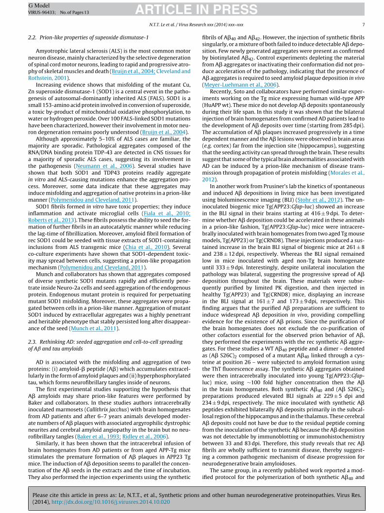
Fig. 3. AFM topological images of different recMoPrP assemblies deposited on mica surfaces after the fibrillization reactions. Preparation #4 was made with 0.1 mg/mLr obtain3 tted l
stpfmiZpiaoe
fHncaoFtcdmee(
adowetelcsc
sot
ecMoPrP, 50 mM acetate buffer, 4 M GndHCl at pH 5.0; samples #19 and #28 were
M GndHCl at pH 5.0, respectively. In the lower panels, height profiles along the do
ame primary structure, exhibiting characteristic biological proper-ies, such as tropism for specific brain areas and incubation period,eculiar neurological signs, as well as distinct physico-chemicaleatures. Considerable evidence argues that the biological infor-
ation of a prion strain is encoded in its conformation [reviewedn Aguzzi et al., 2007b; Collinge and Clarke, 2007; Legname andanusso, 2012; Poggiolini et al., 2013]. As previously described,rion strains can also be efficiently generated synthetically start-
ng from recPrP materials. This has led to the generation of novelmyloid conformations able to transmit TSE in recipient modelrganisms (Colby et al., 2009, 2010; Legname et al., 2004; Makaravat al., 2010; Wang et al., 2010; Zhang et al., 2013).
Bioassay in animals has been the most commonly used methodor assessing infectivity of both natural and synthetic prions in vivo.owever, the use of animal models for the initial screening ofew putative prion strains results in highly expensive and time-onsuming experiments. As an alternative method, cell-basedssays represent a powerful tool to test the ability of synthetic pri-ns to template the conversion of the endogenous PrPC to PrPSc.urthermore, cell-culture systems may provide additional informa-ion about the molecular mechanisms governing prion formation,ell-to-cell prion spread and intracellular prion accumulation. Toissect the mechanism of prion strain transformation, Ghaem-aghami and collaborators have investigated the physicochemical
volution of a mouse synthetic prion strain, MoSP1 (Legnamet al., 2004), after repeated passage in mice and cultured cellsGhaemmaghami et al., 2011).
We recently started a platform of cell-based experiments aimedt elucidating whether different synthetic amyloid preparationsisplay prion features – for instance inducing the conversionf the endogenous PrPC into a PK-resistant PrPSc-like isoform –hen exposed to neuronal cells. As experimental tools, we have
mployed two distinct cellular models: the murine neuroblas-oma N2a and the hypothalamic-derived GT1 cell lines (Solassolt al., 2003). These cells were exposed to different recPrP amy-oid assemblies made using full-length recMoPrP at different bufferonditions. The variables we tested for producing these putativeynthetic prions include: ionic strength, pH and the presence ofhaotropic (i.e. GndHCl) or reducing agents (i.e. DTT).
Please cite this article in press as: Le, N.T.T., et al., Synthetic prions a(2014), http://dx.doi.org/10.1016/j.virusres.2014.10.020
This method shares some aspects of ASA (Colby et al., 2009) –uch as intermittent shaking of full-length recMoPrP and evaluationf the fibrillization kinetics via ThT-fluorescence – combined withhe evaluation of the putative synthetic prions using as read-out
ed in fibrillization buffer containing PBS, 2 M GndHCl at pH 7.4 and 50 mM acetate,ine bars.
the PK-resistance levels of PrPSc-like isoforms present in N2a orGT1 exposed to recPrP aggregates.
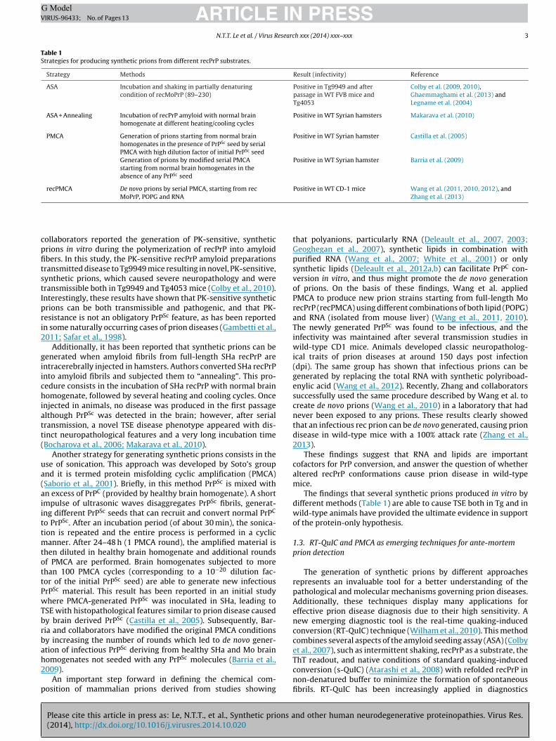
Fig. 2 shows a graphical representation about the experimentalprocedure we employed to screen our bona fide synthetic prions.Briefly, we have produced different recMoPrP amyloid preparationsthat were morphologically characterized by AFM. We found thatbuffer composition strongly affects the structural organization ofrecMoPrP assemblies. For instance, aggregates obtained at acidicpH and high GndHCl concentration display short fibril morphol-ogy with a maximum height distribution of ∼6 nm (see recMoPrPamyloid termed as #4), while those obtained at lower denatu-rant concentration are organized in long fibrils of ∼8 nm height(see amyloid #28). Interestingly, a shift of pH to neutral condi-tions and a reduction of GndHCl concentration result in a newassembly characterized by long ∼120 nm height branches (see #19)(Fig. 3).
To further assess whether our synthetic amyloids display prion-like features, we seeded these synthetic materials in media ofcultured GT1 and N2a cells. Bona fide PrPSc has been operationallydefined as resistant to PK digestion and insoluble to non-denaturingdetergents (Prusiner, 1998). Although this definition has beenreconsidered after the discovery of PK-sensitive prions in vivo(Gambetti et al., 2011; Pastrana et al., 2006; Safar et al., 1998;Tzaban et al., 2002; Zou et al., 2013), PK-based assays still representthe best choice for detecting PrPSc in cellular models (Vilette, 2008).After exposing the cells to recMoPrP amyloid preparations for upto six passages, we were able to detect a characteristic PK-resistantlower molecular weight PrPSc-like fragment (Fig. 4a). Interestingly,cells exposed to these rec materials displayed an increased intra-cellular PrPC accumulation (Fig. 4b), suggesting the de novo prionconversion in N2a or GT1 cells may affect the physiological PrPC
pathway through endocytic compartments as has been observedby other groups (Goold et al., 2013, 2011; Marijanovic et al., 2009;Rouvinski et al., 2014; Yamasaki et al., 2014). Taken together, theseobservations strengthen the current body of evidence that syn-thetic recPrP amyloids may act as real seeds able to template theconversion of PrPC to a prion-like isoform in cell culture experi-ments. With this experimental approach we were able to select upto 34 novel synthetic recMoPrP amyloids, which were character-
nd other human neurodegenerative proteinopathies. Virus Res.
ized on the basis of their morphology and on their ability to inducebona fide PK-resistant PrPSc in cells. This cell-based method maytherefore represent a useful tool for the initial screening of severalamyloid preparations before testing these materials in vivo.

ARTICLE IN PRESSG ModelVIRUS-96433; No. of Pages 13
10 N.T.T. Le et al. / Virus Research xxx (2014) xxx–xxx
F enousa
4
cIcpcbduninogficmd
ooaltiaiaopgod
A
FP
R
A
ig. 4. recMoPrP amyloid preparations induce spontaneous conversion of the endogccumulation (b).
. Concluding remarks
The most fascinating and unique feature of TSE is that they areaused by misfolded protein accumulation in the nervous system.n the latter part of 1980s bovine spongiform encephalopathy inattle and subsequently vCJD, a human prion disease caused byrion-contaminated cattle meat, raised unprecedented public con-ern due to the concrete possibility that prions in animals coulde transmitted to humans through the food chain. For roughly twoecades, prions were under intense scrutiny and many studies werendertaken worldwide. These investigations have led our commu-ity to a better risk assessment and management of prion diseases
n humans and in animals, substantially limiting the possibility ofew prion epidemics in the world. Today, prions have been broughtnce again to the fore after the discovery that a variety of neurode-enerative diseases, in particular AD, PD, Huntington’s disease androntotemporal dementia, share fundamental features with prions,ncluding protein misfolding and aggregation in the brain, cell-to-ell transmission and in vivo infectivity. Therefore, studying prionsight help to understand the pathological mechanism of these
isorders.In this review we have described the state-of-the-art in the field
f prions and a novel cell culture assay as a fast screening methodol-gy to characterize putative infectious materials. The bona fide PrPSc
nd the cell lines infected with that synthetic material has high-ighted the conformational diversity of pathological recPrPSc andheir biological activities, as well as the novel insights in character-zing molecular events involved in mammalian prion conversionnd propagation. This assay represents a feasible tool for study-ng the mechanism of prion conversion and propagation, as wells for developing new therapeutic targets that could be applied tother neurodegenerative diseases. In fact, the finding that syntheticrions, as well as other proteins, such as A�, tau and �-synucleinenerated from rec proteins are able to induce pathology in vivopens new perspectives for future research in neurodegenerativeiseases, especially in AD and PD.
cknowledgement
This work was supported by European Community’s Seventhramework Programme (FP7 grant agreement no. 222887 – theRIORITY project).
eferences
Please cite this article in press as: Le, N.T.T., et al., Synthetic prions a(2014), http://dx.doi.org/10.1016/j.virusres.2014.10.020
bskharon, R.N., Giachin, G., Wohlkonig, A., Soror, S.H., Pardon, E., Legname, G.,Steyaert, J., 2014. Probing the N-terminal beta-sheet conversion in the crystalstructure of the human prion protein bound to a nanobody. J. Am. Chem. Soc.136 (3), 937–944.
PrPC to PK-resistant PrPSc-like forms in both GT1 and N2a cells (a) and intracellular
Aguzzi, A., Heikenwalder, M., Polymenidou, M., 2007a. Insights into prion strainsand neurotoxicity. Nat. Rev. Mol. Cell Biol. 8 (7), 552–561.
Aguzzi, A., Heikenwalder, M., Polymenidou, M., 2007b. Mechanisms of disease –insights into prion strains and neurotoxicity. Nat. Rev. Mol. Cell Biol. 8 (7),552–561.
Amenitsch, H., Benetti, F., Ramos, A., Legname, G., Requena, J.R., 2013. SAXS structuralstudy of PrP reveals ∼11 nm diameter of basic double intertwined fibers. Prion7 (6).
Atarashi, R., Satoh, K., Sano, K., Fuse, T., Yamaguchi, N., Ishibashi, D., Matsubara, T.,Nakagaki, T., Yamanaka, H., Shirabe, S., Yamada, M., Mizusawa, H., Kitamoto, T.,Klug, G., McGlade, A., Collins, S.J., Nishida, N., 2011. Ultrasensitive human priondetection in cerebrospinal fluid by real-time quaking-induced conversion. Nat.Med. 17 (2), 175–178.
Atarashi, R., Wilham, J.M., Christensen, L., Hughson, A.G., Moore, R.A., Johnson, L.M.,Onwubiko, H.A., Priola, S.A., Caughey, B., 2008. Simplified ultrasensitive priondetection by recombinant PrP conversion with shaking. Nat. Methods 5 (3),211–212.
Aulic, S., Le, T.T., Moda, F., Abounit, S., Corvaglia, S., Casalis, L., Gustincich, S., Zur-zolo, C., Tagliavini, F., Legname, G., 2014. Defined alpha-synuclein prion-likemolecular assemblies spreading in cell culture. BMC Neurosci. 15 (1), 69.
Baker, H.F., Ridley, R.M., Duchen, L.W., Crow, T.J., Bruton, C.J., 1993. Evidence for theexperimental transmission of cerebral beta-amyloidosis to primates. Int. J. Exp.Pathol. 74 (5), 441–454.
Barria, M.A., Mukherjee, A., Gonzalez-Romero, D., Morales, R., Soto, C., 2009. De novogeneration of infectious prions in vitro produces a new disease phenotype. PLoSPathog. 5 (5), e1000421.
Baskakov, I.V., Legname, G., Baldwin, M.A., Prusiner, S.B., Cohen, F.E., 2002. Path-way complexity of prion protein assembly into amyloid. J. Biol. Chem. 277 (24),21140–21148.
Benetti, F., Legname, G., 2009. De novo mammalian prion synthesis. Prion 3 (4),213–219.
Bessen, R.A., Kocisko, D.A., Raymond, G.J., Nandan, S., Lansbury, P.T., Caughey, B.,1995. Non-genetic propagation of strain-specific properties of scrapie prionprotein. Nature 375 (6533), 698–700.
Bocharova, O.V., Makarava, N., Breydo, L., Anderson, M., Salnikov, V.V., Baskakov,I.V., 2006. Annealing prion protein amyloid fibrils at high temperatureresults in extension of a proteinase K-resistant core. J. Biol. Chem. 281 (4),2373–2379.
Bousset, L., Pieri, L., Ruiz-Arlandis, G., Gath, J., Jensen, P.H., Habenstein, B., Madiona,K., Olieric, V., Bockmann, A., Meier, B.H., Melki, R., 2013. Structural and functionalcharacterization of two alpha-synuclein strains. Nat. Commun. 4, 2575.
Braak, H., Del Tredici, K., Rub, U., de Vos, R.A., Jansen Steur, E.N., Braak, E., 2003.Staging of brain pathology related to sporadic Parkinson’s disease. Neurobiol.Aging 24 (2), 197–211.
Bruijn, L.I., Miller, T.M., Cleveland, D.W., 2004. Unraveling the mechanisms involvedin motor neuron degeneration in ALS. Annu. Rev. Neurosci. 27, 723–749.
Caiati, M.D., Safiulina, V.F., Fattorini, G., Sivakumaran, S., Legname, G., Cherubini, E.,2013. PrPC controls via protein kinase A the direction of synaptic plasticity inthe immature hippocampus. J. Neurosci. 33 (7), 2973–2983.
Castilla, J., Saa, P., Hetz, C., Soto, C., 2005. In vitro generation of infectious scrapieprions. Cell 121 (2), 195–206.
Caughey, B., Raymond, G.J., Bessen, R.A., 1998. Strain-dependent differences inbeta-sheet conformations of abnormal prion protein. J. Biol. Chem. 273 (48),32230–32235.
Caughey, B.W., Dong, A., Bhat, K.S., Ernst, D., Hayes, S.F., Caughey, W.S., 1991. Sec-ondary structure analysis of the scrapie-associated protein PrP 27–30 in waterby infrared spectroscopy. Biochemistry 30 (31), 7672–7680.
Chia, R., Tattum, M.H., Jones, S., Collinge, J., Fisher, E.M., Jackson, G.S., 2010. Super-
nd other human neurodegenerative proteinopathies. Virus Res.
oxide dismutase 1 and tgSOD1 mouse spinal cord seed fibrils, suggesting apropagative cell death mechanism in amyotrophic lateral sclerosis. PLoS ONE5 (5), e10627.
Cleveland, D.W., Rothstein, J.D., 2001. From Charcot to Lou Gehrig: decipheringselective motor neuron death in ALS. Nat. Rev. Neurosci. 2 (11), 806–819.

ING ModelV
esearc
C
C
C
C
C
D
D
d
D
D
D
D
D
D
D
D
F
F
G
G
G
G
G
G
G
G
G
ARTICLEIRUS-96433; No. of Pages 13
N.T.T. Le et al. / Virus R
obb, N.J., Sonnichsen, F.D., McHaourab, H., Surewicz, W.K., 2007. Molecular archi-tecture of human prion protein amyloid: a parallel, in-register beta-structure.Proc. Natl. Acad. Sci. U. S. A. 104 (48), 18946–18951.
olby, D.W., Giles, K., Legname, G., Wille, H., Baskakov, I.V., DeArmond, S.J., Prusiner,S.B., 2009. Design and construction of diverse mammalian prion strains. Proc.Natl. Acad. Sci. U. S. A. 106 (48), 20417–20422.
olby, D.W., Wain, R., Baskakov, I.V., Legname, G., Palmer, C.G., Nguyen, H.O., Lemus,A., Cohen, F.E., DeArmond, S.J., Prusiner, S.B., 2010. Protease-sensitive syntheticprions. PLoS Pathog. 6 (1), e1000736.
olby, D.W., Zhang, Q., Wang, S., Groth, D., Legname, G., Riesner, D., Prusiner, S.B.,2007. Prion detection by an amyloid seeding assay. Proc. Natl. Acad. Sci. U. S. A.104 (52), 20914–20919.
ollinge, J., Clarke, A.R., 2007. A general model of prion strains and their pathogeni-city. Science 318 (5852), 930–936.
anzer, K.M., Haasen, D., Karow, A.R., Moussaud, S., Habeck, M., Giese, A.,Kretzschmar, H., Hengerer, B., Kostka, M., 2007. Different species of alpha-synuclein oligomers induce calcium influx and seeding. J. Neurosci. 27 (34),9220–9232.
anzer, K.M., Krebs, S.K., Wolff, M., Birk, G., Hengerer, B., 2009. Seeding induced byalpha-synuclein oligomers provides evidence for spreading of alpha-synucleinpathology. J. Neurochem. 111 (1), 192–203.
e Calignon, A., Polydoro, M., Suarez-Calvet, M., William, C., Adamowicz, D.H.,Kopeikina, K.J., Pitstick, R., Sahara, N., Ashe, K.H., Carlson, G.A., Spires-Jones, T.L.,Hyman, B.T., 2012. Propagation of tau pathology in a model of early Alzheimer’sdisease. Neuron 73 (4), 685–697.
e Genst, E.J., Guilliams, T., Wellens, J., O‘Day, E.M., Waudby, C.A., Meehan, S.,Dumoulin, M., Hsu, S.T., Cremades, N., Verschueren, K.H., Pardon, E., Wyns, L.,Steyaert, J., Christodoulou, J., Dobson, C.M., 2010. Structure and properties of acomplex of alpha-synuclein and a single-domain camelid antibody. J. Mol. Biol.402 (2), 326–343.
e Meyer, T., Muyldermans, S., Depicker, A., 2014. Nanobody-based products asresearch and diagnostic tools. Trends Biotechnol. 32 (5), 263–270.
eleault, N.R., Harris, B.T., Rees, J.R., Supattapone, S., 2007. Formation of native pri-ons from minimal components in vitro. Proc. Natl. Acad. Sci. U. S. A. 104 (23),9741–9746.
eleault, N.R., Lucassen, R.W., Supattapone, S., 2003. RNA molecules stimulate prionprotein conversion. Nature 425 (6959), 717–720.
eleault, N.R., Piro, J.R., Walsh, D.J., Wang, F., Ma, J., Geoghegan, J.C., Supattapone,S., 2012a. Isolation of phosphatidylethanolamine as a solitary cofactor for prionformation in the absence of nucleic acids. Proc. Natl. Acad. Sci. U. S. A. 109 (22),8546–8551.
eleault, N.R., Walsh, D.J., Piro, J.R., Wang, F., Wang, X., Ma, J., Rees, J.R., Supat-tapone, S., 2012b. Cofactor molecules maintain infectious conformation andrestrict strain properties in purified prions. Proc. Natl. Acad. Sci. U. S. A. 109(28), E1938–E1946.
eMarco, M.L., Daggett, V., 2004. From conversion to aggregation: protofibril for-mation of the prion protein. Proc. Natl. Acad. Sci. U. S. A. 101 (8), 2293–2298.
esplats, P., Lee, H.J., Bae, E.J., Patrick, C., Rockenstein, E., Crews, L., Spencer, B.,Masliah, E., Lee, S.J., 2009. Inclusion formation and neuronal cell death throughneuron-to-neuron transmission of alpha-synuclein. Proc. Natl. Acad. Sci. U. S. A.106 (31), 13010–13015.
iala, M., Chattopadhay, M., La Cava, A., Tse, E., Liu, G., Lourenco, E., Eskin, A., Liu,P.T., Magpantay, L., Tse, S., Mahanian, M., Weitzman, R., Tong, J., Nguyen, C., Cho,T., Koo, P., Sayre, J., Martinez-Maza, O., Rosenthal, M.J., Wiedau-Pazos, M., 2010.IL-17A is increased in the serum and in spinal cord CD8 and mast cells of ALSpatients. J. Neuroinflamm. 7, 76.
rost, B., Jacks, R.L., Diamond, M.I., 2009. Propagation of tau misfolding from theoutside to the inside of a cell. J. Biol. Chem. 284 (19), 12845–12852.
ambetti, P., Puoti, G., Zou, W.Q., 2011. Variably protease-sensitive prionopathy: anovel disease of the prion protein. J. Mol. Neurosci. 45 (3), 422–424.
asset, M., Baldwin, M.A., Fletterick, R.J., Prusiner, S.B., 1993. Perturbation of thesecondary structure of the scrapie prion protein under conditions that alterinfectivity. Proc. Natl. Acad. Sci. U. S. A. 90 (1), 1–5.
eoghegan, J.C., Valdes, P.A., Orem, N.R., Deleault, N.R., Williamson, R.A., Harris,B.T., Supattapone, S., 2007. Selective incorporation of polyanionic molecules intohamster prions. J. Biol. Chem. 282 (50), 36341–36353.
haemmaghami, S., Colby, D.W., Nguyen, H.O., Hayashi, S., Oehler, A., DeArmond,S.J., Prusiner, S.B., 2013. Convergent replication of mouse synthetic prion strains.Am. J. Pathol. 182 (3), 866–874.
haemmaghami, S., Watts, J.C., Nguyen, H.O., Hayashi, S., DeArmond, S.J., Prusiner,S.B., 2011. Conformational transformation and selection of synthetic prionstrains. J. Mol. Biol. 413 (3), 527–542.
oold, R., McKinnon, C., Rabbanian, S., Collinge, J., Schiavo, G., Tabrizi, S.J., 2013.Alternative fates of newly formed PrPSc upon prion conversion on the plasmamembrane. J. Cell Sci. 126 (Pt 16), 3552–3562.
oold, R., Rabbanian, S., Sutton, L., Andre, R., Arora, P., Moonga, J., Clarke, A.R., Schi-avo, G., Jat, P., Collinge, J., Tabrizi, S.J., 2011. Rapid cell-surface prion proteinconversion revealed using a novel cell system. Nat. Commun. 2, 281.
oormaghtigh, E., Cabiaux, V., Ruysschaert, J.M., 1990. Secondary structure anddosage of soluble and membrane proteins by attenuated total reflection Fourier-transform infrared spectroscopy on hydrated films. Eur. J. Biochem. 193 (2),
Please cite this article in press as: Le, N.T.T., et al., Synthetic prions a(2014), http://dx.doi.org/10.1016/j.virusres.2014.10.020
409–420.ovaerts, C., Wille, H., Prusiner, S.B., Cohen, F.E., 2004. Evidence for assembly of
prions with left-handed beta-helices into trimers. Proc. Natl. Acad. Sci. U. S. A.101 (22), 8342–8347.
PRESSh xxx (2014) xxx–xxx 11
Guo, J.L., Covell, D.J., Daniels, J.P., Iba, M., Stieber, A., Zhang, B., Riddle, D.M., Kwong,L.K., Xu, Y., Trojanowski, J.Q., Lee, V.M., 2013. Distinct alpha-synuclein strainsdifferentially promote tau inclusions in neurons. Cell 154 (1), 103–117.
Guo, J.L., Lee, V.M., 2011. Seeding of normal Tau by pathological Tau conform-ers drives pathogenesis of Alzheimer-like tangles. J. Biol. Chem. 286 (17),15317–15331.
Head, M.W., Ironside, J.W., 2012. Review: Creutzfeldt–Jakob disease: prion proteintype, disease phenotype and agent strain. Neuropathol. Appl. Neurobiol. 38 (4),296–310.
Heise, H., 2008. Solid-state NMR spectroscopy of amyloid proteins. Chembiochem 9(2), 179–189.
Helmus, J.J., Surewicz, K., Nadaud, P.S., Surewicz, W.K., Jaroniec, C.P., 2008. Molecularconformation and dynamics of the Y145Stop variant of human prion protein inamyloid fibrils. Proc. Natl. Acad. Sci. U. S. A. 105 (17), 6284–6289.
Herms, J., Tings, T., Gall, S., Madlung, A., Giese, A., Siebert, H., Schurmann, P., Windl, O.,Brose, N., Kretzschmar, H., 1999. Evidence of presynaptic location and functionof the prion protein. J. Neurosci. 19 (20), 8866–8875.
Hill, A.F., Antoniou, M., Collinge, J., 1999. Protease-resistant prion protein producedin vitro lacks detectable infectivity. J. Gen. Virol. 80 (Pt 1), 11–14.
Horiuchi, M., Yamazaki, N., Ikeda, T., Ishiguro, N., Shinagawa, M., 1995. A cellularform of prion protein (PrPC) exists in many non-neuronal tissues of sheep. J.Gen. Virol. 76 (Pt 10), 2583–2587.
Iba, M., Guo, J.L., McBride, J.D., Zhang, B., Trojanowski, J.Q., Lee, V.M., 2013. Synthetictau fibrils mediate transmission of neurofibrillary tangles in a transgenic mousemodel of Alzheimer’s-like tauopathy. J. Neurosci. 33 (3), 1024–1037.
Imran, M., Mahmood, S., 2011. An overview of animal prion diseases. Virol. J. 8, 493.Jarrett, J.T., Lansbury Jr., P.T., 1993. Seeding “one-dimensional crystallization” of
amyloid: a pathogenic mechanism in Alzheimer’s disease and scrapie? Cell 73(6), 1055–1058.
Khosravani, H., Zhang, Y., Tsutsui, S., Hameed, S., Altier, C., Hamid, J., Chen, L.,Villemaire, M., Ali, Z., Jirik, F.R., Zamponi, G.W., 2008. Prion protein attenuatesexcitotoxicity by inhibiting NMDA receptors. J. Cell Biol. 181 (3), 551–565.
Kocisko, D.A., Come, J.H., Priola, S.A., Chesebro, B., Raymond, G.J., Lansbury, P.T.,Caughey, B., 1994. Cell-free formation of protease-resistant prion protein.Nature 370 (6489), 471–474.
Kordower, J.H., Chu, Y., Hauser, R.A., Freeman, T.B., Olanow, C.W., 2008. Lewy body-like pathology in long-term embryonic nigral transplants in Parkinson’s disease.Nat. Med. 14 (5), 504–506.
Kumar, J., Sreeramulu, S., Schmidt, T.L., Richter, C., Vonck, J., Heckel, A., Glaubitz, C.,Schwalbe, H., 2010. Prion protein amyloid formation involves structural rear-rangements in the C-terminal domain. Chembiochem 11 (9), 1208–1213.
Lashuel, H.A., Overk, C.R., Oueslati, A., Masliah, E., 2013. The many faces of alpha-synuclein: from structure and toxicity to therapeutic target. Nat. Rev. Neurosci.14 (1), 38–48.
Legname, G., Baskakov, I.V., Nguyen, H.O., Riesner, D., Cohen, F.E., DeArmond,S.J., Prusiner, S.B., 2004. Synthetic mammalian prions. Science 305 (5684),673–676.
Legname, G., Giachin, G., Benetti, F., 2012. Structural studies of prion proteinsand prions. In: Rahimi, F., Bitan, G. (Eds.), Non-fibrillar Amyloidogenic ProteinAssemblies – Common Cytotoxins Underlying Degenerative Diseases. Springer,Netherlands, pp. 289–317.
Legname, G., Nguyen, H.O., Baskakov, I.V., Cohen, F.E., Dearmond, S.J., Prusiner, S.B.,2005. Strain-specified characteristics of mouse synthetic prions. Proc. Natl. Acad.Sci. U. S. A. 102 (6), 2168–2173.
Legname, G., Nguyen, H.O., Peretz, D., Cohen, F.E., DeArmond, S.J., Prusiner, S.B., 2006.Continuum of prion protein structures enciphers a multitude of prion isolate-specified phenotypes. Proc. Natl. Acad. Sci. U. S. A. 103 (50), 19105–19110.
Legname, G., Zanusso, G., 2012. Molecular pathogenesis of prion diseases. In: Tanas-escu, R. (Ed.), Miscellanea on Encephalopathies. InTech, pp. 95–112.
Li, J.Y., Englund, E., Holton, J.L., Soulet, D., Hagell, P., Lees, A.J., Lashley, T., Quinn,N.P., Rehncrona, S., Bjorklund, A., Widner, H., Revesz, T., Lindvall, O., Brundin,P., 2008. Lewy bodies in grafted neurons in subjects with Parkinson’s diseasesuggest host-to-graft disease propagation. Nat. Med. 14 (5), 501–503.
Liu, L., Drouet, V., Wu, J.W., Witter, M.P., Small, S.A., Clelland, C., Duff, K., 2012. Trans-synaptic spread of tau pathology in vivo. PLoS ONE 7 (2), e31302.
Luk, K.C., Kehm, V., Carroll, J., Zhang, B., O‘Brien, P., Trojanowski, J.Q., Lee, V.M., 2012a.Pathological alpha-synuclein transmission initiates Parkinson-like neurodegen-eration in nontransgenic mice. Science 338 (6109), 949–953.
Luk, K.C., Kehm, V.M., Zhang, B., O‘Brien, P., Trojanowski, J.Q., Lee, V.M., 2012b.Intracerebral inoculation of pathological alpha-synuclein initiates a rapidly pro-gressive neurodegenerative alpha-synucleinopathy in mice. J. Exp. Med. 209 (5),975–986.
Luk, K.C., Song, C., O‘Brien, P., Stieber, A., Branch, J.R., Brunden, K.R., Trojanowski, J.Q.,Lee, V.M., 2009. Exogenous alpha-synuclein fibrils seed the formation of Lewybody-like intracellular inclusions in cultured cells. Proc. Natl. Acad. Sci. U. S. A.106 (47), 20051–20056.
Maglio, L.E., Perez, M.F., Martins, V.R., Brentani, R.R., Ramirez, O.A., 2004. Hippocam-pal synaptic plasticity in mice devoid of cellular prion protein. Brain Res. Mol.Brain Res. 131 (1-2), 58–64.
Makarava, N., Kovacs, G.G., Bocharova, O., Savtchenko, R., Alexeeva, I., Budka, H.,Rohwer, R.G., Baskakov, I.V., 2010. Recombinant prion protein induces a new
nd other human neurodegenerative proteinopathies. Virus Res.
transmissible prion disease in wild-type animals. Acta Neuropathol. 119 (2),177–187.
Marijanovic, Z., Caputo, A., Campana, V., Zurzolo, C., 2009. Identification of an intra-cellular site of prion conversion. PLoS Pathog. 5 (5), e1000426.

ING ModelV
1 esearc
M
M
M
M
M
M
M
N
N
N
O
P
P
P
P
P
P
PP
R
R
R
R
R
R
S
S
S
ARTICLEIRUS-96433; No. of Pages 13
2 N.T.T. Le et al. / Virus R
asuda-Suzukake, M., Nonaka, T., Hosokawa, M., Oikawa, T., Arai, T., Akiyama, H.,Mann, D.M., Hasegawa, M., 2013. Prion-like spreading of pathological alpha-synuclein in brain. Brain 136 (Pt 4), 1128–1138.
cGuire, L.I., Peden, A.H., Orru, C.D., Wilham, J.M., Appleford, N.E., Mallinson, G.,Andrews, M., Head, M.W., Caughey, B., Will, R.G., Knight, R.S., Green, A.J., 2012.Real time quaking-induced conversion analysis of cerebrospinal fluid in sporadicCreutzfeldt–Jakob disease. Ann. Neurol. 72 (2), 278–285.
eyer-Luehmann, M., Coomaraswamy, J., Bolmont, T., Kaeser, S., Schaefer, C., Kil-ger, E., Neuenschwander, A., Abramowski, D., Frey, P., Jaton, A.L., Vigouret, J.M.,Paganetti, P., Walsh, D.M., Mathews, P.M., Ghiso, J., Staufenbiel, M., Walker,L.C., Jucker, M., 2006. Exogenous induction of cerebral beta-amyloidogenesisis governed by agent and host. Science 313 (5794), 1781–1784.
oda, F., Gambetti, P., Notari, S., Concha-Marambio, L., Catania, M., Park, K.W.,Maderna, E., Suardi, S., Haik, S., Brandel, J.P., Ironside, J., Knight, R., Tagliavini,F., Soto, C., 2014. Prions in the urine of patients with variant Creutzfeldt–Jakobdisease. N. Engl. J. Med. 371 (6), 530–539.
orales, R., Duran-Aniotz, C., Castilla, J., Estrada, L.D., Soto, C., 2012. De novo induc-tion of amyloid-beta deposition in vivo. Mol. Psychiatry 17 (12), 1347–1353.
ouillet-Richard, S., Ermonval, M., Chebassier, C., Laplanche, J.L., Lehmann, S., Lau-nay, J.M., Kellermann, O., 2000. Signal transduction through prion protein.Science 289 (5486), 1925–1928.
unch, C., O‘Brien, J., Bertolotti, A., 2011. Prion-like propagation of mutant super-oxide dismutase-1 misfolding in neuronal cells. Proc. Natl. Acad. Sci. U. S. A. 108(9), 3548–3553.
arkiewicz, J., Giachin, G., Legname, G., 2014. In vitro aggregation assays for thecharacterization of alpha-synuclein prion-like properties. Prion 8 (1).
eumann, M., Sampathu, D.M., Kwong, L.K., Truax, A.C., Micsenyi, M.C., Chou, T.T.,Bruce, J., Schuck, T., Grossman, M., Clark, C.M., McCluskey, L.F., Miller, B.L.,Masliah, E., Mackenzie, I.R., Feldman, H., Feiden, W., Kretzschmar, H.A., Tro-janowski, J.Q., Lee, V.M., 2006. Ubiquitinated TDP-43 in frontotemporal lobardegeneration and amyotrophic lateral sclerosis. Science 314 (5796), 130–133.
onaka, T., Watanabe, S.T., Iwatsubo, T., Hasegawa, M., 2010. Seeded aggregationand toxicity of {alpha}-synuclein and tau: cellular models of neurodegenerativediseases. J. Biol. Chem. 285 (45), 34885–34898.
rru, C.D., Bongianni, M., Tonoli, G., Ferrari, S., Hughson, A.G., Groveman, B.R.,Fiorini, M., Pocchiari, M., Monaco, S., Caughey, B., Zanusso, G., 2014. A testfor Creutzfeldt-Jakob disease using nasal brushings. N. Engl. J. Med. 371 (6),519–529.
an, K.M., Baldwin, M., Nguyen, J., Gasset, M., Serban, A., Groth, D., Mehlhorn, I.,Huang, Z., Fletterick, R.J., Cohen, F.E., et al., 1993. Conversion of alpha-helicesinto beta-sheets features in the formation of the scrapie prion proteins. Proc.Natl. Acad. Sci. U. S. A. 90 (23), 10962–10966.
astrana, M.A., Sajnani, G., Onisko, B., Castilla, J., Morales, R., Soto, C., Requena, J.R.,2006. Isolation and characterization of a proteinase K-sensitive PrPSc fraction.Biochemistry 45 (51), 15710–15717.
oggiolini, I., Saverioni, D., Parchi, P., 2013. Prion protein misfolding, strains, andneurotoxicity: an update from studies on mammalian prions. Int. J. Cell Biol.2013, 910314.
olymenidou, M., Cleveland, D.W., 2011. The seeds of neurodegeneration: prion-likespreading in ALS. Cell 147 (3), 498–508.
rusiner, S.B., 1982. Novel proteinaceous infectious particles cause scrapie. Science216 (4542), 136–144.
rusiner, S.B., 1991. Molecular biology of prion diseases. Science 252 (5012),1515–1522.
rusiner, S.B., 1998. Prions. Proc. Natl. Acad. Sci. U. S. A. 95 (23), 13363–13383.ushie, M.J., Pickering, I.J., Martin, G.R., Tsutsui, S., Jirik, F.R., George, G.N., 2011.
Prion protein expression level alters regional copper, iron and zinc content inthe mouse brain. Metallomics 3 (2), 206–214.
equena, J.R., Wille, H., 2014. The structure of the infectious prion protein: experi-mental data and molecular models. Prion 8 (1).
ey, N.L., Petit, G.H., Bousset, L., Melki, R., Brundin, P., 2013. Transfer of human alpha-synuclein from the olfactory bulb to interconnected brain regions in mice. ActaNeuropathol. 126 (4), 555–573.
idley, R.M., Baker, H.F., Windle, C.P., Cummings, R.M., 2006. Very long term stud-ies of the seeding of beta-amyloidosis in primates. J. Neural Transm. 113 (9),1243–1251.
oberts, K., Zeineddine, R., Corcoran, L., Li, W., Campbell, I.L., Yerbury, J.J., 2013.Extracellular aggregated Cu/Zn superoxide dismutase activates microglia to givea cytotoxic phenotype. Glia 61 (3), 409–419.
ouvinski, A., Karniely, S., Kounin, M., Moussa, S., Goldberg, M.D., Warburg, G.,Lyakhovetsky, R., Papy-Garcia, D., Kutzsche, J., Korth, C., Carlson, G.A., Godsave,S.F., Peters, P.J., Luhr, K., Kristensson, K., Taraboulos, A., 2014. Live imaging ofprions reveals nascent PrPSc in cell-surface, raft-associated amyloid strings andwebs. J. Cell Biol. 204 (3), 423–441.
uan, Y., Zheng, X.Y., Zhang, H.L., Zhu, W., Zhu, J., 2012. Olfactory dysfunctions inneurodegenerative disorders. J. Neurosci. Res. 90 (9), 1693–1700.
aborio, G.P., Permanne, B., Soto, C., 2001. Sensitive detection of pathologicalprion protein by cyclic amplification of protein misfolding. Nature 411 (6839),810–813.
acino, A.N., Brooks, M., McGarvey, N.H., McKinney, A.B., Thomas, M.A., Levites,Y., Ran, Y., Golde, T.E., Giasson, B.I., 2013. Induction of CNS alpha-synuclein
Please cite this article in press as: Le, N.T.T., et al., Synthetic prions a(2014), http://dx.doi.org/10.1016/j.virusres.2014.10.020
pathology by fibrillar and non-amyloidogenic recombinant alpha-synuclein.Acta Neuropathol. Commun. 1 (1), 38.
afar, J., Wille, H., Itrri, V., Groth, D., Serban, H., Torchia, M., Cohen, F.E., Prusiner, S.B.,1998. Eight prion strains have PrPSc molecules with different conformations.Nat. Med. 4 (10), 1157–1165.
PRESSh xxx (2014) xxx–xxx
Salvadores, N., Shahnawaz, M., Scarpini, E., Tagliavini, F., Soto, C., 2014. Detection ofmisfolded Abeta oligomers for sensitive biochemical diagnosis of Alzheimer’sdisease. Cell Rep. 7 (1), 261–268.
Sanders, D.W., Kaufman, S.K., DeVos, S.L., Sharma, A.M., Mirbaha, H., Li, A., Barker,S.J., Foley, A.C., Thorpe, J.R., Serpell, L.C., Miller, T.M., Grinberg, L.T., Seeley, W.W.,Diamond, M.I., 2014. Distinct tau prion strains propagate in cells and mice anddefine different tauopathies. Neuron 82 (6), 1271–1288.
Santuccione, A., Sytnyk, V., Leshchyns’ka, I., Schachner, M., 2005. Prion proteinrecruits its neuronal receptor NCAM to lipid rafts to activate p59fyn and toenhance neurite outgrowth. J. Cell Biol. 169 (2), 341–354.
Serpell, L.C., Berriman, J., Jakes, R., Goedert, M., Crowther, R.A., 2000. Fiber diffractionof synthetic alpha-synuclein filaments shows amyloid-like cross-beta confor-mation. Proc. Natl. Acad. Sci. U. S. A. 97 (9), 4897–4902.
Smirnovas, V., Baron, G.S., Offerdahl, D.K., Raymond, G.J., Caughey, B., Surewicz, W.K.,2011. Structural organization of brain-derived mammalian prions examined byhydrogen-deuterium exchange. Nat. Struct. Mol. Biol. 18 (4), 504–506.
Solassol, J., Crozet, C., Lehmann, S., 2003. Prion propagation in cultured cells. Br. Med.Bull. 66, 87–97.
Spillantini, M.G., Crowther, R.A., Jakes, R., Hasegawa, M., Goedert, M., 1998. alpha-Synuclein in filamentous inclusions of Lewy bodies from Parkinson’s disease anddementia with lewy bodies. Proc. Natl. Acad. Sci. U. S. A. 95 (11), 6469–6473.
Steele, A.D., Emsley, J.G., Ozdinler, P.H., Lindquist, S., Macklis, J.D., 2006. Prion protein(PrPc) positively regulates neural precursor proliferation during developmen-tal and adult mammalian neurogenesis. Proc. Natl. Acad. Sci. U. S. A. 103 (9),3416–3421.
Stohr, J., Condello, C., Watts, J.C., Bloch, L., Oehler, A., Nick, M., DeArmond, S.J., Giles,K., DeGrado, W.F., Prusiner, S.B., 2014. Distinct synthetic Abeta prion strainsproducing different amyloid deposits in bigenic mice. Proc. Natl. Acad. Sci. U. S.A. 111 (28), 10329–10334.
Stohr, J., Watts, J.C., Mensinger, Z.L., Oehler, A., Grillo, S.K., DeArmond, S.J., Prusiner,S.B., Giles, K., 2012. Purified and synthetic Alzheimer’s amyloid beta (Abeta)prions. Proc. Natl. Acad. Sci. U. S. A. 109 (27), 11025–11030.
Tanaka, M., Chien, P., Naber, N., Cooke, R., Weissman, J.S., 2004. Conformational vari-ations in an infectious protein determine prion strain differences. Nature 428(6980), 323–328.
Tycko, R., Savtchenko, R., Ostapchenko, V.G., Makarava, N., Baskakov, I.V., 2010. Thealpha-helical C-terminal domain of full-length recombinant PrP converts to anin-register parallel beta-sheet structure in PrP fibrils: evidence from solid statenuclear magnetic resonance. Biochemistry 49 (44), 9488–9497.
Tycko, R., Wickner, R.B., 2013. Molecular structures of amyloid and prion fibrils:consensus versus controversy. Acc. Chem. Res. 46 (7), 1487–1496.
Tzaban, S., Friedlander, G., Schonberger, O., Horonchik, L., Yedidia, Y., Shaked, G.,Gabizon, R., Taraboulos, A., 2002. Protease-sensitive scrapie prion protein inaggregates of heterogeneous sizes. Biochemistry 41 (42), 12868–12875.
Vázquez-Fernández, E., Vos, M., Cebey, L., Renault, L., Sevillano, A., Peters, P.J., Fer-nández, J.J., Young, H., Wille, H., Requena, J.R., 2014. Recent advances towards anunderstanding of the structure of PrPSc. In: International Prion Congress-Prion,vol. 8, p. 6.
Vilette, D., 2008. Cell models of prion infection. Vet. Res. 39 (4), 10.Volpicelli-Daley, L.A., Luk, K.C., Patel, T.P., Tanik, S.A., Riddle, D.M., Stieber, A.,
Meaney, D.F., Trojanowski, J.Q., Lee, V.M., 2011. Exogenous alpha-synuclein fib-rils induce Lewy body pathology leading to synaptic dysfunction and neurondeath. Neuron 72 (1), 57–71.
Wang, F., Wang, X., Ma, J., 2011. Conversion of bacterially expressed recombinantprion protein. Methods 53 (3), 208–213.
Wang, F., Wang, X., Yuan, C.G., Ma, J., 2010. Generating a prion with bacteriallyexpressed recombinant prion protein. Science 327 (5969), 1132–1135.
Wang, F., Yang, F., Hu, Y., Wang, X., Jin, C., Ma, J., 2007. Lipid interaction converts prionprotein to a PrPSc-like proteinase K-resistant conformation under physiologicalconditions. Biochemistry 46 (23), 7045–7053.
Wang, F., Zhang, Z., Wang, X., Li, J., Zha, L., Yuan, C.G., Weissmann, C., Ma, J., 2012.Genetic informational RNA is not required for recombinant prion infectivity. J.Virol. 86 (3), 1874–1876.
Watts, J.C., Condello, C., Stohr, J., Oehler, A., Lee, J., DeArmond, S.J., Lannfelt, L., Ingels-son, M., Giles, K., Prusiner, S.B., 2014. Serial propagation of distinct strains ofAbeta prions from Alzheimer’s disease patients. Proc. Natl. Acad. Sci. U. S. A. 111(28), 10323–10328.
Watts, J.C., Giles, K., Oehler, A., Middleton, L., Dexter, D.T., Gentleman, S.M., DeAr-mond, S.J., Prusiner, S.B., 2013. Transmission of multiple system atrophy prionsto transgenic mice. Proc. Natl. Acad. Sci. U. S. A. 110 (48), 19555–19560.
White, S.H., Ladokhin, A.S., Jayasinghe, S., Hristova, K., 2001. How membranes shapeprotein structure. J. Biol. Chem. 276 (35), 32395–32398.
Wilham, J.M., Orru, C.D., Bessen, R.A., Atarashi, R., Sano, K., Race, B., Meade-White,K.D., Taubner, L.M., Timmes, A., Caughey, B., 2010. Rapid end-point quantitationof prion seeding activity with sensitivity comparable to bioassays. PLoS Pathog.6 (12), e1001217.
Wille, H., Bian, W., McDonald, M., Kendall, A., Colby, D.W., Bloch, L., Ollesch, J.,Borovinskiy, A.L., Cohen, F.E., Prusiner, S.B., Stubbs, G., 2009. Natural and syn-thetic prion structure from X-ray fiber diffraction. Proc. Natl. Acad. Sci. U. S. A.106 (40), 16990–16995.
Wiltzius, J.J., Landau, M., Nelson, R., Sawaya, M.R., Apostol, M.I., Goldschmidt,
nd other human neurodegenerative proteinopathies. Virus Res.
L., Soriaga, A.B., Cascio, D., Rajashankar, K., Eisenberg, D., 2009. Molecularmechanisms for protein-encoded inheritance. Nat. Struct. Mol. Biol. 16 (9),973–978.
Yamasaki, T., Baron, G.S., Suzuki, A., Hasebe, R., Horiuchi, M., 2014. Characteri-zation of intracellular dynamics of inoculated PrP-res and newly generated

ING ModelV
esearc
Z
Z
ARTICLEIRUS-96433; No. of Pages 13
N.T.T. Le et al. / Virus R
PrP(Sc) during early stage prion infection in Neuro2a cells. Virology 450–451,324–335.
ahn, R., Liu, A., Luhrs, T., Riek, R., von Schroetter, C., Lopez Garcia, F., Billeter, M.,
Please cite this article in press as: Le, N.T.T., et al., Synthetic prions a(2014), http://dx.doi.org/10.1016/j.virusres.2014.10.020
Calzolai, L., Wider, G., Wuthrich, K., 2000. NMR solution structure of the humanprion protein. Proc. Natl. Acad. Sci. U. S. A. 97 (1), 145–150.
anusso, G., Ferrari, S., Cardone, F., Zampieri, P., Gelati, M., Fiorini, M., Farinazzo,A., Gardiman, M., Cavallaro, T., Bentivoglio, M., Righetti, P.G., Pocchiari, M.,Rizzuto, N., Monaco, S., 2003. Detection of pathologic prion protein in the
PRESSh xxx (2014) xxx–xxx 13
olfactory epithelium in sporadic Creutzfeldt–Jakob disease. N. Engl. J. Med. 348(8), 711–719.
Zhang, Z., Zhang, Y., Wang, F., Wang, X., Xu, Y., Yang, H., Yu, G., Yuan, C., Ma, J., 2013.
nd other human neurodegenerative proteinopathies. Virus Res.
De novo generation of infectious prions with bacterially expressed recombinantprion protein. FASEB J. 27 (12), 4768–4775.
Zou, W.-Q., Gambetti, P., Xiao, X., Yuan, J., Langeveld, J., Pirisinu, L., 2013. Pri-ons in variably protease-sensitive prionopathy: an update. Pathogens 2 (3),457–471.