Microbiology Chapter 13 Viruses, Viroids, and Prions 本內容已 ...
48
Copyright © 2010 Pearson Education, Inc. Microbiology Chapter 13 Viruses, Viroids, and Prions 本內容已由授課教師方翠筠修訂
-
Upload
khangminh22 -
Category
Documents
-
view
1 -
download
0
Transcript of Microbiology Chapter 13 Viruses, Viroids, and Prions 本內容已 ...

Copyright © 2010 Pearson Education, Inc.
Microbiology
Chapter 13
Viruses, Viroids, and Prions
本內容已由授課教師方翠筠修訂

Copyright © 2010 Pearson Education, Inc.
General Characteristics of Viruses
Obligatory intracellular parasites Multiply inside living
cells by using the synthesizing machinery of the cell.
Contain nucleic acid (DNA or RNA) (single- or double-
stranded, linear or circular, or divided into several separate
molecules)
Contain a protein coat
Some are enclosed by an envelope
Some viruses have spikes
Most viruses infect only specific types of cells
in one host
Host range is determined by specific host attachment sites
and cellular factors
Copyright © 2010 Pearson Education, Inc. Figure 13.1
Virus Sizes
Viral size is determined by
electron microscopy
Ranges from 20 to
1000 nm in length.

Copyright © 2010 Pearson Education, Inc.
Viral Structure
A virion is a complete, fully developed viral particle
composed of nucleic acid (DNA or RNA) surrounded by a
coat.
Viruses are classified by differences in the structures of these
coats.
The protein coat is called the capsid (蛋白殼).
The capsid is composed of protein subunits call capsomeres.
Capsomeres and be a single type of protein or several types.
The capsid of some virus is enclosed by an envelope (外膜)
consisting of lipids, proteins, and carbohydrates.
Some envelopes are covered with carbohydrate-protein
complexes called spikes (刺突, figure 13.3).
Copyright © 2010 Pearson Education, Inc.
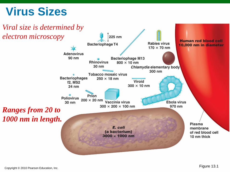
Morphology of a polyhedral virus
Figure 13.2a
Polyhedral viruses (e.g. adenovirus) are many-sided.
Usually the capsid is an icosahedron (二十面體).
美洲白鱘腺病毒屬
蛋白殼
殼粒

Copyright © 2010 Pearson Education, Inc. Figure 13.16a
Polyhedral Viruses
哺乳動物腺病毒屬

Copyright © 2010 Pearson Education, Inc. Figure 13.3
Morphology of an Enveloped Virus •Enveloped viruses are covered by an envelop and are roughly spherical but highly pleomorphic.
•There are also enveloped helical viruses (螺旋病毒 e.g. Influenzavirus, figure 13.3) and enveloped polyhedral viruses (e.g. Herpesvirus).
流行性感冒病毒
刺突
外膜 殼粒
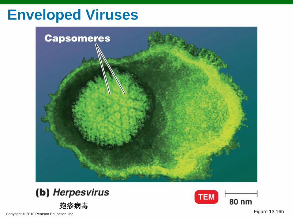
Copyright © 2010 Pearson Education, Inc. Figure 13.16b
Enveloped Viruses
皰疹病毒
Copyright © 2010 Pearson Education, Inc. Figure 13.4
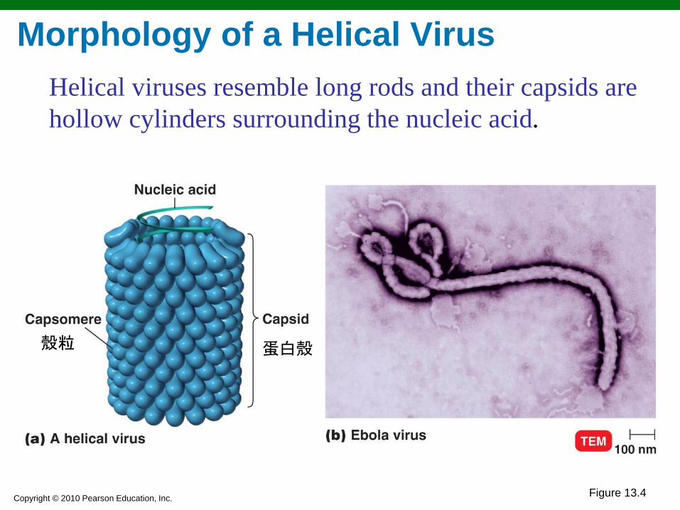
Morphology of a Helical Virus
Helical viruses resemble long rods and their capsids are
hollow cylinders surrounding the nucleic acid.
殼粒 蛋白殼

Copyright © 2010 Pearson Education, Inc. Figure 13.5
Morphology of a Complex Virus
•Complex viruses
have complex
structures. For
example, many
bacteriophages
have a polyhedral
capsid with a
helical tail sheath
attached.
護套;鞘
Copyright © 2010 Pearson Education, Inc.
Taxonomy of Viruses
Order names end in -ales
Family names end in -viridae
Genus names end in -virus
Viral species: A group of viruses sharing the same
genetic information and ecological niche (生態棲位host range). Common names are used for species
Such as human immunodeficiency virus (HIV)人類免疫不全病毒,愛滋病毒
Subspecies are designated by a number
Such as HIV-1

Copyright © 2010 Pearson Education, Inc.
Taxonomy of Viruses
Family: Herpesviridae
Genus: Herpesvirus
Species: human
herpesvirus
Subspecies: 2
HHV-2
Retroviridae
Lentivirus
human
immunodeficiency virus
1
HIV-1

Copyright © 2010 Pearson Education, Inc. Figure 13.6
Growing Viruses
Viruses must be grown
in living cells.
The easiest viruses to
grow are
bacteriophages
The plaque method
mixed bacteriophages
with host bacteria and
nutrient agar.
Bacteriophages form
plaques on a lawn of
bacteria.
• Each plaque originates with a single viral particle; the concentration of viruses is given as plaque-forming units.
Isolation, Cultivation, and Identification
溶菌斑
Copyright © 2010 Pearson Education, Inc. Figure 13.7
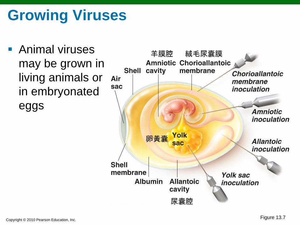
Growing Viruses
Animal viruses
may be grown in
living animals or
in embryonated
eggs
羊膜腔
尿囊腔
絨毛尿囊膜
卵黃囊

Copyright © 2010 Pearson Education, Inc. Figure 13.8
Growing Viruses
Animal and plant viruses may be grown in cell culture.
Primary cell lines (原代細胞株), derived from tissue slices, tend
to die out after only a few generation.
Diploid cell lines, derived from human embryos, can be
maintained for about 100 generations.
Continuous cell lines (連續細胞株, transformed and cancerous,
immortal cell lines) may be maintained indefinitely.
Copyright © 2010 Pearson Education, Inc. Figure 13.9
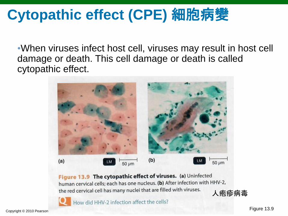
Cytopathic effect (CPE) 細胞病變
•When viruses infect host cell, viruses may result in host cell damage or death. This cell damage or death is called cytopathic effect.
人疱疹病毒
Copyright © 2010 Pearson Education, Inc.
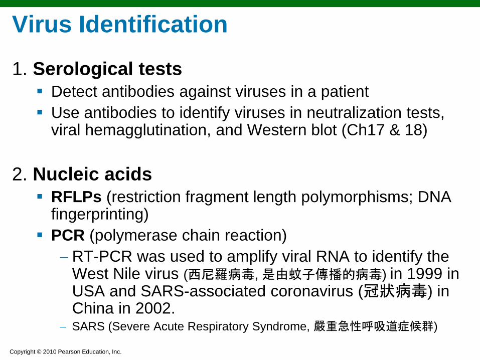
Virus Identification
1. Serological tests Detect antibodies against viruses in a patient
Use antibodies to identify viruses in neutralization tests, viral hemagglutination, and Western blot (Ch17 & 18)
2. Nucleic acids RFLPs (restriction fragment length polymorphisms; DNA
fingerprinting)
PCR (polymerase chain reaction)
RT-PCR was used to amplify viral RNA to identify the West Nile virus (西尼羅病毒, 是由蚊子傳播的病毒) in 1999 in USA and SARS-associated coronavirus (冠狀病毒) in China in 2002.
SARS (Severe Acute Respiratory Syndrome, 嚴重急性呼吸道症候群)

Copyright © 2010 Pearson Education, Inc.
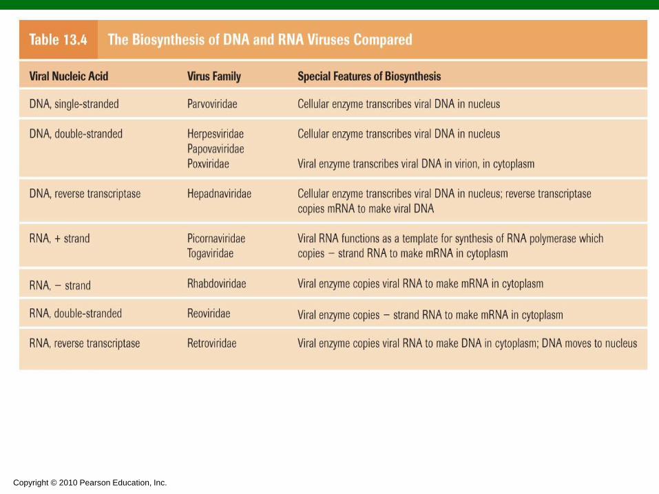
The nucleic acid in a virion contains only a few of
the genes needed for the synthesis of new
viruses.
These include genes for the virion’s structural
components, such as the capsid proteins, and
genes for a few of the enzymes used in the viral
life cycle.
For a virus to multiply, it must invade a host cell
and direct the host’s metabolic machinery to
produce viral enzymes and components.
Viral Multiplication
Copyright © 2010 Pearson Education, Inc. Figure 13.10
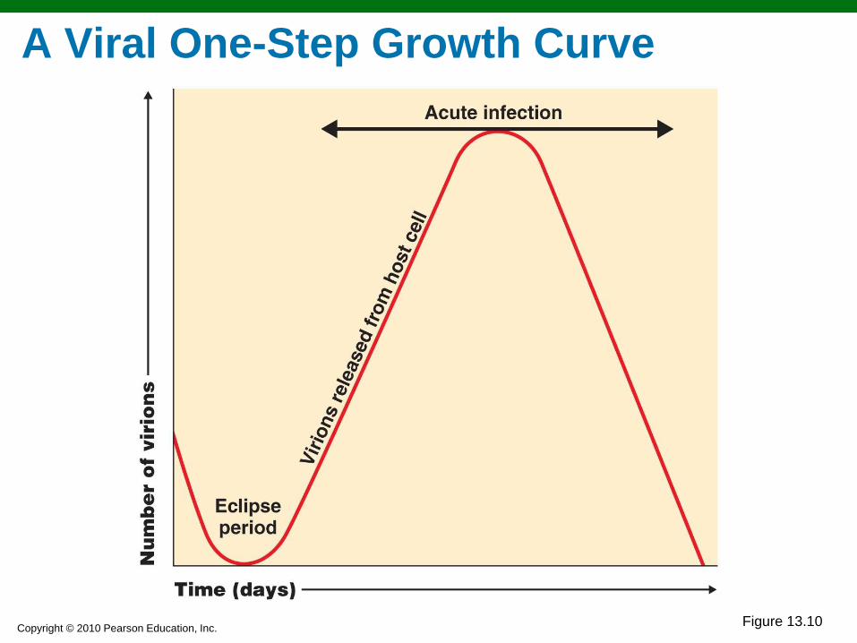
A Viral One-Step Growth Curve

Copyright © 2010 Pearson Education, Inc.
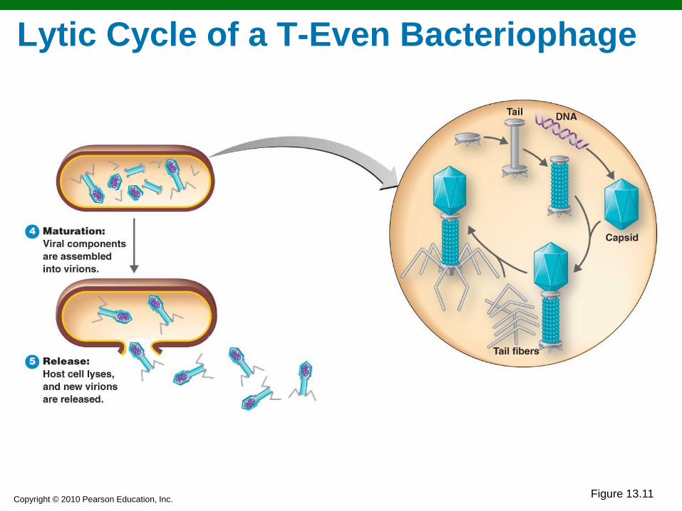
The Lytic Cycle (裂解期): (T-even bacteriophages):
Attachment: Phage attaches by tail fibers to
host cell
Penetration: Phage lysozyme opens cell wall;
tail sheath contracts to force tail core reaching
plasma membrane and injecting DNA into cell
Biosynthesis: Production of phage DNA and
proteins
Maturation: Assembly of phage particles
Release: Phage lysozyme breaks cell wall
Viral Multiplication

Copyright © 2010 Pearson Education, Inc.
Lytic Cycle of a T-Even Bacteriophage
1
2
3
Figure 13.11
Copyright © 2010 Pearson Education, Inc.
4
Figure 13.11
Lytic Cycle of a T-Even Bacteriophage
Copyright © 2010 Pearson Education, Inc.
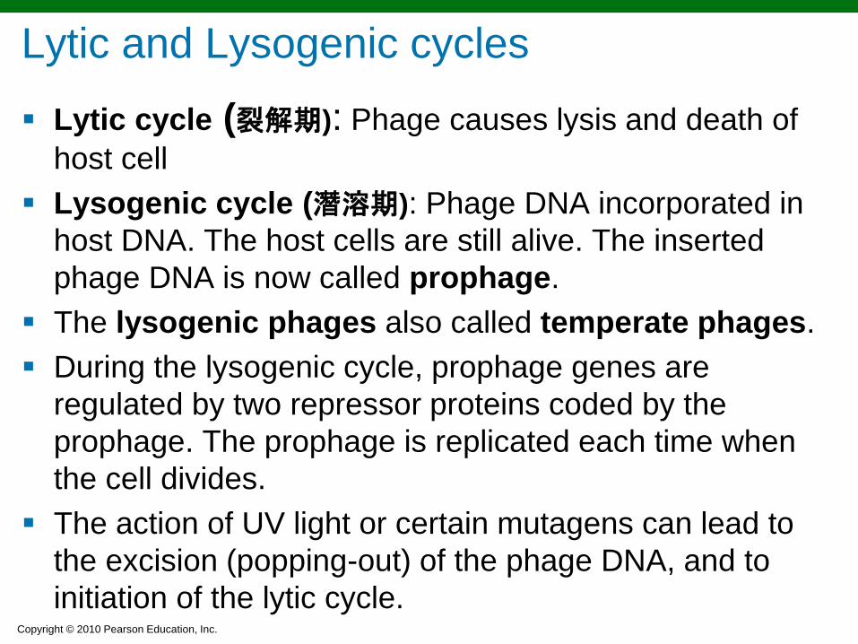
Lytic cycle (裂解期): Phage causes lysis and death of
host cell
Lysogenic cycle (潛溶期): Phage DNA incorporated in
host DNA. The host cells are still alive. The inserted
phage DNA is now called prophage.
The lysogenic phages also called temperate phages.
During the lysogenic cycle, prophage genes are
regulated by two repressor proteins coded by the
prophage. The prophage is replicated each time when
the cell divides.
The action of UV light or certain mutagens can lead to
the excision (popping-out) of the phage DNA, and to
initiation of the lytic cycle.
Lytic and Lysogenic cycles

Copyright © 2010 Pearson Education, Inc. Figure 13.12
The Lysogenic Cycle

Copyright © 2010 Pearson Education, Inc.
1. The lysogenic cells are immune to reinfection by
the same phage. (However, the host cell is not immune to
infection by other phage types.)
2. Phage conversion
The host cell may exhibit new properties, e.g. a toxin
gene carried by a temperate phage
3. Specialized transduction
Only certain bacterial genes can be transferred.
Lysogenic phage packages adjacent bacterial DNA
along with its own DNA in the same capsid.
Any bacterial genes can be transferred by generalized
transduction (Fig. 8.28).
Three results of lysogeny

Copyright © 2010 Pearson Education, Inc.
Results of Multiplication of Bacteriophages Lytic cycle
Phage causes lysis and death of host cell
Lysogenic cycle
The lysogenic cells are immune to reinfection by the same
phage.
Phage conversion
Specialized transduction

Copyright © 2010 Pearson Education, Inc.
2
3
4
5
6
Figure 8.28
Generalized Transduction

Copyright © 2010 Pearson Education, Inc.
Specialized Transduction
Figure 13.13
Prophage exists in galactose-using host (containing the gal gene).
Phage genome excises, carrying with it the adjacent gal gene from the host.
Phage matures and cell lyses, releasing phage carrying gal gene.
1
2
3
Prophage
gal gene
gal gene Bacterial DNA
Galactose-positive donor cell gal gene
Phage infects a cell that cannot utilize galactose (lacking gal gene).
4
Galactose-negative recipient cell
Along with the prophage, the bacterial gal gene becomes integrated into the new host’s DNA.
5
Lysogenic cell can now metabolize galactose.
6
Galactose-positive recombinant cell

Copyright © 2010 Pearson Education, Inc.
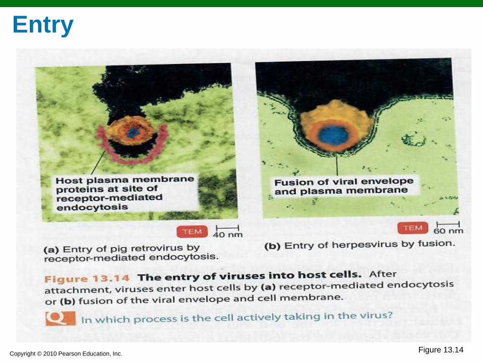
Multiplication of Animal Viruses Attachment Viruses attach to receptor sites on the
cell membrane
Entry By receptor-mediated
endocytosis (胞飲作用) or fusion
(fusion only for enveloped viruses)
Uncoating The separation of the viral nucleic
acid from its protein coat by viral or host enzymes
Biosynthesis Production of nucleic acid and
proteins
Maturation Nucleic acid and capsid proteins
assemble
Release By budding (enveloped viruses) or
rupture
Copyright © 2010 Pearson Education, Inc. Figure 13.14
Entry

Copyright © 2010 Pearson Education, Inc. Figure 13.20a
Release of an enveloped virus by budding
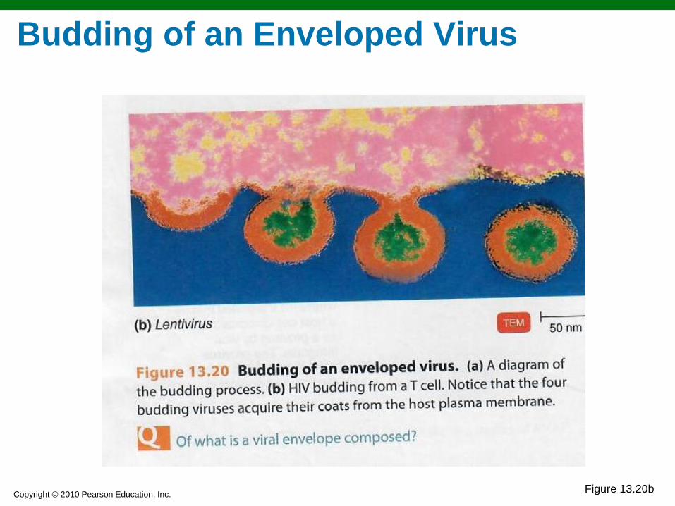
Copyright © 2010 Pearson Education, Inc. Figure 13.20b
Budding of an Enveloped Virus

Copyright © 2010 Pearson Education, Inc. Figure 13.15
Multiplication of DNA-containing Animal Virus
Copyright © 2010 Pearson Education, Inc. Figure 13.17a
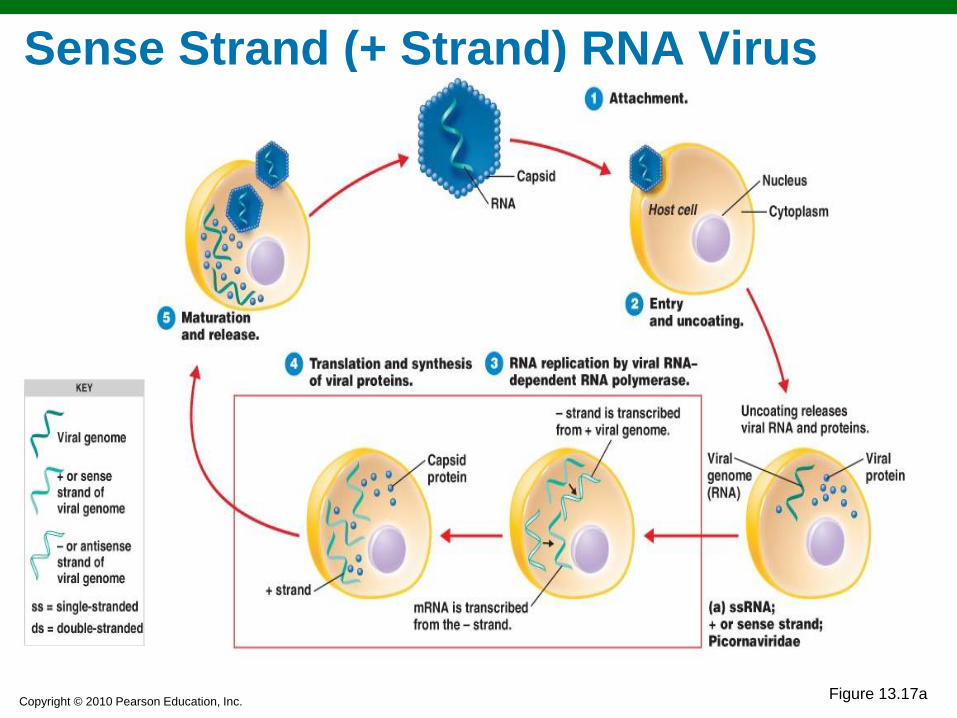
Sense Strand (+ Strand) RNA Virus

Copyright © 2010 Pearson Education, Inc. Figure 13.17b
Antisense Strand (– Strand) RNA Virus
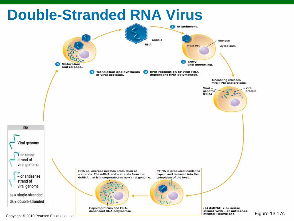
Copyright © 2010 Pearson Education, Inc. Figure 13.17c
Double-Stranded RNA Virus
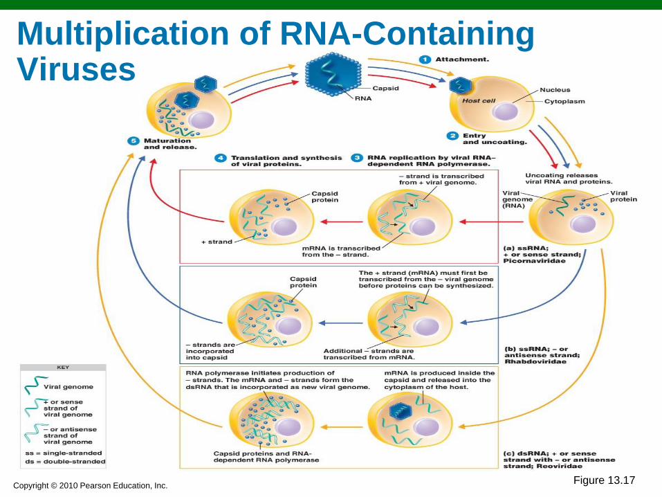
Copyright © 2010 Pearson Education, Inc. Figure 13.17
Multiplication of RNA-Containing Viruses

Copyright © 2010 Pearson Education, Inc. Figure 13.19
Multiplication of a Retrovirus 反轉錄病毒
Copyright © 2010 Pearson Education, Inc.

Copyright © 2010 Pearson Education, Inc.
Viruses and Cancer
The earliest relationship between cancer and viruses
was demonstrated in the early 1900s, when chicken
leukemia and chicken sarcoma were transferred to
healthy animals by cell-free filtrates.
Activated oncogenes transform normal cells into
cancerous cells.
Transformed cells have increased growth, loss of
contact inhibition, contain virus-specific antigens (tumor
specific transplant antigen and T antigen), exhibit
chromosomal abnormalities, and can produce tumors
when injected into susceptible animals..
The genetic material of oncogenic viruses becomes
integrated into the host cell's DNA.
Copyright © 2010 Pearson Education, Inc.
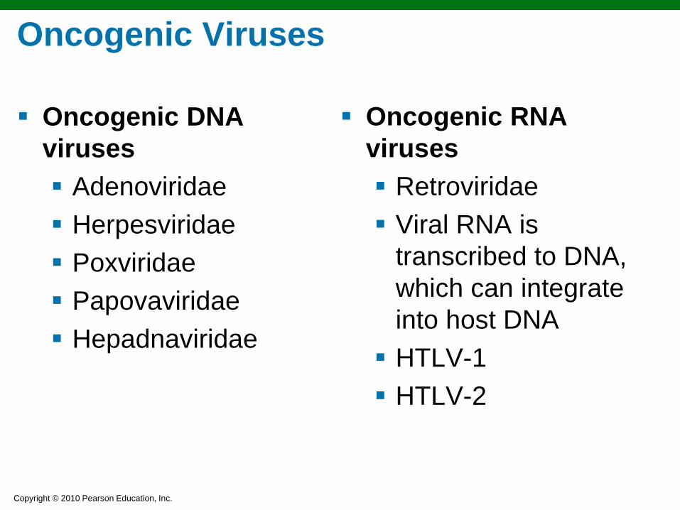
Oncogenic Viruses
Oncogenic DNA
viruses
Adenoviridae
Herpesviridae
Poxviridae
Papovaviridae
Hepadnaviridae
Oncogenic RNA
viruses
Retroviridae
Viral RNA is
transcribed to DNA,
which can integrate
into host DNA
HTLV-1
HTLV-2
Copyright © 2010 Pearson Education, Inc.
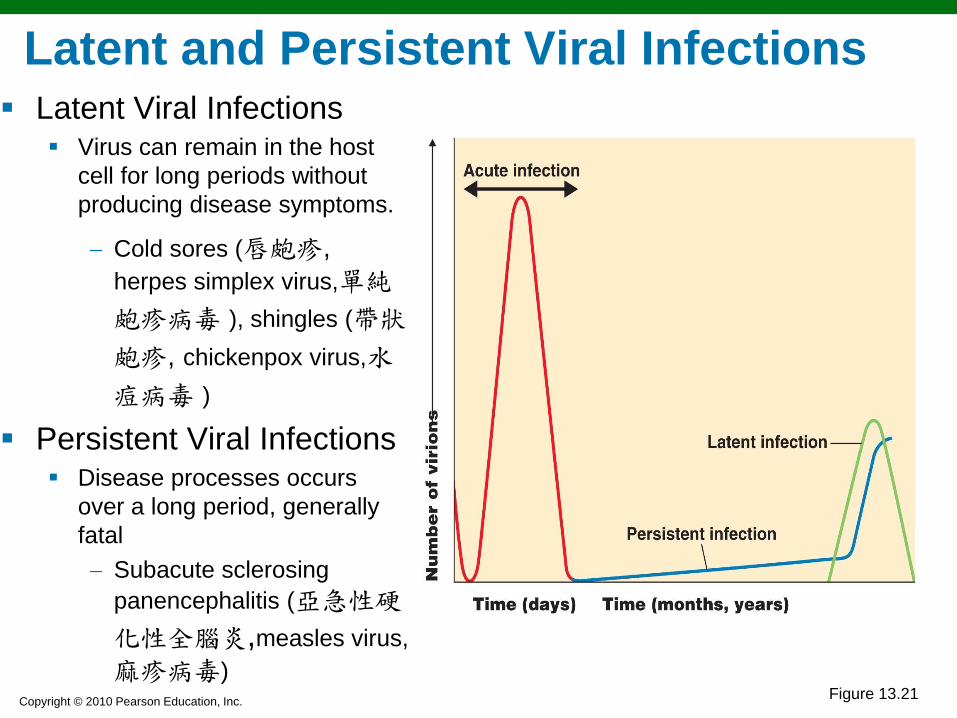
Latent Viral Infections Virus can remain in the host
cell for long periods without
producing disease symptoms.
Cold sores (唇皰疹, herpes simplex virus,單純
皰疹病毒 ), shingles (帶狀
皰疹, chickenpox virus,水
痘病毒 )
Persistent Viral Infections Disease processes occurs
over a long period, generally
fatal
Subacute sclerosing
panencephalitis (亞急性硬
化性全腦炎,measles virus,
麻疹病毒)
Latent and Persistent Viral Infections
Figure 13.21

Copyright © 2010 Pearson Education, Inc.
Prions (傳染性蛋白質):
Infectious proteins discovered in the 1980s
Inherited and transmissible by ingestion, transplant, &
surgical instruments
Spongiform encephalopathies (海綿樣腦病): Sheep
scrapie, Creutzfeldt-Jakob disease (庫賈氏症),
Gerstmann-Sträussler-Scheinker syndrome (GSS氏症),
fatal familial insomnia (致死性家族失眠症), mad cow
disease
All involved degeneration of brain tissue.
Prions, Viroids (類病毒), and Plant Viruses

Copyright © 2010 Pearson Education, Inc.
全家罹罕見「致死性家族失眠症」 已多人死亡 (2016年05月02日, 蘋果日報)
澳洲一個家族,被證實罹患罕見疾病「致死性家族失眠症」(Fatal Familial Insomnia,FFI),目前全球經證實罹病者,一千萬人中不到一人,且無藥可醫。FFI患者無法被預測何時發病,患者一般的表現就像是失眠症,難以進入深層睡眠,但與多數失眠症患者是心理或環境因素造成不同,FFI的患者是因為體內的部分蛋白質產生異常結塊,進而引發神經系統受損,在腦部的丘腦產生如海綿般的空洞,而丘腦正是管理睡眠的重要部位。
30歲的韋伯(Hayley Webb)原本是澳洲《第九頻道》電視台記者,早在青少年時期她就知道自己全家都罹患這種疾病,「十多歲的時候我就知道,我們全家都受到了詛咒」,當時她的祖母率先發病,突然失明,接著出現痴呆症狀,並伴隨長期幻覺,最後連說話功能都喪失,醫生診斷才知道罹患FFI,沒多久就死亡。之後,她的母親、舅舅、阿姨陸續死亡,現在家中就剩下她與28歲弟弟拉克蘭(Lachlan Webb)。目前,兩姐弟已經辭去工作,前往美國加州大學參與一項研究計畫,希望能找出補救措施。(於慶中/綜合外電報導)
Copyright © 2010 Pearson Education, Inc.
Prions
PrPC: Normal cellular prion protein, on cell surface
PrPSc: Scrapie protein; accumulates in brain cells,
forming plaques
Prion diseases are due to an altered protein; the
cause can be a mutation in the normal gene for PrP
or contact with an altered protein (PrPSc).

Copyright © 2010 Pearson Education, Inc. Figure 13.22
How a Protein Can Be Infectious

Copyright © 2010 Pearson Education, Inc. Figure 13.23
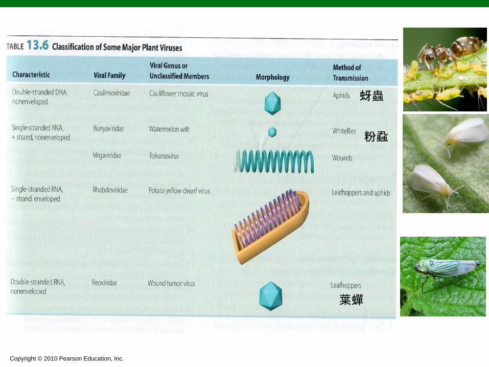
Plant Viruses and Viroids
Plant Viruses Plant viruses enter
through wounds or via insects
Some plant viruses also multiply in insect (vector) cells.
Viroids Viroids are
infectious RNA (short naked RNA, ~300 to 400 nucleotides)
potato spindle tuber disease
Copyright © 2010 Pearson Education, Inc.
蚜蟲
粉蝨
葉蟬

![关键词] 修辞格,修辞格翻译,绝对直译,相对直译,相对意义,绝对意译 , 形式与内 容](https://static.fdokumen.com/doc/165x107/631a407ffd704e1d390a0c19/--1674590328.jpg)