Epigallocatechin-3-gallate modulates Tau Post-translational ...
Upload
khangminh22Category
view
3download
0
REVIEWARTICLE
Like prions: the propagation of aggregated tauand a-synuclein in neurodegeneration
Michel Goedert, Masami Masuda-Suzukake and Benjamin Falcon
The abnormal aggregation of a small number of known proteins underlies the most common human neurodegenerative diseases. In
tauopathies and synucleinopathies, the normally soluble intracellular proteins tau and a-synuclein become insoluble and filament-
ous. In recent years, non-cell autonomous mechanisms of aggregate formation have come to the fore, suggesting that nucleation-
dependent aggregation may occur in a localized fashion in human tauopathies and synucleinopathies, followed by seed-dependent
propagation. There is a long prodromal phase between the formation of protein aggregates and the appearance of the first clinical
symptoms, which manifest only after extensive propagation, opening novel therapeutic avenues.
MRC Laboratory of Molecular Biology, Francis Crick Avenue, Cambridge CB2 0QH, UK
Correspondence to: M. Goedert,
MRC Laboratory of Molecular Biology,
Francis Crick Avenue,
Cambridge CB2 0QH, UK
E-mail: [email protected]
Keywords: Alzheimer’s disease; alpha-synuclein; Parkinson’s disease; prion-like; tau
Abbreviations: CBD = corticobasal degeneration; (v)CJD = (variant) Creutzfeldt-Jakob disease; MSA = multiple system atrophy
IntroductionCommon human neurodegenerative diseases are characterized
by the presence of abundant filamentous inclusions (Goedert,
2015; Walker and Jucker, 2015). Each type of inclusion has
one protein as its major component, with amyloid-b, tau and
a-synuclein being the most commonly affected (Fig. 1). These
proteins undergo a transformation from a soluble to an insol-
uble filamentous state, with a number of intermediates. Most
cases of disease are sporadic, but a small percentage are in-
herited, often in a dominant manner. The latter are caused by
mutations in the genes encoding the proteins that make up the
inclusions, or proteins that increase their production, under-
scoring the importance of inclusion formation for neurodegen-
eration. It constitutes the gain of toxic function that most
probably causes human neurodegenerative diseases.
Mutations in MAPT, the tau gene, cause an inherited
form of frontotemporal dementia and parkinsonism, with
abundant filamentous tau inclusions in the brain.
Mutations in SNCA, the a-synuclein gene, and multiplica-
tions thereof, give rise to familial forms of Parkinson’s dis-
ease and dementia with Lewy bodies. Lewy pathology,
which is made of a-synuclein filaments, defines
Parkinson’s disease and dementia with Lewy bodies at the
neuropathological level.
Until recently, cell autonomous mechanisms were
believed to account for human neurodegenerative diseases.
These mechanisms imply that brain cells degenerate, as a
result of the same aggregation events occurring independ-
ently. While it is easy to see how such mechanisms can lead
to inherited forms of disease (mutant proteins are widely
expressed), it is more difficult to understand how cell au-
tonomous mechanisms could give rise to sporadic diseases.
At death, protein inclusions are present in thousands of
nerve cells. It appears unlikely that the same molecular
events take place independently thousands of times in
doi:10.1093/brain/aww230 BRAIN 2017: 140; 266–278 | 266
Received June 21, 2016. Revised July 13, 2016. Accepted July 15, 2016. Advance Access publication September 21, 2016
� The Author (2016). Published by Oxford University Press on behalf of the Guarantors of Brain. All rights reserved.
For Permissions, please email: [email protected]
Dow
nloaded from https://academ
ic.oup.com/brain/article/140/2/266/2669395 by guest on 30 June 2022

post-mitotic cells. The first inclusions may form in a loca-
lized fashion, from where they propagate to normal cells,
resulting in degeneration (cell non-autonomous mechan-
isms). Even though cell non-autonomous mechanisms are
not necessary for explaining inherited diseases, they may
nonetheless operate. This appears to be the case of patho-
logical huntingtin (Cicchetti et al., 2014; Pecho-Vrieseling
et al., 2014). It has previously been suggested that neuro-
degenerative diseases may involve the transfer of substances
between nerve cells (Saper et al., 1987) and it has been
known for many years that some proteins can move
trans-synaptically (Schwab and Thoenen, 1976).
Spreading is consistent with staging schemes that have
postulated a stereotypical progression of inclusions from a
single site (transentorhinal cortex for the tau inclusions of
Alzheimer’s disease, ambient gyrus for the tau inclusions of
argyrophilic grain disease, cortical sulci for the tau inclu-
sions of chronic traumatic encephalopathy and dorsal
motor nucleus of the vagus nerve, as well as olfactory
bulb, for the a-synuclein inclusions of Parkinson’s disease)
(Braak and Braak, 1991; Braak et al., 2003; Saito et al.,
2004; McKee et al., 2013). However, staging cannot prove
the prion-like spreading of protein aggregates, because it is
also compatible with brain regions being sequentially
affected by aggregate formation (‘just one damned thing
after another’, Bell, 1909). Propagation of tau pathology
is supported by its absence from a piece of disconnected
cerebral cortex in an individual with Alzheimer’s disease
who had undergone an operation to remove a meningioma
27 years earlier (Duyckaerts et al., 1997). The tumour and
the operation disconnected a small piece of frontal cortex.
While there was abundant tau pathology in limbic and
isocortical regions, tau-positive neuritic plaques, neurofib-
rillary tangles and neuropil threads were not present in the
disconnected frontal cortex.
Additional evidence for the existence of prion-like mech-
anisms in the human brain has come from the development
of scattered Lewy pathology in foetal human midbrain neu-
rons that were therapeutically implanted into the striata of
patients with advanced Parkinson’s disease (Fig. 2)
(Kordower et al., 2008; Li et al., 2008). Lewy pathology
was detected in 2–5% of grafted cells in patients who had
survived for 10 or more years, approximately the same
percentage as neurons with Lewy pathology in the pars
compacta of the substantia nigra in Parkinson’s disease.
a-Synuclein immunoreactivity was not seen in nerve cell
bodies 18 months after grafting, but it was present in a
non-aggregated form after 4 years and in an aggregated,
Figure 1 Distribution of amyloid-b, tau and a-synuclein inclusions in human brain. Left: Amyloid-b (Ab) plaques develop first in basal
temporal and orbitofrontal neocortex (Phase 1). They are observed later throughout the neocortex, hippocampal formation, amygdala, di-
encephalon and basal ganglia (Phases 2 and 3). In severe cases of Alzheimer’s disease, amyloid-b plaques are also found in mesencephalon, lower
brainstem and cerebellar cortex (Phases 4 and 5). Middle: Tau inclusions develop in the locus coeruleus, as well as in transentorhinal and entorhinal
regions (Stages I and II). This is followed by their presence in the hippocampal formation and some parts of the neocortex (Stages III and IV),
followed by large parts of the neocortex (Stages V and VI). Right: The first a-synuclein inclusions are present in the olfactory bulb and the dorsal
motor nucleus of the vagal and glossopharyngeal nerves of the medulla oblongata (Stages 1 and 2). From the brainstem, the pathology ascends
through the pons to midbrain and basal forebrain (Stages 3 and 4), followed by the neocortex (Stages 5 and 6). This figure is based on the work of
Braak, Del Tredici, and collaborators. From Goedert (2015).
Tauopathies and synucleinopathies BRAIN 2017: 140; 266–278 | 267
Dow
nloaded from https://academ
ic.oup.com/brain/article/140/2/266/2669395 by guest on 30 June 2022

ubiquitinated state after 14 years (Chu and Kordower,
2010). After 24 years, 11–12% of grafted dopaminergic
neurons exhibited a-synuclein and ubiquitin-positive inclu-
sions (Li et al., 2016). The clinical benefits of transplant-
ation were gradually lost after 14 years. Although these
findings are compatible with the prion-like spreading of
Lewy pathology, it is also possible that the microenviron-
ment of the nigrostriatal system in Parkinson’s disease pre-
disposes to the formation of Lewy pathology.
Over the past 7 years, experimental studies have shown
that the injection of tau and a-synuclein inclusions into
animals induces neurons to form intracellular inclusions
at the injection sites, from where they can spread to distant
brain regions (Clavaguera et al., 2009; Desplats et al.,
2009; Hansen et al., 2011; Goedert, 2015). Unless these
findings are not related to what is happening in the
human brain, cell non-autonomous mechanisms must also
be at work in human neurodegenerative diseases.
These mechanisms are often called ‘prion-like’, referring
to the intercellular spreading of protein aggregates, result-
ing in their propagation through the brain. The acronym
‘prion’ stands for ‘proteinaceous infectious particle’
(Prusiner, 1982), encompassing both intercellular propaga-
tion and inter-organismal transmission. There is no evi-
dence to suggest that Alzheimer’s disease and Parkinson’s
disease can transfer between individuals. Hence the use of
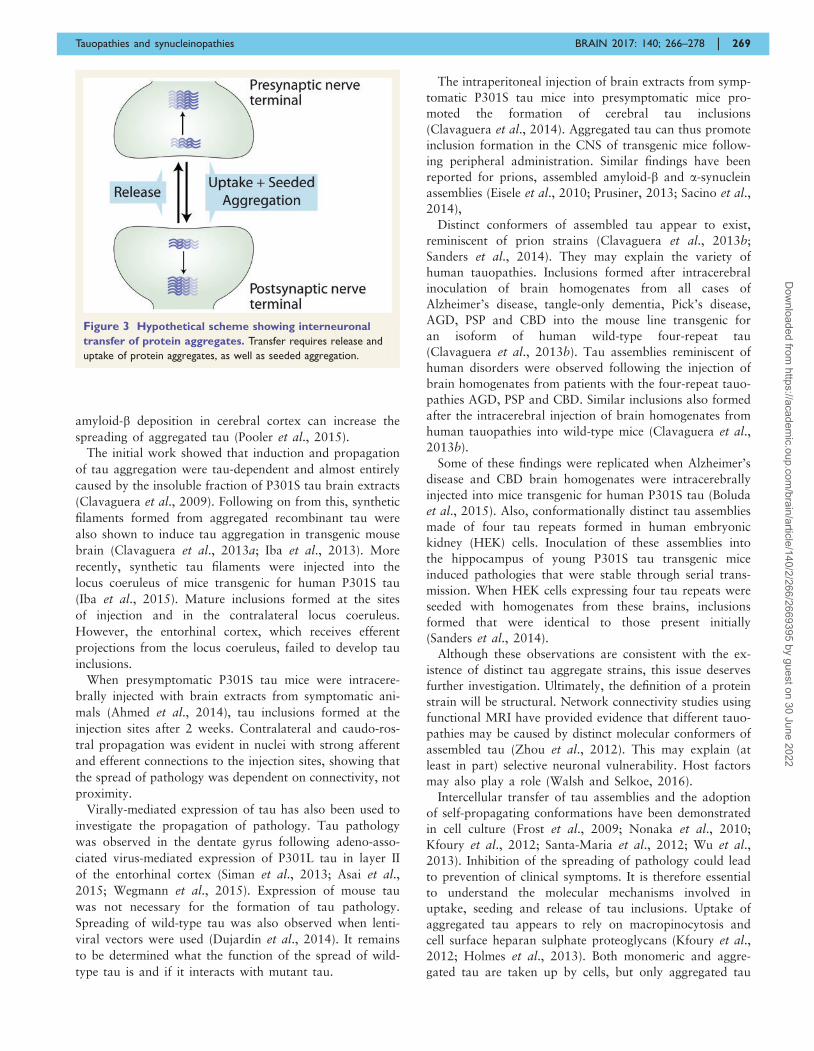
‘prion-like’. Interneuronal spreading of tau and a-synuclein
aggregates requires their release into the extracellular space,
uptake by connected cells and seeded aggregation of sol-
uble protein (Fig. 3). If the spreading of aggregates is linked
to the development of clinical symptoms, it represents a
therapeutic target through the inhibition of aggregate
uptake or release, inhibition of seeding and activation of
the clearance of protein aggregates.
Aggregated tau
Tau aggregates are characteristic of a number of human
neurodegenerative diseases, the so-called tauopathies
(Table 1). They form in a variety of brain regions, where
they are largely neuronal. However, glial tau inclusions are
also found in some diseases, such as progressive supra-
nuclear palsy (PSP) and corticobasal degeneration (CBD).
In white matter tauopathy with globular glial inclusions,
oligodendroglial tau inclusions predominate (Kovacs
et al., 2008).
In adult human brain, six tau isoforms ranging from 352
to 441 amino acids are produced from a single gene
through alternative mRNA splicing (Goedert et al., 1989;
Goedert, 2015). Three isoforms have three repeats each,
and three isoforms have four repeats each. The repeats
and some adjoining sequences constitute the microtubule-
binding domains of tau. They also make up the core of
tau filaments. In Alzheimer’s disease, the most common
human neurodegenerative disease, and in chronic traumatic
encephalopathy, all six tau isoforms are present in the
filaments. In other disease filaments—such as those of
PSP, CBD, argyrophilic grain disease (AGD) and white
matter tauopathy with globular glial inclusion—tau iso-
forms with four repeats are found. In Pick’s disease,
three-repeat tau isoforms predominate in the inclusions.
Unlike Alzheimer’s disease, these diseases lack amyloid-bpathology.
Several lines of investigation in animal models and cell
culture have shown that assembled tau can behave like a
prion. Injection of human mutant tau inclusions into the
brains of mice transgenic for wild-type tau induced the for-
mation of inclusions made of wild-type human tau (Fig. 4),
followed by their spreading to distant brain regions
(Clavaguera et al., 2009). Subsequently, several groups stu-
died the spreading of pathological tau along the entorhinal
cortex/hippocampal pathway. They used mouse models
that apparently expressed human P301L tau only in the
entorhinal cortex (de Calignon et al., 2012; Harris et al.,
2012; Liu et al., 2012). Several months after the appear-
ance of the first tau inclusions, hippocampal neurons de-
veloped filamentous tau pathology. Using this system,
together with a mouse line transgenic for mutant amyloid
precursor protein and presenilin-1, it has been shown that
Figure 2 Possible host-to-graft spreading of Lewy pathology in Parkinson’s disease. The patient received a transplant of foetal human
mesencephalic dopaminergic nerve cells into the putamen 16 years previously. Immunohistochemistry for a-synuclein visualizes Lewy bodies and
neurites in the host substantia nigra (A) and the transplant (B and C). Adapted from Li et al. (2008).
268 | BRAIN 2017: 140; 266–278 M. Goedert et al.
Dow
nloaded from https://academ
ic.oup.com/brain/article/140/2/266/2669395 by guest on 30 June 2022
amyloid-b deposition in cerebral cortex can increase the
spreading of aggregated tau (Pooler et al., 2015).
The initial work showed that induction and propagation
of tau aggregation were tau-dependent and almost entirely
caused by the insoluble fraction of P301S tau brain extracts
(Clavaguera et al., 2009). Following on from this, synthetic
filaments formed from aggregated recombinant tau were
also shown to induce tau aggregation in transgenic mouse
brain (Clavaguera et al., 2013a; Iba et al., 2013). More
recently, synthetic tau filaments were injected into the
locus coeruleus of mice transgenic for human P301S tau
(Iba et al., 2015). Mature inclusions formed at the sites
of injection and in the contralateral locus coeruleus.
However, the entorhinal cortex, which receives efferent
projections from the locus coeruleus, failed to develop tau
inclusions.
When presymptomatic P301S tau mice were intracere-
brally injected with brain extracts from symptomatic ani-
mals (Ahmed et al., 2014), tau inclusions formed at the
injection sites after 2 weeks. Contralateral and caudo-ros-
tral propagation was evident in nuclei with strong afferent
and efferent connections to the injection sites, showing that
the spread of pathology was dependent on connectivity, not
proximity.
Virally-mediated expression of tau has also been used to
investigate the propagation of pathology. Tau pathology
was observed in the dentate gyrus following adeno-asso-
ciated virus-mediated expression of P301L tau in layer II
of the entorhinal cortex (Siman et al., 2013; Asai et al.,
2015; Wegmann et al., 2015). Expression of mouse tau
was not necessary for the formation of tau pathology.
Spreading of wild-type tau was also observed when lenti-
viral vectors were used (Dujardin et al., 2014). It remains
to be determined what the function of the spread of wild-
type tau is and if it interacts with mutant tau.
The intraperitoneal injection of brain extracts from symp-
tomatic P301S tau mice into presymptomatic mice pro-
moted the formation of cerebral tau inclusions
(Clavaguera et al., 2014). Aggregated tau can thus promote
inclusion formation in the CNS of transgenic mice follow-
ing peripheral administration. Similar findings have been
reported for prions, assembled amyloid-b and a-synuclein
assemblies (Eisele et al., 2010; Prusiner, 2013; Sacino et al.,
2014),
Distinct conformers of assembled tau appear to exist,
reminiscent of prion strains (Clavaguera et al., 2013b;
Sanders et al., 2014). They may explain the variety of
human tauopathies. Inclusions formed after intracerebral
inoculation of brain homogenates from all cases of
Alzheimer’s disease, tangle-only dementia, Pick’s disease,
AGD, PSP and CBD into the mouse line transgenic for
an isoform of human wild-type four-repeat tau
(Clavaguera et al., 2013b). Tau assemblies reminiscent of
human disorders were observed following the injection of
brain homogenates from patients with the four-repeat tauo-
pathies AGD, PSP and CBD. Similar inclusions also formed
after the intracerebral injection of brain homogenates from
human tauopathies into wild-type mice (Clavaguera et al.,
2013b).
Some of these findings were replicated when Alzheimer’s
disease and CBD brain homogenates were intracerebrally
injected into mice transgenic for human P301S tau (Boluda
et al., 2015). Also, conformationally distinct tau assemblies
made of four tau repeats formed in human embryonic
kidney (HEK) cells. Inoculation of these assemblies into
the hippocampus of young P301S tau transgenic mice
induced pathologies that were stable through serial trans-
mission. When HEK cells expressing four tau repeats were
seeded with homogenates from these brains, inclusions
formed that were identical to those present initially
(Sanders et al., 2014).
Although these observations are consistent with the ex-
istence of distinct tau aggregate strains, this issue deserves
further investigation. Ultimately, the definition of a protein
strain will be structural. Network connectivity studies using
functional MRI have provided evidence that different tauo-
pathies may be caused by distinct molecular conformers of
assembled tau (Zhou et al., 2012). This may explain (at
least in part) selective neuronal vulnerability. Host factors
may also play a role (Walsh and Selkoe, 2016).
Intercellular transfer of tau assemblies and the adoption
of self-propagating conformations have been demonstrated
in cell culture (Frost et al., 2009; Nonaka et al., 2010;
Kfoury et al., 2012; Santa-Maria et al., 2012; Wu et al.,
2013). Inhibition of the spreading of pathology could lead
to prevention of clinical symptoms. It is therefore essential
to understand the molecular mechanisms involved in
uptake, seeding and release of tau inclusions. Uptake of
aggregated tau appears to rely on macropinocytosis and
cell surface heparan sulphate proteoglycans (Kfoury et al.,
2012; Holmes et al., 2013). Both monomeric and aggre-
gated tau are taken up by cells, but only aggregated tau
Figure 3 Hypothetical scheme showing interneuronal
transfer of protein aggregates. Transfer requires release and
uptake of protein aggregates, as well as seeded aggregation.
Tauopathies and synucleinopathies BRAIN 2017: 140; 266–278 | 269
Dow
nloaded from https://academ
ic.oup.com/brain/article/140/2/266/2669395 by guest on 30 June 2022

is able to seed the aggregation of soluble, monomeric tau
(Falcon et al., 2015). Most seeds are trafficked to lyso-
somes, but some endosomes rupture and seeds enter the
cytoplasm, where they induce tau assembly.
Tau seeds made from aggregated recombinant tau are not
phosphorylated, whereas seeded tau is hyperphosphory-
lated. Seeded tau is also p62-positive, like the tau inclusions
in human brains and in mice transgenic for human P301S
tau (Schaeffer et al., 2012). Expressed tau can only be
seeded when it is aggregation-competent (Falcon et al.,
2015). Aggregation inhibitors may thus be able to reduce
tau-induced seeding and spreading.
Using a cell assay, we found that the seeding potency of
recombinant P301S tau aggregated with heparin was lower
than that of aggregated tau from the brains of mice trans-
genic for human mutant P301S tau (Fig. 5) (Falcon et al.,
2015). Similar discrepancies between recombinant and
brain aggregates have been described for prions, amyloid-
b, a-synuclein and reactive amyloid A (Meyer-Luehmann
et al., 2006; Zhang et al., 2008; Luk et al., 2012a; Stohr
et al., 2012; Prusiner, 2013; Recasens et al., 2014).
Recombinant tau aggregates were more resistant to disag-
gregation by guanidine hydrochloride and digestion by pro-
teinase K than tau aggregates from transgenic mouse brain,
consistent with the view that more stable aggregates possess
lower seeding activity. Recombinant aggregated tau was
Table 1 Diseases with tau inclusions
Alzheimer’s disease
Amyotrophic lateral sclerosis/parkinsonism-dementia complex
Argyrophilic grain disease
Chronic traumatic encephalopathy
Corticobasal degeneration
Diffuse neurofibrillary tangles with calcification
Down’s syndrome
Familial British dementia
Familial Danish dementia
Familial frontotemporal dementia and parkinsonism
Gerstmann-Straussler-Scheinker disease
Guadeloupean parkinsonism
Huntington’s disease
Meningio-angiomatosis
Myotonic dystrophy
Neurodegeneration with brain iron accumulation
Niemann-Pick disease, type C
Non-Guamanian motor neuron disease with neurofibrillary tangles
Pick’s disease
Postencephalitic parkinsonism
Progressive supranuclear palsy
SLC9A6-related mental retardation
Subacute sclerosing panencephalitis
Tangle-only dementia
White matter tauopathy with globular glial inclusions
Figure 4 Induction of filamentous tau pathology in mice transgenic for one isoform of wild-type human tau (line ALZ17)
following injection with brain extract from symptomatic mice transgenic for one isoform of human mutant P301S tau. Staining
of the hippocampal CA3 region of 18-month-old ALZ17 mice with anti-tau antibodies AT8 and AT100 and Gallyas-Braak silver. Non-injected (left),
15 months after injection with brain extract from non-transgenic control mice (middle) and 15 months after injection with brain extract from
6-month-old mice transgenic for human P301S tau (right). The sections were counterstained with haematoxylin. Scale bar = 50 mm.
270 | BRAIN 2017: 140; 266–278 M. Goedert et al.
Dow
nloaded from https://academ
ic.oup.com/brain/article/140/2/266/2669395 by guest on 30 June 2022

also found to be more stable than tau filaments extracted
from Alzheimer’s disease brain (Morozova et al., 2013).
Distinct conformations accounted for differences in seeding
potency. Thus, tau filaments formed from recombinant
P301S tau following seeding with aggregated tau from
transgenic mouse brain showed resistance to guanidine
hydrochloride similar to that of tau seeds from the brains
of mice transgenic for human P301S tau (Falcon et al.,
2015) (Fig. 5). The seeding potency of tau filaments was
like that of brain-derived aggregated P301S tau, consistent
with the prion concept.
We dissected the molecular characteristics of seed-compe-
tent tau from the brains of symptomatic P301S tau trans-
genic mice and found that sucrose gradient fractions from
brain lysates seeded tau aggregation in transfected cells
only when tau aggregates (410mers) were present
(Jackson et al., 2016) (Fig. 6). There was no detectable
seeding by fractions containing small, oligomeric tau aggre-
gates (56mers), despite the presence of a 400 kDa form of
tau on non-denaturing gels. Fractions containing large tau
aggregates induced the formation and spreading of fila-
mentous tau in presymptomatic transgenic mice, whereas
fractions containing tau monomers and small aggregates
were inactive. Seed-competent sucrose gradient fractions
contained aggregated tau species ranging from ring-like
structures to small filaments. These findings demonstrate
that short fibrils are the major seed-competent tau species
in the P301S tau transgenic mouse model.
Cells may interact with particles, rather than protein
monomers. Because oligomeric species may be made of
fewer than a hundred and fibrils of thousands of mono-
mers, at equal concentrations of monomer, many more
oligomers than fibrils were injected. It will be interesting
to see if similar species of aggregated tau underlie seeding,
spreading and neurodegeneration in Alzheimer’s disease
and other human tauopathies.
Less is understood about the mechanisms underlying the
release of aggregated tau. One study concluded that micro-
glial cells promote tau propagation through exosome-de-
pendent mechanisms (Asai et al., 2015). It has been
suggested that exosome-mediated secretion of tau may
play a role in disease (Saman et al., 2012; Polanco et al.,
2016). By contrast, a recent study concluded that tau is
released from cells through a pathway that is distinct
from exosome-mediated secretion (Fontaine et al., 2016).
This pathway requires heat shock cognate 70 (Hsc70), its
co-chaperone DnaJ and the SNARE protein SNAP23.
These findings have suggested that chaperones may be cen-
tral in the removal of misfolded proteins from the cyto-
plasm. However, it remains to be shown that aggregated
tau is released from cells through these mechanisms.
It will be important to know which percentage of
released tau aggregates is membrane-enclosed in different
cells, because antibodies used for immunotherapy need to
have access to tau aggregates. Anti-tau antibodies have
been shown to reduce hyperphosphorylated and aggregated
tau in mice transgenic for human P301S tau (Yanamandra
et al., 2014; Sankaranarayanan et al., 2015). Although it is
known that tau aggregates can spread between connected
neurons, it is unclear if their release is entirely through
synaptic mechanisms. A study using non-neuronal tau in-
clusion donor cells and acceptor hippocampal neurons
showed that synapses enhanced the propagation of tau in-
clusions (Calafate et al., 2015). However, non-synaptic
mechanisms were also at work.
Most studies used transfected non-neuronal cells, some of
which were of human origin, or primary nerve cells from
rodents. However, human neurons derived from induced
pluripotent stem cells also took up tau seeds and exhibited
cytoplasmic seed-induced tau aggregation (Usenovic et al.,
2015; Verheyen et al., 2015).
Aggregated a-synuclein
a-Synuclein aggregates are characteristic of Parkinson’s dis-
ease (the second most common neurodegenerative disease),
dementia with Lewy bodies, Parkinson’s disease dementia,
rapid eye movement sleep behaviour disorder, primary
autonomic failure and multiple system atrophy (MSA)
(Goedert, 2015). In these diseases, a-synuclein, a normally
soluble protein of 140 amino acids, assembles into a fila-
mentous, b-sheet-rich conformation that is able to seed ag-
gregation of soluble a-synuclein (Rodriguez et al., 2015;
Tuttle et al., 2016). Lewy pathology in nerve cells charac-
terizes these diseases, with the exception of MSA, where
filamentous a-synuclein inclusions are present in both
nerve cells and glial cells, in particular oligodendrocytes
and Schwann cells (Goedert et al., 2013; Cykowski et al.,
2015; Nakamura et al., 2015). Pathologically, the brains of
patients with some SNCA mutations (G51D and A53E)
have characteristics of both Parkinson’s disease and MSA
(Kiely et al., 2013; Pasanen et al., 2014)
Lewy pathology appears to spread along neural pathways
in the brain, beginning in dorsal motor nucleus of the
glossopharyngeal and vagal nerves, olfactory bulb and an-
terior olfactory nucleus (Fig. 1) (Braak et al., 2003). Lewy
inclusions are also found in spinal cord, autonomic ganglia,
adrenal medulla, submandibular gland, heart and enteric
nervous system (Goedert et al., 2013). a-Synuclein deposits
may form early in the enteric nervous system, which is
connected to the brain through the vagal nerve, and in
the peripheral nervous system. Disease mechanisms could
originate in the gut and move retrogradely to the brain via
the vagal nerve, or they could start in the vagal dorsal
motor nucleus and move from there to the spinal cord
and the gut in an anterograde fashion. Gut-to-brain spread-
ing would be analogous to variant Creutzfeldt-Jakob dis-
ease (vCJD). In support, vagotomy has been reported to be
associated with a reduced risk of Parkinson’s disease
(Svensson et al., 2015) and Lewy pathology was detected
in the gastrointestinal tract up to 20 years before a clinical
diagnosis of Parkinson’s disease (Stokholm et al., 2016).
In overexpressing rodents, human a-synuclein can transit
to nerve cells grafted into the striatum, reminiscent of what
Tauopathies and synucleinopathies BRAIN 2017: 140; 266–278 | 271
Dow
nloaded from https://academ
ic.oup.com/brain/article/140/2/266/2669395 by guest on 30 June 2022

may happen in grafted Parkinson’s disease cases (Hansen
et al., 2011; Kordower et al., 2011; Angot et al., 2012).
Moreover, acceleration of the formation of aggregated a-
synuclein phosphorylated at S129 and a reduced survival
time have been described in presymptomatic transgenic
mice (line M83) following intracerebral inoculation of
brain homogenates from symptomatic mice (Luk et al.,
2012a; Mougenot et al., 2012). The formation of a-
Figure 5 Conformation determines the seeding potencies and resistance to disaggregation of tau aggregates. (A) Quantitation
by western blotting of insoluble fraction from tau-expressing HEK cells seeded with equivalent amounts of aggregated recombinant P301S tau
(P301S tau + heparin), TgP301S tau aggregates and aggregated P301S tau (P301S tau + 5% TgP301S tau aggregates). (B) Guanidine hydrochloride
(GdnHCl) treatment of tau seeds.
Figure 6 Seeding of tau aggregation with sucrose gradient fractions from the brains of mice transgenic for human mutant
P301S tau in a cell-based assay. The mice were aged 4.4 weeks (no symptoms, no tau filaments) or 24.4 weeks (symptoms, abundant tau
filaments). Sucrose gradient fractions were used to seed aggregation of tau in HEK cells overexpressing 1N4R tau with the P301S mutation. The
pellet from a 100 000 g spin of the seeded cells was analysed by western blotting for total tau and tau phosphorylated at S202/T205 (anti-tau
antibodies DA9 and AT8). Filamentous tau runs at �68 kDa (high molecular weight, HMW); non-filamentous tau runs at �59 kDa (low molecular
weight, LMW). Positive control was seeding with sarkosyl-extracted tau from unfractionated brains of symptomatic transgenic P301S tau mice and
the normalized positive control was seeding with sarkosyl-extracted tau from symptomatic mice, normalized for total tau levels relative to those
of the sucrose gradient fractions. Seeding ability correlated with the presence of the 64 kDa band in 24.4-week-old mice (20–50% sucrose gradient
fractions). No seeding was observed upon addition of sucrose gradient fractions from 4.4-week-old mice.
272 | BRAIN 2017: 140; 266–278 M. Goedert et al.
Dow
nloaded from https://academ
ic.oup.com/brain/article/140/2/266/2669395 by guest on 30 June 2022

synuclein inclusions was accompanied by neurodegenera-
tion. Accelerated pathology and neurodegeneration were
absent in a-synuclein knockout mice that had been injected
with the same brain homogenates.
Using long-term in vivo imaging, it has been shown that
aggregated recombinant a-synuclein can seed the ordered
assembly of expressed a-synuclein (Osterberg et al.,
2015). Inclusion-bearing neurons degenerated, demonstrat-
ing that inclusion formation was linked to cellular toxicity.
However, the toxic a-synuclein species are not known and
could be oligomers. Two types of oligomers formed
in vitro, one of which exhibited proteinase K resistance
and neurotoxicity to rat primary neurons (Cremades
et al., 2012).
Further evidence for assembled a-synuclein behaving like
a prion has come from the injection of MSA brain extracts
into heterozygous mice transgenic for A53T a-synuclein,
which express the human mutant protein predominantly in
nerve cells (Prusiner et al., 2015; Woerman et al., 2015).
Intracerebral injection led to the formation of abundant
a-synuclein inclusions and their spreading, accompanied
by motor impairment. By contrast, the injection of
Parkinson’s disease brain extracts did not cause pathology
or symptoms. Unlike in MSA, where they are also found
in oligodendrocytes, inclusions were only present in nerve
cells, the major site of production of the transgene. It is
unclear why oligodendrocytes develop a-synuclein inclu-
sions in MSA. One possibility is that a-synuclein is nor-
mally produced at low levels by oligodendrocytes, which
overexpress or fail to clear it in MSA. a-Synuclein has
been detected in oligodendrocyte lineage precursors, but
its levels decreased during oligodendrocyte maturation
(Djelloul et al., 2015). Alternatively, a-synuclein aggre-
gates may propagate from neurons to oligodendrocytes.
Experimentally, a-synuclein aggregates have been shown
to transfer from neurons to oligodendrocytes (Reyes
et al., 2014). After injection into hind limb muscles of
heterozygous A53T transgenic mice, recombinant a-synu-
clein assemblies induced cerebral a-synuclein aggregation
and disease symptoms (Sacino et al., 2014). Transection of
the sciatic nerve mitigated these effects.
The intracerebral injection of sarkosyl-insoluble fractions
from the brains of patients with MSA and incidental Lewy
body disease into mice expressing wild-type human a-synu-
clein in nerve cells on a mouse a-synuclein knockout back-
ground, led to the formation of abundant neuronal a-
synuclein inclusions and their spreading, in the absence of
clinical symptoms (Bernis et al., 2015; Prusiner et al.,
2015).
Transport of a-synuclein aggregates from periphery to
brain has been demonstrated in wild-type rats (Peelaerts
et al., 2015). However, it remains to be shown that these
aggregates can seed aggregation of endogenous a-synuclein.
To date, the peripheral injection of aggregated a-synuclein
has not resulted in the formation of brain or spinal cord
inclusions in wild-type animals.
This contrasts with the central injection of a-synuclein
aggregates. Intrastriatal or intranigral injection of recom-
binant a-synuclein assemblies into wild-type rodents gave
rise to a-synuclein inclusions and some brain dysfunction
(Luk et al., 2012b; Masuda-Suzukake et al., 2013, 2014;
Paumier et al., 2015). Depending on the injection sites of a-
synuclein aggregates, spreading to different brain regions
was observed (Masuda-Suzukake et al., 2014). Injection
of a-synuclein assemblies from the brains of patients with
dementia with Lewy bodies and Parkinson’s disease into
wild-type mice also resulted in the formation of inclusions
(Masuda-Suzukake et al., 2013; Recasens et al., 2014).
Moreover, the intracerebral injection of Lewy body extracts
from Parkinson’s disease patients into macaque monkeys
gave rise to a-synuclein inclusions and signs of nerve cell
dysfunction (Recasens et al., 2014). Similar experiments
remain to be carried out using brain extracts from human
tauopathies.
Although these findings support the view that aggregated
a-synuclein interacts directly with monomeric protein and
induces its aggregation, it is also possible that this happens
indirectly. It has been shown that the in vitro assembly of
monomeric a-synuclein requires large numbers of seeds
(Iljina et al., 2016). In cells, reactive oxygen species were
generated in response to a much lower concentration of a-
synuclein aggregates. These findings suggest that effective
seeding of monomeric a-synuclein may require both seeds
and cellular stress.
Morphological differences between disease-associated a-
synuclein filaments have been described (Goedert, 2015).
Polymorphs of recombinant aggregated a-synuclein in the
form of ribbons or fibrils have also been reported (Bousset
et al., 2013). When injected into the rat substantia nigra,
ribbons gave rise to Lewy pathology, whereas fibrils, which
did not seed Lewy pathology, led to the loss of dopamin-
ergic neurons (Peelaerts et al., 2015). It remains to be deter-
mined if ribbons and fibrils have their counterparts in
human synucleinopathies.
In separate work, some a-synuclein filaments seeded both
tau and a-synuclein aggregation, whereas others seeded
only a-synuclein aggregation (Guo et al., 2013). These con-
formers of aggregated a-synuclein exhibited different prop-
erties after proteinase K digestion. They were like prion
strains in that they showed structural variations, differences
in seeding properties and heritability of phenotypic traits.
Studies in cultured cells have shown that a-synuclein fibrils
are internalized, seed aggregation of expressed protein, are
axonally transported, released and taken up by adjoining
neurons (Lee et al., 2008; Guo and Lee, 2014). Like aggre-
gated tau, uptake of aggregated a-synuclein was through
macropinocytosis and required heparan sulphate proteogly-
cans (Hansen et al., 2011; Holmes et al., 2013). Unlike tau
aggregates, induced a-synuclein aggregates were resistant to
degradation and inhibited macroautophagy (Tanik et al.,
2013). Like tau aggregates, they were p62-positive. Fibril-
induced inclusions can be formed from endogenously ex-
pressed a-synuclein (Volpicelli-Daley et al., 2011). Like
Tauopathies and synucleinopathies BRAIN 2017: 140; 266–278 | 273
Dow
nloaded from https://academ
ic.oup.com/brain/article/140/2/266/2669395 by guest on 30 June 2022

tau, a-synuclein has to be aggregation-competent (Danzer
et al., 2009; Luk et al., 2009), suggesting that aggregation
inhibitors can reduce a-synuclein-induced seeding and
spreading. Stabilization of a-synuclein fibrils resulted in a
reduction of seeding, confirming that fibril stability was
inversely related to seeding activity (Lam et al., 2016).
Addition of A30P a-synuclein seeds to cells expressing
wild-type protein led to the generation of fibrils with the
same characteristics as the seeds, consistent with the prion
concept (Yonetani et al., 2009).
In cell culture, a-synuclein is released through unconven-
tional exocytosis (Lee et al., 2005) and it has been reported
that its release is enhanced when lysosomal function is
reduced (Alvarez-Erviti et al., 2011). Dopaminergic neurons
derived from induced pluripotent stem cells harbouring a
triplication of SNCA secrete a-synuclein that is taken up by
neighbouring neurons (Reyes et al., 2015). A fraction of
extracellular a-synuclein is associated with exosomes and
may promote aggregation (Emmanouilidou et al., 2010;
Stuendl et al., 2016).
Misfolding-associated protein secretion (MAPS), which
targets misfolded cytoplasmic proteins for secretion when
proteasome activity is impaired, may also play a role (Lee
et al., 2016). It uses the endoplasmic reticulum-associated
chaperone/deubiquitinase USP19, which has been shown to
promote a-synuclein secretion. Seeded a-synuclein aggre-
gates are ubiquitinated. It will be interesting to see if
released a-synuclein is not ubiquitinated. In this pathway,
de-ubiquitinated cargoes are packaged into endoplasmic re-
ticulum-associated late endosomes and secreted. It is not
known how MAPS (Lee et al., 2016) relates to the secretion
pathway involving Hsc70, DnaJ and SNAP23 (Fontaine
et al., 2016). Given that most secreted a-synuclein is free,
immunotherapy may be beneficial in human synucleinopa-
thies (Bae et al., 2012; Tran et al., 2014). It remains to be
seen if secreted a-synuclein can also be aggregated.
Acquired protein aggregates
Prion diseases are sporadic, inherited or acquired (Prusiner,
2013). Only 1% of cases of CJD are acquired, with 99%
being sporadic or inherited. Acquired cases include iatro-
genic CJD, vCJD and Kuru. Iatrogenic CJD occurred under
unusual circumstances, such as injections of cadaveric
growth hormone and gonadotropin, cadaveric dura mater
grafts, blood transfusions, corneal transplants and implant-
ation of improperly sterilized depth electrodes. Injections of
growth hormone and dura mater grafts gave rise to most
cases of iatrogenic CJD. Recent work on the propagation
of aggregated amyloid-b, tau and a-synuclein has raised the
question if these protein deposits can also be acquired. As
Alzheimer’s disease and Parkinson’s disease are much more
common than prion diseases, even if 1% of cases were
acquired, this would represent hundreds of thousands of
cases.
Between 1958 and 1985, �15 000 individuals with short
stature received intramuscular injections of human growth
hormone extracted from the pituitary glands of pooled ca-
davers (Brown et al., 2012). It has been estimated that
more than 400 000 pituitaries were used and some had
probably been collected from patients with CJD. In the
mid-1980s some recipients of growth hormone died of
CJD many years after cessation of their growth hormone
treatment. Fortunately, recombinantly produced human
growth hormone became available in 1985, at the time
when some of the cadaver-derived hormone was known
to be contaminated with prions. From then on, recombin-
ant growth hormone was mostly used. However, human
pituitary-derived growth hormone continues to be available
on the black market and is being abused, because of its
anabolic and lipolytic properties (Holt and Sonsken,
2008). Unlike recombinant human growth hormone, ca-
daveric growth hormone is indistinguishable from endogen-
ously produced growth hormone.
To date, �220 individuals injected with cadaver-derived
pituitary human growth hormone developed iatrogenic
CJD. Most cases occurred in France, the UK and the
USA. In a recent study, of eight patients dying of CJD be-
tween the ages of 36 and 51 following the injection of
cadaver-derived human growth hormone, four had abun-
dant amyloid-b plaques (Jaunmuktane et al., 2015). Three
patients had smaller amounts of deposits and only one pa-
tient lacked amyloid-b pathology. Three of the four pa-
tients with abundant amyloid-b plaques also had cerebral
amyloid angiopathy. Pituitary extracts may have been con-
taminated with amyloid-b seeds, possibly originating from
the pituitaries of Alzheimer’s disease patients. Tau-positive
inclusions were not present. These findings are reminiscent
of work showing that the intracerebral injection of
Alzheimer’s disease brain extracts into wild-type marmosets
caused the formation of amyloid-b deposits, but not tau
inclusions (Ridley et al., 2006).
Another route of transmission of iatrogenic CJD has been
through cadaveric human dura mater grafts. About 230
individuals, mostly in Japan, developed CJD (Brown
et al., 2012). Some years ago, amyloid-b deposits were de-
tected in a 28-year-old patient who died from CJD 23 years
after receiving a dura mater graft (Preusser et al., 2006).
More recently, of six additional patients with dura grafts
dying from CJD between the ages of 33 and 63, four had
abundant amyloid-b plaques and cerebral amyloid angio-
pathy (Frontzek et al., 2016). They all died more than 20
years after surgery. Patients lacking amyloid-b pathology
died 11 and 12 years after surgery.
Although these studies suggest that amyloid-b seeds can
transfer between individuals, the patients died from iatro-
genic CJD. They did not have Alzheimer’s disease, either
clinically or neuropathologically, possibly because they
lacked abundant tau inclusions. Most patients treated
with cadaver-derived human growth hormone before
274 | BRAIN 2017: 140; 266–278 M. Goedert et al.
Dow
nloaded from https://academ
ic.oup.com/brain/article/140/2/266/2669395 by guest on 30 June 2022

1985 did not develop a prion disease. It will be important
to determine if they have an increased risk of developing
Alzheimer’s disease.
ConclusionOverexpression of tau and a-synuclein in cells does not
result in inclusions. However, the addition of seeds causes
inclusion formation, which makes it possible to investigate
the mechanisms underlying their formation and those caus-
ing nerve cell dysfunction. For example, it has been shown
that mitochondrial stress is downstream of a-synuclein ag-
gregation (Dryanovski et al., 2013), where a-synuclein
oligomers appear to bind to the mitochondrial receptor
TOM20 (Di Maio et al., 2016).
These models do not speak to nucleation-dependent ag-
gregation. The evidence so far indicates that small fibrils
are required for the seeding and spreading of tau and a-
synuclein inclusions, with sonication of seed preparations
increasing the amounts of seeded aggregation, probably
through an increase in free ends. The ends of existing fibrils
are believed to recruit soluble monomers into aggregates.
The propensity of fibrils to fragment is probably an import-
ant determinant of self-propagation (Knowles et al., 2009).
Besides their intrinsic tendency to fragment, fibrils may also
break in cells as a result of cleavage by disaggregases (Gao
et al., 2015; Nillegoda et al., 2015). A fibril that does not
break might be relatively harmless in the context of propa-
gation of pathology. Seeded aggregation of tau and a-synu-
clein must be accompanied by amplification of seeds for
sustained propagation.
In these assays, soluble monomeric protein is the sub-
strate and seeding is concentration-dependent. Not surpris-
ingly, therefore, most studies required overexpression of
monomeric proteins to work well. However, upon intracer-
ebral injection of wild-type mice with aggregates, some tau
or a-synuclein inclusions formed (Clavaguera et al., 2009,
2013b; Luk et al., 2012a; Masuda-Suzukake et al., 2013).
Interestingly, an age-related increase in the staining of
nigral cell bodies for a-synuclein has been described in
humans and in rhesus monkeys (Chu and Kordower,
2007). Might this have something to do with the age-de-
pendent development of Parkinson’s disease, or does it re-
flect a defect in axonal transport resulting from the
aggregation of a-synuclein in nerve terminals? It remains
to be seen if the propagation of protein aggregates changes
with age. This appears likely, because an increase in age
leads to a reduction in proteostasis (Vilchez et al., 2014).
Protein aggregation may take place all the time, but in
most individuals, brain cells may be able to degrade small
aggregates, preventing the formation of large aggregates
and their propagation. With age, the ability of cells to de-
grade aggregates of a particular protein may decrease and
this may differ between cells. Intrinsic differences in the
efficiency of lysosomal clearance may also exist. They
may explain, for instance, the differential vulnerabilities
of dopaminergic neurons in substantia nigra and ventral
tegmental area to toxic concentrations of a-synuclein
(Decressac et al., 2013).
Prion replication and toxicity may be caused by different
species of aggregated PrPSc (Sandberg et al., 2011), akin to
secondary nucleation (Knowles et al., 2009). Subclinical
states and large amounts of PrPSc have been described, as
has neurodegeneration in the presence of small amounts of
PrPSc. It remains to be seen if the species of aggregated tau
and a-synuclein that are responsible for propagation and
neurodegeneration are also different. In transmission ex-
periments, signs of nerve cell dysfunction and neurodegen-
eration have been observed more commonly following the
injection of a-synuclein aggregates than that of aggregated
tau.
FundingOur work is supported by the UK Medical Research
Council (MC_U105184291) and the EU Joint Programme
– Neurodegenerative Disease Research.
ReferencesAlvarez-Erviti L, Seow Y, Schapira AH, Gardiner C, Sargent IL, Wood
MJA, et al. Lysosomal dysfunction increases exosome-mediated
alpha-synuclein release and transmission. Neurobiol Dis 2011; 42:
360–7.
Ahmed Z, Cooper J, Murray TK, Garn K, McNaughton E, Clarke H,
et al. A novel in vivo model of tau propagation with rapid and
progressive neurofibrillary tangle pathology: the pattern of spread
is determined by connectivity, not proximity. Acta Neuropathol
2014; 127: 667–83.
Angot E, Steiner JA, Tome CML, Ekstrom P, Mattson B, Bjorklund A,
et al. Alpha-synuclein cell-to-cell transfer and seeding in grafted
dopaminergic neurons in vivo. PLoS One 2012; 7: e39465.Asai H, Ikezu S, Tsunoda S, Medalla M, Luebke J, Haydar T, et al.
Depletion of microglia and inhibition of exosome synthesis halt tau
propagation. Nat Neurosci 2015; 18: 1584–93.
Bae EJ, Lee HJ, Rockenstein E, Hwo DH, Park EB, Yang NY, et al.
Antibody-aided clearance of extracellular a-synuclein prevents cell-
to-cell aggregate transmission. J Neurosci 2012; 32, 13454–69.
Bell L. The Concentrations of Bee. Boston: LC Page & Co, 1909.
Bernis ME, Babila JT, Breid S, Wusten KA, Wullner U, Tamguney G.
Prion-like propagation of human brain-derived alpha-synuclein in
transgenic mice expressing human wild-type alpha-synuclein. Acta
Neuropathol Commun 2015; 3: 75.
Boluda S, Iba M, Zhang B, Raible KM, Lee VMY, Trojanowski JQ,
et al. Differential induction and spread of tau pathology in young
PS19 tau transgenic mice following intracerebral injections of patho-
logical tau from Alzheimer’s disease or corticobasal degeneration
brains. Acta Neuropathol 2015; 129: 221–37.
Bousset L, Peri L, Ruiz-Arlandis G, Gath J, Jensen PH, Habenstein B,
et al. Structural and functional characterization of two alpha-synu-
clein strains. Nat Commun 2013; 4: 2575.Braak H, Braak E. Neuropathological stageing of Alzheimer-related
changes. Acta Neuropathol 1991; 82: 239–59.
Tauopathies and synucleinopathies BRAIN 2017: 140; 266–278 | 275
Dow
nloaded from https://academ
ic.oup.com/brain/article/140/2/266/2669395 by guest on 30 June 2022
Braak H, Del Tredici K, Rub U, De Vos RA, Jansen Steur EN, Braak
E. Staging of brain pathology related to sporadic Parkinson’s dis-
ease. Neurobiol Aging 2003; 24: 197–211.
Brown P, Brandel JP, Sato T, Nakamura Y, MacKenzie J, Will RG,
et al. Iatrogenic Creutzfeldt-Jakob disease, final assessment. Emerg
Inf Dis 2012; 18: 901–7.
Calafate S, Buist A, Miskiewicz K, Vijayan V, Daneels G, de Strooper
B, et al. Synaptic contacts enhance cell-to-cell tau pathology propa-
gation. Cell Rep 2015; 11: 1176–83.
Chu Y, Kordower JH. Age-associated increases of a-synuclein in mon-
keys and humans are associated with nigrostriatal dopamine deple-
tion: is this the target for Parkinson’s disease? Neurobiol Dis 2007;
25: 134–49.Chu Y, Kordower JH. Lewy body pathology in fetal grafts. Ann NY
Acad Sci 2010; 1184: 55–67.Cicchetti F, Lacroix S, Cisbaniu G, Vallieres N, Saint-Pierre M, St-
Amour I, et al. Mutant huntingtin is present in neuronal grafts in
Huntington’s disease patients. Ann Neurol 2014; 76: 31–42.Clavaguera F, Bolmont T, Crowther RA, Abramowski D, Frank S,
Probst A et al. Transmission and spreading of tauopathy in trans-
genic mouse brain. Nat Cell Biol 2009; 11: 909–13.Clavaguera F, Lavenir I, Falcon B, Frank S, Goedert M, Tolnay M.
“Prion-like” template misfolding in tauopathies. Brain Pathol 2013a;
23: 342–9.
Clavaguera F, Akatsu H, Fraser G, Crowther RA, Frank S, Hench J,
et al. Brain homogenates from human tauopathies induce tau inclu-
sions in mouse brain. Proc Natl Acad Sci USA 2013b; 110: 9535–
40.
Clavaguera F, Hench J, Lavenir I, Schweighauser G, Frank S, Goedert
M, et al. Peripheral administration of tau aggregates triggers intra-
cerebral tauopathy in transgenic mice. Acta Neuropathol 2014; 127:
299–301.
Cremades N, Cohen SIA, Deas E, Abramov AY, Chen AY, Orte A,
et al. Direct observation of the interconversion of normal and toxic
forms of a-synuclein. Cell 2012; 149: 1048–59.Cykowski MD, Coon EA, Powell SZ, Jenkins SM, Benarroch EE, Low
PA, et al. Expanding the spectrum of neuronal pathology in multiple
system atrophy. Brain 2015; 138: 2293–309.Danzer KM, Krebs SK, Wolff M, Birk G, Hengerer B. Seeding induced
by a-synuclein oligomers provides evidence for spreading of a-synu-
clein pathology. J Neurochem 2009; 111: 192–203.
de Calignon A, Polydoro N, Suarez-Calvet M, William C, Adamowicz
DH, Kopeikina KJ, et al. Propagation of tau pathology in a model
of early Alzheimer’s disease. Neuron 2012; 73: 685–97.
Decressac M, Mattson B, Weikop P, Lundblad M,. Jakobsson J,
Bjorklund A. TFEB-mediated autophagy rescues midbrain dopamine
neurons from a-synuclein toxicity. Proc Natl Acad Sci USA 2013;
110: E1817–26.
Desplats P, Lee HJ, Bae E, Patrick C, Rockenstein E, Crews L et al.
Inclusion formation and neuronal cell death through neuron-to-
neuron transmission of a-synuclein. Proc Natl Acad Sci USA
2009; 106: 13010–15.Di Maio R, Barrett PJ, Hoffman EK, Barrett CW, Zharikov A, Borah
A, et al. a-Synuclein binds to TOM20 and inhibits mitochondrial
protein import in Parkinson’s disease. Sci Transl Med 2016; 8:
342ra78.
Djelloul M, Holmqvist S, Boza-Serrano A, Azevedo C, Yeung MS,
Goldwurm S, et al. Alpha-synuclein expression in the oligodendro-
cyte lineage: an in vitro and in vivo study using rodent and human
models. Stem Cell Rep 2015; 5: 174–84.
Dryanovski DI, Guzman JN, Xie Z, Galteri DJ, Volpicelli-Daley LA,
Lee VMY, et al. Calcium entry and a-synuclein inclusions elevate
dendritic mitochondrial oxidant stress in dopaminergic neurons. J
Neurosci 2013; 33: 10154–64.
Dujardin Sl, Lecolle K, Caillierez R, Begard S, Zommer N, Lachaud C,
et al. Neuron-to-neuron wild-type Tau protein transfer through a
trans-synaptic mechanism: relevance to sporadic tauopathies. Acta
Neuropathol Commun 2014; 2: 14.
Duyckaerts C, Uchihara T, Seilhean D, He Y, Hauw JJ. Dissociation
of Alzheimer type pathology in a disconnected piece of cortex. Acta
Neuropathol 1997; 93: 501–7.
Eisele YS, Obermuller U, Heilbronner G, Baumann F, Kaeser SA,
Wolburg H, et al. Peripherally applied Ab-containing inoculates
induce cerebral b-amyloidosis. Science 2010; 330: 980–2.
Emmanouilidou E, Melachroinou K, Roumeliotis T, Garbis SD,
Ntzouni M, Margaritis LH, et al. Cell-produced alpha-synuclein is
secreted in a calcium-dependent manner by exosomes and impacts
neuronal survival. J Neurosci 2010; 30: 6838–51.
Falcon B, Cavallini A, Angers R, Glover S, Murray TK, Barnham L,
et al. Conformation determines the seeding potencies of native and
recombinant tau aggregates. J Biol Chem 2015; 290: 1049–65.
Fontaine SN, Zheng D, Sabbagh JJ, et al. DnaJ/Hsc70 chaperone
complexes control the extracellular release of neurodegenerative-
associated proteins. EMBO J 2016; 35: 1537–49.
Frontzek K, Lutz MI, Aguzzi A, Kovacs GG, Budka H. Amyloid-bpathology and cerebral amyloid angiopathy are frequent in iatro-
genic Creutzfeldt-Jakob disease after dural grafting. Swiss Med
Wkly 2016; 146: w14287.
Frost B, Jacks RL, Diamond MI. Propagation of tau misfolding from
the outside to the inside of a cell. J Biol Chem 2009; 284: 123845–
52.
Gao X, Carroni M, Nussbaum-Krammer C, Mogk A, Nillegoda N,
et al. Human HSP70 disaggregase reverses Parkinson’s-linked a-
synuclein amyloid fibrils. Mol Cell 2015; 59: 781–93.
Goedert M, Spillantini MG, Jakes R, Rutherford D, Crowther RA.
Multiple isoforms of human microtubule-associated protein tau: se-
quences and localization in neurofibrillary tangles of Alzheimer’s
disease. Neuron 1989; 3: 519–26.
Goedert M, Spillantini MG, Del Tredici K, Braak H. 100 years of
Lewy pathology. Nat Rev Neurol 2013; 9: 13–23.
Goedert M. Alzheimer’s and Parkinson’s diseases: the prion concept in
relation to assembled Ab, tau, and a-synuclein. Science 2015; 349:
1255555.
Guo JL, Covell DJ, Daniels JP, Iba M, Stieber A, Zhang B, et al.
Distinct a-synuclein strains differentially promote tau inclusions in
neurons. Cell 2013; 154: 103–17.
Guo JL, Lee VMY. Cell-to-cell transmission of pathogenic proteins in
neurodegenerative diseases. Nat Med 2014; 20: 130–8.
Hansen C, Angot E, Bergstrom AL, Steiner JA, Pieri L, Paul G, et al.
a-Synuclein propagates from mouse brain to grafted dopaminergic
neurons and seeds aggregation in cultured human cells. J Clin Invest
2011; 121: 715–25.
Harris JA, Koyama A, Maeda S, Ho K, Devidze N, Dubal DB, et al.
Human P301L-mutant tau expression in mouse entorhinal-hippo-
campal network causes tau aggregation and presynaptic pathology
but no cognitive defects. PLoS One 2012; 9: e45881.
Holmes BB, De Vos SL, Kfoury N, Li M, Jacks R, Yanamandra K,
et al. Heparan sulphate proteoglycans mediate internalization and
propagation of specific proteopathic seeds. Proc Natl Acad Sci USA
2013; 110: E3138–47.
Holt RIG, Sonksen PH. Growth hormone, IGF-1 and insulin and their
abuse in sport. Br J Pharmacol 2008; 154: 542–56.
Iba M, Guo JL, McBride JD, Zhang B, Trojanowski JQ, Lee VMY.
Synthetic tau fibrils mediate transmission of neurofibrillary tangles
in a transgenic mouse model of Alzheimer’s disease-like tauopathy. J
Neurosci 2013; 33: 1024–37.
Iba M, McBride JD, Guo JL, Zhang B, Trojanowski JQ, Lee VMY,
Tau pathology spread in PS19 tau transgenic mice following locus
coeruleus (LC) injections of synthetic tau fibrils is determined by
LC’s afferent and efferent connections. Acta Neuropathol 2015;
130: 249–62.
Iljina M, Garcia GA, Horrocks MH, Tosatto L, Choi ML, Ganzinger
KA, et al. Kinetic model of the aggregation of alpha-synuclein pro-
vides insights into prion-like spreading. Proc Natl Acad Sci USA
2016; 113: E1206–15.
276 | BRAIN 2017: 140; 266–278 M. Goedert et al.
Dow
nloaded from https://academ
ic.oup.com/brain/article/140/2/266/2669395 by guest on 30 June 2022

Jackson SJ, Kerridge C, Cooper J, Cavallini A, Falcon B, Cella CV,
et al. Short fibrils constitute the major species of seed-competent tau
in the brains of mice transgenic for human P301S tau. J Neurosci
2016; 36: 762–72.
Jaunmuktane Z, Mead S, Ellis M, Wadsworth JD, Nicoll AJ, Kenny J,
Launchbury F, et al. Evidence for human transmission of amyloid-bpathology and cerebral amyloid angiopathy. Nature 2015; 525:
247–50.Kiely AP, Asi Y, Kara E, Lumousin P, Ling H, Lewis P, et al. a-
Synucleinopathy associated with G51D SNCA mutation: a link be-
tween Parkinson’s disease and multiple system atrophy? Acta
Neuropathol 2013; 125: 753–69.
Kfoury N, Holmes BB, Jiang H, Holtzman DM, Diamond M.
Transcellular propagation of tau aggregation by fibrillar species. J
Biol Chem 2012; 287: 19440–51.
Knowles TPJ, Waudby CA, Devlin GL, Cohen SIA, Aguzzi A,
Vendruscolo M, et al. An analytical solution to the kinetics of
breakable filament assembly. Science 2009; 326: 1533–37.
Kordower JH, Chu Y, Hauser RA, Freeman TB, Olanow CW. Lewy
body-like pathology in long-term embryonic neural transplants in
Parkinson’s disease. Nat Med 2008; 14: 504–6.
Kordower JH, Dodiya HB, Kordower AM, Terpstra B, Paumier K,
Madhavan L, et al. Transfer of host-derived alpha synuclein to
grafted dopaminergic neurons in rat. Neurobiol Dis 2011; 43:
552–7.Kovacs GG, Majtenyi K, Spina S, Murrell JR, Gelpi E, Hoftberger R,
et al. White matter tauopathy with globular glial inclusions: a dis-
tinct sporadic frontotemporal lobar degeneration. J Neuropathol
Exp Neurol 2008; 67: 963–75.
Lam HT, Graber MC, Gentry KA, Bieschke J. Stabilization of a-synu-
clein fibril clusters prevents fragmentation and reduces seeding ac-
tivity and toxicity. Biochemistry 2016; 55: 675–85.
Lee HJ, Patel S,Lee SJ. Intravesicular localization and exocytosis of
alpha-synuclein and its aggregates. J Neurosci 2005; 25: 6016–24.
Lee HJ, Suk JE, Bae EJ, Lee JH, Paik SR, Lee SJ. Assembly-dependent
endocytosis and clearance of extracellular a-synuclein. Int J Biochem
Cell Biol 2008; 40: 1835–49.
Lee JG, Takahama S, Zhang G, Tomarev SI, Ye Y. Unconventional
secretion of misfolded proteins promotes adaptation to proteasome
dysfunction in mammalian cells. Nat Cell Biol 2016; 18: 765–76.
DOI: 10.1038/ncb3372.
Li J, Englund E, Holton JL, Soulet D, Hagell P, Lees AJ, et al. Lewy
bodies in grafted neurons in subjects with Parkinson’s disease sug-
gest host-to-graft disease propagation. Nat Med 2008; 14: 501–3.
Li W, Englund E, Widner H, Mattson B, van Westen D, Latt J, et al.
Extensive graft-derived dopaminergic innervation is maintained 24
years after transplantation in the degenerating parkinsonian brain.
Proc Natl Acad Sci USA 2016; 113: 6544–9.Liu L, Drouet V, Wu JW, Witter MP, Small SA, Clelland C. Trans-
synaptic spread of tau pathology in vivo. PloS One 2012; 7: e31302.Luk KC, Song C, O’Brien P, Stieber A, Branch JR, Brunden KR, et al.
Exogenous a-synuclein fibrils seed the formation of Lewy body-like
intracellular inclusions in cultured cells. Proc Natl Acad Sci USA
2009; 106: 20051–6.
Luk KC, Kehm VM, Zhang B, O’Brien P, Trojanowski JQ, Lee VMY.
Intracerebral inoculation of pathological a-synuclein initiates a rap-
idly progressive neurodegenerative a-synucleinopathy in mice. J Exp
Med 2012a; 209: 975–86.
Luk KC, Kehm V, Carroll J, Zhang B, O’Brien P, Trojanowski JQ,
et al. Pathological a-synuclein transmission initiates Parkinson-like
neurodegeneration in nontransgenic mice. Science 2012b; 338:
949–53.McKee AC, Stein TD, Nowinski CJ, Stern RA, Daneshvar DH,
Alvarez VE, et al. The spectrum of disease in chronic traumatic
encephalopathy. Brain 2013; 136: 43–64.Masuda-Suzukake M, Nonaka T, Hosokawa M, Oikawa T, Arai T,
Akiyama H, et al. Prion-like spreading of pathological a-synuclein in
brain. Brain 2013; 136: 1128–38.
Masuda-Suzukake M, Nonaka T, Hosokawa M, Kubo M, Shimozawa
A, Akiyama H, et al. Pathological alpha-synuclein propagates
through neural networks. Acta Neuropathol Commun 2014; 2: 88.
Meyer-Luehmann M, Coomaraswamy J, Bolmont T, Kaeser S,
Schaefer C, Kilger E, et al. Exogenous induction of cerebral b-amy-
loidogenesis is governed by agent and host. Science 2006; 313:
1781–4.
Morozova OA, March ZM, Robinson AS, Colby DW.
Conformational features of tau fibrils from Alzheimer’s disease
brain are faithfully propagated by unmodified recombinant protein.
Biochemistry 2013; 52: 6960–7.
Mougenot AL, Nicot S, Bencsik A, Morignat E, Verchere J, Lakhdar
L, et al. Prion-like acceleration of a synucleinopathy in a transgenic
mouse model. Neurobiol Aging 2012; 33: 2225–8.
Nakamura K, Mori F, Kon T, Tanji K, Miki Y, Tomiyama M, et al.
Filamentous aggregations of phosphorylated alpha-synuclein in
Schwann cells (Schwann cell cytoplasmic inclusions) in multiple
system atrophy. Acta Neuropathol Commun 2015; 3: 29.
Nillegoda NB, Kirstein J, Szlachcic A, Berynskyy M, Stank A, Stengel
F, et al. Crucial HSP70 co-chaperone complex unlocks metazoan
protein disaggregation. Nature 2015; 524: 247–51.
Nonaka T, Watanabe ST, Iwatsubo T, Hasegawa M. Seeded aggrega-
tion and toxicity of a-synuclein and tau. J Biol Chem 2010; 285:
34885–98.
Osterberg VR, Spinelli KJ, Weston LJ, Luk KC, Woltjer RL, Unni VK.
Progressive aggregation of alpha-synuclein and selective degener-
ation of Lewy inclusion-bearing neurons in a mouse model of par-
kinsonism. Cell Rep 2015; 10: 1252–60.
Pasanen P, Myllykangeas L, Siitonen M, Raunio A, Kaakkola S,
Lyytinen J, et al. A novel a-synuclein mutation A53E associated
with atypical multiple system atrophy and Parkinson’s disease-type
pathology. Neurobiol Aging 2014; 35: 2180.e1–e5.
Paumier KL, Luk KC, Manfredsson FP, Kanaan NM, Lipton JW,
Collier TJ, et al. Intrastriatal injection of pre-formed mouse a-synu-
clein fibrils into rats triggers a-synuclein pathology and bilateral
nigrostriatal degeneration. Neurobiol Dis 2015; 82: 185–99.Pecho-Vrieseling E, Rieker C, Fuchs S, Bleckmann D, Esposito MS,
Botta P, et al. Transneuronal propagation of mutant huntingtin con-
tributes to non-cell autonomous pathology in neurons. Nat Neurosci
2014; 17: 1064–72.Peelaerts W, Bousset L, Van der Perren A, Moskalyuk A, Pulizzi R,
Giugliano M, et al. a-Synuclein strains cause distinct synucleinopa-
thies after local and systemic administration. Nature 2015; 522:
340–4.Polanco JC, Scicluna J, Hill AF, Gotz J. Extracellular vesicles
isolated from the brains of rTg4510 mice seed tau protein aggrega-
tion in a threshold-dependent manner. J Biol Chem 2016; 291:
12445–66.
Pooler AM, Polydoro M, Maury EA, Nicholls SB, Reddy SM, et al.
Amyloid accelerates tau propagation and toxicity in a model of early
Alzheimer’s disease. Acta Neuropathol Commun 2015; 3: 14.
Preusser M, Strobel T, Gelpi E, Eiler M, Broessner G, Schmutzhard E,
et al. Alzheimer-type neuropathology in a 28 year old patient with
iatrogenic Creutzfeldt-Jakob disease after dural grafting. J Neurol
Neurosurg Psychiatry 2006; 77: 413–16.
Prusiner SB. Novel proteinaceous infectious particles cause scrapie.
Science 1982; 216: 136–44.
Prusiner SB. Biology and genetics of prions causing neurodegeneration.
Annu Rev Genet 2013; 47: 601–23.Prusiner SB, Woerman AL, Mordes DA, Watts JC, Rampersaud R,
Berry DB, et al. Evidence for a-synuclein prions causing multiple
system atrophy in humans with parkinsonism. Proc Natl Acad Sci
USA 2015; 112: E5308–17.Recasens A, Dehay B, Bove J, Carballo-Carbajal I, Dovero S, Perez-
Villalba A, et al. Lewy body extracts from Parkinson disease brains
trigger a-synuclein pathology and neurodegeneration in mice and
monkeys. Ann Neurol 2014; 75: 351–62.
Tauopathies and synucleinopathies BRAIN 2017: 140; 266–278 | 277
Dow
nloaded from https://academ
ic.oup.com/brain/article/140/2/266/2669395 by guest on 30 June 2022
Reyes JF, Rey NL, Bousset L, Melki R, Brundin P, Angot E. Alpha-synuclein transfers from neurons to oligodendrocytes. Glia 2014; 62:
387–98.
Reyes JF, Olsson TT, Lamberts JT, Devine MJ, Kunath T, Brundin P.
A cell culture model for monitoring a-synuclein cell-to-cell transfer.Neurobiol Dis 2015; 77: 266–75.
Ridley RM, Baker HF, Windle CP, Cummings RM. Very long term
studies of the seeding of b-amyloidosis in primates. J Neural Transm
2006; 113: 1243–51.Rodriguez JA, Ivanova MI, Sawaya MR, Cascio D, Reyes FE, Shi D,
et al. Structure of the toxic core of a-synuclein from invisible crys-
tals. Nature 2015; 525, 486–90.Sacino AN, Brooks M, Thomas MA, McKinney AB, Lee S, Regenhardt
RW, et al. Intramuscular injection of a-synuclein induces CNS a-synu-
clein pathology and a rapid-onset motor phenotype in transgenic
mice. Proc Natl Acad Sci USA 2014; 111: 10732–7.Saito Y, Ruberu NN, Sawabe M, Arai T, Tanaka N, Kakuta Y, et al.
Staging of argyrophilic grains: an age-associated tauopathy. J
Neuropathol Exp Neurol 2004; 63: 911–18.
Saman S, Kim WH, Raya M, Visnick Y, Miro S, Saman S, et al.Exosome-associated tau is secreted in tauopathy models and is se-
lectively phosphorylated in cerebrospinal fluid in early Alzheimer
disease. J Biol Chem 2012; 287: 3842–9.
Sandberg MK, Al-Doujaily H, Sharps B, Clarke AR, Collinge J. Prionpropagation and toxicity in vivo occur in two distinct mechanistic
phases. Nature 2011; 470: 540–2.
Sanders SW, Kaufman SK, De Vos SL, Sharma AM, Mirhaba H, Li A,et al. Distinct tau prion strains propagate in cells and mice and
define different tauopathies. Neuron 2014; 82: 1271–88.
Sankaranarayanan S, Barten DM, Vana L, Devidze N, Yang L,
Cadelina G, et al. Passive immunization with phospho-tau antibo-dies reduces tau pathology and functional deficits in two distinct
mouse tauopathy models. PLoS One 2015; 10: e0125614.
Santa-Maria I, Varghese M, Ksiezak-Reding H, Dzhun A, Wang J,
Pasinetti GM. Paired helical filaments from Alzheimer diseasebrain induce intracellular accumulation of Tau protein in aggre-
somes. J Biol Chem 2012; 287: 20522–33.
Saper CB, Wainer BH, German DC. Axonal and transneuronal trans-port in the transmission of neurological disease: potential role in
system degenerations, including Alzheimer’s disease. Neuroscience
1987; 23: 389–98.
Schaeffer V, Lavenir I, Ozcelik S, Tolnay M, Winkler DT, Goedert M.Stimulation of autophagy reduces neurodegeneration in a mouse
model of human tauopathy. Brain 2012; 135: 2169–77.
Schwab ME, Thoenen H. Electron microscopic evidence for a trans-syn-
aptic migration of tetanus toxin in spinal cord motoneurons: an auto-radiographic and morphometric study. Brain Res 1976; 105: 213–27.
Siman R, Lin YG, Malthankar-Phatak G, Dong Y. A rapid gene de-
livery-based mouse model for early-stage Alzheimer’s disease-typetauopathy. J Neuropathol Exp Neurol 2013; 72: 1062–71.
Steundel A, Kunadt M, Kruse N, et al. Induction of a-synuclein aggre-
gate formation by CSF exosomes from patients with Parkinson’s
disease and dementia with Lewy bodies. Brain 2016; 139: 481–94.Stohr J, Watts JC, Mensinger ZL, Oehler A, Grillo SK, DeArmond SJ,
et al. Purified and synthetic Alzheimer’s amyloidb (Ab) prions. Proc
Natl Acad Sci USA 2012; 109: 11025–30.
Stokholm MS, Danielsen EH, Hamilton-Dutoit SJ, Borghammer P.Pathological alpha-synuclein in gastrointestinal tissues from pro-
dromal Parkinson’s disease patients. Ann Neurol 2016; 79: 940–9.
Svensson E, Horvath-Puho E, Thomsen RW, Djurhuus JC, Pedersen L,
Borghammer P, et al. Vagotomy and subsequent risk of Parkinson’s
disease. Ann Neurol 2015; 78: 522–9.Tanik SA, Scultheiass CE, Volpicelli-Daley LA, Brunden KR, Lee
VMY. Lewy body-like a-synuclein aggregates resist degradation
and impair macroautophagy. J Biol Chem 2013; 288: 15194–210.Tran HT, Chung CHY, Iba M, Zhang B, Trojanowski JQ, et al. a-
Synuclein immunotherapy blocks uptake and template propagation
of misfolded a-synuclein and neurodegeneration. Cell Rep 2014; 7:
2054–65.
Tuttle MD, Comellas G, Nieuwkoop AJ, Covell DJ, Berthold DA,
Kloepper KD, et al. Solid-state NMR structure of a pathogenic
fibril of full-length human a-synuclein. Nat Struct Mol Biol 2016;
23: 409–15.
Usenovic M, Niroomand S, Drolet RE, Yao L, Gaspar RC, Hatcher
NG, et al. Internalized tau oligomers cause neurodegeneration by
inducing accumulation of pathogenic tau in human neurons derived
from induced pluripotent stem cells. J Neurosci 2015; 35: 14234–
50.
Verheyen A, Diels A, Dijkmans J, Oyelami T, Meneghelio G, Mertens
L, et al. Using human iPSC-derived neurons to model TAU aggre-
gation. PLoS One 2015; 10: e0146127.Vilchez D, Saez I, Dillin A. The role of protein clearance mechanisms
in organismal ageing and age-related disease. Nat Commun 2014; 5:
5539.
Volpicelli-Daley LA, Luk KC, Patel TP, Tanik SA, Riddle DM, Stieber
A, et al. Exogenous alpha-synuclein fibrils induce Lewy body path-
ology leading to synaptic dysfunction and neuron death. Neuron
2011; 72: 57–71.
Walker LC, Jucker M. Neurodegenerative diseases: expanding the
prion concept. Annu Rev Neurosci 2015; 38: 87–103.
Walsh DM, Selkoe DJ. A critical appraisal of the pathogenic protein
spread hypothesis of neurodegeneration. Nat Rev Neurosci 2016;
17: 251–60.Wegmann S, Maury EA, Kirk MJ, Saqran L, Roe A, De Vos SL, et al.
Removing endogenous tau does not prevent tau propagation yet
reduces its neurotoxicity. EMBO J 2015; 34: 3028–41.
Woerman AL, Stohr J, Aoyagi A, Rampersaud R, Krejciova Z, Watts
JC, et al. Propagation of prions causing synucleinopathies in cul-
tured cells. Proc Natl Acad Sci USA 2015; 112: E4949–58.
Wu JW, Herman M, Liu L, Simoes S, Acker CM, Figueroa H, et al.
Small misfolded tau species are internalized via bulk endocytosis and
anterogradely and retrogradely transported in neurons. J Biol Chem
2013; 288: 1856–70.
Yanamandra K, Kfoury N, Jiang H, Mahan TE, Ma S, Maloney SE,
et al. Anti-tau antibodies that block tau aggregate seeding in vitro
markedly decrease pathology and improve cognition in vivo.
Neuron 2014; 80: 402–14.Yonetani M, Nonaka M, Masuda M, Inukai Y, Oikawa T, Hisanaga
S, et al. Conversion of wild-type alpha-synuclein into mutant-type
fibrils and its propagation in the presence of A30P mutant. J Biol
Chem 2009; 284: 7940–50.Zhang B, Une Y, Fu X, Yan J, Ge FX, Yao J, et al. Fecal transmission
of AA amyloidosis in the cheetah contributes to high incidence of
disease. Proc Natl Acad Sci USA 2008; 105: 7263–8.
Zhou J, Gennatas ED, Kramer JH, Miller BL, Seeley WW. Predicting
regional neurodegeneration from the healthy brain functional con-
nectome. Neuron 2012; 73: 1216–27.
278 | BRAIN 2017: 140; 266–278 M. Goedert et al.
Dow
nloaded from https://academ
ic.oup.com/brain/article/140/2/266/2669395 by guest on 30 June 2022
Copyright © 2022 FDOKUMEN