T4-like Bacteriophages Isolated from Pig Stools Infect Yersinia ...
Upload
independentCategory
view
1download
0
RESEARCH ARTICLE
Lysis to Kill: Evaluation of the Lytic Abilities,and Genomics of Nine BacteriophagesInfective for Gordonia spp. and TheirPotential Use in Activated Sludge FoamBiocontrolZoe A. Dyson1, Joseph Tucci1, Robert J. Seviour2, Steve Petrovski2*
1 La Trobe Institute of Molecular Sciences, Bundoora, Victoria, Australia, 2 Department of Physiology,Anatomy and Microbiology, La Trobe University, Bundoora, Victoria, Australia
AbstractNine bacteriophages (phages) infective for members of the genusGordonia were isolated
from wastewater and other natural water environments using standard enrichment tech-
niques. The majority were broad host range phages targeting more than oneGordonia spe-cies. When their genomes were sequenced, they all emerged as double stranded DNA
Siphoviridae phages, ranging from 17,562 to 103,424 bp in size, and containing between 27
and 127 genes, many of which were detailed for the first time. Many of these phage
genomes diverged from the expected modular genome architecture of other characterized
Siphoviridae phages and contained unusual lysis gene arrangements. Whole genome
sequencing also revealed that infection with lytic phages does not appear to prevent spon-
taneous prophage induction inGordonia malaquae lysogen strain BEN700. TEM sample
preparation techniques were developed to view both attachment and replication stages of
phage infection.
IntroductionMany isolates of members of the actinobacterial genus Gordonia have been cultured fromwastewater treatment plants [1] where they probably play a key role in degrading the morerecalcitrant influent substrates [2, 3]. They include Gordonia amarae, an organism with a char-acteristic right-angled branching morphology, and among the first foam forming bacteria iso-lated and cultured [1, 4, 5]. Other Gordonia species and members of closely related generashare this distinctive morphology, and so in the absence of more precise identification, thosewith it are commonly referred to as Gordonia amarae-like organisms, or GALO [1].
Members of the Corynebacteriales, which include Gordonia, Nocardia, Rhodococcus, Tsuka-murella andMycobacterium, are often referred to collectively as the Mycolata because they
PLOSONE | DOI:10.1371/journal.pone.0134512 August 4, 2015 1 / 16
a11111
OPEN ACCESS
Citation: Dyson ZA, Tucci J, Seviour RJ, Petrovski S(2015) Lysis to Kill: Evaluation of the Lytic Abilities,and Genomics of Nine Bacteriophages Infective forGordonia spp. and Their Potential Use in ActivatedSludge Foam Biocontrol. PLoS ONE 10(8):e0134512. doi:10.1371/journal.pone.0134512
Editor: Raymond Schuch, ContraFect Corporation,UNITED STATES
Received: April 24, 2015
Accepted: July 9, 2015
Published: August 4, 2015
Copyright: © 2015 Dyson et al. This is an openaccess article distributed under the terms of theCreative Commons Attribution License, which permitsunrestricted use, distribution, and reproduction in anymedium, provided the original author and source arecredited.
Data Availability Statement: The nucleotidesequence for all phages have been depositedGenBank under the following accession numbers;GTE6 (KR053200), GTE8 (KR053201), GMA2(KR063281), GMA3 (KR063279), GMA4(KR053199), GMA5 (KR053198), GMA6(KR063280), GMA7 (KR063278), and GRU3(KR053197).
Funding: This work was supported by La TrobeUniversity.

alone synthesize long chain hydroxylated mycolic acids, organized as an exocellular outermembrane [6]. Their presence renders these cells highly hydrophobic. In activated sludge, highlevels of these Mycolata stabilize foams formed on the surface of aeration tanks and clarifiers[1]. Formation of these stable foams is a global problem that impacts negatively on plant aes-thetics, increases maintenance costs, and complicates sludge management [7]. Some of theMycolata in these foams are opportunistic pathogens, thus posing a potential health hazard toplant operators from their aerosol dispersal [1, 7, 8].
Formation of these stable foams requires air bubbles, surface active agents, and hydrophobicparticles, in this case the Mycolata cells [9]. A successful control strategy must be directed atthe hydrophobic bacteria because neither air bubbles nor detergents can be eliminated fromthe activated sludge process [9]. Current foam control strategies are not effective universally,and no single method reliably controls all foams. This probably is a reflection of how little isknown about the microbial ecology of these causative bacteria [1]. It was Thomas et al., [10]who first proposed that phage therapy could be exploited using the natural lytic cycles ofphages as an attractive and environmentally friendly approach to selectively control their pop-ulation levels without affecting other desirable bacteria in these systems.
Currently (February 2015) 228 phages targeting members of the genusMycobacterium havehad their genomes sequenced, and only four lytic Gordonia phage genome sequences are avail-able. These are phages GTE2 [11], GTE7 [12], GRU1 and GTE5 [13]. All Gordonia phages iso-lated so far have distinctive genome sequences [11–13]. Yet with such a small sample size, it isnot sensible to comment on the general characteristics of Gordonia phages and draw conclu-sions from these as to their suitability or otherwise for foam bio-control. Therefore, more Gor-donia lytic phages are needed, including those from habitats other than activated sludge plants.
This study set out to increase the small existing library of Gordonia phages, and to charac-terize them in terms of their host ranges, morphologies, and genomics. Nine phages infectivefor members of this genus were isolated and their suitability for use in Gordonia foam biologi-cal control was investigated.
Materials and MethodsNo specific permission was required for sample collection from the water locations sampled asall samples were publically available for researchers to collect. All fieldwork conducted in thisstudy did not involve endangered or protected species.
Isolation and preliminary characterization ofGordonia phagesHost strains held in the La Trobe University culture collection used in this study, and the tech-niques for their growth are those detailed by Petrovski et al., [14], together with those listed inTable 1, which were grown in the same manner. All phages were isolated and subsequentlypurified from water samples collected from a variety of locations using enrichment pools ofmultiple host strains, as shown in Table 1 and described previously [14]. Phage host rangespecificity determinations were also carried out as described by [14].
Transmission electron microscopy of virion morphologyGrids for visualization of virions were prepared with the negative stain uranyl acetate [14].Both carbon and formvar coated grids were used (Electron Microscopy Sciences, Australia),with the exception of phage GTE6 which was examined on grids coated with formvar only.Prepared grids were subsequently examined with a JEOL JEM-100CX, JEOL JEM-2010HC, ora Tenaci Fei T30 Transmission Electron Microscope (Table 2).
Characterization ofGordonia Phages
PLOS ONE | DOI:10.1371/journal.pone.0134512 August 4, 2015 2 / 16
Competing Interests: The authors have declaredthat no competing interests exist.

Table 1. Isolation and characterization of nineGordonia phage.
Phage Sample Strain Lab ID Enrichment pool members Host range
GMA2 Activated sludge,Kyneton, Victoria,Australia
G. malaqaue A448 See GTE8 G. terrae (CON34, GOR9, G238), G.malaquae (CON59, CON60, A554,A448), G. hydrophobica (CON65,CON66)
GMA3 Wastewater, Glenelg,South Australia,Australia
G. malaquae BEN700 See GTE8 G. terrae (G238), G. malaquae (BEN700)
GMA4 Puddle water andsediment, Reservoir,Victoria, Australia
G. malaquae BEN700 See GTE8 G. malaquae (BEN700)
GMA5 Activated sludge,Carrum (EasternTreatment Plant),Victoria, Australia
G. malaquae BEN700 See GTE8 G. rubropertincta (CON38), G. terrae(G238, G232), G. malaquae (BEN700)
GMA6 Activated sludge,Bendigo, Victoria,Australia
G. malaquae CON67 See GTE8 G. malaquae (CON59, CON60, CON67,A554, A448, BEN700), G. terrae (G238)
GMA7 Activated sludge,Werribee, Victoria,Australia
G. malaquae CON60 G. terrae (GOR9, G232, G238), G. malaquae(A554, A448, CON60, BEN700), T.paurometabola (CON61)
G. terrae (CON34, GOR9, G238), G.rubropertincta (CON38), G. malaquae(CON59, CON60, A554, A448, BEN700),G. hydrophobica (CON65, CON66)
GRU3 Wastewater, Inverell,Queensland, Australia
G.rubropertincta
CON38 See GTE8 G. rubropertincta (CON38), G. terrae(GOR9, G232)
GTE6 Activated sludge,Nambour, Queensland,Australia
G. terrae CON34 G. terrae (CON34, BEN601, BEN604), G.sputi (CON48, CON49), G. amarae (CON44,CON9)
G. terrae (CON34, GOR9), G. malaquae(CON59, CON60, A554, A448), G.hydrophobica (CON65, CON66)
GTE8 Bendigo creek water,Bendigo, Victoria,Australia
G. terrae G232 G. terrae (CON34, G238, G290, G255,G232, GOR9), G. sputi (CON48, CON49), G.amarae (CON44, CON9), G. hydrophobica(CON65, CON66), G. desulfuricans(CON69), G. polyisoprenovorans (CON71),G. alkanivorans (CON72), G. malaquae(A554, A448, BEN700, CON67), T.inchonensis (BEN701), R. erythropolis(BEN703) G. aicheiensis (CON22)
N. asteroids (CON12), G. terrae (CON34,GOR9, G232), G. rubropertincta(CON38)
doi:10.1371/journal.pone.0134512.t001
Table 2. Gordonia phage virion measurements.
Phage name Capsid diameter (nm) Tail length (nm)
GMA2b 61 ± 4 386 ± 3
GMA4b 54 ± 2 244 ± 2
GMA5 b 37 ± 2 85 ± 9
GMA6 b 62 ± 2 143 ± 7
GMA7 c 63 ± 3 474 ± 9
GRU3 b 43 ± 2 93 ± 10
GTE6 a 48 ± 8 152 ± 12
GTE8 b 56 ± 2 239 ± 12
a electron micrographs obtained using a JEOL JEM-100CX,b electron micrographs obtained using a Tenaci Fei T30,c electron micrographs obtained using a JEOL JEM-2010HC.
doi:10.1371/journal.pone.0134512.t002
Characterization ofGordonia Phages
PLOS ONE | DOI:10.1371/journal.pone.0134512 August 4, 2015 3 / 16
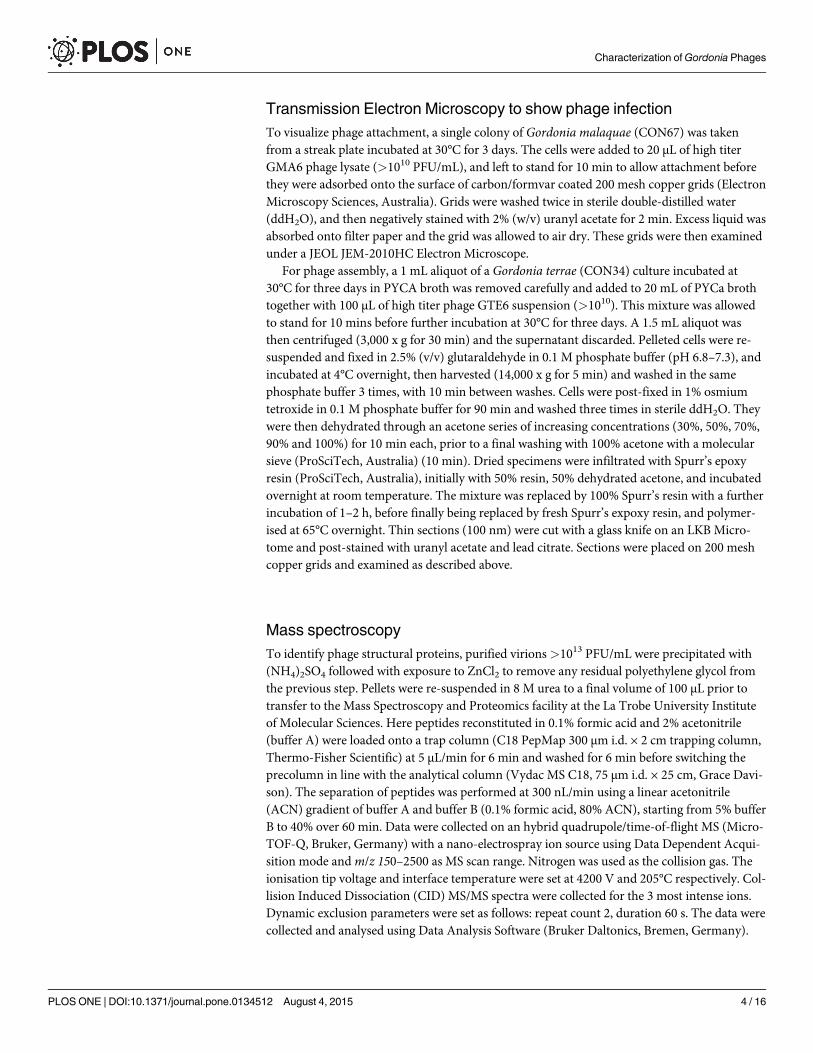
Transmission Electron Microscopy to show phage infectionTo visualize phage attachment, a single colony of Gordonia malaquae (CON67) was takenfrom a streak plate incubated at 30°C for 3 days. The cells were added to 20 μL of high titerGMA6 phage lysate (>1010 PFU/mL), and left to stand for 10 min to allow attachment beforethey were adsorbed onto the surface of carbon/formvar coated 200 mesh copper grids (ElectronMicroscopy Sciences, Australia). Grids were washed twice in sterile double-distilled water(ddH2O), and then negatively stained with 2% (w/v) uranyl acetate for 2 min. Excess liquid wasabsorbed onto filter paper and the grid was allowed to air dry. These grids were then examinedunder a JEOL JEM-2010HC Electron Microscope.
For phage assembly, a 1 mL aliquot of a Gordonia terrae (CON34) culture incubated at30°C for three days in PYCA broth was removed carefully and added to 20 mL of PYCa brothtogether with 100 μL of high titer phage GTE6 suspension (>1010). This mixture was allowedto stand for 10 mins before further incubation at 30°C for three days. A 1.5 mL aliquot wasthen centrifuged (3,000 x g for 30 min) and the supernatant discarded. Pelleted cells were re-suspended and fixed in 2.5% (v/v) glutaraldehyde in 0.1 M phosphate buffer (pH 6.8–7.3), andincubated at 4°C overnight, then harvested (14,000 x g for 5 min) and washed in the samephosphate buffer 3 times, with 10 min between washes. Cells were post-fixed in 1% osmiumtetroxide in 0.1 M phosphate buffer for 90 min and washed three times in sterile ddH2O. Theywere then dehydrated through an acetone series of increasing concentrations (30%, 50%, 70%,90% and 100%) for 10 min each, prior to a final washing with 100% acetone with a molecularsieve (ProSciTech, Australia) (10 min). Dried specimens were infiltrated with Spurr’s epoxyresin (ProSciTech, Australia), initially with 50% resin, 50% dehydrated acetone, and incubatedovernight at room temperature. The mixture was replaced by 100% Spurr’s resin with a furtherincubation of 1–2 h, before finally being replaced by fresh Spurr’s expoxy resin, and polymer-ised at 65°C overnight. Thin sections (100 nm) were cut with a glass knife on an LKB Micro-tome and post-stained with uranyl acetate and lead citrate. Sections were placed on 200 meshcopper grids and examined as described above.
Mass spectroscopyTo identify phage structural proteins, purified virions>1013 PFU/mL were precipitated with(NH4)2SO4 followed with exposure to ZnCl2 to remove any residual polyethylene glycol fromthe previous step. Pellets were re-suspended in 8 M urea to a final volume of 100 μL prior totransfer to the Mass Spectroscopy and Proteomics facility at the La Trobe University Instituteof Molecular Sciences. Here peptides reconstituted in 0.1% formic acid and 2% acetonitrile(buffer A) were loaded onto a trap column (C18 PepMap 300 μm i.d. × 2 cm trapping column,Thermo-Fisher Scientific) at 5 μL/min for 6 min and washed for 6 min before switching theprecolumn in line with the analytical column (Vydac MS C18, 75 μm i.d. × 25 cm, Grace Davi-son). The separation of peptides was performed at 300 nL/min using a linear acetonitrile(ACN) gradient of buffer A and buffer B (0.1% formic acid, 80% ACN), starting from 5% bufferB to 40% over 60 min. Data were collected on an hybrid quadrupole/time-of-flight MS (Micro-TOF-Q, Bruker, Germany) with a nano-electrospray ion source using Data Dependent Acqui-sition mode andm/z 150–2500 as MS scan range. Nitrogen was used as the collision gas. Theionisation tip voltage and interface temperature were set at 4200 V and 205°C respectively. Col-lision Induced Dissociation (CID) MS/MS spectra were collected for the 3 most intense ions.Dynamic exclusion parameters were set as follows: repeat count 2, duration 60 s. The data werecollected and analysed using Data Analysis Software (Bruker Daltonics, Bremen, Germany).
Characterization ofGordonia Phages
PLOS ONE | DOI:10.1371/journal.pone.0134512 August 4, 2015 4 / 16

Genome sequencing ofGordonia phagesGenomic DNA was extracted from phages GTE6, GMA2, and GMA6 and sequenced using aRoche GS FLX genome sequencer and titanium chemistry, as described in Petrovski, Seviour[14]. Genomic DNA extracted from all other phages in the same manner was prepared with anIllumina Nextera XT sample preparation kit as per manufacturers’ instructions. The preparedDNA libraries were sequenced on an Illumina MiSeq as a 150 bp paired end run.
Genome annotationThe genome open reading frames (ORFs) were screened initially using Glimmer (v3.02), whereORFs with a minimum size of 90 bp were detected [15]. All predicted start codons wereinspected for the presence of putative ribosomal binding sites and corrected as necessary.Sequence similarity searches were carried out against the GenBank database, as described byPetrovski et al. [11]. The presence of tRNA and tmRNA were also determined using both ARA-GORN [16], and tRNAScan-SE [17]. Transmembrane domains were predicted with the DASTransmembrane Prediction server [18].
Phage DNA when analyzed by gel electrophoresis gave results consistent with circularly per-muted DNA genomes. Therefore, for consistency the genomes annotations were conductedstarting with the DNA packaging operon.
Nucleotide sequence accession numberThe nucleotide sequences for all phages have been deposited GenBank under the followingaccession numbers; GTE6 (KR053200), GTE8 (KR053201), GMA2 (KR063281), GMA3(KR063279), GMA4 (KR053199), GMA5 (KR053198), GMA6 (KR063280), GMA7(KR063278), and GRU3 (KR053197).
Results and Discussion
Phage isolation and host range characterizationAll Gordonia phages isolated previously were obtained from wastewater, with most comingfrom activated sludge plants on the east coast of Australia [10–13]. While most phage isolatesdescribed here were also from wastewater (Table 1), an additional two phages GMA4 andGTE8, were obtained from puddle sediment (Reservoir, Australia), and creek water (Bendigo,Australia), respectively.
One of these, phage GMA4, lysed a single Mycolata strain, Gordonia malaquae strainBEN700. Phage GMA3 lysed the same G.malaquae strain (BEN700), but, also G. terrae(G238). All the other phage’s lysed multiple Gordonia strains, with phage GMA7 attacking 11strains from four different Gordonia species i.e. G. terrae (CON34, GOR9, G238), G. rubroper-tincta (CON38), G.malaquae (CON59, CON60, A554, A448, BEN700), and G. hydrophobica(CON65, CON66). As well as phage GTE8 lysing three strains of Gordonia terrae (CON34,GOR9, G232) and one of G. rubropertincta (CON38), it could also lyse Nocardia asteroides(CON12).
Phages able to lyse members of both these genera have been reported before. They includephage GRU1, which targets Nocardia nova strain CON47 and Gordonia terrae strains CON34,and G232 and also Gordonia rubropertincta strain CON38 [13]. This outcome might reflectthe close phylogenetic relationship of these host bacteria.
Many of these overlap in their host ranges. For example phages GTE6, GMA2, GMA6, andGMA7 all lysed the same four strains of G.malaquae (CON59, CON60, A554, and A448), aproperty which might make them useful additions to any phage cocktail designed to target
Characterization ofGordonia Phages
PLOS ONE | DOI:10.1371/journal.pone.0134512 August 4, 2015 5 / 16
foaming caused by G.malaquae, especially if they use different host receptor sites. PhageGMA5 was lytic against two of eight G. terrae strains (G238, G232). A similar situation hasbeen reported for other Gordonia phages, including GTE2 [11] that lysed only one of five G.terrae strains. No phages infective for G. amarae were obtained in this study.
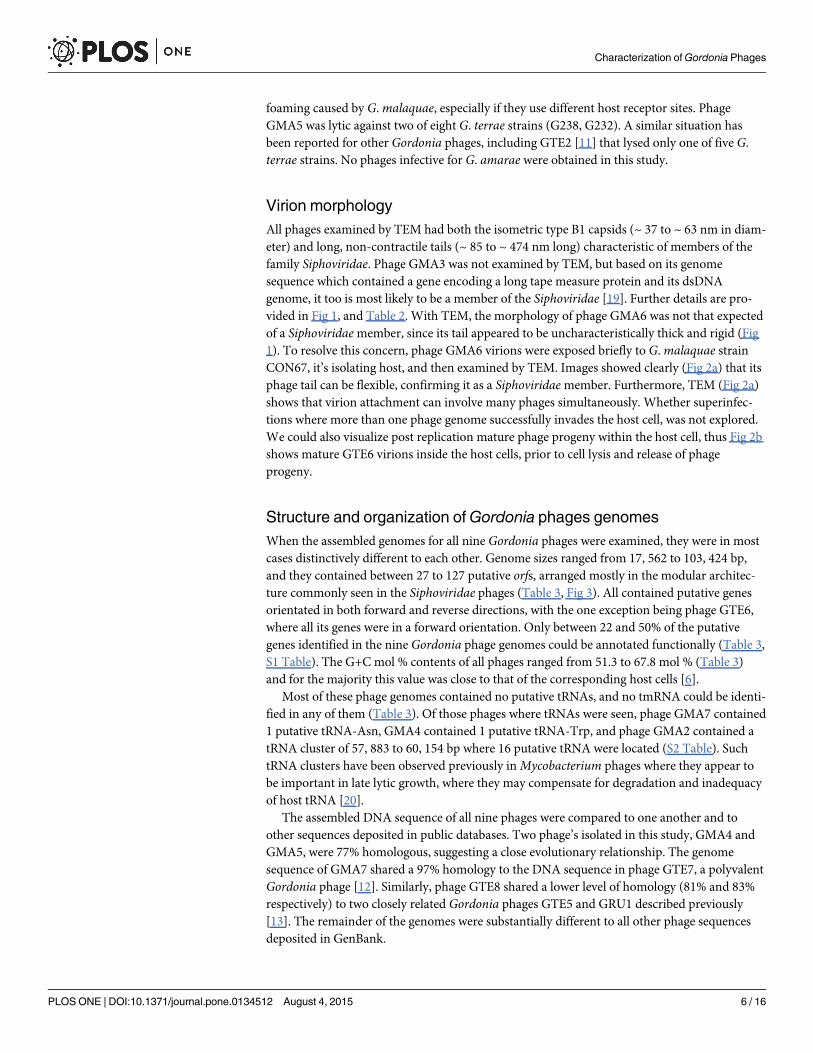
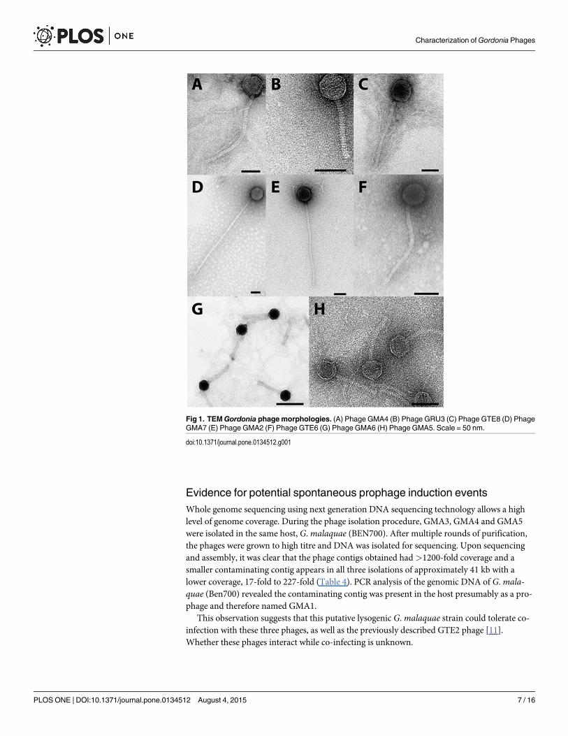
Virion morphologyAll phages examined by TEM had both the isometric type B1 capsids (~ 37 to ~ 63 nm in diam-eter) and long, non-contractile tails (~ 85 to ~ 474 nm long) characteristic of members of thefamily Siphoviridae. Phage GMA3 was not examined by TEM, but based on its genomesequence which contained a gene encoding a long tape measure protein and its dsDNAgenome, it too is most likely to be a member of the Siphoviridae [19]. Further details are pro-vided in Fig 1, and Table 2. With TEM, the morphology of phage GMA6 was not that expectedof a Siphoviridaemember, since its tail appeared to be uncharacteristically thick and rigid (Fig1). To resolve this concern, phage GMA6 virions were exposed briefly to G.malaquae strainCON67, it’s isolating host, and then examined by TEM. Images showed clearly (Fig 2a) that itsphage tail can be flexible, confirming it as a Siphoviridaemember. Furthermore, TEM (Fig 2a)shows that virion attachment can involve many phages simultaneously. Whether superinfec-tions where more than one phage genome successfully invades the host cell, was not explored.We could also visualize post replication mature phage progeny within the host cell, thus Fig 2bshows mature GTE6 virions inside the host cells, prior to cell lysis and release of phageprogeny.
Structure and organization ofGordonia phages genomesWhen the assembled genomes for all nine Gordonia phages were examined, they were in mostcases distinctively different to each other. Genome sizes ranged from 17, 562 to 103, 424 bp,and they contained between 27 to 127 putative orfs, arranged mostly in the modular architec-ture commonly seen in the Siphoviridae phages (Table 3, Fig 3). All contained putative genesorientated in both forward and reverse directions, with the one exception being phage GTE6,where all its genes were in a forward orientation. Only between 22 and 50% of the putativegenes identified in the nine Gordonia phage genomes could be annotated functionally (Table 3,S1 Table). The G+C mol % contents of all phages ranged from 51.3 to 67.8 mol % (Table 3)and for the majority this value was close to that of the corresponding host cells [6].
Most of these phage genomes contained no putative tRNAs, and no tmRNA could be identi-fied in any of them (Table 3). Of those phages where tRNAs were seen, phage GMA7 contained1 putative tRNA-Asn, GMA4 contained 1 putative tRNA-Trp, and phage GMA2 contained atRNA cluster of 57, 883 to 60, 154 bp where 16 putative tRNA were located (S2 Table). SuchtRNA clusters have been observed previously inMycobacterium phages where they appear tobe important in late lytic growth, where they may compensate for degradation and inadequacyof host tRNA [20].
The assembled DNA sequence of all nine phages were compared to one another and toother sequences deposited in public databases. Two phage’s isolated in this study, GMA4 andGMA5, were 77% homologous, suggesting a close evolutionary relationship. The genomesequence of GMA7 shared a 97% homology to the DNA sequence in phage GTE7, a polyvalentGordonia phage [12]. Similarly, phage GTE8 shared a lower level of homology (81% and 83%respectively) to two closely related Gordonia phages GTE5 and GRU1 described previously[13]. The remainder of the genomes were substantially different to all other phage sequencesdeposited in GenBank.
Characterization ofGordonia Phages
PLOS ONE | DOI:10.1371/journal.pone.0134512 August 4, 2015 6 / 16
Evidence for potential spontaneous prophage induction eventsWhole genome sequencing using next generation DNA sequencing technology allows a highlevel of genome coverage. During the phage isolation procedure, GMA3, GMA4 and GMA5were isolated in the same host, G.malaquae (BEN700). After multiple rounds of purification,the phages were grown to high titre and DNA was isolated for sequencing. Upon sequencingand assembly, it was clear that the phage contigs obtained had>1200-fold coverage and asmaller contaminating contig appears in all three isolations of approximately 41 kb with alower coverage, 17-fold to 227-fold (Table 4). PCR analysis of the genomic DNA of G.mala-quae (Ben700) revealed the contaminating contig was present in the host presumably as a pro-phage and therefore named GMA1.
This observation suggests that this putative lysogenic G.malaquae strain could tolerate co-infection with these three phages, as well as the previously described GTE2 phage [11].Whether these phages interact while co-infecting is unknown.
Fig 1. TEMGordonia phagemorphologies. (A) Phage GMA4 (B) Phage GRU3 (C) Phage GTE8 (D) PhageGMA7 (E) Phage GMA2 (F) Phage GTE6 (G) Phage GMA6 (H) Phage GMA5. Scale = 50 nm.
doi:10.1371/journal.pone.0134512.g001
Characterization ofGordonia Phages
PLOS ONE | DOI:10.1371/journal.pone.0134512 August 4, 2015 7 / 16
Sequence repeats inGordonia phage genomesRepeat structures have been reported previously in genome sequences of several relatedphages [13–14]. All nine phage genomes contain between 1 to 18 palindromic sequences ofbetween 14 to 98 bp in length (S3 Table). Some of these are located in what appear to be inter-genic areas, which might support their roles as putative rho-independent transcriptional ter-minators [21]. They also contained 14 to 252 direct repeats ranging in length between 14 and425 bp (S4 Table). Also seen in these genomes were 3 to 87 inverted repeats of 56 to 14 bplong (S4 Table). Inverted repeats may indicate replication origins and transposable elements[22], but neither of these could be identified in any of these phages, and so their roles remainunknown.
Gordonia phage DNA packaging modulesIn Siphoviridae phage genomes, the large terminase subunit protein usually functions in a com-plex with a small terminase subunit, and together these act to mediate cleavage of the phageDNA at specific sites prior to packaging into the prohead [23–24]. The gene encoding the largeterminase subunit was identified in all nine Gordonia phages examined here by either aminoacid sequence homology to other known terminase genes (GMA4, GMA3, GMA5, GMA7, andGTE8) or the presence of the diagnostic conserved motif. In Siphoviridae phages the small ter-minase gene is typically located upstream, and is transcribed in the same direction as the largeterminase [23–24]. In all phages except GMA2 this pattern could be recognized, and in somecases supported by amino acid sequence homology to other known small terminases (S1Table).
Fig 2. Stages inGordonia phage infection cycles. (A) Attachment stage of phage infection cycle between phage GMA6 and hostGordonia malaquaestrain CON67. Scale = 200 nm. (B) Replication of phage GTE6 insideG. terrae strain CON34 cells prior to cell lysis. Arrows indicate phage replicated insidebacterial cells. Scale = 200 nm.
doi:10.1371/journal.pone.0134512.g002
Characterization ofGordonia Phages
PLOS ONE | DOI:10.1371/journal.pone.0134512 August 4, 2015 8 / 16

Gordonia phage structural protein genes and their proteomicsPhage structural protein genes are located typically adjacent to the DNA packaging module,usually beginning with head morphogenesis genes, followed by tail morphogenesis genes [19].Some departures from this gene arrangement were seen in these nine phages. For example, inGMA6, only one (orf8) of the genes identified between the terminase genes (orf2 and orf3) andthe putative portal protein gene (orf11), could be assigned a putative function in encoding anucleoside triphosphate pyrophosphohydrolase. This interpretation was based on the presenceof the cd11541 motif. Seemingly involved in DNAmaintenance, it was seen between the struc-tural and packaging modules, a location different to the typical modulated genome architectureof Siphoviridae phages, where all genes of similar function are clustered together [25]. Further-more, orf14, within the structural gene module of GMA6, appears to encode a HNH endonu-clease based on its amino acid sequence homology to the diagnostic pfam01844 motif. Genearrangements in phage GMA2 suggest that orf21, encoding a putative DNA methyltransferaseis in the structural gene module.
In all nine phages the tape measure proteins were encoded by their longest gene, which isusual in Siphoviridae phages [19] (S1 Table, Fig 3). The only exception was in phage GRU3where orf6 encoding a putative phage head protein was slightly larger in size (657 amino acids)than orf12 encoding its tape measure protein, 622 amino acids long. In most of these (GMA2,GMA3, GMA4, GMA6, GMA7, and GTE8), the two genes preceding that encoding the tape
Table 3. Summary of characters of the nineGordonia phage genomes.
Phagename
Averagecoverage(fold)
Totalreadcount
Genomesize (bp)
G+Ccontent(mol %)
No.putativetRNA
No.putativegenes
No.putativegenes inforwardsorientation
No.functionallyannotatedputativegenes
No.novelgenes
No.palindromes
No.directrepeats
No.invertedrepeats
GMA2a c
1, 212 336,750
103, 424 53.4 16 126 42 42 62 7 22 10
GMA3b e d
1, 200 677,981
77, 779 51.3 0 104 32 27 47 16 18 8
GMA4b e d
1, 981 716,641
45, 537 66.4 1 68 61 22 11 6 40 31
GMA5b f d
6, 793 930,480
17, 562 66.4 0 28 24 14 4 11 28 13
GMA6a c
247 55,269
83, 324 58.2 0 115 109 38 68 1 20 3
GMA7b c
1, 603 947,843
73, 419 56.6 1 101 32 23 5 18 14 5
GRU3b e d
520 89,131
17, 727 66.5 0 26 23 12 6 3 42 16
GTE6a c
915 141,321
56, 982 67.8 0 86 86 23 49 3 252 87
GTE8b c
1, 605 777,336
67, 617 66.0 0 94 67 23 22 5 48 36
a sequenced using 454,b sequenced using Illumina,c reads assembled using CLC workbench (v6.5.1),d reads assembled using CLC workbench (v7.5.1),e reads assembled using Spades (v3.1.0),f reads assembled using ABySS (v1.3.7).
doi:10.1371/journal.pone.0134512.t003
Characterization ofGordonia Phages
PLOS ONE | DOI:10.1371/journal.pone.0134512 August 4, 2015 9 / 16
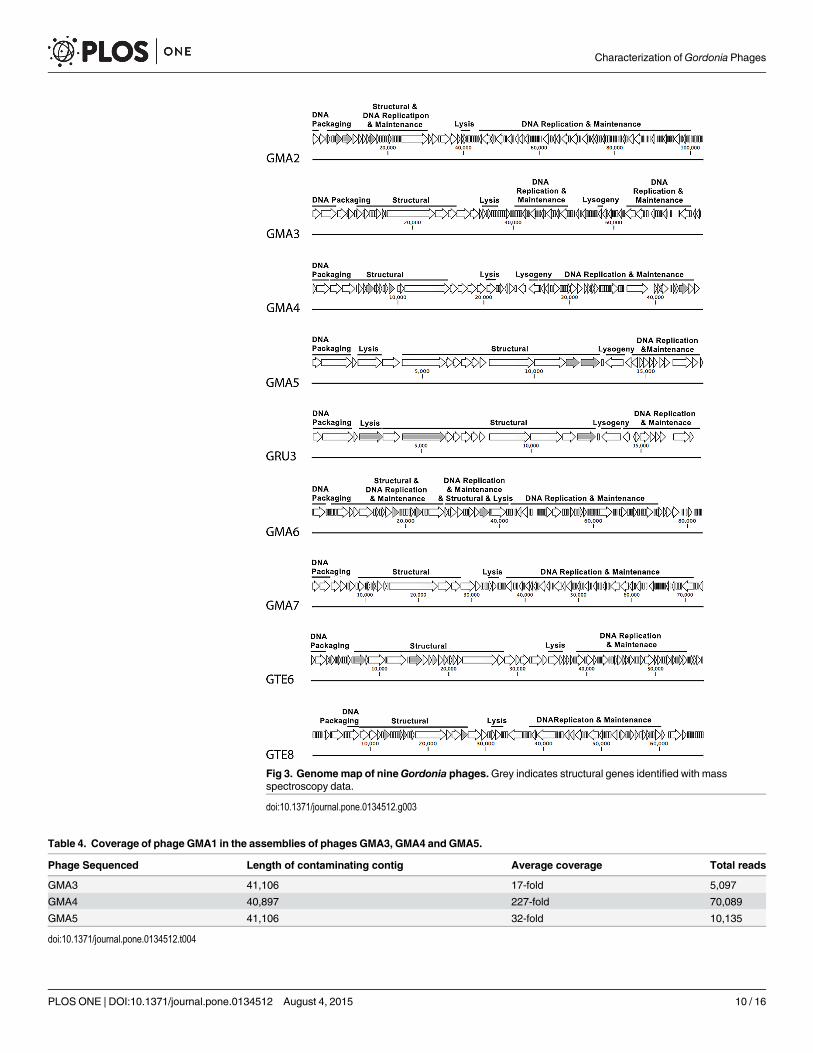
Fig 3. Genomemap of nineGordonia phages.Grey indicates structural genes identified with massspectroscopy data.
doi:10.1371/journal.pone.0134512.g003
Table 4. Coverage of phage GMA1 in the assemblies of phages GMA3, GMA4 and GMA5.
Phage Sequenced Length of contaminating contig Average coverage Total reads
GMA3 41,106 17-fold 5,097
GMA4 40,897 227-fold 70,089
GMA5 41,106 32-fold 10,135
doi:10.1371/journal.pone.0134512.t004
Characterization ofGordonia Phages
PLOS ONE | DOI:10.1371/journal.pone.0134512 August 4, 2015 10 / 16

measure protein were identified as encoding putative tail assembly proteins, where the latterappeared to be translated using a conserved programmed frameshift, a common feature ofSiphoviridae phages [26]. Usually the gene immediately upstream of these is that encoding theputative major tail protein [26].
Mass spectrometry data (S5 Table) seemed to suggest several structural genes are locatedoutside the structural gene module. For example, in phage GMA4 orf66 was located in theDNA replication module, with a translated protein sequence homologous to a hypotheticalprotein from Aeromicrobium marinum, but also a motif for a phage tail fiber protein(COG5301). Similarly, in GMA6, orf43 was located within the DNA replication gene modules.
Gordonia phage lysis gene modules are diverseLysin genes were identified in all nine Gordonia phages, but their locations and numbers var-ied, and as with many already discussed, they often appeared to disrupt the usual and expectedmodular genome architecture of Siphoviridae phages. Phages GMA5 and GRU3 contain a D-alanyl-D-alanine carboxypeptidase encoding gene (orf5 in both) showing amino acid sequencehomology to a hypothetical protein in Gordonia soli, located within what appears to be thephage structural module (S1 Table, Fig 3). A phage lysin motif within the structural gene mod-ule was reported for Rhodococcus phage RRH1, suggesting this is not an uncommon occur-rence [27, 28].
In the genomes of GMA7 and GTE6 their lysin genes were adjacent to their structural pro-tein encoding genes (orf28 –orf29 and orf38, respectively), and unusually, both had additionallysin genes in their DNA replication gene modules (orf41 and orf58, respectively). The samepattern was reported for phage GTE7, [12], to which GMA7 is genetically similar at a nucleo-tide level (97% identity, 95% coverage).
Phages GMA2, GMA3, and GMA6 also had unusual lysin gene arrangements, with highernumbers of such genes than the more common lysin A and B arrangement [29]. Phage GMA2unusually possessed four putative lysin genes (orf35 –orf38), identified from their amino acidhomologies and presence of the diagnostic pfam13529 (peptidase), pfam01510 (N-acetylmura-moyl-L-alanine amidase), and cd02619 (peptidase) motifs in Orf35, Orf36, and Orf38, respec-tively. Phage GMA6 also had four lysin genes (orf34, orf37, orf40, and orf45), many of whichwere separated by genes associated with DNA replication/maintenance and virion morphogen-esis. A similar pattern was seen in phage GMA3, which contained three putative lysin genes(orf22, orf24, and orf26) separated by a putative nuclease gene (orf25), again associated withDNA replication/maintenance (S1 Table, Fig 3).
Orf45 in GMA6 is a more complex lysin gene than those seen in all other actinophages. Italone encodes an unusually high number of different lysin motifs. These include an N-terminalBacA motif of a bacterial lysin from Enterococcus faecalis (cd06418), an N-acetylmuramoyl-L-alanine amidase motif (pfam01510) downstream of this, a peptidase motif (pfam01551) furtherdownstream, and an additional C-terminal motif (pfam13810) of unknown function (S1 Table).
Holins could not be identified in phages GMA4, GMA5, and GRU3 by nucleotide or aminoacid sequence homologies, nor by the criteria of Wang et al. [30], which state that expressedproducts should be less than 150 amino acid residues and contain two or more transmembraneregions. If holins are present in these two phages, it would seem that their genes are novel intheir locations and/or translated amino acid sequences.
DNA replication/maintenance genesDNA replication modules in all other actinophage genomes sequenced so far are arranged in amodular architecture, where genes functioning in DNA replication/maintenance are located
Characterization ofGordonia Phages
PLOS ONE | DOI:10.1371/journal.pone.0134512 August 4, 2015 11 / 16

adjacent to lysin genes [11–14, 27, 28, 31, 32]. In GMA4, GMA7, and GMA2 phages, thisregion contains putative DNA-methylase encoding genes, of these GMA2 appears to possess atleast two (orf21 and orf51). If functional, they may play a role in protecting their DNA fromhost cell restriction attack [33]. Such enzymes have been identified in other Gordonia phagesincluding GTE7 phages [12]. Metagenomic studies by Tamaki et al. [34] have suggested thatmethylase genes are more prevalent in phages within the activated sludge habitat from wheremost actinophages have come. Glycosyltransferase encoding genes are also seen in many phagegenomes [35], including that of GMA2 (orf4 and orf22), and all appear to have similar func-tions to phage methylases where they help protect phage DNA from digestion with restrictionendonucleases from host RM systems [33]. These genes can also have other functions includingserotype conversion in temperate phages [35], and so their purpose here remains unclear.
Lysogeny and lysogenic conversion genesGenomes of phages GMA3, GMA4, GMA5, and GRU3 all contain putative genes that arehomologues of phage integrase genes (orf75, orf29, orf17, and orf17 respectively), based ontheir product amino acid sequence similarities to those of known phage integrases, and the pos-session of the integrase specific motif pfam00589. If functional, their presence suggests thecapability for a lysogenic lifecycle as well as a lytic one.
The GMA4 genome appears to encode several moron genes that may confer a selectiveadvantage to its host. For example, it possesses a gene associated with phage resistance (orf34)[36]. The N-terminal region of Orf34 contains a Rha motif (pfam09669), thought to interferewith further phage infection of bacterial host strains lacking the integration host factor (IHF)[36]. It regulates expression of the rha gene, and so may confer resistance to further phageattack of any bacterial host infected by it in a lysogenic cycle [36].
Unexpected features of theGordonia phagesAs mentioned, most of the nine Gordonia phages sequenced in this study had highly distinctivegenomes, with high percentages of ORFans (5 to 59%) (S1 Table), for which no statistically sig-nificant identifications could be made against sequences held in GenBank. Yet their genesencode motifs suggestive of their putative function. For example, both GMA2 and GMA6 pos-sess a cd00233 motif in their Orf14 and Orf13 putative proteins, respectively. The orf14 andorf13 genes appear to encode a VIP2 family actin-ADP-ribosylating toxin with high specificityagainst the insect pest corn rootworms, and sharing a statistically significant sequence similar-ity with enzymatic components of other binary toxins, including the Clostridium botulinum C2toxin, C. perfringens iota toxin, C. piroforme toxin, C. piroforme toxin and C. difficile toxin [37].
Furthermore, phage GTE6 genome appeared to contain a gene (orf12), encoding a host cellsurface-exposed lipoprotein since its expressed amino acid sequence shares homology with thepfram07553 motif. Such motifs are usually involved in superinfection exclusion, acting at thestage of DNA release from the phage head into the host cell. These motifs have been associatedwith Superinfection exclusion (Sie) systems in temperate phages, where they interfere with co-infections involving other phages [33, 38, 39]. Presence of such a motif in what appears to bean obligatory lytic phage is unexpected. Equally unexpected is that orf21 in phage GTE6encodes a putative Eppstein-Barr nuclear antigen (Orf21), showing 35% amino acid sequencesimilarity to that of Saccharomonospora phage PIS 136 in this region [40]. Whether this homol-ogy reflects a similar function for the pair, or a distant evolutionary relationship between them,is unknown. The orf4 of phage GMA7 also appears to encode an unexpected motif (cd12820)normally associated with a putative adhesion virulence factor, forming a matrix on the bacterialouter membrane, which mediates binding to collagen and epithelial cells [41].
Characterization ofGordonia Phages
PLOS ONE | DOI:10.1371/journal.pone.0134512 August 4, 2015 12 / 16
Evolutionary ancestry ofGordonia phagesFrom the data presented here, it is clear that phages GTE8, isolated from creek water andGMA7, from activated sludge are genetically very similar to phages GTE5/GRU1, and GTE7,respectively. It is reasonable to propose that these similarities reflect a closely shared ancestralpast. Similar comments apply to phage GMA5 and GRU3. Despite not sharing nucleotidesequence identity with phage GTE7 DNA, the expressed amino acid sequences of phageGMA3 expressed genes are highly similar to it. Nine genes of GMA3 were most similar tothose from GTE7, while 23 other genes were most similar to those from phage ReqiDocB7, towhich GTE7 genome is closely related at an amino acid level [12, 42]. As a similar closenesswas not reflected at the nucleotide sequence level, one suggestion might be that more distantevolutionary relationships exist between GTE7, ReqiDocB7, and GMA3. GMA3 contains agene showing homology to a putative RNA-binding gene from the chimpanzee Pan troglodytes(orf91), and a centromere protein F-like gene from the banana plantMusa acuminata (orf74).The expected values for these matches are borderline statistically significant (2e-04 and 6e-04,respectively), so whether these data reflect real distant evolutionary relationships remainsunresolved.
Each individual Gordonia phage genome sequence was unique, but given the close geneticrelationships between the Mycolata host genera, attempts were made to classify these accordingto the system of Hatfull, Jacobs-Sera [43] designed to show evolutionary relationships theMycobacteriophages. It was not possible to place these Gordonia phages into any of their pre-existing clusters. For example, while GMA7 is highly similar to GTE7 at a nucleotide sequencelevel, and GMA3 contains genes encoding several putative proteins also similar to those ofGTE7, none could be grouped with any Mycobacteriophages. Instead they emerge as singletonssince none of the existing clusters embraced them.
Suitability of these phages for use in foam bio-controlOf the nine phages examined in this study, GMA3, GMA5, GMA5 and GRU3 contain putativeintegrase genes, suggesting that they may undertake a lysogenic lifecycle. If these genes arefunctional, then these are probably undesirable candidates for standard phage therapy for acti-vated sludge foam control.
Of the remaining five phages GMA2 and GMA6 both appear to contain a putative VIP2family actin-ADP-ribosylating toxin gene, which target eukaryotic proteins upon infection.Consequently, neither phage would be considered being suitable for bio-control strategies.Their release into the environment may potentially result in the spread of these undesirablegenes and an increased virulence of other bacteria hosts.
Other Gordonia phages GMA7, GTE6, and GTE8 appear to be obligatory lytic. Of these,GMA7 and GTE8 seem particularly attractive as both have impressive broad host ranges. Forinstance, GMA7 targets eleven strains of Gordonia including those of G. terrae, G.malaquae,G. rubropertincta and G. hydrophobica. Similarly, GTE8 targets several species including G. ter-rae (CON34, GOR9, G232) and G. rubropertincta (CON38) and Nocardia asteroids (strainCON12) (Table 1). Furthermore, phage GMA7 contains a putative DNA methylase gene(orf38) containing a pfam01555 motif. If this gene is functional, then this phage may evadecleavage by host defense RM systems [33, 38] and thus become an even more powerful additionto any phage therapy cocktail.
ConclusionsNine phages infective for members of the genus Gordonia were isolated from wastewater andnatural water environments, several of which had broad host ranges. Methods for visualization
Characterization ofGordonia Phages
PLOS ONE | DOI:10.1371/journal.pone.0134512 August 4, 2015 13 / 16

of the phage infection cycle using TEM were successful and may prove to be useful in studies ofmechanisms of phage infection. Whole genome sequencing of these phages revealed that theirgenomes were all distinctively different, failing to cluster with those of known Mycobacterio-phages, based on both nucleotide and amino acid sequence similarities. Some are less modularin their genomic architecture than those characterized previously, and contain a higher num-ber of lysin genes seen in other Actinophage genomes previously. Of these nine phages, threebroad host range phages GMA7, GTE6, and GTE8 appear obligatory lytic and hence poten-tially suitable candidates for phage therapy cocktails to control activated sludge foaming.
Supporting InformationS1 Table. Genome annotations of nine phages infecting Gordonia spp. a ORFs were num-bered consecutively, b The most closely related gene (only if named) and the name of theorganism, c Percentage identity is based on the best match when a BLAST P analysis is per-formed, d The probability of obtaining a match by chance as determined by BLAST analysis.Only values less than 10−4 were considered significant, e Predicted function is based on aminoacid identity, conserved motifs, and gene location within functional modules.(DOCX)
S2 Table. Putative tRNA detected in Gordonia phage genomes.(DOCX)
S3 Table. Palindromes in the genome sequence of nine Gordonia spp. phages.(DOCX)
S4 Table. Repeats in the genome sequences of nine Gordonia spp. phages. I indicatesinverted repeat, D indicates direct repeat.(DOCX)
S5 Table. Summary of Gordonia phage structural genes identified by mass spectroscopy.(DOCX)
AcknowledgmentsWe wish to thank Dr. Robert Glaishier (LIMS), Dr. Sabine Wilkens (LIMS), Ms. Glenys Shirley(LIMS), Dr. Eric Hanssen (Bio21 Institute) and Dr. Christopher Adda (LIMS) for assistancewith Transmission Electron Microscopy, Dr. Pierre Faou (LIMS) for assistance with mass spec-troscopy, and Dr. Daniel Tillett for useful discussions.
Author ContributionsConceived and designed the experiments: RS ZD SP. Performed the experiments: ZD. Ana-lyzed the data: ZD SP. Contributed reagents/materials/analysis tools: JT. Wrote the paper: RSJT SP ZD.
References1. de los Reyes FL III. 2010. Foaming. p 215–258. In Seviour RJ, Nielsen PH (ed), Microbial Ecology of
Activated Sludge. IWA Publishing, London.
2. Drzyzga O. The strengths and weaknesses of Gordonia: a review of an emerging genus with increasingbiotechnological potential. Crit Rev Microbiol. 2012; 38(4):300–16. doi: 10.3109/1040841X.2012.668134 PMID: 22551505
3. Arenskötter M, Bröker D, Steinbüchel A. Biology of the Metabolically Diverse Genus Gordonia. AppEnviron Microbiol. 2004; 70:3195–204.
Characterization ofGordonia Phages
PLOS ONE | DOI:10.1371/journal.pone.0134512 August 4, 2015 14 / 16

4. Klatte S, Rainey FA, Kroppenstedt RM. Transfer of Rhodococcus aichiensis Tsukamura 1982 andNocardia amarae Lechevalier and Lechevalier 1974 to the GenusGordonia asGordona aichiensiscomb. nov. andGordona amarae comb. nov. Int J Syst Bacteriol. 1994; 44(4):769–73. PMID: 7981103
5. Lechevalier MP, Lechevalier HA. Nocardia amarae sp. nov., an Actinomycete Common in FoamingActivated Sludge. Nati J Syst Bacteriol. 1974; 24(2):278–88.
6. Goodfellow M, Kumar V, Maldonado LA. Genus II. Gordonia (Tsukamura 1971) Stackbrandt, Smidaand Collins 1988, 345VP. In: Goodfellow M, Kampfer P, Busse H-J, Trujillo ME, Suzuki K-I, LudwigW,et al., editors. Bergey's Manual of Systematic Bacteriology. 5. 2 ed. New York: Springer; 2012. p.419–35.
7. Soddell JA, Seviour RJ. Microbiology of foaming in activated sludge plants. J App Bacteriol. 1990;69:145–76.
8. Soddell JA. Foaming. In: Seviour RJ, editor. The microbiology of activated sludge. Dordrecht: KluwerAcademic Publishers; 1999.
9. Petrovski S, Dyson ZA, Quill ES, McIlroy SJ, Tillett D, Seviour RJ. An examination of the mechanismsfor stable foam formation in activated sludge systems. Water Res. 2011; 45(5):2146–54. doi: 10.1016/j.watres.2010.12.026 PMID: 21239035
10. Thomas JA, Soddell JA, Kurtböke DÍ. Fighting foam with phages. Water Sci Technol. 2002; 46:511–53. PMID: 12216679
11. Petrovski S, Seviour RJ, Tillett D. Characterization of the Genome of the Polyvalent Lytic Bacterio-phage GTE2, Which Has Potential for Biocontrol ofGordonia, Rhodococcus, andNocardia StabilizedFoams in Activated Sludge Plants. App Environ Microbiol. 2011; 77(12):3923–9.
12. Petrovski S, Seviour RJ, Tillett D. Prevention ofGordonia andNocardia Stabilized Foam Formation byUsing Bacteriophage GTE7. App Environ Microbiol. 2011; 77(21):7864–7
13. Petrovski S, Tillett D, Seviour RJ. Genome Sequences and Characterization of the RelatedGordoniaPhages GTE5 and GRU1 and Their Use as Potential Biocontrol Agents. App Environ Microbiol. 2012;78(1):42–7.
14. Petrovski S, Seviour RJ, Tillett D. Genome sequence and characterization of the Tsukamurella bacteri-ophage TPA2. Appl Environ Microbiol. 2011; 77(4):1389–98. doi: 10.1128/AEM.01938-10 PMID:21183635
15. Delcher AL, Bratke KA, Powers EC, Salzberg SL. Identifying bacterial genes and endosymbiont DNAwith Glimmer. Bioinformatics. 2007; 23(6):673–9. PMID: 17237039
16. Laslett D, Canback B. ARAGORN, a program to detect tRNA genes and tmRNA genes in nucleotidesequences. Nucleic Acids Res. 2004; 32(1):11–6. PMID: 14704338
17. Schattner P, Brooks AN, Lowe TM. The tRNAscan-SE, snoscan and snoGPS web servers for thedetection of tRNAs and snoRNAs. Nucleic Acids Res. 2005; 33(Web Server issue):W686–9. PMID:15980563
18. Cserzo M, Wallin E, Simon I, Heijne Gv, Elofsson A. Prediction of transmembrane alpha-helices in pro-cariotic membrane proteins: the Dense Alignment Surface method. Prot Eng. 1997; 10(6):673–6.
19. Pedulla ML, Ford ME, Houtz JM, Karthikeyan T, Wadsworth C, Lewis JA, et al. Origins of Highly MosaicMycobacteriophage Genomes. Cell. 2003; 113:171–82. PMID: 12705866
20. PopeWH, Anders KR, Baird M, Bowman CA, Boyle MM, Broussard GW, et al. Cluster M Mycobacterio-phages Bongo, PegLeg, and Rey with Unusually Large Repertoires of tRNA Isotypes. J Virol. 2014; 88(5):2461–80. doi: 10.1128/JVI.03363-13 PMID: 24335314
21. Lesnik EA, Sampath R, Levene HB, Henderson TJ, McNeil JA, Ecker DJ. Prediction of rho-independenttranscriptional terminators in Escherichia coli. Nucleic Acids Res. 2001; 29(17):3583–94. PMID:11522828
22. Mott ML, Berger JM. DNA replication initiation: mechanisms and regulation in bacteria. Nat Rev Micro-biol. 2007; 5:343–54. PMID: 17435790
23. Catalano CE. The terminase enzyme from bacteriophage lambda: a DNA-packaging machine. Cell MolLife Sci. 2000; 57:128–48. PMID: 10949585
24. Fujisawa H, Morita M. Phage DNA packaging. Genes Cells. 1997; 2(9):537–45. PMID: 9413995
25. Hatfull GF. Bacteriophage Genomics. Curr opin microbiol. 2008; 11(5):447–53. doi: 10.1016/j.mib.2008.09.004 PMID: 18824125
26. Xu J, Hendrix RW, Duda RL. Conserved translational frameshift in dsDNA bacteriophage tail assemblygenes. Mol Cell. 2004; 16(1):11–21. PMID: 15469818
27. Petrovski S, Seviour RJ, Tillett D. Characterization and whole genome sequences of the Rhodococcusbacteriophages RGL3 and RER2. Arch Virol. 2013; 158(3):601–9. doi: 10.1007/s00705-012-1530-5PMID: 23129131
Characterization ofGordonia Phages
PLOS ONE | DOI:10.1371/journal.pone.0134512 August 4, 2015 15 / 16
28. Petrovski S, Dyson ZA, Seviour RJ, Tillett D. Small but sufficient: the Rhodococcus phage RRH1 hasthe smallest known Siphoviridae genome at 14.2 kilobases. J Virol. 2012; 86(1):358–63. doi: 10.1128/JVI.05460-11 PMID: 22013058
29. Payne K, Sun Q, Sacchettini J, Hatfull GF. Mycobacteriophage Lysin B is a novel mycolylarabinogalac-tan esterase. Mol microbiol. 2009; 73(3):367–81. doi: 10.1111/j.1365-2958.2009.06775.x PMID:19555454
30. Wang IN, Smith DL, Young R. Holins: the protein clocks of bacteriophage infections. Annu Rev Micro-biol. 2000; 54:799–825. PMID: 11018145
31. Petrovski S, Seviour RJ, Tillett D. Genome sequence and characterization of a Rhodococcus equiphage REQ1. Virus Genes. 2013; 46(3):588–90. doi: 10.1007/s11262-013-0887-1 PMID: 23381579
32. Petrovski S, Seviour RJ, Tillett D. Genome sequence of the Nocardia bacteriophage NBR1. Arch Virol.2014; 159(1):167–73. doi: 10.1007/s00705-013-1799-z PMID: 23913189
33. Labrie SJ, Samson JE, Moineau S. Bacteriophage resistance mechanisms. Nat Rev Microbiol. 2010; 8(5):317–27. doi: 10.1038/nrmicro2315 PMID: 20348932
34. Tamaki H, Zhang R, Angly FE, Nakamura S, Hong P-Y, Yasunaga T, et al. Metagenomic analysis ofDNA viruses in a wastewater treatment plant in tropical climate. Environ Microbiol. 2012; 14(2):441–52. doi: 10.1111/j.1462-2920.2011.02630.x PMID: 22040222
35. Markine-Goriaynoff N, Gillet L, Van Etten JL, Korres H, Verma N, Vanderplasschen A. Glycosyltrans-ferases encoded by viruses. J Gen Virol. 2004; 85(10):2741–54.
36. Henthorn KS, Friedman DI. Identification of related genes in phages phi 80 and P22 whose productsare inhibitory for phage growth in Escherichia coli IHF mutants. J Bacteriol. 1995; 177(11):3185–90.PMID: 7768817
37. Han S, Arvai AS, Clancy SB, Tainer JA. Crystal structure and novel recognition motif of Rho ADP-ribo-sylating C3 exoenzyme from Clostridium botulinum: structural insights for recognition specificity andcatalysis1. Journal of Mol Biol. 2001; 305(1):95–107.
38. Samson JE, Magadan AH, Sabri M, Moineau S. Revenge of the phages: defeating bacterial defences.Nat Rev Micro. 2013; 11(10):675–87.
39. Ali Y, Koberg S, Heßner S, Sun X, Rabe B, Back A, et al. Temperate Streptococcus thermophilusphages expressing superinfection exclusion proteins of the Ltp type. Front Microbiol. 2014; 5.
40. Bajpai R, Soni V, Khandrika L, Jangir PK, Sharma R, Agrawal P. Genome Sequence of a Novel Acti-nophage PIS136 Isolated from a Strain of Saccharomonospora sp. J Virol. 2012; 86(17):9552. doi: 10.1128/JVI.01529-12 PMID: 22879621
41. El Tahir Y, Skurnik M. YadA, the multifaceted Yersinia adhesin. International Journal of Medical Micro-biology. 2001; 291(3):209–18. PMID: 11554561
42. Summer EJ, Liu M, Gill JJ, Grant M, Chan-Cortes TN, Ferguson L, et al. Genomic and functional analy-ses of Rhodococcus equi phages ReqiPepy6, ReqiPoco6, ReqiPine5, and ReqiDocB7. Appl EnvironMicrobiol. 2011; 77(2):669–83. doi: 10.1128/AEM.01952-10 PMID: 21097585
43. Hatfull GF, Jacobs-Sera D, Lawrence JG, PopeWH, Russell DA, Ko CC, et al. Comparative genomicanalysis of 60 Mycobacteriophage genomes: genome clustering, gene acquisition, and gene size. JMol Biol. 2010; 397(1):119–43. doi: 10.1016/j.jmb.2010.01.011 PMID: 20064525
Characterization ofGordonia Phages
PLOS ONE | DOI:10.1371/journal.pone.0134512 August 4, 2015 16 / 16
Copyright © 2022 FDOKUMEN