
Tumor Lysis Syndrome: An Endless Challenge in Onco ... - MDPI
18
Citation: Lupus , oru, G.; Ailinc˘ ai, I.; Fr˘ at , il˘ a, G.; Ungureanu, O.; Andronesi, A.; Lupus , oru, M.; Banu, M.; V ˘ ac˘ aroiu, I.; Dina, C.; Sinescu, I. Tumor Lysis Syndrome: An Endless Challenge in Onco-Nephrology. Biomedicines 2022, 10, 1012. https://doi.org/10.3390/ biomedicines10051012 Academic Editors: Silvio Maringhini and Paola Pontrelli Received: 10 February 2022 Accepted: 26 April 2022 Published: 28 April 2022 Publisher’s Note: MDPI stays neutral with regard to jurisdictional claims in published maps and institutional affil- iations. Copyright: © 2022 by the authors. Licensee MDPI, Basel, Switzerland. This article is an open access article distributed under the terms and conditions of the Creative Commons Attribution (CC BY) license (https:// creativecommons.org/licenses/by/ 4.0/). biomedicines Review Tumor Lysis Syndrome: An Endless Challenge in Onco-Nephrology Gabriela Lupus , oru 1,2,† , Ioana Ailincăi 1,2,† , Georgiana Frăt , ilă 1,2 , Oana Ungureanu 1,2 , Andreea Andronesi 1,2, * , Mircea Lupus , oru 3, * , Mihaela Banu 4,† , Ileana Văcăroiu 1,5 , Constantin Dina 6 and Ioanel Sinescu 1,7 1 Department of Nephrology, Carol Davila University of Medicine and Pharmacy, 020021 Bucharest, Romania; [email protected] (G.L.); [email protected] (I.A.); [email protected] (G.F.); [email protected] (O.U.); [email protected] (I.V.); [email protected] (I.S.) 2 Department of Nephrology, Fundeni Clinical Institute, 022328 Bucharest, Romania 3 Department of Physiology, Carol Davila University of Medicine and Pharmacy, 020021 Bucharest, Romania 4 Department of Anatomy, Carol Davila University of Medicine and Pharmacy, 020021 Bucharest, Romania; [email protected] 5 Department of Nephrology, Sf Ioan Clinical Emergency Hospital, 042122 Bucharest, Romania 6 Department of Anatomy, Ovidius University, 900527 Constant , a, Romania; [email protected] 7 Center for Uronephrology and Kidney Transplantation, Fundeni Clinical Institute, 022328 Bucharest, Romania * Correspondence: [email protected] (A.A.); [email protected] (M.L.) † These authors contributed equally to this work. Abstract: Tumor lysis syndrome (TLS) is a common cause of acute kidney injury in patients with malignancies, and it is a frequent condition for which the nephrologist is consulted in the case of the hospitalized oncological patient. Recognizing the patients at risk of developing TLS is essential, and so is the prophylactic treatment. The initiation of treatment for TLS is a medical emergency that must be addressed in a multidisciplinary team (oncologist, nephrologist, critical care physician) in order to reduce the risk of death and that of chronic renal impairment. TLS can occur spontaneously in the case of high tumor burden or may be caused by the initiation of highly efficient anti-tumor therapies, such as chemotherapy, radiation therapy, dexamethasone, monoclonal antibodies, CAR-T therapy, or hematopoietic stem cell transplantation. It is caused by lysis of tumor cells and the release of cellular components in the circulation, resulting in electrolytes and metabolic disturbances that can lead to organ dysfunction and even death. The aim of this paper is to review the scientific data on the updated definition of TLS, epidemiology, pathogenesis, and recognition of patients at risk of developing TLS, as well as to point out the recent advances in TLS treatment. Keywords: cancer; chemotherapy; toxicity; tumor lysis syndrome 1. Introduction Patients with malignancies are at increased risk of developing acute kidney injury (AKI) due to multiple causes: vomiting related to chemotherapy, nephrotoxicity of anti- neoplastic drugs, or direct kidney involvement caused by the underlying malignancy or urinary tract obstruction. AKI may be severe enough to eventually require renal replace- ment therapies, which will increase the morbidity and mortality of these patients [1]. Tumor lysis syndrome (TLS) is the result of a series of events leading to the rapid death of a high number of malignant cells. Lysis of these cells leads to the release of intra- cellular ions and metabolic byproducts into the bloodstream, resulting in hyperuricemia, hyperkalemia, hyperphosphatemia, and hypocalcemia. All these disturbances may cause serious complications such as AKI, cardiac arrhythmias, seizures, and even death. TLS is an oncological emergency with high morbidity and mortality, especially if the diagnosis is delayed and treatment measures are not instituted promptly [2]. The most important aspect is to rapidly identify the patients at risk for TLS, in order to start the Biomedicines 2022, 10, 1012. https://doi.org/10.3390/biomedicines10051012 https://www.mdpi.com/journal/biomedicines
-
Upload
khangminh22 -
Category
Documents
-
view
3 -
download
0
Transcript of Tumor Lysis Syndrome: An Endless Challenge in Onco ... - MDPI

Citation: Lupus, oru, G.; Ailincai, I.;
Frat,ila, G.; Ungureanu, O.;
Andronesi, A.; Lupus, oru, M.; Banu,
M.; Vacaroiu, I.; Dina, C.; Sinescu, I.
Tumor Lysis Syndrome: An Endless
Challenge in Onco-Nephrology.
Biomedicines 2022, 10, 1012.
https://doi.org/10.3390/
biomedicines10051012
Academic Editors: Silvio Maringhini
and Paola Pontrelli
Received: 10 February 2022
Accepted: 26 April 2022
Published: 28 April 2022
Publisher’s Note: MDPI stays neutral
with regard to jurisdictional claims in
published maps and institutional affil-
iations.
Copyright: © 2022 by the authors.
Licensee MDPI, Basel, Switzerland.
This article is an open access article
distributed under the terms and
conditions of the Creative Commons
Attribution (CC BY) license (https://
creativecommons.org/licenses/by/
4.0/).
biomedicines
Review
Tumor Lysis Syndrome: An Endless Challengein Onco-NephrologyGabriela Lupus, oru 1,2,† , Ioana Ailincăi 1,2,†, Georgiana Frăt,ilă 1,2, Oana Ungureanu 1,2, Andreea Andronesi 1,2,* ,Mircea Lupus, oru 3,* , Mihaela Banu 4,†, Ileana Văcăroiu 1,5, Constantin Dina 6 and Ioanel Sinescu 1,7
1 Department of Nephrology, Carol Davila University of Medicine and Pharmacy, 020021 Bucharest, Romania;[email protected] (G.L.); [email protected] (I.A.);[email protected] (G.F.); [email protected] (O.U.);[email protected] (I.V.); [email protected] (I.S.)
2 Department of Nephrology, Fundeni Clinical Institute, 022328 Bucharest, Romania3 Department of Physiology, Carol Davila University of Medicine and Pharmacy, 020021 Bucharest, Romania4 Department of Anatomy, Carol Davila University of Medicine and Pharmacy, 020021 Bucharest, Romania;
[email protected] Department of Nephrology, Sf Ioan Clinical Emergency Hospital, 042122 Bucharest, Romania6 Department of Anatomy, Ovidius University, 900527 Constant,a, Romania; [email protected] Center for Uronephrology and Kidney Transplantation, Fundeni Clinical Institute, 022328 Bucharest, Romania* Correspondence: [email protected] (A.A.); [email protected] (M.L.)† These authors contributed equally to this work.
Abstract: Tumor lysis syndrome (TLS) is a common cause of acute kidney injury in patients withmalignancies, and it is a frequent condition for which the nephrologist is consulted in the case ofthe hospitalized oncological patient. Recognizing the patients at risk of developing TLS is essential,and so is the prophylactic treatment. The initiation of treatment for TLS is a medical emergency thatmust be addressed in a multidisciplinary team (oncologist, nephrologist, critical care physician) inorder to reduce the risk of death and that of chronic renal impairment. TLS can occur spontaneouslyin the case of high tumor burden or may be caused by the initiation of highly efficient anti-tumortherapies, such as chemotherapy, radiation therapy, dexamethasone, monoclonal antibodies, CAR-Ttherapy, or hematopoietic stem cell transplantation. It is caused by lysis of tumor cells and the releaseof cellular components in the circulation, resulting in electrolytes and metabolic disturbances thatcan lead to organ dysfunction and even death. The aim of this paper is to review the scientific dataon the updated definition of TLS, epidemiology, pathogenesis, and recognition of patients at risk ofdeveloping TLS, as well as to point out the recent advances in TLS treatment.
Keywords: cancer; chemotherapy; toxicity; tumor lysis syndrome
1. Introduction
Patients with malignancies are at increased risk of developing acute kidney injury(AKI) due to multiple causes: vomiting related to chemotherapy, nephrotoxicity of anti-neoplastic drugs, or direct kidney involvement caused by the underlying malignancy orurinary tract obstruction. AKI may be severe enough to eventually require renal replace-ment therapies, which will increase the morbidity and mortality of these patients [1].
Tumor lysis syndrome (TLS) is the result of a series of events leading to the rapiddeath of a high number of malignant cells. Lysis of these cells leads to the release of intra-cellular ions and metabolic byproducts into the bloodstream, resulting in hyperuricemia,hyperkalemia, hyperphosphatemia, and hypocalcemia. All these disturbances may causeserious complications such as AKI, cardiac arrhythmias, seizures, and even death.
TLS is an oncological emergency with high morbidity and mortality, especially if thediagnosis is delayed and treatment measures are not instituted promptly [2]. The mostimportant aspect is to rapidly identify the patients at risk for TLS, in order to start the
Biomedicines 2022, 10, 1012. https://doi.org/10.3390/biomedicines10051012 https://www.mdpi.com/journal/biomedicines

Biomedicines 2022, 10, 1012 2 of 18
proper prophylactic and curative treatment. It commonly occurs in patients with high-gradehematological malignancies, such as acute leukemia and Burkitt’s lymphoma, but also inlarge and rapidly growing solid organ tumors, especially after starting chemotherapy [3,4].It is a life-threatening condition, being responsible for increasing the in-hospital mortality ofthe cancer patient by up to 79% in cases of acute myeloid leukemia (AML) during inductiontherapy [5,6]. It may occur either spontaneously, or after antineoplastic therapy such asconventional chemotherapy, corticosteroids, molecular-targeted therapy, immunotherapy,and even after radiotherapy and chemoembolization [7–18].
2. Definition and Classification
Hande and Garrow classified TLS in 1993 in two categories: laboratory and clinicalTLS [19]. They used some specific parameters of which variation are usually observedduring the first four days after starting antineoplastic therapy. Their definition was notincluding patients with spontaneous TLS, and it was modified by Cairo and Bishop in 2004by summing up the clinical and laboratory changes that appear within 3 to 7 days after theinitiation of chemotherapy, thus including patients who already have TLS at presentation,as well as those who are developing it later on (Table 1) [20]. Additionally, it is necessary toexclude other causes of AKI.
Table 1. Cairo–Bishop criteria for defining tumor lysis syndrome (modified after [20]).
Cairo–Bishop Definition of Tumor Lysis Syndrome
Laboratory TLS= modification of at least2 parameters within 24 h
- Uric acid ≥ 8 mg/dL- Potassium ≥ 6 mg/dL- Phosphate ≥ 4.5 mg/dL
Or 25% increase
within 3 to 7 days afterchemotherapy initiation
- Calcium ≤ 7 mg/dL Or 25% decrease
Clinical TLS= laboratory TLS + 1 organ
dysfunction or death
- Renal dysfunction (creatinine > 1.5 X normal values)- Cardiac involvement (arrhythmias)- Neurological involvement (seizures, tetany)- Death
TLS—tumor lysis syndrome.
Howard and colleagues made several amendments to the Cairo–Bishop definitionin 2011:
• changes of the laboratory parameters must be simultaneous within 24 h because thepatient may develop one abnormality, and later on another, unrelated to TLS (e.g.,hypocalcemia associated with sepsis);
• symptomatic hypocalcemia has to be a criterion for clinical TLS, even when thedecrease in calcium level is less than 25% of baseline;
• a 25% variation of a parameter is significant for the diagnosis only if it causes symp-toms or if the value is not within the normal range [3].
Moreover, according to some authors [7], the definition of renal dysfunction has to bethe same as the AKI definition, meaning an increase in serum creatinine by ≥0.3 mg/dL oran increase to ≥1.5 times baseline or oliguria (defined as a decrease in urine output below0.5 mL/kg/h for 6–12 h) [21]. This will help to distinguish the patients with pre-existingkidney impairment from those who develop AKI secondary to TLS.
3. Pathogenesis
Pathogenic mechanisms are based on the release of potassium, phosphorus, andnucleic acids during tumor cell lysis in quantities higher than the body’s homeostaticmechanisms (Figure 1).

Biomedicines 2022, 10, 1012 3 of 18
Biomedicines 2022, 10, x FOR PEER REVIEW 3 of 19
3. Pathogenesis Pathogenic mechanisms are based on the release of potassium, phosphorus, and nu-
cleic acids during tumor cell lysis in quantities higher than the body’s homeostatic mech-anisms (Figure 1).
Figure 1. Pathogenesis of tumor lysis syndrome, resulting in acute kidney injury.
3.1. Hyperuricemia Nucleic acids (adenosine monophosphate—AMP, guanosine monophosphate—
GMP) are metabolized into adenine and guanine and then to hypoxanthine and xanthine, which is finally converted into uric acid under the influence of xanthine oxidase. In mam-mals, uric acid is further metabolized into allantoin (a molecule 5 to 10 times more soluble than uric acid) that is excreted by the kidneys. This catabolic pathway needs the presence of urate oxidase (OU), an enzyme lacking in humans and higher primates (Figure 2). Due to abnormal high levels, uric acid may precipitate into the renal tubules, especially in as-sociation with an acidic urine, contributing to renal dysfunction [22].
Figure 1. Pathogenesis of tumor lysis syndrome, resulting in acute kidney injury.
3.1. Hyperuricemia
Nucleic acids (adenosine monophosphate—AMP, guanosine monophosphate—GMP)are metabolized into adenine and guanine and then to hypoxanthine and xanthine, whichis finally converted into uric acid under the influence of xanthine oxidase. In mammals,uric acid is further metabolized into allantoin (a molecule 5 to 10 times more soluble thanuric acid) that is excreted by the kidneys. This catabolic pathway needs the presence ofurate oxidase (OU), an enzyme lacking in humans and higher primates (Figure 2). Dueto abnormal high levels, uric acid may precipitate into the renal tubules, especially inassociation with an acidic urine, contributing to renal dysfunction [22].

Biomedicines 2022, 10, 1012 4 of 18Biomedicines 2022, 10, x FOR PEER REVIEW 4 of 19
Figure 2. Uric acid metabolism and mechanisms of action of hypouricemic drugs. AMP—adenosine monophosphate; GMP—guanosine monophosphate; XO—xanthine oxydase; UO—urate oxydase.
Tubular obstruction leads to progressive increase in proximal and distal tubule pres-sure and therefore to increased peritubular capillary pressure and vascular resistance. Thus, uric acid can cause kidney injury also through crystal-independent mechanisms, such as hemodynamic changes (renal vasoconstriction) [7,23] and impaired autoregula-tion caused by a low nitric oxide level with vasoconstriction [24]. Furthermore, high levels of uric acid cause smooth muscle cells to release cytokines that cause systemic inflamma-tory response syndrome: monocyte chemotactic protein 1 (MCP1), tumor necrosis factor (TNF-α), mitogen-activated protein kinases (MAPK), and nuclear factor kappa-light-chain-enhancer of activated B cells (NFkB) [25]. Proliferation of proximal tubular cells and endothelial cells is inhibited once renal injury is produced [26].
3.2. Hyperkalemia Tumor cell lysis releases large amounts of potassium into the circulation, and the
uptake capacity by muscle and liver is exceeded. It is even more pronounced in the setting of chronic kidney disease (CKD) or pre-existing AKI. It can lead to muscle fatigue, paral-ysis, arrhythmia, and death.
3.3. Hyperphosphatemia and Hypocalcemia Cell lysis releases significant amounts of phosphate, leading to hyperphosphatemia.
As in the case of hyperkalemia, hyperphosphatemia is even more severe in the setting of pre-existing kidney impairment. Malignant cells have a four times higher level of phos-phate than normal cells [27]. It seems that hyperphosphatemia is less common in sponta-neous TLS, as excessive phosphate is rapidly uptaken by the remaining highly metaboli-cally active tumor cells [28–31].
High levels of serum phosphate can lead to precipitation of calcium phosphate in the renal tissue (nephrocalcinosis), especially in patients with an alkaline urine. Calcium
Figure 2. Uric acid metabolism and mechanisms of action of hypouricemic drugs. AMP—adenosinemonophosphate; GMP—guanosine monophosphate; XO—xanthine oxydase; UO—urate oxydase.
Tubular obstruction leads to progressive increase in proximal and distal tubule pres-sure and therefore to increased peritubular capillary pressure and vascular resistance. Thus,uric acid can cause kidney injury also through crystal-independent mechanisms, such ashemodynamic changes (renal vasoconstriction) [7,23] and impaired autoregulation causedby a low nitric oxide level with vasoconstriction [24]. Furthermore, high levels of uricacid cause smooth muscle cells to release cytokines that cause systemic inflammatory re-sponse syndrome: monocyte chemotactic protein 1 (MCP1), tumor necrosis factor (TNF-α),mitogen-activated protein kinases (MAPK), and nuclear factor kappa-light-chain-enhancerof activated B cells (NFkB) [25]. Proliferation of proximal tubular cells and endothelial cellsis inhibited once renal injury is produced [26].
3.2. Hyperkalemia
Tumor cell lysis releases large amounts of potassium into the circulation, and theuptake capacity by muscle and liver is exceeded. It is even more pronounced in the settingof chronic kidney disease (CKD) or pre-existing AKI. It can lead to muscle fatigue, paralysis,arrhythmia, and death.
3.3. Hyperphosphatemia and Hypocalcemia
Cell lysis releases significant amounts of phosphate, leading to hyperphosphatemia.As in the case of hyperkalemia, hyperphosphatemia is even more severe in the setting ofpre-existing kidney impairment. Malignant cells have a four times higher level of phosphatethan normal cells [27]. It seems that hyperphosphatemia is less common in spontaneousTLS, as excessive phosphate is rapidly uptaken by the remaining highly metabolicallyactive tumor cells [28–31].

Biomedicines 2022, 10, 1012 5 of 18
High levels of serum phosphate can lead to precipitation of calcium phosphate inthe renal tissue (nephrocalcinosis), especially in patients with an alkaline urine. Calciumphosphate can also precipitate in the conduction system of the heart, leading to conduc-tion abnormalities and sometimes fatal arrhythmias. Hypouricemic therapy has evolvedtremendously during the past years, so the major mechanism of AKI in TLS is representedby hyperphosphatemia and not by hyperuricemia.
Another mechanism of phosphate toxicity is the binding of the calcium to the phos-phate. Hypocalcemia may become symptomatic, causing neuromuscular excitability withtetany, seizures, arrhythmia, and death. Hypocalcemia may persist even after the resolutionof hyperphosphatemia, possibly due to 1.25-vitamin D deficiency [32].
When a calcium x phosphate product is higher than 60 mg2/dL2, there is an increasedrisk of calcium phosphate precipitation in the renal tubules, leading to AKI. Calcium xphosphate product over 70 mg2/dL2 is an indication for dialysis.
4. Epidemiology
The incidence of TLS varies from sporadic cases up to high incidence (Table 2). How-ever, even in tumors with low risk for TLS, the patient should be closely monitored asdiseases such as multiple myeloma may develop TLS due to highly efficient modernanticancer therapy [33].
There is a great heterogeneity in reporting the incidence of TLS, mainly because mostof the studies preceding the Cairo–Bishop criteria were retrospective and also because of theabsence of uniform diagnostic criteria (some considering only clinical TLS as valid criteria).
A study including 788 adults and pediatric patients with acute leukemia or non-Hodgkin’s lymphoma demonstrated how incidence of TLS varied according to laboratoryor clinical TLS—18.9% versus 5%, respectively. The incidences of laboratory and clinicalTLS based on the tumor type were 21.4% and 5.2% in acute lymphoblastic leukemia (ALL),14.7% and 3.4% in AML, and 19.6% and 6.1% in non-Hodgkin lymphoma, respectively [34].In other studies, TLS incidence varied from 26% in ALL [35] to 32% in AML [36]. Thehighest risk of developing TLS is among patients with large and fast-growing tumors (e.g.,Burkitt’s lymphoma B-cell ALL and AML with leukocytosis of over 100,000/mm3). Thelowest incidence is among malignancies with a low multiplication rate, such as chronicleukemia and multiple myeloma. Large tumors (over 10 cm) and those with local invasionor metastases also have a high risk of TLS. Cases of catastrophic TLS in low-risk tumorsare also described, for example, the reported cases of a patient receiving cetuximab formetastatic colon cancer [37], a patient with chronic leukemia who developed TLS duringthe first 24 h after starting imatinib [38], and a patient with renal cell carcinoma treated withsunitinib [39]. To the contrary, the TLS incidence may be significantly reduced in high-risktumors (e.g., Burkitt’s lymphoma) due to aggressive prophylactic therapy with hydrationand Rasburicase.
Table 2. Solid tumors associated with tumor lysis syndrome [40].
Germ cell tumors
Neuro- and medulla blastomas
Small cell carcinoma and other lung tumors
Breast, ovarian, and vulvar neoplasms
Hepatoblastoma and hepatocellular carcinoma
Colorectal and gastric carcinoma
Melanoma
Sarcoma

Biomedicines 2022, 10, 1012 6 of 18
Tumor sensitivity to chemotherapy, tumor extension and metastases, and associatedclinical conditions (CKD, hypovolemia, hypotension) also contribute to increased riskof TLS.
5. Identification of Patients at Risk
TLS is associated with significant morbidity and mortality. Proper assessment of thepatients with appropriate risk stratification is of major importance for a more efficient ther-apeutic approach. Several risk-stratification models for TLS have been developed [41,42],most of them taking into account different patients’ characteristics (including other comor-bidities) and type of neoplasia (Table 3).
Table 3. Risk factors for tumor lysis syndrome [6,34,43–45].
Tumor Risk Factors Patient-Related Risk Factors
Type of tumor Male gender
Tumor volume (tumors > 10 cm) Age > 65 years
Metastatic disease Pretreatment serum creatinine > 1.4 mg/dL
Tumor growth rate (LDH > 2 times NV) Renal obstruction
Level of leukocytosis (>25,000/mm3) Pretreatment serum uric acid > 7.5 mg/dL
Sensitivity to chemotherapy (germ cell tumors,small cell lung cancer, etc.)
Associated conditions (hypotension, hypovolemia,nephrotoxic drugs, CKD)
LDH—lactate dehydrogenase; CKD—chronic kidney disease; NV—normal value.
5.1. Tumor Risk Factors
Tumor-related risk factors include the type of neoplasia, sensitivity to chemotherapy,tumor volume, tumor growth rate (LDH level of more than two times the NV), andleukocytosis higher than 25,000/mm3 (Table 3).
Increased incidence of TLS is observed in tumors with high sensitivity to chemotherapy,such as endometrial cancer, hepatocellular carcinomas, or chronic leukemia (treated with bi-ological therapy), cancers which are otherwise associated with low risk forthis complication [3].
5.2. Risk Factors Related to Therapy
Intrathecal chemotherapy, interferon, steroids, radiation therapy, and bortezomib–cyclophosphamide–dexamethasone combination (in patients with multiple myeloma), aswell as fludarabine, rituximab, or ibrutinib for chronic lymphocytic leukemia (CLL), areoften associated with TLS [46,47].
Other substances and drugs (such as alcohol, caffeine, thiazide diuretics, acetylsalicylicacid, cisplatin, methyldopa, theophylline, pyrazinamide, diazoxide, ethambutol) thatincrease the level of serum uric acid can also increase the risk of TLS [48].
5.3. Tumor Lysis Syndrome Risk Stratification
The initial risk stratification model was based on the type of tumor and the level ofleukocytosis [42]. Later on, other criteria were added, such as patient’s age, disease stage,histological aspects, anti-tumor therapy (cytotoxic agents and biological therapy), andkidney function [41,49].
Patients are broadly classified into three risk categories: high risk (>5% of patientsdevelop TLS), intermediate risk (1–5% of patients develop TLS), and low risk (<1% ofpatients develop TLS).
A special situation is that of CLL, AML, and ALL, which are classified as high, low,or intermediate risk, respectively, according to the level of leukocytosis and LDH. Non-Hodgkin’s lymphomas are classified as high, intermediate, or low risk for TLS according topatient’s age, disease stage, and LDH level.

Biomedicines 2022, 10, 1012 7 of 18
6. Tumor Lysis Syndrome Management
Management should focus on identifying patients at risk to develop TLS and onprompt prophylaxis. An adequate management has the purpose of reducing the risk ofAKI and preventing the major electrolyte disorders.
6.1. Prophylaxis
The key of prophylaxis is to maintain an adequate urine output and to decrease theblood levels of uric acid, potassium, and phosphate. The monitoring of biological valuesare recommended to be done with the following frequency:
• every 4 to 6 h after antitumor therapy initiation for patients at high risk;• every 8 to 12 h for patients at intermediate risk;• daily for patients at low risk.
In addition, it is recommended:
• to avoid the nephrotoxic drugs (NSAIDs, contrast agents);• to stop the treatment with angiotensin-converting enzyme inhibitors and angiotensin
receptor blockers.
6.1.1. Volume Expansion
Volume expansion is accomplished by crystalloids solutions, which increase the urineoutput and thus the phosphate, potassium, and uric acid excretion. Apart from thiseffect, salt delivery to the distal tubules increases potassium secretion and lowers kalemia.Decreasing the urinary calcium x phosphate product also prevents the precipitation of thecrystals. Therefore, it is recommended to administer 2500–3500 mL/m2/day to children(200 mL/kg/day for those whose weight is under 10 kg) and 3000 mL/day to adults bymouth or intravenously for 2 to 3 days before chemotherapy. The urine output that shouldbe obtained is
• over 100 mL/m2/h or a diuresis of 2.5 L/day for adults;• over 4 mL/kg/h for children.
6.1.2. Diuretics
Diuretics are not routinely recommended because they induce volume depletion, thuscompromising the renal hemodynamics even more. Diuretics are used only in the caseof symptomatic hypervolemia. The loop diuretics are preferred, because, in addition toincreasing the urinary flow, they also increase the potassium secretion. Thiazides arecontraindicated because they increase the blood levels of uric acid.
6.1.3. Urine Alkalinization
Urine alkalinization favors the conversion of the uric acid to urate and decreasestubular crystals precipitation (uric acid has a solubility of 0.15 mg/dL when pH is 5,whereas urate has a solubility of 2.2 mg/dL when pH is 7). However, alkalinizationdecreases calcium phosphate solubility and favors crystals precipitation in renal tubules andsoft tissues. Moreover, alkalosis increases the amount of calcium that is bound to albuminand favors arrhythmia and tetany. Therefore, urine alkalinization is not recommended as itmay even be dangerous.
6.1.4. Calcium Supplementation
Calcium is not routinely recommended, because it increases the precipitation of cal-cium in the soft tissues and it aggravates AKI. Calcium administration is recommendedonly in certain conditions: severe and symptomatic hypocalcemia (tetany, Chvostek sign,muscular fasciculation, bronchospasm, laryngospasm, seizures), changes of the electrocar-diogram, and arrhythmia. In these cases, treatment is administered in order to alleviate thesymptoms and not to normalize the calcemia.

Biomedicines 2022, 10, 1012 8 of 18
6.1.5. Treating Hyperkalemia
Hyperkalemia must be promptly treated because it can induce life threatening ar-rhythmias. When potassium value increases with more than 25% compared to baselinevalue or when kalemia reaches 6 mmol/L, cardiac monitoring is recommended, altogetherwith the standard treatment: beta-adrenergic agonists (albuterol), glucose-insulin solution,short calcium gluconate infusion for myocardial protection, loop diuretics, and potassiumbinding resins in order to increase digestive loss. When kalemia exceeds 7 mmol/L, dialysisis recommended. Special attention is needed for the drugs that may increase the potassiumserum values (Table 4).
Table 4. Drugs that increase the serum potassium level.
Drugs That Increase the Release of the Potassium from the Cells
Beta-adrenergic blockers
Digoxin
Verapamil
Mannitol
Drugs that interfere with the renin–angiotensin–aldosterone system
Angiotensin-converting enzyme inhibitors and angiotensin receptor blockers
NSAIDs
Calcineurin inhibitors
Heparin
Potassium sparing diuretics
Trimethoprim
Drugs that contain potassium
G penicillin
Frozen blood productsNSAIDs—non-steroidal anti-inflammatory drugs.
6.1.6. Allopurinol
Allopurinol is a purine analogue and is the isomer of hypoxanthine. It is metabolizedby xanthine oxidase to oxypurinol (the active form of allopurinol), which is a competitiveinhibitor of xanthine oxidase. Oxypurinol has a half time of 24 h and it is excreted bythe kidneys, which makes the adjustments of the doses necessary in the case of kidneydysfunction. Oxypurinol decreases the production of uric acid from xanthine, but it has noeffect on the uric acid that has been already synthetized [50]. This leads to a weak responseto treatment in patients with TLS and severe hyperuricemia. Moreover, in this case, theserum concentration of hypoxanthine and xanthine increases after allopurinol and xanthinecan precipitate into the renal tubules, leading to AKI. Therefore, it is recommended to useallopurinol as a prophylactic treatment and not in established TLS, where it can be usedonly if the patient is allergic to rasburicase or has glucose-6-phosphate dehydrogenase(G6PD) deficiency.
Doses:
• for adults: 200–400 mg/m2/day, divided in 1 to 3 doses, to maximum 800 mg/day;• for children: 300–450 mg/m2/day in 3 doses, to maximum 400 mg/day [41].
In chronic kidney disease, the dose is adjusted according to estimated glomerularfiltration ratio (eGFR):
• 20–50 mL/min/1.73 m2: 200–300 mg/day;• 10–20 mL/min/1.73 m2: 100–200 mg/day;• <10 mL/min/1.73 m2: 100 mg/day or every 2 days.

Biomedicines 2022, 10, 1012 9 of 18
Prophylactic therapy must begin at least 24 h prior to chemotherapy initiation andmust be continued for at least 7 days.
Allopurinol has multiple drug interactions; therefore, it is recommended to lowerthe doses for 6-mercaptopurine and azathioprine with 65–75% when these drugs areadministered together with allopurinol. Moreover, other drugs for which doses shouldbe adjusted in this situation are thiazides, cyclophosphamide, cyclosporine, ampicillin,and amoxicillin.
Side effects are rare but may be life-threatening: from Steven–Johnson syndrome upto toxic epidermal necrolysis, acute toxic hepatitis, small vessel vasculitis, bone marrowaplasia, and DRESS syndrome (eosinophilia, rash, fever, lymphadenopathy, acute hepatitis,acute interstitial nephritis) [51]. Previous studies have shown that these side effects areidiosyncratic reactions [52,53].
6.1.7. Febuxostat
Febuxostat is a new inhibitor of xanthine oxidase. It does not induce the hypersensi-tivity reactions seen with allopurinol and it does not necessitate dose adjustment accordingto eGFR, so it is an alternative to allopurinol in certain groups of patients. The FLORENCEtrial tested the efficiency of febuxostat compared to allopurinol in patients with bloodmalignancies and medium/high risk for developing TLS [54]. The study showed thatfebuxostat in a single daily dose of 120 mg/day was more efficient in preventing TLS.Another study found that febuxostat had the same efficiency as allopurinol in loweringserum and urinary uric acid levels in 45 children with ages between 5 and 15 years andhematological neoplasia, with medium and high risk of TLS [55]. In this study, febuxostatwas administered in a single daily dose of 10 mg/day, a dose too small to demonstrateits better effect over allopurinol. A recent meta-analysis included six studies and wasperformed in order to evaluate the efficacy and safety of febuxostat compared to allop-urinol as a prophylaxis for TLS; more than half of all the patients from this meta-analysiscame from the FLORENCE trial. Both drugs showed a similar TLS incidence (OR 1.01,95%CI: 0.56–1.81) and response rate (OR 1.01, 95%CI: 0.55–3.51) [56].
6.1.8. Rasburicase
Rasburicase is a recombinant variant of UO, derived from Aspergillus flavus. Itwas approved by the Food and Drug Administration (FDA) for pediatric patients withhematological malignancies undergoing chemotherapy in 2002 and for adults at risk fordeveloping TLS in 2009 [57]. Rasburicase can be used for TLS prevention in high-riskpatients and for the treatment of already-constituted TLS.
The efficacy and safety of rasburicase for the prevention and treatment of TLS inhigh-risk patients have been tested in a series of studies (Table 5) [58–72]. In one of them, itwas tested in terms of its efficiency in treating hyperuricemia in patients with malignancies(1069 adults and children) and showed lower uric acid level in 99% of children and 100% ofadults. Renal replacement therapies were necessary in only 2.8% of cases [73]. In a phaseIII study on patients at risk for TLS, Cortes et al. evaluated the uric acid lowering effect ofrasburicase versus rasburicase followed by allopurinol administration, versus allopurinol.The study showed a better response for the patients treated exclusively with rasburicase.The uric acid was reduced in 87% of cases treated with rasburicase, versus 78% cases treatedwith rasburicase followed by allopurinol, versus 66% of cases treated with allopurinol [58].
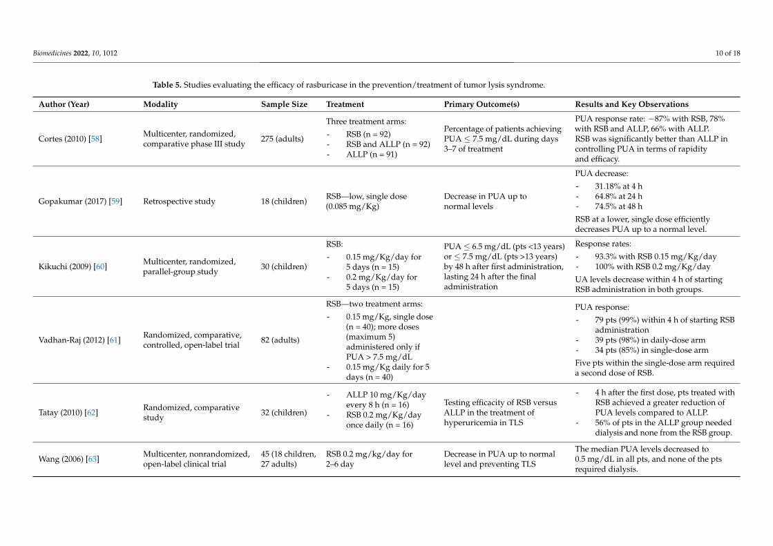
Biomedicines 2022, 10, 1012 10 of 18
Table 5. Studies evaluating the efficacy of rasburicase in the prevention/treatment of tumor lysis syndrome.
Author (Year) Modality Sample Size Treatment Primary Outcome(s) Results and Key Observations
Cortes (2010) [58] Multicenter, randomized,comparative phase III study 275 (adults)
Three treatment arms:
- RSB (n = 92)- RSB and ALLP (n = 92)- ALLP (n = 91)
Percentage of patients achievingPUA ≤ 7.5 mg/dL during days3–7 of treatment
PUA response rate: −87% with RSB, 78%with RSB and ALLP, 66% with ALLP.RSB was significantly better than ALLP incontrolling PUA in terms of rapidityand efficacy.
Gopakumar (2017) [59] Retrospective study 18 (children) RSB—low, single dose(0.085 mg/Kg)
Decrease in PUA up tonormal levels
PUA decrease:
- 31.18% at 4 h- 64.8% at 24 h- 74.5% at 48 h
RSB at a lower, single dose efficientlydecreases PUA up to a normal level.
Kikuchi (2009) [60] Multicenter, randomized,parallel-group study 30 (children)
RSB:
- 0.15 mg/Kg/day for5 days (n = 15)
- 0.2 mg/Kg/day for5 days (n = 15)
PUA ≤ 6.5 mg/dL (pts <13 years)or ≤ 7.5 mg/dL (pts >13 years)by 48 h after first administration,lasting 24 h after the finaladministration
Response rates:
- 93.3% with RSB 0.15 mg/Kg/day- 100% with RSB 0.2 mg/Kg/day
UA levels decrease within 4 h of startingRSB administration in both groups.
Vadhan-Raj (2012) [61] Randomized, comparative,controlled, open-label trial 82 (adults)
RSB—two treatment arms:
- 0.15 mg/Kg, single dose(n = 40); more doses(maximum 5)administered only ifPUA > 7.5 mg/dL
- 0.15 mg/Kg daily for 5days (n = 40)
PUA response:
- 79 pts (99%) within 4 h of starting RSBadministration
- 39 pts (98%) in daily-dose arm- 34 pts (85%) in single-dose arm
Five pts within the single-dose arm requireda second dose of RSB.
Tatay (2010) [62] Randomized, comparativestudy 32 (children)
- ALLP 10 mg/Kg/dayevery 8 h (n = 16)
- RSB 0.2 mg/Kg/dayonce daily (n = 16)
Testing efficacity of RSB versusALLP in the treatment ofhyperuricemia in TLS
- 4 h after the first dose, pts treated withRSB achieved a greater reduction ofPUA levels compared to ALLP.
- 56% of pts in the ALLP group neededdialysis and none from the RSB group.
Wang (2006) [63] Multicenter, nonrandomized,open-label clinical trial
45 (18 children,27 adults)
RSB 0.2 mg/kg/day for2–6 day
Decrease in PUA up to normallevel and preventing TLS
The median PUA levels decreased to0.5 mg/dL in all pts, and none of the ptsrequired dialysis.

Biomedicines 2022, 10, 1012 11 of 18
Table 5. Cont.
Author (Year) Modality Sample Size Treatment Primary Outcome(s) Results and Key Observations
Galardy (2013) [64] Multicenter, randomized,open-trial 76 (children)
RSB 0.2 mg/kg/day,minimum 1 dose prior tocytoreduction therapy andevery 24 h if needed for up tomaximum 5 doses
Decrease in PUA to a normallevel and preventing LTLSand CTLS
Following RSB there was 9% incidence ofLTLS and 5% incidence of CTLS.There was a significant improvement inestimated GFR following RSB.Only 1.3% pts required dialysis.
Goldman (2001) [65] Multicenter, randomized,comparative trial 52 (children)
- RSB (n = 27)- ALLP (n = 25)
RSB versus ALLP in decreasingPUA levels during the first 5days of chemotherapy and thereduction of PUA at 4 h after thefirst dose of treatment
- Pts receiving RSB experienced a2.6-fold less PUA during the first 96 hof therapy.
- There was an 86% reduction in PUAlevels after 4 h of the first dose of RSBcompared to only 12% for ALLP.
Digumarti (2014) [66] Multicenter, open-label,phase-III study 88 (adults) RSB 0.2 mg/kg/day for
4 days
- Percentage of reduction inPUA at 4 h after RSB
- PUA AUC (0–96 h)- Incidence of adverse events
- There was a 75.3 ± 28.5% of reductionin PUA at 4 h as compared to baseline.
- The PUA AUC (0–96 h) was 259.9 ±215.5 mg/dL/h.
- 29 pts had mild-to-moderate adverseevents.
Bosly (2003) [67] Multicenter, randomizedstudy
278 (166children,112 adults)
RSB 0.2 mg/kg/day for1–7 days
Decrease in PUA to ≤6.5 mg/dL(pts < 13 years) or ≤7.5 mg/dL(pts > 13 years)
- There was a significant decrease inmean PUA level among both adult andpediatric pts, regardless of whetherthey were hyperuricemic atpresentation or not.
- After RSB treatment, all pts had PUAlevels within the normal range; theresponse rate was 100%.
Vachhani (2021) [68] Prospective, randomizedstudy 24 (adults)
RSB—two treatment arms:
- 1.5 mg—day 1 (Arm A)- 3 mg day 1 (Arm B)
ALLP 300 mg on days 1–6 toall pts
The efficacity of two lower, singledoses of RSB in decreasingUA levels
- 83% pts in both arms achievedPUA < 7.5 mg/dL by 24 hafter therapy.
- 21% pts required additional dosesof RSB.
- 23/24 of pts achieved UA goals after1–2 doses of RSB.

Biomedicines 2022, 10, 1012 12 of 18
Table 5. Cont.
Author (Year) Modality Sample Size Treatment Primary Outcome(s) Results and Key Observations
Rényi (2007) [69] Multicenter, prospective,open-label, phase-IV study
- 36(children)
- 14 pts(historiccohort)
- RSB 0.2 mg/kg for5 days (n = 36)
- ALLP 300 mg/m2 dailydose (historic cohort of14 pts)
The efficacity of RSB to decreaseUA level and prevent AKI in ptsat risk for TLS
- RSB decreased the PUA level by 4 hafter first dose of treatment from343 micromol/L to 58 micromol/L.
- ALLP significantly decreased PUAlevel by 61 h after treatment.
- In the RSB group, 1 patient requireddialysis compared to 3 pts thatexperienced AKI (1 requiring dialysis)in the ALLP group.
Coiffier (2003) [70] Prospective, randomizedstudy 100 (adults) RSB 0.2 mg/kg/day for
3–7 days
Testing efficacity and safety ofRSB in prevention and treatmentof hyperuricaemia duringinduction chemotherapy ofnon-Hodgkin’s lymphoma
- RSB treatment controlled the PUAlevel within 4 h after first dose.
- No patient experienced increasedcreatinine levels or required dialysis.
Shin (2006) [71] Prospective, open-label,phase-IV study 37 (children) RSB 0.2 mg/kg/day, once
daily for 3–5 daysPUA ≤ 7 mg/dL and safetyof RSB
- 36/37 (97.3%) pts reachedPUA endpoint.
- Drug-related toxicities were mildand reversible.
Malaguarnera (2009) [72]Pilot randomized clinical trial(comparison of RSBand placebo)
38 (elderly)
- RSB 4.5 mg in 100-ccphysiological saline
or
- Placebo (physiologicalsaline)
Reducing PUA and improvingrenal function
- In the RSB group, mean PUA haddecreased 93% at day 1, 73% at day 7,and remained within a normal valueafter 1 month of therapy.
- 2 months after RSB, there was asignificant reduction of urate andcreatinine and an increase in creatinineclearance and urate clearance.
RSB—rasburicase; ALLP—allopurinol; n—number of patients; pts—patients; PUA—plasma uric acid; TLS—tumor lysis syndrome; LTLS—laboratory tumor lysis syndrome;CTLS—clinical tumor lysis syndrome; AUC—area under the curve; GFR—glomerular filtration ratio; AKI—acute kidney injury.
Biomedicines 2022, 10, 1012 13 of 18
Rasburicase converts uric acid in allantoin, carbon dioxide and hydrogen peroxide. Ac-cumulation of hydrogen peroxide in patients with G6PD deficiency leads to methemoglobinaccumulation leading to hemolytic anemia [74]. Prior to starting rasburicase administration,patients should be tested for G6PD deficiency. An initial dose of 0.05 mg/kg can be admin-istered intravenously whenever testing is not possible. Regardless of this, these patientsmust be closely monitored for signs of hemolytic anemia. According to the guidelines formanaging TLS in adult and pediatric patients with hematological malignancies elaboratedby the British Committee of Standards in Haematology, it is recommended to administerrasburicase as a prophylaxis to high-risk patients in a single dose of 3 mg to adults and0.2 mg/kg to children. Rasburicase administration can be repeated daily for 5–7 days whennecessary (lack of response or increase in the uric acid level) [48].
Rasburicase is very well tolerated with only few side effects, has less drug interactionscompared to allopurinol, and has a rapid effect (it lowers uric acid level in 4 h). Rarely, it caninduce rash, fever, headache, nausea, vomiting, or hepatic cytolysis and it is contraindicatedin pregnancy, lactating women, or patients with G6PD deficiency. Rasburicase continues tobe active ex vivo, thus leading to false decreased values of uric acid if the blood sample isnot immediately delivered to the laboratory in ice recipients in order to be processed in nomore than 4 h after blood collection.
6.1.9. Treating Hyperphosphatemia
The main therapeutic measure is the increase in phosphaturia by volume expansionwith isotonic solutions. The management should also focus on dietary phosphorus restric-tions and the administration of oral phosphate chelating agents (e.g., sevelamer). Dialysis isindicated when the value of the calcium x phosphate product exceeds 70 mg2/dL2, despitethe therapeutic and prophylactic measures.
6.1.10. Future PerspectivesPrevention through Chemotherapy Modulation
The risk of TLS can be diminished through choosing less aggressive chemotherapyregimens in order to slowly reduce the tumor burden, allowing enough time for thecompensatory kidney mechanisms to remove the products derived from tumor lysis.For example, the Berlin–Frankfurt–Muenster group treated a child with ALL by initiallyadministering prednisone for one week before initiating induction chemotherapy [75]. Forpatients with Burkitt’s lymphoma, there are regimens that include a first week of treatmentwith low doses of vincristine, prednisone, and cyclophosphamide, followed by full doses.
In the future, TLS prophylaxis may be improved by means of interfering with ma-lignant cells multiplication and metastasis. For example, it is possible to decrease theinvasive behavior by targeting the expression of desmosome junction proteins throughdrugs that increase the expression of these desmosomes [76]. Desmosomes are intercellularjunctions whose main function is strong adhesion; +9most of the mechanisms that regulatetheir assembly and stability in migratory epithelial cells are still unknown [77]. A seriesof studies have reported a correlation between reduced desmosome expression and theinvasiveness of tumors, such as transitional cell carcinoma of the bladder and non-smallcell lung cancer, conditions that may be associated with TLS [78–80], and thus it is toopremature to close the chapter focusing on the prophylaxis of TLS.
Other Options for Enzyme Replacement Therapy
Due to the limited efficiency of xanthine oxidase inhibitors and side effects of intra-venous rasburicase, during the past years, studies were aimed at developing oral engineereduricase. Although the kidney is the major site of elimination for uric acid, the gut alsoplays an important role in urate metabolism. Oral recombinant urate oxidase (ALLN-346),which targets intestinal degradation of urate, was studied in mice deficient in uricase withsevere hyperuricemia and crystalline obstructive nephropathy [81]. ALLN-346 determineda significant reduction of hyperuricemia by 44% and the normalization of uricosuria.

Biomedicines 2022, 10, 1012 14 of 18
Pegloticase is a third-line treatment that was authorized by the European MedicinesAgency in 2016 for the treatment of severe, refractory gout. It is given as an intravenousinfusion every two weeks. Pegloticase is a recombinant porcine uricase that is pegylated toincrease its elimination half-life and to decrease the immunogenicity. Like rasburicase, it iscontraindicated in patients with G6PD deficiency. NCT04745910 is a phase-IV clinical trialthat will soon start patients’ recruitment. Its aim is to assess the efficiency of pegloticasecompared to rasburicase in reducing uric acid levels in patients with hyperuricemia causedby TLS [82].
6.2. Treatment
Once established, TLS necessitates a multidisciplinary approach and a careful mon-itoring of some key elements: regularly checking the patient because his general statusmay change from one hour to another, and monitoring diuresis, laboratory tests, andpossible complications.
It is recommended to maintain a urine output of at least 100 mL/m2/h for adults and4 mL/kg/h for children. Urine alkalinization is not recommended (level 1C recommendation) [48].
It is recommended to administer rasburicase and not xanthine oxidase inhibitors,since they do not have any effect on the uric acid that has already been produced. Theonly indications for xanthine oxidase inhibitors are known rasburicase allergy and G6PDdeficiency [48]. As a therapy, the recommended dose of rasburicase is 0.2 mg/kg/day, andthe treatment duration must be established according to clinical response, but no more than3 to 7 days. In contrast to the prophylactic treatment, when administration of low doses ofrasburicase is supported by multiple studies, there are not enough data to support a fixed,low dose of rasburicase compared to weight adapted doses when dealing with an alreadyconstituted TLS. Some studies still recommend a fixed, single dose of 6 mg, which wasfound to be as effective as the weight-adapted dose [74]. Therefore, for financial reasons,this alternative treatment may be reasonable and suitable for daily administration.
Renal Replacement Therapy
The necessity for renal replacement therapy (RRT) decreased dramatically in the eraof hypouricemic drugs, especially in the countries where rasburicase is available for theprevention and treatment of TLS. RRT is indicated when kidney dysfunction is aggravatingdespite therapeutic measures, when the patient develops hypervolemia, or when electrolytedisturbances are refractory to medical treatment.
The options for RRT are:
• daily hemodialysis;• continuous veno-venous hemofiltration;• combination of intermittent hemodialysis and continuous hemofiltration/hemodiafiltration
for an efficient clearance of phosphate, which is time dependent. These techniques usedialysis membranes with large pores, which allow for rapid clearance of moleculesthat otherwise are not efficiently removed by conventional hemodialysis.
Peritoneal dialysis is not adequate, because it offers a less efficient clearance of uricacid and phosphate. Moreover, patients may associate abdominal complications related toneoplasia (peritoneal carcinomatosis, compartment syndrome), which are contraindicationsfor this procedure.
When necessary, RRT should be performed until urine output and electrolyte valuesreturn to normal.
7. Conclusions
TLS is a constellation of metabolic disorders that occur as a result of malignant cellslysis, related either to the treatment or to the increased rate of cells proliferation. TLS is anonco-nephrology emergency, with AKI being one of the most important predictors of short-and long-term mortality in these patients. According to TLS risk stratification, an early,aggressive, and multidisciplinary approach is mandatory in order to limit the occurrence

Biomedicines 2022, 10, 1012 15 of 18
of this condition and consequently of AKI. Nowadays, new advances in chemotherapypose the risk of TLS developing even in patients with malignancies that were previouslyclassified as having a low risk for this complication.
Author Contributions: Conceptualization, G.L., I.A., A.A., M.L., M.B. and I.S.; resources, G.F., O.U.,I.V., C.D. and I.S.; data curation, G.F., A.A., M.B., I.V., M.L., C.D. and I.S.; writing—original draftpreparation, G.L., A.A., M.B., G.F., O.U., and I.V.; writing—review and editing, G.L., M.L., I.A., C.D.and I.S.; visualization, O.U.; supervision, G.L.; project administration, G.L. All authors have read andagreed to the published version of the manuscript.
Funding: This research received no external funding.
Data Availability Statement: Not applicable.
Conflicts of Interest: The authors declare no conflict of interest.
References1. Lupusoru, M.; Lupusoru, G.; Ailincăi, I.; Frăt, ilă, G.; Andronesi, A.; Micu, E.; Banu, M.; Costea, R.; Ismail, G. Renal Replacement
Therapy in Cancer Patients with Acute Kidney Injury (Review). Exp. Ther. Med. 2021, 22, 864. [CrossRef]2. Rastegar, M.; Kitchlu, A.; Shirali, A.C. Tumor Lysis Syndrome. In Onco-Nephrology; Finkel, K.M., Perazella, M.A., Cohen, E.P., Eds.;
Elsevier Inc.: Amsterdam, The Netherlands, 2020; pp. 275–280.e3.3. Howard, S.C.; Jones, D.P.; Pui, C.-H. The Tumor Lysis Syndrome. N. Eng. J. Med. 2011, 364, 1844–1854. [CrossRef]4. Abu-Alfa, A.K.; Younes, A. Tumor Lysis Syndrome and Acute Kidney Injury: Evaluation, Prevention, and Management. Am. J.
Kidney Dis. 2010, 55, S1–S13. [CrossRef]5. Matuszkiewicz-Rowinska, J.; Malyszko, J. Prevention and Treatment of Tumor Lysis Syndrome in the Era of Onco-Nephrology
Progress. Kidney Blood Press. Res. 2020, 45, 645–660. [CrossRef]6. Montesinos, P.; Lorenzo, I.; Martin, G.; Sanz, J.; Perez-Sirvent, M.L.; Martinez, D.; Orti, G.; Algarra, L.; Martinez, J.;
Moscardo, F.; et al. Tumor Lysis Syndrome in Patients with Acute Myeloid Leukemia: Identification of Risk Factors andDevelopment of a Predictive Model. Haematologica 2008, 93, 67–74. [CrossRef]
7. Wilson, F.P.; Berns, J.S. Onco-Nephrology: Tumor Lysis Syndrome. Clin. J. Am. Soc. Nephrol. 2012, 7, 1730–1739. [CrossRef]8. McDonnell, C.; Barlow, R.; Campisi, P.; Grant, R.; Malkin, D. Fatal Peri-Operative Acute Tumour Lysis Syndrome Precipitated by
Dexamethasone. Anaesthesia 2008, 63, 652–655. [CrossRef]9. Furtado, M.; Simon, R. Bortezomib-Associated Tumor Lysis Syndrome in Multiple Myeloma. Leuk. Lymphoma 2008, 49, 2380–2382.
[CrossRef]10. Lipstein, M.; O’Connor, O.; Montanari, F.; Paoluzzi, L.; Bongero, D.; Bhagat, G. Bortezomib-Induced Tumor Lysis Syndrome in a
Patient with HIV-Negative Plasmablastic Lymphoma. Clin. Lymphoma Myeloma Leuk. 2010, 10, E43–E46. [CrossRef]11. Fuente, N.; Mañe, J.M.; Barcelo, R.; Muñoz, A.; Perez-Hoyos, T.; Lopez-Vivanco, G. Tumor Lysis Syndrome in a Multiple Myeloma
Treated with Thalidomide. Ann. Oncol. 2004, 15, 537. [CrossRef]12. Lee, C.-C.; Wu, Y.-H.; Chung, S.-H.; Chen, W.-J. Acute Tumor Lysis Syndrome after Thalidomide Therapy in Advanced
Hepatocellular Carcinoma. Oncologist 2006, 11, 87–88. [CrossRef]13. Francescone, S.A.; Murphy, B.; Fallon, J.T.; Hammond, K.; Pinney, S. Tumor Lysis Syndrome Occurring after the Administration
of Rituximab for Posttransplant Lymphoproliferative Disorder. Transplant. Proc. 2009, 41, 1946–1948. [CrossRef]14. Noh, G.Y.; Choe, D.H.; Kim, C.H.; Lee, J.C. Fatal Tumor Lysis Syndrome during Radiotherapy for Non–Small-Cell Lung Cancer.
J. Clin. Oncol. 2008, 26, 6005–6006. [CrossRef]15. Fleming, D.R.; Henslee-Downey, P.J.; Coffey, C. Radiation Induced Acute Tumor Lysis Syndrome in the Bone Marrow Transplant
Setting. Bone Marrow Transplant. 1991, 8, 235–236.16. Al-Kali, A.; Farooq, S.; Tfayli, A. Tumor Lysis Syndrome after Starting Treatment with Gleevec in a Patient with Chronic
Myelogenous Leukemia. J. Clin. Pharm. Ther. 2009, 34, 607–610. [CrossRef]17. Hsieh, P.-M.; Hung, K.-C.; Chen, Y.-S. Tumor Lysis Syndrome after Transarterial Chemoembolization of Hepatocellular Carcinoma:
Case Reports and Literature Review. World J. Gastroenterol. 2009, 15, 4726. [CrossRef]18. Simmons, E.D.; Somberg, K.A. Acute Tumor Lysis Syndrome after Intrathecal Methotrexate Administration. Cancer 1991, 67,
2062–2065. [CrossRef]19. Hande, K.R.; Garrow, G.C. Acute Tumor Lysis Syndrome in Patients with High-Grade Non-Hodgkin’s Lymphoma. Am. J. Med.
1993, 94, 133–139. [CrossRef]20. Cairo, M.S.; Bishop, M. Tumour Lysis Syndrome: New Therapeutic Strategies and Classification. Br. J. Haematol. 2004, 127, 3–11.
[CrossRef]21. Mehta, R.L.; Kellum, J.A.; Shah, S.V.; Molitoris, B.A.; Ronco, C.; Warnock, D.G.; Levin, A. Acute Kidney Injury Network: Report
of an Initiative to Improve Outcomes in Acute Kidney Injury. Crit. Care 2007, 11, R31. [CrossRef]

Biomedicines 2022, 10, 1012 16 of 18
22. Shimada, M.; Johnson, R.J.; May, W.S.; Lingegowda, V.; Sood, P.; Nakagawa, T.; Van, Q.C.; Dass, B.; Ejaz, A.A. A Novel Rolefor Uric Acid in Acute Kidney Injury Associated with Tumour Lysis Syndrome. Nephrol. Dial. Transplant. 2009, 24, 2960–2964.[CrossRef]
23. Conger, J.D.; Falk, S.A. Intrarenal Dynamics in the Pathogenesis and Prevention of Acute Urate Nephropathy. J. Clin. Investig.1977, 59, 786–793. [CrossRef]
24. Kang, D.-H.; Park, S.-K.; Lee, I.-K.; Johnson, R.J. Uric Acid–Induced C-Reactive Protein Expression: Implication on CellProliferation and Nitric Oxide Production of Human Vascular Cells. J. Am. Soc. Nephrol. 2005, 16, 3553–3562. [CrossRef]
25. Cirillo, P.; Gersch, M.S.; Mu, W.; Scherer, P.M.; Kim, K.M.; Gesualdo, L.; Henderson, G.N.; Johnson, R.J.; Sautin, Y.Y.Ketohexokinase-Dependent Metabolism of Fructose Induces Proinflammatory Mediators in Proximal Tubular Cells. J. Am. Soc.Nephrol. 2009, 20, 545–553. [CrossRef]
26. Han, H.J.; Lim, M.J.; Lee, Y.J.; Lee, J.H.; Yang, I.S.; Taub, M. Uric Acid Inhibits Renal Proximal Tubule Cell Proliferation via atLeast Two Signaling Pathways Involving PKC, MAPK, CPLA2, and NF-KB. Am. J. Physiol. Ren. Physiol. 2007, 292, F373–F381.[CrossRef]
27. Larson, R.A.; Pui, C.-H. Tumor Lysis Syndrome: Definition, Pathogenesis, Clinical Manifestations, Etiology and Risk Factors.Available online: https://www.uptodate.com/contents/tumor-lysis-syndrome-definition-pathogenesis-clinical-manifestations-etiology-and-risk-factors (accessed on 15 March 2020).
28. Riccio, B.; Mato, A.; Olson, E.M.; Berns, J.S.; Luger, S. Spontaneous Tumor Lysis Syndrome in Acute Myeloid Leukemia: TwoCases and a Review of the Literature. Cancer Biol. Ther. 2006, 5, 1614–1617. [CrossRef]
29. Agnani, S.; Gupta, R.; Atray, N.K.; Vachharajani, T.J. Marked Hyperuricemia with Acute Renal Failure: Need to Consider OccultMalignancy and Spontaneous Tumour Lysis Syndrome. Int. J. Clin. Pract. 2005, 60, 364–366. [CrossRef]
30. Hsu, H.; Lin, J.; Hsu, S.; Chan, Y.; Hunag, C. Neglected Cause of Renal Failure in Cancer Patients: Spontaneous Tumor LysisSyndrome Inducing Acute Uric Acid Nephropathy. Dial. Transplant. 2004, 33, 316–325.
31. Filippatos, T.D.; Milionis, H.J.; Elisaf, M.S. Alterations in Electrolyte Equilibrium in Patients with Acute Leukemia. Eur. J.Haematol. 2005, 75, 449–460. [CrossRef]
32. Dunlay, R.W.; Camp, M.A.; Allon, M.; Fanti, P.; Malluche, H.H.; Llach, F. Calcitriol in Prolonged Hypocalcemia Due to the TumorLysis Syndrome. Ann. Intern. Med. 1989, 110, 162. [CrossRef]
33. Andronesi, A.G.; Tanase, A.D.; Sorohan, B.M.; Craciun, O.G.; Stefan, L.; Varady, Z.; Lipan, L.; Obrisca, B.; Truica, A.; Ismail, G.Incidence and Risk Factors for Acute Kidney Injury Following Autologous Stem Cell Transplantation for Multiple Myeloma.Cancer Med. 2019, 8, 3278–3285. [CrossRef]
34. Annemans, L.; Moeremans, K.; Lamotte, M.; Garcia Conde, J.; van den Berg, H.; Myint, H.; Pieters, R.; Uyttebroeck, A. Incidence,Medical Resource Utilisation and Costs of Hyperuricemia and Tumour Lysis Syndrome in Patients with Acute Leukaemia andNon-Hodgkin’s Lymphoma in Four European Countries. Leuk. Lymphoma 2003, 44, 77–83. [CrossRef]
35. Bahoush, G.R.; Yazdi, E.; Ansari, S.H.; Arjmandi, K.H.; Vossough, P. Identification of Children with Acute LymphoblasticLeukemia at Low Risk for Tumor Lysis Syndrome. J. Blood Disord. Transfus. 2015, 6, 2–5. [CrossRef]
36. Ejaz, A.A.; Pourafshar, N.; Mohandas, R.; Smallwood, B.A.; Johnson, R.J.; Hsu, J.W. Uric Acid and the Prediction Models of TumorLysis Syndrome in AML. PLoS ONE 2015, 10, e0119497. [CrossRef]
37. Krishnan, G.; D’Silva, K.; Al-Janadi, A. Cetuximab-Related Tumor Lysis Syndrome in Metastatic Colon Carcinoma. J. Clin. Oncol.2008, 26, 2406–2408. [CrossRef]
38. Keane, C.; Henden, A.; Bird, R. Catastrophic Tumour Lysis Syndrome Following Single Dose of Imatinib. Eur. J. Haematol. 2009,82, 244–245. [CrossRef]
39. Michels, J.; Lassau, N.; Gross-Goupil, M.; Massard, C.; Mejean, A.; Escudier, B. Sunitinib Inducing Tumor Lysis Syndrome in aPatient Treated for Renal Carcinoma. Investig. New Drugs 2010, 28, 690–693. [CrossRef]
40. Baeksgaard, L.; Sørensen, J.B. Acute Tumor Lysis Syndrome in Solid Tumors—a Case Report and Review of the Literature. CancerChemother. Pharmacol. 2003, 51, 187–192. [CrossRef]
41. Cairo, M.S.; Coiffier, B.; Reiter, A.; Younes, A. Recommendations for the Evaluation of Risk and Prophylaxis of Tumour LysisSyndrome (TLS) in Adults and Children with Malignant Diseases: An Expert TLS Panel Consensus. Br. J. Haematol. 2010, 149,578–586. [CrossRef]
42. Coiffier, B.; Altman, A.; Pui, C.-H.; Younes, A.; Cairo, M.S. Guidelines for the Management of Pediatric and Adult Tumor LysisSyndrome: An Evidence-Based Review. J. Clin. Oncol. 2008, 26, 2767–2778. [CrossRef]
43. Wössmann, W.; Schrappe, M.; Meyer, U.; Zimmermann, M.; Reiter, A. Incidence of Tumor Lysis Syndrome in Children withAdvanced Stage Burkitt’s Lymphoma/Leukemia before and after Introduction of Prophylactic Use of Urate Oxidase. Ann.Hematol. 2003, 82, 160–165. [CrossRef]
44. Mato, A.R.; Riccio, B.E.; Qin, L.; Heitjan, D.F.; Carroll, M.; Loren, A.; Porter, D.L.; Perl, A.; Stadtmauer, E.; Tsai, D.; et al.A Predictive Model for the Detection of Tumor Lysis Syndrome during AML Induction Therapy. Leuk. Lymphoma 2006, 47,877–883. [CrossRef]
45. Truong, T.; Beyene, J.; Hitzler, J.; Abla, O.; Maloney, A.; Weitzman, S.; Sung, L. Features of Presentation Predict Children withAcute Lymphoblastic Leukemia at Low Risc for Tumor Lysis Syndrome. Cancer 2007, 110, 1832–1839. [CrossRef]
46. Belay, Y.; Yirdaw, K.; Enawgaw, B. Tumor Lysis Syndrome in Patients with Hematological Malignancies. J. Oncol. 2017, 2017,9684909. [CrossRef]

Biomedicines 2022, 10, 1012 17 of 18
47. Kaur, V.; Mehta, P.; Johnsurd, J.; Govindarajan, R. Ibrutinib-Associated Tumor Lysis Syndrome in a Patient with ChronicLymphocytic Leukemia. Blood 2014, 124, 3503–3505. [CrossRef]
48. Jones, G.L.; Will, A.; Jackson, G.H.; Webb, N.J.A.; Rule, S. Guidelines for the Management of Tumour Lysis Syndrome in Adultsand Children with Haematological Malignancies on Behalf of the British Committee for Standards in Haematology. Br. J. Haematol.2015, 169, 661–671. [CrossRef]
49. Williams, S.M.; Killeen, A.A. Tumor Lysis Syndrome. Arch. Pathol. Lab. Med. 2019, 143, 386–393. [CrossRef]50. Bellinghieri, G.; Santoro, D.; Saviva, D. Pharmacological Treatment of Acute and Chronic Hyperuricemia in Kidney Diseased
Patients. Contrib. Nephrol. 2005, 149, 149–160. [CrossRef]51. Arellano, F.; Sacristán, J.A. Allopurinol Hypersensitivity Syndrome: A Review. Ann. Pharmacother. 1993, 27, 337–343. [CrossRef]52. Vazquez-Mellado, J.; Morales, E.M.; Pacheco-Tena, C.; Burgos-Vargas, R. Relation between Adverse Events Associated with
Allopurinol and Renal Function in Renal Patients with Gout. Ann. Rheumatol. Dis. 2001, 60, 981–983. [CrossRef]53. Hung, S.-I.; Chung, W.-H.; Liou, L.-B.; Chu, C.-C.; Lin, M.; Huang, H.-P.; Lin, Y.-L.; Lan, J.-L.; Yang, L.-C.; Hong, H.-S.; et al.
HLA-B*5801 Allele as a Genetic Marker for Severe Cutaneous Adverse Reactions Caused by Allopurinol. Proc. Natl. Acad. Sci.USA 2005, 102, 4134–4139. [CrossRef] [PubMed]
54. Spina, M.; Nagy, Z.; Ribera, J.M.; Federico, M.; Aurer, I.; Jordan, K.; Borsaru, G.; Pristupa, A.S.; Bosi, A.; Grosicki, S.; et al.Florence: A Randomised, Double Blind, Phase III Pivotal Study of Febuxostat versus Allopurinol for the Prevention of TumorLysis Syndrome (TLS) in Patients with Hematologic Malignancies at Intermediate to High TLS Risc. Ann. Oncol. 2015, 26,2155–2161. [CrossRef]
55. Kishimoto, K.; Kobayashi, R.; Hori, D.; Sano, H.; Suzuki, D.; Kobayashi, K. Febuxostat as a Prophylaxis for Tumor Lysis Syndromein Children with Hematological Malignancies. Anticancer Res. 2017, 37, 5845–5849. [CrossRef]
56. Bellos, I.; Kontzoglou, K.; Psyrri, A.; Pergialiotis, V. Febuxostat administration for the prevention of tumour lysis syndrome:A meta-analysis. J. Clin. Pharm. Ther. 2019, 44, 525–533. [CrossRef]
57. Edeani, A.; Shirali, A. Tumor Lysis Syndrome. Available online: https://www.asn-online.org/education/distancelearning/curricula/onco/Chapter4.pdf (accessed on 15 March 2020).
58. Jeha, S.; Kantarjian, H.; Irwin, D.; Shen, V.; Shenoy, S.; Blaney, S.; Camitta, B.; Pui, C.-H. Efficacy and Safety of Rasburicase,a Recombinant Urate Oxidase (Elitek t), in the Management of Malignancy-Associated Hyperuricemia in Pediatric and AdultPatients: Final Results of a Multicenter Compassionate Use Trial. Leukemia 2005, 19, 34–38. [CrossRef]
59. Cortes, J.; Moore, J.O.; Maziarz, R.T.; Wetzler, M.; Craig, M.; Matous, J.; Luger, S.; Dey, B.R.; Schiller, G.J.; Pham, D.; et al. Controlof Plasma Uric Acid in Adults at Risk for Tumor Lysis Syndrome: Efficacy and Safety of Rasburicase Alone and RasburicaseFollowed by Allopurinol Compared With Allopurinol Alone—Results of a Multicenter Phase III Study. J. Clin. Oncol. 2010, 28,4207–4213. [CrossRef]
60. Gopakumar, K.G.; Thankamony, P.; Seetharam, S.; Kusumakumary, P. Treatment of Tumor Lysis Syndrome in Children withLeukemia/Lymphoma in Resource-Limited Settings—Efficacy of a Fixed Low-Dose Rasburicase. Pediatr. Hematol. Oncol. 2017, 34,206–211. [CrossRef]
61. Kikuchi, A.; Kigasawa, H.; Tsurusawa, M.; Kawa, K.; Kikuta, A.; Tsuchida, M.; Nagatoshi, Y.; Asami, K.; Horibe, K.;Makimoto, A.; et al. A Study of Rasburicase for the Management of Hyperuricemia in Pediatric Patients with Newly DiagnosedHematologic Malignancies at High Risk for Tumor Lysis Syndrome. Int. J. Hematol. 2009, 90, 492–500. [CrossRef]
62. Vadhan-Raj, S.; Fayad, L.E.; Fanale, M.A.; Pro, B.; Rodriguez, A.; Hagemeister, F.B.; Bueso-Ramos, C.E.; Zhou, X.; McLaughlin, P.W.;Fowler, N.; et al. A Randomized Trial of a Single-Dose Rasburicase versus Five-Daily Doses in Patients at Risk for Tumor LysisSyndrome. Ann. Oncol. 2012, 23, 1640–1645. [CrossRef]
63. Sánchez Tatay, V.; López Castilla, J.D.; Carmona Ponce, J.M.; Pérez Hurtado, J.M.; Quiroga Cantero, E.; Loscertales Abril, M.Rasburicasa versus Alopurinol Como Tratamiento de La Hiperuricemia En El Síndrome de Lisis Tumoral [Rasburicase versusallopurinol in the treatment of hyperuricaemia in tumour lysis syndrome]. An. Pediatr. 2010, 72, 103–110. [CrossRef]
64. Wang, L.-Y.; Shih, L.-Y.; Chang, H.; Jou, S.-T.; Lin, K.-H.; Yeh, T.-C.; Lin, S.-F.; Liang, D.-C. Recombinant Urate Oxidase(Rasburicase) for the Prevention and Treatment of Tumor Lysis Syndrome in Patients with Hematologic Malignancies. ActaHaematol. 2006, 115, 35–38. [CrossRef]
65. Galardy, P.J.; Hochberg, J.; Perkins, S.L.; Harrison, L.; Goldman, S.; Cairo, M.S. Rasburicase in the Prevention of Labora-tory/Clinical Tumour Lysis Syndrome in Children with Advanced Mature B-NHL: A Children’s Oncology Group Report.Br. J. Haematol. 2013, 163, 365–372. [CrossRef]
66. Goldman, S.C.; Holcenberg, J.S.; Finklestein, J.Z.; Hutchinson, R.; Kreissman, S.; Johnson, F.L.; Tou, C.; Harvey, E.; Morris, E.;Cairo, M.S. A Randomized Comparison between Rasburicase and Allopurinol in Children with Lymphoma or Leukemia at HighRisk for Tumor Lysis. Blood 2001, 97, 2998–3003. [CrossRef]
67. Digumarti, R.; Sinha, S.; Nirni, S.; Patil, S.; Pedapenki, R. Efficacy of Rasburicase (Recombinant Urate Oxidase) in the Preventionand Treatment of Malignancy-Associated Hyperuricemia: An Indian Experience. Indian J. Cancer 2014, 51, 180. [CrossRef]
68. Bosly, A.; Sonet, A.; Pinkerton, C.R.; McCowage, G.; Bron, D.; Sanz, M.A.; van den Berg, H. Rasburicase (Recombinant UrateOxidase) for the Management of Hyperuricemia in Patients with Cancer. Cancer 2003, 98, 1048–1054. [CrossRef]
69. Vachhani, P.; Baron, J.; Freyer, C.W.; Miller, A.; Wetzler, M.; Thompson, J.E.; Griffiths, E.A.; Wang, E.S. A Phase 2 Trial of SingleLow Doses of Rasburicase for Treatment of Hyperuricemia in Adult Patients with Acute Leukemia. Leuk. Res. 2021, 107, 106588.[CrossRef]

Biomedicines 2022, 10, 1012 18 of 18
70. Rényi, I.; Bárdi, E.; Udvardi, E.; Kovács, G.; Bartyik, K.; Kajtár, P.; Masát, P.; Nagy, K.; Galántai, I.; Kiss, C. Prevention andTreatment of Hyperuricemia with Rasburicase in Children with Leukemia and Non-Hodgkin’s Lymphoma. Pathol. Oncol. Res.2007, 13, 57–62. [CrossRef]
71. Coiffier, B.; Mounier, N.; Bologna, S.; Fermé, C.; Tilly, H.; Sonet, A.; Christian, B.; Casasnovas, O.; Jourdan, E.; Belhadj, K.; et al.Efficacy and Safety of Rasburicase (Recombinant Urate Oxidase) for the Prevention and Treatment of Hyperuricemia duringInduction Chemotherapy of Aggressive Non-Hodgkin’s Lymphoma: Results of the GRAAL1 (Groupe d’Etude Des Lymphomesde l’Adulte Trial on Rasburicase Activity in Adult Lymphoma) Study. J. Clin. Oncol. 2003, 21, 4402–4406. [CrossRef]
72. Shin, H.Y.; Kang, H.J.; Park, E.S.; Choi, H.S.; Ahn, H.S.; Kim, S.Y.; Chung, N.G.; Kim, H.K.; Kim, S.Y.; Kook, H.; et al. RecombinantUrate Oxidase (Rasburicase) for the Treatment of Hyperuricemia in Pediatric Patients with Hematologic Malignancies: Results ofa Compassionate Prospective Multicenter Study in Korea. Pediatr. Blood Cancer 2006, 46, 439–445. [CrossRef]
73. Malaguarnera, M.; Vacante, M.; Russo, C.; Dipasquale, G.; Gargante, M.P.; Motta, M. A Single Dose of Rasburicase in ElderlyPatients with Hyperuricaemia Reduces Serum Uric Acid Levels and Improves Renal Function. Expert Opin. Pharmacother. 2009,10, 737–742. [CrossRef]
74. Browning, L.A.; Kruse, J.A. Hemolysis and Methemoglobinemia Secondary to Rasburicase Administration. Ann. Pharmacother.2005, 39, 1932–1935. [CrossRef]
75. Howard, S.; Pui, C.-H.; Ribeiro, R. Tumor Lysis Syndrome. In Renal Diseases in Cancer Patients; Finkel, K.W., Howard, S.C., Eds.;Academic Press: Cambridge, MA, USA, 2014; pp. 39–64. [CrossRef]
76. Galoian, K.; Qureshi, A.; Wideroff, G.; Temple, H.T. Restoration of Desmosomal Junction Protein Expression and Inhibition ofH3K9-Specific Histone Demethylase Activity by Cytostatic Proline-Rich Polypeptide-1 Leads to Suppression of TumorigenicPotential in Human Chondrosarcoma Cells. Mol. Clin. Oncol. 2015, 3, 171–178. [CrossRef]
77. Rusu, M.C.; Pop, F.; Mănoiu, V.M.; Lupusoru, M.O.; Didilescu, A.C. Zipper-like Series of Desmosomes Supported by Subplas-malemmal Actin Belts in Thymic Epithelial Reticular Cells in the Rat. Ann. Anat.-Anat. 2013, 195, 359–364. [CrossRef]
78. Chidgey, M.A.J. Histology and Histopathology from Cell Biology to Tissue Engineering Desmosomes and Disease. Histol.Histopathol. 1997, 12, 1159–1168. [CrossRef]
79. Wang, J. Tumor Lysis Syndrome Associated with Urothelial Cancer: A Case Series. Arch. Clin. Case Stud. 2019, 1, 515. [CrossRef]80. Mirrakhimov, A.E.; Ali, A.M.; Khan, M.; Barbaryan, A. Tumor Lysis Syndrome in Solid Tumors: An up to Date Review of the
Literature. Rare Tumors 2014, 6, 68–76. [CrossRef]81. Pierzynowska, K.; Deshpande, A.; Mosiichuk, N.; Terkeltaub, R.; Szczurek, P.; Salido, E.; Pierzynowski, S.; Grujic, D. Oral
Treatment with an Engineered Uricase, ALLN-346, Reduces Hyperuricemia, and Uricosuria in Urate Oxidase-Deficient Mice.Front. Med. 2020, 24, 569215. [CrossRef]
82. ClinicalTrials.gov. Available online: https://clinicaltrials.gov/ct2/show/NCT04745910 (accessed on 1 April 2022).


![Samenvatting urban challenge[1]](https://static.fdokumen.com/doc/165x107/6313d00f3ed465f0570ad8b4/samenvatting-urban-challenge1.jpg)

