Effect of Metals on the Lytic Cycle of the Coccolithovirus, EhV86
10
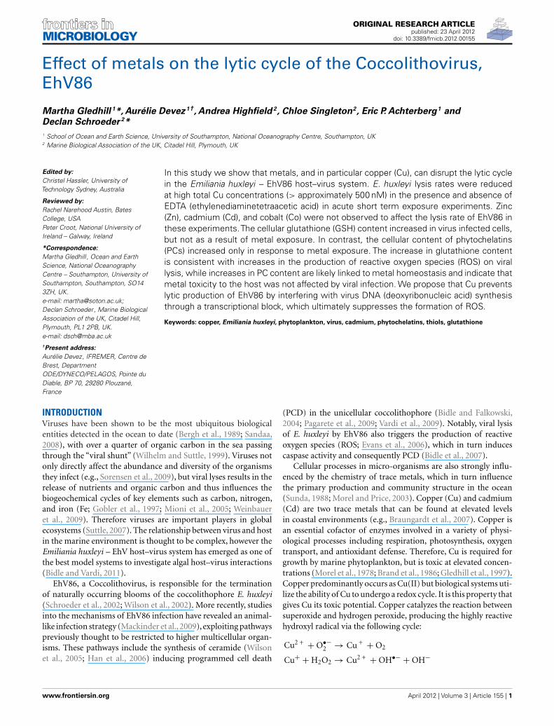
ORIGINAL RESEARCH ARTICLE published: 23 April 2012 doi: 10.3389/fmicb.2012.00155 Effect of metals on the lytic cycle of the Coccolithovirus, EhV86 Martha Gledhill 1 *, Aurélie Devez 1† , Andrea Highfield 2 , Chloe Singleton 2 , Eric P. Achterberg 1 and Declan Schroeder 2 * 1 School of Ocean and Earth Science, University of Southampton, National Oceanography Centre, Southampton, UK 2 Marine Biological Association of the UK, Citadel Hill, Plymouth, UK Edited by: Christel Hassler, University of Technology Sydney, Australia Reviewed by: Rachel Narehood Austin, Bates College, USA Peter Croot, National University of Ireland – Galway, Ireland *Correspondence: Martha Gledhill , Ocean and Earth Science, National Oceanography Centre – Southampton, University of Southampton, Southampton, SO14 3ZH, UK. e-mail: [email protected]; Declan Schroeder, Marine Biological Association of the UK, Citadel Hill, Plymouth, PL1 2PB, UK. e-mail: [email protected] † Present address: Aurélie Devez, IFREMER, Centre de Brest, Department ODE/DYNECO/PELAGOS, Pointe du Diable, BP 70, 29280 Plouzané, France In this study we show that metals, and in particular copper (Cu), can disrupt the lytic cycle in the Emiliania huxleyi – EhV86 host–virus system. E. huxleyi lysis rates were reduced at high total Cu concentrations (> approximately 500 nM) in the presence and absence of EDTA (ethylenediaminetetraacetic acid) in acute short term exposure experiments. Zinc (Zn), cadmium (Cd), and cobalt (Co) were not observed to affect the lysis rate of EhV86 in these experiments.The cellular glutathione (GSH) content increased in virus infected cells, but not as a result of metal exposure. In contrast, the cellular content of phytochelatins (PCs) increased only in response to metal exposure. The increase in glutathione content is consistent with increases in the production of reactive oxygen species (ROS) on viral lysis, while increases in PC content are likely linked to metal homeostasis and indicate that metal toxicity to the host was not affected by viral infection.We propose that Cu prevents lytic production of EhV86 by interfering with virus DNA (deoxyribonucleic acid) synthesis through a transcriptional block, which ultimately suppresses the formation of ROS. Keywords: copper, Emiliania huxleyi, phytoplankton, virus, cadmium, phytochelatins, thiols, glutathione INTRODUCTION Viruses have been shown to be the most ubiquitous biological entities detected in the ocean to date (Bergh et al., 1989; Sandaa, 2008), with over a quarter of organic carbon in the sea passing through the “viral shunt” (Wilhelm and Suttle, 1999). Viruses not only directly affect the abundance and diversity of the organisms they infect (e.g., Sorensen et al., 2009), but viral lyses results in the release of nutrients and organic carbon and thus influences the biogeochemical cycles of key elements such as carbon, nitrogen, and iron (Fe; Gobler et al., 1997; Mioni et al., 2005; Weinbauer et al., 2009). Therefore viruses are important players in global ecosystems (Suttle, 2007). The relationship between virus and host in the marine environment is thought to be complex, however the Emiliania huxleyi – EhV host–virus system has emerged as one of the best model systems to investigate algal host–virus interactions (Bidle and Vardi, 2011). EhV86, a Coccolithovirus, is responsible for the termination of naturally occurring blooms of the coccolithophore E. huxleyi (Schroeder et al., 2002; Wilson et al., 2002). More recently, studies into the mechanisms of EhV86 infection have revealed an animal- like infection strategy (Mackinder et al., 2009), exploiting pathways previously thought to be restricted to higher multicellular organ- isms. These pathways include the synthesis of ceramide (Wilson et al., 2005; Han et al., 2006) inducing programmed cell death (PCD) in the unicellular coccolithophore (Bidle and Falkowski, 2004; Pagarete et al., 2009; Vardi et al., 2009). Notably, viral lysis of E. huxleyi by EhV86 also triggers the production of reactive oxygen species (ROS; Evans et al., 2006), which in turn induces caspase activity and consequently PCD (Bidle et al., 2007). Cellular processes in micro-organisms are also strongly influ- enced by the chemistry of trace metals, which in turn influence the primary production and community structure in the ocean (Sunda, 1988; Morel and Price, 2003). Copper (Cu) and cadmium (Cd) are two trace metals that can be found at elevated levels in coastal environments (e.g., Braungardt et al., 2007). Copper is an essential cofactor of enzymes involved in a variety of physi- ological processes including respiration, photosynthesis, oxygen transport, and antioxidant defense. Therefore, Cu is required for growth by marine phytoplankton, but is toxic at elevated concen- trations (Morel et al., 1978; Brand et al., 1986; Gledhill et al., 1997). Copper predominantly occurs as Cu(II) but biological systems uti- lize the ability of Cu to undergo a redox cycle. It is this property that gives Cu its toxic potential. Copper catalyzes the reaction between superoxide and hydrogen peroxide, producing the highly reactive hydroxyl radical via the following cycle: Cu 2+ + O •− 2 → Cu + + O 2 Cu + + H 2 O 2 → Cu 2+ + OH •− + OH − www.frontiersin.org April 2012 |Volume 3 | Article 155 | 1
Transcript of Effect of Metals on the Lytic Cycle of the Coccolithovirus, EhV86
ORIGINAL RESEARCH ARTICLEpublished: 23 April 2012
doi: 10.3389/fmicb.2012.00155
Effect of metals on the lytic cycle of the Coccolithovirus,EhV86
Martha Gledhill 1*, Aurélie Devez 1†, Andrea Highfield 2, Chloe Singleton2, Eric P. Achterberg1 and
Declan Schroeder 2*
1 School of Ocean and Earth Science, University of Southampton, National Oceanography Centre, Southampton, UK2 Marine Biological Association of the UK, Citadel Hill, Plymouth, UK
Edited by:
Christel Hassler, University ofTechnology Sydney, Australia
Reviewed by:
Rachel Narehood Austin, BatesCollege, USAPeter Croot, National University ofIreland – Galway, Ireland
*Correspondence:
Martha Gledhill , Ocean and EarthScience, National OceanographyCentre – Southampton, University ofSouthampton, Southampton, SO143ZH, UK.e-mail: [email protected];Declan Schroeder , Marine BiologicalAssociation of the UK, Citadel Hill,Plymouth, PL1 2PB, UK.e-mail: [email protected]†Present address:
Aurélie Devez, IFREMER, Centre deBrest, DepartmentODE/DYNECO/PELAGOS, Pointe duDiable, BP 70, 29280 Plouzané,France
In this study we show that metals, and in particular copper (Cu), can disrupt the lytic cyclein the Emiliania huxleyi – EhV86 host–virus system. E. huxleyi lysis rates were reducedat high total Cu concentrations (> approximately 500 nM) in the presence and absence ofEDTA (ethylenediaminetetraacetic acid) in acute short term exposure experiments. Zinc(Zn), cadmium (Cd), and cobalt (Co) were not observed to affect the lysis rate of EhV86 inthese experiments.The cellular glutathione (GSH) content increased in virus infected cells,but not as a result of metal exposure. In contrast, the cellular content of phytochelatins(PCs) increased only in response to metal exposure. The increase in glutathione contentis consistent with increases in the production of reactive oxygen species (ROS) on virallysis, while increases in PC content are likely linked to metal homeostasis and indicate thatmetal toxicity to the host was not affected by viral infection. We propose that Cu preventslytic production of EhV86 by interfering with virus DNA (deoxyribonucleic acid) synthesisthrough a transcriptional block, which ultimately suppresses the formation of ROS.
Keywords: copper, Emiliania huxleyi, phytoplankton, virus, cadmium, phytochelatins, thiols, glutathione
INTRODUCTIONViruses have been shown to be the most ubiquitous biologicalentities detected in the ocean to date (Bergh et al., 1989; Sandaa,2008), with over a quarter of organic carbon in the sea passingthrough the “viral shunt” (Wilhelm and Suttle, 1999). Viruses notonly directly affect the abundance and diversity of the organismsthey infect (e.g., Sorensen et al., 2009), but viral lyses results in therelease of nutrients and organic carbon and thus influences thebiogeochemical cycles of key elements such as carbon, nitrogen,and iron (Fe; Gobler et al., 1997; Mioni et al., 2005; Weinbaueret al., 2009). Therefore viruses are important players in globalecosystems (Suttle, 2007). The relationship between virus and hostin the marine environment is thought to be complex, however theEmiliania huxleyi – EhV host–virus system has emerged as one ofthe best model systems to investigate algal host–virus interactions(Bidle and Vardi, 2011).
EhV86, a Coccolithovirus, is responsible for the terminationof naturally occurring blooms of the coccolithophore E. huxleyi(Schroeder et al., 2002; Wilson et al., 2002). More recently, studiesinto the mechanisms of EhV86 infection have revealed an animal-like infection strategy (Mackinder et al., 2009), exploiting pathwayspreviously thought to be restricted to higher multicellular organ-isms. These pathways include the synthesis of ceramide (Wilsonet al., 2005; Han et al., 2006) inducing programmed cell death
(PCD) in the unicellular coccolithophore (Bidle and Falkowski,2004; Pagarete et al., 2009; Vardi et al., 2009). Notably, viral lysisof E. huxleyi by EhV86 also triggers the production of reactiveoxygen species (ROS; Evans et al., 2006), which in turn inducescaspase activity and consequently PCD (Bidle et al., 2007).
Cellular processes in micro-organisms are also strongly influ-enced by the chemistry of trace metals, which in turn influencethe primary production and community structure in the ocean(Sunda, 1988; Morel and Price, 2003). Copper (Cu) and cadmium(Cd) are two trace metals that can be found at elevated levelsin coastal environments (e.g., Braungardt et al., 2007). Copper isan essential cofactor of enzymes involved in a variety of physi-ological processes including respiration, photosynthesis, oxygentransport, and antioxidant defense. Therefore, Cu is required forgrowth by marine phytoplankton, but is toxic at elevated concen-trations (Morel et al., 1978; Brand et al., 1986; Gledhill et al., 1997).Copper predominantly occurs as Cu(II) but biological systems uti-lize the ability of Cu to undergo a redox cycle. It is this property thatgives Cu its toxic potential. Copper catalyzes the reaction betweensuperoxide and hydrogen peroxide, producing the highly reactivehydroxyl radical via the following cycle:
Cu2 + + O•−2 → Cu + + O2
Cu+ + H2O2 → Cu2 + + OH•− + OH−
www.frontiersin.org April 2012 | Volume 3 | Article 155 | 1
Gledhill et al. Metals, EhV86, and Emiliania huxleyi
The reaction with hydrogen peroxide will compete with thefaster reaction between Cu+ and O•−
2 (Voelker et al., 2000; Hellerand Croot, 2010, 2011):
Cu+ + O•−2 → Cu2+ + H2O2
so that the production of the hydroxide radical will be influencedby the ambient redox environment (which is reducing within cells;Schafer and Buettner, 2001) and the relative concentrations of Cuand superoxide.
Hydroxyl radicals can cause oxidative damage to cellular com-ponents such as deoxyribonucleic acid (DNA), proteins, and lipids.For example, Cu causes damage to DNA by binding near guaninebases, where it is reduced to Cu(I) and then reoxidized to Cu(II)by reaction with hydrogen peroxide producing hydroxyl radicals.The radicals then mediate DNA strand breakage in close proximityto the bound Cu (Sagripanti and Kraemer, 1989; Aruoma et al.,1991). The redox properties of Cu also allow the metal to bindto several types of amino acid residues and therefore Cu couldbe inappropriately incorporated into proteins and enzymes thatnormally bind other metal ions. This results in a loss of func-tion through inactivation or changes in conformational fold. Inhumans for example, Cu may contribute to the development ofAlzheimer’s disease, along with Fe and zinc (Zn), as it has beenfound to induce aggregation of the β-amyloid (Aβ) protein andhas been found in high quantities (0.44 mM) in Alzheimer plaques(Lovell et al., 1998; Curtain et al., 2001). Moreover, these high Culevels are linked to an increase in oxidative stress which plays acentral role in neurodegenerative disorders (Permyakov, 2009).
Copper has long been known to possess antimicrobial andantiviral properties, and in the last decade studies have suggestedthat Cu surfaces could be reintroduced into hospitals to reducethe transmission of microbes such as methicillin resistant Staphy-lococcus aureus (Noyce et al., 2006a), Escherichia coli O157 (Noyceet al., 2006b), and influenza A virus (Noyce et al., 2007). The mech-anisms of Cu disruption of virus infection may vary dependingon the virus and have yet to be fully understood (Karlstrom andLevine, 1991a,b; Sagripanti et al., 1997; Horie et al., 2008), how-ever there is some evidence to suggest that inactivation of virusescan proceed via Cu mediated DNA damage as described above(Levinson et al., 1973; Sagripanti et al., 1997).
Cadmium (Cd) had been considered a non-essential metal, butmore recently a unique biological role for Cd has been identifiedin marine diatoms. It has been shown that Cd can replace Zn asa metal of the Zn carbonic anhydrase (Price and Morel, 1990),and that Cd carbonic anhydrases can play a role in the acquisitionof inorganic carbon for photosynthesis in the oceans (Lane et al.,2005). High levels of anthropogenic Cd in the coastal environmenthas also led to toxicological effects in exposed marine organisms.For example, Cd is reported to reduce reproduction rates in phy-toplankton (Brand et al., 1986). The mechanism of Cd toxicity isknown in animal systems where Cd complexes glutathione (GSH)and protein-bound sulfhydryl groups, resulting in enhanced pro-duction of ROS such as superoxide ion, hydroxyl radicals, andhydrogen peroxide (Stohs et al., 2001). Cadmium has been shownto inhibit the enzymatic activity of the Cu/Zn-superoxide dismu-tase (Cu/Zn-SOD) from rat liver (Hussain et al., 1987) and human
Cu/Zn-SOD (Huang et al., 2006). This occurs through replace-ment of the normally bound Zn(II) ion with a Cd(II) ion at theactive site.
Elevated Cu and Cd concentrations in many marine eukary-otic phytoplankton are tolerated through the induction of phy-tochelatins, thiols of the general formula (γ-Glu-Cys)n-Gly, wheren commonly ranges between 2 and 4 (Ahner and Morel, 1995;Ahner et al., 1995; Kawakami et al., 2006b,c; Devez et al., 2009).Phytochelatins and other thiols may also be released into the sur-rounding media, reducing free metal concentrations, and poten-tially affecting metal bioavailability (Lee et al., 1996; Leal et al.,1999; Vasconcelos and Leal, 2001; Vasconcelos et al., 2002). Phy-tochelatins are synthesized from GSH, which is also known torespond to oxidative stress. However, while the intracellular abun-dance of PCs is thought to be linked to metal concentrations in thesurrounding water (e.g.,Ahner and Morel, 1995; Ahner et al., 1995;Morelli and Scarano, 2001; Dupont and Ahner, 2005; Le Faucheuret al., 2005; Kawakami et al., 2006b; Pawlik-Skowronska et al.,2007; Morelli and Fantozzi, 2008; Devez et al., 2009), cellular GSHabundance, and metal concentrations are not necessarily directlyrelated (Kawakami et al., 2006c; Scheidegger et al., 2011). Glu-tathione has many metabolic roles (Mendoza-Cozatl et al., 2005),however, exogenous GSH is known to affect replication of Herpessimplex virus type 1 (HSV-1) by interfering with the very late stagesof the virus life cycle, without otherwise affecting host cellularmetabolism (Palamara et al., 1995).
Metals thus have the potential to impact host–virus interac-tions. However, to our knowledge, the effects of metals have notyet been assessed for any marine host–virus system. The aim ofthis study was therefore to undertake a preliminary investigationinto interactions between trace metals and the E. huxleyi – EhV86system. We subsequently examined (1) the impact of elevated Cuand Cd concentrations on the EhV86 lytic cycle and (2) the cellularmechanism involved in these interactions.
MATERIALS AND METHODSSterile trace metal clean techniques were used for culturing. Glass-ware and polycarbonate bottles (Nalgene) were acid washed (1 MHCl) for at least 24 h prior to use, 4 L polycarbonate culture vessels(Nalgene) were double bagged (Nalgene autoclavable plastic bags)prior to autoclaving at 120˚C for 30 min.
CULTURE CONDITIONSEmiliania huxleyi (strain CCMP 1516) was obtained from theProvasoli-Guillard Center for Culture of Marine Phytoplank-ton (CCMP). Experiments reported here focused on acute shortterm (4 days) effects. E. huxleyi was batch cultured in f/2 minusSi medium prepared using 0.2 μm filtered seawater collectedfrom the North Atlantic Gyre in the Canary Basin (between24.1 and 29.5˚N and 23.4 and 27.6˚W). The culture medium(pH = 7.8 ± 0.1) was allowed to equilibrate overnight and then fil-tered sterilized (0.2 μm, Sartorius) prior to seeding with E. huxleyi.Although it was possible that viruses already present in the seawa-ter would have passed through the 0.2-μm filter, in practice we didnot observed any evidence of lysis of E. huxleyi in our control cul-tures, indicating that this was not a problem in these experiments.Concentrations of the nutrients nitrate (NaNO3) and phosphate
Frontiers in Microbiology | Microbiological Chemistry April 2012 | Volume 3 | Article 155 | 2

Gledhill et al. Metals, EhV86, and Emiliania huxleyi
(NaH2PO4) added to the seawater were 3 × 10⁻⁴ and 1 × 10⁻⁵ M,respectively. Concentrations of trace metals added to the sea-water were 10 nM Cu, 100 nM molybdenum, 4 nM Zn, 2.5 nMcobalt (Co), 23 nM manganese, 450 nM Fe, and 10 nM selenium.Media used for initial experiments with 2.5 μM added Cd, Co,Cu, and Zn were carried out in the presence of 5 μM ethylene-diaminetetraacetic acid (EDTA). Experiments with different Cuconcentrations were carried out in the presence and absence of5 μM EDTA, and experiments investigating thiol production andRNA expression were carried out in the absence of EDTA. Cultureswere maintained at 15 ± 1˚C under a light/dark cycle of 12:12 hand at an illumination of 150 μmol photons m−2 s−1 in a growthcabinet (MLR-350, Sanyo).
VIRUS CULTURE MAINTENANCEThe Coccolithovirus EhV86 was propagated by using acclimatedand synchronized batch cultures of E. huxleyi 1516 grown in f/2medium without EDTA and Si (Schroeder et al., 2002). The occur-rence of lysis was generally indicated by a change in the cultureappearance, from a green to a chalky white color. The new virusstock solution was obtained from an E. huxleyi culture grown to acell density of approximately 1 × 10⁶ cells mL−1 at a multiplicityof infection of approximately 10. The new virus stock solution waslabeled and stored in the dark at 4˚C until required.
METAL AND VIRUS ADDITIONThe virus and the single studied metal (Cd, Co, Cu, Zn) were addedsimultaneously. The addition of EhV86 virus, in excess for infec-tion, was done to exponentially growing E. huxleyi host culturesapproximately 4 days after subculturing. In initial experimentsinvestigating effects of Cd, Co, Cu, and Zn, metals were addedat a concentration of 2.5 μM in excess of concentrations alreadypresent in the media. A second experiment investigated a rangeof Cu concentrations between 125 nM and 1 μM. For the finalexperiment investigating the mechanism of the Cu virus interac-tion, Cu was added at a total concentration of 1.25 μM and Cd at5.0 μM. Non-infected cultures with and without metal were usedas a control in parallel for each virus/metal treatment.
Growth of the cultures was monitored daily by enumeratingcells (Multisizer™ II coulter counter). Cell numbers were used toguide subsequent sampling frequency for PCs and viral ribonu-cleic acid (RNA). Cultures were sampled daily for virus counts andon alternate days for thiol content. RNA expression was sampledon days 5, 7, and 10 post infection for Cu and daily up to day8 post infection for Cd. All analyses were carried out in dupli-cate. For virus counts, 1 mL was sampled and fixed using 50 μLof polyoxymethylene (paraformaldehyde, Sigma Aldrich, 1% finalconcentration) and subsequently stored at −80˚C for later analysisby flow cytometry. For thiol analysis, 500 mL of culture solutionwas filtered (0.45 μm pore size nitrocellulose membrane filters,Whatman) under gentle vacuum pressure and stored at −80˚C. Forisolation of RNA, E. huxleyi cells were harvested via centrifugationand RNA was extracted from the pellets using the RNeasy Mini Kit(Qiagen) according to the manufacturer’s instructions. Total RNAwas DNase treated (Promega) to remove any DNA contaminationand then quantified using the NanoDrop 1000 spectrophotometer(Thermo Scientific).
Cell counts from the coulter counter were used to calculate theaverage growth (μ) rates for E. huxleyi over the period of viralinfection from the slope of a graph of ln(cells) against time. Theviral lysis rate is then calculated from
μ+virus = μ − γlysis
where γlysis is the E. huxleyi lysis rate,μ is the growth rate of E. hux-leyi in control cultures or treatments containing the added metal,and μ+virus is the growth rate of E. huxleyi in infected cultures orinfected cultures containing the added metal.
FLOW CYTOMETRYDetermination of the abundance of viral particles and E. huxleyicells was performed simultaneously (FACSort, Becton DickinsonBiosciences). EhV86 were discriminated based on their green flu-orescence and side scatter. E. huxleyi cells were counted based ontheir red and orange fluorescence signatures upon staining withSYBR Green I DNA dye (Schroeder et al., 2002; Wilson et al., 2002).
Comparison of fresh (coulter counter) and fixed (Flow Cytom-etry) E. huxleyi cell counts showed that a good agreement wasobserved between these two counting approaches (t test, p > 0.05,n = 48).
DETERMINATION OF PARTICULATE THIOLSThe total concentrations of glutathione (GSH) and phytochelatins(PCs) in metal and virus exposed E. huxleyi cultures were deter-mined according to the method reported by Kawakami et al.(2006a). Intracellular thiol measurements were performed induplicate by reverse-phase high performance liquid chromatog-raphy (HPLC) with fluorescence detection.
Thiols were extracted on ice (5 min), following addition of1.2 mL solution of 0.1 M HCl containing 5 mM diethylenetriaminepentaacetic acid (DTPA, Fluka Biochemica) to a 2-mL microcen-trifuge tube (Fisher) containing the filter with E. huxleyi biomass.The extract was centrifuged (13000 g/20 min at 4˚C) and syringefiltered (0.2 μm pore size cellulose membrane, Minisart RC4,Sartorius) prior to reduction (25 μL of a 20-mM 2-carboxyethyl-phosphine hydrochloride, 5 min, TCEP, Sigma). Further oxidationwas minimized (5 mM DTPA) and the extract was buffered at pH8.2 (200 mM N-2-hydroxyethylpiperazine-N′-2-ethanesulphonicacid, HEPES). After a further 5 min, 10 μL of a 100-mM of a sulfur-specific fluorescent tag monobromobimane was added (MBrB inacetonitrile, Fisher) followed by 465 μL of the HEPES/DTPA pH8.2 solution. The derivatization procedure was carried out in a darkroom under dim red light conditions. After 15 min, the reactionwas stabilized and the derivatization of thiols by MBrB, stoppedby addition of 100 μL of 1 M methanesulfonic acid (99%, Sigma).Vials were stored in the dark at 4˚C until HPLC analysis.
Thiols were analyzed by reverse-phase HPLC with fluorescencedetection (Kawakami et al., 2006a). The HPLC comprised a sys-tem controller (Shimadzu SCL-10A) and two pumps (ShimadzuLC-10ADvp), an autosampler (Shimadzu SIL-10ADVP) and a flu-orescence detector (Shimadzu RF-10A XL) operating at 380 nm(excitation) and 470 nm (emission) wavelengths. Separation ofthe thiols was carried out using a 150 mm × 2.1 mm C-18 HPLCcolumn (Ascentis, Supelco) with a 3-μm particle size and a gradi-ent program of 0–5 min, 10% B; 5–18 min, 10–22% B; 18–40 min,
www.frontiersin.org April 2012 | Volume 3 | Article 155 | 3
Gledhill et al. Metals, EhV86, and Emiliania huxleyi
22–35% B; 40–50 min, 35–100%; 50–55 min, isocratic 100% B;55–58 min, 100–10% B; 58–60 min 10% B, where A was 0.1% tri-fluoroacetic acid (TFA, Fluka) and B was acetonitrile. The flowrate was 0.2 mL min−1.
Phytochelatin concentrations were standardized with GSH(reduced form, purity 99%, Sigma) assuming that the fluores-cence response was directly proportional to the number of thiolgroups (Kawakami et al., 2006a). We used PCs directly produced byPhaeodactylum tricornutum under metal stress for identificationof retention times for PC2, PC3, and PC4. GSH eluted at 11.4 minand PC2, 3, and 4 at 18.8, 21.7, and 24.2 min, respectively. CellularGSH and PC concentrations were normalized to the number ofcells and are thus expressed in amol SH cell−1. The limit of detec-tion, calculated from three times SD of a 5-pmol GSH standard,was 0.1 pmol with a 100-μL injection volume. Analytical variabil-ity within standards and samples was less than 10%. The recoveryof GSH added to samples prior to derivatization was determinedto be 86 ± 29% (n = 11).
ONE-STEP REVERSE TRANSCRIPTION-PCRRT-PCR detection of virus-related gene expression was under-taken using primers designed to amplify four viral genes, DNApolymerase (DNA pol), Helicase, proliferating cell nuclear anti-gen (PCNA) protein, and major capsid protein (MCP; Table 1).One-step RT-PCR was used to amplify 10 ng RNA in 25 μL reac-tions containing 1 × One-step sensimix QPCR mix (with SYBRgreen), 7.5 pmol forward primer, 7.5 pmol reverse primer, and5 units RNase inhibitor. Reactions were carried out in a Rotor-gene 6000 QPCR machine (Corbett Research) using the followingconditions: reverse transcription at 49˚C for 10 min, polymeraseactivation at 95˚C for 10 min, followed by 40 cycles of 95˚C for 15 s,54˚C for 15 s (60˚C for MCP), and 72˚C for 15 s. Fluorescence wasacquired at the end of each extension step on the green channel.RT-PCR reactions were subjected to melt curve analysis to ensurea single product had been generated by gradual melting from 72to 95˚C and fluorescence acquisition at each 1˚C increment. RT-PCR products were verified by gel electrophoresis on a 2% (w/v)agarose gel in 1 × TAE buffer and viewed on a UV transilluminator(Syngene).
STATISTICAL ANALYSISAs analysis was only performed in duplicate, estimates of errorsin growth, and lysis rates were calculated from the square routeof the sum of squares of the SEs of the slopes of ln(cells) against
time. The lack of experimental replication meant that the statisti-cal significance of our results could not be tested and comparisonsbetween treatments are thus qualitative.
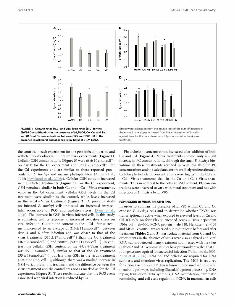
RESULTSE. HUXLEYI – EhV86 INFECTION DYNAMICS IN THE PRESENCE OFVARIOUS METAL IONSPreliminary experiments, carried out with a range of metals (Cu,Zn, Co, and Cd) on the E. huxleyi – EhV86 host–virus systemindicated that viral lysis of E. huxleyi cells was disrupted in thepresence of Cu, but was similar to controls for the metals Cd,Co, and Zn (Figure 1A). In this study we have interpreted ourdata qualitatively as lack of sufficient replicates in our experimen-tal design precludes more quantitative estimates of the statisticalsignificance of our results. However, Cu was consistently observedto disrupt viral lysis of E. huxleyi in all the experiments under-taken as part of this study. Furthermore varying the concentrationof Cu in the absence and presence of EDTA indicated that virallysis rates decreased with increasing Cu concentration and werelowest in the absence of EDTA (Figure 1D). Copper is known tobe toxic to marine algae at high concentrations (e.g., Sunda andGuillard, 1976; Brand et al., 1986; Gledhill et al., 1997; Levy et al.,2007, 2008; Debelius et al., 2009), and indeed the growth rate of E.huxleyi was reduced at the highest Cu concentration when com-pared to control cultures (Figures 1A,C). However the cumulativeeffect of metal plus virus on host growth was only observed inCu treatments and thus appeared to be a specific effect of Cu.Further short term exposure experiments aimed at understandingthe interaction between Cu and the E. huxleyi virus–host systemfocused on Cd and Cu as these two metals are both known tobe toxic and they exhibited contrasting behaviors in our prelim-inary experiments. Furthermore EDTA was omitted in order tomaximize the effect of trace metals on both host and virus.
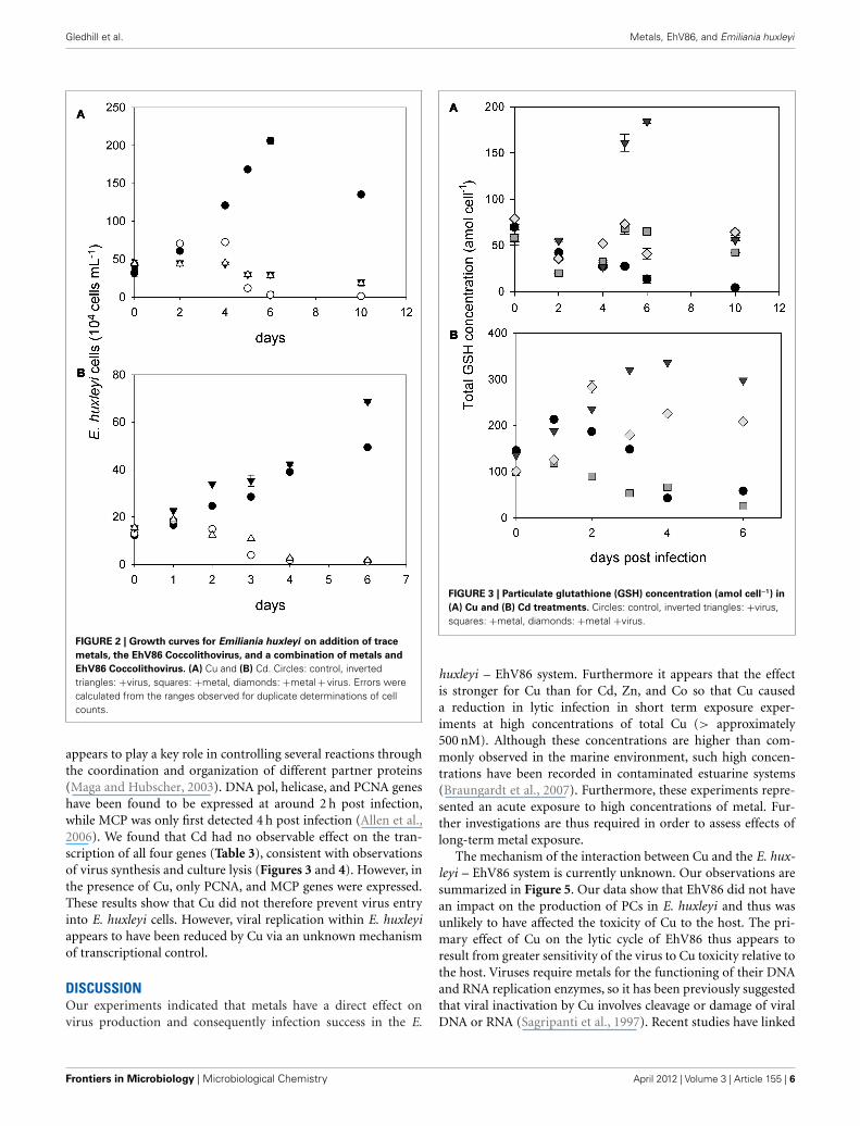
PRODUCTION OF GLUTATHIONE AND PHYTOCHELATINS BY E. HUXLEYIThe intracellular content of GSH and PCs in infected and non-infected E. huxleyi cells were determined in order to investigateoxidative stress and trace metal homeostasis during the course ofthe experiments. Post virus and metal addition growth curves forE. huxleyi in the thiol expression experiments are presented inFigure 2. Initial cell numbers were different for each experimentwhen virus and metals were added (4.0 ± 0.6 × 10⁵ cells mL−1
for the Cu experiment and 1.4 ± 0.2 × 10⁵ cells mL−1 for the Cdexperiment), however, calculated growth rates were similar for
Table 1 | Primers used in this study.
Primer name Target (CDS) Sequence (5′–3′) Amplicon size (bp) Reference
EhVpol_F DNA polymerase (ehv030) TATAATGCACGCCAACTTGC 98 This study
EhVpol_R GCAATTGCACCAAGTGGATA
EhVpcna_F PCNA (ehv440) GGGCATTTCATTTGCCATAC 157 This study
EhVpcna_R ATTCTCCGTCGACAATACGC
EhVhel_F Helicase (ehv104) GCCAACTGGTACAGGGAAAA 184 This study
EhVhel_R CATCCATGCATGTGTGACAA
MCP_F2 MCP (ehv085) GACCTTTAGGCCAGGGAG 134 Schroeder et al. (2002)
MCP_R2 GTTCGCGCTCGAGTCGAT
Frontiers in Microbiology | Microbiological Chemistry April 2012 | Volume 3 | Article 155 | 4
Gledhill et al. Metals, EhV86, and Emiliania huxleyi
FIGURE 1 | Growth rates (A,C) and viral lysis rates (B,D) for the
EhV86 Coccolithovirus in the presence of (A,B) Cd, Co, Cu, and Zn
and (C,D) at Cu concentrations between 125 and 1000 nM in the
presence (black bars) and absence (gray bars) of 5 μM EDTA.
Errors were calculated from the square root of the sum of squares ofthe errors in the slopes obtained from linear regression of ln(cells)against time for the period over which lysis occurred in the +virustreatment.
the controls in each experiment for the post infection period andreflected results observed in preliminary experiments (Figure 1).Cellular GSH concentrations (Figure 3) were 66 ± 10 amol cell−1
on day 0 for the Cu experiment and 120 ± 20 amol cell−1 forthe Cd experiment and are similar to those reported previ-ously for E. huxleyi and marine phytoplankton (Ahner et al.,1995; Kawakami et al., 2006c). Cellular GSH content increasedin the infected treatments (Figure 3). For the Cu experiment,GSH remained similar in both Cu and +Cu +Virus treatments,while in the Cd experiment, cellular GSH levels in the Cdtreatment were similar to the control, while levels increasedin the +Cd +Virus treatment (Figure 3). A previous studyon infected E. huxleyi cells indicated an increased intracel-lular occurrence of ROS and oxidative stress (Evans et al.,2006). The increase in GSH in virus infected cells in this studyis consistent with a response to increased oxidative stress onviral infection. Glutathione content in the +Cd +Virus treat-ment increased to an average of 216 ± 11 amol cell−1 betweendays 4 and 6 after infection and was closer to that of thevirus treatment (316 ± 27 amol cell−1) than the Cd treatment(46 ± 29 amol cell−1) and control (50 ± 11 amol cell−1). In con-trast the cellular GSH content of the +Cu +Virus treatmentwas 55 ± 16 amol cell−1, similar to that of the Cu treatment(55 ± 19 amol cell−1), but less than GSH in the virus treatment(124 ± 85 amol cell−1), although there was a marked increase inGSH variability in this treatment and the difference between thevirus treatment and the control was not as marked as for the Cdexperiment (Figure 3). These results indicate that the ROS eventassociated with viral infection is reduced by Cu.
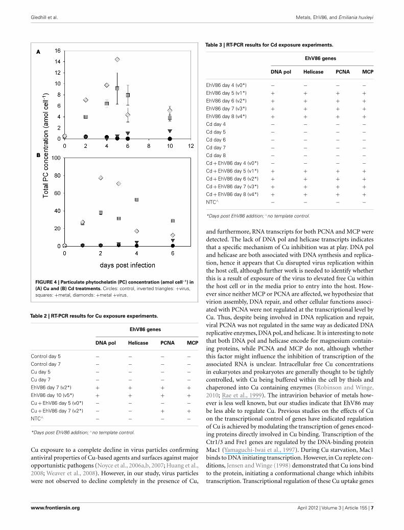
Phytochelatin concentrations increased after addition of bothCu and Cd (Figure 4). Virus treatments showed only a slightincrease in PC concentrations, although the small E. huxleyi bio-volume in these treatments resulted in very low absolute PCconcentrations and the calculated errors are likely underestimated.Cellular phytochelatin concentrations were higher in the Cd and+Cd +Virus treatments than in the Cu or +Cu +Virus treat-ments. Thus in contrast to the cellular GSH content, PC concen-trations were observed to vary with metal treatment and not withinfection of E. huxleyi by EhV86.
EXPRESSION OF VIRUS-RELATED RNAIn order to confirm the presence of EhV86 within Cu and Cdexposed E. huxleyi cells and to determine whether EhV86 wastranscriptionally active when exposed to elevated levels of Cu andCd, RT-PCR on four EhV86 encoded genes – DNA dependentDNA pol – ehv030, PCNA protein – ehv440, Helicase – ehv104and MCP – ehv085 – was carried out in duplicate before and aftertreatment (Tables 2 and 3). Particulate material from Cu and Cdexperiments in the absence of virus were also analyzed and viralRNA was not detected in any treatment not infected with the virus(Tables 2 and 3). Genomic studies have previously revealed that allfour genes are required for successful infection (Wilson et al., 2005;Allen et al., 2006). DNA pol and helicase are required for DNAsynthesis and therefore virus replication. The MCP is requiredfor virion assembly and PCNA is known to be involved in severalmetabolic pathways, including Okazaki fragment processing, DNArepair, translesion DNA synthesis, DNA methylation, chromatinremodeling, and cell cycle regulation. PCNA in mammalian cells
www.frontiersin.org April 2012 | Volume 3 | Article 155 | 5
Gledhill et al. Metals, EhV86, and Emiliania huxleyi
FIGURE 2 | Growth curves for Emiliania huxleyi on addition of trace
metals, the EhV86 Coccolithovirus, and a combination of metals and
EhV86 Coccolithovirus. (A) Cu and (B) Cd. Circles: control, invertedtriangles: +virus, squares: +metal, diamonds: +metal + virus. Errors werecalculated from the ranges observed for duplicate determinations of cellcounts.
appears to play a key role in controlling several reactions throughthe coordination and organization of different partner proteins(Maga and Hubscher, 2003). DNA pol, helicase, and PCNA geneshave been found to be expressed at around 2 h post infection,while MCP was only first detected 4 h post infection (Allen et al.,2006). We found that Cd had no observable effect on the tran-scription of all four genes (Table 3), consistent with observationsof virus synthesis and culture lysis (Figures 3 and 4). However, inthe presence of Cu, only PCNA, and MCP genes were expressed.These results show that Cu did not therefore prevent virus entryinto E. huxleyi cells. However, viral replication within E. huxleyiappears to have been reduced by Cu via an unknown mechanismof transcriptional control.
DISCUSSIONOur experiments indicated that metals have a direct effect onvirus production and consequently infection success in the E.
FIGURE 3 | Particulate glutathione (GSH) concentration (amol cell−1) in
(A) Cu and (B) Cd treatments. Circles: control, inverted triangles: +virus,squares: +metal, diamonds: +metal +virus.
huxleyi – EhV86 system. Furthermore it appears that the effectis stronger for Cu than for Cd, Zn, and Co so that Cu causeda reduction in lytic infection in short term exposure exper-iments at high concentrations of total Cu (> approximately500 nM). Although these concentrations are higher than com-monly observed in the marine environment, such high concen-trations have been recorded in contaminated estuarine systems(Braungardt et al., 2007). Furthermore, these experiments repre-sented an acute exposure to high concentrations of metal. Fur-ther investigations are thus required in order to assess effects oflong-term metal exposure.
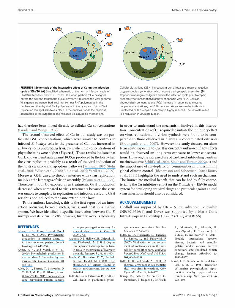
The mechanism of the interaction between Cu and the E. hux-leyi – EhV86 system is currently unknown. Our observations aresummarized in Figure 5. Our data show that EhV86 did not havean impact on the production of PCs in E. huxleyi and thus wasunlikely to have affected the toxicity of Cu to the host. The pri-mary effect of Cu on the lytic cycle of EhV86 thus appears toresult from greater sensitivity of the virus to Cu toxicity relative tothe host. Viruses require metals for the functioning of their DNAand RNA replication enzymes, so it has been previously suggestedthat viral inactivation by Cu involves cleavage or damage of viralDNA or RNA (Sagripanti et al., 1997). Recent studies have linked
Frontiers in Microbiology | Microbiological Chemistry April 2012 | Volume 3 | Article 155 | 6
Gledhill et al. Metals, EhV86, and Emiliania huxleyi
FIGURE 4 | Particulate phytochelatin (PC) concentration (amol cell−1) in
(A) Cu and (B) Cd treatments. Circles: control, inverted triangles: +virus,squares: +metal, diamonds: +metal +virus.
Table 2 | RT-PCR results for Cu exposure experiments.
EhV86 genes
DNA pol Helicase PCNA MCP
Control day 5 − − − −Control day 7 − − − −Cu day 5 − − − −Cu day 7 − − − −EhV86 day 7 (v2*) + + + +EhV86 day 10 (v5*) + + + +Cu + EhV86 day 5 (v0*) − − − −Cu + EhV86 day 7 (v2*) − − + +NTC∧ − − − −
*Days post EhV86 addition; ∧no template control.
Cu exposure to a complete decline in virus particles confirmingantiviral properties of Cu-based agents and surfaces against majoropportunistic pathogens (Noyce et al., 2006a,b, 2007; Huang et al.,2008; Weaver et al., 2008). However, in our study, virus particleswere not observed to decline completely in the presence of Cu,
Table 3 | RT-PCR results for Cd exposure experiments.
EhV86 genes
DNA pol Helicase PCNA MCP
EhV86 day 4 (v0*) − − − −EhV86 day 5 (v1*) + + + +EhV86 day 6 (v2*) + + + +EhV86 day 7 (v3*) + + + +EhV86 day 8 (v4*) + + + +Cd day 4 − − − −Cd day 5 − − − −Cd day 6 − − − −Cd day 7 − − − −Cd day 8 − − − −Cd + EhV86 day 4 (v0*) − − − −Cd + EhV86 day 5 (v1*) + + + +Cd + EhV86 day 6 (v2*) + + + +Cd + EhV86 day 7 (v3*) + + + +Cd + EhV86 day 8 (v4*) + + + +NTC∧ − − − −
*Days post EhV86 addition; ∧no template control.
and furthermore, RNA transcripts for both PCNA and MCP weredetected. The lack of DNA pol and helicase transcripts indicatesthat a specific mechanism of Cu inhibition was at play. DNA poland helicase are both associated with DNA synthesis and replica-tion, hence it appears that Cu disrupted virus replication withinthe host cell, although further work is needed to identify whetherthis is a result of exposure of the virus to elevated free Cu withinthe host cell or in the media prior to entry into the host. How-ever since neither MCP or PCNA are affected, we hypothesize thatvirion assembly, DNA repair, and other cellular functions associ-ated with PCNA were not regulated at the transcriptional level byCu. Thus, despite being involved in DNA replication and repair,viral PCNA was not regulated in the same way as dedicated DNAreplicative enzymes, DNA pol, and helicase. It is interesting to notethat both DNA pol and helicase encode for magnesium contain-ing proteins, while PCNA and MCP do not, although whetherthis factor might influence the inhibition of transcription of theassociated RNA is unclear. Intracellular free Cu concentrationsin eukaryotes and prokaryotes are generally thought to be tightlycontrolled, with Cu being buffered within the cell by thiols andchaperoned into Cu containing enzymes (Robinson and Winge,2010; Rae et al., 1999). The intravirion behavior of metals how-ever is less well known, but our studies indicate that EhV86 maybe less able to regulate Cu. Previous studies on the effects of Cuon the transcriptional control of genes have indicated regulationof Cu is achieved by modulating the transcription of genes encod-ing proteins directly involved in Cu binding. Transcription of theCtr1/3 and Fre1 genes are regulated by the DNA-binding proteinMac1 (Yamaguchi-Iwai et al., 1997). During Cu starvation, Mac1binds to DNA initiating transcription. However, in Cu replete con-ditions, Jensen and Winge (1998) demonstrated that Cu ions bindto the protein, initiating a conformational change which inhibitstranscription. Transcriptional regulation of these Cu uptake genes
www.frontiersin.org April 2012 | Volume 3 | Article 155 | 7
Gledhill et al. Metals, EhV86, and Emiliania huxleyi
FIGURE 5 | Schematic of the interactive effect of Cu on the infection
cycle of EhV86. (A) Simplified schematic of the normal infection cycle ofEhV86 (after Mackinder et al., 2009). The virion particle (blue hexagon)enters the cell and targets the nucleus where it releases the viral genome.Viral genes are transcribed (red) first by host RNA polymerase in thenucleus and then by viral RNA polymerase in the cytoplasm. Virus DNAreplication (orange) also takes place in the nucleus, while the capsid isassembled in the cytoplasm and released via a budding mechanism.
Cellular glutathione (GSH) increases (green arrow) as a result of reactiveoxygen species generation, which occurs during capsid assembly. (B)
Copper down-regulates (green arrow) the infection cycle prior to capsidassembly via transcriptional control of specific viral RNA. Cellularphytochelatin concentrations (PCs) increase in response to elevatedcopper concentrations, but GSH concentrations are similar to those inuninfected cells as capsid assembly is highly reduced. The ultimate resultis a reduction in virus production.
has therefore been linked directly to cellular Cu concentrations(Graden and Winge, 1997).
The second observed effect of Cu in our study was on par-ticulate GSH concentrations, which were similar to controls ininfected E. huxleyi cells in the presence of Cu, but increased inE. huxleyi cells undergoing lysis, even when the concentrations ofphytochelatins were higher (Figure 3). These results indicate thatGSH, known to mitigate against ROS, is produced by the host whenthe virus replicates probably as a result of the viral induction ofthe hosts ceramide and apoptosis pathways (Schwarz, 1996; Stohset al., 2001; Wilson et al., 2005; Bidle et al., 2007; Vardi et al., 2009).Moreover, GSH can also directly interfere with virus replication,mainly at the late stages of virion assembly (Palamara et al., 1995).Therefore, in our Cu exposed virus treatments, GSH productiondecreased when compared to virus treatments because the viruswas unable to complete its replication and infection cycle and GSHwas thus not induced to the same extent in the host.
To the authors knowledge, this is the first report of an inter-action occurring between metals, virus, and host in a marinesystem. We have identified a specific interaction between Cu, E.huxleyi and its virus EhV86, however, further work is necessary
in order to understand the mechanism involved in this interac-tion. Concentrations of Cu required to initiate the inhibitory effecton virus replication and virion synthesis were found to be com-parable to those observed in highly Cu contaminated estuaries(Braungardt et al., 2007). However the study focused on shortterm acute exposure to Cu. It is currently unknown if any effectswould be observed on long-term exposure to lower concentra-tions. However, the increased use of Cu-based antifouling paints inmarine systems (Schiff et al., 2004; Singh and Turner, 2009a,b) andthe importance of phytoplankton communities in underpinningglobal climate control (Richardson and Schoeman, 2004; Bouvyet al., 2011) highlights the need to understand such mechanisms.The immediate medical benefit that can be had by fully charac-terizing the Cu inhibitory effort on the E. huxleyi – EhV86 modelsystem for developing antiviral drugs and protocols against animalvirus infections should also be considered.
ACKNOWLEDGMENTSGledhill was supported by UK – NERC Advanced Fellowship(NE/E013546/1) and Devez was supported by a Marie CurieIntra-European Fellowship (FP6-023215-QWSTRESS).
REFERENCESAhner, B. A., Kong, S., and Morel,
F. M. M. (1995). Phytochelatinproduction in marine algae. 1.An interspecies comparison. Limnol.Oceanogr. 40, 649–657.
Ahner, B. A., and Morel, F. M. M.(1995). Phytochelatin production inmarine algae 2. Induction by var-ious metals. Limnol. Oceanogr. 40,658–665.
Allen, M. J., Forster, T., Schroeder, D.C., Hall, M., Roy, D., Ghazal, P., andWilson, W. H. (2006). Locus-specificgene expression pattern suggests
a unique propagation strategy fora giant algal virus. J. Virol. 80,7699–7705.
Aruoma, O. I., Halliwell, B., Gajewski, E.,and Dizdaroglu, M. (1991). Copperion dependent damage to the basesin DNA in the presence of hydrogenperoxide. Biochem. J. 273, 601–604.
Bergh, O., Borsheim, K. Y., Bratbak,G., and Heldal, M. (1989). Highabundance of viruses found inaquatic environments. Nature 340,467–468.
Bidle, K. D., and Falkowski, P. G. (2004).Cell death in planktonic, photo-
synthetic microorganisms. Nat. Rev.Microbiol. 2, 643–655.
Bidle, K. D., Haramaty, L., Barcelos,E., Ramos, J., and Falkowski, P.(2007). Viral activation and recruit-ment of metacaspases in the uni-cellular coccolithophore, Emilianiahuxleyi. Proc. Natl. Acad. Sci. U.S.A.104, 6049–6054.
Bidle, K. D., and Vardi, A. (2011). Achemical arms race at sea mediatesalgal host-virus interactions. Curr.Opin. Microbiol. 14, 449–457.
Bouvy, M., Bettarel, Y., Bouvier, C.,Domaizon, I., Jacquet, S., Le Floc’h,
E., Montanie, H., Mostajir, B.,Sime-Ngando, T., Torreton, J. P.,Vidussi, F., and Bouvier, T. (2011).Trophic interactions betweenviruses, bacteria and nanofla-gellates under various nutrientconditions and simulated climatechange. Environ. Microbiol. 13,1842–1857.
Brand, L. E., Sunda, W. G., and Guil-lard, R. R. L. (1986). Reductionof marine phytoplankton repro-duction rates by copper and cad-mium. J. Exp. Mar. Biol. Ecol. 96,225–250.
Frontiers in Microbiology | Microbiological Chemistry April 2012 | Volume 3 | Article 155 | 8
Gledhill et al. Metals, EhV86, and Emiliania huxleyi
Braungardt, C. B., Achterberg, E. P.,Gledhill, M., Nimmo, M., Elbaz-Poulichet, F., Cruzado, A., andVelasquez, Z. (2007). Chemical spe-ciation of dissolved Cu, Ni, and Co ina contaminated estuary in southwestSpain and its influence on planktoncommunities. Environ. Sci. Technol.41, 4214–4220.
Curtain, C. C., Ali, F., Volitakis, I.,Cherny, R. A., Norton, R. S.,Beyreuther, K., Barrow, C. J., Mas-ters, C. L., Bush, A. I., and Barn-ham, K. J. (2001). Alzheimer’sdisease amyloid-beta binds cop-per and zinc to generate anallosterically ordered membrane-penetrating structure containingsuperoxide dismutase-like subunits.J. Biol. Chem. 276, 20466–20473.
Debelius, B., Forja, J. M., Delvalls, A.,and Lubian, L. M. (2009). Toxic-ity and bioaccumulation of copperand lead in five marine microal-gae. Ecotoxicol. Environ. Saf. 72,1503–1513.
Devez, A., Achterberg, E. P., and Gled-hill, M. (2009). “Metal ion-bindingproperties of phytochelatins andrelated ligands,” in Metal Ions in LifeSciences, eds A. Sigel, H. Sigel, and K.O. Sigel (Cambridge: Royal Societyof Chemistry), 441–481.
Dupont, C. L., and Ahner, B. A. (2005).Effects of copper, cadmium, and zincon the production and exudation ofthiols by Emiliania huxleyi. Limnol.Oceanogr. 50, 508–515.
Evans, C., Malin, G., Mills, G. P., andWilson, W. H. (2006). Viral infec-tion of Emiliania huxleyi (Prymne-siophyceae) leads to elevated pro-duction of reactive oxygen species.J. Phycol. 42, 1040–1047.
Gledhill, M., Nimmo, M., Hill, S. J.,and Brown, M. T. (1997). The tox-icity of copper(II) species to marinealgae, with particular reference tomacroalgae. J. Phycol. 33, 2–11.
Gobler, C. J., Hutchins, D. A., Fisher,N. S., Cosper, E. M., and Sanudo-Wilhelmy, S. (1997). Release andbioavailability of C, N, P, Se andFe following viral lysis of a marinechrysophyte. Limnol. Oceanogr. 42,1492–1504.
Graden, J. A., and Winge, D. R.(1997). Copper-mediated repressionof the activation domain in theyeast Mac1p transcription factor.Proc. Natl. Acad. Sci. U.S.A. 94,5550–5555.
Han, G., Gable, K., Yan, L., Allen, M.J., Wilson, W. H., Moitra, P., Har-mon, J. M., and Dunn, T. M. (2006).Expression of a novel marine viralsingle-chain serine palmitoyltrans-ferase and construction of yeast and
mammalian single-chain chimera. J.Biol. Chem. 281, 39935–39942.
Heller, M. I., and Croot, P. L. (2010).Superoxide decay kinetics in theSouthern Ocean. Environ. Sci. Tech-nol. 44, 191–196.
Heller, M. I., and Croot, P. L. (2011).Superoxide decay as a probe for spe-ciation changes during dust disso-lution in Tropical Atlantic surfacewaters near Cape Verde. Mar. Chem.126, 37–55.
Horie, M., Ogawa, H., Yoshida, Y.,Yamada, K., Hara, A., Ozawa, K.,Matsuda, S., Mizota, C., Tani, M.,Yamamoto, Y., Yamada, M., Naka-mura, K., and Imai, K. (2008). Inac-tivation and morphological changesof avian influenza virus by copperions. Arch. Virol. 153, 1467–1472.
Huang, H.-I., Shih, H.-Y., Lee, C.-M., Yang, T. C., Lay, J.-J., andLin, Y. E. (2008). In vitro effi-cacy of copper and silver ionsin eradicating Pseudomonas aerugi-nosa, Stenotrophomonas maltophiliaand Acinetobacter baumannii: impli-cations for on-site disinfection forhospital infection control. Water Res.42, 73–80.
Huang, Y. H., Shih, C. M., Huang, C.J., Lin, C. M., Chou, C. M., Tsai, M.L., Liu, T. P., Chiu, J. F., and Chen,C. T. (2006). Effects of cadmiumon structure and enzymatic activityof Cu, Zn-SOD and oxidative statusin neural cells. J. Cell. Biochem. 98,577–589.
Hussain, T., Shukla, G. S., and Chan-dra, S. V. (1987). Effects of cad-mium on superoxide-dismutase andlipid peroxidation in liver and kid-ney of growing rats – in vivo andin vitro studies. Pharmacol. Toxicol.60, 355–358.
Jensen, L. T., and Winge, D. R. (1998).Identification of a copper-inducedintramolecular interaction in thetranscription factor Mac1 from Sac-charomyces cerevisiae. EMBO J. 17,5400–5408.
Karlstrom, A. R., and Levine, R. L.(1991a). Copper inhibits the HIV-1protease by both oxygen-dependentand oxygen independent mecha-nisms. FASEB J. 5, A452–A452.
Karlstrom, A. R., and Levine, R. L.(1991b). Copper inhibits the pro-tease from human immunodefi-ciency virus-1 by both cysteine-dependent and cysteine independentmechanisms. Proc. Natl. Acad. Sci.U.S.A. 88, 5552–5556.
Kawakami, S. K., Gledhill, M., andAchterberg, E. P. (2006a). Determi-nation of phytochelatins and glu-tathione in phytoplankton fromnatural waters using HPLC with
fluorescence detection. Trends Ana-lyt. Chem. 25, 133–142.
Kawakami, S. K., Gledhill, M., andAchterberg, E. P. (2006b). Effectsof metal combinations on the pro-duction of phytochelatins and glu-tathione by the marine diatomPhaeodactylum tricornutum. Bio-metals 19, 51–60.
Kawakami, S. K., Gledhill, M., andAchterberg, E. P. (2006c). Produc-tion of phytochelatins and glu-tathione by marine phytoplanktonin response to metal stress. J. Phycol.42, 975–989.
Lane, T. W., Saito, M. A., George, G.N., Pickering, I. J., Prince, R. C.,and Morel, F. M. M. (2005). Acadmium enzyme from a marinediatom. Nature 435, 42–42.
Le Faucheur, S. V., Behra, R., and Sigg, L.(2005). Thiol and metal contents inperiphyton exposed to elevated cop-per and zinc concentrations: a fieldand microcosm study. Environ. Sci.Technol. 39, 8099–8107.
Leal, M. F. C., Vasconcelos, M. T. S. D.,and van den Berg, C. M. G. (1999).Copper-induced release of complex-ing ligands similar to thiols by Emil-iania huxleyi in seawater cultures.Limnol. Oceanogr. 44, 1750–1762.
Lee, J., Ahner, B. A., and Morel, F. M.M. (1996). Export of cadmium andphytochelatin by the marine diatomThalassiosira weissflogii. Environ. Sci.Technol. 30, 1814–1821.
Levinson, W., Faras, A., Woodson, B.,Jackson, J., and Bishop, J. M. (1973).Inhibition of RNA-dependent DNApolymerase of Rous sarcoma virusby thiosemicarbazones and severalcations. Proc. Natl. Acad. Sci. U.S.A.70, 164–168.
Levy, J. L., Angel, B. M., Stauber, J. L.,Poon, W. L., Simpson, S. L., Cheng,S. H., and Jolley, D. F. (2008). Uptakeand internalisation of copper bythree marine microalgae: compari-son of copper-sensitive and copper-tolerant species. Aquat. Toxicol. 89,82–93.
Levy, J. L., Stauber, J. L., and Jolley,D. F. (2007). Sensitivity of marinemicroalgae to copper: the effect ofbiotic factors on copper adsorptionand toxicity. Sci. Total Environ. 387,141–154.
Lovell, M. A., Robertson, J. D., Tees-dale, W. J., Campbell, J. L., andMarkesbery, W. R. (1998). Copper,iron and zinc in Alzheimer’s diseasesenile plaques. J. Neurol. Sci. 158,47–52.
Mackinder, L. C. M., Worthy, C. A.,Biggi, G., Hall, M., Ryan, K. P.,Varsani, A., Harper, G. M., Wilson,W. H., Brownlee, C., and Schroeder,
D. C. (2009). A unicellular algalvirus, Emiliania huxleyi virus 86,exploits an animal-like infectionstrategy. J. Gen. Virol. 90, 2306–2316.
Maga, G., and Hubscher, U. (2003).Proliferating cell nuclear antigen(PCNA): a dancer with many part-ners. J. Cell. Sci. 116, 3051–3060.
Mendoza-Cozatl, D., Loza-Tavera,H., Hernandez-Navarro, A., andMoreno-Sanchez, R. (2005). Sul-fur assimilation and glutathionemetabolism under cadmium stressin yeast, protists and plants. FEMSMicrobiol. Rev. 29, 653–671.
Mioni, C., Poorvin, L., and Wil-helm, S. W. (2005). Virus andsiderophore-mediated transfer ofavailable Fe between heterotrophicbacteria: characterisation using aniron specific reporter. Aquat. Microb.Ecol. 41, 233–245.
Morel, F. M. M., and Price, N. M. (2003).The biogeochemical cycles of tracemetals in the oceans. Science 300,944–947.
Morel, N. M. L., Rueter, J. G., and Morel,F. M. M. (1978). Copper toxicityto Skeletonema costatum (Bacillario-phyceae). J. Phycol. 14, 43–48.
Morelli, E., and Fantozzi, L. (2008).Phytochelatins in the diatom Phaeo-dactylum tricornutum Bohlin: anevaluation of their use as biomarkersof metal exposure in marine waters.Bull. Environ. Contam. Toxicol. 81,236–241.
Morelli, E., and Scarano, G. (2001). Syn-thesis and stability of phytochelatinsinduced by cadmium and lead inthe marine diatom Phaeodactylumtricornutum. Mar. Environ. Res. 52,383–395.
Noyce, J. O., Michels, H., and Keevil, C.W. (2006a). Potential use of coppersurfaces to reduce survival of epi-demic meticillin-resistant Staphy-lococcus aureus in the healthcareenvironment. J. Hosp. Infect. 63,289–297.
Noyce, J. O., Michels, H., and Keevil, C.W. (2006b). Use of copper cast alloysto control Escherichia coli O157cross-contamination during foodprocessing. Appl. Environ. Microbiol.72, 4239–4244.
Noyce, J. O., Michels, H., and Keevil, C.W. (2007). Inactivation of influenzaA virus on copper versus stainlesssteel surfaces. Appl. Environ. Micro-biol. 73, 2748–2750.
Pagarete, A., Allen, M. J., Wilson, W.H., Kimmance, S. A., and De Var-gas, C. (2009). Host-virus shift ofthe sphingolipid pathway along anEmiliania huxleyi bloom: survival ofthe fattest. Environ. Microbiol. 11,2840–2848.
www.frontiersin.org April 2012 | Volume 3 | Article 155 | 9
Gledhill et al. Metals, EhV86, and Emiliania huxleyi
Palamara, A. T., Perno, C. F., Ciriolo,M. R., Dini, L., Balestra, E., Dagos-tini, C., Difrancesco, P., Favalli, C.,Rotilio, G., and Garaci, E. (1995).Evidence for antiviral activity of glu-tathione – in vitro inhibition of Her-pes simplex virus type-1 replication.Antiviral Res. 27, 237–253.
Pawlik-Skowronska, B., Pirszel, J., andBrown, M. T. (2007). Concentra-tions of phytochelatins and glu-tathione found in natural assem-blages of seaweeds depend on speciesand metal concentrations of thehabitat. Aquat. Toxicol. 83, 190–199.
Permyakov, E. (2009). Metallopro-teomics. Hoboken, NJ: John Wiley.
Price, N. M., and Morel, F. M. M. (1990).Cadmium and cobalt substitutionfor zinc in a marine diatom. Nature344, 658–660.
Rae, T. D., Schmidt, P. J., Pufahl, R. A.,Culotta, V. C., and O’Halloran, T.V. (1999). Undetectable intracellu-lar free copper: the requirement ofa copper chaperone for superoxidedismutase. Science 284, 805–808.
Richardson, A. J., and Schoeman, D. S.(2004). Climate impact on planktonecosystems in the Northeast Atlantic.Science 305, 1609–1612.
Robinson, N. J., and Winge, D.R. (2010). “Copper metallochaper-ones,” in Annual Review of Biochem-istry, Vol. 79, eds R. D. Kornberg,C. R. H. Raetz, J. E. Rothman, andJ. W. Thorner (Palo Alto: AnnualReviews), 537–562.
Sagripanti, J. L., and Kraemer, K. H.(1989). Site specific oxidative DNAdamage at polyguanosines producedby copper plus hydrogen peroxide. J.Biol. Chem. 264, 1729–1734.
Sagripanti, J. L., Routson, L. B.,Bonifacino, A. C., and Lytle, C.D. (1997). Mechanism of copper-mediated inactivation of Herpessimplex virus. Antimicrob. AgentsChemother. 41, 812–817.
Sandaa, R. A. (2008). Burden or ben-efit? Virus-host interactions in themarine environment. Res. Microbiol.159, 374–381.
Schafer, F. Q., and Buettner, G. R.(2001). Redox environment of thecell as viewed through the redoxstate of the glutathione disul-fide/glutathione couple. Free Radic.Biol. Med. 30, 1191–1212.
Scheidegger, C., Behra, R., and Sigg,L. (2011). Phytochelatin formationkinetics and toxic effects in the fresh-water alga Chlamydomonas rein-hardtii upon short- and long-termexposure to lead(II). Aquat. Toxicol.101, 423–429.
Schiff, K., Diehl, D., and Valkirs, A.(2004). Copper emissions fromantifouling paint on recreationalvessels. Mar. Pollut. Bull. 48,371–377.
Schroeder, D. C., Oke, J., Malin, G.,and Wilson, W. H. (2002). Coccol-ithovirus (Phycodnaviridae): char-acterisation of a new large dsDNAalgal virus that infects Emilianiahuxleyi. Arch. Virol. 147, 1685–1698.
Schwarz, K. B. (1996). Oxidative stressduring viral infection: a review. FreeRadic. Biol. Med. 21, 641–649.
Singh, N., and Turner, A. (2009a).Leaching of copper and zinc fromspent antifouling paint particles.Environ. Pollut. 157, 371–376.
Singh, N., and Turner, A. (2009b). Tracemetals in antifouling paint particlesand their heterogeneous contami-nation of coastal sediments. Mar.Pollut. Bull. 58, 559–564.
Sorensen, G., Baker, A. C., Hall, M. J.,Munn, C. B., and Schroeder, D. C.(2009). Novel virus dynamics in anEmiliania huxleyi bloom. J. PlanktonRes. 31, 787–791.
Stohs, S. J., Bagchi, D., Hassoun, E.,and Bagchi, M. (2001). Oxida-tive mechanisms in the toxicity ofchromium and cadmium ions. J.Environ. Pathol. Toxicol. Oncol. 20,77–88.
Sunda, W. G. (1988). Trace metal inter-actions with marine phytoplankton.Biol. Oceanogr. 6, 411–442.
Sunda, W. G., and Guillard, R. R. L.(1976). The relationship betweencupric ion activity and toxicity of
copper to phytoplankton. J. Mar.Res. 34, 511–529.
Suttle, C. A. (2007). Marine viruses –major players in the global ecosys-tem. Nat. Rev. Microbiol. 5, 801–812.
Vardi, A., Van Mooy, B. A. S., Fredricks,H. F., Popendorf, K. J., Ossolinski,J. E., Haramaty, L., and Bidle, K.D. (2009). Viral glycosphingolipidsinduce lytic infection and cell deathin marine phytoplankton. Science326, 861–865.
Vasconcelos, M., and Leal, M. F. C.(2001). Adsorption and uptake ofCu by Emiliania huxleyi in naturalseawater. Environ. Sci. Technol. 35,508–515.
Vasconcelos, M., Leal, M. F. C., and vanden Berg, C. M. G. (2002). Influ-ence of the nature of the exudatesreleased by different marine algaeon the growth, trace metal uptake,and exudation of Emiliania huxleyiin natural seawater. Mar. Chem. 77,187–210.
Voelker, B. M., Sedlak, D. L., andZafiriou, O. C. (2000). Chemistry ofsuperoxide radical in seawater: reac-tions with organic Cu complexes.Environ. Sci. Technol. 34, 1036–1042.
Weaver, L., Michels, H. T., and Keevil, C.W. (2008). Survival of Clostridiumdifficile on copper and steel: futur-istic options for hospital hygiene. J.Hosp. Infect. 68, 145–151.
Weinbauer, M. G., Arrieta, J. M.,Griebler, C., and Herndl, G. J.(2009). Enhanced viral produc-tion and infection of bacterioplank-ton during an iron-induced phy-toplankton bloom in the South-ern Ocean. Limnol. Oceanogr. 54,774–784.
Wilhelm, S. W., and Suttle, C. A.(1999). Viruses and nutrient cyclesin the sea – viruses play criticalroles in the structure and functionof aquatic food webs. Bioscience 49,781–788.
Wilson, W. H., Schroeder, D. C., Allen,M. J., Holden, M. T. G., Parkhill, J.,Barrell, B. G., Churcher, C., Harn-lin, N., Mungall, K., Norbertczak,
H., Quail, M. A., Price, C., Rabbi-nowitsch, E., Walker, D., Craigon,M., Roy, D., and Ghazal, P. (2005).Complete genome sequence andlytic phase transcription profileof a Coccolithovirus. Science 309,1090–1092.
Wilson, W. H., Tarran, G. A., Schroeder,D., Cox, M., Oke, J., and Malin, G.(2002). Isolation of viruses respon-sible for the demise of an Emilianiahuxleyi bloom in the English chan-nel. J. Mar. Biolog. Assoc. U.K. 82,369–377.
Yamaguchi-Iwai, Y., Serpe, M., Haile, D.,Yang, W. M., Kosman, D. J., Klaus-ner, R. D., and Dancis, A. (1997).Homeostatic regulation of copperuptake in yeast via direct binding ofMAC1 protein to upstream regula-tory sequences of FRE1 and CTR1.J. Biol. Chem. 272, 17711–17718.
Conflict of Interest Statement: Theauthors declare that the research wasconducted in the absence of any com-mercial or financial relationships thatcould be construed as a potential con-flict of interest.
Received: 08 December 2011; paperpending published: 01 February 2012;accepted: 04 April 2012; published online:23 April 2012.Citation: Gledhill M, Devez A, High-field A, Singleton C, Achterberg EPand Schroeder D (2012) Effect of met-als on the lytic cycle of the Coccol-ithovirus, EhV86. Front. Microbio. 3:155.doi: 10.3389/fmicb.2012.00155This article was submitted to Frontiers inMicrobiological Chemistry, a specialty ofFrontiers in Microbiology.Copyright © 2012 Gledhill, Devez,Highfield, Singleton, Achterberg andSchroeder. This is an open-access articledistributed under the terms of the Cre-ative Commons Attribution Non Com-mercial License, which permits non-commercial use, distribution, and repro-duction in other forums, provided theoriginal authors and source are credited.
Frontiers in Microbiology | Microbiological Chemistry April 2012 | Volume 3 | Article 155 | 10