Light Transmittance of Ocular Media in Living Rabbit Eyes
6
Light Transmittance of Ocular Media in Living Rabbit Eyes Peep V. Algvere, Per-Arne L. Torstensson* and Bjo'rn M. Tengroth Purpose. The purpose of this study was to measure the transmittance of electromagnetic radia- tion, particularly visible light, through the ocular media of living and whole rabbit eyes. Pre- vious determinations have been carried out on excised cadaver eyes. Methods. A specially designed fiberoptic probe (outer diameter, 0.9 mm) was placed in the vitreous in front of the retina using a microsurgical technique. In eight living albino rabbits (under general anesthesia), ocular transmittance was determined in the wavelength range 350 to 1 100 nni using a reversed beam path (from vitreous to cornea). Results. A maximum optical transmittance of 94% to 96% (standard deviation, 2%-3%) was found between 630 and 730 nm (reflection losses in the cornea-air interface excluded). In the blue portion of the spectrum, transmittance decreased rapidly for shorter wavelengths, and was 50% at 400 nm and less than 1% at 380 nm. In the infrared part of the spectrum, transmit- tance was close to 90% up to 900 nm but declined at longer wavelengths, coinciding with the absorption in pure water. Calibration recordings showed a 1% to 2% accuracy of the method. Conclusions. This experimental technique using an intraocular fiberoptic probe yields a high accuracy and indicates that light transmittance is very high in vivo and superior to that re- ported from cadaver eyes. Invest Ophthalmol Vis Sci. 1993;34:349-354. Otudies on ocular transmittance characteristics with respect to electromagnetic radiation 1 " 8 have formed the basis for interpretation of photoeffects on the eye. 9 Some reports are compatible with each other, whereas others do not agree. All measurements up until now have, however, been carried out on dead (enucleated) eyes, and it has not been fully investigated how early postmortem changes affect the optical char- acteristics of the eye. Boynton et al 3 and Demott et al 5 found considerable time dependence in postmortem changes, whereas Boettner and Wolter 6 reported a minor, almost negligible time dependence. One possi- ble explanation of these incongruities may be the vary- From the Department of Ophthalmology, Karolinska Institute, and St. Erik Eye Hospital, Stockholm, and *Radians Innova, Inc., Gothenburg, Sweden. Supported by the National Defense Research Agency, Stockholm, Sweden. Submitted for publication: June 16, 1992; accepted September 11, 1992. Proprietary interest category: N (PVA); C3 (P-ALT); N (11MT). Reprint requests: Peep V. Algvere, St. Erik Eye Hospital, Fleminggatan 22, 112 82 Stockholm, Sweden. ing drying effects in the specimen due to different handling techniques. To overcome these discrepancies, we worked out an in vivo method for measurement of transmittance of ocular media. A special fiberoptic probe was devel- oped for insertion into the vitreous cavity of a living eye. With this intraocular fiberoptic technique, it is possible to perform several types of experiments, in- cluding measurements of radiation transmittance, and studies of fluorescence phenomena and optical scat- tering in different parts of the ocular media. In the current report, we describe the results of intraocular light transmittance in the cornea, aqueous, lens, and vitreous using this fiberoptic probe technique. MATERIALS AND METHODS Instrumentation A fiberoptic probe, designed and developed for these experiments, was inserted into the vitreous cavity close to the retina and adjusted to the optical axis of the eye. Investigative Ophthalmology & Visual Science, February 1993, Vol. 34, No. 2 Copyright © Association for Research in Vision and Ophthalmology 349
-
Upload
independent -
Category
Documents
-
view
2 -
download
0
Transcript of Light Transmittance of Ocular Media in Living Rabbit Eyes

Light Transmittance of Ocular Media in Living Rabbit Eyes
Peep V. Algvere, Per-Arne L. Torstensson* and Bjo'rn M. Tengroth
Purpose. The purpose of this study was to measure the transmittance of electromagnetic radia-tion, particularly visible light, through the ocular media of living and whole rabbit eyes. Pre-vious determinations have been carried out on excised cadaver eyes.
Methods. A specially designed fiberoptic probe (outer diameter, 0.9 mm) was placed in thevitreous in front of the retina using a microsurgical technique. In eight living albino rabbits(under general anesthesia), ocular transmittance was determined in the wavelength range 350to 1 100 nni using a reversed beam path (from vitreous to cornea).
Results. A maximum optical transmittance of 94% to 96% (standard deviation, 2%-3%) wasfound between 630 and 730 nm (reflection losses in the cornea-air interface excluded). In theblue portion of the spectrum, transmittance decreased rapidly for shorter wavelengths, andwas 50% at 400 nm and less than 1% at 380 nm. In the infrared part of the spectrum, transmit-tance was close to 90% up to 900 nm but declined at longer wavelengths, coinciding with theabsorption in pure water. Calibration recordings showed a 1% to 2% accuracy of the method.
Conclusions. This experimental technique using an intraocular fiberoptic probe yields a highaccuracy and indicates that light transmittance is very high in vivo and superior to that re-ported from cadaver eyes. Invest Ophthalmol Vis Sci. 1993;34:349-354.
Otudies on ocular transmittance characteristics withrespect to electromagnetic radiation1"8 have formedthe basis for interpretation of photoeffects on theeye.9 Some reports are compatible with each other,whereas others do not agree. All measurements upuntil now have, however, been carried out on dead(enucleated) eyes, and it has not been fully investigatedhow early postmortem changes affect the optical char-acteristics of the eye. Boynton et al3 and Demott et al5
found considerable time dependence in postmortemchanges, whereas Boettner and Wolter6 reported aminor, almost negligible time dependence. One possi-ble explanation of these incongruities may be the vary-
From the Department of Ophthalmology, Karolinska Institute, and St.Erik Eye Hospital, Stockholm, and *Radians Innova, Inc., Gothenburg,Sweden.Supported by the National Defense Research Agency, Stockholm, Sweden.Submitted for publication: June 16, 1992; accepted September 11, 1992.Proprietary interest category: N (PVA); C3 (P-ALT); N (11MT).Reprint requests: Peep V. Algvere, St. Erik Eye Hospital, Fleminggatan22, 112 82 Stockholm, Sweden.
ing drying effects in the specimen due to differenthandling techniques.
To overcome these discrepancies, we worked outan in vivo method for measurement of transmittanceof ocular media. A special fiberoptic probe was devel-oped for insertion into the vitreous cavity of a livingeye. With this intraocular fiberoptic technique, it ispossible to perform several types of experiments, in-cluding measurements of radiation transmittance, andstudies of fluorescence phenomena and optical scat-tering in different parts of the ocular media. In thecurrent report, we describe the results of intraocularlight transmittance in the cornea, aqueous, lens, andvitreous using this fiberoptic probe technique.
MATERIALS AND METHODS
InstrumentationA fiberoptic probe, designed and developed for theseexperiments, was inserted into the vitreous cavity closeto the retina and adjusted to the optical axis of the eye.
Investigative Ophthalmology & Visual Science, February 1993, Vol. 34, No. 2Copyright © Association for Research in Vision and Ophthalmology 349

350 Investigative Ophthalmology & Visual Science, February 1993, Vol. 34, No. 2
interaction distance
beam path
optical fiber
fiber probe
FIGURE 1. Location of fiberoptic probe (outer diameter, 0.9mm) inside the vitreous cavity, close to the retina, centeredto the optical axis of eye. Test light enters the eye throughthe optical fiber and is reflected from the tip of the probe inthe vitreous to exit through the pupil and cornea.
The fiberoptic cable was connected to a test lightsource, a halogen lamp (Intralux 250 HL Volpi AG,Zurich, Switzerland) or He-Ne laser (model 106-1, 10mW; Spectra Physics, Carlsbad, CA). The intraocularfiberoptic probe makes it possible, and preferable, touse a reversed beam path through the eye because thisgreatly facilitates the optical alignment in living eyes.The beam path from the tip of the fiberoptic probewas directed anteriorly through the pupil to exit theeye through the cornea (Fig. 1).
Basically, the probe consists of a standard, multi-mode optical fiber (HCG-365, numerical aperture= 0.2; Ensign Bickford, Avon, CT). The optical parts(core and cladding) of this fiber are made of purequartz, resulting in very high transmittance within thewavelength range of 300 to 2000 nm.
The fiber was first fixed into an approximately 50-mm long injection steel needle with an outer diameterof 0.9 mm. The needle was bent to an appropriateradius to fit the rabbit eye retinal curvature. A criticalpart of the probe manufacturing was the preparationof the optical needle end. To achieve a well confinedbeam output, it was necessary to cut and polish thefiber tip to an angle of 45° with respect to the opticalaxis of the fiber. To serve as a mirror, this surface hadto be coated with aluminum to avoid leakage of radia-tion in unwanted directions (Fig. 2). The outcouplingsurface of the fiber tip also was made flat to achieve awell confined beam lobe with low divergence. Thehalf-angle of the output lobe was 14° in air and lessthan 10° (numerical aperture = 0.18) in water (vitre-ous), which is small enough to avoid any obstruction ofthe beam path by the dilated eye pupil aperture duringthe measurements.
The light beam exiting the pupil was directed, us-ing a multimode quartz fiber, through a monochroma-tor (Oriel [Stratford, CT] 77200, with gratings 1200lines/mm #77998 and 600 lines/mm #77999) to aphotodiode (Photodyne XL-88 [Westlake Village, CA]with detector #250) and an RG XYt recorder (PM8277; Philips, Eindhoven, Holland). The experimentalset-up is shown in Figure 3.
Surgical Technique
To place the fiberoptic probe (outer diameter, 0.9mm) into the vitreous cavity, a microsurgical tech-nique using an operating microscope was adopted.The pupil of the eye was maximally dilated with eyedrops (atropine, phenylephrine). General anesthesiawas induced with intramuscular injections of ketamine(35 mg/kg body weight) and xylazine (5 mg/kg body
flat surface
mirror surface
steel
Toptical axis
optical fiber
optical epoxy
FIGURE 2. Appearance of Lhetip of the fiberoptic probe.Optical fiber inside a 20 Gsteel needle ends in a mirroredsurface that reflects light in thedirection of the optical axis ofLhe eye. Divergence angle (a) is10° in water.

Light Transmittance in Living Eyes 351
FIGURE 3. Experimental set-upfor measurements of transmit-tance in rabbit eyes. Lightsource (LSI) delivers light to amultimode quartz fiber (OF1)inside the vitreous cavity.Light exiting the eye passesachromatic lenses (LI, L2) andmirror (MR). Multimodequartz fiber (OF2) leads intomonochromator (MC) andphotodiode instrument (OD I),the signal of which is fed into aXYt recorder (RC). The flexi-ble arm fixture (ST1) for theoptical fiber, the adjustablesupport (ST2) for the rabbitbed (*), and the microscope(MS) are auxiliary tools.
weight) and, if necessary, additional anesthetics weregiven every 30 to 45 min.
The conjunctiva was cut free (limbal incision)from the upper lateral quadrant of the eye. The eyewall was perforated with a stiletto (sclerotomy 1.4 mm)in the region of the equator of the eye and the stilettowas directed toward the posterior pole of the eye. Thefiberoptic probe (20 G) fits tightly to the sclerotomy.The probe was inserted into the vitreous and guided tothe posterior pole of the eye, close to the retina, undermicroscopic control (using a corneal contact lens). Thefront surface of the probe was positioned less than 2mm from the retina and centered to the optical axis.The position of the mirror of the probe was checkedby connecting the fiberoptic cable to a He-Ne laserthat illuminates the mirror in a "retrograde" direc-tion. The probe, attached to a flexible arm fixture, wasfixed in such a position that the light beam enteringthe eye pupil was focused to the optical tip of theprobe. This position was maintained throughout theexperiment.
Care was taken to avoid drying of the corneal epi-thelium, and balanced salt solution was dropped regu-larly on the cornea. The transparency of the ocularmedia remained excellent. No hypotony occurred be-cause the sclerotomy remained water-tight. Experi-ments were run over a 2-hr time course without com-plications. Intraocular hemorrhages or retinal detach-ment did not occur during the experiments. When thefiberoptic probe was removed from the eye, the scle-rotomy and conjunctiva were sutured.
Animals
Measurements were performed on 12 eyes from 12New Zealand white (albino) rabbits. The rabbits' ages
ranged from 4 mo to 2.5 yr (median, 9 months). Therabbits were handled in accordance with the ARVOResolution on the Use of Animals in Research. Wewere able successfully to examine eight eyes. In fourcases, the recordings were not satisfactory, partly dueto insufficient immobilization of the animal, with varia-tions in breathing movements.
Measurement ProcedureThe measurements were divided into two procedures.First, the relative spectral transmission of the eye wasrecorded over the whole spectral range (350-1100nm). After that, the absolute spectral transmittance ata specific wavelength (633 nm) was measured.
In the first procedure, we used the white lightsource (halogen lamp) and a scanning monochroma-tor for measurement of the optical power transmissionas a function of the wavelength with (Pieye[X]) andwithout (Prsys[X]) the eye present in the optical system.The optical power as a function of wavelength wasrecorded graphically with the XY recorder. The slitopening of the monochromator was set to 0.8 mm,corresponding to an optical bandwidth of 4 nm. Asequence always started with a calibration, recordingthe spectral characteristics of the complete systemwithout the eye (Prsys[X]). Immediately after this cali-bration, we inserted the fiberoptic probe into the al-ready prepared rabbit eye and made a new recording(Preye[X]). The relative spectral transmission (T,.el) ofthe rabbit eye then was calculated as the ratio of thetransmission values with and without the eye:
Trel(X) = Crel X Preye(X)/Prsys(X) (1)
where (Crd) is a wavelength-independent scale factor.
352 Investigative Ophthalmology & Visual Science, February 1993, Vol. 34, No. 2
The constant (C,.e|) differs from eye to eye due to minordeviations in the beam path alignment and detectoramplification adjustments.
In the second procedure, we used the He-Ne laser(633 nm) as a light source. This facilitated the visualalignment and measurement procedures. The reasonfor using a laser instead of a conventional light sourcewas that it is much easier to launch high optical powerconfined in a narrow wavelength band into an opticalfiber with a laser. The absolute calibration referencelevel of the optical transmission was measured for thecomplete optical system with the fiberoptic probe im-mersed in 1 mm pure water (Pasys[X = 633]). Immedi-ately after the calibration in water, the probe was in-serted into the eye and the optical power wasmeasured again (Paeye[X
= 633]). The absolute trans-mittance (Tabs[X = 633]) is simply the quotient (Paeye[X= 633]/Pasys[X = 633]). Note that the absolute trans-mittance depends only on absorption and scattering oflight inside the eye because we cancel out the effects ofreflections in the interfaces between probe tip and vit-reous humor as well as the interface between the cor-nea and air using the calibration procedure with theprobe immersed in pure water. The influence of ab-sorption and scattering in water with a path length of 1mm at 633 nm can be ignored safely. The refractiveindex of the cornea and vitreous humor is very closethe refractive index of water (n = 1.33). Thus, thetransmittances presented in this report do not includethe effects of reflection of light in the cornea-air in-terface, which was calculated to be 2% according tothe well known formula for optical power reflection(R) at perpendicular incidence:
= ([n, - no]/[n, + n0])2 (2)
where n, = 1.33 for water and cornea and n0 = 1 forair.
The absolute transmittance for any wavelength inthe measured range for a specific eye can now be cal-culated using the scale factor (Cabs) according to thefollowing expressions:
Tabs(X) = Cabs X Trel(X) (3)
Trel(X) = Crel X (Preye[X]/Prsys[X]) (4)
C;ibs = (Paeye[X = 633]/Pasys[X = 633])
X (Prsys[X = 633]/Preye[X = 633]) X 1/Crel (5)
Accuracy of the Method
Repeated calibration recordings indicated an accuracyof 1% to 2% for the whole system. In some recordingswith the optical probe inside a living rabbit eye, how-ever, the accuracy typically could be 3% to 6% because
of disturbances caused by the rabbit's breathing move-ments. The depth of the anesthesia was found to be acritical factor.
RESULTS
Visible Light
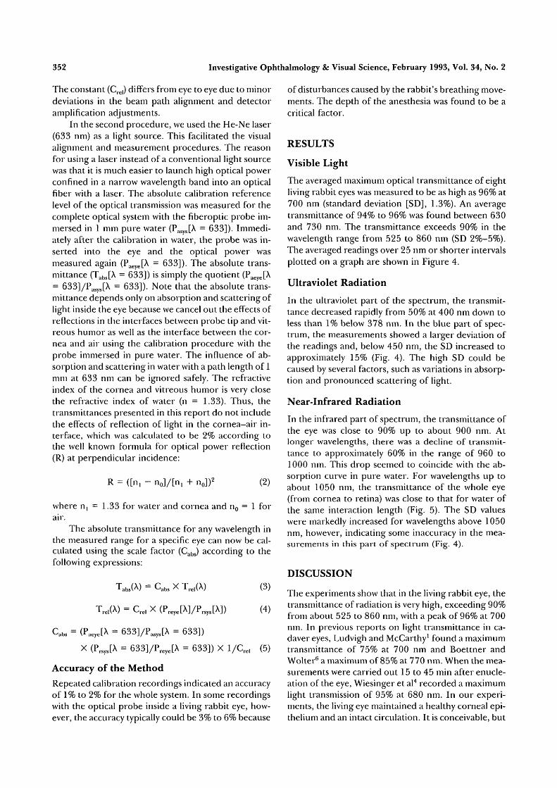
The averaged maximum optical transmittance of eightliving rabbit eyes was measured to be as high as 96% at700 nm (standard deviation [SD], 1.3%). An averagetransmittance of 94% to 96% was found between 630and 730 nm. The transmittance exceeds 90% in thewavelength range from 525 to 860 nm (SD 2%-5%).The averaged readings over 25 nm or shorter intervalsplotted on a graph are shown in Figure 4.
Ultraviolet Radiation
In the ultraviolet part of the spectrum, the transmit-tance decreased rapidly from 50% at 400 nm down toless than 1% below 378 nm. In the blue part of spec-trum, the measurements showed a larger deviation ofthe readings and, below 450 nm, the SD increased toapproximately 15% (Fig. 4). The high SD could becaused by several factors, such as variations in absorp-tion and pronounced scattering of light.
Near-Infrared Radiation
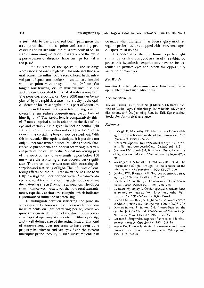
In the infrared part of spectrum, the transmittance ofthe eye was close to 90% up to about 900 nm. Atlonger wavelengths, there was a decline of transmit-tance to approximately 60% in the range of 960 to1000 nm. This drop seemed to coincide with the ab-sorption curve in pure water. For wavelengths up toabout 1050 nm, the transmittance of the whole eye(from cornea to retina) was close to that for water ofthe same interaction length (Fig. 5). The SD valueswere markedly increased for wavelengths above 1050nm, however, indicating some inaccuracy in the mea-surements in this part of spectrum (Fig. 4).
DISCUSSION
The experiments show that in the living rabbit eye, thetransmittance of radiation is very high, exceeding 90%from about 525 to 860 nm, with a peak of 96% at 700nm. In previous reports on light transmittance in ca-daver eyes, Ludvigh and McCarthy1 found a maximumtransmittance of 75% at 700 nm and Boettner andWolter6 a maximum of 85% at 770 nm. When the mea-surements were carried out 15 to 45 min after enucle-ation of the eye, Wiesinger et al4 recorded a maximumlight transmission of 95% at 680 nm. In our experi-ments, the living eye maintained a healthy corneal epi-thelium and an intact circulation. It is conceivable, but

Light Transmittance in Living Eyes
100
353
300
Q
T3
(0
CO
400 500 600 700 800 900
Wavelength [nm]1000 1100
FIGURE 4. Radiation transmittance, average of eight rabbit eyes. Transmittance (circles) is94% to 96% between 630 and 730 nm. Within these wavelengths, the SD (crosses) is 2% to 3%(plotted at base of graph).
not shown in this study, that the optical transparencyof the living eye is superior to that postmortem.
The current method enables a thorough position-ing of the detector probe, the tip of which is adjustedin the vitreous under microscopic control. In addition,a He-Ne laser beam was used as an adjunct measure tomonitor the accurate positioning of the probe. The
output lobe of the fiberoptic probe subtends a half-angle of 10° in water, and delivers the light beamthrough the eye pupil and cornea. This experimentaldesign yields an accurate alignment of the test light tothe recording system with minimal losses of radiation.The main reflection loss (calculated to be 2%) ema-nates from the interface between the cornea and air. It
300 400 500 600 700 800 900 1000 1100 1200Wavelength [nm]
FIGURE 5. Radiation absorption in 12 mm of pure water (circles) and transmittance of rabbiteye (crosses). The two curves coincide up to about 1050 nm.
354 Investigative Ophthalmology & Visual Science, February 1993, Vol. 34, No. 2
is justifiable to use a reversed beam path given theassumption that the absorption and scattering pro-cesses in the eye are isotropic. Measurements of oculartransmission using radiations that traversed the eye ina posteroanterior direction have been performed inthe past.2
In the extremes of the spectrum, the readingswere associated with a high SD. This indicates that sev-eral factors may influence the results here. In the infra-red part of spectrum, ocular transmittance coincidedwith absorption in water up to about 1050 nm. Forlonger wavelengths, ocular transmittance declinedand the curve deviated from that of water absorption.The poor correspondence above 1050 nm can be ex-plained by the rapid decrease in sensitivity of the opti-cal detector for wavelengths in this part of spectrum.
It is well known that age-related changes in thecrystalline lens reduce transmittance, particularly ofblue light.1011 The rabbit lens is comparatively thick(6-7 mm in optical axis) in relation to the size of theeye and certainly has a great impact on ocular lighttransmittance. Thus, individual or age-related varia-tions in the crystalline lens cannot be ruled out. Withthis intraocular fiberoptic technique, it is possible notonly to measure transmittance, but also to study fluo-rescence phenomena and optical scattering in differ-ent parts of the ocular media. A most interesting partof the spectrum is the wavelength region below 450nm where the scattering effects become very signifi-cant. The transmittance decreases with increasing ab-sorption and scattering of light. The influence of scat-tering effects on the total transmittance has not beenfully investigated. Boettner and Wolter6 measured di-rect and total transmittance in an attempt to separatethe scattering effects from pure absorption. The directtransmittance was much lower than the total transmit-tance, especially at short wavelengths, which indicatesa pronounced influence of scattering.
To distinguish between scattering and pure ab-sorption effects, however, it is necessary to performmeasurements on light scattering per se, which re-quire an accurate definition of the direct beam, a verysmall optical aperture at the detector fiber optic tip,and a well defined size of the retinal image. This kindof measurement does not seem to have been doneproperly in living or cadaver eyes. With the currentfiberoptic probe technique, such measurements can
be made when the system has been slightly modified(eg, the probe must be equipped with a very small opti-cal aperture at its tip).
It is conceivable that the human eye has lighttransmittance that is as good as that of the rabbit. Toprove this hypothesis, experiments have to be ex-tended to primate eyes and, when the opportunityarises, to human eyes.
Key Words
intravitreal probe, light transmittance, living eyes, quartzoptical fiber, wavelength, whole eyes.
Acknowledgments
The authors thank Professor Bengt KJeman, Chalmers Insti-tute of Technology, Gothenburg, for valuable advice anddiscussions, and Dr. Jianming Ren, St. Erik Eye Hospital,Stockholm, for surgical assistance.
References
1. Ludvigh E, McCarthy EF. Absorption of the visiblelight by the refractive media of the human eye. ArchOphthalmol. 1938; 20:37-51.
2. Kinsey VE. Spectral transmission of the eye to ultravio-let radiations. Arch Ophthalmol. 1948; 39:508-513.
3. Boynton RM, Enoch JM, Bush WR. Physical measureof light in excised eyes./ Opt Soc Am. 1954;44:879-883
4. Wiesinger H, Schmidt FH, Williams RC, et al. Thetransmission of light through the ocular media of therabbit eye. Am] Ophthalmol. 1956;42:907-910
5. DeMott DW, Boynton RM. Sources of entoptic straylight. J Opt Soc Am. 1958;48:120-125
6. Boettner EA, Wolter JR. Transmission of the ocularmedia. Invest Ophthalmol. 1962; 1:776-783
7. Geeraets WJ, Berry R. Ocular spectral characteristicsas related to hazards from lasers and other lightsources. AmJ Ophthalmol. 1968; 66:15-20
8. Beems EM, van Best JA. Light transmission of corneain whole human eyes. Exp Eye Res. 1990;50:393-395
9. Dayhaw-Barker P, Barker FM. Photoeffects on theeye. In: Jackson EM, ed. Photobiology of Skin and Eye.New York: Marcel Dekker; 1986: J17-147
10. Lerman S. Biophysical aspects of corneal and lenticu-lar transparency. Curr Eye Res. 1984; 3:3-14
11. Weale RA. Human lenticular fluorescence and trans-missivity, and their effects on vision. Exp Eye Res.1985;41:457-473.