Influence of electrolyte pH on TiO2 nanotube formation by Ti anodization
6
Journal of Alloys and Compounds 485 (2009) 478–483 Contents lists available at ScienceDirect Journal of Alloys and Compounds journal homepage: www.elsevier.com/locate/jallcom Influence of electrolyte pH on TiO 2 nanotube formation by Ti anodization Srimala Sreekantan a,∗ , Zainovia Lockman a , Roshasnorlyza Hazan a , Minoo Tasbihi b , Lee Keat Tong b , Abdul Rahman Mohamed b a School of Materials & Mineral Resource Engineering, Universiti Sains Malaysia (Engineering Campus), 14300, Nibong Tebal, Seberang Perai Selatan, Malaysia b School of Chemical Engineering, Universiti Sains Malaysia (Engineering Campus), 14300, Nibong Tebal, Seberang Perai Selatan, Malaysia article info Article history: Received 23 October 2008 Received in revised form 26 May 2009 Accepted 31 May 2009 Available online 6 June 2009 Keywords: TiO2 nanotube Electrolyte pH Anodization Anatase Rutile Phenol degradation abstract This paper investigates the effect of pH and anodization time on the formation of titania (TiO 2 ) nanotubes. Well-organized TiO 2 nanotubes were electrochemically prepared by anodizing titanium foil in 1 M Na 2 SO 4 containing 0.3 g of NH 4 F. It was found that TiO 2 nanotubes with uniform pore diameters can be produced by manipulating electrochemical conditions. This study showed that nanotube lengths ranging from ∼0.7 to 2.5 m could be formed by altering the electrolytes pH. The rate of formation depends upon the specific pH level applied as shown in the following results: pH 3 at a rate of 23 nm min −1 , 0.7 m; pH 5 at a rate of 15nmmin −1 , 0.6 m; pH 7 at a rate of 8nmmin −1 , 0.5 m. In the as-anodized condition, the nanotubes had an amorphous structure. Annealing the nanotube arrays promoted the formation of anatase and rutile phases depending upon the temperature. Furthermore, the sample annealed at 500 ◦ C showed better photocatalytic activity. The activity is also believed to be closely related to the degree of crystallinity and the crystallography structure of the nanotube, which consists of the anatase (76%) and rutile (24%) phases. © 2009 Elsevier B.V. All rights reserved. 1. Introduction Over the years, TiO 2 has been extensively studied owing to its many unique properties that make it an active component in photocatalysis [1,2], photovoltaics [3], biological coatings [4], and photoelectrolysis [5] applications. Studies have indicated that for these applications, well-arrayed TiO 2 nanotubes are of great impor- tance due to their one-dimensional (1D) nature, ease of handling, and simple preparation. Furthermore, the band gap of nanotube materials can be altered because of the quantum confinement effect. Usually, higher ratios of diameter to length of the tubes are preferred because this provides a larger surface area for photon absorption. Moreover, TiO 2 nanotubes contain free spaces in their interiors that can be filled with active materials such as chemi- cal compounds, enzymes, and noble metals, enabling them to be engineered to produce advanced materials in terms of multifunc- tionality. To date, TiO 2 nanotubes have been produced through a variety of methods which include the sol–gel technique [6], metallor- ganic chemical vapor deposition (MOCVD) [7], templating [8] and anodization [9,10]. Among these methods, the anodization of tita- nium is the only process that allows the formation of highly ordered nanotube arrays, which is its most remarkable property. In relation ∗ Corresponding author. Tel.: +60 4 5995255; fax: +60 4 5941011. E-mail address: [email protected] (S. Sreekantan). to this, Mor et al. initially reported the generation of TiO 2 nanotube arrays in an aqueous hydrofluoric (HF) acid-based electrolyte [11]. However, as a result of the high dissolution rate, only a limited length (200–300 nm) of the nanotube was produced. To increase the length of the nanotubes, Macak et al. [12] and Taveira et al. [13] subsequently proposed the use of ammonium fluoride (NH 4 F) solution instead of HF. In 2007, Shankar et al. were able to pro- duce long nanotubes (220 m) using NH 4 F or HF in a variety of polar organic electrolytes [14], but the time required to achieve such length was 17 h. Therefore, it can be said that most works reported on extremely long durations for the formation of long nanotubes. Other than electrolyte composition, pH value is also one of the most important factors that affect the length of the tubes. For instance, Wu et al. [15] have reported the formation of nanotubes with 1.8 m length after 6 h of anodization in aqueous electrolyte with pH 4.5–5. In Yunhuai’s work [16], it was found that nanotubes are not formed when the pH value was above 6. In contrast, Feng et al. [17] showed that the basic environment (pH 8–9) is much more efficient for the formation of longer nanotubes than the commonly used acidic condition. In the present work, we have shown that nanotubes can be prepared not only in acidic conditions but also in neutral conditions for short anodization times. This result is in agreement with Feng’s finding on the successful formation of nan- otubes at pH above 6. Furthermore, in our work, the same length of tubes was obtained in neutral condition pH 7, which is of high signif- icance for future robust industrial production. This study therefore aims to investigate the possibility of achieving a well-ordered TiO 2 0925-8388/$ – see front matter © 2009 Elsevier B.V. All rights reserved. doi:10.1016/j.jallcom.2009.05.152
-
Upload
independent -
Category
Documents
-
view
0 -
download
0
Transcript of Influence of electrolyte pH on TiO2 nanotube formation by Ti anodization

Journal of Alloys and Compounds 485 (2009) 478–483
Contents lists available at ScienceDirect
Journal of Alloys and Compounds
journa l homepage: www.e lsev ier .com/ locate / ja l l com
Influence of electrolyte pH on TiO2 nanotube formation by Ti anodization
Srimala Sreekantan a,∗, Zainovia Lockman a, Roshasnorlyza Hazan a, Minoo Tasbihi b,Lee Keat Tong b, Abdul Rahman Mohamed b
a School of Materials & Mineral Resource Engineering, Universiti Sains Malaysia (Engineering Campus), 14300, Nibong Tebal, Seberang Perai Selatan, Malaysiab School of Chemical Engineering, Universiti Sains Malaysia (Engineering Campus), 14300, Nibong Tebal, Seberang Perai Selatan, Malaysia
a r t i c l e i n f o
Article history:Received 23 October 2008Received in revised form 26 May 2009Accepted 31 May 2009Available online 6 June 2009
Keywords:
a b s t r a c t
This paper investigates the effect of pH and anodization time on the formation of titania (TiO2) nanotubes.Well-organized TiO2 nanotubes were electrochemically prepared by anodizing titanium foil in 1 M Na2SO4
containing 0.3 g of NH4F. It was found that TiO2 nanotubes with uniform pore diameters can be producedby manipulating electrochemical conditions. This study showed that nanotube lengths ranging from∼0.7 to 2.5 �m could be formed by altering the electrolytes pH. The rate of formation depends upon thespecific pH level applied as shown in the following results: pH 3 at a rate of 23 nm min−1, 0.7 �m; pH
−1 −1
TiO2 nanotubeElectrolyte pHAnodizationAnataseRutileP5 at a rate of 15 nm min , 0.6 �m; pH 7 at a rate of 8 nm min , 0.5 �m. In the as-anodized condition,the nanotubes had an amorphous structure. Annealing the nanotube arrays promoted the formation ofanatase and rutile phases depending upon the temperature. Furthermore, the sample annealed at 500 ◦Cshowed better photocatalytic activity. The activity is also believed to be closely related to the degree ofcrystallinity and the crystallography structure of the nanotube, which consists of the anatase (76%) and
1
ippttamepaicet
ogann
0d
henol degradation rutile (24%) phases.
. Introduction
Over the years, TiO2 has been extensively studied owing tots many unique properties that make it an active component inhotocatalysis [1,2], photovoltaics [3], biological coatings [4], andhotoelectrolysis [5] applications. Studies have indicated that forhese applications, well-arrayed TiO2 nanotubes are of great impor-ance due to their one-dimensional (1D) nature, ease of handling,nd simple preparation. Furthermore, the band gap of nanotubeaterials can be altered because of the quantum confinement
ffect. Usually, higher ratios of diameter to length of the tubes arereferred because this provides a larger surface area for photonbsorption. Moreover, TiO2 nanotubes contain free spaces in theirnteriors that can be filled with active materials such as chemi-al compounds, enzymes, and noble metals, enabling them to bengineered to produce advanced materials in terms of multifunc-ionality.
To date, TiO2 nanotubes have been produced through a varietyf methods which include the sol–gel technique [6], metallor-
anic chemical vapor deposition (MOCVD) [7], templating [8] andnodization [9,10]. Among these methods, the anodization of tita-ium is the only process that allows the formation of highly orderedanotube arrays, which is its most remarkable property. In relation∗ Corresponding author. Tel.: +60 4 5995255; fax: +60 4 5941011.E-mail address: [email protected] (S. Sreekantan).
925-8388/$ – see front matter © 2009 Elsevier B.V. All rights reserved.oi:10.1016/j.jallcom.2009.05.152
© 2009 Elsevier B.V. All rights reserved.
to this, Mor et al. initially reported the generation of TiO2 nanotubearrays in an aqueous hydrofluoric (HF) acid-based electrolyte [11].However, as a result of the high dissolution rate, only a limitedlength (200–300 nm) of the nanotube was produced. To increasethe length of the nanotubes, Macak et al. [12] and Taveira et al.[13] subsequently proposed the use of ammonium fluoride (NH4F)solution instead of HF. In 2007, Shankar et al. were able to pro-duce long nanotubes (220 �m) using NH4F or HF in a variety ofpolar organic electrolytes [14], but the time required to achieve suchlength was 17 h. Therefore, it can be said that most works reportedon extremely long durations for the formation of long nanotubes.
Other than electrolyte composition, pH value is also one of themost important factors that affect the length of the tubes. Forinstance, Wu et al. [15] have reported the formation of nanotubeswith 1.8 �m length after 6 h of anodization in aqueous electrolytewith pH 4.5–5. In Yunhuai’s work [16], it was found that nanotubesare not formed when the pH value was above 6. In contrast, Feng etal. [17] showed that the basic environment (pH 8–9) is much moreefficient for the formation of longer nanotubes than the commonlyused acidic condition. In the present work, we have shown thatnanotubes can be prepared not only in acidic conditions but alsoin neutral conditions for short anodization times. This result is in
agreement with Feng’s finding on the successful formation of nan-otubes at pH above 6. Furthermore, in our work, the same length oftubes was obtained in neutral condition pH 7, which is of high signif-icance for future robust industrial production. This study thereforeaims to investigate the possibility of achieving a well-ordered TiO2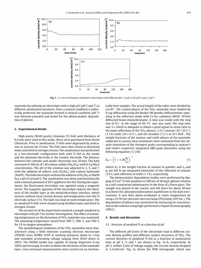
S. Sreekantan et al. / Journal of Alloys and Compounds 485 (2009) 478–483 479
yte wi
ndtwt
2
0Ctwiabccw(hwisneean
eiTf
a(a1(l
Fig. 1. I–t curve of titanium anodized in electrol
anotube by utilizing an electrolyte with a high pH (pH 5 and 7) atifferent anodization durations. Since a neutral condition is indus-rially preferred, the nanotube formed in neutral condition (pH 7)as thermal-annealed and tested for the photocatalytic degrada-
ion of phenol.
. Experimental details
High purity (99.6% purity) titanium (Ti) foils with thickness of.2 mm were used in this study; these were purchased from Stremhemicals. Prior to anodization, Ti foils were degreased by sonica-ion in acetone for 15 min. The foils were then rinsed in deionizedater and dried in nitrogen stream. The anodization was performed
n a two-electrode configuration bath with Ti foil as the anodend the platinum electrode as the counter electrode. The distanceetween the cathodic and anodic electrodes was 30 mm. The bathonsisted of 100 mL of 1 M sodium sulfate (Na2SO4) with 0.3 g NH4Foncentration. The pH of the solution was adjusted to 3, 5, and 7,ith the addition of sulfuric acid (H2SO4) and sodium hydroxide
NaOH). The bulk electrolyte without the addition of H2SO4 or NaOHas a pH of around 6. The anodization was done potentiostaticallyith constant potential of 20 V applied to the foil. During the exper-
ment, the fluorinated electrolyte was agitated using a magnetictirrer. The magnetic agitation of the electrolyte reduces the thick-ess of the double layer at the metal/electrolyte interface, whichnsures uniform local current density and temperature over the Tilectrode surface [11]. The bath was kept at room temperature. Thes-anodized Ti foils were cleaned using distilled water and dried initrogen stream.
The second set of the experiment involved a sample anodized inlectrolyte with pH 7 for further investigation. The effect of anneal-ng temperature on the formation of TiO2 nanotube was examined.he annealing temperature varied from 300 ◦C, 500 ◦C, and 700 ◦Cor 2 h in oxygen atmosphere.
The morphological conditions of the TiO2 nanotubes were char-cterized using a field emission scanning electron microscope
FESEM) Zeiss SUPRA 35VP at working distances down to 1 mmnd extended accelerating voltage ranging from 30 kV down to00 V. The FESEM model was capable of energy dispersive X-rayEDX) spectroscopy. In order to obtain the thickness of the nanotubeayer, cross-sectional measurements were carried out on mechan-th different pHs: (a) pH 3, (b) pH 5 and (c) pH 7.
ically bent samples. The actual length of the tubes were divided bycos 45◦. The crystal phases of the TiO2 nanotube were studied byX-ray diffraction using the Bruker D8 powder diffractometer oper-ating in the reflection mode with Cu K� radiation (40 kV, 30 mA)diffracted beam monochromator. A step scan mode with the stepsize of 0.1◦ in the range of 20–75◦ was also used. The step timewas 3 s, which is adequate to obtain a good signal-to-noise ratio inthe main reflections of the TiO2 phases, (1 0 1) anatase (2� = 25.3◦),(1 1 0) rutile (2� = 27.5◦), and the brookite (1 2 1) at 2� = 30.8◦. Theweight fractions of the anatase and rutile phases of the nanotubesubjected to various heat treatments were estimated from the rel-ative intensities of the strongest peaks corresponding to anatase’sand rutile’s respective integrated XRD peak intensities using thefollowing equation (1) [18]:
XA =(
1 + 1.26IRIA
)−1(1)
where XA is the weight fraction of anatase in powder, and IA andIR are the X-ray integrated intensities of the reflection of anatase(1 0 1) and reflection of rutile (1 1 0), respectively.
The photocatalytic degradation studies were performed by dip-ping 0.5 cm2 Ti foil anodized in 500 mL of 50 mg/L phenol solutionin a self-constructed photoreactor in the form of a Pyrex glass. Thesample was placed in the reactor and left there for about 30 minto achieve the adsorption/desorption equilibrium in the dark envi-ronment. It was then photo-irradiated at room temperature byusing a 15-W low-pressure mercury lamp (PCQ lamp, UVP, Inc.). Thedegradation of phenol was monitored by measuring its concentra-tion in the solution using high-performance liquid chromatography(HPLC).
3. Results and discussion
3.1. Structure of anodized Ti as a function of pH
The different pH levels of the electrolyte lead to different cur-
rent density profiles and different surface structures of TiO2. Thecurrent densities of anodized Ti foil as a function of anodizationtime at pH 3, 5, and 7 are shown in Fig. 1a–b, respectively. AtpH 3, within 5 min of voltage supply, the current density droppedto 5.3 mA/cm2. Fig. 2a shows the SEM micrograph, which was
480 S. Sreekantan et al. / Journal of Alloys and Compounds 485 (2009) 478–483
F t timd
otTrimiorsa(satol3oipr[
ig. 2. SEM images of the titanium foil anodized in electrolyte with pH 3 at differenis at a magnification of 50 000× shows the length of the nanotubes.)
btained within 5 min of voltage supply. It confirms that the dras-ic drop in current density is attributable to the oxidation of thei foil to form TiO2. The presence of TiO2 was confirmed with EDXesults (not shown) [19]. However, with the time of anodizationncreased to 10 min, the current density also increased to reach a
aximum value of 5.6 mA/cm2. The increase in current densitys caused by the formation of pits and enlargement of the pitsn the oxide layer as shown in Fig. 2b. Beyond this point, cur-ent density drops slowly, indicating the growth of the nanotubetructure. A well-ordered TiO2 nanotube with a diameter of 90 nmnd a length of 700 nm was formed after 30 min of anodizationFig. 2c). After 30 min of anodization, the resulting current den-ity showed a very small periodic fluctuation between 4.2 mA/cm2
nd 4.3 mA/cm2. This is probably due to the competition betweenhe growth and dissolution of the oxide layer to form a stable nan-tube. No significant changes were observed with regard to the
ength of the nanotube produced in a time period ranging from0 min to 120 min (Fig. 2d). Nevertheless, some overlapping nan-
tubes were observed in the sample anodized for 120 min, which isn agreement with the observation of Zhao and Wang [20]. Newores inside existing pores were created (inset in Fig. 2d) as aesult of the breakdown of repassivated layer inside the pores20].
es: (a) 5 min, (b) 10 min, (c) 30 min and (d) 120 min. (The inset micrograph in c and
At pH 5 and 7, the drops in current density were more significantas compared with pH 3. The current density dropped specificallyto 1.9 mA/cm2 (Fig. 1b) at pH 5 and 0.3 mA/cm2 (Fig. 1c) at pH 7.This indicates that the oxide layer formed in pH 5 (Fig. 3a) andpH 7 (Fig. 4a) is denser as compared to that in pH 3. Dense oxidelayer results in high ohmic resistance, which ultimately reduces thecurrent density. The formation of less dense oxide at a low pH canbe explained using the following equations (Eqs. (I) and (II)):
2H2O → O2 + 4H+ + 4e (I)
Ti + O2 → TiO2 (II)
Consequently, a low pH (high concentration of H+) reducesthe hydrolysis ability (Eq. (I)) of the electrolyte. This subsequentlyresults in lower concentration of oxygen, which is required in theformation of a dense oxide layer.
The SEM micrographs shown in Figs. 3b and 4b were taken atpH 5 and 7 for 10 min and 30 min of anodization time, respectively.
Results indicated that a high pH requires a longer anodization timefor the formation of pits. Furthermore, it was observed that the rateof nanotube formation is closely related to the pH of the electrolyte.At pH 3, the length of the tube formed within 30 min was 700 nm(inset in Fig. 2c). At pH 5, the length of the tube was shorter ∼600 nm
S. Sreekantan et al. / Journal of Alloys and Compounds 485 (2009) 478–483 481
F t timd
(tTAaid[e
T
abeiCtn
wp
ig. 3. SEM images of the titanium foil anodized in electrolyte with pH 5 at differenis at a magnification of 50 000× shows the length of the nanotubes.)
inset in Fig. 3c). On the other hand, at pH 7, the average length ofhe tube formed within 60 min was around 500 nm (inset in Fig. 4c).hus, the rate of formation at pH 3 was more or less 23 nm min−1.t pH 5, the rate was approximately 15 nm min−1, while pH 7 hadbout 8 nm min−1. The reduced rate of nanotube formation withncreased pH is probably associated with the decreased chemicalissolubility of the electrolyte. As explained by Grimes and Shumiki11,12], the mechanism of pit formation is given in the followingquation (Eq. (III)):
iO2 + 6F− + 4H+ → [TiF6]2− + 2H2O (III)
Based on this equation, it is apparent that the oxide layer isttacked in fluoride media under the formation of highly solu-le [TiF6]2− complex, which is closely related to the pH of thelectrolyte. As for a high pH, the formation of [TiF6]2− complexs reduced, resulting in a relatively weak chemical dissolubility.onsequently, it requires longer anodization time for the forma-
ion of pits as well as for the formation of stable and well-orderedanotubes.Nevertheless, at pH 5 and 7, it is noted that the tube’s length andall thickness are significantly altered by the anodization time. AtH 5, by prolonging anodization time to 120 min (inset in Fig. 3d),
es: (a) 5 min, (b) 10 min, (c) 30 min and (d) 120 min. (The inset micrograph in c and
an average length of 1.8 �m with a wall thickness of 20 nm (Fig. 3d)was obtained. At pH 7, the average length of 2.5 �m with parts of theregion having a length of 3 �m (inset Fig. 4d) was achieved within360 min. The thickness of the wall was ∼25 nm. It is of note thatthe formation of TiO2 nanotube at pH 7 is in disagreement with theresults of other previous studies, which indicate that nanotubes canonly be formed in an acidic condition [21–23].
3.2. Structure of anodized Ti as a function annealing temperature
Fig. 5 shows the crystal structure of the TiO2 nanotube pro-duced at pH 7 for 2 h as a function of annealing temperature. Theresult shows that the crystal structure of TiO2 is dependent on theannealing temperature. The XRD of the anodized sample withoutannealing (Fig. 5a) indicated that the self-organized TiO2 nanotubesdid have an amorphous structure since only Ti-peaks were shownand originated from the foil. However, the sample treated at 300 ◦C
showed Ti-peaks and indistinct anatase peaks, indicating the pres-ence of anatase phase with a low degree of crystallinity (Fig. 5b).Ti-peaks again presented because the information from the sub-strate is revealed. Polycrystalline anatase- and rutile-type TiO2could be obtained by heating up to 500 ◦C (Fig. 5c). The weight frac-
482 S. Sreekantan et al. / Journal of Alloys and Compounds 485 (2009) 478–483
Fig. 4. SEM images of the titanium foil anodized in electrolyte with pH 7 at different timed is at a magnification of 50 000× shows the length of the nanotubes.)
Fig. 5. XRD patterns of (a) un-annealed sample; (b) annealed at 300 ◦C, (c) 500 ◦Cand (d) 700 ◦C [(�)Ti, (♣) rutile and (�) anatase].
s: (a) 5 min, (b) 30 min, (c) 60 min and (d) 360 min. (The inset micrograph in c and
tions of the anatase and rutile phases of the nanotube subjected to500 ◦C were 76% and 24%, respectively. When the annealing temper-ature was increased to 700 ◦C (Fig. 5d), the anatase peak vanished,while rutile as a major phase was observed.
3.3. Photocatalytic degradation of phenol
Fig. 6 shows the use of TiO2 nanotube, which was produced inneutral condition and annealed at various temperatures, in the pho-tocatalytic degradation of phenol, a toxic and harmful pollutant. Atypical run without UV irradiation confirms no photo-oxidation tak-ing place (region A) for all samples, but there was an adsorption ofa small amount of phenol on the sample. After 120 min of UV irra-diation, the sample annealed at 300 ◦C, 500 ◦C and 700 ◦C showed adecrease in phenol concentration of 62.5%, 77.5%, and 54%, respec-tively. The un-annealed sample showed a 40% decrease in phenolconcentration. At a later stage (after 240 min of UV irradiation), thesample annealed at 500 ◦C showed complete degradation of phenol,while the sample annealed at 300 ◦C, 700 ◦C, and the un-annealed
sample showed a degradation of 87%, 76%, and 70%, respectively.Based on the results obtained, it appears that the crystal struc-ture and the degree of crystallinity are the important factors thataffecting photocatalytic activity. Although it is generally acceptedthat the anatase phase is a good candidate for the degradation of

S. Sreekantan et al. / Journal of Alloys and
Fa
aaaar
ocisoaaawt
artrb(1
4
i
[
[[
[[
[[[26] S.V. Awate, K. Suzuki, Adsorption 7 (2001) 326.[27] Y.H. Hsien, C.F. Chang, Y.H. Chen, S. Cheng, Appl. Catal. B 31 (2001) 241.
ig. 6. The phenol degradation of titania nanotube (a) without annealing, (b) afternnealing at 300 ◦C, (c) 500 ◦C and (d) 700 ◦C.
n organic pollutant [24–28], this results of this study demonstratedecreased activity in phenol degradation for a nanotube with thenatase phase. This is probably associated with the low degree ofnatase crystallinity, which increases the number of charge carrierecombination center.
In contrast, the sample annealed at 500 ◦C, which consistsf high crystalline anatase and rutile, exhibited the most effi-ient photocatalytic activity for the degradation of phenol. Thiss in agreement with the results of Hurum’s work [29], whichuggested that within a mixed-phase TiO2, there is a morphol-gy of nanoclusters containing rutile crystallites interwoven withnatase crystallites; this allows rapid electron transfer from rutile tonatase, which hinders recombination. As a consequence, rutile actss an antenna to extend the photoactivity into visible wavelengths,ith the structural arrangement creating catalytic “hot spots” at
he rutile–anatase interface.This also explains the behavior of the sample which was
nnealed at 700 ◦C. This sample, which comprise consists of 100%utile phase, showed less activity because of lack of anatase phasehat could assist in reducing the rapid rate of recombination inutile. However, the sample with pure anatase (300 ◦C) showedetter photoactivity in phenol degradation as compared to rutile700 ◦C) provided sufficient time is allocated (in this case, it took20 min to achieve sufficient degradation of phenol).
. Conclusion
The results of this study demonstrated the possibility of obtain-ng TiO2 nanotubes in 1 M Na2SO4 solution containing 3 wt% NH4F
[
[
Compounds 485 (2009) 478–483 483
at different pH levels. The rate of nanotube formation varies withdifferent pH levels. At pH 3, the rate of formation was 23 nm min−1;at pH 5, it was 15 nm min−1; and at pH 7, it was 8 nm min−1. Controlover the rate of the nanotubes formation was, therefore, success-fully established in this work simply by controlling electrolyte pH.The nanotube produced has an amorphous form, which can betransformed into anatase and rutile upon annealing. The samplethat was annealed at 500 ◦C, which consisted of high crystallineanatase (76%) and rutile (24%), has been acknowledged as the mostefficient form for degradation of phenol.
Acknowledgments
The author would like to thank Universiti Sains Malaysia forsponsoring this work under Short Term Grant 2007 (6035227),Ministry of Higher Education for FRGS (6070020) and L’Oreal forFellowship (6050136).
References
[1] A. Fujishima, T.N. Rao, D.A. Tryk, J. Photochem. Photobiol. C 1 (2000) 1.[2] R. Asahi, Y. Taga, W. Mannstadt, A.J. Freeman, Phys. Rev. B 61 (2000) 7459.[3] M. Adachi, Y. Murata, I. Okada, S. Yoshikawa, J. Electrochem. Soc. 150 (2003)
G488.[4] S.H. Oh, R.R. Finones, C. Daraio, L.H. Chen, S.H. Jin, Biomaterials 26 (2005) 4938.[5] A. Fujishima, K. Honda, Nature 37 (1972) 238.[6] M. Gotic, M. Ivanda, A. Sekulic, S. Music, S. Popovic, A. Turkovic, K. Furic, Mater.
Lett. 28 (1996) 225.[7] W. Li, S.I. Shah, C.-P. Haung, C. Ni, Mater. Sci. Eng. B 96 (2002) 247.[8] J. Choi, R.B. Wehrspohn, J. Lee, U. Gösele, Electrochim. Acta 49 (2004) 2645.[9] D. Gong, C.A. Grimes, O.K. Varghese, W. Hu, R.S. Singh, Z. Chen, E.C. Dickey, J.
Mater. Res. 16 (2001) 3331.[10] R. Beranek, H. Hildebrand, P. Schmuki, Electrochem. Solid-State Lett. 6 (2003)
B12.[11] G.K. Mor, O.K. Varghese, M. Paulose, K. Shankar, C.A. Grimes, Sol. Energy Mater.
Sol. Cells 90 (2006) 2011.12] J.M. Macak, K. Sirotna, P. Schmuki, Electrochim. Acta 50 (2005) 3679.
[13] L. Taveira, J.M. Macak, H. Tsuchiya, L.F.P. Dick, P. Schmuki, J. Electrochem. Soc.152 (2005) B405.
[14] K. Shankar, G.K. Mor, H.E. Prakasan, S. Yoriya, M. Paulose, O.K. Varghese, C.A.Grimes, Nanotechnology 18 (2007).
[15] J. Wu, L. Shi, S. Yuan, Y. Chen, Y. Zhang, Chem. React. Eng. Technol. 24 (2008)147.
[16] Z. Yunhuai, H.U. Fu, X. Peng, F. Xiaoyan, Int. J. Mod. Phys. B 21 (2007) 3506.[17] X. Feng, J.M. Macak, P. Schmuki, Chem. Mater. 19 (2007) 1534.[18] R.A. Spurr, H. Myers, Anal. Chem. 29 (1957) 760.[19] S. Sreekantan, M.H. Ling, Z. Lockman, Z.A. Ahmad, Proceedings of the Inter-
national Conference on Advancement of Materials and Nanotechnology,Langkawi, Malaysia, 29 May–1 June, 2007, p. 92.
20] J. Zhao, X. Wang, R. Chen, L. Li, Solid State Commun. 134 (2005) 705.21] G.K. Mor, O.K. Varghese, M. Paulose, N. Mukherjee, C.A. Grimes, J. Mater. Res. 18
(2003) 2588.22] J.M. Macak, H. Tsuchiya, P. Schmuki, Angew. Chem. Int. Ed. 44 (2005) 2100.23] J.M. Macak, H. Tsuchiya, L. Taveira, S. Aldabergerova, P. Schmuki, Angew. Chem.
Int. Ed. 44 (2005) 7463.24] A. Hagfeldt, M. Gratzel, Chem. Rev. 95 (1995) 49.25] K. Gurunathan, J. Mol. Catal. A 156 (2000) 67.
28] J. Pichat, J. Disdier, C. Hoang-Van, D. Mas, G. Goutailler, C. Gaysse, Catal. Today63 (2000) 363.
29] D.C. Hurum, A.G. Agrios, K.A. Gray, T. Rajh, M.C. Thurnauer, J. Phys. Chem. B 107(2003) 4545.