Critical role of formaldehyde during methanol conversion to ...
Upload
independentCategory
view
3download
0
International Journal for Pharmaceutical Research Scholars (IJPRS)
V-2, I-3, 2013 ISSN No: 2277 - 7873 RESEARCH ARTICLE
© Copyright reserved by IJPRS 74
Induction of Apoptosis in Human Colon Cancer Cells by Methanol Fraction of
Leaves of Plectranthus amboinicus (Lour) Spreng Pillai PG1*, Suresh P2, Gitanjali M3
*1KVM College of Pharmacy, Cherthala, Kerala, India. 2Department of Pharmacy, GITAM Institute of Pharmacy, GITAM University, Rushikonda,
Visakhapatnam- 530045, Andhra Pradesh, India. 3Department of Zoology, Berhampur University, Bhanja-Bihar, Orissa, India.
Manuscript No: IJPRS/V2/I3/00135, Received On: 08/08/2013, Accepted On: 22/08/2013
ABSTRACT To evaluate the cytotoxicity and apoptosis inducing activities of the methanol extract from leaves of Plectranthus amboinicus (Lour) Spreng in an attempt to determine whether the medicinal uses are supported by pharmacological effects. Cytotoxicity was determined by sulforhodamine B assay method. Cytotoxicity and apoptosis inducing effect were evaluated in- vitro using human colon cancer cell line, COLO 205. There was statistically significant cell growth inhibition at the doses of 10, 20, 40 and 80 µg/ml methanol extract of Plectranthus amboinicus (Lour) Spreng. Similarly, Plectranthus amboinicus at the doses of 50 and 100 µg/ml induced apoptosis in COLO 205 cells. The results suggest that the methanol extract of Plectranthus amboinicus (Lour) Spreng possesses anti- proliferative and apoptosis inducing activities, supporting the folk use of this medicinal species.
KEYWORDS Plectranthus amboinicus (Lour) Spreng, Cytotoxicity, apoptosis, COLO 205
INTRODUCTION Colon cancer is the third most common type of cancer worldwide. The highest prevalence of colon cancer occurs in North America, Europe, Australia, and New Zealand and accounts for almost 65% of the total global incidence1. The lowest incidence was seen in Central and South America, Asia and Africa, but it is now increasing in these regions. Epidemiological studies suggest that the consumption of vegetables and fruits is inversely associated with colon cancer risk2. Much of the protective effects of vegetables and fruits have been attributed to biologically active secondary plant
metabolites which are non-nutrient constituents such as the carotenoids, the phenolic acids and flavonoids 2,3. The Plectranthus amboinicus (Lour) Spreng (Lamiaceae) is a native species from Asia and distributed in America. It is commonly known as country borage in English and its use is frequent in folk medicine against urinary diseases, asthma, chronic bronchitis, hepatopathy, malarial fever4.
Previous pharmacological studies showed that Plectranthus amboinicus possesses urolithiatic5, anti-epileptic6, anti-tumor and anti-mutagenic7, neuropharmacological8, radio protective9, antioxidant10, anti-microbial11,12 anti-bacterial and anti-fungal properties13,14.
*Address for Correspondence: Dr. Preeja G Pillai KVM College of Pharmacy, Cherthala, Alappuzha (DT) Kerala, India-680583. E-Mail Id: [email protected]

Induction of Apoptosis in Human Colon Cancer Cells by Methanol Fraction of Leaves of Plectranthus amboinicus (Lour) Spreng
© Copyright reserved by IJPRS 75
The leaves of this species contain essential oils, flavonoids, terpens and cinnamic derivates15. In Brazil, especially in the Northeast Region, the use of this medicinal species is a practice widely employed, mainly in the form of crude extracts and infusions, to the treatment of several diseases including inflammatory diseases and cancer15,16. There are pharmacological evidences that support this medicinal species for anti-inflammatory and anti-tumor activity against Sarcoma-180 ascite and Ehrlich ascite carcinoma7. Therefore, it is necessary to investigate the anti-proliferative activity and the molecular mechanism of Plectranthus amboinicus in order to determine whether the medicinal uses are supported by pharmacological effects. We report here the results of in- vitro experimental studies with the methanol extract from the leaves of Plectranthus amboinicus on COLO 205 colon cancer cells by sulforhodamine B (SRB) assay protocol.
MATERIALS AND METHODS Plant Material The leaves of Plectranthus amboinicus (Lour) Spreng were authenticated by Dr. A.K.Pradeep, Reader, Dept of Botany, and University of Calicut. A voucher specimen (No.3/386/2008/Adm.7646) was deposited in GITAM Institute of Pharmacy, Andhra Pradesh.
Extraction The shade dried powdered leaves of Plectranthus amboinicus (Lour) Spreng were subjected to Soxhlet extraction successively with 95% methanol. Methanolic Extract of Plectranthus amboinicus (Lour) Spreng (MEPA) was concentrated in a rotary flash evaporator. The residue was dried in desiccator over sodium sulfite.
Cell Lines Human colon cancer cell line, COLO 205 culture was obtained from the National Cancer Institute (USA). RPMI-1640 medium and fetal bovine serum (FBS) was purchased from Gibco Corp. (NJ). Dulbecco’s Modified Eagle’s
Medium (DMEM) was purchased from Himedia Laboratory, Mumbai, India. Sulforhodamine B dye was bought from Sigma Chemical Co. All sterile plastic-wares were procured from Nunc Inc. (Denmark). All other reagents and solvents used were procured locally and were of analytical grade. Cells-cDNA kit was purchased from Ambion Inc (Austin Tex, USA).
In vitro Cytotoxicity Studies The cells were seeded in 96-well microtitre-plates (at a concentration of 1 × 10⁴ cells/well) and incubated for 24 h and MEPA were added in triplicate in different lanes of the wells, Cell cultures were fixed by slow addition of cold 50% trichloroacetic acid (TCA) and SRB solution (0.4% w/v in 1% acetic acid) was added to each of the wells and kept at room temperature for 20 min. The unbound dye was removed by washing with 1% acetic acid and bound dye was then extracted by addition of 10mM Tris base (pH 10.5) to each of the wells. Optical density was measured at 540 nm on an ELISA microplate reader 17.
Effect on Cell Cycling of COLO205 Cells One million COLO 205 cells suspended in DMEM with 10% FBS and were seeded in- to 4 culture flasks. These cells were incubated for 4, 48 and 72 hours at 370C in CO2 incubator with extract (100 µg/ml). After incubation the cells were analyzed for cell cycle changes in flow-cytometer using Becton-Dickson kit as per the manufacture’s protocol 18.
Effect on the Apoptosis of COLO205 cells Morphological and Nuclei Changes COLO 205 cells suspended in RPMI containing 20% FBS were cultured in 96 -well plates in the presence of 50,100 µg/ml of MEPA at 370C for 24 hrs. About 60l of the medium was removed from the wells and the same amount of the diluted dye was added. The cells were then incubated at 37ºc for 10 minutes in a 5% CO2 incubator. 60l of the medium was removed from the wells and observed under Nikon inverted fluorescent microscope and fluorescent images were captured. Apoptotic nucleus with

Induction of Apoptosis in Human Colon Cancer Cells by Methanol Fraction of Leaves of Plectranthus amboinicus (Lour) Spreng
© Copyright reserved by IJPRS 76
condensed chromatin was scored in percentage from 200 - 300 cells/sample 19.
DNA Fragmentation Analysis One million COLO 205 cells were treated with different concentrations of MEPA (50, 100 μg/ml) for 48 hrs. After incubation , the cells were treated with 0.1 ml lysis buffer (100 mmol/L Tris-HCl, pH 8.0, containing 0.2% Triton-X 100, and 1mmol/L EDTA) for 10 minutes at -200C. DNA was extracted using Phenol- Chloroform method, precipitated with chilled ethanol and resuspended in Tris/EDTA buffer (10 mmol/L Tris-HCl, pH 8.0 and 1 mmol/L EDTA). DNA samples were separated by electrophoresis in 1% agarose gels. DNA was stained with ethidium bromide and photographed under UV light 18.
Terminal Deoxynucleotidyl Transferase dUTP Nick End Labeling (TUNEL) Assay TUNEL assay was done to detect apoptosis via DNA fragmentation using Apoptag Peroxidase in situ Apoptosis detection kit, (CHEMICO). Cells (5,000 cells/ well) suspended in DMEM supplemented with 10% FCS, 100μg/ml streptomycin and penicillin and 2mmol/l glutamine were plated in 96-well flat bottom titer plates and incubated for 24 h at 37°C in a 5% CO2 incubator. After 24 h, different concentrations of MEPA (50, 100 μg/ml) were added to the cells, which were then incubated further for 48 h under the same conditions. The cells were washed in PBS and stained as per manufacturer’s protocol 18,20.
Effect of MEPA on Expression of Caspase 3, 8, 9, Bax, Bid and p53 genes The effect of MEPA on the genes regulating cell cycling and apoptosis of COLO 205 cells were analyzed by RT-PCR method. COLO 205 cells (2x10⁴ cells) suspended in serum free DMEM (250 μl) were seeded in 96 well titer plate and incubated for 24 h at 370 C in 5% CO2 incubator. Extract at a concentration of 100μg/ml were added in to the wells and incubation was continued for another 4h. cDNA was synthesized after 4 hrs incubation using cell
to cDNA kit (Ambion Inc.). Gene expression analysis was done by PCR18.
Statistical Analysis Data were presented as mean± SD. Differences between the mean of control and treatment groups were analyzed using one-way analysis of variance followed by Dunnet test and Tukey’s Kramer test. Statistical significance was determined at the level of P< 0.05 or P< 0.01 or P<0.001.
RESULTS Thin Layer Chromatography (TLC) Qualitative phytochemical evaluation of the extract was carried out in a systematic way. MEPA showed the presence of carbohydrate, glycoside, flavonoids, sterols, terpenoids, fixed oils and tannins. The Chromatography was performed by TLC using different solvent systems were carried out. TLC of flavonoid showed the presence of one spot corresponding to compounds with flavonoid behavior (yellow-brown fluorescensce, after observed under UV). Standard (std) Quercetin (Rf = 0.87), MEPA (Rf =0.87). The Rf value of std was found to be 0.87 in all tracks. The chromatogram of MEPA was very similar to the std. TLC of MEPA using Toluene: Ethyl acetate (93:7) gave 6 spots at different Rf value, TLC of MEPA using Toluene: Ethyl acetate: Methanol (15:3:1.5) showed bands at different Rf 0.66, 0.488, 0.44, 0.35, 0.31 and 0.62, 0.57, 0.48, 0.42, 0.35, 0.28, 0.24 respectively (Table 1 and Figure1).
In-vitro Cell Proliferation Both MEPA and Adiramycin (Adr) showed very good anti-proliferative activity (>50% cell growth inhibition) towards human colon cancer cells tested, in-vitro. The activity of extract was concentration dependent, maximum activity being obtained at the highest concentration tested (80µg/ml), whereas standard drug Adr showed cytotoxic effect even at lower concentration. The MEPA was less toxic than Adr in that the viable cell number decreased to less than 40% at concentrations 10 μg/ml.

Induction of Apoptosis in Human Colon Cancer Cells by Methanol Fraction of Leaves of Plectranthus amboinicus (Lour) Spreng
© Copyright reserved by IJPRS 77
Table 1: Thin layer chromatography of MEPA
Metabolite Solvent system Detection No. of spots Rf Values Presence/
Absence
Flavonoid
Ethyl Acetate: Formic acid:
Glacial Acetic Acid: Water
(100:11:11:27)
UV light 1 0.87 +
Monoterpenoids
Toluene: Ethyl acetate (93:7)
Vanillin-Sulphuric
acid reagent 6
0.789, 0.54, 0.47, 0.3508, 0.2456,
0.087 +
Terpenoids and Steroids
Toluene: Ethyl acetate: Methanol
(15:3:1.5)
Liebermann-Burchard reagent
5 0.66, 0.488, 0.44, 0.35,0.31 +
Figure 1: Thin Layer Chromatography of methanol extract of Plectranthus amboinicus (Lour) Spreng
(MEPA)

Induction of Apoptosis in Human Colon Cancer Cells by Methanol Fraction of Leaves of Plectranthus amboinicus (Lour) Spreng
© Copyright reserved by IJPRS 78
The Inhibitory concentration (IC50) for MEPA and Adr on COLO205 cells was 74.07± 1.4 μg/ml and 8.19 ± 0.02 μg/ml respectively. However, in the case of COLO 205 cancer cells, the anti-proliferatory effect of MEPA was found to be improved but statistically significant (two way analysis of variance, P <0.05) as compared with that std Adr [Figure 2]. MEPA extract showed more than 50% inhibition of cell growth at a concentration of 80µg/ml.
Figure 2: Percentage cell growth inhibition of COLO 205 human colon cancer cells
Each data point and error bars represent mean ± S.D of three replicates. Statistical analysis was carried out by two way ANOVA* considered significant at P< 0.05 compared with std Adr.
Effect on Cell Cycling of COLO 205 Cells In untreated control 3.29% cells were in sub- G0/G1, 56.1% of the cells were in G0/G1- phase (M1), 14.79% of the cells were in S- phase and 17.27% of the cells were in G2/M- phase (M2). Treatment with 100 μg/ml of MEPA increased the percentage of cells in sub- G0/G1- phase to 15.13%, 24.7%, and 60.75% at 4, 48 and 72 hours of incubation respectively. But the cells in G0/G1 phase, S phase, G2/M phase decreased considerably (21.01%, 15.25%, 2.5%) respectively for MEPA (Figure 3).
Figure 3: Effect of MEPA on COLO 205 cell
cycle
Effect on the Apoptosis of COLO205 cells Morphological and Nuclei Changes In order to show the cytotoxicity of MEPA is mediated by the induction of apoptosis, chromatin condensation was analyzed in COLO 205 cells by Hoechst staining. COLO 205 cells were treated with 50, 100 μg/ml of MEPA. At the end of the incubation period, the cells were observed under the inverted fluorescent microscope for chromatin condensation. Cells treated with 50, 100 μg/mL of MEPA showed evident morphological changes as well as the presence of apoptotic bodies. Such morphological change was not seen with control cells (without the MEPA treatment). However, the nucleus of MEPA treated cells showed significant condensed chromatin. Chromatin condensation and destructive fragmentation of the nucleus with intact cell membrane was seen with COLO 205 cells which had been treated with 50, 100 μg/ml of MEPA indicated late apoptotic event. Apoptotic cells showed clear difference from non-apoptotic cells as the former were bright and their nuclei were condensed. Percentage of apoptosis was increasing with increase in concentration of extract (Figure 4 and 5).
Induction of Apoptosis in Human Colon Cancer Cells by Methanol Fraction of Leaves of Plectranthus amboinicus (Lour) Spreng
© Copyright reserved by IJPRS 79
Figure 4: Effect of MEPA on the nuclear
condensation of COLO 205 cells. (A): Hoechst stained untreated control cells, (B &C): Hoechst
stained cells were examined for nuclei morphology in the presence of 50, 100 μg/ml of
Plectranthus amboinicus (Lour) Spreng
Figure 5: Percentage of apoptosis in cells was
treated with 50 and 100 μg/ml of MEPA
Control and treatment groups were analyzed using one-way ANOVA followed by Dunnet test and Tukey multiple comparison test. The values are expressed as mean ± SD; a P< 0.05 considered statistically significant.
The untreated COLO 205 cells exhibited typical growth patterns and a smooth, flattened morphology with normal nuclei. When treated with MEPA, the COLO 205 cells exhibited condensed chromatin, apoptotic morphological changes with cytoplasmic blebbing and
detachment from the surface (Figure 4). Most MEPA treated cells showed apoptotic bodies with cytoplasmic condensation indicating apoptosis like changes.
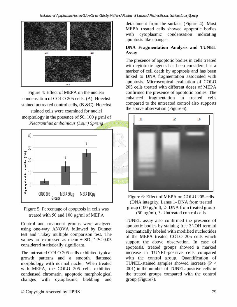
DNA Fragmentation Analysis and TUNEL Assay The presence of apoptotic bodies in cells treated with cytotoxic agents has been considered as a marker of cell death by apoptosis and has been linked to DNA fragmentation associated with apoptosis. Microscopical evaluation of COLO 205 cells treated with different doses of MEPA confirmed the presence of apoptotic bodies. The enhanced fragmentation in treated cells compared to the untreated control also supports the above observation (Figure 6).
Figure 6: Effect of MEPA on COLO 205 cells (DNA integrity. Lanes 1- DNA from treated
group (100 μg/ml), 2- DNA from treated group (50 μg/ml), 3- Untreated control cells
TUNEL assay also confirmed the presence of apoptotic bodies by staining free 3’-OH termini enzymatically labeled with modified nucleotides of the MEPA treated COLO 205 cells which support the above observation. In case of apoptosis, treated groups showed a marked increase in TUNEL-positive cells compared with the control group. Quantification of TUNEL-stained samples showed increase (P < .001) in the number of TUNEL-positive cells in the treated groups compared with the control group (Figure7).

Induction of Apoptosis in Human Colon Cancer Cells by Methanol Fraction of Leaves of Plectranthus amboinicus (Lour) Spreng
© Copyright reserved by IJPRS 80
Figure 7: Effect of the apoptosis of COLO 205
cells by TUNEL assay
Effect of MEPA on Expression of Caspase 3, 8, 9, Bax, Bid, p53 genes To determine the mechanism of MEPA-induced apoptosis, the expression of anti- and pro-apoptotic proteins following MEPA treatment was examined by RT-PCR method. It was clear from the results that the expression of p53, Caspase-3, 8, 9, and bax were up regulated in MEPA treated cells compared to the untreated control cells. Bid gene was expressed both in control and treated cells (Figure 8).
Caspase 3
Caspase 8
Caspase 9
Bax
p53
Bid
Control Treated
Figure 8: Effects of MEPA on expression of p53, Caspases, Bax, and Bid in COLO 205 cells,
Left- untreated control, Right- MEPA treated (100 μg/ml)
DISCUSSION In the present study, we showed that MEPA treatment of human colon COLO 205 cells induced cytotoxic effects in a dose dependent fashion. The SRB assay showed significantly reduced rate of proliferation of COLO 205 cells after the treatment with MEPA. The p53-dependent apoptosis pathway is of particular interest in cancer therapy since it can be used to eliminate tumors. The involvement of p53 in multiple biological pathways suggests that its loss may have dramatic consequence for the cell and that it may be an important event in cancer development. The study of p53 gene knockout mice [21] and the analysis of the status of the p53 gene in human tumors show that the loss of p53 activity is a key event in cancer development. Here in this study we found a clear up regulation of the p53 gene in COLO 205 cells after the treatment with MEPA. Kuttan et al. also found that the loss of p53 activity is a key event in cancer development.
Apoptosis is characterized by the activation of a specific family of cystein proteases, the caspases, followed by a series of caspase mediated morphological changes such as shrinkage of the cells, the condensation of chromatin and membrane blebbing22. The presence of cells with apoptotic bodies in cultured cells has been considered as a marker of cell death by apoptosis and has been linked to DNA fragmentation. Here the cells after treatment of MEPA showed chromatin condensation, apoptotic body formation and membrane blebbing in a dose dependent manner.
Caspase-3 is an effector caspase which once activated, cleaves many substrate proteins and structural proteins to generate the characteristic apoptotic morphology. The data from the reverse transcription PCR and agarose gel electrophoresis showed clear band of caspase-3. In cancerous cells, the expression of caspase-3 is much suppressed and in the present study the untreated control COLO 205 cells did not exhibit its expression. Caspase-3 is a most likely candidate to mediate MEPA induced apoptosis

Induction of Apoptosis in Human Colon Cancer Cells by Methanol Fraction of Leaves of Plectranthus amboinicus (Lour) Spreng
© Copyright reserved by IJPRS 81
in COLO 205 cells as evidenced by increased expression of this gene upon treatment with MEPA. DNA extracts from COLO 205 cells incubated with MEPA displayed characteristic ladder pattern of discontinuous DNA fragments which support the above results. The increased sub G0/G1 population of COLO 205 cells in cell cycle analysis and increased number of stained cells in the TUNEL assay also support MEPA induced apoptosis and DNA fragmentation in MEPA treated cells. Several studies have reported that Plectranthus amboinicus (Lour) Spreng possesses anti-tumor, anti-mutagenic, anti-inflammatory and antioxidant properties7,10,15,16. However, to the best of our knowledge, this is the first report on the effect of MEPA on apoptosis and DNA fragmentation of COLO 205 cells. Therefore the role of Plectranthus amboinicus in apoptosis induction and DNA fragmentation in COLO205 cells as seen in the present study may be in part due to the antioxidant and anti-inflammatory effects.
CONCLUSION The above data clearly demonstrate that MEPA induced apoptosis in COLO 205 cells by activating p53 induced caspase-3 apoptotic signaling. This could be a promising chemotherapeutic agent and may also serve as a model to develop and design new derivatives which may be more potent.
REFERENCES 1. World Health Organization, “World Cancer
Report”, Lyon: IARC Press, 2003. 2. Terry P, Giovannucci E, Michels KB,
Bergkvist L, Hansen H, Holmberg L, Wolk A, “ Fruit, vegetables, dietary fiber, and risk of colorectal cancer”, Journal of National Cancer Institutes, 2001, 93, 525-533.
3. Maria-Elena MC, Souad B, Francine G, Carole M, Annelise L, Francis R, “Differential Induction of Apoptosis by Apple Procyanidins in TRAIL-Sensitive Human Colon Tumor Cells and Derived TRAIL- Resistant Metastatic Cells”, Journal of Cancer Molecules, 2009, 5(1), 21-30.
4. Lukhoba CW, Simmonds MSJ, Paton AJ, “Plectranthus: a review of ethno botanical uses”, Journal of Ethnopharmacology, 2006, 103, 1–24.
5. Jose A, Ibrahim M, Janardhanan S, “Modulatory effect of Plectranthus amboinicus Lour on ethylene glycol induced nephrolithiasis in rats” Indian Journal of Pharmacology, 2005, 37, 43–44.
6. Buznego MT, Perez-Saad H, “Antiepileptic effect of Plectranthus amboinicus (Lour.) Spring”, Revista de Neurologia, 1999, 29, 229–232.
7. Annapurna S, Priya R, “Anti-mutagenic and anti-genotoxic effects of polyphenol extracts of selected medicinal plants” Indian Journal of Nutrition and Dietetics, 1999, 36, 431–435.
8. Perez SH, Buznego MT, Lianio M, Fernandez P, Menedez R, “Neuropharmacological profile of Plectranthus amboinicus (Lour) Spreng (Indian borage)” Revista de Neurologia, 2003, 36, 98–99.
9. Rao BS, Shanbhoge R, Upadhya D, Iagetia GC, Adiga SK, Kumar P, “Antioxidant, anticlastogenic and radio protective effect of Coleus aromaticus on Chinese hamster fibroblast cells (V79) exposed to gamma radiation” Mutagenesis, 2006, 21, 237–242.
10. Kumaran A, Karunakaran JR, “Antioxidant and free radical scavenging activity of an aqueous extract of Coleus aromaticus”, Food Chemistry, 2006, 97, 109–114.
11. Rao A, Baby P, Prasad RY, “Leaf oil of Coleus amboinicus Lour: the in vitro anti-microbial studies”, Perfume Kosmetik, 1991, 72, 744–745.
12. Deena MJ, Sreeranjini K, Thoppil JE, “Antimicrobial screening of essential oils of Coleus aromaticus and Coleus zeylanicus”, International Journal of Aromatherapy, 2002, 12, 105–107.
13. Vijaya Kumar S, Syed MA, Bdami S, Anil TM, David B, “Antibacterial activity of

Induction of Apoptosis in Human Colon Cancer Cells by Methanol Fraction of Leaves of Plectranthus amboinicus (Lour) Spreng
© Copyright reserved by IJPRS 82
aqueous extract of Coleus amboinicus”, Pharmacology Online, 2008, 3, 224-226.
14. Murthy PS, Murthy RK, Srinivas P, “Fungitoxic activity of Indian borage (Plectranthus amboinicus) volatiles”, Food chemistry, 2009, 114, 1014-1018.
15. Diniz MF, Memento fitoterápico: as plantas como alternativa terapêutica: conhecimentos populares ecientíficos. Ed Universitária/UFPB, João Pessoa 1997, 205.
16. Lorenzi H, Matos FJA. Plantas medicinais no Brasil: nativas exóticas. Institute Plantarum, Nova Odessa 2002.
17. Kale Rajendrakumar, Saraf Madhusudan, Juvekar Aarti, Tayade Pralhad, “Decreased B16F10 Melanoma Growth and Impaired Tumour Vascularization in BDF1 Mice with Quercetin–Cyclodextrin Binary System”, Journal of Pharmacy and Pharmacology, 2006, 58, 1351-1358.
18. Kanjoormana AM, Kuttan G, “Punarnavine Induction of Apoptosis in B16F-10 Cells by
Inhibition of NF-κB Signaling”,Asia Pacific Journal of Cancer Prevention, 2009, 10, 1031-1037.
19. Merlin NJ, Parthasarathy V, Santhoshkumar TR, “Induction of apoptosis in human breast cancer cell line MCF-7 by phytochemicals from Gmelina asiatica”, African Journal of Biotechnology, 2010, 9(28), 4451-4456.
20. Kaur M, Velmurugan B, Tyagi A, Agarwal C, Singh RP, Agarwal R, “Silibinin suppresses growth of human Colorectal carcinoma SW480 cells in culture and xenograft through down regulation of β-Catenin-dependent signaling”, Neoplasia, 2010, 12(5), 415-424.
21. Clarke PG, Clarke S, “Historic apoptosis,” Nature, 1995, 378, 230.
22. Kothakota S, Azuma T, Reinhard C, “Caspase-3- generated fragment of gelsolin: effector of morphological change in apoptosis,” Science, 1997, 278, 294-298.
Copyright © 2022 FDOKUMEN