Indolequinone inhibitors of NRH:quinone oxidoreductase 2. Characterization of the mechanism of...
11
Published: June 30, 2011 r2011 American Chemical Society 6678 dx.doi.org/10.1021/bi2002967 | Biochemistry 2011, 50, 6678–6688 ARTICLE pubs.acs.org/biochemistry Indolequinone Inhibitors of NRH:Quinone Oxidoreductase 2. Characterization of the Mechanism of Inhibition in both Cell-Free and Cellular Systems Chao Yan, † Marine Dufour, ‡ David Siegel, † Philip Reigan, † Joe Gomez, † Biehuoy Shieh, † Christopher J. Moody,* ,‡ and David Ross* ,† † Department of Pharmaceutical Sciences, School of Pharmacy, University of Colorado Denver, Aurora, Colorado 80045, United States ‡ School of Chemistry, University of Nottingham, University Park, Nottingham NG7 2RD, U.K. b S Supporting Information T here are two quinone reductases that occur in mammalian systems, NAD(P)H:quinone oxidoreductase 1 (NQO1, EC 1.6.99.2) and NRH:quinone oxidoreductase 2 (NQO2, EC 1.10.99.2). NQO1 was originally characterized by Ernster and Navazio 1,2 and was probably identical to an enzyme isolated by Martius a few years earlier. 3,4 Interestingly, NQO2 was cloned and fully characterized by Jaiswal et al. 5 but as highlighted by Zhao et al. 6 was also found to be identical to a flavoprotein isolated 30 years earlier. 7 Both NQO1 and NQO2 are homo- dimeric flavoproteins, containing one FAD site per monomer, that utilize pyridine nucleotide cofactors to catalyze the direct two-electron reduction of a broad range of quinone substrates. 6,8,9 However, NQO2 differs from NQO1 in that it utilizes dihydro- nicotinamide riboside (NRH) instead of NAD(P)H as the cofactor. In addition, in comparison to NQO1, which is usually strongly expressed in solid tumors, 10 higher levels of NQO2 expression are found in red blood cells 11 and in leukemias. 12 With respect to quinone substrates, NQO2 has been suggested to preferentially reduce o-quinones derived from catecholamines and estrogen, which has led to its proposed involvement in neurodegenerative diseases and breast cancer. 13,14 NQO2 has been shown to reduce numerous antitumor quinones in vitro, including mitomycin C, 15 RH1, 16 and the HSP90 inhibitor 17AAG, 17 while the antitumor activity of CB1954, a non-quin- one dinitrobenzamide-containing compound currently in clinical trials, relies on targeted activation by NQO2 via nitroreduction. 18 The identification of inhibitors for NQO2 has generated considerable interest. Despite structural similarities between NQO2 and NQO1, commonly used NQO1 inhibitors such as dicoumarol 19 and ES936 20 are extremely poor inhibitors of NQO2, while conversely, inhibitors of NQO2 such as resveratrol and quercetin have been shown to selectively inhibit NQO2 but not NQO1. 21 23 Previous studies have shown that resve- ratrol, 21,22 quercetin, 23 chloroquine, 11,24 and melatonin 9,25 can inhibit the catalytic activity of NQO2 but do so reversibly. In addition to inhibiting NQO2, these compounds have also been Received: February 26, 2011 Revised: May 10, 2011 ABSTRACT: We describe a series of indolequinones as efficient mech- anism-based inhibitors of NRH:quinone oxidoreductase 2 (NQO2) for use either in cellular or cell-free systems. Compounds were designed to be reduced in the active site of the enzyme leading to loss of a substituted phenol leaving group and generation of a reactive iminium electrophile. Inhibition of NQO2 activity was assessed in both cell-free systems and the human leukemia K562 cell line. Inhibition of recombinant human NQO2 by the indolequinones was NRH-dependent, with kinetic parameters characteristic of mechanism-based inhibition and partition ratios as low as 2.0. Indolequinones inhibited NQO2 activity in K562 cells at nanomolar concentrations that did not inhibit NQO1 and were nontoxic to cells. Computation-based molecular modeling simulations demonstrated favorable conformations of indolequinones positioned directly above and in parallel with the isoalloxazine ring of FAD, and mass spectrometry extended our previous finding of adduction of the FAD in the active site of NQO2 by an indolequinone-derived iminium electrophile to the wider series of indolequinone inhibitors. Modeling combined with biochemical testing identified key structural parameters for effective inhibition, including a 5-aminoalkylamino side chain. Hydrogen bonding of the terminal amine nitrogen in the aminoalkylamino side chain was found to be critical for the correct orientation of the inhibitors in the active site. These indolequinones were irreversible inhibitors and were found to be at least 1 order of magnitude more potent than any previously documented competitive inhibitors of NQO2 and represent the first mechanism-based inhibitors of NQO2 to be characterized in cellular systems.
-
Upload
elcentrocollege -
Category
Documents
-
view
1 -
download
0
Transcript of Indolequinone inhibitors of NRH:quinone oxidoreductase 2. Characterization of the mechanism of...
Published: June 30, 2011
r 2011 American Chemical Society 6678 dx.doi.org/10.1021/bi2002967 | Biochemistry 2011, 50, 6678–6688
ARTICLE
pubs.acs.org/biochemistry
Indolequinone Inhibitors of NRH:Quinone Oxidoreductase 2.Characterization of the Mechanism of Inhibition in both Cell-Freeand Cellular SystemsChao Yan,† Marine Dufour,‡ David Siegel,† Philip Reigan,† Joe Gomez,† Biehuoy Shieh,†
Christopher J. Moody,*,‡ and David Ross*,†
†Department of Pharmaceutical Sciences, School of Pharmacy, University of Colorado Denver, Aurora, Colorado 80045, United States‡School of Chemistry, University of Nottingham, University Park, Nottingham NG7 2RD, U.K.
bS Supporting Information
There are two quinone reductases that occur in mammaliansystems, NAD(P)H:quinone oxidoreductase 1 (NQO1, EC
1.6.99.2) and NRH:quinone oxidoreductase 2 (NQO2, EC1.10.99.2). NQO1 was originally characterized by Ernster andNavazio1,2 and was probably identical to an enzyme isolated byMartius a few years earlier.3,4 Interestingly, NQO2 was clonedand fully characterized by Jaiswal et al.5 but as highlighted byZhao et al.6 was also found to be identical to a flavoproteinisolated 30 years earlier.7 Both NQO1 and NQO2 are homo-dimeric flavoproteins, containing one FAD site per monomer,that utilize pyridine nucleotide cofactors to catalyze the directtwo-electron reduction of a broad range of quinone substrates.6,8,9
However, NQO2 differs from NQO1 in that it utilizes dihydro-nicotinamide riboside (NRH) instead of NAD(P)H as thecofactor. In addition, in comparison to NQO1, which is usuallystrongly expressed in solid tumors,10 higher levels of NQO2expression are found in red blood cells11 and in leukemias.12
With respect to quinone substrates, NQO2 has been suggested topreferentially reduce o-quinones derived from catecholaminesand estrogen, which has led to its proposed involvement in
neurodegenerative diseases and breast cancer.13,14 NQO2 hasbeen shown to reduce numerous antitumor quinones in vitro,including mitomycin C,15 RH1,16 and the HSP90 inhibitor17AAG,17 while the antitumor activity of CB1954, a non-quin-one dinitrobenzamide-containing compound currently in clinicaltrials, relies on targeted activation byNQO2 via nitroreduction.18
The identification of inhibitors for NQO2 has generatedconsiderable interest. Despite structural similarities betweenNQO2 and NQO1, commonly used NQO1 inhibitors such asdicoumarol19 and ES93620 are extremely poor inhibitors ofNQO2, while conversely, inhibitors of NQO2 such as resveratroland quercetin have been shown to selectively inhibit NQO2 butnot NQO1.21�23 Previous studies have shown that resve-ratrol,21,22 quercetin,23 chloroquine,11,24 and melatonin9,25 caninhibit the catalytic activity of NQO2 but do so reversibly. Inaddition to inhibiting NQO2, these compounds have also been
Received: February 26, 2011Revised: May 10, 2011
ABSTRACT: We describe a series of indolequinones as efficient mech-anism-based inhibitors of NRH:quinone oxidoreductase 2 (NQO2) foruse either in cellular or cell-free systems. Compounds were designed to bereduced in the active site of the enzyme leading to loss of a substitutedphenol leaving group and generation of a reactive iminium electrophile.Inhibition of NQO2 activity was assessed in both cell-free systems and thehuman leukemia K562 cell line. Inhibition of recombinant humanNQO2by the indolequinones was NRH-dependent, with kinetic parameterscharacteristic of mechanism-based inhibition and partition ratios as low as2.0. Indolequinones inhibited NQO2 activity in K562 cells at nanomolarconcentrations that did not inhibit NQO1 and were nontoxic to cells. Computation-based molecular modeling simulationsdemonstrated favorable conformations of indolequinones positioned directly above and in parallel with the isoalloxazine ring ofFAD, and mass spectrometry extended our previous finding of adduction of the FAD in the active site of NQO2 by anindolequinone-derived iminium electrophile to the wider series of indolequinone inhibitors. Modeling combined with biochemicaltesting identified key structural parameters for effective inhibition, including a 5-aminoalkylamino side chain. Hydrogen bonding ofthe terminal amine nitrogen in the aminoalkylamino side chain was found to be critical for the correct orientation of the inhibitors inthe active site. These indolequinones were irreversible inhibitors and were found to be at least 1 order of magnitude more potentthan any previously documented competitive inhibitors of NQO2 and represent the first mechanism-based inhibitors of NQO2 tobe characterized in cellular systems.
6679 dx.doi.org/10.1021/bi2002967 |Biochemistry 2011, 50, 6678–6688
Biochemistry ARTICLE
shown to inhibit other enzymes and have direct antioxidantactivities. Most recently, NQO2 has been found to be the majornon-kinase target of imatinib in leukemia cells,12,26 suggesting itmay play an as yet uncharacterized role in leukemia and/orimatinib pharmacodynamics. All of these studies point to anemerging role for NQO2 in diverse physiological and diseaseprocess, but one major obstacle in defining the role of NQO2 incomplex cellular systems has been the absence of potent andspecific inhibitors of the enzyme. We have recently examined thestructural requirements for selective inhibition of NQO2 relativeto NQO127 and proposed a novel mechanism of inhibitioninvolving flavin adduction. In this study, we have characterizeda series of indolequinones as mechanism-based inhibitors ofNQO2 that can be utilized in both cell-free and cellular systems.In addition, we have utilized molecular modeling in combinationwith biochemical studies and mass spectrometry to define thestructural parameters of this indolequinone series that arenecessary for effective inhibition of NQO2.
’MATERIALS AND METHODS
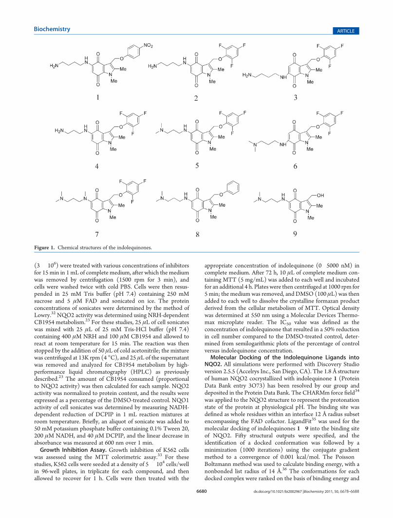
Materials. NADH, FAD, 2,6-dichlorophenolindophenol(DCPIP), 3-(4,5-dimethylthiazol-2-yl)-2,5-diphenyltetrazoliumbromide (MTT), menadione, resveratrol, chloroquine, querce-tin, and melatonin were obtained from Sigma (St. Louis, MO).Imatinib mesylate was purchased from LC laboratories (Woburn,MA). The indolequinones 5-(4-aminobutyl)amino-1,2-dimethyl-3-[(4-nitrophenoxy)methyl]indole-4,7-dione (1), 5-(4-amino-butyl)amino-1,2-dimethyl-3-[(2,4,6-trifluorophenoxy)methyl]in-dole-4,7-dione (2), 6-(4-aminobutyl)amino-1,2-dimethyl-3-[(2,-4,6-trifluorophenoxy)methyl]indole-4,7-dione (3), 5-(3-amino-propyl)amino-1,2-dimethyl-3-[(2,4,6-trifluorophenoxy)methyl]indole-4,7-dione (4), 5-(3-dimethylamino)propylamino-1,2-di-methyl-3-[(2,4,6-trifluorophenoxy)methyl]indole-4,7-dione (5), 6-(3-dimethylamino)propylamino-1,2-dimethyl-3-[(2,4,6-trifluor-ophenoxy)methyl]indole-4,7-dione (6), 5-(3-dimethylamino)propylmethylamino-1,2-dimethyl-3-[(2,4,6-trifluorophenoxy)methyl]indole-4,7-dione (7), 5-(3-dimethylamino)propylamino-1,2-dimethyl-3-(phenoxymethyl)indole-4,7-dione (8), and 5-(3-dimethylamino)propylamino-1,2-dimethyl-3-(hydroxymethyl)-indole-4,7-dione (9) were synthesized using published methods27
except that indolequinones 3 and 6were prepared as described intheSupporting Information.Recombinant humanNQO1(rhNQO1)was purified from Escherichia coli using Cibacron blue affinitychromatography as previously described.28 Recombinant humanNQO2 (rhNQO2) was purchased from Sigma, dissolved in250 mM sucrose, and stored at �80 �C. Dihydronicotinamideriboside (NRH) was prepared from NADH using previouslyreported methods.16,29
Cell Lines.Human leukemia cell line K562 was obtained fromATCC (Manassas, VA) and grown in complete RPMI1640medium supplemented with 4 mM L-glutamine, 10% (v/v) fetalbovine serum, 100 units/mL penicillin, and 100 μg/mL strepto-mycin. Cells were maintained in a humidified incubator contain-ing 5% carbon dioxide at 37 �C.Inhibitor Screening for NQO2. Mechanism-based inactiva-
tion of NQO2 by this indolequinone series was assayedusing purified human recombinant NQO2 (rhNQO2). In thesereactions (final volume of 0.5 mL), rhNQO2 (4 μg/mL) wasincubated with 0.1�10.0 μM indolequinone in the absence andpresence of 0.2 mMNRH in 50 mM potassium phosphate buffer(pH 7.4) containing 5 μM FAD, 125 mM NaCl, and 1 mg/mL
bovine serum albumin at room temperature. After 5 min, a 50 μLaliquot was removed and diluted 50-fold in stop buffer [50 mMpotassium phosphate buffer (pH 7.4) containing 250 mM su-crose, 5 μM FAD, 0.1% (v/v) Tween 20, and 0.25 mg/mLMTT]. An aliquot (960 μL) of the mixture was then transferredto a cuvette andmixed with 200 μMNRH and 10 μMmenadione(final volume of 1 mL), and the linear increase in absorbance wasmonitored spectrophotometrically at 550 nm for 2 min at roomtemperature. NQO2 activity was measured as NRH-dependentmenadione reductase activity using MTT as the final electronacceptor.23
Inhibitor Characterization. Partition ratios for inactivation ofrhNQO2 by the indolequinones that demonstrated mechanism-based inhibition were determined by using the methods des-cribed above except that the indolequiones and rhNQO2 wereincubated in the presence of 0.2 mM NRH for 15 min, withdefined molar ratios of indolequinone to rhNQO2 monomer(active sites) (range from 0.25:1 to 120:1). The percentof activity remaining for each concentration was then plottedversus the molar ratio; a straight line was generated using linearregression and extrapolated, and the x-intercept of the linerepresents the partition ratio (number of molecules of inhibitormetabolized to inactivate one molecule of enzyme).30
The concentration- and time-dependent inactivation of NQO2by the selected indolequinone 1 and 5 was examined to con-firm mechanism-based inhibition. For these studies, inactiva-tion of NQO2 by indolequinone 1 or 5 was performed at 4 �C ina reaction system (final volume of 1 mL) containing 50 mMpotassium phosphate buffer (pH 7.4), 0.1% Tween 20, 125 mMNaCl, 200 μM NRH, 5 μM FAD, and 4 μg/mL rhNQO2.Indolequinones 1 and 5 were then added independently (finalconcentration from 0 to 2 μM), and at the indicated time points,an aliquot (40 μL) was removed and diluted 100-fold in stopbuffer [50 mM potassium phosphate buffer (pH 7.4) containing250 mM sucrose, 5 μM FAD, 200 μM NRH, and 0.1% (v/v)Tween 20] and frozen immediately in liquid nitrogen. NQO2activity was then measured at room temperature in samples(960 μL) supplemented with 10 μMmenadione and 0.1 mg/mLMTT (final volume of 1 mL), and the linear increase in absor-bance at 550 nm was monitored for 3 min. The data generatedwere analyzed using the Kitz and Wilson plot,31 and the Ki andkinact values were calculated accordingly.Comparison of Indolequinones with Known Competitive
Inhibitors of NQO2.The ability of various compounds to inhibitNQO2 was compared in an assay adapted for a 96-well formatusing resazurin as the NQO2 substrate. A 100 μL portion of thereaction mixture was set up for each drug concentration contain-ing 50mMpotassium phosphate buffer (pH 7.4), 1 mg/mL BSA,125 mM NaCl, 5 μM FAD, 200 μMNRH, and 20 ng of NQO2.Drugs were then introduced at final concentrations from 0.0025to 250μMand themixtures incubated at room temperature for 5min;10 μL of the resazurin stock solution was then added into eachwell to achieve a final concentration of 15 μM. The fluorescenceof reduced resazurin was then monitored with 530 nm excitationand 590 nm emission for 5 min using a Molecular DevicesSpectraMax fluorescence plate reader. The rate of change influorescence per minute was calculated and the NQO2 activityremaining expressed as a percentage of the DMSO-treatedcontrol.Inactivation of NQO2 and NQO1 in K562 Cells. The
inhibition of NQO2 activity in K562 cells by these indolequi-nones was determined using the following procedure. K562 cells
6680 dx.doi.org/10.1021/bi2002967 |Biochemistry 2011, 50, 6678–6688
Biochemistry ARTICLE
(3� 106) were treated with various concentrations of inhibitorsfor 15 min in 1 mL of complete medium, after which the mediumwas removed by centrifugation (1500 rpm for 3 min), andcells were washed twice with cold PBS. Cells were then resus-pended in 25 mM Tris buffer (pH 7.4) containing 250 mMsucrose and 5 μM FAD and sonicated on ice. The proteinconcentrations of sonicates were determined by the method ofLowry.32 NQO2 activity was determined using NRH-dependentCB1954 metabolism.23 For these studies, 25 μL of cell sonicateswas mixed with 25 μL of 25 mM Tris-HCl buffer (pH 7.4)containing 400 μM NRH and 100 μM CB1954 and allowed toreact at room temperature for 15 min. The reaction was thenstopped by the addition of 50 μL of cold acetonitrile; the mixturewas centrifuged at 13K rpm (4 �C), and 25 μL of the supernatantwas removed and analyzed for CB1954 metabolism by high-performance liquid chromatography (HPLC) as previouslydescribed.23 The amount of CB1954 consumed (proportionalto NQO2 activity) was then calculated for each sample. NQO2activity was normalized to protein content, and the results wereexpressed as a percentage of the DMSO-treated control. NQO1activity of cell sonicates was determined by measuring NADH-dependent reduction of DCPIP in 1 mL reaction mixtures atroom temperature. Briefly, an aliquot of sonicate was added to50 mM potassium phosphate buffer containing 0.1% Tween 20,200 μM NADH, and 40 μM DCPIP, and the linear decrease inabsorbance was measured at 600 nm over 1 min.Growth Inhibition Assay. Growth inhibition of K562 cells
was assessed using the MTT colorimetric assay.33 For thesestudies, K562 cells were seeded at a density of 5� 104 cells/wellin 96-well plates, in triplicate for each compound, and thenallowed to recover for 1 h. Cells were then treated with the
appropriate concentration of indolequinone (0�5000 nM) incomplete medium. After 72 h, 10 μL of complete medium con-taining MTT (5 mg/mL) was added to each well and incubatedfor an additional 4 h. Plates were then centrifuged at 1000 rpm for5 min; the medium was removed, and DMSO (100 μL) was thenadded to each well to dissolve the crystalline formazan productderived from the cellular metabolism of MTT. Optical densitywas determined at 550 nm using a Molecular Devices Thermo-max microplate reader. The IC50 value was defined as theconcentration of indolequinone that resulted in a 50% reductionin cell number compared to the DMSO-treated control, deter-mined from semilogarithmic plots of the percentage of controlversus indolequinone concentration.Molecular Docking of the Indolequinone Ligands into
NQO2. All simulations were performed with Discovery Studioversion 2.5.5 (Accelrys Inc., San Diego, CA). The 1.8 Å structureof human NQO2 cocrystallized with indolequinone 1 (ProteinData Bank entry 3O73) has been resolved by our group anddeposited in the Protein Data Bank. The CHARMm force field34
was applied to the NQO2 structure to represent the protonationstate of the protein at physiological pH. The binding site wasdefined as whole residues within an interface 12 Å radius subsetencompassing the FAD cofactor. LigandFit35 was used for themolecular docking of indolequinones 1�9 into the binding siteof NQO2. Fifty structural outputs were specified, and theidentification of a docked conformation was followed by aminimization (1000 iterations) using the conjugate gradientmethod to a convergence of 0.001 kcal/mol. The Poisson�Boltzmann method was used to calculate binding energy, with anonbonded list radius of 14 Å.36 The conformations for eachdocked complex were ranked on the basis of binding energy and
Figure 1. Chemical structures of the indolequinones.
6681 dx.doi.org/10.1021/bi2002967 |Biochemistry 2011, 50, 6678–6688
Biochemistry ARTICLE
interactions examined. The top-ranked poses were subsequentlychosen on the basis of binding energies and interactions knownto be essential for efficient mechanism-based inhibition, includ-ing hydrogen bonding and π-stacking between the indolequi-none and FAD cofactor.20,27,37,38
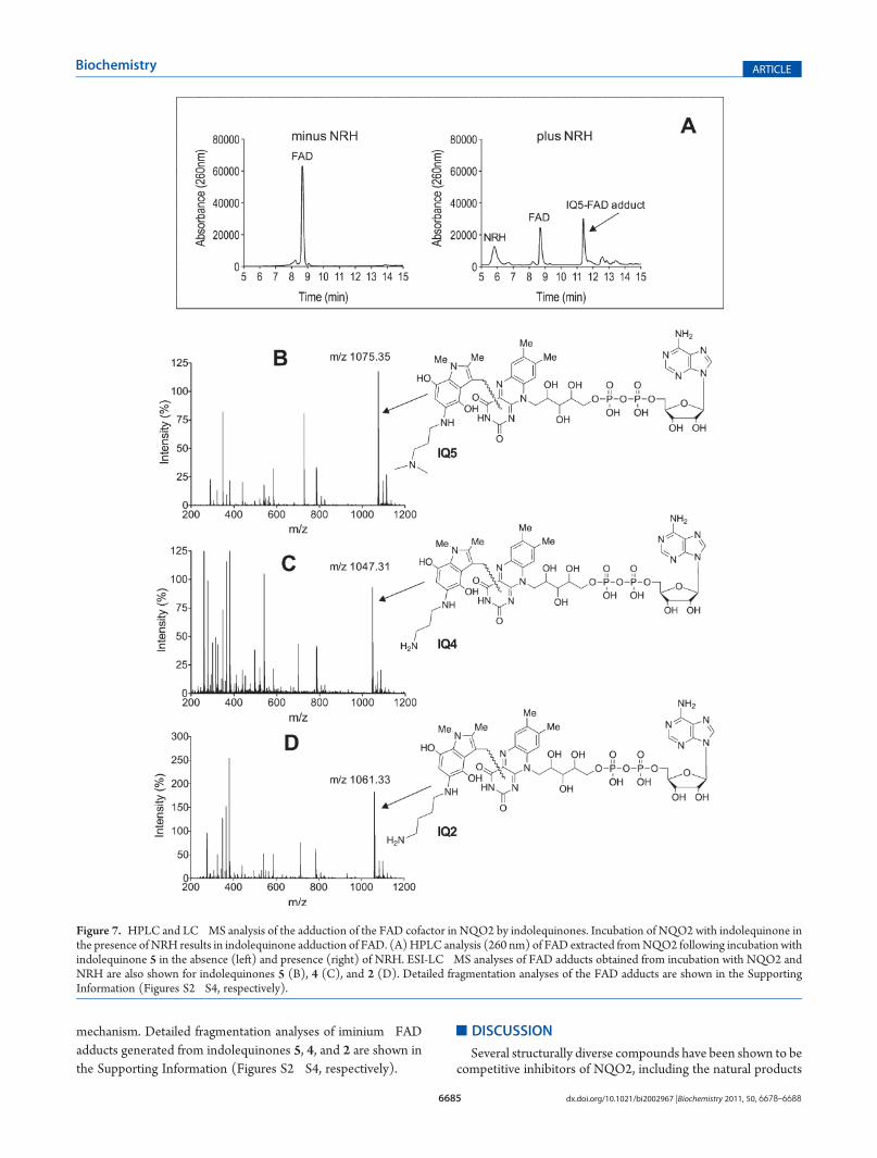
HPLC and Mass Spectrometry Analysis of FAD Adducts.Indolequinone-inactivated human recombinant NQO2 proteinfor HPLC and MS analysis was prepared by incubating 25 μg ofrhNQO2 with a 10-fold molar excess of indolequinones in25 mM potassium phosphate buffer (pH 7.4, final volume of40 μL) in the presence or absence of 0.5 mM NRH. Reactionswere stopped after 10 min by the addition of 60 μL of ice-coldmethanol to denature NQO2 and release FAD. Precipitated
protein was removed by centrifugation (13000 rpm for 5 minat 4 �C), and 50 μL of the supernatant was then loaded onto aC18 reverse-phase HPLC column [Luna C18(2), 5 μm, 250 mm�4.6 mm, Phenomenex, Torrance, CA] and eluted at a rateof 1 mL/min with a mobile phase of 10 mM ammonium acetate(A) and 100% acetonitrile (B) with a linear gradient from 5 to50% B over 15 min using a detection wavelength of 260 nm.For LC�MS analysis, rhNQO2 was inactivated and FADextracted as described above and then concentrated undervacuum (SpeedVac, Savant AES-1000), and 8 μL of the samplewas introduced into an Agilent 1100 capillary HPLC systemutilizing a 1.0 mm � 150 mm Grace Vydac C18 MS column(218MS5115) and a mobile phase of 10 mM ammonium acetate(A) and 10 mM ammonium acetate in acetonitrile (B) with alinear gradient from 5 to 60% B over 10 min at a flow rate of50 μL/min. Spectra were recorded on a Waters QTOFII massspectrometer in positive mode, scanning from m/z 200 to 1500using a 1 s scan time.
’RESULTS
Indolequinones. A series of indolequinones were selected forthis study varying in both the 5- and 6-substituents and theleaving group at the 3-position. The 5- and 6-substituents werechosen to examine the effect of structural variations in the sidechain on inhibition of NQO2 and the impact of 5- versus6-substitutions. Compounds were designed to have either verygood leaving groups at the indole 3-position, such as 4-nitrophe-noxy and 2,4,6-trifluorophenoxy, or very poor leaving groupsunder physiological conditions (3-phenoxy and 3-hydroxy).Chemical structures of the indolequinones used in these studiesare shown in Figure 1.NRH-Dependent NQO2 Inhibition by Indolequinones.
The ability of indolequinones to inhibit recombinant humanNQO2 protein was examined in the presence and absence of thecofactor NRH. For these studies, rhNQO2 was incubated with
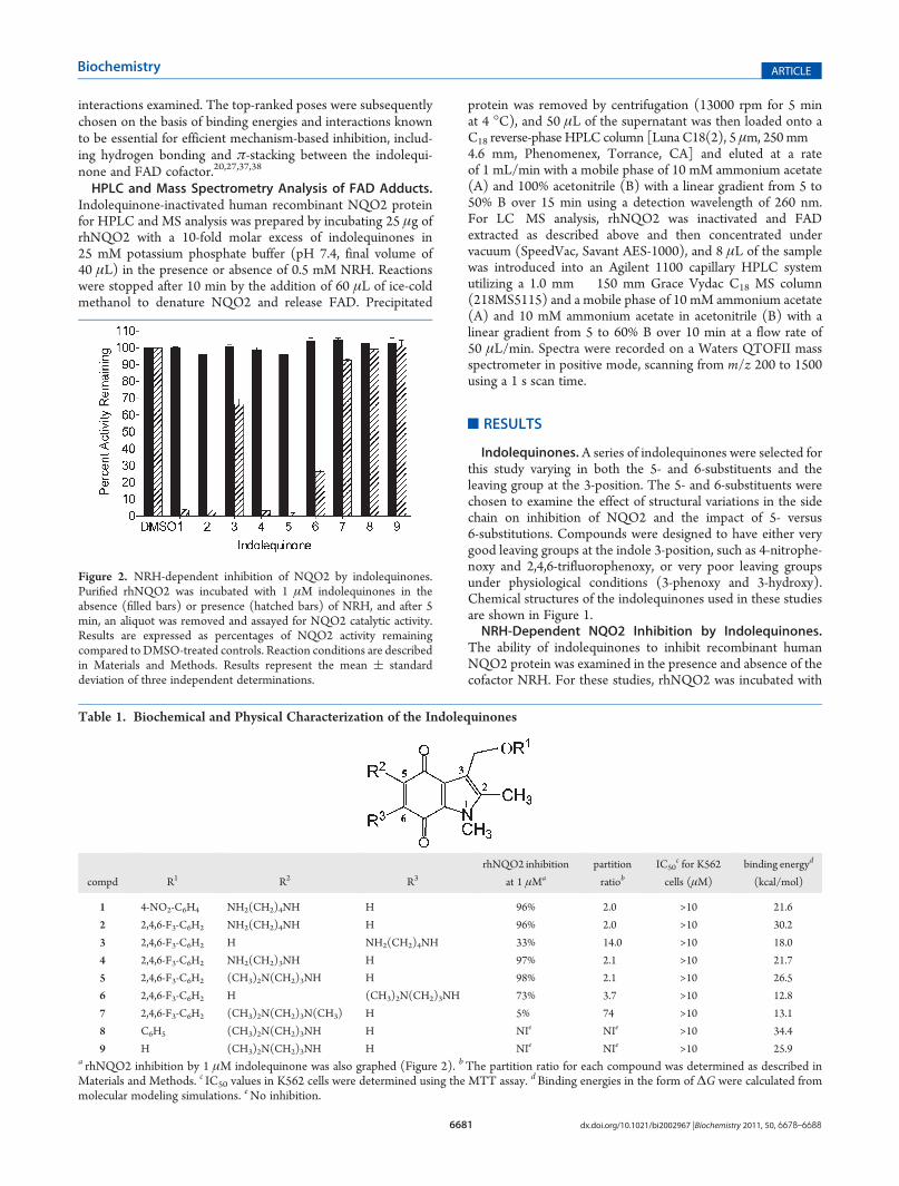
Figure 2. NRH-dependent inhibition of NQO2 by indolequinones.Purified rhNQO2 was incubated with 1 μM indolequinones in theabsence (filled bars) or presence (hatched bars) of NRH, and after 5min, an aliquot was removed and assayed for NQO2 catalytic activity.Results are expressed as percentages of NQO2 activity remainingcompared to DMSO-treated controls. Reaction conditions are describedin Materials and Methods. Results represent the mean ( standarddeviation of three independent determinations.
Table 1. Biochemical and Physical Characterization of the Indolequinones
compd R1 R2 R3
rhNQO2 inhibition
at 1 μMa
partition
ratiobIC50
c for K562
cells (μM)
binding energyd
(kcal/mol)
1 4-NO2-C6H4 NH2(CH2)4NH H 96% 2.0 >10 �21.6
2 2,4,6-F3-C6H2 NH2(CH2)4NH H 96% 2.0 >10 �30.2
3 2,4,6-F3-C6H2 H NH2(CH2)4NH 33% 14.0 >10 �18.0
4 2,4,6-F3-C6H2 NH2(CH2)3NH H 97% 2.1 >10 �21.7
5 2,4,6-F3-C6H2 (CH3)2N(CH2)3NH H 98% 2.1 >10 �26.5
6 2,4,6-F3-C6H2 H (CH3)2N(CH2)3NH 73% 3.7 >10 �12.8
7 2,4,6-F3-C6H2 (CH3)2N(CH2)3N(CH3) H 5% 74 >10 �13.1
8 C6H5 (CH3)2N(CH2)3NH H NIe NIe >10 �34.4
9 H (CH3)2N(CH2)3NH H NIe NIe >10 �25.9a rhNQO2 inhibition by 1 μM indolequinone was also graphed (Figure 2). bThe partition ratio for each compound was determined as described inMaterials and Methods. c IC50 values in K562 cells were determined using the MTT assay. dBinding energies in the form of ΔG were calculated frommolecular modeling simulations. eNo inhibition.
6682 dx.doi.org/10.1021/bi2002967 |Biochemistry 2011, 50, 6678–6688
Biochemistry ARTICLE
indolequinones in the presence and absence of NRH for 5 min,after which a sample of the reaction mixture was removedand diluted and NQO2 activity was measured spectrophotome-trically (550 nm) using NRH-dependent menadione reductioncoupled toMTTreduction.23The incubationof1,2,4, and5 (1μM)with NRH and rhNQO2 resulted in greater than 95% inhibitionof NQO2 catalytic activity (Figure 2 and Table 1), and thisinactivation was dependent upon NRH, suggestive of mechanism-based inhibition. These data demonstrated that variations in the5-position amino alkyl chain length, (CH2)n where n = 3(compounds 4 and 5) or n = 4 (compounds 1 and 2), as wellas substitutions on the terminal amine (compound 5) were toleratedand resulted in marked NQO2 inhibition. However, aminoalk-ylamino substitutions at the 6-position (compounds 3 and 6)resulted in compounds that were much poorer inhibitors whencompared to indolequinones substituted at the 5-position. Noinhibition of rhNQO2was observed with indolequinones 7�9 inthe presence or absence of NRH. Data for compound 7 illustratethat branching of the proximal nitrogen in the aminoalkylaminochain is not optimal for inhibition, while the studies withcompounds 8 and 9 demonstrated that a leaving group isrequired at the 3-position for inhibition. Inhibition of NQO2by the indolequinones appeared to be irreversible becausedesalting of the indolequinone-inhibited protein using the Milli-poreMicroconMolecular Cutoff device did not result in recoveryof enzyme activity (data not shown). To determine if thesecompounds could also inhibit the related enzyme NQO1, weincubated rhNQO1with indolequinones (1 μM) in the presenceand absence of NADH, and in these studies, no inhibition ofNQO1 was detected.27
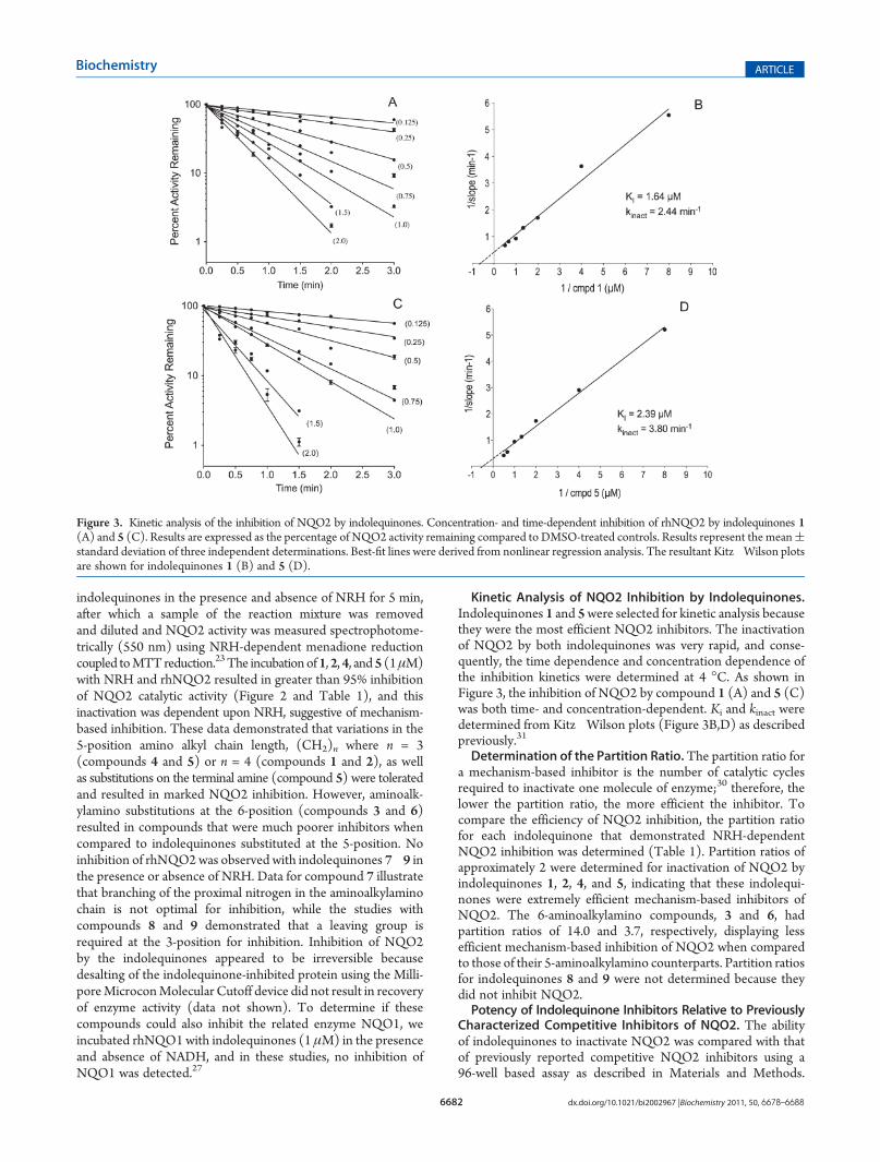
Kinetic Analysis of NQO2 Inhibition by Indolequinones.Indolequinones 1 and 5were selected for kinetic analysis becausethey were the most efficient NQO2 inhibitors. The inactivationof NQO2 by both indolequinones was very rapid, and conse-quently, the time dependence and concentration dependence ofthe inhibition kinetics were determined at 4 �C. As shown inFigure 3, the inhibition of NQO2 by compound 1 (A) and 5 (C)was both time- and concentration-dependent. Ki and kinact weredetermined from Kitz�Wilson plots (Figure 3B,D) as describedpreviously.31
Determination of the Partition Ratio.The partition ratio fora mechanism-based inhibitor is the number of catalytic cyclesrequired to inactivate one molecule of enzyme;30 therefore, thelower the partition ratio, the more efficient the inhibitor. Tocompare the efficiency of NQO2 inhibition, the partition ratiofor each indolequinone that demonstrated NRH-dependentNQO2 inhibition was determined (Table 1). Partition ratios ofapproximately 2 were determined for inactivation of NQO2 byindolequinones 1, 2, 4, and 5, indicating that these indolequi-nones were extremely efficient mechanism-based inhibitors ofNQO2. The 6-aminoalkylamino compounds, 3 and 6, hadpartition ratios of 14.0 and 3.7, respectively, displaying lessefficient mechanism-based inhibition of NQO2 when comparedto those of their 5-aminoalkylamino counterparts. Partition ratiosfor indolequinones 8 and 9 were not determined because theydid not inhibit NQO2.Potency of Indolequinone Inhibitors Relative to Previously
Characterized Competitive Inhibitors of NQO2. The abilityof indolequinones to inactivate NQO2 was compared with thatof previously reported competitive NQO2 inhibitors using a96-well based assay as described in Materials and Methods.
Figure 3. Kinetic analysis of the inhibition of NQO2 by indolequinones. Concentration- and time-dependent inhibition of rhNQO2 by indolequinones 1(A) and 5 (C). Results are expressed as the percentage of NQO2 activity remaining compared to DMSO-treated controls. Results represent the mean(standard deviation of three independent determinations. Best-fit lines were derived from nonlinear regression analysis. The resultant Kitz�Wilson plotsare shown for indolequinones 1 (B) and 5 (D).
6683 dx.doi.org/10.1021/bi2002967 |Biochemistry 2011, 50, 6678–6688
Biochemistry ARTICLE
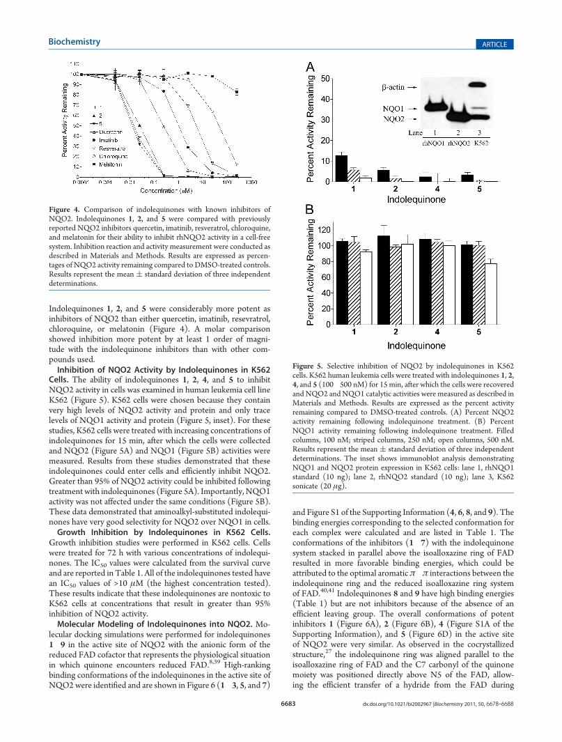
Indolequinones 1, 2, and 5 were considerably more potent asinhibitors of NQO2 than either quercetin, imatinib, resevratrol,chloroquine, or melatonin (Figure 4). A molar comparisonshowed inhibition more potent by at least 1 order of magni-tude with the indolequinone inhibitors than with other com-pounds used.Inhibition of NQO2 Activity by Indolequinones in K562
Cells. The ability of indolequinones 1, 2, 4, and 5 to inhibitNQO2 activity in cells was examined in human leukemia cell lineK562 (Figure 5). K562 cells were chosen because they containvery high levels of NQO2 activity and protein and only tracelevels of NQO1 activity and protein (Figure 5, inset). For thesestudies, K562 cells were treated with increasing concentrations ofindolequinones for 15 min, after which the cells were collectedand NQO2 (Figure 5A) and NQO1 (Figure 5B) activities weremeasured. Results from these studies demonstrated that theseindolequinones could enter cells and efficiently inhibit NQO2.Greater than 95% of NQO2 activity could be inhibited followingtreatment with indolequinones (Figure 5A). Importantly, NQO1activity was not affected under the same conditions (Figure 5B).These data demonstrated that aminoalkyl-substituted indolequi-nones have very good selectivity for NQO2 over NQO1 in cells.Growth Inhibition by Indolequinones in K562 Cells.
Growth inhibition studies were performed in K562 cells. Cellswere treated for 72 h with various concentrations of indolequi-nones. The IC50 values were calculated from the survival curveand are reported in Table 1. All of the indolequinones tested havean IC50 values of >10 μM (the highest concentration tested).These results indicate that these indolequinones are nontoxic toK562 cells at concentrations that result in greater than 95%inhibition of NQO2 activity.Molecular Modeling of Indolequinones into NQO2. Mo-
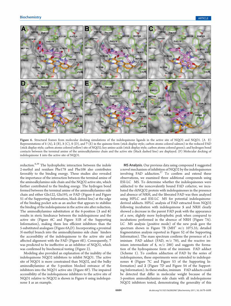
lecular docking simulations were performed for indolequinones1�9 in the active site of NQO2 with the anionic form of thereduced FAD cofactor that represents the physiological situationin which quinone encounters reduced FAD.8,39 High-rankingbinding conformations of the indolequinones in the active site ofNQO2were identified and are shown in Figure 6 (1�3, 5, and 7)
and Figure S1 of the Supporting Information (4, 6, 8, and 9). Thebinding energies corresponding to the selected conformation foreach complex were calculated and are listed in Table 1. Theconformations of the inhibitors (1�7) with the indolequinonesystem stacked in parallel above the isoalloxazine ring of FADresulted in more favorable binding energies, which could beattributed to the optimal aromaticπ�π interactions between theindolequinone ring and the reduced isoalloxazine ring systemof FAD.40,41 Indolequinones 8 and 9 have high binding energies(Table 1) but are not inhibitors because of the absence of anefficient leaving group. The overall conformations of potentinhibitors 1 (Figure 6A), 2 (Figure 6B), 4 (Figure S1A of theSupporting Information), and 5 (Figure 6D) in the active siteof NQO2 were very similar. As observed in the cocrystallizedstructure,27 the indolequinone ring was aligned parallel to theisoalloxazine ring of FAD and the C7 carbonyl of the quinonemoiety was positioned directly above N5 of the FAD, allow-ing the efficient transfer of a hydride from the FAD during
Figure 4. Comparison of indolequinones with known inhibitors ofNQO2. Indolequinones 1, 2, and 5 were compared with previouslyreported NQO2 inhibitors quercetin, imatinib, resveratrol, chloroquine,and melatonin for their ability to inhibit rhNQO2 activity in a cell-freesystem. Inhibition reaction and activity measurement were conducted asdescribed in Materials and Methods. Results are expressed as percen-tages of NQO2 activity remaining compared to DMSO-treated controls.Results represent the mean ( standard deviation of three independentdeterminations.
Figure 5. Selective inhibition of NQO2 by indolequinones in K562cells. K562 human leukemia cells were treated with indolequinones 1, 2,4, and 5 (100�500 nM) for 15 min, after which the cells were recoveredand NQO2 andNQO1 catalytic activities were measured as described inMaterials and Methods. Results are expressed as the percent activityremaining compared to DMSO-treated controls. (A) Percent NQO2activity remaining following indolequinone treatment. (B) PercentNQO1 activity remaining following indolequinone treatment. Filledcolumns, 100 nM; striped columns, 250 nM; open columns, 500 nM.Results represent the mean ( standard deviation of three independentdeterminations. The inset shows immunoblot analysis demonstratingNQO1 and NQO2 protein expression in K562 cells: lane 1, rhNQO1standard (10 ng); lane 2, rhNQO2 standard (10 ng); lane 3, K562sonicate (20 μg).
6684 dx.doi.org/10.1021/bi2002967 |Biochemistry 2011, 50, 6678–6688
Biochemistry ARTICLE
reduction.8,39 The hydrophobic interaction between the indole2-methyl and residues Phe178 and Phe106 also contributesfavorably to the binding energy. These studies also revealedthe importance of the interaction between the terminal amine ofthe aminoalkylamino side chain and the NQO2 active site, whichfurther contributed to the binding energy. The hydrogen bondformed between the terminal amine of the aminoalkylamino sidechain and either Gln122, Glu193, or FAD (Figure 6 and FigureS1 of the Supporting Information, black dotted line) at the edgeof the binding pocket acts as an anchor that appears to stabilizethe binding of the indolequinone in the active site after reduction.The aminoalkylamino substitution at the 6-position (3 and 6)results in steric hindrance between the indolequinone and theactive site (Figure 6C and Figure S1B of the SupportingInformation), making them less efficient inhibitors than the5-substituted analogues (Figure 6A,D). Incorporating a proximalN-methyl branch into the aminoalkylamino side chain7 hindersthe accessibility of the indolequinone to the active site andaffected alignment with the FAD (Figure 6E). Consequently, 7was predicted to be ineffective as an inhibitor of NQO2, whichwas confirmed by biochemical testing (Figure 2).Modeling also provided an explanation of the inability of the
indolequinone NQO2 inhibitors to inhibit NQO1. The activesite of NQO1 is more constrained than NQO2, and the bulkyaminoalkylamino at the 5-position blocked entrance of theinhibitors into the NQO1 active site (Figure 6F). The impairedaccessibility of the indolequinone inhibitors to the active site ofNQO1 relative to NQO2 is shown in Figure 6 using indolequi-none 1 as an example.
MS Analysis.Our previous data using compound 1 suggesteda novel mechanism of inhibition of NQO2 by the indolequinonesinvolving FAD adduction.27 To confirm and extend theseobservations, we examined three additional compounds usingESI-LC�MS. To determine whether the indolequinones wereadducted to the noncovalently bound FAD cofactor, we incu-bated the rhNQO2 protein with indolequinones in the presenceand absence of NRH, and the liberated FAD was then analyzedusing HPLC and ESI-LC�MS for potential indolequinone-derived adducts. HPLC analysis of FAD extracted from NQO2following incubation with indolequinone 5 and NRH clearlyshowed a decrease in the parent FAD peak with the appearanceof a new, slightly more hydrophobic peak when compared toincubations performed in the absence of NRH (Figure 7A).LC�MS analysis (positive mode) of the new peak gave thespectrum shown in Figure 7B (MH+ m/z 1075.35; detailedfragmentation analysis reported in Figure S2 of the SupportingInformation). The mass spectrum confirms the presence of a 5iminium�FAD adduct (FAD, m/z 785, and the reactive im-inium intermediate of 5, m/z 288) and suggests the forma-tion of the hydroquinone form of the iminium�FAD adduct(Scheme 1). To confirm adduction of FAD by the series ofindolequinones, these experiments were extended to indolequi-nones 4 (Figure 7C and Figure S3 of the Supporting In-formation) and 2 (Figure 7D and Figure S4 of the Support-ing Information). In these studies, iminium�FAD adducts couldbe detected that differ in molecular weight because of the5-position aminoalkylamino side chain with all indolequinoneNQO2 inhibitors tested, demonstrating the generality of this
Figure 6. Structural frames from molecular docking simulations of the indolequinone ligands in the active site of NQO2 and NQO1. (A�E)Representations of 1 (A), 2 (B), 3 (C), 5 (D), and 7 (E) in the quinone form (stick display style; carbon atoms colored salmon) in the reduced FAD(stick display style; carbon atoms colored yellow) site of NQO2; key amino acids (stick display style; carbon atoms colored green); and hydrogen bondcontacts between the terminal amine of the aminoalkylamino chain and the active site (black dashed line) are displayed. (F) Molecular docking ofindolequinone 1 into the active site of NQO1.
6685 dx.doi.org/10.1021/bi2002967 |Biochemistry 2011, 50, 6678–6688
Biochemistry ARTICLE
mechanism. Detailed fragmentation analyses of iminium�FADadducts generated from indolequinones 5, 4, and 2 are shown inthe Supporting Information (Figures S2�S4, respectively).
’DISCUSSION
Several structurally diverse compounds have been shown to becompetitive inhibitors of NQO2, including the natural products
Figure 7. HPLC and LC�MS analysis of the adduction of the FAD cofactor in NQO2 by indolequinones. Incubation of NQO2 with indolequinone inthe presence of NRH results in indolequinone adduction of FAD. (A)HPLC analysis (260 nm) of FAD extracted fromNQO2 following incubation withindolequinone 5 in the absence (left) and presence (right) of NRH. ESI-LC�MS analyses of FAD adducts obtained from incubation with NQO2 andNRH are also shown for indolequinones 5 (B), 4 (C), and 2 (D). Detailed fragmentation analyses of the FAD adducts are shown in the SupportingInformation (Figures S2�S4, respectively).
6686 dx.doi.org/10.1021/bi2002967 |Biochemistry 2011, 50, 6678–6688
Biochemistry ARTICLE
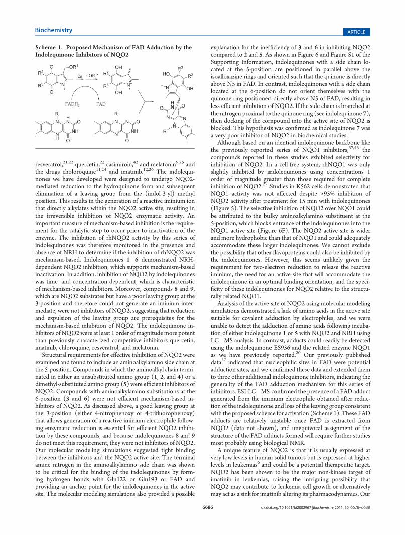
resveratrol,21,22 quercetin,23 casimiroin,42 and melatonin9,25 andthe drugs choloroquine11,24 and imatinib.12,26 The indolequi-nones we have developed were designed to undergo NQO2-mediated reduction to the hydroquinone form and subsequentelimination of a leaving group from the (indol-3-yl) methylposition. This results in the generation of a reactive iminium ionthat directly alkylates within the NQO2 active site, resulting inthe irreversible inhibition of NQO2 enzymatic activity. Animportant measure of mechanism-based inhibition is the require-ment for the catalytic step to occur prior to inactivation of theenzyme. The inhibition of rhNQO2 activity by this series ofindolequinones was therefore monitored in the presence andabsence of NRH to determine if the inhibition of rhNQO2 wasmechanism-based. Indolequinones 1�6 demonstrated NRH-dependent NQO2 inhibition, which supports mechanism-basedinactivation. In addition, inhibition of NQO2 by indolequinoneswas time- and concentration-dependent, which is characteristicof mechanism-based inhibitors. Moreover, compounds 8 and 9,which are NQO2 substrates but have a poor leaving group at the3-position and therefore could not generate an iminium inter-mediate, were not inhibitors of NQO2, suggesting that reductionand expulsion of the leaving group are prerequisites for themechanism-based inhibition of NQO2. The indolequinone in-hibitors of NQO2were at least 1 order of magnitudemore potentthan previously characterized competitive inhibitors quercetin,imatinib, chloroquine, resveratrol, and melatonin.
Structural requirements for effective inhibition of NQO2wereexamined and found to include an aminoalkylamino side chain atthe 5-position. Compounds in which the aminoalkyl chain termi-nated in either an unsubstituted amino group (1, 2, and 4) or adimethyl-substituted amino group (5) were efficient inhibitors ofNQO2. Compounds with aminoalkylamino substitutions at the6-position (3 and 6) were not efficient mechanism-based in-hibitors of NQO2. As discussed above, a good leaving group atthe 3-position (either 4-nitrophenoxy or 4-trifluorophenoxy)that allows generation of a reactive iminium electrophile follow-ing enzymatic reduction is essential for efficient NQO2 inhibi-tion by these compounds, and because indolequinones 8 and 9do notmeet this requirement, they were not inhibitors of NQO2.Our molecular modeling simulations suggested tight bindingbetween the inhibitors and the NQO2 active site. The terminalamine nitrogen in the aminoalkylamino side chain was shownto be critical for the binding of the indolequinones by form-ing hydrogen bonds with Gln122 or Glu193 or FAD andproviding an anchor point for the indolequinones in the activesite. The molecular modeling simulations also provided a possible
explanation for the inefficiency of 3 and 6 in inhibiting NQO2compared to 2 and 5. As shown in Figure 6 and Figure S1 of theSupporting Information, indolequinones with a side chain lo-cated at the 5-position are positioned in parallel above theisoalloxazine rings and oriented such that the quinone is directlyabove N5 in FAD. In contrast, indolequinones with a side chainlocated at the 6-position do not orient themselves with thequinone ring positioned directly above N5 of FAD, resulting inless efficient inhibition of NQO2. If the side chain is branched atthe nitrogen proximal to the quinone ring (see indolequinone 7),then docking of the compound into the active site of NQO2 isblocked. This hypothesis was confirmed as indolequinone 7 wasa very poor inhibitor of NQO2 in biochemical studies.
Although based on an identical indolequinone backbone likethe previously reported series of NQO1 inhibitors,37,43 thecompounds reported in these studies exhibited selectivity forinhibition of NQO2. In a cell-free system, rhNQO1 was onlyslightly inhibited by indolequinones using concentrations 1order of magnitude greater than those required for completeinhibition of NQO2.27 Studies in K562 cells demonstrated thatNQO1 activity was not affected despite >95% inhibition ofNQO2 activity after treatment for 15 min with indolequinones(Figure 5). The selective inhibition of NQO2 over NQO1 couldbe attributed to the bulky aminoalkylamino substituent at the5-position, which blocks entrance of the indolequinones into theNQO1 active site (Figure 6F). The NQO2 active site is widerand more hydrophobic than that of NQO1 and could adequatelyaccommodate these larger indolequinones. We cannot excludethe possibility that other flavoproteins could also be inhibited bythe indolequinones. However, this seems unlikely given therequirement for two-electron reduction to release the reactiveiminium, the need for an active site that will accommodate theindolequinone in an optimal binding orientation, and the speci-ficity of these indolequinones for NQO2 relative to the structu-rally related NQO1.
Analysis of the active site of NQO2 using molecular modelingsimulations demonstrated a lack of amino acids in the active sitesuitable for covalent adduction by electrophiles, and we wereunable to detect the adduction of amino acids following incuba-tion of either indolequinone 1 or 5 with NQO2 and NRH usingLC�MS analysis. In contrast, adducts could readily be detectedusing the indolequinone ES936 and the related enzyme NQO1as we have previously reported.20 Our previously publisheddata27 indicated that nucleophilic sites in FAD were potentialadduction sites, and we confirmed these data and extended themto three other additional indolequinone inhibitors, indicating thegenerality of the FAD adduction mechanism for this series ofinhibitors. ESI-LC�MS confirmed the presence of a FAD adductgenerated from the iminium electrophile obtained after reduc-tion of the indolequinone and loss of the leaving group consistentwith the proposed scheme for activation (Scheme 1). These FADadducts are relatively unstable once FAD is extracted fromNQO2 (data not shown), and unequivocal assignment of thestructure of the FAD adducts formed will require further studiesmost probably using biological NMR.
A unique feature of NQO2 is that it is usually expressed atvery low levels in human solid tumors but is expressed at higherlevels in leukemias6 and could be a potential therapeutic target.NQO2 has been shown to be the major non-kinase target ofimatinib in leukemias, raising the intriguing possibility thatNQO2 may contribute to leukemia cell growth or alternativelymay act as a sink for imatinib altering its pharmacodynamics. Our
Scheme 1. Proposed Mechanism of FAD Adduction by theIndolequinone Inhibitors of NQO2
6687 dx.doi.org/10.1021/bi2002967 |Biochemistry 2011, 50, 6678–6688
Biochemistry ARTICLE
data indicate that indolequinones were nontoxic to K562 cells atthe concentrations where complete NQO2 inhibition wasachieved, and although this was not the major goal of our study,it does suggest that inhibition of NQO2 alone is not sufficient tocause leukemia cell death. However, it is possible that NQO2inhibition may have a synergistic effect with Bcr-Abl inhibition inthe presence of imatinib, or alternatively that NQO2 inhibitionby indolequinone could potentiate imatinib toxicity by increasingthe concentration of free drug. Both genetic and pharmaco-logic approaches are currently underway in our laboratory in aneffort to fully understand the role of NQO2 in leukemias and inimatinib pharmacology.
In summary, we have generated and characterized a series ofindolequinone compounds as mechanism-based inhibitors ofNQO2 in cell-free and cellular systems. The indolequinonesare the most effective inhibitors of NQO2 described to date, andutilizing modeling, biochemical studies, and mass spectrometry,we have characterized both their mechanism of inhibition andstructural parameters required for effective inhibition of NQO2.
’ASSOCIATED CONTENT
bS Supporting Information. Structural frames from the mo-lecular docking simulations andmass spectrometry data for someof the indolequinones and the methods for the preparation ofindolequinones 3 and 6. This material is available free of chargevia the Internet at http://pubs.acs.org.
’AUTHOR INFORMATION
Corresponding Author*D.R. (biology): phone, (303) 724-7265; fax, (303) 724-7266;e-mail, [email protected]. C.J.M. (chemistry): phone,+44 115 846 8500; fax, +44 115 951 3564; e-mail, [email protected].
Author ContributionsC.Y. and M.D. contributed equally to this work.
Funding SourcesThis work was funded in part by National Institutes of HealthGrant R21 HL095995.
NotesC.J.M., D.R., and D.S. are cofounders and stockholders in QGentaInc., which holds an option to license molecules described inthis article.
’ABBREVIATIONS
NQO1, NADH:quinone oxidoreductase 1; NQO2, NRH:qui-none oxidoreductase 2; NRH, nicotinamide riboside;MTT, 3-(4,5-dimethylthiazol-2-yl)-2,5-diphenyltetrazolium bromide; ESI-LC�MS, electrospray ionization liquid chromatography�massspectrometry.
’REFERENCES
(1) Ernster, L., and Navazio, F. (1958) Soluble diaphorase in animaltissues. Acta Chem. Scand. 12, 595–602.(2) Ernster, L., Ljunggren, M., and Danielson, L. (1960) Purification
and some properties of a highly dicumarol-sensitive liver diaphorase.Biochem. Biophys. Res. Commun. 2, 88–92.(3) Martius, C. (1954) [The place of vitamin K1 in respiratory chain;
preliminary report.]. Biochem. Z. 326, 26–27.
(4) Ernster, L. (1987) DT-diaphorase: A historical review.Chem. Scr.27A, 1–13.
(5) Jaiswal, A. K., Burnett, P., Adesnik, M., and McBride, O. W.(1990) Nucleotide and deduced amino acid sequence of a human cDNA(NQO2) corresponding to a secondmember of the NAD(P)H:quinoneoxidoreductase gene family. Extensive polymorphism at the NQO2 genelocus on chromosome 6. Biochemistry 29, 1899–1906.
(6) Zhao, Q., Yang, X. L., Holtzclaw, W. D., and Talalay, P. (1997)Unexpected genetic and structural relationships of a long-forgottenflavoenzyme to NAD(P)H:quinone reductase (DT-diaphorase). Proc.Natl. Acad. Sci. U.S.A. 94, 1669–1674.
(7) Liao, S., Dulaney, J. T., and Williams-Ashman, H. G. (1962)Purification and properties of a flavoprotein catalyzing the oxidation ofreduced ribosyl nicotinamide. J. Biol. Chem. 237, 2981–2987.
(8) Bianchet, M. A., Faig, M., and Amzel, L. M. (2004) Structure andmechanism of NAD[P]H:quinone acceptor oxidoreductases (NQO).Methods Enzymol. 382, 144–174.
(9) Vella, F., Ferry, G., Delagrange, P., and Boutin, J. A. (2005)NRH:quinone reductase 2: An enzyme of surprises and mysteries.Biochem. Pharmacol. 71, 1–12.
(10) Siegel, D., and Ross, D. (2000) Immunodetection of NAD-(P)H:quinone oxidoreductase 1 (NQO1) in human tissues. Free RadicalBiol. Med. 29, 246–253.
(11) Graves, P. R., Kwiek, J. J., Fadden, P., Ray, R., Hardeman, K.,Coley, A. M., Foley, M., and Haystead, T. A. (2002) Discovery of noveltargets of quinoline drugs in the human purine binding proteome. Mol.Pharmacol. 62, 1364–1372.
(12) Bantscheff, M., Eberhard, D., Abraham, Y., Bastuck, S., Boesche,M., Hobson, S., Mathieson, T., Perrin, J., Raida, M., Rau, C., Reader, V.,Sweetman, G., Bauer, A., Bouwmeester, T., Hopf, C., Kruse, U.,Neubauer, G., Ramsden, N., Rick, J., Kuster, B., and Drewes, G. (2007)Quantitative chemical proteomics reveals mechanisms of action ofclinical ABL kinase inhibitors. Nat. Biotechnol. 25, 1035–1044.
(13) Fu, Y., Buryanovskyy, L., and Zhang, Z. (2008) Quinone redu-ctase 2 is a catechol quinone reductase. J. Biol. Chem. 283, 23829–23835.
(14) Gaikwad, N. W., Yang, L., Rogan, E. G., and Cavalieri, E. L.(2009) Evidence for NQO2-mediated reduction of the carcinogenicestrogen ortho-quinones. Free Radical Biol. Med. 46, 253–262.
(15) Jamieson, D., Tung, A. T., Knox, R. J., and Boddy, A. V. (2006)Reduction of mitomycin C is catalysed by human recombinant NRH:quinone oxidoreductase 2 using reduced nicotinamide adenine dinu-cleotide as an electron donating co-factor. Br. J. Cancer 95, 1229–1233.
(16) Yan, C., Kepa, J. K., Siegel, D., Stratford, I. J., and Ross, D.(2008) Dissecting the role of multiple reductases in bioactivation andcytotoxicity of the antitumor agent 2,5-diaziridinyl-3-(hydroxymethyl)-6-methyl-1,4-benzoquinone (RH1). Mol. Pharmacol. 74, 1657–1665.
(17) Siegel, D., Reigan, P., Guo, W., and Ross, D. (2007) Themetabolism of antitumor quinones by NRH:quinone oxidoreductase 2(NQO2). In Proceedings of the American Association for Cancer Research,American Association for Cancer Research, Los Angeles.
(18) Knox, R. J., Jenkins, T. C., Hobbs, S.M., Chen, S., Melton, R. G.,and Burke, P. J. (2000) Bioactivation of 5-(aziridin-1-yl)-2,4-dinitro-benzamide (CB 1954) by human NAD(P)H quinone oxidoreductase 2:A novel co-substrate-mediated antitumor prodrug therapy. Cancer Res.60, 4179–4186.
(19) Hollander, P. M., and Ernster, L. (1975) Studies on the reactionmechanism of DT diaphorase. Action of dead-end inhibitors and effectsof phospholipids. Arch. Biochem. Biophys. 169, 560–567.
(20) Winski, S. L., Faig, M., Bianchet, M. A., Siegel, D., Swann, E.,Fung, K., Duncan, M. W., Moody, C. J., Amzel, L. M., and Ross, D.(2001) Characterization of a mechanism-based inhibitor of NAD(P)H:quinone oxidoreductase 1 by biochemical, X-ray crystallographic, andmass spectrometric approaches. Biochemistry 40, 15135–15142.
(21) Buryanovskyy, L., Fu, Y., Boyd, M., Ma, Y., Hsieh, T. C., Wu,J. M., and Zhang, Z. (2004) Crystal structure of quinone reductase 2 incomplex with resveratrol. Biochemistry 43, 11417–11426.
(22) Wang, Z., Hsieh, T. C., Zhang, Z., Ma, Y., andWu, J. M. (2004)Identification and purification of resveratrol targeting proteins using
6688 dx.doi.org/10.1021/bi2002967 |Biochemistry 2011, 50, 6678–6688
Biochemistry ARTICLE
immobilized resveratrol affinity chromatography. Biochem. Biophys. Res.Commun. 323, 743–749.(23) Wu, K., Knox, R., Sun, X. Z., Joseph, P., Jaiswal, A. K., Zhang, D.,
Deng, P. S., and Chen, S. (1997) Catalytic properties of NAD(P)H:quinone oxidoreductase-2 (NQO2), a dihydronicotinamide ribosidedependent oxidoreductase. Arch. Biochem. Biophys. 347, 221–228.(24) Kwiek, J. J., Haystead, T. A., and Rudolph, J. (2004) Kinetic
mechanism of quinone oxidoreductase 2 and its inhibition by theantimalarial quinolines. Biochemistry 43, 4538–4547.(25) Mailliet, F., Ferry, G., Vella, F., Berger, S., Coge, F., Chomarat,
P., Mallet, C., Guenin, S. P., Guillaumet, G., Viaud-Massuard, M. C.,Yous, S., Delagrange, P., and Boutin, J. A. (2005) Characterization of themelatoninergic MT3 binding site on the NRH:quinone oxidoreductase2 enzyme. Biochem. Pharmacol. 71, 74–88.(26) Winger, J. A., Hantschel, O., Superti-Furga, G., and Kuriyan, J.
(2009) The structure of the leukemia drug imatinib bound to humanquinone reductase 2 (NQO2). BMC Struct. Biol. 9, 7.(27) Dufour, M., Yan, C., Siegel, D., Colucci, M. A., Jenner, M.,
Oldham, N. J., Gomez, J., Reigan, P., Li, Y., Matteis, C. I. D., Ross, D., andMoody, C. J. (2011) Mechanism-based inhibition of quinone reductase2 (NQO2). Selectivity for NQO2 over NQO1 and structural basis forflavoprotein inhibition. ChemBioChem 12, 1203–1208.(28) Beall, H. D., Mulcahy, R. T., Siegel, D., Traver, R. D., Gibson,
N. W., and Ross, D. (1994) Metabolism of bioreductive antitumorcompounds by purified rat and human DT-diaphorases. Cancer Res.54, 3196–3201.(29) Friedlos, F., Jarman, M., Davies, L. C., Boland, M. P., and Knox,
R. J. (1992) Identification of novel reduced pyridinium derivatives assynthetic co-factors for the enzyme DT diaphorase (NAD(P)H dehy-drogenase (quinone), EC 1.6.99.2). Biochem. Pharmacol. 44, 25–31.(30) Silverman, R. B. (1988) Mechanism based enzyme inhibition.
In Chemistry and Enzymology, pp 3�30, CRC Press, Boca Raton, FL.(31) Kitz, R., andWilson, I. B. (1962) Esters of methanesulfonic acid as
irreversible inhibitors of acetylcholinesterase. J. Biol. Chem. 237, 3245–3249.(32) Lowry, O. H., Rosebrough, N. J., Farr, A. L., and Randall, R. J.
(1951) Protein measurement with the Folin phenol reagent. J. Biol.Chem. 193, 265–275.(33) Mosmann, T. (1983) Rapid colorimetric assay for cellular
growth and survival: Application to proliferation and cytotoxicity assays.J. Immunol. Methods 65, 55–63.(34) Brooks, B. R., Bruccoleri, R. E., Olafson, B. D., States, D. J., and
Swaminathan, S. (1983) CHARMM: A program for macromolecularenergy, minimization, and dynamics calculations. J. Comput. Chem.4, 187–217.(35) Luty, B. A., Wasserman, Z. R., Stouten, P. F. W., Hodge, C. N.,
Zacharias, M., and McCammon, J. A. (1995) A molecular mechanics/grid method for evaluation of ligand-receptor interactions. J. Comput.Chem. 16, 454–464.(36) Gilson, M. K., and Honig, B. (1988) Calculation of the total
electrostatic energy of a macromolecular system: Solvation energies,binding energies, and conformational analysis. Proteins 4, 7–18.(37) Reigan, P., Colucci, M. A., Siegel, D., Chilloux, A., Moody, C. J.,
and Ross, D. (2007) Development of indolequinone mechanism-basedinhibitors of NAD(P)H:quinone oxidoreductase 1 (NQO1): NQO1inhibition and growth inhibitory activity in human pancreatic MIAPaCa-2 cancer cells. Biochemistry 46, 5941–5950.(38) Nolan, K. A., Zhao, H., Faulder, P. F., Frenkel, A. D., Timson,
D. J., Siegel, D., Ross, D., Burke, T. R., Jr., Stratford, I. J., and Bryce, R. A.(2007) Coumarin-based inhibitors of human NAD(P)H:quinone oxi-doreductase-1. Identification, structure-activity, off-target effects and invitro human pancreatic cancer toxicity. J. Med. Chem. 50, 6316–6325.(39) Cavelier, G., and Amzel, L. M. (2001) Mechanism of NAD-
(P)H:quinone reductase: Ab initio studies of reduced flavin. Proteins43, 420–432.(40) Zhou, Z., Fisher, D., Spidel, J., Greenfield, J., Patson, B., Fazal,
A., Wigal, C., Moe, O. A., and Madura, J. D. (2003) Kinetic and dockingstudies of the interaction of quinones with the quinone reductase activesite. Biochemistry 42, 1985–1994.
(41) Suleman, A., and Skibo, E. B. (2002) A comprehensive study ofthe active site residues of DT-diaphorase: Rational design of benzimi-dazolediones as DT-diaphorase substrates. J. Med. Chem. 45, 1211–1220.
(42) Maiti, A., Reddy, P. V., Sturdy, M., Marler, L., Pegan, S. D.,Mesecar, A. D., Pezzuto, J. M., and Cushman, M. (2009) Synthesis ofcasimiroin and optimization of its quinone reductase 2 and aromataseinhibitory activities. J. Med. Chem. 52, 1873–1884.
(43) Colucci, M. A., Reigan, P., Siegel, D., Chilloux, A., Ross, D., andMoody, C. J. (2007) Synthesis and evaluation of 3-aryloxymethyl-1,2-dimethylindole-4,7-diones as mechanism-based inhibitors of NAD-(P)H:quinone oxidoreductase 1 (NQO1) activity. J. Med. Chem. 50,5780–5789.