Lactamase Inhibitors in -Lactam-Producing Actinobacteria
18
Citation: Martin, J.F.; Alvarez-Alvarez, R.; Liras, P. Penicillin-Binding Proteins, β-Lactamases, and β-Lactamase Inhibitors in β-Lactam-Producing Actinobacteria: Self-Resistance Mechanisms. Int. J. Mol. Sci. 2022, 23, 5662. https://doi.org/10.3390/ ijms23105662 Academic Editor: Carlos Juan Nicolau Received: 10 April 2022 Accepted: 16 May 2022 Published: 18 May 2022 Publisher’s Note: MDPI stays neutral with regard to jurisdictional claims in published maps and institutional affil- iations. Copyright: © 2022 by the authors. Licensee MDPI, Basel, Switzerland. This article is an open access article distributed under the terms and conditions of the Creative Commons Attribution (CC BY) license (https:// creativecommons.org/licenses/by/ 4.0/). International Journal of Molecular Sciences Review Penicillin-Binding Proteins, β-Lactamases, and β-Lactamase Inhibitors in β-Lactam-Producing Actinobacteria: Self-Resistance Mechanisms Juan F. Martin, Ruben Alvarez-Alvarez and Paloma Liras * Departamento de Biología Molecular, Universidad de León, 24071 León, Spain; [email protected] (J.F.M.); [email protected] (R.A.-A.) * Correspondence: [email protected]; Tel.: +34-987-209451 Abstract: The human society faces a serious problem due to the widespread resistance to antibiotics in clinical practice. Most antibiotic biosynthesis gene clusters in actinobacteria contain genes for intrinsic self-resistance to the produced antibiotics, and it has been proposed that the antibiotic resistance genes in pathogenic bacteria originated in antibiotic-producing microorganisms. The model actinobacteria Streptomyces clavuligerus produces the β-lactam antibiotic cephamycin C, a class A β-lactamase, and the β lactamases inhibitor clavulanic acid, all of which are encoded in a gene supercluster; in addition, it synthesizes the β-lactamase inhibitory protein BLIP. The secreted clavulanic acid has a synergistic effect with the cephamycin produced by the same strain in the fight against competing microorganisms in its natural habitat. High levels of resistance to cephamycin/cephalosporin in actinobacteria are due to the presence (in their β-lactam clusters) of genes encoding PBPs which bind penicillins but not cephalosporins. We have revised the previously reported cephamycin C and clavulanic acid gene clusters and, in addition, we have searched for novel β-lactam gene clusters in protein databases. Notably, in S. clavuligerus and Nocardia lactamdurans, the β-lactamases are retained in the cell wall and do not affect the intracellular formation of isopenicillin N/penicillin N. The activity of the β-lactamase in S. clavuligerus may be modulated by the β-lactamase inhibitory protein BLIP at the cell-wall level. Analysis of the β-lactam cluster in actinobacteria suggests that these clusters have been moved by horizontal gene transfer between different actinobacteria and have culminated in S. clavuligerus with the organization of an elaborated set of genes designed for fine tuning of antibiotic resistance and cell wall remodeling for the survival of this Streptomyces species. This article is focused specifically on the enigmatic connection between β-lactam biosynthesis and β-lactam resistance mechanisms in the producer actinobacteria. Keywords: antibiotic resistance; penicillin-binding proteins; β-lactamases; Streptomyces clavuligerus; clavulanic acid; cephamycin C; clusters organization and evolution; superclusters 1. Introduction Resistance to Antibiotics and Its Relation to Antibiotic-Producing Bacteria Antibiotics have saved thousands of human lives since their clinical introduction in the fourth decade of last century [1,2]; however, at present, there is a serious problem of antibi- otic resistance in many pathogenic bacteria, which is a challenge in the fight against bacterial diseases [3–5]. The problem includes resistance to β-lactams, aminoglycosides, tetracyclins, and macrolides [6,7], and is being also observed against antibiotics introduced more re- cently in clinical practice such as fluoroquinolones [8] and imipenem [9]. The appearance of antibiotic resistance was partially associated with the widespread and frequently indis- criminate use of antibiotics in medicine. In parallel, the finding of β-lactamases and other antibiotic resistance genes forming part of the antibiotic biosynthesis gene clusters (GC) led to the hypothesis that the modern resistance determinants might have originated from those that protect the antibiotic-producing bacteria [10–12]. Recent advances in molecular Int. J. Mol. Sci. 2022, 23, 5662. https://doi.org/10.3390/ijms23105662 https://www.mdpi.com/journal/ijms
-
Upload
khangminh22 -
Category
Documents
-
view
1 -
download
0
Transcript of Lactamase Inhibitors in -Lactam-Producing Actinobacteria
Citation: Martin, J.F.;
Alvarez-Alvarez, R.; Liras, P.
Penicillin-Binding Proteins,
β-Lactamases, and β-Lactamase
Inhibitors in β-Lactam-Producing
Actinobacteria: Self-Resistance
Mechanisms. Int. J. Mol. Sci. 2022, 23,
5662. https://doi.org/10.3390/
ijms23105662
Academic Editor: Carlos Juan
Nicolau
Received: 10 April 2022
Accepted: 16 May 2022
Published: 18 May 2022
Publisher’s Note: MDPI stays neutral
with regard to jurisdictional claims in
published maps and institutional affil-
iations.
Copyright: © 2022 by the authors.
Licensee MDPI, Basel, Switzerland.
This article is an open access article
distributed under the terms and
conditions of the Creative Commons
Attribution (CC BY) license (https://
creativecommons.org/licenses/by/
4.0/).
International Journal of
Molecular Sciences
Review
Penicillin-Binding Proteins, β-Lactamases, and β-LactamaseInhibitors in β-Lactam-Producing Actinobacteria:Self-Resistance MechanismsJuan F. Martin, Ruben Alvarez-Alvarez and Paloma Liras *
Departamento de Biología Molecular, Universidad de León, 24071 León, Spain; [email protected] (J.F.M.);[email protected] (R.A.-A.)* Correspondence: [email protected]; Tel.: +34-987-209451
Abstract: The human society faces a serious problem due to the widespread resistance to antibiotics inclinical practice. Most antibiotic biosynthesis gene clusters in actinobacteria contain genes for intrinsicself-resistance to the produced antibiotics, and it has been proposed that the antibiotic resistance genesin pathogenic bacteria originated in antibiotic-producing microorganisms. The model actinobacteriaStreptomyces clavuligerus produces the β-lactam antibiotic cephamycin C, a class A β-lactamase,and the β lactamases inhibitor clavulanic acid, all of which are encoded in a gene supercluster; inaddition, it synthesizes the β-lactamase inhibitory protein BLIP. The secreted clavulanic acid has asynergistic effect with the cephamycin produced by the same strain in the fight against competingmicroorganisms in its natural habitat. High levels of resistance to cephamycin/cephalosporin inactinobacteria are due to the presence (in their β-lactam clusters) of genes encoding PBPs whichbind penicillins but not cephalosporins. We have revised the previously reported cephamycin C andclavulanic acid gene clusters and, in addition, we have searched for novel β-lactam gene clusters inprotein databases. Notably, in S. clavuligerus and Nocardia lactamdurans, the β-lactamases are retainedin the cell wall and do not affect the intracellular formation of isopenicillin N/penicillin N. Theactivity of the β-lactamase in S. clavuligerus may be modulated by the β-lactamase inhibitory proteinBLIP at the cell-wall level. Analysis of the β-lactam cluster in actinobacteria suggests that theseclusters have been moved by horizontal gene transfer between different actinobacteria and haveculminated in S. clavuligerus with the organization of an elaborated set of genes designed for finetuning of antibiotic resistance and cell wall remodeling for the survival of this Streptomyces species.This article is focused specifically on the enigmatic connection between β-lactam biosynthesis andβ-lactam resistance mechanisms in the producer actinobacteria.
Keywords: antibiotic resistance; penicillin-binding proteins; β-lactamases; Streptomyces clavuligerus;clavulanic acid; cephamycin C; clusters organization and evolution; superclusters
1. IntroductionResistance to Antibiotics and Its Relation to Antibiotic-Producing Bacteria
Antibiotics have saved thousands of human lives since their clinical introduction in thefourth decade of last century [1,2]; however, at present, there is a serious problem of antibi-otic resistance in many pathogenic bacteria, which is a challenge in the fight against bacterialdiseases [3–5]. The problem includes resistance to β-lactams, aminoglycosides, tetracyclins,and macrolides [6,7], and is being also observed against antibiotics introduced more re-cently in clinical practice such as fluoroquinolones [8] and imipenem [9]. The appearanceof antibiotic resistance was partially associated with the widespread and frequently indis-criminate use of antibiotics in medicine. In parallel, the finding of β-lactamases and otherantibiotic resistance genes forming part of the antibiotic biosynthesis gene clusters (GC)led to the hypothesis that the modern resistance determinants might have originated fromthose that protect the antibiotic-producing bacteria [10–12]. Recent advances in molecular
Int. J. Mol. Sci. 2022, 23, 5662. https://doi.org/10.3390/ijms23105662 https://www.mdpi.com/journal/ijms
Int. J. Mol. Sci. 2022, 23, 5662 2 of 18
genetics, proteomics, and transcriptomic studies of antibiotic-producing strains indicatethat although this hypothesis might be partially true, the evolution of antibiotic resistancegenes is very complex and they respond to elaborate horizontal transfer mechanisms [13].In this article, we focus on the available information about the complex and enigmaticinteractions between β-lactam antibiotics, penicillin-binding proteins, β-lactamases, andβ-lactamase inhibitors in actinobacteria.
2. Overview of Resistance Mechanisms to Antibiotics inAntibiotic-Producing Actinobacteria
Actinobacteria constitute a large group of Gram-positive bacteria that have a com-plex vegetive cycle, forming mycelium that usually differentiates into spores, althoughsome actinobacteria do not form spores. Actinobacteria have been widely studied be-cause they produce a large number of antibiotics and other pharmacologically activesecondary metabolites.
There is great interest in understanding the molecular mechanism by which, in gen-eral, the antibiotic-producing bacteria avoid suicide [14–16]. An important mechanism ofantibiotic resistance is the modification of the antibiotic target in the antibiotic-producingbacteria [17,18]. In β-lactam-producing actinobacteria, an intrinsic self-resistance mech-anism has been attributed to modified penicillin-binding proteins (PBPs) [17] with lowaffinity for the β-lactam produced by the strain. A second mechanism is the breakingdown or inactivation of the antibiotic, usually by enzyme-mediated cleavage or modifi-cation of the antibiotic molecule. This is the case of the cleavage of the β-lactam ring ofpenicillin and cephalosporin by β-lactamases [19,20]. A third frequent mechanism is theactive secretion of the antibiotic, thus preventing its inhibitory effect on the growth of theproducing strain [21,22]. Genes encoding β-lactam efflux pumps, putatively mediatingβ-lactam secretion, have been located in the cephamycin GCs of Nocardia lactamdurans [23]and S. clavuligerus, although there is still limited information of the effect of these β-lactamefflux pumps on the resistance mechanism [24].
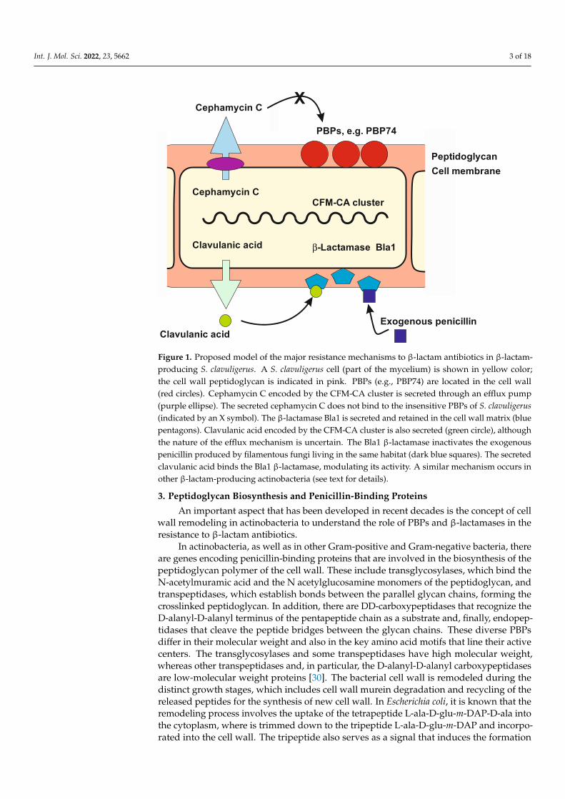
Those actinobacteria that produce β-lactam antibiotics, e.g. S. clavuligerus, Strepto-myces jumonjinensis, and Streptomyces cattleya, face the challenge of avoiding the toxicityof the antibiotics that they produce. S. clavuligerus, a producer of cephamycin C, is com-pletely insensitive to cephalosporin or cefoxitin (MIC 7.0 mg/mL cephalosporin), while thecephamycin non-producers Streptomyces albus J1074, Streptomyces flavogriseus ATCC 33,331,and Streptomyces coelicolor M1146 are not able to grow in 2.0–3.0 mg/mL cephalosporinC [25]. When the cephamycin C gene cluster that contains antibiotic resistance genes isintroduced into other Streptomyces strains, it increases the resistance to cephalosporin C inthe transformants [25]. Figure 1 shows a model of the resistance mechanisms to β-lactamantibiotic in β-lactam-producing actinobacteria.
Similar mechanisms of resistance to antibiotics have been acquired either by muta-tion [26] or by horizontal gene transfer [27,28] in a variety of Gram-positive and Gram-negative bacteria. This resistance to antibiotics is exerted by distinct mechanisms that havebeen widely described [6,7,26,29]. However, in this article, we restrict the study to therelationship between β-lactamases, β-lactamase inhibitors, and penicillin-binding proteinsas mechanisms of resistance in β-lactam-producing actinobacteria, which are discussed indetail in the following sections.
Int. J. Mol. Sci. 2022, 23, 5662 3 of 18Int. J. Mol. Sci. 2022, 23, x FOR PEER REVIEW 3 of 18
PBPs, e.g. PBP74
Cell membranePeptidoglycan
Cephamycin C
Cephamycin C
CFM-CA cluster
Clavulanic acid
Clavulanic acid
β-Lactamase Bla1
Exogenous penicillin
X
Figure 1. Proposed model of the major resistance mechanisms to β-lactam antibiotics in β-lactam-producing S. clavuligerus. A S. clavuligerus cell (part of the mycelium) is shown in yellow color; the cell wall peptidoglycan is indicated in pink. PBPs (e.g., PBP74) are located in the cell wall (red cir-cles). Cephamycin C encoded by the CFM-CA cluster is secreted through an efflux pump (purple ellipse). The secreted cephamycin C does not bind to the insensitive PBPs of S. clavuligerus (indicated by an X symbol). The β-lactamase Bla1 is secreted and retained in the cell wall matrix (blue penta-gons). Clavulanic acid encoded by the CFM-CA cluster is also secreted (green circle), although the nature of the efflux mechanism is uncertain. The Bla1 β-lactamase inactivates the exogenous peni-cillin produced by filamentous fungi living in the same habitat (dark blue squares). The secreted clavulanic acid binds the Bla1 β-lactamase, modulating its activity. A similar mechanism occurs in other β-lactam-producing actinobacteria (see text for details).
Similar mechanisms of resistance to antibiotics have been acquired either by muta-tion [26] or by horizontal gene transfer [27,28] in a variety of Gram-positive and Gram-negative bacteria. This resistance to antibiotics is exerted by distinct mechanisms that have been widely described [6,7,26,29]. However, in this article, we restrict the study to the relationship between β-lactamases, β-lactamase inhibitors, and penicillin-binding pro-teins as mechanisms of resistance in β-lactam-producing actinobacteria, which are dis-cussed in detail in the following sections.
3. Peptidoglycan Biosynthesis and Penicillin-Binding Proteins An important aspect that has been developed in recent decades is the concept of cell
wall remodeling in actinobacteria to understand the role of PBPs and β-lactamases in the resistance to β-lactam antibiotics.
In actinobacteria, as well as in other Gram-positive and Gram-negative bacteria, there are genes encoding penicillin-binding proteins that are involved in the biosynthesis of the peptidoglycan polymer of the cell wall. These include transglycosylases, which bind the N-acetylmuramic acid and the N acetylglucosamine monomers of the peptidoglycan, and transpeptidases, which establish bonds between the parallel glycan chains, forming the crosslinked peptidoglycan. In addition, there are DD-carboxypeptidases that recognize the D-alanyl-D-alanyl terminus of the pentapeptide chain as a substrate and, finally, en-dopeptidases that cleave the peptide bridges between the glycan chains. These diverse PBPs differ in their molecular weight and also in the key amino acid motifs that line their active centers. The transglycosylases and some transpeptidases have high molecular
Figure 1. Proposed model of the major resistance mechanisms to β-lactam antibiotics in β-lactam-producing S. clavuligerus. A S. clavuligerus cell (part of the mycelium) is shown in yellow color;the cell wall peptidoglycan is indicated in pink. PBPs (e.g., PBP74) are located in the cell wall(red circles). Cephamycin C encoded by the CFM-CA cluster is secreted through an efflux pump(purple ellipse). The secreted cephamycin C does not bind to the insensitive PBPs of S. clavuligerus(indicated by an X symbol). The β-lactamase Bla1 is secreted and retained in the cell wall matrix (bluepentagons). Clavulanic acid encoded by the CFM-CA cluster is also secreted (green circle), althoughthe nature of the efflux mechanism is uncertain. The Bla1 β-lactamase inactivates the exogenouspenicillin produced by filamentous fungi living in the same habitat (dark blue squares). The secretedclavulanic acid binds the Bla1 β-lactamase, modulating its activity. A similar mechanism occurs inother β-lactam-producing actinobacteria (see text for details).
3. Peptidoglycan Biosynthesis and Penicillin-Binding Proteins
An important aspect that has been developed in recent decades is the concept of cellwall remodeling in actinobacteria to understand the role of PBPs and β-lactamases in theresistance to β-lactam antibiotics.
In actinobacteria, as well as in other Gram-positive and Gram-negative bacteria, thereare genes encoding penicillin-binding proteins that are involved in the biosynthesis of thepeptidoglycan polymer of the cell wall. These include transglycosylases, which bind theN-acetylmuramic acid and the N acetylglucosamine monomers of the peptidoglycan, andtranspeptidases, which establish bonds between the parallel glycan chains, forming thecrosslinked peptidoglycan. In addition, there are DD-carboxypeptidases that recognize theD-alanyl-D-alanyl terminus of the pentapeptide chain as a substrate and, finally, endopep-tidases that cleave the peptide bridges between the glycan chains. These diverse PBPsdiffer in their molecular weight and also in the key amino acid motifs that line their activecenters. The transglycosylases and some transpeptidases have high molecular weight,whereas other transpeptidases and, in particular, the D-alanyl-D-alanyl carboxypeptidasesare low-molecular weight proteins [30]. The bacterial cell wall is remodeled during thedistinct growth stages, which includes cell wall murein degradation and recycling of thereleased peptides for the synthesis of new cell wall. In Escherichia coli, it is known that theremodeling process involves the uptake of the tetrapeptide L-ala-D-glu-m-DAP-D-ala intothe cytoplasm, where is trimmed down to the tripeptide L-ala-D-glu-m-DAP and incorpo-rated into the cell wall. The tripeptide also serves as a signal that induces the formation
Int. J. Mol. Sci. 2022, 23, 5662 4 of 18
of β-lactamases [31]. Recent studies in the model actinobacteria S. coelicolor indicate thatthere is an extensive degradation and reconstruction of the cell wall, resulting in its re-modeling, particularly during the conversion of mycelium to spores [32]. Several enzymesact in the cell wall remodeling in actinobacteria, including autolysines, carboxypepti-dases, and penicillin-binding proteins [33]. Chemical studies using high-performanceliquid chromatography associated to mass spectrometry (HPLC-MS) on the changes ofcell wall polymers in vegetative mycelium reveal large amounts of free disaccharide N-acetylglucosamine-N-acetylmuramic acid and free pentapeptide L-ala-D-glu-LL-DAP(orglycine)-D-ala-D-ala [32,34]. These results indicate that extensive changes in the crosslinkingmediated by PBPs occur during growth and differentiation in Streptomyces species.
4. Clusters of Genes for β-Lactam Antibiotics in Actinobacteria
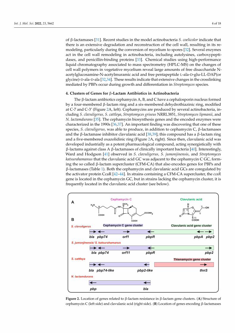
The β-lactam antibiotics cephamycin A, B, and C have a cephalosporin nucleus formedby a four-membered β-lactam ring and a six-membered dehydrothiazinic ring, modifiedat C-7 and C-3’ (Figure 2A, left). Cephamycins are produced by several actinobacteria, in-cluding S. clavuligerus, S. cattleya, Streptomyces griseus NRRL3851, Streptomyces lipmanii, andN. lactamdurans [35]. The cephamycin biosynthesis genes and the encoded enzymes werecharacterized in the 1990s [36,37]. An important finding was discovering that one of thesespecies, S. clavuligerus, was able to produce, in addition to cephamycin C, β-lactamasesand the β-lactamase inhibitor clavulanic acid [38,39]; this compound has a β-lactam ringand a five-membered oxazolidinic ring (Figure 2A, right). Since then, clavulanic acid wasdeveloped industrially as a potent pharmacological compound, acting synergistically withβ-lactams against class A β-lactamases of clinically important bacteria [40]. Interestingly,Ward and Hodgson [41] observed in S. clavuligerus, S. jumonjinensis, and Streptomyceskatsurahamanus that the clavulanic acid GC was adjacent to the cephamycin C GC, form-ing the so called β-lactam supercluster (CFM-CA) that also encodes genes for PBPs andβ-lactamases (Table 1). Both the cephamycin and clavulanic acid GCs are coregulated bythe activator protein CcaR [42–44]. In strains containing a CFM-CA supercluster, the ccaRgene is located in the cephamycin GC, but in strains lacking the cephamycin cluster, it isfrequently located in the clavulanic acid cluster (see below).
Int. J. Mol. Sci. 2022, 23, x FOR PEER REVIEW 5 of 18
bla pbp74 orf1 pbpR pbpA pbp2
bla pbp74 orf11 pbpR pbp2
bla pbp74-like pbp2-like thnS
pbp bla
S. clavuligerus
S. jumonjinensis/ S. katsurahamanus
S. cattleya
N. lactamdurans
Cephamycin C gene cluster Clavulanic acid gene cluster
Thienamycin gene cluster
OH
N
COOH
OH
O76 5
3 2
9
8
10 COOH
O
SH2N
COOHO
NH
N OCONH2
OCH3D
Cephamycin C Clavulanic acidA
B
Figure 2. Location of genes related to β-lactam resistance in β-lactam gene clusters. (A) Structure of cephamycin C (left side) and clavulanic acid (right side). (B) Location of genes encoding β-lac-tamases or PBPs in the cephamycin C gene clusters (purple), clavulanic acid gene clusters (green), and thienamycin gene cluster (pink) of S. clavuligerusm S, jumonjinensis, S. katsurahamanus, S. cattleya, and N. lactamdurans.
Clavulanic acid clusters have been studied also in Streptomyces flavogriseus (also named Streptomyces pratensis) and Saccharomonospora viridis [45]. The organization of these clusters is different from that of S. clavuligerus, and the scientific bases for their lack of clavulanic acid production have been thoroughly studied [45].
Most actinobacteria have several copies of PBPs [17]; in particular, in β-lactam pro-ducers, the antibiotic biosynthesis clusters contain several PBPs (Figure 2B). In order to determine whether clavulanic acid and/or cephamycin C clusters in other Streptomyces strains carried genes for β-lactamases or PBP proteins, we searched in actinobacteria da-tabases for proteins orthologous to those of S. clavuligerus, with percentages of identity higher than 60%. This analysis resulted in the finding of complete sets of clavulanic acid genes in different Streptomyces strains (see below), although the production of clavulanic acid has not yet been demonstrated in many of them.
5. Genes Encoding PBP Proteins in S. clavuligerus and Other Actinobacteria β-Lactam Gene Clusters
For many years, it was suggested that β-lactam-producing actinobacteria are re-sistant to penicillin/cephalosporins due to evolutive modification of the PBPs [17,46]. Elec-trophoretic studies of penicillin-labelled proteins showed that both Streptomyces lividans and the cephamycin producer N. lactamdurans have eight PBPs. One of them, PBP-4, is encoded by a gene located in the N. lactamdurans cephamycin cluster. In vitro competition binding experiments showed that PBP-4 lacks affinity for cephamycin C [23], providing experimental support for the Ogawara [46] hypothesis.
The best studied PBPs in the β-lactam GC are those of S. clavuligerus. A bioinformatic analysis of the S. clavuligerus genome revealed the presence of at 8 eight PBPs [47]; in con-trast, Ogawara et al. [17] listed 12: 3 of the class A and 9 of class B, not including PBP-74 [17]. Four S. clavuligerus PBPs are encoded by genes located in the CFM-CA supercluster. One of them, named pbp74, is located at one end in the cephamycin cluster [24]; the other two, named pbpA and pbp2, are located at the distal end in the clavulanic acid cluster
Figure 2. Location of genes related to β-lactam resistance in β-lactam gene clusters. (A) Structure ofcephamycin C (left side) and clavulanic acid (right side). (B) Location of genes encoding β-lactamases
Int. J. Mol. Sci. 2022, 23, 5662 5 of 18
or PBPs in the cephamycin C gene clusters (purple), clavulanic acid gene clusters (green), andthienamycin gene cluster (pink) of S. clavuligerusm S, jumonjinensis, S. katsurahamanus, S. cattleya, andN. lactamdurans.
Clavulanic acid clusters have been studied also in Streptomyces flavogriseus (also namedStreptomyces pratensis) and Saccharomonospora viridis [45]. The organization of these clustersis different from that of S. clavuligerus, and the scientific bases for their lack of clavulanicacid production have been thoroughly studied [45].
Most actinobacteria have several copies of PBPs [17]; in particular, in β-lactam pro-ducers, the antibiotic biosynthesis clusters contain several PBPs (Figure 2B). In order todetermine whether clavulanic acid and/or cephamycin C clusters in other Streptomycesstrains carried genes for β-lactamases or PBP proteins, we searched in actinobacteriadatabases for proteins orthologous to those of S. clavuligerus, with percentages of identityhigher than 60%. This analysis resulted in the finding of complete sets of clavulanic acidgenes in different Streptomyces strains (see below), although the production of clavulanicacid has not yet been demonstrated in many of them.
5. Genes Encoding PBP Proteins in S. clavuligerus and Other Actinobacteria β-LactamGene Clusters
For many years, it was suggested that β-lactam-producing actinobacteria are resis-tant to penicillin/cephalosporins due to evolutive modification of the PBPs [17,46]. Elec-trophoretic studies of penicillin-labelled proteins showed that both Streptomyces lividansand the cephamycin producer N. lactamdurans have eight PBPs. One of them, PBP-4, isencoded by a gene located in the N. lactamdurans cephamycin cluster. In vitro competitionbinding experiments showed that PBP-4 lacks affinity for cephamycin C [23], providingexperimental support for the Ogawara [46] hypothesis.
The best studied PBPs in the β-lactam GC are those of S. clavuligerus. A bioinformaticanalysis of the S. clavuligerus genome revealed the presence of at 8 eight PBPs [47]; incontrast, Ogawara et al. [17] listed 12: 3 of the class A and 9 of class B, not includingPBP-74 [17]. Four S. clavuligerus PBPs are encoded by genes located in the CFM-CAsupercluster. One of them, named pbp74, is located at one end in the cephamycin cluster [24];the other two, named pbpA and pbp2, are located at the distal end in the clavulanic acidcluster (Figure 2B). In addition, the gene pbpR (also designated pcbR) is located between thecephamycin and the clavulanic acid GCs [48–50].
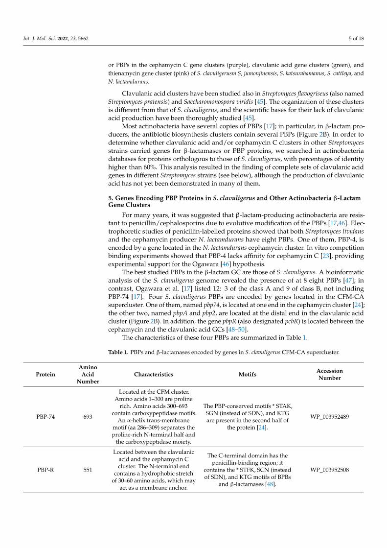
The characteristics of these four PBPs are summarized in Table 1.
Table 1. PBPs and β-lactamases encoded by genes in S. clavuligerus CFM-CA supercluster.
ProteinAminoAcid
NumberCharacteristics Motifs Accession
Number
PBP-74 693
Located at the CFM cluster.Amino acids 1–300 are proline
rich. Amino acids 300–693contain carboxypeptidase motifs.
An α-helix trans-membranemotif (aa 286–309) separates theproline-rich N-terminal half and
the carboxypeptidase moiety.
The PBP-conserved motifs * STAK,SGN (instead of SDN), and KTGare present in the second half of
the protein [24].
WP_003952489
PBP-R 551
Located between the clavulanicacid and the cephamycin Ccluster. The N-terminal end
contains a hydrophobic stretchof 30–60 amino acids, which may
act as a membrane anchor.
The C-terminal domain has thepenicillin-binding region; it
contains the * STFK, SCN (insteadof SDN), and KTG motifs of BPBs
and β-lactamases [48].
WP_003952508
Int. J. Mol. Sci. 2022, 23, 5662 6 of 18
Table 1. Cont.
ProteinAminoAcid
NumberCharacteristics Motifs Accession
Number
PBP-A 494
Located at the end of the ACcluster. Contains a pfam0095
transpeptidase domain and anATP/GTP-binding motif.
Contains the * STFK, STN, andKTG motifs, but lacks the EPELN
motif [49].WP_003952525
PBP-2 717 Located at the end of the ACcluster. Domain pbp2_mrdA.
Contains SIFK and FTG (instead ofKTG) [49]. WP_003952526
Bla1 332 Located at the CFM cluster.ClassA β-lactamase.
Contain the active center 70 STFKand the motifs 130 SDG (instead ofSDN), 166 EPELN, and 234 KTG of
class A β-lactamases [51].
WP_003952487
BlaB1 338 Located at the CFM cluster [42].Class B β-lactamase [this work]. Motif 117 HGHFD121. WP_003952502
5.1. The PBP-74 Protein
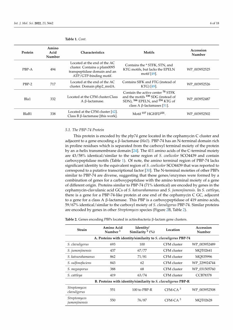
This protein is encoded by the pbp74 gene located in the cephamycin C cluster andadjacent to a gene encoding a β-lactamase (bla1). PBP-74 has an N-terminal domain richin proline residues which is separated from the carboxyl terminal moiety of the proteinby an α-helix transmembrane domain [24]. The 411 amino acids of the C-terminal moietyare 43/58% identical/similar to the same region of S. coelicolor SCO4439 and containcarboxypeptidase motifs (Table 1). Of note, the amino terminal region of PBP-74 lackssignificant identity to the equivalent region of S. coelicolor SCO4439 that was reported tocorrespond to a putative transcriptional factor [30]. The N-terminal moieties of other PBPssimilar to PBP-74 are diverse, suggesting that these genes/enzymes were formed by acombination of genes for a carboxypeptidase with the amino terminal moiety of a geneof different origin. Proteins similar to PBP-74 (71% identical) are encoded by genes in thecephamycin-clavulanic acid GCs of S. katsurahamanus and S. jumonjinensis. In S. cattleya,there is a gene for a PBP-74-like protein at one end of the cephamycin C GC, adjacentto a gene for a class A β-lactamase. This PBP is a carboxypeptidase of 419 amino acids,59/67% identical/similar to the carboxyl moiety of S. clavuligerus PBP-74. Similar proteinsare encoded by genes in other Streptomyces species (Figure 2B, Table 2).
Table 2. Genes encoding PBPs located in actinobacteria β-lactam gene clusters.
Strain Amino AcidNumber 1
Identity/Similarity 2 (%) Location Accession
Number
A. Proteins with identity/similarity to S. clavuligerus PBP-74
S. clavuligerus 693 100 CFM cluster WP_003952489
S. jumonjinensis 437 67/77 CFM cluster MQT02641
S. katsurahamanus 862 71/81 CFM cluster MQS35996
S. sulfonofaciens 843 62 CFM cluster WP_229924744
S. megasporus 388 68 CFM cluster WP_031505760
S. cattleya 419 63/74 CFM cluster CCB78378
B. Proteins with identity/similarity to S. clavuligerus PBP-R
Streptomycesclavuligerus 551 100 to PBP-R CFM-CA 3 WP_003952508
Streptomycesjumonjinensis 550 76/87 CFM-CA 3 MQT02628

Int. J. Mol. Sci. 2022, 23, 5662 7 of 18
Table 2. Cont.
Strain Amino AcidNumber 1
Identity/Similarity 2 (%) Location Accession
Number
B. Proteins with identity/similarity to S. clavuligerus PBP-R
Streptomyceskatsurahamanus 550 76/87 CFM-CA 3 MQS35981
Streptomycesfulvorobeus 555 66/77 CA cluster WP_173313670
Streptomycesalbiflavescens 548 71/83 CA cluster WP_189188591
Streptomyces sp.M41 548 72/83 CA cluster WP_081218566
Streptomyces sp.SID14446 548 71/82 CA cluster WP_164372891
Streptomyces sp.B93 549 71/83 CA cluster WP_210923884
Streptomyces sp.SID2888 547 67/79 CA cluster WP_161240914
Streptomyces sp.NRRL 24051 553 61/74 CA cluster WP_014152699
Streptomyces sp.S-325 553 61/74 CA cluster WP_014152699
Streptomyces sp.SM10 556 61/74 CA cluster WP_103513573
Streptomyces sp.PMAC2608 553 61/74 CA cluster WP_014152699
Streptomycesflavovirens 553 60/74 CA cluster WP_030636769
Streptomycesflavogriseus 551 61/74 CA cluster MBD28344891
Streptomyces finlayi 550 67/79 CA cluster WP_185300304
Streptomycessulfonofaciens 549 70/80 CFM cluster WP_189933512
C. Proteins with identity/similarity to S. clavuligerus PBP-A
Streptomycesclavuligerus 494 100 CA cluster WP_003952525
Streptomyces sp.B93 494 78/89 CA cluster WP_210923906
D. Proteins with identity/similarity to S. clavuligerus PBP-2
Streptomycesclavuligerus 764 100 CA cluster WP_003952526
Streptomycesjumonjinensis 721 80 CA cluster MQ535963
Streptomyceskatsurahamanus 721 80 CA cluster MQT02611
Streptomycescattleya 695 66/78 CFM cluster CCB78364
1 The identity is calculated for the carboxyl terminal end of PBP-74 (i.e., amino acids 330–693); 2 The % ofidentity/similarity was obtained using the NCBI global alignment Needleman–Wunsch Program; 3 Gene locatedbetween the CA and the CFM clusters.

Int. J. Mol. Sci. 2022, 23, 5662 8 of 18
5.2. The PBP-R Protein
A PBP of 551 amino acids is encoded by a pbpR gene that separates the cephamycinand clavulanic acid GCs. The carboxyl end of PBP-R contains a transpeptidase domain. TheN-terminal moiety has a hydrophobic transmembrane domain, suggesting that this proteinis membrane bound and, indeed, using anti-PBP-R antibodies it was located in the cellmembrane [48]. A pbpR-disrupted mutant was not affected in clavulanic acid. S. clavuligerusis naturally more resistant to cephalosporin and cephalothin production. This mutantshowed lower resistance to penicillin and cephalosporin than the parental strain [48];this suggests that PBP-R is involved in antibiotic resistance/cell wall biosynthesis thanto penicillin [25], which might be partially due to the lack of affinity of PBP-R towardscephalosporin. In Streptomyces strains carrying the clavulanic acid GCs, but not the adjacentcephamycin GC, the pbpR gene is located at the end of the clavulanic acid GC (Figure 2).Expression of pbpR in S. flavogriseus is low but greatly increases in S. flavogriseus strainstransformed with the S. clavuligerus cephamycin GC, independently of the presence of theactivator ccaR or the bla gene. This correlates well with the higher resistance to penicillin Gand cefoxitin observed in these transformants [45], confirming that the pbpR gene presentin the transformants confers resistance to β-lactam antibiotics.
5.3. PBP-A and PBP-2
Two other PBPs, encoded by genes located at the other end of S. clavuligerus clavulanicacid cluster (Table 1, Figure 2B), are named PBP-A and PBP-2 [49,52]. These two PBPs have494 and 717 amino acids, respectively, and only share 20% overall identity. PBP-A containsa transpeptidase domain and an ATP/GTP-binding motif in addition to the classical PBPmotifs STKF, STN, and KTG, but lacks the EPELN motif. In both PBP-A and PBP-2, a singlehydrophobic region confers the ability to bind cell membranes, and the two proteins werefound to be associated with membranes when expressed in E. coli. PBP-2 appears to confermore resistance to penicillin G than PBP-A [52]. The PBP-A amino acid sequence showshigh similarity (82%) with the S. coelicolor PBP-A protein [49]; of note, the pbpA gene is notpresent in the supercluster of S. jumonjinensis and S. katsurahamanus. In S. cattleya, adjacentto the ccaR gene for the cluster positive regulator, there is a gene encoding a PBP proteinof 695 amino acids with a transpeptidase domain which is 66/78% identical/similar toS. clavuligerus PBP-2. Genes encoding proteins similar to PBP-A and PBP-2 are frequent inStreptomyces species, but are rarely associated with β-lactam clusters (Table 2). As occurswith pbpR, the heterologous expression of S. clavuligerus pbpA in S. flavogriseus leads tohigher resistance to penicillin G and cefoxitin [45], thus confirming that the PBPs aredeterminants for β-lactam resistance.
6. β-Lactamases
β-lactamases exist in Gram-negative and Gram-positive bacteria, including actinobac-teria [53]. These enzymes cleave the β-lactam ring of penicillins and cephalosporins, andmany of them may be inactivated by β-lactamase inhibitors. Bacterial β-lactamases areextracellular enzymes secreted to protect the cell against external β-lactam antibiotics pro-duced by filamentous fungi or other microorganisms that live together with actinobacteriain their natural habitats.
β-lactamases are divided into four classes (from A to D) depending on their structureand the mechanism of their β-lactam bond hydrolysis.
Classes A, C, and D are serine hydrolases in which a serine residue in the activecenter (* STFK) of the protein exerts a nucleophilic attack on the β-lactam ring, forming anacyl–enzyme intermediate through the carbonyl group of the open β-lactam ring [54,55].The acylated β-lactamase protein is then deacylated with the help of the highly conservedglutamic acid (166 E) residue that activates a water molecule for the hydrolysis. Indeed,mutation of this glutamic acid residue results in loss of the deacylation step, and thisinactivates the β-lactamase [56,57]. Class A β-lactamases hydrolyze penicillins, a type ofβ-lactams containing a five-membered thiazolidine ring in addition to the four-membered
Int. J. Mol. Sci. 2022, 23, 5662 9 of 18
β-lactam ring, and are inactivated by the β-lactamase inhibitor clavulanic acid (Section 7.1,Figure 2A). In contrast, class C β-lactamases cleave the β-lactam ring of cephalosporin andsome semisynthetic derivatives, which contain the β-lactam ring fused to a six-membereddehydrothiazinic ring. Class D β-lactamases are mainly active towards oxacillins.
Of note, class B β-lactamases are metalloenzymes that have a different mechanism ofaction: they use a zinc-coordinated water molecule to attack and cleave the β-lactam ring.
6.1. Analysis and Role of β-Lactamases Located in the S. clavuligerus β-Lactam Supercluster
Genes encoding type A β-lactamases have been found in most of the cephamycinGCs in actinobacteria. An important question is whether the β-lactamases associatedwith these clusters are somehow different from other class A β-lactamases occurring inactinobacteria that do not produce β-lactams. A related question concerns the role of theβ-lactamases produced by cephamycin-producing actinobacteria and whether they destroythe isopenicillin N and penicillin N intermediates of the cephamycin pathway. Of particularinterest are the β-lactamases encoded in the genome of S. clavuligerus, which serves asa model of actinobacteria, due to its ability to simultaneously produce the β-lactamaseinhibitor clavulanic acid. In S. clavuligerus, Ogawara [52] bioinformatically identified threeclass A β-lactamases, eight class B β-lactamases, and six class C β-lactamases (Table S1).
Class A β-Lactamases of S. clavuligerus
This class includes the enzyme encoded by the bla gene (hereafter named bla1), locatedin the cephamycin GC (see below), and the β-lactamase-like protein encoded by the orf12gene, located in the clavulanic acid cluster [49,50].
The S. clavuligerus bla1 gene is located at the cephamycin end of the CFM-CA su-percluster, contiguous to pbp74 [24]. The encoded protein has 332 amino acids and is aclass A β-lactamase containing an active center at position 70 STFK73 and three additionalconserved motifs of class A β-lactamases: SDG (instead of SDN), EPELN, and KTG [51].The standard bacterial β-lactamases are secreted proteins and their activity is detectedin the supernatant broth. However, there is no β-lactamase activity in the supernatantof S. clavuligerus cultures, and, as occurs with the β-lactamase of N. lactamdurans (seebelow), S. clavuligerus Bla1 remains located between the membrane and cell wall and isonly released after protoplast formation. The purified enzyme has a Km of 11 µM forbenzylpenicillin, but the Km for cephalosporin C or semisynthetic cephalosporins rises to5000 µM. Bla1 binds and retains labelled [H3]-benzylpenicillin in contrast to the efficientrelease of the hydrolyzed penicillin by other bacterial β-lactamases; this behavior correlateswith a low penicillin deacylation rate and suggests that this enzyme has some properties ofpenicillin-binding proteins that are known to retain the bound penicillin [57]. This indicatesthat the β-lactamase in the CFM-CA supercluster is somehow different from extracellularβ-lactamases from other bacteria. This similarity suggests that S. clavuligerus Bla1 mayhave a role in the biosynthesis/regulation or remodeling of the cell wall.
The Orf12 protein, encoded in S. clavuligerus clavulanic CG, has a β-lactamase domain,but the pure protein lacks β-lactamase activity [58,59] and the orf12-disrupted mutants areunable to produce clavulanic acid. Therefore, the Orf12 protein appears to be a biosyntheticenzyme that is not included among the standard S. clavuligerus β-lactamases.
6.2. Class A β-Lactamases Associated with β-Lactam Clusters in Other Actinobacteria
The N. lactamdurans β-lactamase was the first of this type of enzyme reported to beencoded by a gene located in a cephamycin GC [23,60]. This β-lactamase has 302 aminoacids and contains the classical active center * STFK and all the conserved motifs of classA β-lactamases. The N. lactamdurans Bla1 protein has an N-terminal putative leaderpeptide of 29 amino acids ending in AAA29, but surprisingly, there is no β-lactamaseactivity detectable in the supernatant of N. lactamdurans cultures. The enzyme is trappedbetween the membrane and the cell wall peptidoglycan, and the β-lactamase activity isonly detected after protoplast formation. The enzyme released after this treatment is very
Int. J. Mol. Sci. 2022, 23, 5662 10 of 18
active on benzylpenicillin and less active on isopenicillin N, an intermediate of cephamycinbiosynthesis; in contrast, it is poorly active against cephalosporins and has no activityagainst cephamycin, the final product of its biosynthetic pathway. The N. lactamduransBla1 enzyme is inhibited by clavulanic acid (50% inhibition by 0.5 µg/ml CA). Mutantsdisrupted in bla1 are more sensitive to penicillin, but do not show significant differences insensitivity to cephalosporin C or cephamycin. Conversely, N. lactamdurans transformantswith a bla1 amplified copy number are more resistant to benzylpenicillin, indicating that themajor role of this β-lactamase is the defense against exogenous benzylpenicillin rather thandegradation of the cytosolic isopenicillin N/penicillin N synthesized by this strain [60].
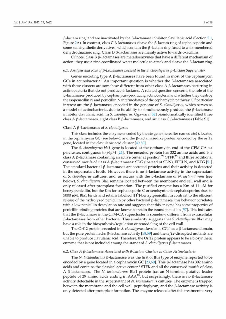
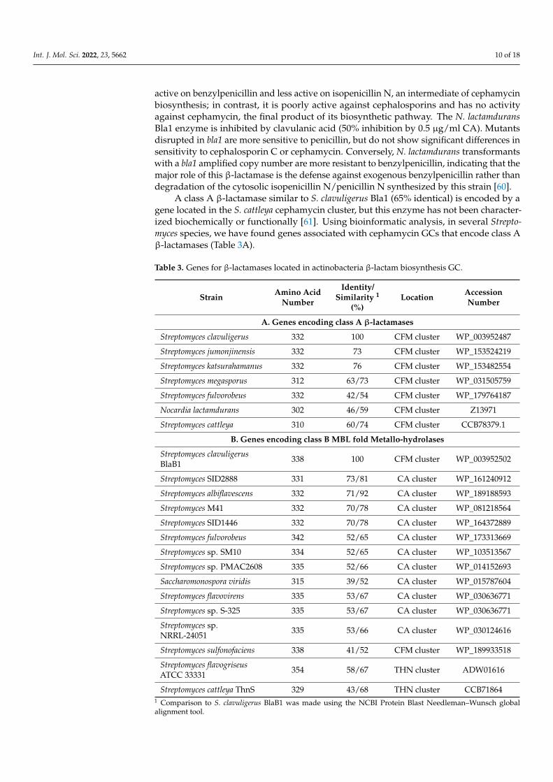
A class A β-lactamase similar to S. clavuligerus Bla1 (65% identical) is encoded by agene located in the S. cattleya cephamycin cluster, but this enzyme has not been character-ized biochemically or functionally [61]. Using bioinformatic analysis, in several Strepto-myces species, we have found genes associated with cephamycin GCs that encode class Aβ-lactamases (Table 3A).
Table 3. Genes for β-lactamases located in actinobacteria β-lactam biosynthesis GC.
Strain Amino AcidNumber
Identity/Similarity 1
(%)Location Accession
Number
A. Genes encoding class A β-lactamases
Streptomyces clavuligerus 332 100 CFM cluster WP_003952487
Streptomyces jumonjinensis 332 73 CFM cluster WP_153524219
Streptomyces katsurahamanus 332 76 CFM cluster WP_153482554
Streptomyces megasporus 312 63/73 CFM cluster WP_031505759
Streptomyces fulvorobeus 332 42/54 CFM cluster WP_179764187
Nocardia lactamdurans 302 46/59 CFM cluster Z13971
Streptomyces cattleya 310 60/74 CFM cluster CCB78379.1
B. Genes encoding class B MBL fold Metallo-hydrolases
Streptomyces clavuligerusBlaB1 338 100 CFM cluster WP_003952502
Streptomyces SID2888 331 73/81 CA cluster WP_161240912
Streptomyces albiflavescens 332 71/92 CA cluster WP_189188593
Streptomyces M41 332 70/78 CA cluster WP_081218564
Streptomyces SID1446 332 70/78 CA cluster WP_164372889
Streptomyces fulvorobeus 342 52/65 CA cluster WP_173313669
Streptomyces sp. SM10 334 52/65 CA cluster WP_103513567
Streptomyces sp. PMAC2608 335 52/66 CA cluster WP_014152693
Saccharomonospora viridis 315 39/52 CA cluster WP_015787604
Streptomyces flavovirens 335 53/67 CA cluster WP_030636771
Streptomyces sp. S-325 335 53/67 CA cluster WP_030636771
Streptomyces sp.NRRL-24051 335 53/66 CA cluster WP_030124616
Streptomyces sulfonofaciens 338 41/52 CFM cluster WP_189933518
Streptomyces flavogriseusATCC 33331 354 58/67 THN cluster ADW01616
Streptomyces cattleya ThnS 329 43/68 THN cluster CCB718641 Comparison to S. clavuligerus BlaB1 was made using the NCBI Protein Blast Needleman–Wunsch globalalignment tool.

Int. J. Mol. Sci. 2022, 23, 5662 11 of 18
6.3. Class B β-Lactamases
The six class B β-lactamases identified in S. clavuligerus by Ogawara [52] are encodedby genes scattered in the genome. In addition, a seventh gene, located in the cephamycin Ccluster, encodes an MBL-fold hydrolase, hereafter named BlaB1 (acronym for β-lactamaseclass B). The blaB1 gene is contiguous to ccaR, a gene encoding the positive regulator thatcontrols cephamycin C and clavulanic acid production. When this protein was initially dis-covered, no similarity to other proteins was found [42]; therefore, it was not further studied.It was only by recent searching in the databases that we found that this gene encodes ahydrolase of the metallo-β-lactamase family. This protein has the 117HGHFD121 motif andhistidine in the expected positions (193H, 265H) for a class B β-lactamase (Tables 1 and 3).
Searching in the databases shows the presence of genes encoding class B β-lactamasesin most of the clavulanic acid GCs of other actinobacteria (Table 3B, Figure 3) suggestingthat this gene may have a direct/indirect role in clavulanic acid biosynthesis or regulation.Unfortunately, no blaB1-disrupted mutants have ever been reported. The S. cattleya thnSgene, located in the thienamycin GC, encodes a class B β-lactamase which is 43/68%identical/similar to S. clavuligerus BlaB1. ThnS is involved in resistance to thienamycin inthe producer strain [62].
6.4. Class C β-Lactamases
Six putative class C β-lactamases have been found bioinformatically in the genomeof S. clavuligerus and annotated as cephalosporinases (Table S1) [52]. However, none ofthese genes are located in a β-lactam biosynthesis cluster. Most of these proteins containall the conserved domains of classical type C β-lactamases, and one of them is truncated.However, S. clavuligerus lacks significant extracellular cephalosporinase activity.
7. β-Lactamase Inhibitors and β-Lactamase Inhibitory Proteins
Two distinct components of the β-lactamase regulatory system in actinobacteria arethe β-lactamase inhibitors and the β-lactamase inhibitory proteins (BLIP).
7.1. β-Lactamases Inhibitors
An important strategy to improve the action of β-lactam antibiotics was the sys-tematic search of β-lactamase inhibitors [40]. This search focused on finding inhibitorsstructurally related to penicillin that might acylate the β-lactamases but will not deacylatethem easily [63]. This resulted in the finding of compounds that bind β-lactamase in anirreversible or partially reversible form, such as CA and the olivanic acid family [38]. Theirreversible inhibitors permanently inactivated the β-lactamases by reaction with the activesite of the cognate β-lactamases [64]. Clavulanic acid contains a β-lactam ring fused toan oxygen-containing five-membered oxazolidinic ring (Figure 2A right) that mimics thepenicillin structure [39]. CA combined with amoxicillin or other penicillins has potentβ-lactamase inhibitory activity against class A β-lactamases and low activity against classesB, C, and D [40,63]; this compound is the more frequently used in clinical practice. TheCA molecule binds the serine in the active site of β-lactamases and performs the acylationstep, but the acylated β-lactamase–CA complex is resistant to the subsequent deacyla-tion; therefore, the acylated β-lactamases remain inactive. The biosynthesis of CA and itsmolecular genetics have been well studied [65–67]. The role of autogenous CA in β-lactam-producing actinobacteria is enigmatic; CA may act as a synergistic metabolite in the fightagainst soil bacterial. In addition, CA inactivates the purified class A β-lactamase Bla1 ofS. clavuligerus or N. lactamdurans, which suggests that CA may modulate these enzymes’activity in vivo [57,60]. Clavulanic acid has weak antibiotic activity and in E. coli interactswith the PBP2 protein [68]. This interaction affects the cell wall remodeling and results inalteration of the rod shape of E. coli cells. Similarly, in S. clavuligerus, CA may be also a finemodulator of the cell wall remodeling, although experimental work needs to be done tosupport this hypothesis.
Int. J. Mol. Sci. 2022, 23, 5662 12 of 18
7.2. The BLIP Protein of S. clavuligerus and Other Actinobacteria
Some β-lactamase-producing actinobacteria, such as S. clavuligerus, contain a sep-arated gene, named blip, encoding a small protein that is a strong inhibitor of class Aβ-lactamases [69]. The blip gene is located far from the clavulanic acid GC [70] and encodesan 18 KDa protein that is found in the supernatant of S. clavuligerus cultures, in which itis a major protein. Proteins similar to BLIP (74% identical) are encoded in the genomesof S. jumonjinensis and S. katsurahamanus, but not in other Streptomyces species. Structuralstudies reveal that BLIP contains a tandem domain forming a polar concave protrudingregion that interacts with a conserved convex loop-helix region adjacent to the active centerof class A β-lactamases, inhibiting its β-lactamase activity [71–73]. The BLIP protein mayhave a modulation role in the β-lactamase activity in S. clavuligerus.
Two other similar proteins, BLIP-I and BLIP-II, were subsequently discovered inculture supernatants of Streptomyces exfoliatus [74,75]. BLIP-II is a 28 KDa protein; itfolds forming a seven-bladed β-propeller structure that interacts with the same regionof β-lactamases adjacent to the active center as BLIP does. Thermodynamic studies haveshown that BLIP-II has a very high affinity for the β-lactamases and an extremely lowdissociation constant. It has been proposed that BLIP-II plays a role in the remodeling ofthe cell wall in S. exfoliatus [74].
8. Distinct Arrangement of the Clavulanic Acid Cluster in Actinobacteria
There are different arrangements of the genes in the β-lactam gene clusters in antibiotic-poducing actinobacteria [45,66,76].
8.1. Is a PBP Gene in the β-Lactam Gene Cluster Required for Self Resistance?
Since PBPs are determinants for resistance to the β-lactam antibiotics, but only someof them are located in the CFM-CA supercluster, an interesting question is whether any ofthe PBP genes in the clavulanic or cephamycin GC are required for self-resistance.
In our bioinformatic study for this article of the CFM-CA gene clusters in actinobacteria,we found considerable differences in the organization of these clusters. This search revealed15 complete and 4 incomplete CA clusters (Figure 3). Only three of the studied strains(S. clavuligerus, S. katsurahamanus, and S. jumonjinensis) contain the complete CFM-CAsupercluster [41]; all other strains lack an adjacent cephamycin cluster. Four strains (S.albiflavescens, Streptomyces sp. SID14446, Streptomyces sp. M41, and Streptomyces sp. SID288)have identical organization to the previous strains, but contain the blaB1 and ccaR genesadjacent to the CA cluster. In the third block of strains (Streptomyces flavovirens, S. flavogriseus,Streptomyces sp. S-325, Streptomyces sp. PAMC 2608, and Streptomyces sp. NRRL 2401), twoblocks of genes (from oppA1 to gcaS and from oat2 to cas2) are conserved, and the blaB1and ccaR genes are inserted between them. Finally, the gene organization is different inStreptomyces fulveorobeos, Streptomyces finlayi, and Sacc. viridis (Figure 3) and incomplete inthe other four species, although in all of them, the four genes (ceaS-pah-bls-cas) for the earlysteps of the pathway leading to clavaminic acid formation are conserved. Regarding thepresence of pbp genes associated with resistance to β-lactam antibiotics, the only gene thatis present in most clavulanic acid clusters (89% of the cases) is pbpR (Table 2, Figure 3). Thepbp74 gene is always associated with the cephamycin C cluster, either when the cluster isadjacent to the clavulanic acid cluster or when it stands alone; finally, the pbpA and pbp2genes occur rarely and are associated either with the clavulanic GC or with the cephamycinGCs. These results suggest that PBP-R is the more important protein required for themaintenance of resistance in the clavulanic acid clusters, and that PBP-74 exerts a similarfunction in the cephamycin clusters.

Int. J. Mol. Sci. 2022, 23, 5662 13 of 18Int. J. Mol. Sci. 2022, 23, x FOR PEER REVIEW 13 of 18
S. clavuligerusS. katsurahamanusS. jumonjinensis
Streptomyces sp. M41S. albiflavescensStreptomyces sp. SID14446
Streptomyces sp. SID2888
pbpR ceaS bls pah cas2 oat2
S. fulvorobeus
S. finlayi
Streptomyces sp. PAMC2608
S. flavovirens NRRL B-2182 Streptomyces sp. NRRL S-325
Streptomyces sp NRRL24051
Complete CFM gene cluster
Sac. viridis DSM43017
Streptomyces sp. B93 blaB1-ccaR-pcbR-ceaS-bls-pah-cas-oat2-oppA1-claR
ccaR-blaB1-oat-ceaS-bls-pah-cas-pbpR
ceaS-bls-pah-cas-oat
orfA-X-X-ceaS-bls-pah-cas-oat-X-X-gcaS
Streptomyces sp. M10 Streptomyces sp SID 10853 Kitasatospora MBT63
blaB1oat2 claR PBP
cas pah bls ceaS claR
blaR1 ccaR pbpR ceaS bls pah cas2 oat2
pbpR cas2 pah bls ceaS oat2 blaB1 ccaR
S. flavogriseus ATCC33331
cas2 pah bls ceaS claR oppA1 car cyp-fd bla pcbC pcbAB lat cefD pcd cmcT
1.75 Mb
oppA1 claR car cyp-fd orf12 orf13 orf14 oppA2 orf16 gcaS
orf12 orf13 orf14 oppA2 orf16 gcaS pbpR blaB1 oat ccaR
oppA1 car fd orf12 orf13 orf14 oppA2 gcaS pbpR oat
oppA1 ceaS bls gcaS pah cas2 orf16 oppA2 orf14 orf13 orf12 fd-cyp car
oppA1 claR car cyp-fd orf12 orf13 orf14 oppA2 orf16 gcaS
oppA1 claR car cyp-fd orf12 orf13 orf14 oppA2 orf16 gcaS pbpA pbp2
Figure 3. Organization of the clavulanic acid gene clusters of different actinobacteria. The name of the strain is indicated in the left column. The genes are color-coded and their names are indicated. Genes in black color are not related to cephamycin or clavulanic acid biosynthesis. The initial search of CA gene clusters was made using the essential S. clavuligerus proteins GcaS and CeaS2 as probes. Subsequently, all the CA biosynthesis proteins were searched using the homologous proteins of S. clavuligerus as probe. The organization of the genes was obtained using the NCBI Genome Blast tool. The gene clusters of clavulanic acid in S. clavuligerus, S. jumonjinensis-S.katsurahamanus, S. flavo-griseus, and Sacc. viridis have been previously reported [45,66,76]. The CA clusters of all other ac-tinobacteria shown in Figure 3 are reported for the first time in this work.
8.2. Distribution of β-Lactamase and Blip Genes in the Cephamycin and Clavulanic Acid GC Genes for class A β-lactamases are always associated with cephamycin GCs, while
the gene for the class B β-lactamase blaB1 is most frequently associated with the clavulanic acid gene cluster, suggesting that this gene may play a role, directly or indirectly, in clavu-lanic acid biosynthesis. Genes similar to either blip or blipII are very rare in actinobacteria [69,74], and only orthologous proteins with less than 40% identity have been found in some of the new strains carrying CA clusters indicated in this work (Figure 3).
8.3. Are Some Genes in the Clavams or Cephamycin Clusters Required for Biosynthesis of Clavulanic Acid
In addition to clavulanic acid and cephamycin C GCs, S. clavuligerus contains two different clusters for the biosynthesis of 3S, 5S antifungal clavams that share the initial intermediates with the clavulanic acid pathway up to clavaminic acid (Figure S1), but do not isomerize the 3S, 5S clavams to the 3R, 5R configuration characteristic of clavulanic acid [77,78].
It has been suggested that the clavulanic acid cluster was initially formed by dupli-cation of a 3S, 5S clavam cluster [79]. In that article, the authors hypothesized that after the clavam cluster was duplicated, one of the copies acquired some genes to convert the 3S, 5S intermediates to the 3R, 5R final compound. The formation of clavulanic acid was
Figure 3. Organization of the clavulanic acid gene clusters of different actinobacteria. The name ofthe strain is indicated in the left column. The genes are color-coded and their names are indicated.Genes in black color are not related to cephamycin or clavulanic acid biosynthesis. The initialsearch of CA gene clusters was made using the essential S. clavuligerus proteins GcaS and CeaS2 asprobes. Subsequently, all the CA biosynthesis proteins were searched using the homologous proteinsof S. clavuligerus as probe. The organization of the genes was obtained using the NCBI GenomeBlast tool. The gene clusters of clavulanic acid in S. clavuligerus, S. jumonjinensis-S.katsurahamanus,S. flavogriseus, and Sacc. viridis have been previously reported [45,66,76]. The CA clusters of all otheractinobacteria shown in Figure 3 are reported for the first time in this work.
8.2. Distribution of β-Lactamase and Blip Genes in the Cephamycin and Clavulanic Acid GC
Genes for class A β-lactamases are always associated with cephamycin GCs, while thegene for the class B β-lactamase blaB1 is most frequently associated with the clavulanic acidgene cluster, suggesting that this gene may play a role, directly or indirectly, in clavulanicacid biosynthesis. Genes similar to either blip or blipII are very rare in actinobacteria [69,74],and only orthologous proteins with less than 40% identity have been found in some of thenew strains carrying CA clusters indicated in this work (Figure 3).
8.3. Are Some Genes in the Clavams or Cephamycin Clusters Required for Biosynthesis ofClavulanic Acid
In addition to clavulanic acid and cephamycin C GCs, S. clavuligerus contains twodifferent clusters for the biosynthesis of 3S, 5S antifungal clavams that share the initialintermediates with the clavulanic acid pathway up to clavaminic acid (Figure S1), but donot isomerize the 3S, 5S clavams to the 3R, 5R configuration characteristic of clavulanicacid [77,78].
It has been suggested that the clavulanic acid cluster was initially formed by duplica-tion of a 3S, 5S clavam cluster [79]. In that article, the authors hypothesized that after theclavam cluster was duplicated, one of the copies acquired some genes to convert the 3S,

Int. J. Mol. Sci. 2022, 23, 5662 14 of 18
5S intermediates to the 3R, 5R final compound. The formation of clavulanic acid was alsoproposed to be a response to the presence and expression of a CFM cluster in the genomeand to the production of cephamycin by the strains [79].
This raises the question of whether there are actinobacteria able to produce clavulanicacid without the presence of 3S, 5S clavam clusters. The clavam GC is present in someStreptomyces species, (e.g., Streptomyces antibioticus) which do not contain the CA genecluster and therefore produce clavams but not clavulanic acid [80,81]. In a search forclavam biosynthesis genes in the novel strains carrying clavulanic acid clusters (Figure 3),we could not find evidence for clavam clusters. The lack of 3S, 5S clavam clusters in all theanalyzed strains suggests that the clavam cluster was formed in the clavulanic producerstrains to increase the supply of enzymes and intermediates for clavulanic acid production.
A different question is whether the genetic information encoded by the cephamycin GCis required for the occurrence or expression of the clavulanic acid genes. Here, we discussnovel information that sheds light on this intriguing question. Early fermentation studiesindicated that it is possible to dissociate the cephamycin and clavulanic acid production [82],and that the lack of cephamycin formation does not affect significantly clavulanic acidproduction [83]. While S. clavuligerus, S. katsurahamanus, and S. jumonjinensis have theCFM-CA supercluster, in the novel strains analyzed here, only carrying the CA cluster, oneof them, Streptomyces fulvorobeos, contains an incomplete truncated cephamycin GC. Thistruncated cluster lacks the genes for the medium and late cephamycin biosynthesis steps;therefore, the final product of this cluster should be penicillin N. The cephamycin cluster ofS. fulvorobeos is separated by 1.75 Mb from the CA cluster.
In strains carrying the CFM-CA supercluster, the only gene located in the cephamycinGC required for CA formation is that encoding the activator CcaR, which regulates theexpression of the genes in both subclusters. Of note, this gene has been preserved in strainsthat lack the cephamycin cluster and is located in the clavulanic acid GC (Figure 3), thusallowing expression of the CA biosynthesis genes without requiring a cephamycin GC. Insummary, the complete cephamycin cluster is not required to produce clavulanic acid, butthe presence of the ccaR gene is essential for triggering the production of clavulanic acid.
9. Conclusions and Future Outlook
The origin of antibiotic resistance in ancient times may be due to gene transfer fromantibiotic-producing actinobacteria (in which these resistance genes occur) to sensitivebacteria; however, this is difficult to demonstrate experimentally. As reviewed in thisarticle, some cephamycin-producing actinobacteria contain cephalosporin-resistant PBPs;in addition, they include class A β-lactamases, a gene cluster for the β-lactamase inhibitorclavulanic acid, and also genes for β-lactamase inhibitory proteins. All these componentsmay have evolved to form an elaborate biosynthetic and regulatory system that preventssuicide of the producer strain and contributes to the fine tuning of the biosynthesis of cellwall components in the β-lactam-producing actinobacteria. Many actinobacteria containthe cephamycin gene cluster, and some contain the clavulanic acid gene cluster, but only afew have the integrated CFM-CA supercluster, which has likely evolved to coordinate thebiosynthesis of these two compounds.
Regarding the molecular mechanism of resistance of β-lactam-producing actinobacte-ria to their own antibiotics, it is now evident that one of the major resistance determinantsis the presence of modified PBPs. In addition, the efflux transport contributes to the protec-tion against the toxic metabolite [84]. There is still limited information about the β-lactamantibiotic secretion process, although disruption of one of the putative transport systemsindicates that the efflux of the antibiotic has an important role in the secretion to avoidsuicide [50]. Further molecular genetics and protein structure/functional analysis arerequired to provide a better understanding of the complex resistance systems and theirregulation in actinobacteria.
Int. J. Mol. Sci. 2022, 23, 5662 15 of 18
Supplementary Materials: The following supporting information can be downloaded at: https://www.mdpi.com/article/10.3390/ijms23105662/s1.
Author Contributions: Conceptualization, J.F.M. and P.L.; software, P.L.; writing, J.F.M. and P.L.,supervision, J.F.M. and P.L.; bioinformatic analysis, P.L. and R.A.-A. All authors have read and agreedto the published version of the manuscript.
Funding: This research received no external funding.
Institutional Review Board Statement: Not applicable.
Informed Consent Statement: Not applicable.
Data Availability Statement: Not applicable.
Acknowledgments: J.F.M. and P.L. acknowledge the support of the University of León (Spain) forthe use of its bioinformatic facilities.
Conflicts of Interest: The authors declare no conflict of interest.
Abbreviations
AC—Clavulanic acid; BLIP—β-lactamases inhibitory protein; CFM—cephamycin C; GC—Genecluster, MRSA—Methicillin-resistant Staphylococcus aureus; MBL—Metallo β-lactamase fold hydro-lase; PBP—Penicillin-binding-protein; Thn—thienamycin.
References1. Waksman, S.A.; Reilly, H.C.; Schatz, A. Strain specificity and production of antibiotic substances. V. Strain resistance of bacteria to
antibiotic substances, especially streptomycin. Proc. Natl. Acad. Sci. USA 1945, 31, 157–164. [CrossRef]2. Demain, A.L. Antibiotics: Natural products essential to human health. Med. Res. Rev. 2009, 29, 821–842. [CrossRef]3. Davies, J.; Davies, D. Origins and evolution of antibiotic resistance. Microbiol. Mol. Biol. Rev. 2010, 74, 417–433. [CrossRef]4. Wells, V.; Piddock, L.J.V. Addressing antimicrobial resistance in the UK and Europe. Lancet Infect. Dis. 2017, 17, 1230–1231.
[CrossRef]5. Cox, J.A.G.; Worthington, T. The ‘antibiotic apocalypse’—Scaremongering or scientific reporting? Trends Microbiol. 2017, 25,
167–169. [CrossRef]6. Wright, G.D. The antibiotic resistome: The nexus of chemical and genetic diversity. Nat. Rev. Microbiol. 2007, 5, 175–186.
[CrossRef]7. D’Costa, V.M.; Griffiths, E.; Wright, G.D. Expanding the soil antibiotic resistome: Exploring environmental diversity. Curr. Opin.
Microbiol. 2007, 10, 481–489. [CrossRef]8. Azargun, R.; Gholizadeh, P.; Sadeghi, V.; Hosainzadegan, H.; Tarhriz, V.; Memar, M.Y.; Pormohammad, A.; Eyvazi, S. Molecular
mechanisms associated with quinolone resistance in Enterobacteriaceae: Review and update. Trans. R. Soc. Trop. Med. Hyg. 2020,114, 770–781. [CrossRef]
9. Gomis-Font, M.A.; Cabot, G.; Sánchez-Diener, I.; Fraile-Ribot, P.A.; Juan, C.; Moya, B.; Zamorano, L.; Oliver, A. In vitro dynamicsand mechanisms of resistance development to imipenem and imipenem/relebactam in Pseudomonas aeruginosa. J. Antimicrob.Chemother. 2020, 75, 2508–2515. [CrossRef]
10. Benveniste, R.; Davies, J. Mechanisms of antibiotic resistance in bacteria. Annu. Rev. Biochem. 1973, 42, 471–506. [CrossRef]11. Barlow, M.; Hall, B.G. Phylogenetic analysis shows that the OXA beta-lactamase genes have been on plasmids for millions of
years. J. Mol. Evol. 2002, 55, 314–321. [CrossRef]12. Wencewicz, T.A. Crossroads of Antibiotic Resistance and Biosynthesis. J. Mol. Biol. 2019, 431, 3370–3399. [CrossRef]13. Jiang, X.; Ellabaan, M.M.H.; Charusanti, P.; Munck, C.; Blin, K.; Tong, Y.; Weber, T.; Sommer, M.O.A.; Lee, S.Y. Dissemination of
antibiotic resistance genes from antibiotic producers to pathogens. Nat. Commun. 2017, 8, 15784. [CrossRef]14. Cundliffe, E. How antibiotic-producing organisms avoid suicide. Annu. Rev. Microbiol. 1989, 43, 207–233. [CrossRef]15. Cundliffe, E.; Demain, A.L. Avoidance of suicide in antibiotic-producing microbes. J. Ind. Microbiol. Biotechnol. 2010, 37, 643–672.
[CrossRef]16. Hopwood, D.A. How do antibiotic-producing bacteria ensure their self-resistance before antibiotic biosynthesis incapacitates
them? Mol. Microbiol. 2007, 63, 937–940. [CrossRef]17. Ogawara, H. Penicillin-binding proteins in Actinobacteria. J. Antibiot. 2015, 68, 223–245. [CrossRef]18. Ogawara, H. Comparison of Antibiotic Resistance Mechanisms in Antibiotic-Producing and Pathogenic Bacteria. Molecules 2019,
24, 3430. [CrossRef]19. Frére, J.M. β-Lactamases and bacterial resistance to antibiotics. Mol. Microbiol. 1995, 16, 385–395. [CrossRef]
Int. J. Mol. Sci. 2022, 23, 5662 16 of 18
20. King, D.T.; Sobhanifar, S.; Strynadka, N.C. One ring to rule them all: Current trends in combating bacterial resistance to theβ-lactams. Protein Sci. 2016, 25, 787–803. [CrossRef]
21. Piddock, L.J. Multidrug-resistance efflux pumps—Not just for resistance. Nat. Rev. Microbiol. 2006, 4, 629–636. [CrossRef][PubMed]
22. Severi, E.; Thomas, G.H. Antibiotic export: Transporters involved in the final step of natural product production. Microbiology2019, 165, 805–818. [CrossRef]
23. Coque, J.J.; Liras, P.; Martín, J.F. Genes for a beta-lactamase, a penicillin-binding protein and a transmembrane protein areclustered with the cephamycin biosynthetic genes in Nocardia lactamdurans. EMBO J. 1993, 12, 631–639. [CrossRef]
24. Pérez-Llarena, F.J.; Rodríguez-García, A.; Enguita, F.J.; Martín, J.F.; Liras, P. The pcd gene encoding piperideine-6-carboxylatedehydrogenase involved in biosynthesis of alpha-aminoadipic acid is located in the cephamycin cluster of Streptomyces clavuligerus.J. Bacteriol. 1998, 180, 4753–4756. [CrossRef]
25. Martínez-Burgo, Y.; Álvarez-Álvarez, R.; Pérez-Redondo, R.; Liras, P. Heterologous expression of Streptomyces clavuligerus ATCC27064 cephamycin C gene cluster. J. Biotechnol. 2014, 186, 21–29. [CrossRef]
26. Munita, J.M.; Arias, C.A. Mechanisms of Antibiotic Resistance. Microbiol. Spectr. 2016, 4, 1–30. [CrossRef]27. Perry, J.A.; Wright, G.D. The antibiotic resistance “mobilome”: Searching for the link between environment and clinic. Front.
Microbiol. 2013, 4, 138. [CrossRef]28. Forsberg, K.J.; Reyes, A.; Wang, B.; Selleck, E.M.; Sommer, M.O.; Dantas, G. The shared antibiotic resistome of soil bacteria and
human pathogens. Science 2012, 337, 1107–1111. [CrossRef]29. Durão, P.; Balbontín, R.; Gordo, I. Evolutionary Mechanisms Shaping the Maintenance of Antibiotic Resistance. Trends Microbiol.
2018, 26, 677–691. [CrossRef]30. Rioseras, B.; Yagüe, P.; López-García, M.T.; González-Quiñonez, N.; Binda, E.; Marinelli, F.; Manteca, A. Characterization
of SCO4439, a D-alanyl-D-alanine carboxypeptidase involved in spore cell wall maturation, resistance, and germination inStreptomyces coelicolor. Sci. Rep. 2016, 6, 21659. [CrossRef]
31. Jacobs, C.; Huang, L.J.; Bartowsky, E.; Normark, S.; Park, J.T. Bacterial cell wall recycling provides cytosolic muropeptides aseffectors for β-lactamase induction. EMBO J. 1994, 13, 4684–4694. [CrossRef] [PubMed]
32. Van der Aart, L.T.; Spijksm, G.K.; Harms, A.; Vollmer, W.; Hankemeier, T.; van Wezel, G.P. High-Resolution Analysis of thePeptidoglycan Composition in Streptomyces coelicolor. J. Bacteriol. 2018, 200, e00290-18. [CrossRef] [PubMed]
33. Haiser, H.J.; Yousef, M.R.; Elliot, M.A. Cell wall hydrolases affect germination, vegetative growth, and sporulation in Streptomycescoelicolor. J. Bacteriol. 2009, 191, 6501–6512. [CrossRef]
34. Hugonnet, J.E.; Haddache, N.; Veckerle, C.; Dubost, L.; Marie, A.; Shikura, N.; Mainardi, J.L.; Rice, L.B.; Arthur, M. Peptidoglycancross-linking in glycopeptide-resistant Actinomycetales. Antimicrob. Agents Chemother. 2014, 58, 1749–1756. [CrossRef] [PubMed]
35. Stapley, E.O.; Jackson, M.; Hernández, S.; Zimmerman, S.B.; Currie, S.A.; Mochales, S.; Mata, J.M.; Woodruff, H.B.; Hendlin, D.Cephamycins, a new family of beta-lactam antibiotics I. Production by actinomycetes, including Streptomyces lactamdurans sp.Antimicrob. Agents Chemother. 1972, 2, 122–131. [CrossRef]
36. Aharonowitz, Y.; Cohen, G.; Martín, J.F. Penicillin and cephalosporin biosynthetic genes: Structure, organization, regulation, andevolution. Annu. Rev. Microbiol. 1992, 46, 461–495. [CrossRef]
37. Liras, P. Biosynthesis and Molecular Genetics of Cephamycins. Ant. Van Leeuwenhoek 1999, 75, 109–124. [CrossRef]38. Brown, A.G.; Butterworth, D.; Cole, M.; Hanscomb, G.; Hood, J.D.; Reading, C.; Rolinson, G.N. Naturally-occurring beta-lactamase
inhibitors with antibacterial activity. J. Antibiot. 1976, 29, 668–669. [CrossRef]39. Reading, C.; Cole, M. Clavulanic acid: A beta-lactamase-inhibiting beta-lactam from Streptomyces clavuligerus. Antimicrob. Agents
Chemother. 1977, 11, 852–857. [CrossRef]40. Drawz, S.M.; Bonomo, R.A. Three decades of beta-lactamase inhibitors. Clin. Microbiol. Rev. 2010, 23, 160–201. [CrossRef]41. Ward, J.M.; Hodgson, J.E. The biosynthetic genes for clavulanic acid and cephamycin production occur as a ‘super-cluster’ in
three Streptomyces. FEMS Microbiol. Lett. 1993, 110, 239–242. [CrossRef] [PubMed]42. Pérez-Llarena, F.J.; Liras, P.; Rodríguez-García, A.; Martín, J.F. A regulatory gene (ccaR) required for cephamycin and clavulanic
acid production in Streptomyces clavuligerus: Amplification results in overproduction of both beta-lactam compounds. J. Bacteriol.1997, 179, 2053–2059. [CrossRef] [PubMed]
43. Alexander, D.C.; Jensen, S.E. Investigation of the Streptomyces clavuligerus cephamycin C gene cluster and its regulation by theCcaR protein. J. Bacteriol. 1998, 180, 4068–4079. [CrossRef] [PubMed]
44. Santamarta, I.; López-García, M.T.; Kurt, A.; Nárdiz, N.; Alvarez-Álvarez, R.; Pérez-Redondo, R.; Martín, J.F.; Liras, P. Character-ization of DNA-binding sequences for CcaR in the cephamycin-clavulanic acid supercluster of Streptomyces clavuligerus. Mol.Microbiol. 2011, 81, 968–981. [CrossRef]
45. Álvarez-Álvarez, R.; Martínez-Burgo, Y.; Pérez-Redondo, R.; Braña, A.F.; Martín, J.F.; Liras, P. Expression of the endogenous andheterologous clavulanic acid cluster in Streptomyces flavogriseus: Why a silent cluster is sleeping. Appl. Microb. Biotechnol. 2013, 97,9451–9463. [CrossRef]
46. Ogawara, H. Antibiotic resistance in pathogenic and producing bacteria, with special reference to β-lactam antibiotics. Microbiol.Rev. 1981, 45, 591–619. [CrossRef]
Int. J. Mol. Sci. 2022, 23, 5662 17 of 18
47. Medema, M.H.; Trefzer, A.; Kovalchuk, A.; van den Berg, M.; Müller, U.; Heijne, W.; Wu, L.; Alam, M.T.; Ronning, C.M.; Nierman,W.C.; et al. The sequence of a 1.8-mb bacterial linear plasmid reveals a rich evolutionary reservoir of secondary metabolicpathways. Genome Biol. Evol. 2010, 2, 212–224. [CrossRef]
48. Paradkar, A.S.; Aidoo, K.A.; Wong, A.; Jensen, S.E. Molecular analysis of a beta-lactam resistance gene encoded within thecephamycin gene cluster of Streptomyces clavuligerus. J. Bacteriol. 1996, 178, 6266–6274. [CrossRef]
49. Mellado, E.; Lorenzana, L.M.; Rodríguez-Sáiz, M.; Díez, B.; Liras, P.; Barredo, J.L. The clavulanic acid biosynthetic clusterof Streptomyces clavuligerus: Genetic organization of the region upstream of the car gene. Microbiology 2002, 148, 1427–1438.[CrossRef]
50. Jensen, S.E.; Paradkar, A.S.; Mosher, R.H.; Anders, C.; Beatty, P.H.; Brumlik, M.J.; Griffin, A.; Barton, B. Five additional genes areinvolved in clavulanic acid biosynthesis in Streptomyces clavuligerus. Antimicrob. Agents Chemother. 2004, 48, 192–202. [CrossRef]
51. Pérez-Llarena, F.; Martín, J.F.; Galleni, M.; Coque, J.J.; Fuente, J.L.; Frère, J.M.; Liras, P. The bla gene of the cephamycin cluster ofStreptomyces clavuligerus encodes a class A beta-lactamase of low enzymatic activity. J. Bacteriol. 1997, 179, 6035–6040. [CrossRef][PubMed]
52. Ishida, K.; Hung, T.V.; Liou, K.; Lee, H.C.; Shin, C.-H.; Sohng, J.K. Characterization of pbpA and pbp2 encoding penicillin-bindingproteins located on the downstream of clavulanic acid gene cluster in Streptomyces clavuligerus. Biotechnol. Lett. 2006, 28, 409–417.[CrossRef] [PubMed]
53. Ogawara, H. Self-resistance in Streptomyces, with Special Reference to β-Lactam Antibiotics. Molecules 2016, 10, 605. [CrossRef][PubMed]
54. Christensen, H.; Martin, M.T.; Waley, S.G. Beta-lactamases as fully efficient enzymes. Determination of all the rate constants in theacyl-enzyme mechanism. Biochem. J. 1990, 266, 853–861. [PubMed]
55. Strynadka, N.C.; Adachi, H.; Jensen, S.E.; Johns, K.; Sielecki, A.; Betzel, C.; Sutoh, K.; James, M.N. Molecular structure of theacyl-enzyme intermediate in beta-lactam hydrolysis at 1.7 A resolution. Nature 1992, 359, 700–705. [CrossRef]
56. Adachi, H.; Ohta, T.; Matsuzawa, H. Site-directed mutants, at position 166, of RTEM-1 beta-lactamase that form a stableacyl-enzyme intermediate with penicillin. J. Biol. Chem. 1991, 266, 3186–3191. [CrossRef]
57. Guillaume, G.; Vanhove, M.; Lamotte-Brasseur, J.; Ledent, P.; Jamin, M.; Joris, B.; Frère, J.M. Site-directed mutagenesis of glutamate166 in two beta-lactamases: Kinetic and molecular modelling studies. J. Biol. Chem. 1997, 272, 5438–5444. [CrossRef]
58. Valegård, K.; Iqbal, A.; Kershaw, N.J.; Ivison, D.; Généreux, C.; Dubus, A.; Blikstad, C.; Demetriades, M.; Hopkinson, R.J.; Lloyd,A.J.; et al. Structural and mechanistic studies of the orf12 gene product from the clavulanic acid biosynthesis pathway. ActaCrystallogr. Sect. D Biol. Crystallogr. 2013, 69, 1567–1579. [CrossRef]
59. Srivastava, S.K.; King, K.S.; AbuSara, N.F.; Malayny, C.J.; Piercey, B.M.; Wilson, J.A.; Tahlan, K. In vivo functional analysis ofa class A β-lactamase-related protein essential for clavulanic acid biosynthesis in Streptomyces clavuligerus. PLoS ONE 2019,14, e0215960. [CrossRef]
60. Kumar, V.; de la Fuente, J.L.; Leitão, A.L.; Liras, P.; Martín, J.F. Effect of amplification or targeted disruption of the beta-lactamasegene of Nocardia lactamdurans on cephamycin biosynthesis. Appl. Microbiol. Biotechnol. 1996, 45, 621–628. [CrossRef]
61. Barbe, V.; Bouzon, M.; Mangenot, S.; Badet, B.; Poulain, J.; Segurens, B.; Vallenet, D.; Marlière, P.; Weissenbach, J. Completegenome sequence of Streptomyces cattleya NRRL 8057, a producer of antibiotics and fluorometabolites. J. Bacteriol. 2011, 193,5055–5056. [CrossRef] [PubMed]
62. Diene, S.M.; Pinault, L.; Baron, S.A.; Azza, S.; Armstrong, N.; Hadjadj, L.; Chabrière, E.; Rolain, J.M.; Pontarotti, P.; Raoult, D. Ametallo-beta-lactamase enzyme for internal detoxification of the antibiotic thienamycin. Sci. Rep. 2021, 11, 10062. [CrossRef][PubMed]
63. Fisher, J.; Belasco, J.G.; Charnas, R.L.; Khosla, S.; Knowles, J.R. β-Lactamase inactivation by mechanism-based reagents. Philos.Trans. R. Soc. Lond. B Biol. Sci. 1980, 289, 309–319.
64. Bush, K. β-Lactamase inhibitors from laboratory to clinic. Clin. Microbiol. Rev. 1988, 1, 109–123. [CrossRef] [PubMed]65. Jensen, S.E.; Paradkar, A.S. Biosynthesis and molecular genetics of clavulanic acid. Ant. Van Leeuwenhoek 1999, 75, 125–133.
[CrossRef]66. Liras, P.; Rodríguez-García, A. Clavulanic acid, a β-lactamase inhibitor: Biosynthesis and molecular genetics. Appl. Microbiol.
Biotechnol. 1999, 54, 467–475. [CrossRef]67. Liras, P.; Martín, J.F. Streptomyces clavuligerus: The Omics Era. J. Ind. Microbiol. Biotechnol. 2021, 48, kuab072. [CrossRef]68. Spratt, B.G.; Jobanputra, V.; Zimmermann, W. Binding of thienamycin and clavulanic acid to the penicillin-binding proteins of
Escherichia coli K-12. Antimicrob. Agents Chemother. 1977, 12, 406–409. [CrossRef]69. Strynadka, N.C.; Jensen, S.E.; Johns, K.; Blanchard, H.; Page, M.; Matagne, A.; Frère, J.M.; James, M.N. Structural and kinetic
characterization of a beta-lactamase-inhibitor protein. Nature 1994, 368, 657–660. [CrossRef]70. Doran, J.L.; Leskiw, B.K.; Aippersbach, S.; Jensen, S.E. Isolation and characterization of a beta-lactamase-inhibitory protein from
Streptomyces clavuligerus and cloning and analysis of the corresponding gene. J. Bacteriol. 1990, 172, 4909–4918. [CrossRef]71. Zhang, Z.; Palzkill, T. Dissecting the protein-protein interface between beta-lactamase inhibitory protein and class A beta-
lactamases. J. Biol. Chem. 2004, 279, 42860–42866. [CrossRef] [PubMed]72. Yuan, J.; Huang, W.; Chow, D.-C.; Palzkill, T. Fine mapping of the sequence requirements for binding of beta-lactamase inhibitory
protein (BLIP) to TEM-1 beta-lactamase using a genetic screen for BLIP function. J. Mol. Biol. 2009, 389, 401–412. [CrossRef][PubMed]

Int. J. Mol. Sci. 2022, 23, 5662 18 of 18
73. Gretes, M.; Lim, D.C.; de Castro, L.; Jensen, S.E.; Kang, S.G.; Lee, K.J.; Strynadka, N.C. Insights into positive and negativerequirements for protein-protein interactions by crystallographic analysis of the beta-lactamase inhibitory proteins BLIP, BLIP-I,and BLP-II. J. Mol. Biol. 2009, 389, 289–305. [CrossRef] [PubMed]
74. Park, H.U.; Lee, K.J. Cloning and heterologous expression of the gene for BLIP-II, a β-lactamase-inhibitory protein fromStreptomyces exfoliatus SMF19. Microbiology 1998, 144, 2161–2167. [CrossRef] [PubMed]
75. Lim, D.; Park, H.U.; De Castro, L.; Kang, S.G.; Lee, H.S.; Jensen, S.; Lee, K.J.; Strynadka, N.C. Crystal structure and kinetic analysisof beta-lactamase inhibitor protein-II in complex with TEM-1 beta-lactamase. Nat. Struct. Biol. 2001, 8, 848–852. [CrossRef]
76. AbuSara, N.F.; Piercey, B.M.; Moore, M.A.; Shaikh, A.A.; Nothias, L.F.; Srivastava, S.K.; Cruz-Morales, P.; Dorrestein, P.C.;Barona-Gómez, F.; Tahlan, K. Comparative Genomics and Metabolomics Analyses of Clavulanic Acid-Producing StreptomycesSpecies Provides Insight Into Specialized Metabolism. Front. Microbiol. 2019, 10, 2550. [CrossRef] [PubMed]
77. Tahlan, K.; Park, H.U.; Jensen, S.E. Three unlinked gene clusters are involved in clavam metabolite biosynthesis in Streptomycesclavuligerus. Can. J. Microbiol. 2004, 50, 803–810. [CrossRef]
78. Tahlan, K.; Park, H.U.; Wong, A.; Beatty, P.H.; Jensen, S.E. Two sets of paralogous genes encode the enzymes involved in the earlystages of clavulanic acid and clavam metabolite biosynthesis in Streptomyces clavuligerus. Antimicrob. Agents Chemother. 2004, 48,930–939. [CrossRef]
79. Challis, G.L.; Hopwood, D.A. Synergy and contingency as driving forces for the evolution of multiple secondary metaboliteproduction by Streptomyces species. Proc. Nat. Acad. Sci. USA 2003, 100, 14555–14561. [CrossRef]
80. Egan, L.A.; Busby, R.W.; Iwata-Reuyl, D.; Townsend, C.A. Probable role of clavaminic acid as the terminal intermediate in thecommon pathway to clavulanic acid and the antipodal clavam metabolites. J. Am. Chem. Soc. 1997, 119, 2348–2355. [CrossRef]
81. Jensen, S.E. Biosynthesis of clavam metabolites. J. Ind. Microbiol. Biotechnol. 2012, 39, 1407–1419. [CrossRef] [PubMed]82. Romero, J.; Liras, P.; Martín, J.F. Dissociation of cephamycin and clavulanic acid biosynthesis in Streptomyces clavuligerus. Appl.
Microbiol. Biotechnol. 1984, 20, 318–325. [CrossRef]83. Pérez-Redondo, R.; Rodríguez-García, A.; Martín, J.F.; Liras, P. The claR gene of Streptomyces clavuligerus, encoding a LysR-type
regulatory protein controlling clavulanic acid biosynthesis, is linked to the clavulanate-9-aldehyde reductase (car) gene. Gene1998, 211, 311–321. [CrossRef]
84. Martín, J.F.; Casqueiro, J.; Liras, P. Secretion systems for secondary metabolites: How producer cells send out messages ofintercellular communication. Curr. Opin. Microbiol. 2005, 8, 282–293. [CrossRef] [PubMed]