Molecular Dynamics Simulations of the TEM-1 β-Lactamase Complexed with Cephalothin
12
Molecular Dynamics Simulations of the TEM-1 -Lactamase Complexed with Cephalothin Natalia Dı ´az, Dimas Sua ´ rez, Kenneth M. Merz, Jr.,* ,‡ and Toma ´s L. Sordo* , Departamento de Quı ´mica Fı ´sica y Analı ´tica, Universidad de Oviedo, C/Julia ´ n Claverı ´a 8, 33006 Oviedo, Spain, and Department of Chemistry, The Pennsylvania State University, 152 Davey Laboratory, University Park, Pennsylvania 16802-6300 Received August 5, 2004 Herein, we present theoretical results aimed at elucidating the origin of the kinetic preference for penicillins over cephalosporins characteristic of the TEM/SHV subgroup of class A -lactamases. First, we study the conformational properties of cephalothin showing that the C2-down conformer of the dihydrothiazine ring is preferred over the C2-up one by ∼2 kcal/mol in solution (0.4-1.4 kcal/mol in the gas phase). Second, the TEM-1 -lactamase complexed with cephalothin is investigated by carrying out a molecular dynamics simulation. The ΔG binding energy is then estimated using molecular mechanics Poisson-Boltzmann surface area (MM-PBSA) and quantum chemical PBSA (QM-PBSA) computational schemes. The preferential binding of benzylpenicillin over cephalothin is reproduced by the different energetic calculations, which predict relative ΔΔG binding energies ranging from 1.8 to 5.7 kcal/mol. The benzylpenicillin/ cephalothin ΔΔG binding energy is most likely due to the lower efficacy of cephalosporins than that of penicillins in order to simultaneously bind the “carboxylate pocket” and the “oxyanion hole” in the TEM-1 active site. Introduction Production of -lactamase enzymes is the most im- portant mechanism through which bacteria have be- come resistant to -lactam antibiotics. 1,2 The -lacta- mases are usually grouped into four classes, A, B, C, and D, following a molecular structure classification proposed in the early 1980s although other functional classification schemes have been proposed. 3 Classes A, C, and D are serine hydrolases whose catalytic action is characterized by a simple acyl-enzyme pathway. In the first step, an acyl-enzyme intermediate is formed between the -lactam moiety and the conserved active site serine residue. In the second step, the acyl-enzyme intermediate is hydrolyzed by a water molecule, and finally, the active site is regenerated for the next turnover by product loss. The class B enzymes are zinc- metalloenzymes which catalyze the hydrolysis of nearly all -lactams including the versatile broad-spectrum antibacterial carbapenem derivatives. However, the serine -lactamases outnumber the zinc-enzymes and are considered a more immediate threat that compro- mises the future therapeutic usefulness of the -lactam antibacterial agents. 4 Moreover, many extended-spec- trum serine -lactamases, which have emerged during the past decade, are able to efficiently hydrolyze third- generation cephalosporins and carbapenems. The class A enzymes are the major culprits when it comes to destroying -lactams, and therefore, they have been intensively studied. 5 Initially, the class A -lacta- mases were thought to be better penicillinases than cephalosporinases. However, it has become apparent that class A -lactamases are responsible for extended- spectrum antibiotic resistance in an increasing number of pathogenic bacteria including the Serrattia, Entero- bacter, and Pseudomonas genera. Thus, it has been proposed that class A enzymes should be categorized into three subgroups according to their kinetic and structural properties as penicillinases (TEM/SHV group), cephalosporinases (PER group), and carbapenemases (NMC-A). 6 All these enzymes possess the same catalytic machinery in which the nucleophilic residue Ser70 is surrounded by several conserved residues including Lys73, Ser130, Glu166, and Lys234 and a water mol- ecule (Wat1) bridging the Glu166 carboxylate with the Ser70 hydroxyl group (the sequence numbering of Ambler et al. is used 7 ). In an effort to increase our understanding of the structure and function of the class A -lactamases, many X-ray structures of the apoenzymes have been solved, like TEM-1 8 and TOHO-1 9 from Escherichia coli, BS3 from Bacillus licheniformis, 10 PC1 from Staphylococcus aureus, 11,12 PSE-4 13 and PER-1 6 from Pseudomonas aeruginosa, NMC-A from Enterobacter clocae, 14 and so forth. In addition, high-resolution crystal structures for the acyl-enzymes have been reported. All these crystal- lographic structures complemented with kinetic mea- surements have provided structural explanations for the broadened substrate profile of the class A group of -lactamases. For example, it has been shown that two of the most conserved structural motifs lining the active site of class A enzymes (the Ω loop and the 3 strain) are significantly altered in the PER-1 enzyme. 6 The new fold of the Ω loop and the insertion of four residues at the edge of the -strand 3 generate a large cavity in the PER-1 enzyme that can easily accommodate the bulky substituents of extended-spectrum cephalo- sporins. Similarly, the TEM-like -lactamase SHV-2, * To whom correspondence should be addressed. For K.M.M.: phone, 814-865-3623; fax, 814-863-8403; e-mail, [email protected]. For T.L.S.: phone, +34-985103475; fax, +34-985103125; e-mail, [email protected]. Universidad de Oviedo. ‡ The Pennsylvania State University. 780 J. Med. Chem. 2005, 48, 780-791 10.1021/jm0493663 CCC: $30.25 © 2005 American Chemical Society Published on Web 01/14/2005
-
Upload
independent -
Category
Documents
-
view
4 -
download
0
Transcript of Molecular Dynamics Simulations of the TEM-1 β-Lactamase Complexed with Cephalothin
Molecular Dynamics Simulations of the TEM-1 â-Lactamase Complexed withCephalothin
Natalia Dıaz,† Dimas Suarez,† Kenneth M. Merz, Jr.,*,‡ and Tomas L. Sordo*,†
Departamento de Quımica Fısica y Analıtica, Universidad de Oviedo, C/Julian Claverıa 8, 33006 Oviedo, Spain, andDepartment of Chemistry, The Pennsylvania State University, 152 Davey Laboratory,University Park, Pennsylvania 16802-6300
Received August 5, 2004
Herein, we present theoretical results aimed at elucidating the origin of the kinetic preferencefor penicillins over cephalosporins characteristic of the TEM/SHV subgroup of class Aâ-lactamases. First, we study the conformational properties of cephalothin showing that theC2-down conformer of the dihydrothiazine ring is preferred over the C2-up one by ∼2 kcal/molin solution (0.4-1.4 kcal/mol in the gas phase). Second, the TEM-1 â-lactamase complexedwith cephalothin is investigated by carrying out a molecular dynamics simulation. The ∆Gbindingenergy is then estimated using molecular mechanics Poisson-Boltzmann surface area(MM-PBSA) and quantum chemical PBSA (QM-PBSA) computational schemes. The preferentialbinding of benzylpenicillin over cephalothin is reproduced by the different energetic calculations,which predict relative ∆∆Gbinding energies ranging from 1.8 to 5.7 kcal/mol. The benzylpenicillin/cephalothin ∆∆Gbinding energy is most likely due to the lower efficacy of cephalosporins thanthat of penicillins in order to simultaneously bind the “carboxylate pocket” and the “oxyanionhole” in the TEM-1 active site.
Introduction
Production of â-lactamase enzymes is the most im-portant mechanism through which bacteria have be-come resistant to â-lactam antibiotics.1,2 The â-lacta-mases are usually grouped into four classes, A, B, C,and D, following a molecular structure classificationproposed in the early 1980s although other functionalclassification schemes have been proposed.3 Classes A,C, and D are serine hydrolases whose catalytic actionis characterized by a simple acyl-enzyme pathway. Inthe first step, an acyl-enzyme intermediate is formedbetween the â-lactam moiety and the conserved activesite serine residue. In the second step, the acyl-enzymeintermediate is hydrolyzed by a water molecule, andfinally, the active site is regenerated for the nextturnover by product loss. The class B enzymes are zinc-metalloenzymes which catalyze the hydrolysis of nearlyall â-lactams including the versatile broad-spectrumantibacterial carbapenem derivatives. However, theserine â-lactamases outnumber the zinc-enzymes andare considered a more immediate threat that compro-mises the future therapeutic usefulness of the â-lactamantibacterial agents.4 Moreover, many extended-spec-trum serine â-lactamases, which have emerged duringthe past decade, are able to efficiently hydrolyze third-generation cephalosporins and carbapenems.
The class A enzymes are the major culprits when itcomes to destroying â-lactams, and therefore, they havebeen intensively studied.5 Initially, the class A â-lacta-mases were thought to be better penicillinases thancephalosporinases. However, it has become apparent
that class A â-lactamases are responsible for extended-spectrum antibiotic resistance in an increasing numberof pathogenic bacteria including the Serrattia, Entero-bacter, and Pseudomonas genera. Thus, it has beenproposed that class A enzymes should be categorizedinto three subgroups according to their kinetic andstructural properties as penicillinases (TEM/SHV group),cephalosporinases (PER group), and carbapenemases(NMC-A).6 All these enzymes possess the same catalyticmachinery in which the nucleophilic residue Ser70 issurrounded by several conserved residues includingLys73, Ser130, Glu166, and Lys234 and a water mol-ecule (Wat1) bridging the Glu166 carboxylate with theSer70 hydroxyl group (the sequence numbering ofAmbler et al. is used7).
In an effort to increase our understanding of thestructure and function of the class A â-lactamases, manyX-ray structures of the apoenzymes have been solved,like TEM-18 and TOHO-19 from Escherichia coli, BS3from Bacillus licheniformis,10 PC1 from Staphylococcusaureus,11,12 PSE-413 and PER-16 from Pseudomonasaeruginosa, NMC-A from Enterobacter clocae,14 and soforth. In addition, high-resolution crystal structures forthe acyl-enzymes have been reported. All these crystal-lographic structures complemented with kinetic mea-surements have provided structural explanations for thebroadened substrate profile of the class A group ofâ-lactamases. For example, it has been shown that twoof the most conserved structural motifs lining the activesite of class A enzymes (the Ω loop and the â3 strain)are significantly altered in the PER-1 enzyme.6 The newfold of the Ω loop and the insertion of four residues atthe edge of the â-strand â3 generate a large cavity inthe PER-1 enzyme that can easily accommodate thebulky substituents of extended-spectrum cephalo-sporins. Similarly, the TEM-like â-lactamase SHV-2,
* To whom correspondence should be addressed. For K.M.M.: phone,814-865-3623; fax, 814-863-8403; e-mail, [email protected]. For T.L.S.:phone, +34-985103475; fax, +34-985103125; e-mail, [email protected].
† Universidad de Oviedo.‡ The Pennsylvania State University.
780 J. Med. Chem. 2005, 48, 780-791
10.1021/jm0493663 CCC: $30.25 © 2005 American Chemical SocietyPublished on Web 01/14/2005

which exhibits activity against third-generation cepha-losporins, accomplishes this via a Gly238Ser mutation(relative to the SHV-1 case) which displaces the â-strandâ3 away from the Ω loop and results in an expansion ofthe â-lactam binding site.15 However, there are otherstructures in which either structural changes or residuesubstitutions appear at regions relatively distant fromthe active site (e.g., the presence of an extra disulfidelink in the NMC-A enzyme,16 the Glu240Cys mutationof the TEM-1 enzyme,17 etc.). In these cases, structuralinformation alone has not provided a solution to thequestion of the origin of extended-substrate capabilitiesof these enzymes. Moreover, the origin of the kineticpreference for penicillins over cephalosporins in theTEM/SHV group of â-lactamases is still an open ques-tion as shown in a crystallographic study of the acyl-enzyme complexes of the S. aureus PC1 enzyme withbenzylpenicillin and cephaloridine,12 showing that sub-strate specificity is not determined at the acyl-enzymestate. This result suggests that the preference forpenicillins is determined prior to the cleavage of theâ-lactam ring, when the rigid fused-ring systems of thepenicillins and cephalosporins form different inter-actions with the active site.12
Clearly, the specificity for one â-lactam antibiotic oranother may largely depend on the structural anddynamical properties of the Michaelis complexes formedbetween the class A enzymes and the substrate mol-ecules. Hence, molecular dynamics (MD) simulationsconstitute a valuable tool capable of determining themobility of the active site residues, the nature andrelative stability of the enzyme-substrate contacts, thelocation of the catalytic water molecules, the abundanceof prereactive conformations, the enzyme-substratebinding energy, and so forth. In a previous work, weanalyzed MD simulations of the TEM-1 â-lactamase inaqueous solution.18 Both the free form of the enzymeand its complex with benzylpenicillin were studiedshowing that the conformation of the Ω loop, theinterresidue contacts defining the dense H-bond net-work in the active site, and the enzyme-substrateinteractions were very stable during the simulationtime. Our simulations also gave insight into the possiblepathways for proton abstraction from the Ser70 hy-droxyl group.
In a continuation of our previous work,18 we under-took studies to compare the mode of binding of penicil-lins and cephalosporins to class A enzymes belongingto the TEM/SHV group (penicillinases). We consideredthe Michaelis complexes of the TEM-1 enzyme withbenzylpenicillin (BP) and cephalothin (CEF) (see Scheme1). Both substrates are negatively charged and haveneutral side chains at the C6 (BP) or C3 and C7 (CEF)
positions; that is, they mainly differ in the presence ofthe thiazolidine ring in BP, which is replaced by thedihydrothiazine ring in CEF. Nevertheless, the overallcatalytic efficiency kcat/KM of the TEM-1 enzyme isdecreased by a factor of ∼50 for cephalothin hydrolysiswith respect to benzylpenicillin.17,19 In this article, wehave investigated the structural and dynamical differ-ences between benzylpenicillin and cephalothin interac-tions with the TEM-1 â-lactamase from E. coli takingadvantage of the availability of MD trajectories18 of theTEM-1/benzylpenicillin complex (TEM1-BP trajectory).First, we developed and tested a new molecular me-chanics (MM) representation for cephalothin that takesinto account the conformational properties of its dihy-drothiazine ring and its C3 and C7 side chains. The newset of parameters for cephalothin was used to carry outan MD simulation of this antibiotic in aqueous solution.Subsequently, we computed an MD trajectory of theTEM-1/cephalothin complex (TEM1-CEF trajectory)from which the enzyme-substrate interactions werecharacterized and compared with those observed in theTEM1-BP complex. Relative binding free energies ofthe TEM1-CEF system with respect to the TEM1-BPone were obtained using the so-called molecular me-chanics Poisson-Boltzmann surface area (MM-PBSA)approach that combines molecular mechanics and Pois-son-Boltzmann solvation energies with molecular me-chanics normal mode calculations, which account forentropic effects.20 We also considered a variant of theMM-PBSA approach by using semiempirical quantumchemical methodologies (AM1 and PM3) to computeenthalpies and solvation energies for protein sub-systems.21,22 Comparative analyses of the enzyme-substrate contacts and binding energies in the TEM1-CEF and TEM1-BP MD trajectories allowed us toelucidate the origin of the thermodynamic effects as-sociated with the benzylpenicillin f cephalothin sub-stitution at the TEM-1 active site. Overall, these resultsgive insight into the structural and dynamical changesthat evolution has selected to broaden the substratespectrum of the class A â-lactamases.
Results
Conformational Properties of Cephalothin: QMand MD Calculations. The bicyclic system of thecephalosporins can adopt two different conformations,termed C2-up and C2-down, because the C2 atom of thedihydrothiazine ring can flip between an up and a downposition with respect to the plane formed by the otheratoms in the ring.23,24 The C2-up and C2-down confor-mations are equivalent to the axial and equatorialconformations of penicillins, respectively. For penicillins,both the crystallographic data and theoretical calcula-tions indicate that the equatorial conformer of penicil-lins is biologically active. Similarly, the majority of X-raystructures of cephalosporins adopt the C2-down confor-mation, which suggests the biological relevance of thisconformation.
To better understand the actual impact of the C2-down T C2-up conformational equilibrium on thebiological activity of the cephalosporins, we character-ized the dihydrothiazine puckering using differenttheoretical approaches. Thus, we located two series ofC2-down/C2-up conformers of CEF at the HF/6-31G*
Scheme 1
Dynamics Simulations of TEM-1 â-Lactamase Journal of Medicinal Chemistry, 2005, Vol. 48, No. 3 781

level (i.e., four structures), differing in the orientationof the two side chains at the C3 and C7 atoms (seeFigure 1). The 1C2-down and 2C2-up structures, in whichthe C3 and C7 side chains are in a relatively compactconformation, are very close in energy (the C2-up formis 0.4 kcal/mol above the C2-down one at the MP2/6-31+G**//HF/6-31G* level). When the side chains adoptan extended form, the C2-down conformer is 1.4 kcal/mol (MP2/6-31+G**//HF/6-31G*) more stable than theC2-up conformer (see 3C2-down and 4C2-up in Figure 1).Hence, it turns out that the energy preference for theC2-down conformation in the gas phase is moderate anddepends on the conformation of the antibiotic sidechains. We used the ab initio data of the four CEFconformers to derive the corresponding AMBER param-eters for CEF (RESP charges and structural data). Ofcourse, the resulting CEF parametrization was testedby minimizing the geometry of the four conformers, theoptimized MM structures being similar to the HF/6-31G* ones. For example, the root-mean-square deviationof the heavy atoms in the bicyclic nucleus between theAMBER and HF/6-31G* structures is 0.31, 0.27, 0.26,and 0.23 Å for 1C2-down, 2C2-up, 3C2-down, and 4C2-up,respectively. Similarly, the Cohen distance25 (c) betweenthe â-lactam O9- and C15-carboxyl atoms agrees rea-sonably well at the AMBER and HF/6-31G* levels oftheory (see Figure 1). Figure 1 also shows that thestructural parameters c and w for both C2-down con-formers compare reasonably well with the values ob-tained from the crystallographic structures of selectedcephalosporins. Energetically, the AMBER ∆E termsare in good agreement with the MP2/6-31+G**//HF/6-31G* calculations.
Single-point self-consistent-reaction-field (SCRF) cal-culations on the gas-phase geometries offer an estimateof solvent effects on the conformational properties ofCEF: the computed solvation energies (∆Gsolv in kcal/mol) of the CEF conformers were -72.7 (1C2-down),
-70.4 (2C2-up), -77.6 (3C2-down), and -75.1 (4C2-up).These figures suggest that the intrinsic preference ofcephalosporins to adopt the C2-down conformation isreinforced by ∼2-3 kcal/mol in aqueous solution andthat the extended form of the antibiotic side chainswould be stabilized preferentially. To further investigatethe influence of solute-solvent interactions, we carriedout a 5 ns MD simulation of CEF fully solvated by abox of water molecules. The system was describedclassically using our AMBER-like force field for CEFand the TIP3P potential for water molecules. The largemajority of the analyzed MD snapshots present the CEFmolecule in its C2-down conformation (97%). In fact theC2-up structures were observed only in a short timesegment (∼135 ps) at the beginning of the productionrun. The free energy of the C2-up conformer withrespect to the C2-down one (∆GC2-up/C2-down) can bedirectly estimated from the population of each conformer(NC2-up and NC2-down) along the MD simulations byusing the following equation:
which gives a ∆GC2-up/C2-down value of 2.0 kcal/mol inaqueous solution at 300 K. This result is in agreementwith the ab initio data, confirming that solvent effectsand the dynamic behavior of the puckering motions ofCEF preferentially stabilize the C2-down conformation.
In contrast to the relatively rigid C2-down conforma-tion of the dihydrothiazine ring, more conformationalstates for both the C3 and C7 side chains were popu-lated as revealed by the superposition of the mostimportant “representative clusters” of CEF in solution(see Figure 2). On the basis of their root-mean-squaresimilarity, the representative structures account for 60%of the sampled snapshots (the thickness of the indi-vidual models is proportional to the population of the
Figure 1. View of the HF/6-31G* optimized structures for the C2-down/C2-up equilibrium of cephalothin. Quantum chemical(HF/6-31G* and MP2/6-31+G**//HF/6-31G*), molecular mechanics (in italics), and SCRF solvation (∆Gsolv) energies (in kcal/mol)are given with respect to 1C2-down. The Cohen distance (c, in Å) and the dihydrothiazine S1-C2-C3-C4 puckering torsion angle(w, in degrees) are also indicated for the calculated minima and for the crystallographic structures (in parentheses) of cephaloridine,cephaloglycine, and cefuroxime.65,66
∆GC2-up/C2-down ) -RT ln( NC2-up
NC2-down)
782 Journal of Medicinal Chemistry, 2005, Vol. 48, No. 3 Dıaz et al.

corresponding clusters). In these structures, the twoCEF side chains adopt “extended” conformations.
MD Simulation of the TEM1-CEF Complex.Table 1 collects the heavy atom root-mean-squaredeviations (rmsd) of the TEM1-CEF trajectory relativeto the 1BLT crystal structure as well as the rmsflexibility (rmsf) of the TEM-1 protein as calculated bycomparing the instantaneous protein structure to theaverage one.26 Both the rmsd and rmsf values weresegregated into distinct structural elements (the R andR/â domains, the Ω loop, etc.). For comparative pur-poses, data from the TEM1-BP simulation18 are alsoincluded. In general, we note that the structural changesin the protein were not large during the course of theTEM1-CEF simulation and that the overall proteinarchitecture of the TEM1-BP and TEM1-CEF modelswas practically identical. Furthermore, the TEM1-CEFsimulation gives rmsf’s that hardly differ from thoseobserved for the TEM1-BP model.
Figure 3 shows the structure of the active site regionin the TEM-1 enzyme complexed with cephalothin andschematically identifies the most significant H-bond andhydrophobic contacts between the substrate and theenzyme residues. In Table 2, the enzyme-substrateH-bond contacts were characterized in terms of dis-tances between heavy atoms and their percentage ofoccurrence.
The overall architecture of the active site in thepresence of cephalothin is very similar to those of thefree and benzylpenicillin-complexed forms of the en-zyme. For example, the polar cluster around the Lys73-Glu166 pair including the Glu166-Wat1-Ser70 bridgeas well as the Ser130-Lys234-Ser235 H-bonding se-quence was stable in the presence of the substrate (see
Figure 3a). Similarly, a water bridge connects theArg244 guanidinium group with the carbonyl group ofVal216 throughout the MD simulation. These and othercontacts in the active site region have been thoroughlydiscussed.18 However, it is interesting to note that theTEM1-CEF simulation shows a shorter average dis-tance between the catalytically important Glu166 car-boxylate and the nucleophilic hydroxyl group of Ser70(Oγ@Ser70‚‚‚Oε2@Glu166 is 3.88 ( 0.36 in TEM1-BPand 3.40 ( 0.39 in TEM1-CEF).
Figure 2 displays the most important “representativeclusters” for the accessible conformations of the cepha-lothin molecule during the TEM1-CEF simulation,which accounts for 75% of the sampled snapshots. Thus,we see in Figure 2 that the active site of the TEM-1enzyme does not alter the preference of the dihydrothi-azine ring to adopt the C2-down conformation. It isinteresting to note that, as in the case of the equatorialconformation of penicillins, the C2-down form of theCEF substrate avoids a possible steric clash with themethyl group of Ala237 and simultaneously favors thedirect interaction between the substrate carboxylate andArg244. On the other hand, the flexibility of the C3 andC7 side chains of CEF is significantly reduced uponsubstrate binding (see Figure 2). The C3 side chain ismainly oriented toward the solvent with its carbonylgroup interacting with the Arg244 guanidinium group.The C7-acylamino side chain is partially confined byH-bond interactions with important residues (Asn132and Ala237, see below), but flip motions of the five-membered thiophene ring occur frequently during theTEM1-CEF trajectory. In fact, the relatively largemobility of the thiophene ring prevents the CEF C7 sidechain from forming a stable π-π complex with thearomatic ring of Tyr105 as that observed in the TEM1-BP trajectory.18
As shown in Figure 3, three parts of the CEFantibiotic (i.e., the dihydrothiazine ring, the four-membered â-lactam ring, and the C7-acylamino sidechain) contribute to anchor the substrate within theactive site cleft. Remarkably, the negatively chargedcarboxylate group interacts with an array of polar andcharged residues that constitute the “carboxylate pocket”.Thus, the O16@CEF and O17@CEF atoms give directH-bond interactions (X‚‚‚Y distances ) 2.7-3.0 Å,∼100% abundance) with Ser130, Ser235, and Arg244,while the weakest interaction corresponds to the CEF-COO-‚‚‚+
3HN-Lys234 contact (∼3.4 Å), which is presentin only 64% of the computed snapshots. On the otherhand, the Ala237 NH group and the Ser70 backboneconstitute the so-called “oxyanion hole”. In the TEM1-
Figure 2. Superposition of the most populated representative structures derived from the clustering analyses of the free andTEM-1-complexed simulations of CEF in solution. Thickness of the models corresponds to the number of snapshots representedby each model.
Table 1. Summary of the Root-Mean-Square Deviations,Radius of Gyration, and Root-Mean-Square Fluctuationsa
TEM1-CEF TEM1-BP
Root-Mean-Square Deviationtotal 1.30 ( 0.04 1.27 ( 0.04backbone 0.81 ( 0.05 0.82 ( 0.06subdomain R/â 1.13 ( 0.06 1.20 ( 0.07subdomain R 1.34 ( 0.06 1.21 ( 0.05Ω loop 0.98 ( 0.13 1.00 ( 0.09
Radius of Gyrationb
17.79 ( 0.04 17.79 ( 0.04
Root-Mean-Square Fluctuationtotal 0.90 ( 0.05 0.75 ( 0.05backbone 0.68 ( 0.05 0.55 ( 0.05subdomain R/â 0.81 ( 0.06 0.70 ( 0.06subdomain R 0.88 ( 0.07 0.74 ( 0.06Ω loop 0.64 ( 0.08 0.62 ( 0.10
a All data are in angstroms. b X-ray value ) 18.1.
Dynamics Simulations of TEM-1 â-Lactamase Journal of Medicinal Chemistry, 2005, Vol. 48, No. 3 783
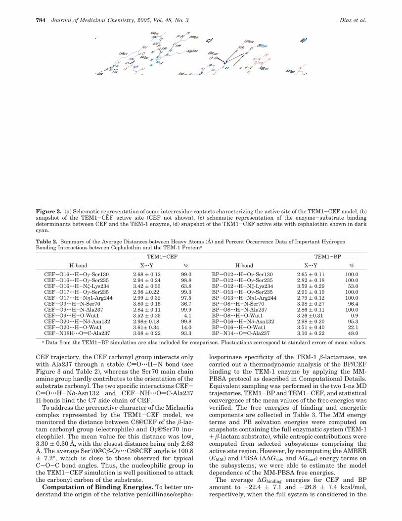
CEF trajectory, the CEF carbonyl group interacts onlywith Ala237 through a stable CdO‚‚‚H-N bond (seeFigure 3 and Table 2), whereas the Ser70 main chainamino group hardly contributes to the orientation of thesubstrate carbonyl. The two specific interactions CEF-CdO‚‚‚H-Nδ-Asn132 and CEF-NH‚‚‚OdC-Ala237H-bonds bind the C7 side chain of CEF.
To address the prereactive character of the Michaeliscomplex represented by the TEM1-CEF model, wemonitored the distance between C8@CEF of the â-lac-tam carbonyl group (electrophile) and Oγ@Ser70 (nu-cleophile). The mean value for this distance was low,3.30 ( 0.30 Å, with the closest distance being only 2.63Å. The average Ser70@Câ-Oγ‚‚‚C8@CEF angle is 100.8( 7.2°, which is close to those observed for typicalC-O-C bond angles. Thus, the nucleophilic group inthe TEM1-CEF simulation is well positioned to attackthe carbonyl carbon of the substrate.
Computation of Binding Energies. To better un-derstand the origin of the relative penicillinase/cepha-
losporinase specificity of the TEM-1 â-lactamase, wecarried out a thermodynamic analysis of the BP/CEFbinding to the TEM-1 enzyme by applying the MM-PBSA protocol as described in Computational Details.Equivalent sampling was performed in the two 1-ns MDtrajectories, TEM1-BP and TEM1-CEF, and statisticalconvergence of the mean values of the free energies wasverified. The free energies of binding and energeticcomponents are collected in Table 3. The MM energyterms and PB solvation energies were computed onsnapshots containing the full enzymatic system (TEM-1+ â-lactam substrate), while entropic contributions werecomputed from selected subsystems comprising theactive site region. However, by recomputing the AMBER(EMM) and PBSA (∆∆Gsolv and ∆Gsurf) energy terms onthe subsystems, we were able to estimate the modeldependence of the MM-PBSA free energies.
The average ∆Gbinding energies for CEF and BPamount to -22.4 ( 7.1 and -26.8 ( 7.4 kcal/mol,respectively, when the full system is considered in the
Figure 3. (a) Schematic representation of some interresidue contacts characterizing the active site of the TEM1-CEF model, (b)snapshot of the TEM1-CEF active site (CEF not shown), (c) schematic representation of the enzyme-substrate bindingdeterminants between CEF and the TEM-1 enzyme, (d) snapshot of the TEM1-CEF active site with cephalothin shown in darkcyan.
Table 2. Summary of the Average Distances between Heavy Atoms (Å) and Percent Occurrence Data of Important HydrogenBonding Interactions between Cephalothin and the TEM-1 Proteina
TEM1-CEF TEM1-BP
H-bond X‚‚‚Y % H-bond X‚‚‚Y %
CEF-O16‚‚‚H-Oγ-Ser130 2.68 ( 0.12 99.0 BP-O12‚‚‚H-Oγ-Ser130 2.65 ( 0.11 100.0CEF-O16‚‚‚H-Oγ-Ser235 2.94 ( 0.24 98.8 BP-O12‚‚‚H-Oγ-Ser235 2.82 ( 0.18 100.0CEF-O16‚‚‚H-Nú-Lys234 3.42 ( 0.33 63.8 BP-O12‚‚‚H-Nú-Lys234 3.59 ( 0.29 53.0CEF-O17‚‚‚H-Oγ-Ser235 2.98 (0.22 99.3 BP-O13‚‚‚H-Oγ-Ser235 2.91 ( 0.19 100.0CEF-O17‚‚‚H-Nη1-Arg244 2.99 ( 0.32 97.5 BP-O13‚‚‚H-Nη1-Arg244 2.79 ( 0.12 100.0CEF-O9‚‚‚H-N-Ser70 3.80 ( 0.15 36.7 BP-O8‚‚‚H-N-Ser70 3.38 ( 0.27 96.4CEF-O9‚‚‚H-N-Ala237 2.84 ( 0.11 99.9 BP-O8‚‚‚H-N-Ala237 2.86 ( 0.11 100.0CEF-O9‚‚‚H-O-Wat1 3.52 ( 0.25 4.1 BP-O8‚‚‚H-O-Wat1 3.26 (0.31 0.9CEF-O20‚‚‚H-Nδ-Asn132 2.98( 0.18 99.8 BP-O16‚‚‚H-Nδ-Asn132 2.98 ( 0.20 95.3CEF-O20‚‚‚H-O-Wat1 3.61( 0.34 14.0 BP-O16‚‚‚H-O-Wat1 3.51 ( 0.40 22.1CEF-N18H‚‚‚OdC-Ala237 3.08 ( 0.22 93.3 BP-N14‚‚‚OdC-Ala237 3.10 ( 0.22 48.0
a Data from the TEM1-BP simulation are also included for comparison. Fluctuations correspond to standard errors of mean values.
784 Journal of Medicinal Chemistry, 2005, Vol. 48, No. 3 Dıaz et al.

MM-PBSA calculations. Truncation effects decrease theabsolute ∆Gbinding energies to -18.1 ( 7.0 (CEF) and-20.2 ( 7.0 (BP) kcal/mol. Nevertheless, the mean∆Gbinding values obtained from both the full system andsubsystem MM-PBSA calculations predict that therelative binding affinity of CEF is lower than that ofBP by 4.4 (full system) and 2.1 (subsystem) kcal/mol.Inspection of the free energy components shows that therelative binding ability of the penicillin and the cepha-losporin is governed by a balance of intraprotein andsolvent effects: benzylpenicillin establishes strongerenzyme-substrate interactions, while binding of CEFis favored by a lower desolvation penalty than BP.
The thermodynamic analyses were repeated for theprotein subsystems in the TEM1-BP and TEM1-CEFtrajectories by applying the quantum chemical meth-odologies Poisson-Boltzmann surface area (QM-PBSA)computational scheme, which is based on extensivelinear scaling QM SCRF calculations on structures thatwere partially relaxed by means of QM/MM energyminimizations. Both the AM1 and PM3 semiempiricalHamiltonians were used. The average values of someQM/MM distances between the CEF/BP substrate andimportant residues in the TEM-1 active site are collectedin Table S5 (see Supporting Information). In general,the QM/MM TEM-1 complexes were structurally similarto those generated during the MD simulations using theMM force field representation.
The QM-PBSA results are collected in Table 4. In theabsence of the Lennard-Jones energy term (see Com-putational Details), we found that the QM-PBSA abso-lute values of the ∆Gbinding energies were largely positive(∼42 kcal/mol) for the two semiempirical methods.Nevertheless, the relative ∆Gbinding values estimated bythe QM-PBSA calculations point out that benzylpeni-cillin is a better TEM-1 ligand than cephalothin by 1.8(AM1) and 2.0 (PM3) kcal/mol in agreement with theprediction made by the standard MM-PBSA approach.It may be noteworthy that the AM1 and PM3 methodsdiffer in the relative importance of enzyme-substrate(∆Hbinding) and desolvation (∆∆Gsolv) effects that deter-
mine the relative penicillin/cephalosporin affinity ofTEM-1. While the lower desolvation penalty in theTEM1-CEF system cannot be compensated by theweaker enzyme-substrate interactions according to thePM3-PBSA calculations, both the enzyme-substrateforces and the desolvation penalty contribute to thereduced cephalosporin binding to the TEM-1 enzymeaccording to the AM1-based energies (see Table 4).
When the attractive Lennard-Jones energy term isincluded (account for the dispersion interactions notincluded in uncorrelated QM methods), the QM-PBSA∆Gbinding energies become clearly negative and havevalues close to the MM-PBSA ones (-25, -31 kcal/mol;see Table 4), showing the importance of the dispersionenergy to the total ∆Gbinding. The relative ∆∆Gbindingenergies were also altered by the inclusion of the LJ/R6
term, the binding energy of the BP substrate beingfavored by 5.6 kcal/mol over CEF.
DiscussionComparison between Theoretical and Experi-
mental Binding Energies. The significance of kcat/KMand KM in the acyl-enzyme mechanism characteristicof the class A â-lactamases has been examined byChristensen et al. in their study of â-lactamase as fullyefficient enzymes.27 These authors have found that thesimilarity of all the first-order rate constants thatcharacterize the general acyl-enzyme kinetic mecha-nism makes KM approximately equal to the true dis-sociation constant KS. Kinetic parameters of the TEM-1â-lactamase reacting with benzylpenicillin and cepha-lothin under comparable experimental conditions areavailable from two previous works. First, Raquet et al.have reported that the thiazolidine f dihydrothiazinesubstitution affects both the rate of catalysis and themode of substrate binding.19 These authors obtained thefollowing kinetic parameters: kcat ) 1600 s-1 and KM) 19 µM for BP; kcat ) 160 s-1 and KM ) 246 µM forCEF. The absolute binding energies derived from theKM values amount to -6.5 and -4.9 kcal/mol for BP andCEF, respectively (using ∆Gbinding ) RT ln KS). In a
Table 3. Average MM-PBSA Free Energy Componentsa in the TEM-1/â-Lactam Complexesb
∆EMM ∆Eelec ∆EvdW ∆∆GsolvPB ∆Gsurf -T∆Sbinding
c ∆Gbindingc
TEM1-BP full system -90.9 ( 7.5 -52.5 ( 7.1 -35.4 ( 2.9 43.2 ( 4.8 -3.3 ( 0.1 -26.8 ( 7.4d
subsystem -149.1 ( 7.0 -119.7 ( 5.1 -29.5 ( 2.8 107.5 ( 4.5 -2.9 ( 0.1 22.9 ( 4.3 -20.2 ( 7.0TEM1-CEF full system -79.2 ( 8.8 -40.4 ( 8.9 -38.7 ( 2.7 37.4 ( 7.4 -3.6 ( 0.2 -22.4 ( 7.1d
(11.7) (12.1) (-3.3) (-5.8) (-0.3) (4.4)subsystem -138.3 ( 8.7 -107.6 ( 8.8 -30.6 ( 2.9 100.2 ( 8.2 -3.0 ( 0.1 21.2 ( 4.6 -18.1 ( 7.0
(11.0) (12.1) (-1.1) (-7.3) (-0.1) (-1.7) (2.1)a In kilocalories per mole. b Relative differences of the TEM1-CEF mean values with respect to the TEM1-BP ones are in parentheses.
Fluctuations correspond to standard errors of mean values. c The standard state is to be taken as 1 M. d Using the entropy correctionsfrom normal mode calculations on subsystems.
Table 4. Average AM1- and PM3-Based Free Energy Componentsa in the TEM-1/â-Lactam Complexesb
∆Hbinding
∆Hbindingincluding LJ/R6 ∆∆Gsolv ∆Gbinding
c∆Gbinding
c
including LJ/R6
TEM1-BP AM1 -79.6 ( 6.1 -154.0 ( 8.3 100.3 ( 4.3 43.2 ( 6.0 -31.2 ( 8.5PM3 -86.7 ( 5.1 -161.1 ( 6.7 106.2 ( 4.1 42.1 ( 5.8 -32.3 ( 7.9
TEM1-CEF AM1 -78.6 ( 6.9 -148.7 ( 8.8 101.9 ( 7.6 45.0 ( 5.5 -25.6 ( 7.5(1.0) (5.3) (1.6) (1.8) (5.6)
PM3 -82.8 ( 6.7 -152.9 ( 8.5 105.1 ( 7.7 44.0 ( 6.6 -26.6 ( 4.8(3.9) (8.2) (-1.1) (2.0) (5.7)
a In kilocalories per mole. b Both data without and with the dispersion energy correction as estimated by the AMBER Lennard-Jonesterm are shown. Relative differences of the TEM1-CEF mean values with respect to the TEM1-BP ones are in parentheses. Fluctuationscorrespond to standard errors of mean values. c Including the entropy corrections from MM normal mode calculations on subsystems.The standard state is to be taken as 1 M.
Dynamics Simulations of TEM-1 â-Lactamase Journal of Medicinal Chemistry, 2005, Vol. 48, No. 3 785
more recent work, Cantu et al. have studied cepha-losporin binding to the TEM-1 â-lactamase.17 The cor-responding kinetic parameters for BP and CEF obtainedin this work (kcat ) 1284 ( 20 s-1 and KM ) 75 ( 8 µMfor BP; kcat ) 115 ( 3 s-1 and KM ) 347 ( 14 µM)roughly reproduce those previously reported by Raquetet al. and lead to ∆Gbinding values of -5.6 and -4.7 kcal/mol for BP and CEF, respectively. Hence, according toCantu et al., BP is a better TEM-1 ligand than CEF by0.9 kcal/mol.
When the theoretical and experimental ∆Gbindingenergies are compared, it turns out that the MM-PBSAand QM-PBSA energies overestimate the binding en-ergy by 15 or more kcal/mol. This is not surprising giventhat absolute binding free energy calculations are stillvery demanding and provide a stringent test to theunderlying methodology used to obtain energy contribu-tions.28 In practice, the estimation of relative ∆∆Gbindingvalues of related systems can be carried out with muchmore confidence using the MM-PBSA and QM-PBSAmethods (see below). Thus, the preferential binding ofBP over CEF predicted by our energetic analyses, whichranges from 1.8 to 5.7 kcal/mol (see Table 4), is inreasonable agreement with the experimental estimatesof 0.9-1.5 kcal/mol. Particularly, both the MM-PBSAand pure QM-PBSA (i.e., no LJ/R6 energy terms in-cluded) calculations on protein subsystems give small∆∆Gbinding energies around 2.0 kcal/mol, which are closeto the experimental estimates.
The observed difference in the free energies can benow interpreted in terms of the energy components andthe observed TEM-1 substrate binding interactions. Inthis respect, we note that the larger energy differencesbetween BP and CEF arise either in the molecularmechanical ∆Eelec terms or in the semiempirical∆Hbinding energies. This indicates that the penicillinaseability of TEM-1 is a consequence of better electrostaticand hydrogen-bond interactions when BP binds to theactive site.
Consistency of the MM-PBSA and QM-PBSACalculations. In recent years, the combination of MDsimulations with a subsequent analysis of energetic andentropic components has been applied to computeabsolute binding energies and relative binding affinitiesbetween diverse ligands. These are very importantproblems in computational biochemistry because of theirpotential impact on rational drug discovery and design.In this respect, the results presented herein can be ofmethodological interest because they further confirm therobustness and reliability of the MM-PBSA method inorder to predict small energy differences (∼1-2 kcal/mol) in the binding energy of dissimilar substratemolecules. This ability of the MM-PBSA/QM-PBSAcalculations to reproduce small ∆∆Gbinding effects likelystems from three different factors. First, the computa-tionally efficient particle-mesh-Ewald (PME) algo-rithm and the extended simulations result in realisticaverage structures of biomolecules in aqueous solution.20
Second, the computation of free energies combining awell-balanced description of specific electrostatic inter-actions throughout the whole protein system with therobustness and reliability of the electrostatic continuummethods is based on the linearized Poisson-Boltzmannequation in order to take into account solvent effects.20
Third, systematic errors in the computed molecularmechanical energies/heats of formation and solvationfree energies of one complex (e.g., TEM1-BP) are likelyto partially cancel those of the other complex (TEM1-CEF).
Our results show that QM-based energies can be usedto account for enzyme-substrate interactions usingstandard semiempirical Hamiltonians with only a mod-erate computational cost. In effect, the sampling andcomputational protocols, which favor cancellation oferrors, appear to attenuate the well-known limitationsof the semiempirical Hamiltonians (too low rotationalbarriers around the amide bonds, poor binding energiesfor hydrogen bonds, lack of dispersion interactions,etc.)29,30 so that the pure QM-PBSA method can predict∆∆Gbinding energies. It is also interesting to note howthe QM calculations, which incorporate charge-transferand polarization effects, provide a softer description ofthe energy changes involved in the binding process thanthe MM-PBSA calculations. Additionally, the QM-PBSAapproach provides a complete description of the struc-ture and electrostatics of many classes of ligands andprotein active sites, including metalloenzymes or reac-tive intermediates which are difficult to accuratelymodel using molecular mechanics. Overall, the presentresults and former results on the development of aQM-based scoring function22 indicate that the use ofQM methodologies will allow us to go beyond thelimitations of molecular mechanical force fields whenevaluating protein-ligand interactions.
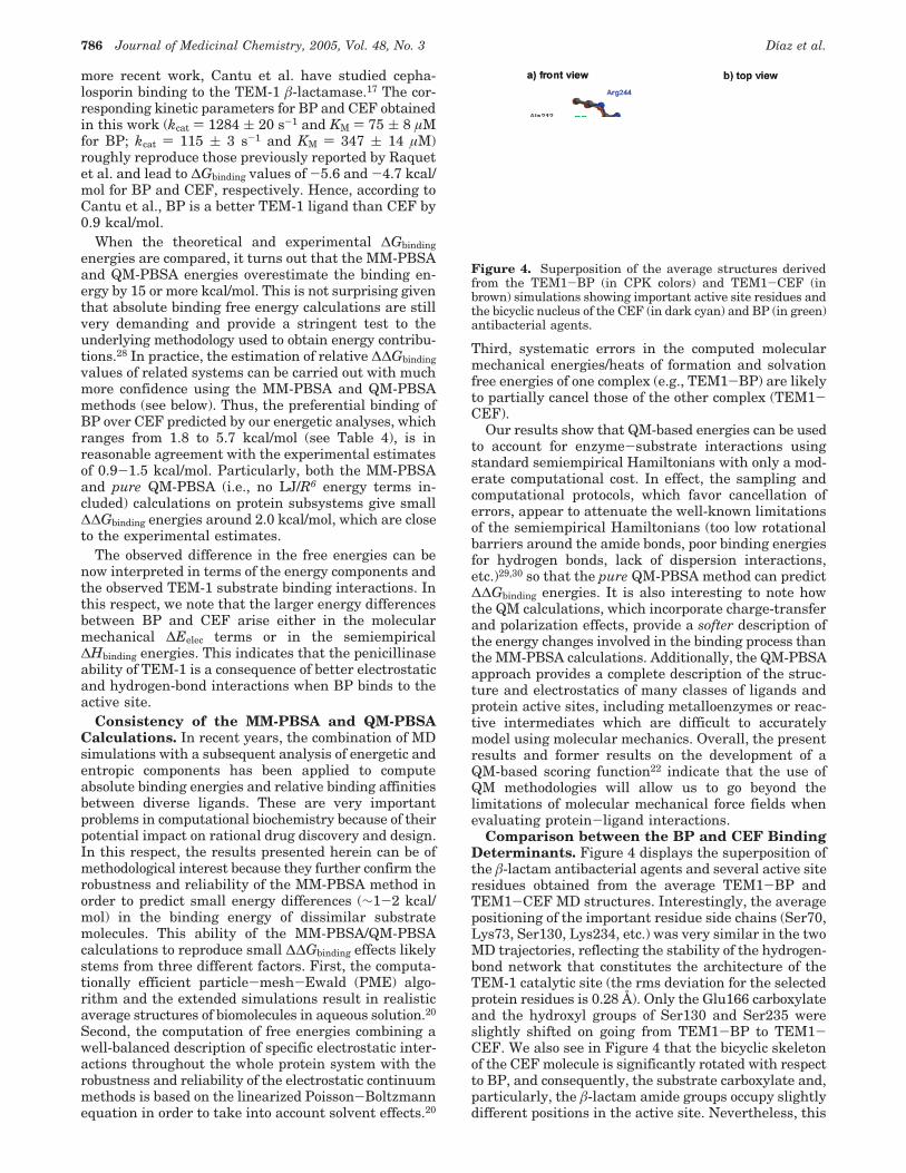
Comparison between the BP and CEF BindingDeterminants. Figure 4 displays the superposition ofthe â-lactam antibacterial agents and several active siteresidues obtained from the average TEM1-BP andTEM1-CEF MD structures. Interestingly, the averagepositioning of the important residue side chains (Ser70,Lys73, Ser130, Lys234, etc.) was very similar in the twoMD trajectories, reflecting the stability of the hydrogen-bond network that constitutes the architecture of theTEM-1 catalytic site (the rms deviation for the selectedprotein residues is 0.28 Å). Only the Glu166 carboxylateand the hydroxyl groups of Ser130 and Ser235 wereslightly shifted on going from TEM1-BP to TEM1-CEF. We also see in Figure 4 that the bicyclic skeletonof the CEF molecule is significantly rotated with respectto BP, and consequently, the substrate carboxylate and,particularly, the â-lactam amide groups occupy slightlydifferent positions in the active site. Nevertheless, this
Figure 4. Superposition of the average structures derivedfrom the TEM1-BP (in CPK colors) and TEM1-CEF (inbrown) simulations showing important active site residues andthe bicyclic nucleus of the CEF (in dark cyan) and BP (in green)antibacterial agents.
786 Journal of Medicinal Chemistry, 2005, Vol. 48, No. 3 Dıaz et al.

different location of BP and CEF does not affect thebinding of their respective carboxylate moieties, whichgive comparable long-lived hydrogen bonds with thepolar residues constituting the “carboxylate pocket”throughout the TEM1-BP and TEM1-CEF MD simu-lations (see Table 2). In contrast, the orientation of theâ-lactam carbonyl group within the “oxyanion hole” isdifferent when comparing the average TEM1-BP andTEM1-CEF structures. Moreover, the CEF substratedoes not give the CdO‚‚‚HN-Ser70 interaction, whileBP gives a stable H-bond with the backbone aminogroup of Ser70 during the TEM1-BP simulation (seeTable 2 and Figure 4). Therefore, it can be concludedthat the lower ∆Gbinding energy of CEF with respect toBP is likely related to the worse interaction of CEF withthe TEM-1 “oxyanion hole”.
A closer examination of the superimposed â-lactamstructures shows that the different orientation of cepha-losporin with respect to penicillin is due to theirdifferent geometrical properties. Thus, cephalosporinshave the carboxylate group closer to the â-lactam amidegroup than penicillins as measured by their correspond-ing Cohen distances,25 which have typical values of ∼4.5(O8‚‚‚C11) and ∼3.4 (O9‚‚‚C15) Å for penicillins andcephalosporins, respectively. This suggests that cepha-losporins are less capable than penicillins to simulta-neously bind the two anchorage points in the TEM-1active site (i.e., the “carboxylate pocket” and the “oxy-anion hole” for the â-lactam carbonyl). This is indeedthe case according to our simulations and energyanalyses: the strength and flexibility of the “carboxylatepocket” dominate over the “oxyanion hole” in theTEM1-CEF configuration, resulting in a good inter-action between the CEF carboxylate and the protein atthe cost of weakening the interaction between the CEFcarbonyl group and the “oxyanion hole”. From theseresults, a clear structure-activity relationship regard-ing the penicillin/cephalosporin affinity exhibited by theTEM-1 â-lactamase can be outlined: the poorer bindingof cephalosporins with respect to penicillins to TEM-1is due to unbalanced interactions of the “carboxylatepocket”/“oxyanion hole” with the â-lactam carboxylate/amide groups determined by the molecular geometry ofthe cephalosporins.
Penicillinase versus Cephalosporinase Activityof the Class A â-Lactamases. In their X-ray structuralstudy of acyl-enzyme complexes of mutant forms ofthe S. aureus â-lactamase with benzylpenicillin andcephaloridine, Chen and Herzberg concluded that theacyl-enzyme structures do not account for the morerapid hydrolysis of benzylpenicillin.12 Consequently,these authors suggested that the relative penicillinase/cephalosporinase activity of the class A â-lactamasesmust be determined prior to acyl-enzyme formationand that, apparently, the structural framework of theS. aureus enzyme is better suited to accommodate boththe reactive amide group of penicillins and the â-lactamcarboxylate group interacting with the “oxyanion hole”and the Lys234 ammonium group, respectively.
The theoretical results discussed above give supportto the hypothesis proposed by Chen and Herzberg andshed further light on the subtle molecular detailsdetermining the preferential penicillinase activity of theclass A â-lactamases. Thus, comparisons of the com-
puted ∆Gbinding energies of BP and CEF bound to theTEM-1 active site as well as the different “oxyanionhole”-substrate interactions emphasize the nearly per-fect match between the penicillin skeleton and theTEM-1 active site crevice, which results in optimumenzyme-substrate interactions and in a carbonyl groupwell poised for nucleophilic attack. For cephalosporinssuch as cephalothin, we found that the more compactarrangement of the â-lactam amide and carboxylategroups changes the orientation of the â-lactam groupwithin the oxyanion hole and decreases ∆Gbinding. Thistheoretical observation relevant to the Michaelis com-plexes rationalizes the experimental data on the dif-ferential binding of benzylpenicillin and cephalothin toTEM-1. However, it has also direct mechanistic implica-tions because during nucleophilic attack of the Ser70hydroxyl on the â-lactam carbonyl C atom the stabiliza-tion of the developing negative charge at the carbonylO atom of the â-lactam by the backbone amide groupsof Ala237 and Ser70 could be lower in the case ofcephalosporins, which weakly interact with the NHgroup of Ser70, relative to the penicillins. This couldexplain the experimentally observed decrease in kcat bya factor of ∼10 when comparing BP and CEF.
Finally, we comment on the small structural changesand/or point residue mutations that presumably perturbthe substrate specificity of class A â-lactamases in asubtle way by modifying the “carboxylate pocket” andthe “oxyanion hole” binding sites. Particularly, weanalyzed some selected interatomic distances in a seriesof X-ray structures of class A carbapenemases in orderto measure the relative location of the “carboxylatepocket” with respect to the backbone N atom at position237. In doing so we found that the “carboxylate pocket”is similarly rearranged in enzymes such as BS3,10
TOHO-1,9 NMC-A,14 and the TEM-64 mutant.31 Thus,Table 5 indicates that the guanidinium group of Arg244(Arg220 in NMC-A) is consistently moved away ∼0.2-0.5 Å from the backbone amide group of residue 237,while the Lys234 ammonium group is 0.3-0.7 Å closerrelative to the equivalent Arg244 and Lys234 residuesin the TEM-1 enzyme (in TOHO-1 the guanidinumgroup is deleted by the Arg244Thr mutation). Thespatial rearrangement experienced by the “carboxylatepocket” seems to be due to the accumulation of severalchanges: point mutations at the 235-237 positions, thepresence of a disulfide bridge connecting the cysteineresidues at positions 69 and 238, and so forth. Hence,the spatial rearrangement of the “carboxylate pockets”,which implies the projection of the Lys234 ammoniumgroup toward the “oxyanion hole” and the separation of
Table 5. Summary of Some Significant Interatomic Distances(Å) between “Carboxylate Pocket” Groups and the Backbone NAtom at the 237 Position in the Active Site of Various Class Aâ-Lactamases As Observed in the Corresponding X-rayStructuresa
distanceTEM-1(1BLT)
BS3(1I2S)
TOHO-1(1IYS)
NMC-A(1BUE)
TEM-64(1JWZ)
Nú@Lys234‚‚‚N@237 7.30 7.06 6.82 6.64 6.96Oγ@Ser130‚‚‚N@237 6.64 6.81 6.56 6.51 6.47Nη1@Arg244‚‚‚N@237 4.03 4.24 4.46c 4.22Oγ@Ser235‚‚‚N@237 6.31 6.24b 6.12 5.64c 6.03
a PDB ID codes are shown in parentheses. b Ala237 is replacedby Thr in the BS3 enzyme. c In the NMC-A enzymes, Ser235 isreplaced by Thr, while Arg220 is placed at the “carboxylate pocket”instead of Arg244.
Dynamics Simulations of TEM-1 â-Lactamase Journal of Medicinal Chemistry, 2005, Vol. 48, No. 3 787
Arg244/Arg240, would be a prerequisite for a goodbinding of the carboxylate group of penems/carba-penems/cephalosporins while simultaneously preservingthe optimal strength and orientation of the interactionbetween the â-lactam carbonyl and the backbone aminogroups of the “oxyanion hole”.
Computational Details
Quantum Mechanical Calculations. Initial coordinatesfor cephalothin were built by using the crystallographicstructure of cephaloridine as a template. Subsequently, HF/6-31G* optimizations were carried out in the gas phasefollowed by single-point MP2/6-31+G** energy calculationsusing the Gaussian 98 suite of programs.32 To take into accountcondensed-phase effects,33 we carried out single-point MP2/6-31+G** Poisson-Boltzmann self-consistent-reaction-field(SCRF) calculations using the local MP2 and SCRF methodsimplemented in the Jaguar program.34,35
Molecular Mechanical Parametrization of Cephal-othin. Four different conformers of CEF differing in thepuckering of the dihydrothiazine ring and/or the conformationof the side chains at C3 and C7 were considered in thecomputation of the atomic point charges using the RESPmethodology.36 Most of the bond, angle, and dihedral param-eters of CEF were available from the AMBER force field.However, some structural data required to represent theequilibrium geometry of the bicyclic skeleton of CEF wereextracted from the HF/6-31G* optimized structures. The vander Waals (vdW) parameters were taken from the closestexisting AMBER atom types using electronic similarity as aguide.
To further test our CEF parametrization, we computed a5.0 ns MD trajectory of CEF fully solvated in a periodic box of3885 TIP3P waters using the SANDER program of theAMBER 5.0 package.37 The time step was chosen to be 1.5 fs,and the SHAKE algorithm38 was used to constrain all bondsinvolving hydrogen atoms. Periodic boundary conditions wereapplied, and the pressure (1 atm) was controlled by Berends-en’s method. To include the contributions of long-rangeinteractions, the particle-mesh-Ewald (PME) method39 wasused with a grid size of 54 × 48 × 48 (grid spacing <1 Å)combined with a fourth-order B-spline interpolation to computethe potential and forces in between grid points. A cutoff of 10Å was used to compute the vdW forces.
Sampled conformations from the MD trajectory were clus-tered using the NMRCLUST program.40 This program usesthe method of average linkage to define how clusters areconstructed, followed by application of a penalty function whichsimultaneously minimizes (1) the number of clusters and (2)the spread across each cluster. A minimum distance of 1.5 Åwas used to select representative structures from each cluster.
MD Simulation of the TEM1-CEF Complex. The de-termination of an initial TEM1-CEF complex was necessaryto obtain a starting configuration for the MD studies. Weemployed the algorithm developed for AutoDock41,42 which usesa Monte Carlo simulated annealing technique to explore theconfiguration space of the enzyme-ligand complex in conjunc-tion with a rapid energy evaluation using grid-based molecularinteraction potentials (built from van der Waals and electro-static contributions). The CEF substrate was docked in thestatic binding site of the TEM-1 â-lactamase. During thedocking process, the internal bonds of the CEF side chainswere allowed to rotate. The coordinates of the protein atomswere taken from the TEM-1 1.85 Å crystal structure of Jelschet al. (PDB ID 1BTL).43
The most stable enzyme-substrate complex predicted byAUTODOCK showed enzyme-substrate contacts that werefavorable for binding and catalysis (e.g., the â-lactam carboxy-late and carbonyl groups pointed toward Lys234 and theAla237 amino group, respectively). This enzyme-substratecomplex and the crystallographic water molecules were sur-rounded by a periodic box of TIP3P water molecules,44 which
extended 10 Å from the protein and substrate atoms. Six Na+
counterions were placed by the LEaP program in order toneutralize the simulation box, which was then minimizedusing the parm96 version of the AMBER force field (2500 stepsfor the water molecules followed by 2500 steps for the entiresystem).45
MD simulations were carried out using the SANDERmodule of the AMBER 5.0 suite of programs.37 The time stepwas 1.5 fs, and the SHAKE algorithm38 was used to constrainall bonds involving hydrogen atoms. A nonbonded cutoff of 10Å was used, and the nonbonded pair list was updated every25 time steps.46 Periodic boundary conditions were applied tosimulate a continuous system. The pressure (1 atm) and thetemperature (300 K) of the system were controlled during theMD simulation by Berendsen’s method.47 To include thecontributions of long-range interactions, the particle-mesh-Ewald (PME) method39 was used with a grid size of 64 × 64 ×64 (grid spacing of ∼1 Å) combined with a fourth-order B-splineinterpolation to compute the potential and forces in betweengrid points. The estimated root-mean-square deviations of thePME force errors48 during the simulation were lower than 10-4.
For the TEM1-CEF model, an equilibration period of 200ps resulted in a stable trajectory as evidenced by the conver-gence of the dimensions of the simulation box and the evolutionof the total energy of the system. Subsequently, a 1 nstrajectory was computed and coordinates were saved foranalysis every 50 time steps. All of the MD results wereanalyzed using the CARNAL module of AMBER 5.0 and someother specific trajectory analysis software developed locally.The root-mean-square (rms) coordinate deviations between twostructures I and J (σIJ) and the radius of gyration (Rgyr) of theprotein complexes were computed according to the followingformulas:26
where N is the total number of atoms, wi is the atomic massof the i-atom, and RC is the center of mass of the protein-substrate complex.
MM-PBSA Energetic Analyses. The molecular mechanicsPoisson-Boltzmann surface area (MM-PBSA) approach inprinciple can perform several types of ∆Gbinding calculations(enzyme-substrate, protein-protein, DNA-protein).20,28,49 Ba-sically, MM-PBSA calculations predict mean values of interac-tion free energies as estimated over a series of representative(∼50-100) snapshots extracted from classical MD simulations.The snapshots are postprocessed through the removal of allsolvent and counterions. Then, one calculates the average freeenergy of the set of structures according to the followingequation:
where Gh is the calculated average free energy, and Eh MM is theaverage molecular mechanics energy,
where these correspond to the bond, angle, torsion, van derWaals, and electrostatic terms in the molecular mechanicsforce field. The term 3RT in eq 2 corresponds to the enthalpyof the six translation and rotational degrees of freedom in theclassical limit. Gh PBSA is the solvation free energy obtained fromPoisson-Boltzmann electrostatic calculations augmented withan estimate of the nonpolar free energy via molecular area,and TSMM is the solute entropy which can be estimated bymolecular mechanics normal mode calculations and standardstatistical mechanical formulas.20 Subsequently, one can es-
σIJ ) x∑i)1
N
wi(riI - ri
J)2
∑i)1
N
wi
Rgyr ) x∑i)1
N
wi(riI - RC)2
∑i)1
N
wi
(1)
Gh ≈ Eh MM + 3RT + Gh PBSA - TSh MM (2)
Eh MM ) Eh bond + Eh angle + Eh tors + Eh vdW + Eh elec (3)
788 Journal of Medicinal Chemistry, 2005, Vol. 48, No. 3 Dıaz et al.
timate the ∆G for ligand association to proteins using thefollowing equation:
where the three G terms are usually evaluated using thesnapshots from a single MD trajectory of the complex (the onetrajectory approximation). Here, the binding free energies arecomputed for a standard state of 1 M. As a consequence, thetranslational entropy for each component (complex, protein,ligand) is 6.4 cal mol-1 K-1 smaller than the entropy valueobtained for the standard state of an ideal gas owing to thechange in concentration from 0.045 M (ideal gas) to 1 M(solution).28
In this work a set of 50 representative structures extractedevery 20 ps along the TEM1-CEF MD trajectory and a secondset of equivalent snapshots from the TEM1-BP simulation18
were postprocessed to calculate the binding free energies ofcephalothin and benzylpenicillin, respectively, using the MM-PBSA approach. For each system, two series of MM-PBSAcalculations were performed: the first array of calculationsincluded the entire enzyme-substrate system, while thesecond one deals with a protein subsystem. This subsystemwas formed by all residues within a distance of 9 Å toOγ@Ser70 including most of the residues in the Ω loop(residues: 68-76, 103-107, 125-135, 161-174, 234-248).Terminal N-methylamine or acetyl groups were placed at theC and N backbone atoms of those residues cleaved from theprotein main chain by the truncation process. In addition, theBP/CEF substrate and the Wat1 molecule were also extracted.
In the MM-PBSA calculations, the AMBER force field wasused to compute (no cutoff) the EMM terms defined in eq 3.The electrostatic contribution to the solvation free energy(∆G°solv) of the TEM1-BP and TEM1-CEF states weredetermined with the Poisson-Boltzmann approach whichrepresents the protein-substrate complexes as a low dielectriccontinuum (a value of εint ) 1 was used in the calculations)with embedded charges and the solvent as a high dielectriccontinuum (εout ) 80) with no salt. Atomic charges were takenfrom the AMBER MM representation of the TEM1-BP andTEM1-CEF states. Because of the small size of the hydrogenatoms in the AMBER force field, the van der Waals surfaceused in the PB calculations was constructed using DREIDINGvan der Waals radii for C, H, N, O, and S atoms.50 Thedielectric boundary is the contact surface between the radii ofthe solute and the radius (1.4 Å) of a water probe molecule.The DELPHI 2.0 program was employed to solve the linearizedPB equation on a cubic lattice by using an iterative finite-difference method (500 iterations were performed for eachcalculation).51 The cubic lattice had a grid spacing of 0.5 Å andwas scaled such that its dimensions were 80% larger than thelongest dimension of the solute. The points at the boundaryof the grid were set to the sum of Debye-Huckel poten-tials.
Solute entropic contributions were estimated only for thesubsystem series (∼1100 atoms) using the nmode module ofthe AMBER 5.0 package. This program uses the normal modesand standard statistical thermodynamic formulas to estimateentropic contributions. Prior to the normal mode calculations,the geometries of the TEM1-BP/TEM1-CEF subsystemsdescribed by their AMBER representations were minimizeduntil the root-mean-square deviation of the elements in thegradient vector was less than 10-5 kcal/(mol Å). The ROAR2.0 program52 was used to carry out the geometry optimiza-tions driven by a limited memory BFGS minimizer.53 Allminimizations and normal mode calculations were carried outwith a distance-dependent dielectric constant (ε ) 4r) to mimicsolvent screening with no cutoff for the nonbonded inter-actions. As noted in previous work,54 this normal mode analysisonly approximately estimates solute entropy.
Semiempirical QM-PBSA Calculations. SemiempiricalQM calculations can also be used to estimate the relativeenergies of the different protein configurations. This can bedone by using linear scaling QM methodologies.55 Thus, the
free energy of the enzyme-substrate systems can be estimatedaccording to the following equation:
where Gh is the calculated average free energy, Hh QM is theaverage QM heat of formation of the solute which accountsfor intraprotein and enzyme-substrate effects, ∆Gh solv is theaverage solvation energy, which can be calculated using theQM Hamiltonian coupled to a continuum model, and TSh MM isthe solute entropy which can be estimated by molecularmechanics normal mode calculations. To complement thesemiempirical QM energy, dispersive nonpolar interactions canbe accounted for22 by adding the attractive part of the Lennard-Jones potential using the AMBER force field to eq 4,
Both eqs 5 and 6 were considered in the QM-PBSA energycalculations.
Prior to the QM-PBSA calculations, the selected set ofTEM1-BP and TEM1-CEF snapshots were subject to QM/MM energy minimization in which the BP/CEF substrates, theWat1 molecule, and the side chains of Ser70, Ser130, Glu166,Lys73, Lys234, and Arg244 (QM region) were relaxed, whilethe rest of the protein and a solvent cap of 1500 watermolecules centered on the Oγ@Ser70 atom (MM region) wereheld fixed. In these calculations, both the AM156 and PM357
Hamiltonians were used to describe the QM region, and theAMBER force field was used for the rest of the system.Hydrogen link atoms were placed at the corresponding Câatoms to cap exposed valence sites due to bonds which crossthe QM-MM boundary. The ROAR 2.0 program52 was usedto carry out the QM/MM minimizations.
From the QM/MM relaxed structures, we built the enzyme-substrate subsystems described above. Then single-point AM1and PM3 calculations were performed on these subsystemsusing the divide and conquer (D&C) approach.58-60 Incorpora-tion of solvent effects within a QM methodology was ac-complished by merging the D&C algorithm with the PBequation.61 In these QM-PB calculations, the solute wasrepresented by charge model 2 (CM2) atomic charges.62 Theset of DREIDING atomic radii was used again in the QM-PBcalculations. An additional “nonpolar” contribution due to thecreation of a solute cavity in the continuum was accountedfor by a term proportional to the solvent accessible surface areaof the solute as in the MM-PBSA approach. The DivCon99program63 was employed to perform the D&C semiempiricalcalculations using the dual buffer layer scheme (inner bufferlayer of 4.0 Å and an outer buffer layer of 2.0 Å) with oneprotein residue per core. This D&C subsetting with a totalbuffer region of 6.0 Å gives accurate relative energies.64 Acutoff of 9.0 Å was used for the off-diagonal elements of theFock, one-electron, and density matrices. Solute entropiccontributions were taken from the AMBER normal modecalculations on the subsystems taken from the trajectories.
Acknowledgment. The authors are grateful to theCICyT (Spain) for a generous allocation of computertime at the CESCA and the CIEMAT. Financial supportby MCyT (Spain) via Grant No. SAF2001-3526 is alsoacknowledged. K.M.M thanks the NIH for support ofthis project via Grant No. GM44974.
Supporting Information Available: Tables showingselected interatomic distances in the TEM-1 active site duringthe TEM1-CEF simulation, the rms flexibilities of importantresidues, the first peak position of g(r) and its integrated value,selected interatomic distances in the QM/MM minimizedstructures, and so forth and a ZIP file containing the cepha-lothin parameters in a format suitable for the LEaP program.
∆G ) Gh complex - Gh protein - Gh ligand (4) Gh ≈ Hh QM + ∆Gh solv - TSh MM (5)
Gh ≈ Hh QM + LJR6
+ ∆Gh solv - TSh MM (6)
Dynamics Simulations of TEM-1 â-Lactamase Journal of Medicinal Chemistry, 2005, Vol. 48, No. 3 789
This material is available free of charge via the Internet athttp://pubs.acs.org.
References(1) Walsh, C. Molecular mechanisms that confer antibacterial drug
resistance. Nature 2000, 406, 775-781.(2) Wright, G. D. Resisting resistance: New chemical strategies for
battling superbugs. Chem. Biol. 2000, 7, 127-132.(3) Bush, K. A functional classification scheme for â-lactamases and
its correlation with molecular structure. Antimicrob. AgentsChemother. 1995, 39, 1211-1233.
(4) Maiti, S. N.; Philips, O. A.; Micetich, R. G.; Livermore, D. M.â-Lactamase inhibitors: Agents to overcome bacterial resistance.Curr. Med. Chem. 1998, 5, 441-456.
(5) Matagne, A.; Lamotte-Brasseur, J.; Frere, J. M. Catalyticproperties of class A â-lactamases: Efficiency and diversity.Biochem. J. 1998, 330, 581-598.
(6) Tranier, S.; Bouthors, A.-M.; Maveyraud, L.; Guillet, V.; Sou-gakoff, W.; Samama, J.-P. The high resolution crystal structurefor class A â-lactamase PER-1 reveals the bases for its increasein breadth of activity. J. Biol. Chem. 2000, 275, 28075-28082.
(7) Ambler, R. P.; Coulson, A. F.; Frere, J. M.; Ghuysen, J. M.;Jaurin, B.; Joris, B.; Levesque, R.; Tiraby, G.; Waley, S. G. Astandard numbering scheme for the class A â-lactamases.Biochem. J. 1991, 276, 269-272.
(8) Fonze, E.; Charlier, P.; To’th, Y.; Vermiere, M.; Raquet, X.;Dubus, A.; Frere, J.-M. TEM1 â-lactamase structure solved bymolecular replacament and refined structure of the S235Amutant. Acta Crystallogr., Sect. D 1995, 51, 682-694.
(9) Shimamura, T.; Ibuka, A.; Fushinobu, S.; Wakagi, T.; Ishiguro,M.; Ishii, Y.; Matsuzawa, H. Acyl-intermediate structures of theextended-spectrum class A â-lactamase, TOHO-1, in complexwith cefotaxime, cephalothin, and benzylpenicillin. J. Biol.Chem. 2002, 277, 46601-46608.
(10) Fonze, E.; Vanhove, M.; Dive, G.; Sauvage, E.; Frere, J.-M.;Charlier, P. Crystal structure of the Bacillus licheniformis BS3class A â-lactamase and of the acyl-enzyme adduct formed withcefoxitin. Biochemistry 2002, 41, 1877-1885.
(11) Herzberg, O. Refined crystal structure of â-lactamase fromStaphylococcus aureus at 2.0 Å resolution. J. Mol. Biol. 1991,217, 701-719.
(12) Chen, C. C. H.; Herzberg, O. Structures of the acyl-enzymecomplexes of the Staphylococcus aureus â-lactamase mutantglu166asp:asn170gln with benzylpenicillin and cephaloridine.Biochemistry 2001, 40, 2351-2358.
(13) Lim, D.; Sanschagrin, F.; Passmore, L.; De Castro, L.; Levesque,R. C.; Strydnadka, N. C. J. Insights into the molecular basis forthe carbenicillinase activity of PSE-4 â-lactamase from crystal-lographic and kinetic studies. Biochemistry 2001, 40, 395-402.
(14) Swarem, P.; Maveyraud, L.; Raquet, X.; Cabantous, S.; Duez,C.; Pedelacq, J.-D.; Mariotte-Boter, S.; Mourey, L.; Labia, R.;Nicolas-Chanoine, M.-H.; Nordmann, P.; Frere, J.-M.; Samama,J.-P. X-ray analysis of the NMC-A â-lactamase at 1.64 angstromresolution, a class A carbapenemase with broad substratespecificity. J. Biol. Chem. 1998, 273, 26714-26721.
(15) Nukaga, M.; Mayama, K.; Hujer, A. M.; Bonomo, R. A.; Knox,J. R. Ultrahigh resolution structure of a class A â-lactamase:On the mechanism and specificity of the extended-spectrumSHV-2 enzyme. J. Mol. Biol. 2003, 329, 289-301.
(16) Raquet, X.; Lamotte-Brasseur, J.; Bouilleme, F.; Frere, J.-M. Adisulfide bridge near the active site of carbapenem-hydrolyzingclass A â-lactamases might explain their unusual substrateprofile. Proteins: Struct., Funct., Genet. 1997, 27, 47-58.
(17) Cantu, C.; Huang, W.; Palzkill, T. Cephalosporin substratespecificity determinants of TEM-1 â-lactamase. J. Biol. Chem.1997, 272, 29144-29150.
(18) Dıaz, N.; Sordo, T. L.; Merz, K. M. J.; Suarez, D. Insights intothe acylation mechanism of class A â-lactamases from moleculardynamics simulations of the TEM-1 enzyme complexed withbenzylpenicillin. J. Am. Chem. Soc. 2003, 125, 672-684.
(19) Raquet, X.; Lamotte-Brasseur, J.; Fonze, E.; Goussard, S.;Couvalin, P.; Frere, J.-M. TEM â-lactamase mutants hydrolisingthird-generation cephalosporins. J. Mol. Biol. 1994, 244, 625-639.
(20) Kollman, P. A.; Massova, I.; Reyes, C.; Kuhn, B.; Huo, S.; Chong,L.; Lee, M.; Lee, T.; Duan, Y.; Wang, W.; Donini, O.; Cieplak,P.; Srinivasan, J.; Case, D. A.; Cheatham, T. E. Calculatingstructures and free energies of complex molecules: Combiningmolecular mechanics and continuum models. Acc. Chem. Res.2000, 33, 889-897.
(21) Suarez, D.; Brothers, E. N.; Merz, K. M. J. Quantum chemicalcalculations and molecular dynamics simulations of the di-nuclear zinc-â-lactamase from Bacteroides fragilis. Biochemistry2002, 41, 6615-6630.
(22) Raha, K.; Merz, K. M., Jr. A quantum mechanics-based scoringfunction: Study of zinc ion-mediated ligand binding. J. Am.Chem. Soc. 2004, 126, 1020-1021.
(23) Boyd, D. B. Chemistry and Biology of â-Lactam Antibiotics;Academic Press: New York, 1982; pp 437-545.
(24) Frau, J.; Donoso, J.; Munoz, F.; Garcıa Blanco, F. Theoreticalcalculations of â-lactam antibiotics part 2. AM1, MNDO, andMINDO/3 calculations of some cephalosporins. J. Mol. Struct.(THEOCHEM) 1991, 251, 205-218.
(25) Cohen, N. C. â-lactam antibiotics: Geometrical requirements forantibacterial activities. J. Med. Chem. 1983, 26, 259-264.
(26) Field, M. J. A practical introduction to the simulation ofmolecular systems; Cambridge University Press: Cambridge,U.K., 1999; pp 25-42.
(27) Christensen, H.; Martin, M. T.; Waley, S. G. â-lactamases asfully efficient enzymes. Determination of all the rate constantsin the acylenzyme mechanism. Biochem. J. 1990, 266, 853-861.
(28) Gohlke, H.; Case, D. A. Converging free energy estimates: MM-PB(GB)SA studies on the protein-protein complex ras-raf. J.Comput. Chem. 2003, 25, 238-250.
(29) Mohle, K.; Hofmann, H.-J.; Thiel, W. Description of peptide andprotein secondary structures employing semiempirical methods.J. Comput. Chem. 2001, 22, 509-520.
(30) Winget, P. C. S.; Horn, A. H. M.; Martin, B.; Clark, T. Towardsa ‘‘next generation” neglect of diatomic differential overlap basedsemiempirical molecular orbital technique. Theor. Chem. Acc.2003, 10, 254-266.
(31) Wang, X.; Minasov, G.; Blazquez, J.; Caselli, E.; Prati, F.;Shoichet, B. K. Recognition and resistance in TEM â-lactamase.Biochemistry 2003, 42, 8434-8444.
(32) Frisch, M. J.; Trucks, G. W.; Schlegel, H. B.; Scuseria, G. E.;Robb, M. A.; Cheeseman, J. R.; Zakrzewski, V. G.; Montgomery,J. A., Jr.; Stratmann, R. E.; Burant, J. C.; Dapprich, S.; Millam,J. M.; Daniels, A. D.; Kudin, K. N.; Strain, M. C.; Farkas, O.;Tomasi, J.; Barone, V.; Cossi, M.; Cammi, R.; Mennucci, B.;Pomelli, C.; Adamo, C.; Clifford, S.; Ochterski, J.; Petersson, G.A.; Ayala, P. Y.; Cui, Q.; Morokuma, K.; Malick, D. K.; Rabuck,A. D.; Raghavachari, K.; Foresman, J. B.; Cioslowski, J.; Ortiz,J. V.; Stefanov, B. B.; Liu, G.; Liashenko, A.; Piskorz, P.;Komaromi, I.; Gomperts, R.; Martin, R. L.; Fox, D. J.; Keith, T.;Al-Laham, M. A.; Peng, C. Y.; Nanayakkara, A.; Gonzalez, C.;Challacombe, M.; Gill, P. M. W.; Johnson, B. G.; Chen, W.; Wong,M. W.; Andres, J. L.; Head-Gordon, M.; Replogle, E. S.; Pople,J. A. Gaussian 98, revision A.6; Gaussian, Inc.: Pittsburgh, PA,1998.
(33) Tomasi, J.; Persico, M. Molecular interactions in solution: Anoverview of methods based on continuous distributions of thesolvent. Chem. Rev. 1994, 94, 2027-2094.
(34) Tannor, D. J.; Marten, B.; Murphy, R.; Friesner, R. A.; Sitkoff,D.; Nicholls, A.; Ringnalda, M.; Goddard, I. W. A.; Honig, B.Accurate first principles calculation of molecular charge distri-butions and solvation energies from ab initio quantum mechan-ics and continuum dielectric theory. J. Am. Chem. Soc. 1994,116, 11875-11882.
(35) Jaguar, version 3.5; Schrodinger, Inc.: Portland, OR, 1998.(36) Bayly, C. A.; Cieplak, P.; Cornell, W. D.; Kollman, P. A. A well
behaved elestrostatic potential based method using chargerestraints for deriving atomic charges: The RESP model. J.Phys. Chem. 1993, 97, 10269-10280.
(37) Case, D. A.; Pearlman, D. A.; Caldwell, J. W.; Cheatham, T. E.,II; Ross, W. S.; Simmerling, C. L.; Darden, T. A.; Merz, K. M.,Jr.; Stanton, R. V.; Cheng, A. L.; Vincent, J. J.; Crowley, M.;Ferguson, D. M.; Radmer, R. J.; Seibel, G. L.; Singh, U. C.;Weiner, P. K.; Kollman, P. A. Amber, 5th ed.; University ofCalifornia: San Francisco, CA, 1997.
(38) van Gunsteren, W. F.; Berendsen, H. J. C. Algorithm formacromolecular dynamics and constraint dynamics. Mol. Phys.1977, 34, 1311-1327.
(39) Essman, V.; Perera, L.; Berkowitz, M. L.; Darden, T.; Lee, H.;Pedersen, L. G. A smooth particle-mesh-Ewald method. J. Chem.Phys. 1995, 103, 8577-8593.
(40) Kelley, L. A.; Gardner, S. P.; Sutcliffe, M. J. An automatedapproach for clustering an ensemble of NMR-derived proteinstructures into conformationally-related subfamilies. ProteinEng. 1996, 9, 1063-1065.
(41) Morris, G. M.; Goodsell, D. S.; Halliday, R. S.; Huey, R.; Hart,W. E.; Belew, R. K.; Olson, A. J. Automated docking using aLamarckian genetic algorithm and empirical binding free energyfunction. J. Comput. Chem. 1998, 19, 1639-1662.
(42) Goodsell, D. S.; Olson, A. J. Automated docking of substrates toproteins by simulated annealing. Proteins: Struct., Funct., Genet.1990, 8, 195-202.
(43) Jelsch, C.; Mourey, L.; Masson, J. M.; Samama, J. P. Crystalstructure of Escherichia coli TEM-1 â-lactamase at 1.8 Åresolution. Proteins: Struct., Funct., Genet. 1993, 16, 364-383.
(44) Jorgensen, W. L.; Chandrasekhar, J.; Madura, J.; Impey, R. W.;Klein, M. L. Comparison of simple potential functions for thesimulation of liquid water. J. Chem. Phys. 1983, 79, 926-935.
790 Journal of Medicinal Chemistry, 2005, Vol. 48, No. 3 Dıaz et al.
(45) Kollman, P. A.; Dixon, R.; Cornell, W.; Fox, T.; Chipot, C.;Pohorille, A. The development/application of a “minimalist”organic/biochemical molecular mechanic force field using acombination of ab initio calculations and experimental data.Computer Simulation of Biomolecular Systems; Kluger: Dor-drecht, The Netherlands, 1997; pp 83-96.
(46) Allen, M. P.; Tildesley, D. J. Computer simulation of liquids;Clarendon Press: Oxford, U.K., 1987.
(47) Berendsen, H. J. C.; Potsma, J. P. M.; van Gunsteren, W. F.;DiNola, A. D.; Haak, J. R. Molecular dynamics with coupling toand external bath. J. Chem. Phys. 1984, 81, 3684-3690.
(48) Petersen, H. G. Accuracy and efficiency of the particle-mesh-ewald method. J. Chem. Phys. 1995, 103, 3668-3679.
(49) Baginski, M.; Fogolari, F.; Briggs, J. M. Electrostatic and non-electrostatic contributions to the binding free energies of an-thracycline antibiotics to DNA. J. Mol. Biol. 1997, 274, 253-267.
(50) Rappe, A. K.; Casewit, C. J.; Colwell, K. S.; Goddard, W. A., III;Skiff, W. M. UFF, a full periodic table force field for molecularmechanics and molecular dynamics simulations. J. Am. Chem.Soc. 1992, 114, 10024-10035.
(51) Sharp, K. A.; Honig, B. Electrostatic interactions in macromole-culessTheory and applications. Annu. Rev. Biophys. Biophys.Chem. 1990, 19, 301-332.
(52) Cheng, A.; Stanton, R. S.; Vincent, J. J.; van der Vaart, A.;Damodaran, K. V.; Dixon, S. L.; Hartsough, D. S.; Mori, M.; Best,S. A.; Monard, G.; Garcia, M.; Van Zant, L. C.; Merz, K. M. J.Roar 2.0; The Pennsylvania State University: University Park,PA, 1999.
(53) Lui, D. C.; Nocedal, J. On the limited memory BFGS methodfor large scale optimization. Math. Programming 1989, 45, 503-528.
(54) Chong, L. T.; Duan, Y.; Wang, L.; Massova, I.; Kollman, P. A.Molecular dynamics and free energy calculations applied toaffinity maturation in antibody 48g7. Proc. Natl. Acad. Sci.U.S.A. 1999, 96, 14330-14335.
(55) Goedecker, S. Linear scaling electronic structure methods. Rev.Mod. Phys. 1999, 71, 1085-1123.
(56) Dewar, M. J. S.; Zoebisch, E. G.; Healy, E. F.; Stewart, J. J. P.AM1: A new general purpose quantum mechanical molecularmodel. J. Am. Chem. Soc. 1985, 107, 3902-3909.
(57) Stewart, J. J. P. Optimization of parameters for semiempiricalmethods I. Method. J. Comput. Chem. 1989, 10, 209-220.
(58) Yang, W.; Lee, T.-S. A density-matrix form of the divide-and-conquer approach for electronic structure calculations of largemolecules. J. Chem. Phys. 1995, 103, 5674-5678.
(59) Dixon, S. L.; Merz, K. M., Jr. Semiempirical molecular orbitalcalculations with linear system size scaling. J. Chem. Phys. 1996,104, 6643-6649.
(60) Dixon, S. L.; Merz, K. M., Jr. Fast, accurate semiempiricalmolecular orbital calculations for macromolecules. J. Chem.Phys. 1997, 107, 879-893.
(61) Gogonea, V.; Merz, K. M., Jr. Fully quantum mechanicaldescription of proteins in solution. Combining linear scalingquantum mechanical methodologies with the Poisson-Boltzmannequation. J. Phys. Chem. A 1999, 103, 5171-5178.
(62) Li, J.; Cramer, C. J.; Truhlar, D. G. New class IV charge modelfor extracting accurate partial charges from wave functions. J.Phys. Chem. A 1998, 102, 1820-1831.
(63) Dixon, S. L.; van der Vaart, A.; Gogonea, V.; Vincent, J. J.;Brothers, E. N.; Suarez, D.; Westerhoff, L. M.; Merz, K. M. J.Divcon99; The Pennsylvania State University: University Park,PA, 1999.
(64) van der Vaart, A.; Suarez, D.; Merz, K. M., Jr. Critical assess-ment of the performance of the semiempirical divide and conquermethod for single point calculations and geometry optimizationsof large chemical systems. J. Chem. Phys. 2000, 113, 10512-10523.
(65) Sweet, R. M.; Dahl, L. F. Molecular architecture of the cepha-losporins. Insights into biological activity based on structuralinvestigations. J. Am. Chem. Soc. 1970, 92, 5489-5507.
(66) Gordon, E.; Mouz, N.; Duee, E.; Dideberg, O. The crystalstructure of the penicillin binding protein 2x from Streptococcuspneumoniae and its acylenzyme form: Implication in drugresistance. J. Mol. Biol. 2000, 299, 477-485.
JM0493663
Dynamics Simulations of TEM-1 â-Lactamase Journal of Medicinal Chemistry, 2005, Vol. 48, No. 3 791