Design of an In Situ Bioremediation Scheme of Chlorinated ...
Upload
independentCategory
view
1download
0
Journal of Tropical Forest Science 26(1): 153–162 (2014) Hema TG et al.
153© Forest Research Institute Malaysia
ACTINOBACTERIA ISOLATES FROM TIN TAILINGS AND FOREST SOIL FOR BIOREMEDIATION OF HEAVY METALS
TG Hema1, K Getha1, *, GYA Tan2, H Lili Sahira1, A Muhd Syamil1 & MY Nur Fairuz1
1Natural Products Division, Forest Research Institute Malaysia, 52109 Kepong, Selangor Darul Ehsan, Malaysia2Universiti Malaya, 50603 Kuala Lumpur, Malaysia
Received April 2013
HEMA TG, GETHA K, TAN GYA, LILI SAHIRA H, MUHD SYAMIL A & NUR FAIRUZ MY. 2014. Actinobacteria isolates from tin tailings and forest soil for bioremediation of heavy metals. The aim of this study was to screen actinobacteria isolated from Bidor tin tailings for heavy metal tolerance as an early exploration into their bioremediation capabilities. As a comparison, isolates from forest soil in Pasoh were also studied for heavy metal tolerance. At the highest test concentration, rapid metal tolerance screening showed that the numbers of Hg2+, Cd2+ and Cu2+ tolerant isolates were less than 7% of the total isolates screened for both sites. More than 10% of total isolates screened from both sites showed tolerance to Pb2+, As3+ and Zn2+. The number of Ni2+ tolerant isolates was significantly higher for Bidor (12.6%) compared with Pasoh (6.6%). Metal toxicity assay carried out on tolerant isolates from Bidor showed that the final metal concentrations supporting actinobacteria growth were 138.9 mmol L-1 for Ni2+, 43.2 mmol L-1 for Pb2+, 21.8 mmol L-1 for Cd2+, 0.8 mmol L-1 for Hg2+, 182 mmol L-1 for As3+ and 14.1 mmol L-1 for Cu2+. For Pasoh isolates, tolerance was observed at 21.8 mmol L-1 for Cd2+ and 182 mmol L-1 for As3+. Compared with rapid metal tolerance screening, metal toxicity assay clearly showed that metal tolerant isolates from Bidor were capable of tolerating heavy metals at a higher concentration compared with Pasoh isolates. Isolates TY046-021 and TY046-078, which showed multimetal tolerance to As3+, Pb2+ and Ni2+ and TY046-017, the only Hg2+ tolerant isolate from Bidor, should be further studied for heavy metal uptake and removal.
Keywords: Actinomycetes, metal toxicity, metal uptake, metal removal, tolerance, ex-mining, Bidor
HEMA TG, GETHA K, TAN GYA, LILI SAHIRA H, MUHD SYAMIL A & NUR FAIRUZ1 MY. 2014. Pencilan aktinobakteria daripada tanah bekas lombong dan tanah hutan untuk bioremediasi logam berat. Kajian ini bertujuan untuk menyelidik toleransi pencilan aktinobakteria daripada tanah bekas lombong di Bidor terhadap logam berat sebagai tinjauan awal kepada keupayaan bioremediasinya. Sebagai perbandingan, pencilan aktinobakteria daripada tanah hutan di Pasoh juga dikaji untuk toleransi logam berat. Saringan pantas toleransi logam berat menunjukkan bahawa pada kepekatan tertinggi, kurang daripada 7% jumlah pencilan di kedua-dua tapak menunjukkan toleransi terhadap logam berat Hg2+, Cd2+ dan Cu2+. Lebih daripada 10% pencilan daripada kedua-dua tapak menunjukkan toleransi terhadap Pb2+, As3+ dan Zn2+. Bilangan pencilan yang menunjukkan toleransi ke atas Ni2+ adalah secara signifikan lebih tinggi di Bidor (12.6%) berbanding dengan Pasoh (6.6%). Asai toksisiti logam yang dijalankan terhadap pencilan aktinobakteria dari Bidor menunjukkan bahawa kepekatan logam akhir yang menyokong pertumbuhan aktinobakteria ialah 138.9 mmol L-1 untuk Ni2+, 43.2 mmol L-1 untuk Pb2+, 21.8 mmol L-1 untuk Cd2+, 0.8 mmol L-1 untuk Hg2+, 182 mmol L-1 untuk As3+ dan 14.1 mmol L-1 untuk Cu2+ . Bagi pencilan dari Pasoh pula, toleransi dicerap pada 21.8 mmol L-1 untuk Cd2+ dan 182 mmol L-1 untuk As3+. Jika dibandingkan dengan saringan pantas toleransi logam berat, asai toksisisti logam jelas menunjukkan bahawa pencilan aktinobakteria dari Bidor mampu bertahan terhadap logam berat pada kepekatan yang lebih tinggi berbanding dengan pencilan dari Pasoh. Pencilan TY046-021 dan TY046-078 yang menunjukkan toleransi pelbagai logam terhadap As3+, Pb2+ dan Ni2+ serta TY046-071 dari Bidor yang hanya menunjukan toleransi terhadap Hg2+ patut dikaji lebih lanjut untuk menyelidik pengambilan dan penyingkiran logam berat.
INTRODUCTION
Heavy metal contamination in soil is mostly due to the use of fertiliser and agrochemicals, mining activities, industrial wastewater irrigation and sewage irrigation (Luo 2009). Even forest soil
which is close to industrial emission sources is contaminated with heavy metal deposits (Shparyk & Parpan 2004). Most heavy metals are toxic to plants, animals and humans. Metals such as

Journal of Tropical Forest Science 26(1): 153–162 (2014) Hema TG et al.
154© Forest Research Institute Malaysia
cadmium, manganese, arsenic, lead, zinc, nickel, copper and antimony are easily accumulated in vital organs and therefore threaten human health (Shivika 2011). For example, itai-itai disease is caused by cadmium poisoning due to mining activity in Japan (Masanori 2006) and minamata disease is caused by mercury poisoning (Toshihide & Takashi 2011). In plants, toxic levels of heavy metal ions induce several cellular stress responses and damage different cellular components such as membranes, proteins and DNA (Waisberg et al. 2003, Jimi et al. 2004). Thus, heavy metal pollution in soil is a growing environmental problem, which requires immediate attention. It has been reported that fruits from trees grown on ex-mining soil contain high levels of heavy metals exceeding the permissible limits (Ang et al. 2000). Many timber species such as Acacia mangium, Hopea odorata, Intsia palembanica and Swietenia macrophylla have been used as phytoremediation agents to extract heavy metals from contaminated soil in order to ensure safe food chain between plants, animals and humans and to maximise the usage of metal-contaminated land for agriculture and forest plantation (Ang et al. 2010). Microorganisms such as Aspergillus spp., Bacillus spp., Staphylococcus spp., Pseudomonas spp. (Kumar et al. 2010), Trichoderma spp. (Sen & Charaya 2010) and Streptomyces spp. (Sineriz et al. 2009) have been reported to be able to tolerate high concentration of various heavy metals (Shivika 2011). Microorganisms have been used as biological agents to degrade toxic wastes from the environment (Milic et al. 2009). Studies have shown that the filamentous Gram-positive actinobacteria are widely studied for bioremediation of heavy metals such as copper, chromium, cadmium (Amoroso & Abate 2012), plumbum (Kumar et al. 2011a) and zinc (Lin et al. 2012). This is because many genera of actinobacteria are able to survive in extreme conditions such as high temperature, low moisture and nutrient starvation to produce biosur factants which increase pollutant biodisponibility and facilitate biodegradation process (Alvarez et al. 2011). In high metal concentration environments, homeostasis within the bacteria cell is maintained to keep reactive heavy metals at an optimal subtoxic level. Thus,
bacteria possess resistance mechanisms assisted by enzymes such as superoxide dismutases (Kim et al. 2003), efflux transporters (Anton et al. 1999, Mergeay et al. 2003) and metal-binding proteins (Silver & Phung et al. 2005) in order to survive. The aim of this study was to screen heavy metal tolerance in actinobacteria isolated from Bidor tin tailings as an early exploration into their bioremediation capabilities. The actinobacteria isolates were isolated from tin tailings collected from an ex-mining area in Bidor, Perak (4°6' N latitude and 101°16' E longitude). Previous studies showed that tin tailings in Bidor could be classified into slime, sandy and sandy slime tailings. The tailings contain few potentially toxic elements such as arsenic, mercury, lead, copper, cadmium, nickel and zinc (Ang & Ang 1997, Ang & Ng 2000). As a comparison, actinobacteria isolated from forest soil in Pasoh, Negeri Sembilan were studied for heavy metal tolerance.
MATERIALS AND METHODS
Actinobacteria cultures
A total of 238 actinobacteria isolates from Bidor tin tailings and 183 isolates from Pasoh forest soil isolated previously by Getha et al. (2008) were used. Cultures were stored at -80 °C in cryovials containing 20% glycerol and maintained at the FRIM Actinobacteria Culture Collection. A quick thawing was done on these glycerol stock cultures and inoculated into sterile ISP2 (International Streptomyces Project 2) (Shirling & Gottlieb 1966) medium containing per litre: glucose 2 g, malt extract 5 g, yeast extract 2 g and agar 18 g (pH 7.3). A standard 10 cm Petri dish was used to prepare the agar which was dried before bacterial inoculation. All 421 isolates from the two study sites were inoculated into ISP2 agar plates. After incubation at 28 ± 2 °C for 2 weeks, a preliminary grouping of isolates into Streptomyces-like (S) and non-Streptomyces (nS) groups was carried out (Numata & Nimura 2003). Isolates were tentatively dereplicated based on their macromorphological and cultural characteristics such as presence of abundant and thick aerial mycelia, diffusible pigment colour
Journal of Tropical Forest Science 26(1): 153–162 (2014) Hema TG et al.
155© Forest Research Institute Malaysia
and spore mass colour for the S group while surface growth colour (without aerial mycelia), spore mass colour (with slight aerial mycelia) and penetration of colonies for the nS group. Colour grouping of isolates was done based on in-house colour chart.
Rapid metal tolerance screening
Seven different types of heavy metal salts (Ni2+, Pb2+, Zn2+, Cd2+, Cu2+, Hg2+, As3+) were used for the metal tolerance assay. Metal solutions were prepared in phosphate buffer saline, pH 6.8 to maintain the pH of the metal solutions (Selvin et al. 2007) and were stored at 4 °C for no longer than 1 month. Three different concentrations of each metal salt were used in the experiment (HgCl2: 0.04, 0.4, 4.0 mmol L-1; As2O3: 5.0, 25.0, 51.0 mmol L-1; PbCl2: 0.4, 4.0, 18.0 mmol L-1; Cu2SO4: 6, 31, 63 mmol L-1; CdCl2: 0.06, 0.6, 6.0 mmol L-1; NiCl2: 8.0, 36.0, 77.0 mmol L-1 and ZnCl2: 7, 37, 73 mmol L-1). The used glasswares were leached in 2 N HNO3 and rinsed with distilled water for a few times before washing to avoid metal contamination. Rapid metal tolerance screening was carried out based on direct agar diffusion assay modified from Chandy (1998). Minimal medium agar that contained per litre: glucose 10 g, L-asparagine 0.5 g, K2HPO4 0.5 g, MgSO4.7H2O 0.2 g, FeSO4.7H2O 0.01 g and agar 16.0 g was prepared in Petri dish. The aerial growth of 7–10-day- old actinobacteria culture was suspended in 5 mL sterile 20% (v/v) glycerol. An aliquot of 50 µL of the suspension was lawned on minimal medium agar and test plates were left to dry for 30 min before use. Surface of the lawned test plates were divided into four quadrants, three areas corresponding to different concentrations of metal solutions and one area for the control sterile PBS solution without metal salt. An amount of 10 µL each of heavy metal and control solutions was spotted on the test plates using a micropipette. Plates were incubated at 28 °C for 7 days and examined for the appearance of clear zone after 5, 6, 7 and 8 days. The diameter of inhibition zone (cm) at the spotted area was measured. Each test was done in replicate plates and mean of two readings was calculated and used in heavy metal tolerance assessment. Presence of inhibition zone indicates that the isolates are sensitive towards the heavy metal, whereas absence indicates heavy metal tolerance.
Metal toxicity assay
Metal toxicity assay was carried out on 40 tolerant isolates selected from the rapid metal tolerance screening to study the minimal metal concentration inhibiting bacterial growth in broth culture using serial dilution method. Seven-day-old ISP2 cultures of actinobacteria were used as inocula. Inocula were prepared as described above and 0.5 mL was inoculated into test tubes containing 5 mL minimal media supplemented with different concentrations of metal salt. Six different types of metal salts at five different concentrations were studied (NiCl2: 17.4, 34.7, 69.45, 138.9, 277.8 mmol L-1; PbCl2: 2.7, 5.4, 10.8, 21.6, 43.2 mmol L-1; CdCl2: 1.4, 2.7, 5.45, 10.9, 21.8 mmol L-1; CuSO4: 14.1, 28.2, 56.4, 112.8, 225.6 mmol L-1; HgCl2: 0.4, 0.8, 1.5, 3.0, 6.0 mmol L-1 and As2O3: 11.4, 22.8, 45.5, 91.0, 182.0 mmol L-1). Test tubes were incubated in an orbital incubator shaker at 28 °C and 220 rpm. After 10 days, 10 µL culture broth grown in different metal concentrations were inoculated onto fresh ISP2 agar plates to determine minimal concentration of metal in media that supported bacterial growth.
Statistical analysis
SPSS version 16.0 was used for statistical analysis. Chi square test was used to analyse the relationship between sampling sites and percentage of heavy metal tolerant isolates towards different metals. Mann Whitney test was used to determine the difference between site and percentage of heavy metal tolerant isolates belonging to Streptomyces-like and non-Streptomyces groups from two different sites.
RESULTS AND DISCUSSION
Rapid metal tolerance screening in actinobacteria
Actinobacteria isolates were tentatively grouped and dereplicated into Streptomyces-like and non-Streptomyces groups based on macromorphological and cultural characteristics. The Streptomyces-like group was distinguished from non-Streptomyces group by the presence of abundant aerial mycelium with powdery spore mass on isolate surface growth in agar plates (Getha et al. 2004, Zhao et al. 2005). Isolates classified under the

Journal of Tropical Forest Science 26(1): 153–162 (2014) Hema TG et al.
156© Forest Research Institute Malaysia
non-Streptomyces group could belong to different genera such as Micromonospora, Actinoplanes, Norcardia and other rare genera characterised by colonies having none or sparse aerial mycelium, with or without the presence of diffusible pigments in agar cultures. More than 50% of the isolates from Bidor and Pasoh belonged to the Streptomyces group (Table 1). Spore mass colours of Streptomyces-like isolates from both sites were mainly grey, white and brown. Non-Streptomyces isolates showed a variety of surface growth colours with sparse or no aerial mycelium. A higher number of isolates producing diffusible pigments obtained from both study sites were from the Streptomyces-like group (Table 1). Actinobacteria, especially Streptomyces spp., are known to produce a range of diffusible pigment colours such as blue, violet, red, rose, yellow, green, brown and black. Some of the antibiotics produced by actinobacteria include many of these pigments (Shaaban 2013). Pigments produced by S. hygroscopicus showed antibacterial activity against drug resistant pathogens such as methicillin-resistant Staphylococcus aureus, vancomycin-resistant S. aureus and extended-spectrum beta lactamase strains (Selvameenal 2009). Figure 1 shows the tolerance of isolates from two different sites towards different
types of heavy metals tested at three different concentrations. The order of metal toxicity observed at the highest test concentration among the actinobacteria isolates could be arranged as Hg > Cd = Cu > Ni = Pb > As = Zn (for isolates obtained from Bidor tin tailings) and Hg > Cu > Cd = Ni > Pb > As = Zn (for isolates obtained from Pasoh forest soil). Less than 1% of the isolates from Bidor and Pasoh showed tolerance to 4 mmol L-1 Hg2+ (Figure 1a). Only 3–5% of isolates from both sites tolerated Cd2+ at 6 mmol L-1 (Figure 1b) and 2–4% of isolates tolerated Cu2+ at 63 mmol L-1 (Figure 1c). On the other hand, more than 10% of isolates from both sites could tolerate As3+, Pb2+ and Zn2+ at the highest test concentrations (Figures 1d, e, f). Zn2+ was the least toxic among the tested metals. In the case of Zn2+, the highest test concentration of 73 mmol L-1 may still be lower than the inhibitory concentration for actinobacteria since this metal is essential for their growth as cofactors for enzymes (Schmidt et al. 2005). A significantly higher number of Bidor isolates showed tolerance to Ni2+ compared with Pasoh (Figure 1g). Moreover, a large number of these Ni2+ tolerant isolates showed metal phenotype of co-tolerance. However, this phenotype was less common among the Pasoh isolates with Ni2+ tolerance. In Bidor, nickel was reported to be
Table 1 Grouping of actinobacteria isolates into Streptomyces-like and non-Streptomyces groups based on spore mass/surface growth colour and presence of diffusible pigment
Sampling site
Spore mass/surface growth
colour
Number of isolates
Streptomyces-like Non-Streptomyces
Pigment producer
Non-pigment producer
Total isolates Pigment producer
Non-pigment producer
Total isolates
Bidor (238) Grey 36 55 91 2 60 62White 3 25 28 4 7 11Brown 3 2 5 4 18 22Orange – – – – 11 11Black – – – 1 4 5Colourless – – – – 3 3Total isolates 42 82 124 (52.1%) 11 103 114 (47.9%)
Pasoh (183) Grey 32 36 68 1 17 18White 2 36 38 3 4 7Brown – 1 1 7 22 29Orange – – – 2 5 7Black – – – 1 1 2Maroon – – – – 5 5Colourless – – – – 8 8Total isolates 34 73 107 (58.5%) 14 62 76 (41.5%)

Journal of Tropical Forest Science 26(1): 153–162 (2014) Hema TG et al.
157© Forest Research Institute Malaysia
(g)
% o
f iso
late
s
15
2520
1050
8 36 77
Bidor
Pasoh
**
Figure 1 Comparison between Bidor and Pasoh on percentage of isolates showing resistance to different types of heavy metal salts at three different concentrations; **p < 0.05 (χ2 test)
Concentration (mmol L-1)
% o
f iso
late
s
864
20
0.04 0.4 4
Bidor
Pasoh % o
f iso
late
s%
of i
sola
tes
% o
f iso
late
s
% o
f iso
late
s20
20
10
15
40
40
202530
25
30
30
15
20
10
10
5
10
0
0
0
50
0.060.06
55
0.4
0.6
2531
4
6
5163
18 7 37 73
Bidor
Bidor
Pasoh
Pasoh
Concentration (mmol L-1)
Concentration (mmol L-1) Concentration (mmol L-1)
Concentration (mmol L-1) Concentration (mmol L-1)
(a) (b)
(c) (d)
(e) (f)
Concentration (mmol L-1)
Bidor
Bidor Bidor
Pasoh
Pasoh Pasoh% o
f iso
late
s
15
2520
1050
Journal of Tropical Forest Science 26(1): 153–162 (2014) Hema TG et al.
158© Forest Research Institute Malaysia
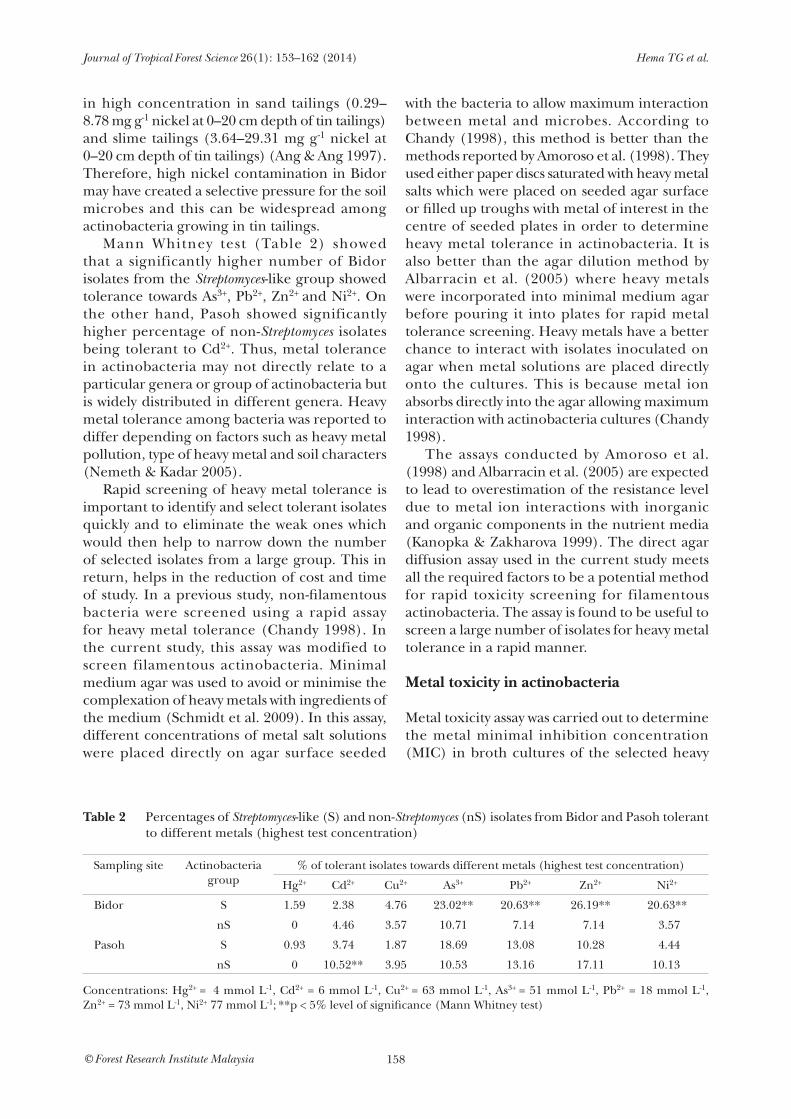
in high concentration in sand tailings (0.29– 8.78 mg g-1 nickel at 0–20 cm depth of tin tailings) and slime tailings (3.64–29.31 mg g-1 nickel at 0–20 cm depth of tin tailings) (Ang & Ang 1997). Therefore, high nickel contamination in Bidor may have created a selective pressure for the soil microbes and this can be widespread among actinobacteria growing in tin tailings. Mann Whitney test (Table 2) showed that a significantly higher number of Bidor isolates from the Streptomyces-like group showed tolerance towards As3+, Pb2+, Zn2+ and Ni2+. On the other hand, Pasoh showed significantly higher percentage of non-Streptomyces isolates being tolerant to Cd2+. Thus, metal tolerance in actinobacteria may not directly relate to a particular genera or group of actinobacteria but is widely distributed in different genera. Heavy metal tolerance among bacteria was reported to differ depending on factors such as heavy metal pollution, type of heavy metal and soil characters (Nemeth & Kadar 2005). Rapid screening of heavy metal tolerance is important to identify and select tolerant isolates quickly and to eliminate the weak ones which would then help to narrow down the number of selected isolates from a large group. This in return, helps in the reduction of cost and time of study. In a previous study, non-filamentous bacteria were screened using a rapid assay for heavy metal tolerance (Chandy 1998). In the current study, this assay was modified to screen filamentous actinobacteria. Minimal medium agar was used to avoid or minimise the complexation of heavy metals with ingredients of the medium (Schmidt et al. 2009). In this assay, different concentrations of metal salt solutions were placed directly on agar surface seeded
with the bacteria to allow maximum interaction between metal and microbes. According to Chandy (1998), this method is better than the methods reported by Amoroso et al. (1998). They used either paper discs saturated with heavy metal salts which were placed on seeded agar surface or filled up troughs with metal of interest in the centre of seeded plates in order to determine heavy metal tolerance in actinobacteria. It is also better than the agar dilution method by Albarracin et al. (2005) where heavy metals were incorporated into minimal medium agar before pouring it into plates for rapid metal tolerance screening. Heavy metals have a better chance to interact with isolates inoculated on agar when metal solutions are placed directly onto the cultures. This is because metal ion absorbs directly into the agar allowing maximum interaction with actinobacteria cultures (Chandy 1998). The assays conducted by Amoroso et al. (1998) and Albarracin et al. (2005) are expected to lead to overestimation of the resistance level due to metal ion interactions with inorganic and organic components in the nutrient media (Kanopka & Zakharova 1999). The direct agar diffusion assay used in the current study meets all the required factors to be a potential method for rapid toxicity screening for filamentous actinobacteria. The assay is found to be useful to screen a large number of isolates for heavy metal tolerance in a rapid manner.
Metal toxicity in actinobacteria
Metal toxicity assay was carried out to determine the metal minimal inhibition concentration (MIC) in broth cultures of the selected heavy
Table 2 Percentages of Streptomyces-like (S) and non-Streptomyces (nS) isolates from Bidor and Pasoh tolerant to different metals (highest test concentration)
Sampling site Actinobacteriagroup
% of tolerant isolates towards different metals (highest test concentration)
Hg2+ Cd2+ Cu2+ As3+ Pb2+ Zn2+ Ni2+
Bidor S 1.59 2.38 4.76 23.02** 20.63** 26.19** 20.63**
nS 0 4.46 3.57 10.71 7.14 7.14 3.57
Pasoh S 0.93 3.74 1.87 18.69 13.08 10.28 4.44
nS 0 10.52** 3.95 10.53 13.16 17.11 10.13
Concentrations: Hg2+ = 4 mmol L-1, Cd2+ = 6 mmol L-1, Cu2+ = 63 mmol L-1, As3+ = 51 mmol L-1, Pb2+ = 18 mmol L-1, Zn2+ = 73 mmol L-1, Ni2+ 77 mmol L-1; **p < 5% level of significance (Mann Whitney test)

Journal of Tropical Forest Science 26(1): 153–162 (2014) Hema TG et al.
159© Forest Research Institute Malaysia
metal tolerant isolates. Of the 40 isolates tested, 20 isolates showed capability to tolerate high concentration of at least one metal in the assay (Table 3). Growth was observed only in Bidor isolates at concentration of 43.2 mmol L-1 for Pb2+ (six isolates) and 138.9 mmol L-1 for Ni2+ (four isolates). For metals such as As3+ and Cd2+, growth was observed in Bidor and Pasoh isolates at the highest concentrations of 182.0 mmol L-1
and 21.8 mmol L-1 respectively. Only one isolate each from Bidor and Pasoh could tolerate the high test concentration of 21.8 mmol L-1 for Cd2+. Six isolates from Bidor and two from Pasoh were tolerant to As3+ at 182 mmol L-1. Final concentrations of Pb2+, Ni2+, As3+ and Cd2+ that isolates could tolerate in the metal toxicity assay were higher than concentrations showing bacterial tolerance in the rapid metal tolerance assay. The concentrations of metal
supporting bacterial growth in metal toxicity assay were higher than those from previous studies: 60–90 mmol L-1 for As3+ and 4–10 mmol L-1 for Pb2+ (Kermanshahi et al. 2007), 10 mmol L-1 for Cd2+ (Amoroso et al. 1998) and 42.6 mmol L-1 for Ni2+ (Van Nostrand et al. 2007). Only one isolate from Bidor could tolerate Hg2+ at 0.8 mmol L-1 and four isolates tolerated Cu2+ at 14 mmol L-1 obtained from both Bidor and Pasoh. Isolates that showed tolerance to Cu2+ and Hg2+ in the rapid metal tolerance screening were able to tolerate higher metal concentrations compared with results obtained in the metal toxicity assay (Table 3). However, concentrations that supported the actinobacteria growth in metal toxicity assay were still much higher than results reported by Albarracin et al. (2005) which was 6 mmol L-1 for Cu2+ and Prithviraj et al. (2012) which was 0.1 mmol L-1 for Hg2+. In the metal
Table 3 Metal MIC values of highly tolerant isolates from Bidor and Pasoh
Sampling site
Isolate code Metal MIC value for different heavy metals obtained in metal toxicity assay(mmol L-1)
Metal tolerance level obtained from rapid metal tolerance screening (mmol L-1)
Pb2+ Ni2+ As3+ Cd2+ Cu2+ Hg2+
Bidor TY045-023 43.2* 34.7 22.8 nt nt nt Pb2+(18); Ni2+(77); As3+(51) #
TY046-016 43.2* 69.45 11.4 nt nt nt Pb2+(18); Ni2+(77); As3+(51)
TY048-049 43.2* nt nt nt nt nt Pb2+(18)
TY046-079 43.2* nt 182.0* nt nt nt Pb2+(18); As3+(51)
TY046-021 43.2* 138.9* 182.0* 10.9 nt nt Pb2+(18); Ni2+(77); As3+(51); Cd2+(6)
TY046-078 43.2* 138.9* 182.0* nt nt nt Pb2+(18); Ni2+(77); As3+(51)
TY046-071 5.4 138.9* 45.5 2.7 nt nt Pb2+(18); Ni2+(77); As3+(51); Cd2+(6)
TY046-027 5.4 138.9* 11.4 nt nt nt Pb2+(18); Ni2+(77); As3+(51)
TY046-073 2.7 17.4 182.0* nt nt nt Pb2+(18); Ni2+(77); As3+(51)
TY047-009 nt nt 182.0* nt nt nt As3+(51)
TY047-063 nt nt 182.0* nt nt nt As3+(51)
TY049-057 5.4 nt 11.4 21.8* nt nt Pb2+(18); As3+(51); Cd2+(6)
TY046-070 nt 34.7 22.8 nt 14.1* nt Ni2+(77); As3+(51); Cu2+(63)
TY047-024 nt nt 17.4 nt 14.1* nt As3+(51); Cu2+(63)
TY046-017 nt nt nt nt nt 0.8* Hg2+(4)
Pasoh TY029-008 2.7 nt 182.0* 2.7 nt nt Pb2+(18); As3+(51); Cd2+(6)
TY029-014 5.4 nt 182.0* 1.4 nt nt Pb2+(18); As3+(51); Cd2+(6)
TY028-047 nt nt 91 21.8* nt nt As3+(51); Cd2+(6)
TY028-019 nt nt nt nt 14.1* nt Cu2+(63)
TY028-043 nt nt nt nt 14.1* nt Cu2+(63)
# Test concentration showing bacterial tolerance (Figure 1); * MIC values supporting actinobacteria growth in metal toxicity assay; MIC = minimal inhibition concentration; nt = not tested

Journal of Tropical Forest Science 26(1): 153–162 (2014) Hema TG et al.
160© Forest Research Institute Malaysia
toxicity assay, liquid medium was used as the test medium for bacterial growth whereas in the rapid metal tolerance screening, solid agar medium was used. Thus, the conditions of diffusion, complexation and availability of metals are different in both methods. Hassen et al. (1998) who tested the levels of tolerance in Pseudomonas aeruginosa to different concentrations of divalent metal ions in nutrient broth showed that the test in liquid media was sensitive at concentrations 10 to 1000 times lower than those obtained in solid media. This might be the reason why isolates tolerant to Hg2+ and Cu2+ in rapid metal tolerance screening became sensitive to metal ions in the metal toxicity assay. Various studies conducted on heavy metal removal indicated the severity of heavy metal pollution and its danger to the environment and humans. Currently available commercial remediation methods for reducing the harmful effects at heavy metal contaminated sites include solidification/stabilisation technologies, soil washing, soil flushing and pytoremediation to extract the metals from the soil (Evanko & Dzombak 1997). However, these new technologies of remediation are becoming uneconomical and unfavourable to remove heavy metals and still failing to provide the needed requirements for a safe, cheap, effective and environmentally-friendly metal remediation technique (Kumar et al. 2011b). Actinobacteria have been widely used in bioremediation. They are used in biotransformation, biodegradation and other purposes. They cause degradation of herbicides, pesticides, chromium (IV), petrochemicals as well as nitroaromatic and 2,4,6-trinitrotoluene compounds (Syed Amir 2011). Thus, the metal tolerant isolates obtained from the current study could be further explored for use in heavy metal removal to fulfil the needed requirements such as safe, ecofriendly, cheap and effective methods of bioremediation. In order to survive and overcome heavy metal stress in the environment, bacteria possess various resistance mechanisms. Bacterial plasmids have been described in encoding mercuric reductase and transport proteins which are involved in resistance towards mercury (Ravel et al. 1998). These plasmids have also genes for resistance to many other toxic ions of heavy metals such as Ag+, Cd2+, Co2+, Cu2+, Ni2+, Pb2+ and Zn2+. There are reports on Streptomyces spp. showing Cr (VI) reduction ability (Pattanapipitpaisal et al.
2001, Laxman & More 2002). Streptomyces is also known for possessing two superoxide dismutases which are iron- and nickel-containing enzymes, regulated by nickel (Kim et al. 1998). The current study showed that rapid metal tolerance screening had been useful to narrow down from a large number of isolates to obtain metal tolerant actinobacteria in a fast manner. In the metal toxicity assay, two multimetal resistant isolates TY046-021 and TY046-078 obtained from Bidor showed tolerance towards more than three heavy metals at the highest test concentration. Both of these isolates were tolerant to As3+, Pb2+ and Ni2+. Compared with rapid metal tolerance screening, the metal toxicity assay clearly showed that metal tolerant isolates from Bidor were able to tolerate and had multimetal tolerating capability to higher concentration of heavy metals compared with Pasoh isolates. The distribution of metal tolerance in actinobacteria to specific metal ions is heavily influenced by environmental conditions from where the isolates are isolated. The two multimetal resistant isolates TY046-021 and TY046-078 and the only Hg2+ tolerant isolate TY046-017 obtained from Bidor should be further studied for heavy metal uptake and removal.
ACKNOWLEDGEMENTS
The authors thank the Ministry of Science, Technology and Innovation of Malaysia for the eScienceFund grant (02-03-10-SF0108), Forest Research Institute Malaysia for facilities, LH Ang (Bidor Research Station) for help and Universiti Malaya for grants PS 167-2009A and HIR 005.
REFERENCES
AlbArrAcin VH, Amoroso MJ & AbAte CM. 2005. Isolation and characterization of indigenous copper-resistant actinomycete strains. Chemi der Erde-Geochemistry 65: 6145–6156.
AlvArez VM, MArques JM, Korenblum E & Seldin L. 2011. Comparative bioremediation of crude oil-amended tropical soil microcosms by natural attenuation, bioaugmentation or bioenrichment. Applied and Environmental Soi l Sc ience : doi 10.1155/2011/156320.
Amoroso MJ & AbAte CM. 2012. Bioremediation of copper, chromium and cadmium by actinomycetes from contaminated soil. Soil Biology 31: 349–364.
Amoroso MJ, CAstro G, CArlino, Federico J, Romero NC, Hill R & Oliver G. 1998. Screening of heavy metal tolerant actinomycetes isolates from the Sali River. Journal of General Applied Microbiology 44: 29–32.

Journal of Tropical Forest Science 26(1): 153–162 (2014) Hema TG et al.
161© Forest Research Institute Malaysia
Ang LH & Ang TB. 1997. Greening the tin tailings—are we ready? Pp 195–205 in Appanah S & Khoo KC (eds)Proceedings of Conference on Forestry and Forest Products Research. 2–4 October 1997, Forest Research Institute Malaysia, Kepong.
Ang LH, Ho WM, RAmli MO, MAimon A, Chung PY & Ng LT. 2000. The update of potentially toxic elements in some economically important plants and fish produced from ex-mining sites in Bidor, Perak. Pp 296–303 in Proceedings of the Malaysian Science Technology Convention 2000. 18–20 September 2000, Kota Kinabalu.
Ang LH & Ng LT. 2000. Trace element concentration in mango (Mangifera indica L), seedless guava (Psidium guajava L) and papaya (Carica papaya L) grown on agricultural and ex-mining lands of Bidor, Perak. Pertanika Journal of Tropical Agriculture Science 23: 15–22.
Ang LH, TAng LK, Ho WM, Hui TF & TheseirA GW. 2010. Pyhotoremediation of Cd and Pb by four tropical timber species grown on an ex-tin mine in Peninsular Malaysia. World Academy of Science, Engineering and Technology 62: 459–463.
Anton A, Grobe C, ReimAnn J, Pribyl T & Nies DH. 1999. CzcD is a heavy metal transporter involved in regulation of heavy metal resistance in Ralstonia sp. strain CH34. Journal of Bacteriology 181: 6876–6881.
ChAndy JP. 1998. Heavy metal tolerance chromogenic and non-chromogenic marine bacteria from Arabian gulf. Environmental Monitoring and Assessment 59: 321–330.
EvAnko CR & DzombAk DA. 1997. Remediation of Metals-Contaminated Soils and Groundwater. Technology Evaluation Report TE-97-01. UESPA Ground-Water Remediation Technology Analysis Center, Pittsburgh.
GethA K, Mohd IlhAm A, Lee SS, ChAng YS, NimurA S & HAtsu M. 2008. Exploratory studies of actinomycetes biodiversity of FRIM forests in aid of drug discovery. Pp 105–109 in Highlights of FRIM’s MOSTI Projects 2007, FRIM, Kepong. 21–22 January 2008, Ayer Keroh.
GethA K, VikineswAry S, Wong WH, Seki T, WArd A & Goodfellow M. 2004. Characterization of selected isolates of indigenous Streptomyces species and evaluation of their antifungal activity against selected plant pathogenic fungi. Malaysian Journal of Science 23: 37–47.
HAssen A, SAidi N, Cherif M & BoudAbous A. 1998. Resistance of environmental bacteria to heavy metals. Biosource Technology 64: 7–15.
Jimi E, Aoki K, SAito H, D’Acquisto F, MAy MJ & NAkAmurA I. 2004. Selective inhibition of NF-kappa B blocks osteoclasto-genesis and prevents inflammatory bone destruction in vivo. Nature Medicine 10: 617–624.
KAnopkA A & ZAkhArovA T. 1999. Quantification of bacterial lead resistance via activity assays. Journal of Microbiological Methods 37: 17–22.
KermAnshAhi KR, GhAzifArd A & TAvAkoli A. 2007. Identification of bacteria to heavy metals in the soils of Isfahan Province. Iranian Journal of Science and Technology 31: A1.
Kim EJ, Chung HJ, Suh HJ, HAh YC & Roe JH. 1998. Transcriptional and post-transcriptional regulation by nickel of sodN gene encoding nickel containing
superoxide dismutase from Streptomyces coelicolor Muller. Molecular Microbiology 27: 187–195.
Kim JS, KAng SO & Lee KJ. 2003. The protein complex composed of nickel-binding SrnQ and DNA binding motif-bearing SrnR of Streptomyces griseus represses sodF transcription in the presence of nickel. The Journal of Biological Chemistry 278: 18455–18463.
KumAr A, Bisht BS & Joshi VD. 2010. Biosorption of heavy metals by four acclimated microbial species, Bacilius spp., Pseudomonas spp., Staphylococcus spp. and Aspergillus niger. Journal of Biology and Environmental Science 4: 97–108.
KumAr A, Bisht BS & Joshi VD. 2011a. Bioremediation potential of three acclimated bacteria with reference to heavy metal removal from waste. International Journal of Environmental Sciences 2: 896–908.
KumAr A, Bisht BS, Joshi VD & dhewA T. 2011b. Review on bioremediation of polluted environment: a management tool. International Journal of Environmental Sciences 1: 1079–1093.
LAxmAn SR & More S. 2002. Reduction of hexavalent chromium by Streptomyces griseus. Minerals Engineering 15: 831–837.
Lin Y, WAng X, WAng B, MohAmAd O & Wei G. 2012. Bioaccumulation characterization of zinc and cadmium by Streptomyces zinciresistens, a novel actinomycete. Ecotoxicology and Environmental Safety 77: 7–17.
Luo YM. 2009. Current research and development in soil remediation technologies. Progress in Chemistry 21: 558–565.
MAsAnori. 2006. Expert and citizen participation in the pollution control: the case of itai-itai disease in Japan. Minamata’s legacy after 50 years. The Japan Times, May 2006.
MergeAy M, Monchy S, VAllAeys T, Auquier V, BenotmAne A, Bertin P, TAghAvi S, Dunn J, VAn Der Lelie D & WAttiez R. 2003. Ralstonia metallidurans, a bacterium specifically adapted to toxic metals towards a catalogue of metal-responsive genes. FEMS Microbiology Reviews 27: 385–410.
Milic JS, Beskosko VP, Ilic ML, Ali SAM, Gojgic-Cvijovic GD & Vrvic MM. 2009. Bioremediation of soil heavily contaminated with crude oil and its products: composition of the microbial consortium. Journal of the Serbian Chemical Society 71: 455–460.
Nemeth T & KAdAr I. 2005. Leaching of microelement contaminants: a long-term field study. Journal of Biosciences 60: 260–264.
NumAtA K & NimurA S. 2003. Access to soil actinomycetes in Malaysian tropical rain forests. Actinomycetologica 17: 54–56.
PAttAnApipitpAisAl NL, Brown NL & MecAskie LE. 2001. Chromate reduction by Microbacterium liquefaciens immobilised in polyvinyl alcohol. Biotechnology Letters 23: 61–65.
PrithvirAj D, DivyA P, PAtel K, AursAng R, Neelu N, BAlAsAheb K, MAdhukAr K & Abul M. 2012. Biosorption of heavy metals by actinomycetes for treatment of industrial effluents. Pp 389–393 in Proceedings of the UMT 11th International Annual Symposium on Sustainability Science and Management. 9 –11 July 2012, Kuala Terengganu.

Journal of Tropical Forest Science 26(1): 153–162 (2014) Hema TG et al.
162© Forest Research Institute Malaysia
RAvel J, Amoroso MJ, Colwell R & Hill RT. 1998. Mercury-resistant actinomycetes from Chesapeake Bay. FEMS Microbiology Letters 162: 177–184.
Schmidt A, HAferburg G, Schmidt A, Lischke U, Merten D, Ghergel F, Buchel G & Kothe E. 2009. Heavy metal resistance to the extreme: Streptomyces strains from a former uranium mining area. Chemi der Erde-Geochemistry 69: 35–44.
Schmidt A, HAferburg G, Sineriz M, Merten D, Buchel G & Kothe E. 2005. Heavy metal resistance in Actinobacteria for survival in AMD contaminated soils. Chemi der Erde-Geochemistry 65: 131–144.
SelvAmeenAl L, RAdhAkrishnAn M & BAlAgurunAthAn R. 2009. Antibiotic pigment from desert soil actinomycetes: biological activity, purification and chemical screening. Indian Journal of Pharmaceutical Sciences 71: 499–504.
Selvin J, ShAnmughA PS, SeghAl KG, ThAngAvelu T & SApnA BAi N. 2007. Sponge-associated marine bacteria as indicators of heavy metal pollution. Microbiological Research 164: 352–363.
Sen S & ChArAyA MU. 2010. Copper tolerant microfungi for bioremediation. Progressive Agriculture 10: 68–71.
ShAAbAn MT, El-SAbbAgh SMM & AlAm A. 2013. Studies on an actinomycete producing a melanin pigment inhibiting aflatoxin B1 production by Aspergillus flavus. Life Science Journal 10: 1437–1448.
Shirling EB & Gottlieb D. 1966. Methods for characterization of Streptomyces species. International Journal of Systematic Bacteriology 16: 313–340.
S h i v i k A B . 2 0 1 1 . H e a v y m e t a l r e s i s t a n c e o f microorganisms. Http://www.biotecharticles.com EnvironmentalBiotechnologyArticle/Heavy-Metal Resistance-of-Microorganisms-1099.html.
ShpAryk YS & PArpAn VI. 2004. Heavy metal pollution and forest health in the Ukrainian Carpathians. Environmental Pollution 130: 55–63.
Sineriz ML, Kothe E & AbAte CM. 2009. Cadmium biosorption by Streptomyces sp. F4 isolated from former uranium mime. Journal of Basic Microbiology 1: 55–62.
Silver S & Phung LT. 2005. A bacterial view of the periodic table: genes and proteins for toxic inorganic ions. Journal of Industrial Microbiology and Biotechnology 32: 587–605.
Syed Amir M. 2011. Actinomycetes and bioremediation. Http://www.biotecharticles.com/Environmental-Biotechnology Article/Actinomycetes-and-Bioremediation-1091.html.
Toshihide T & TAkAshi Y. 2011. The history of minamata disease and public health policy. Epidemiology 22: S99.
VAn NostrAnd JD, KhijniAk TV, Gentry TJ, NovAk MT, Sowdwr AG, Zhou JZ, Bertsch PM & Morris PJ. 2007. Isolation and characterization of four Gram positive nickel tolerant microorganisms from contaminated sediments. Microbial Ecology 53: 670–682.
WAisberg M, Joseph P, HAle B & BeyersmAnn D. 2003. Molecular and cellular mechanisms of cadmium carcinogenesis. Toxicology 192: 95–117.
ZhAo HJ, PArry RL, Ellis DI, Griffith GW & GoodcAcre R. 2005. The rapid differentiation of Streptomyces isolates using Fourier transform infrared spectroscopy. Vibrational Spectroscopy 40: 213–218.
Copyright © 2022 FDOKUMEN