Caspase3 Expression by Cerebellar Granule Neurons Is Regulated by Calcium and Cyclic AMP

Identification of miRNAs Differentially Expressed inHuman Epilepsy with or without Granule Cell PathologySilvia Zucchini1,2,3., Gianluca Marucci4., Beatrice Paradiso1,2,5, Giovanni Lanza5, Paolo Roncon1,2,
Pierangelo Cifelli1,6, Manuela Ferracin3,7, Marco Giulioni8, Roberto Michelucci9, Guido Rubboli9,10"¤,
Michele Simonato1,2,3"*
1 Department of Medical Sciences, Section of Pharmacology and Neuroscience Center, University of Ferrara, Ferrara, Italy, 2 National Institute of Neuroscience, Torino,
Italy, 3 Laboratory for Technologies of Advanced Therapies (LTTA), University of Ferrara, Ferrara, Italy, 4 Department of Biomedical and NeuroMotor Sciences (DiBiNeM),
Section of Pathology, Bellaria Hospital, Bologna, Italy, 5 Department of Morphology, Surgery and Experimental Medicine, Section of Pathology, University of Ferrara,
Ferrara, Italy, 6 Ri.MED Foundation, Palermo, Italy, 7 Department of Morphology, Surgery and Experimental Medicine, Section of Pathology, Oncology and Experimental
Biology, University of Ferrara, Ferrara, Italy, 8 IRCCS Institute of Neurological Sciences, Section of Neurosurgery, Bellaria Hospital, Bologna, Italy, 9 IRCCS Institute of
Neurological Sciences, Section of Neurology, Bellaria Hospital, Bologna, Italy, 10 Danish Epilepsy Center, Epilepsihospital, Dianalund, Denmark
Abstract
The microRNAs (miRNAs) are small size non-coding RNAs that regulate expression of target mRNAs at post-transcriptionallevel. miRNAs differentially expressed under pathological conditions may help identifying mechanisms underlying thedisease and may represent biomarkers with prognostic value. However, this kind of studies are difficult in the brain becauseof the cellular heterogeneity of the tissue and of the limited access to fresh tissue. Here, we focused on a pathologyaffecting specific cells in a subpopulation of epileptic brains (hippocampal granule cells), an approach that bypasses theabove problems. All patients underwent surgery for intractable temporal lobe epilepsy and had hippocampal sclerosisassociated with no granule cell pathology in half of the cases and with type-2 granule cell pathology (granule cell layerdispersion or bilamination) in the other half. The expression of more than 1000 miRNAs was examined in the laser-microdissected dentate granule cell layer. Twelve miRNAs were differentially expressed in the two groups. One of these,miR487a, was confirmed to be expressed at highly differential levels in an extended cohort of patients, using RT-qPCR.Bioinformatics searches and RT-qPCR verification identified ANTXR1 as a possible target of miR487a. ANTXR1 may bedirectly implicated in granule cell dispersion because it is an adhesion molecule that favors cell spreading. Thus, miR487acould be the first identified element of a miRNA signature that may be useful for prognostic evaluation of post-surgicalepilepsy and may drive mechanistic studies leading to the identification of therapeutic targets.
Citation: Zucchini S, Marucci G, Paradiso B, Lanza G, Roncon P, et al. (2014) Identification of miRNAs Differentially Expressed in Human Epilepsy with or withoutGranule Cell Pathology. PLoS ONE 9(8): e105521. doi:10.1371/journal.pone.0105521
Editor: Giuseppe Biagini, University of Modena and Reggio Emilia, Italy
Received May 8, 2014; Accepted July 22, 2014; Published August 22, 2014
Copyright: � 2014 Zucchini et al. This is an open-access article distributed under the terms of the Creative Commons Attribution License, which permitsunrestricted use, distribution, and reproduction in any medium, provided the original author and source are credited.
Data Availability: The authors confirm that all data underlying the findings are fully available without restriction. All relevant data are within the paper.
Funding: This work has been supported by grants from the European Community [FP7-PEOPLE-2011-IAPP project 285827 (EPIXCHANGE) and FP7-HEALTHproject 602102 (EPITARGET), to MS], and from the Ri.MED foundation (to PC). The funders had no role in study design, data collection and analysis, decision topublish, or preparation of the manuscript.
Competing Interests: The authors have declared that no competing interests exist.
* Email: [email protected]
. These authors contributed equally to this work.
" These authors are joint senior authors on this work.
¤ Current address: Institute of Clinical Medicine, University of Copenhagen, Copenhagen, Denmark
Introduction
The microRNAs (miRNAs) are small size endogenous non-
coding RNAs that regulate the expression of target mRNAs at
post-transcriptional level [1]. To date, more than 1000 human
miRNAs have been identified, about 50% of which are expressed
in the brain. miRNAs have been demonstrated to be involved in
several brain functions, many of which may be implicated in
epilepsy and epileptogenesis, like cell death, neurogenesis, synaptic
plasticity [2],[3]. Indeed, silencing miR-134 using a specific
antagomir exerted prolonged seizure-suppressant and neuropro-
tective actions in a murine model [4]. Thus, understanding which
specific miRNAs are differentially expressed in epilepsy may help
to identify the mechanisms underlying the disease. Moreover,
differentially expressed miRNAs may represent biomarkers that
identify specific subpopulations of epileptic patients, holding a
prognostic value [5].
Microarray platforms allow screening and identifying miRNAs
differentially expressed under pathological conditions. Experimen-
tal studies have profiled miRNA expression in animal models of
epilepsy [6],[7],[8],[9] and profiling studies have been also
recently published using hippocampi resected from temporal lobe
epilepsy (TLE) patients [10],[11]. However, some outstanding
obstacles make difficult the interpretation of data from microarray
analysis of human brain samples. First, in most studies tissue is
derived from autopsies or, potentially even worse, pathological
tissue is from surgery samples and control tissue from autopsies.
PLOS ONE | www.plosone.org 1 August 2014 | Volume 9 | Issue 8 | e105521

Post-mortem modifications are very likely to dramatically alter the
molecular composition of the tissue, making the results question-
able. Second, each brain area has a specific and complex cellular
composition that changes (often markedly) in the course of
diseases. Again, this makes interpretation of molecular data very
difficult, because analysis of heterogeneous tissue homogenates
does not allow identification of the cells where changes occur and
because up-regulation of a molecule in one cell population may be
obscured by down-regulation in another cell population.
One approach to overcome these problems is focusing on a
well-defined cell population. For example, we focused here on a
TLE-associated pathology of the granule cells of the hippocampus.
Drug-resistant TLE is the most common type of epilepsy requiring
surgical treatment, with a favorable postsurgical outcome in 60-
70% of the patients. Based on the underlying etiology, TLE
subtypes with different surgical prognosis have been described.
Neuropathological classifications of epileptogenic lesions, includ-
ing focal cortical dysplasias (FCD) [12], hippocampal sclerosis (HS)
[13] and granule cell pathology (GCP) [14], define histopatholog-
ical features and subtypes, allowing attempts to correlate clinical
and pathological findings. Correlations with molecular markers,
however, are still unavailable.
All patients included in this study underwent surgery for
pharmacoresistant TLE and had HS type 1 [13]. All were similar
for age, gender, clinical features of the disease. The most relevant
difference was that half of the patients had no granule cell
pathology (no GCP), whereas the other half had granule cell
dispersion or bilamination (GCP type 2) [14], i.e. the single
differential pathological feature was in a specific, isolable cell
population. Therefore, the granule cell layer was laser-microdis-
sected from all samples, total RNA was extracted from dissected
tissues and the miRNAome profile was obtained using a miRNA
microarray.
Materials and Methods
PatientsThis study was approved by the Ethics Committee of Bologna
(full name: Comitato Etico Indipendente dell’Azienda USL dellaCitta di Bologna). A comprehensive written informed consent (also
approved by the Ethics Committee of Bologna) was signed for the
surgical treatment that produced the tissue samples, the related
diagnostic procedures and the research use. All information
regarding the human material used in this study was managed
using anonymous numerical codes and samples were handled in
compliance with the Helsinki declaration (http://www.wma.net/
en/30publications/10policies/b3/).
Fourteen drug-resistant TLE patients candidate to epilepsy
surgery were collected at the Epilepsy Surgery Center of the
IRCCS Institute of Neurological Sciences of Bologna. All patients
underwent detailed epileptological evaluation and wakefulness/
sleep EEG. All patients also underwent continuous (24 hours)
long-term video-EEG monitoring for seizure recording. Analysis of
ictal clinical and EEG semiology and electroclinical correlations
aimed to identify the epileptogenic area were performed.
Three Tesla MRI, and brain CT scan when necessary, were
carried out. Electroclinical, neuroimaging, and neuropsychological
data were discussed by the Epilepsy Surgery Team (epileptologists,
neuroradiologists, neuropsychologists, neurosurgeons) to establish
the site of the epileptogenic area and the surgical strategy.
SurgeryAll patients underwent tailored temporal lobe resection to
remove the epileptogenic area, according to the data obtained
during pre-surgical investigation. Essentially, surgery consisted of
removing the temporal pole, the anterior neocortical lateral
cortex, the uncus–entorhinal area and the hippocampus and
parahippocampal gyrus. The main surgical specimens (hippocam-
pus and/or temporal pole) were removed ‘‘en bloc’’ and spatially
oriented to allow a proper histopathological examination.
Histology and microdissectionSpecimens were formalin fixed and paraffin embedded. They
were de-waxed using Bio-Clear (Bio-Optica, Milan, Italy), washed
in ethanol and stained with hematoxylin and eosin for histological
diagnosis. Neuropathological evaluation was performed using the
most recent classifications of HS, GCP and FCD [12],[13],[14],
applying the recommended histochemical and immunohistochem-
ical stains. Specimens either displayed no GCP or GCP type 2.
Four different types of neuropathological features can define GCP
type 2: 1) dispersion: rows of granule cells spread into the
molecular layer and the distance between granule cells is
increased; 2) ectopic granule cells: single ectopic granule cells are
dispersed into the molecular layer; 3) clusters: ectopic granule cells
form clusters within the molecular layer; 4) bilaminar: two granule
cell layers, separated by a cell-free gap [14]. While patterns of
granule cell loss (thinning and/or cell free gaps, GCP 1) occur
isolated, patterns of architectural abnormalities (GCP 2) can came
along with cell loss. Therefore, only sections in which no cell loss
was detected (based on NeuN staining) where included in analysis.
Ten-micron-thick sections were cut using a microtome and the
dentate granule layer of the dentate gyrus was laser-dissected
(Fig. 1) using the SL microcut microtest dissector (Nikon, Tokyo,
Japan). Microdissected cells were captured in microcut transfer
film (Nikon). Granule cells were collected in this manner from at
least 3–4 slices per patient, in order to obtain an adequate quantity
of RNA. Material from all sections of the same patient was pooled
together, and total RNA extracted using an RNA purification kit
(RecoverAll Total Nucleic Acid Isolation Kit, Ambion Life
Technologies, CA, USA). Approximately 1.5 mg total RNA were
obtained from each patient. Since miRNAs are more stable than
mRNAs, they can be used for microarray analysis from formalin-
fixed paraffin-embedded tissues [15]. We performed quality
control checks on microarray hybridizations using the Agilent
quality control (QC) tool in the Feature Extraction software. All
samples passed the QC check.
MicroarrayTotal RNA was used for microarray analysis (Human micro-
RNA Microarray V3, #G4470C, Agilent Technologies, Santa
Clara, CA, USA). This chip consists of 60-mer DNA probes and
allows simultaneous analysis of almost 1200 human miRNA
obtained from the Sanger miR-BASE database (Release 10.1). We
employed approximately 100 ng total RNA per sample in each
experiment. RNA labeling and hybridization were performed in
accordance to manufacturer’s indications. Agilent scanner and the
Feature Extraction 10.5 software (Agilent Technologies) were used
to obtain the microarray raw-data.
Microarray results were analyzed using the GeneSpring GX 12
software (Agilent Technologies). Data transformation was applied
to set all the negative raw values at 1.0, followed by Quantile
normalization and log2 transformation. Filters on gene expression
were used to keep only the miRNAs detected in at least one sample
(n = 536). The number of expressed miRNAs was 493 in the no
GCP group, 464 in the GCP 2 group. Differentially expressed
miRNAs were identified by comparing GCP type 2 vs. no GCP
samples. A 2 fold-change filter (n = 141) and the unpaired t-test
were applied (p,0.05; False Discovery Rate-FDR = 7%). Differ-
miRNAs in Temporal Lobe Epilepsy
PLOS ONE | www.plosone.org 2 August 2014 | Volume 9 | Issue 8 | e105521
entially expressed genes were employed in Cluster Analysis, using
the Pearson correlation as a measure of similarity. For Cluster
image generation, an additional step of normalization on gene
median across all samples was added.
miRNA qRT-PCRQuantitative real-time PCR (qRT-PCR) analysis of hsa-miR-
338-3p, 219-5p and 487a was performed using a TaqMan miRNA
assay kit (Applied Biosystems) according to the manufacturer’s
instructions. Samples were run in triplicate at 95uC for 15 s and
60uC for 1 min using a CFX96 Touch Real-Time PCR Detection
System (Applied Biosystems). Analysis was performed by the
comparative threshold cycle (CT) method. rRNA U48 was used as
reference gene. The relative amount of each miRNA in epileptic
samples was calculated using the equation RQ = 22CT, where
CT = (CT miRNA 2 CT U6 RNA). Similar results were
obtained using rRNA U6 as reference gene.
BioinformaticsTarget prediction was performed by comparative analysis of
several databases, using an open-source database [16].
ANTXR1 qRT-PCRmRNA levels of antrax receptor 1 (ANTXR1) (assay ID:
Hs01120394) and neuronal enolase 2 (ENO 2) (assay ID:
Hs01102367), were determined using TaqMan Real-Time PCR,
according to manufacturer’s instructions (Applied Biosystems).
Ten ng of total RNA were retro-transcribed using iScript Reverse
Transcription Supermix (BIO-RAD). cDNA templates were
amplified with TaqMan PreAmp Master Mix (Applied Biosys-
tems), using pooled assay mix for ANTXR1 and ENO 2 and then
assayed for gene expression as described previously. Each sample
was analyzed in triplicate, in two independent experiments. The
level of each mRNA was measured using Ct (threshold cycle) and
the amount of target was calculated as described above for
miRNAs. Gene expression levels were normalized using ENO 2
expression, as reported previously for this particular tissue [17].
ANTXR1 immunohistochemistryANTXR1 immunostaining was performed by an automatic and
clinically validated instrument based on Ventana Benchmark
Ultra systems from Roche Tissue Diagnostics. This immunohis-
tochemistry technique takes advantage of a new enhanced
sensitivity biotin-free multimer technology system, based on direct
linkers between peroxidase and secondary antibodies (ultraView
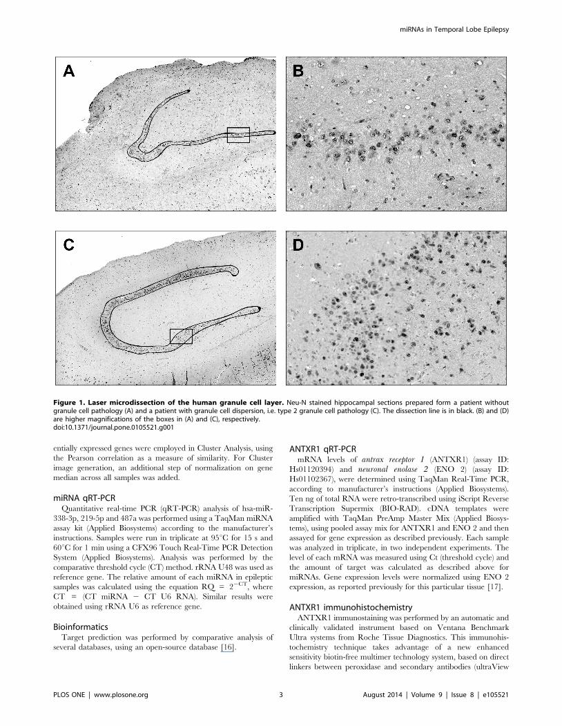
Figure 1. Laser microdissection of the human granule cell layer. Neu-N stained hippocampal sections prepared form a patient withoutgranule cell pathology (A) and a patient with granule cell dispersion, i.e. type 2 granule cell pathology (C). The dissection line is in black. (B) and (D)are higher magnifications of the boxes in (A) and (C), respectively.doi:10.1371/journal.pone.0105521.g001
miRNAs in Temporal Lobe Epilepsy
PLOS ONE | www.plosone.org 3 August 2014 | Volume 9 | Issue 8 | e105521

Universal DAB Detection Kit, Ventana Medical System). The
protocol provided for the automatic CC1 Ventana pre-treatment
(Cell Conditioning Solution, Ventana) for 52 min, then the
incubation for 2 hours with the anti-ANTXR1 antibody by
titration (rabbit polyclonal, by ThermoFisher Scientific; 1: 100).
Staining was visualized with the UltraView DAB procedure by
Benchmark Ultra System. Sections were then counterstained with
haematoxylin. Negative controls were treated identically except
that the primary antibody was omitted. Sections of metastatic
breast cancer were used as positive controls [18],[19]. Evaluation
of data was performed by two expert neuropathologists (GM and
BP) under double-blind conditions.
Statistical analysisFor qRT-PCR data, comparisons between experimental groups
were performed by using the Mann–Whitney U test. Differences
between groups were considered significant when P , 0.05.
Results
PatientsTissues from patients indicated in numbers in Table 1 were
employed for microarray analysis. Neuropathological examination
evidenced that these patients had HS type 1 [13], which was
associated with no granule cell pathology (no GCP) in 5 patients
and with granule cell pathology (GCP type 2) in the other 5 [14]:
GCP consisted of granule cell dispersion in 4 cases and bilaminar
granule cell layer in one. The no GCP group was composed of 3
males and 2 females, with mean age at surgery of 44 (33–60), mean
years after epilepsy diagnosis of 24 (7–38) and approximately 10
seizures per month before surgery (2 to .30); the GCP group was
composed of 5 females, with mean age at surgery of 33 (31–37),
mean years after epilepsy diagnosis of 23 (10–35) and approxi-
mately 10 seizures per month before surgery (3 to 15). An
epileptogenic insult could be identified in only one of the no GCP
cases (febrile convulsions), whereas all GCP cases had a history of
febrile convulsions, one also a possible brain trauma (Table 1).
miRNA microarrayTwelve miRNAs were differentially expressed in patients
without GCP compared with patients with type 2 GCP (Fig. 2).
Of these, 6 had relatively higher expression in tissue from patients
without GCP and 6 were higher in those with GCP 2 (Fig. 2).
Differential expression of a subset of 3 miRNAs (namely miR-338-
3p, miR-219-5p and miR-487a) was validated in an extended
cohort of patients (the ten original patients plus another 2 per
group, indicated in roman numbers in Table 1) using qRT-PCR.
Expression levels of all these miRNAs were apparently different in
the two groups, confirming microarray findings, but data were
dispersed for miR-338-3p and miR-219-5p, not reaching signif-
icance level (Fig. 3A and 3B). In contrast, miR-487a expression
was confirmed to be highly significantly reduced in GCP 2
(Fig. 3C).
Target identification and validation for miR-487aComparative analysis of several databases [16] indicated at least
10 highly likely targets for miR-487a, namely FAM126A,
ANTXR1, NUDCD1, AP1S3, AP3D1, AFTPH, KIAA1217,
ZNF57 PAG1 and PTGER3. All these mRNAs are expressed in
the brain (www.genecards.org). A subset of these are associated
with vesicle trafficking (AP1S3, AP3D1, AFTPH), others code for
receptors (PTGER3), intracellular signaling (FAM126A) or
transcription factors (ZNF57; www.genecards.org). More interest-
ingly, two of these mRNAs (PAG1 and ANTXR1) may be
associated with cell adhesion (www.genecards.org). In particular,
ANTXR1 (also known as tumor endothelial marker 8, TEM8) is
expressed in the mouse dentate gyrus granule cell layer (Allen
atlas; http://mouse.brain-map.org/gene/show/45380) and has
been reported to promote cell spreading in human tumor tissues
[20],[21]: therefore, it was hypothesized that reduced expression of
miR487a will increase ANTXR1 levels, leading to granule cell
spreading (i.e. dispersion or bilamination, i.e. GCP 2).
Evidence in support of this hypothesis has been pursued by
analyzing ANTXR1 mRNA and protein levels in the same
samples employed for validation of miR-487a. As predicted, using
qRT-PCR ANTXR1 mRNA levels were increased in the GCP 2
group (Fig. 4A). Moreover, a clear increase of ANTXR1
immunoreactivity was observed in the granule cell layer of
patients with type-2 GCP, as compared with those with no GCP
(Fig. 4B–C).
Discussion
The main findings of this study were: (1) the identification of 12
miRNAs differentially expressed in the hippocampal granule cell
layer of patients with hippocampal sclerosis associated with GCP 2
as compared with patients with no GCP; (2) the RT-qPCR
confirmation of one of these, miR-487a, in an extended cohort of
patients; (3) the identification of ANTXR1 as a possible target of
miR-487a.
An important issue in evaluation of this data is the possible
influence of medical treatments on miRNA expression. Indeed,
there is evidence that antiepileptic drugs can interfere with
miRNA expression: it has been reported that valproate can
modulate miR-24, miR-34a, and miR-128 [22] and that
phenobarbital can down-regulate miR-122 [23]. Although two
patients in the no-GCP group were treated with valproate and five
patients (three in the no-GCP group and two in the GCP 2 group)
were treated with phenobarbital, none of the above miRNAs was
found to be differentially expressed. More in general, a systematic
bias due to pharmacological treatments seems unlikely, because all
patients in both groups were using many drugs in combination.
Therefore, although we cannot rule out an influence of the
antiepileptic treatment on miRNAs expression, it seems more
likely that the changes we observed are due to the pathology.
The prognosis of patients undergoing epilepsy surgery has been
hypothesized to depend on the absence or presence of GCP but,
thus far, results have been inconsistent. While some studies
reported that GCP does not affect post-surgical outcome [24],[25],
others suggested association between GCP and a favorable
prognosis [14],[26]. Identification of molecular biomarkers that
parallel and/or integrate the pathology findings would provide a
valuable prognostic element, and miR-487a may represent a first
component of this molecular signature that will allow better
patient stratification. In the cohort of patients we analyzed in this
study, however, no clear distinction of the outcome was observed
in the early timeframe of post-surgical follow-up. Therefore,
extension of the follow-up will be needed to verify this possibility.
MiR-487a has been reported to be down-regulated in
Alzheimer disease [27] and up-regulated in schizophrenia [28].
Could it play a role in GCP? All miRNAs can have hundreds of
targets, but target prediction based bioinformatics approaches is
difficult for many reasons, most of all because of imperfect
complementarity. However, comparative analysis of several
databases [16] indicates at least 10 highly likely targets for miR-
487a. ANTXR1 emerged as the most interesting, because it has
been reported to promote cell spreading: ANTXR1 is a
transmembrane protein that functions as an adhesion molecule,
miRNAs in Temporal Lobe Epilepsy
PLOS ONE | www.plosone.org 4 August 2014 | Volume 9 | Issue 8 | e105521

Ta
ble
1.
Pat
ien
tsin
clu
de
din
the
stu
dy.
Pa
tie
nt
nu
mb
er
Ge
nd
er
Ag
ea
tsu
rge
ryE
pil
ep
tog
en
icin
sult
Ye
ars
aft
er
dia
gn
osi
sS
eiz
ure
sp
er
mo
nth
Dru
gth
era
py
(cu
rre
nt)
Pa
tho
log
yM
TS
[29
]P
ath
olo
gy
MT
S[3
0]
Pa
tho
log
yG
CP
[14
]O
utc
om
e[3
1]
01
M6
0n
on
e3
8.
30
VP
A,
CB
Z,
TG
BG
rad
eIV
MT
S1
Bn
oG
CP
Ia
02
M4
4n
on
e1
25
–9
TP
M,
LVT
Gra
de
IIIM
TS
1A
no
GC
PIa
03
M3
6n
on
e7
8–
10
LVT
,P
B,
CLB
Gra
de
IIIM
TS
1A
no
GC
PIa
04
F4
7n
on
e3
32
–3
PB
,C
BZ
Gra
de
IIIM
TS
1A
no
GC
PIc
05
F3
3fe
bri
leco
nvu
lsio
ns
30
5–
10
TP
M,
CB
Z,
VP
A,
PB
Gra
de
IIIM
TS
1A
no
GC
PIa
IM
55
no
ne
51
10
–1
5O
XC
,LT
G,
CLB
Gra
de
IIIM
TS
1A
no
GC
PIa
IIF
31
no
ne
16
3–
4C
BZ
,Z
NS
Gra
de
IIIM
TS
1A
no
GC
PIa
06
F3
1fe
bri
leco
nvu
lsio
ns
10
8–
12
PB
,T
PM
Gra
de
IIIM
TS
1A
GC
P2
IIa
07
F3
3fe
bri
leco
nvu
lsio
ns
24
3–
4LT
G,
LVT
,P
BG
rad
eIII
MT
S1
AG
CP
2Ia
08
F3
2fe
bri
leco
nvu
lsio
ns
27
4–
10
CB
ZG
rad
eIII
MT
S1
AG
CP
2Ia
09
F3
2fe
bri
leco
nv.
(tra
um
a?)
20
9–
10
OX
C,
LVT
Gra
de
IVM
TS
1B
GC
P2
Ia
10
F3
7fe
bri
leco
nvu
lsio
ns
35
12
–1
5C
BZ
,T
PM
Gra
de
IVM
TS
1B
GC
P2
Ia
IIIF
41
feb
rile
con
vuls
ion
s3
83
–4
CB
Z,
PG
BG
rad
eIII
MT
S1
AG
CP
2Ia
IVM
36
feb
rile
con
vuls
ion
s2
25
–6
TP
M,
LVT
Gra
de
IIIM
TS
1A
GC
P2
Ia
CB
Z,
carb
amaz
ep
ine
;C
LB,
clo
baz
am;
LTG
,la
mo
trig
ine
;LV
T,
leve
tira
ceta
m;
OX
C,
oxc
arb
aze
pin
e;
PB
,p
en
tob
arb
ital
;T
GB
,ti
agab
ine
;T
PM
,to
pir
amat
e;
VP
A,
valp
roic
acid
.d
oi:1
0.1
37
1/j
ou
rnal
.po
ne
.01
05
52
1.t
00
1
miRNAs in Temporal Lobe Epilepsy
PLOS ONE | www.plosone.org 5 August 2014 | Volume 9 | Issue 8 | e105521

coupling binding of an immobilized extracellular ligand and cell
spreading through association to the actin cytoskeleton [20]. Thus,
reduced expression of miR-487a could increase ANTXR1 mRNA
and protein levels and thereby favor granule cell dispersion. Here,
we have provided circumstantial evidence that this could indeed
be the case. Further studies in vitro and in animal models are
currently ongoing to directly demonstrate this hypothesis.
In conclusion, miR-487a may be the first identified element of a
miRNA signature that may be useful for a prognostic evaluation of
post-surgical epilepsy and form the basis for mechanistic studies
Figure 2. miRNAs differentially expressed in patients without granule cell pathology (no GCP) or with type 2 GCP (GCP 2). Heat-maprepresentation of the average expression of the 12 differentially expressed miRNAs in no GCP and GCP 2 from ten different tissues. The colors of thegenes represented on the heat map correspond to the expression values normalized on miRNA mean expression across all samples: green indicatesdown-regulated; red indicates up-regulated in the tissue. Patients are identified by numbers (in parenthesis) that correspond to those reported in theTable 1.doi:10.1371/journal.pone.0105521.g002
Figure 3. Relative expression of miR-338-3p (A), miR-219-5p (B) and miR-487a (C), evaluated by qRT-PCR in patients withoutgranule cell pathology (no GCP, blue bars) or with type-2 GCP (black bars). Seven patients per group. **P,0.01 Mann-Whitney U test.doi:10.1371/journal.pone.0105521.g003
miRNAs in Temporal Lobe Epilepsy
PLOS ONE | www.plosone.org 6 August 2014 | Volume 9 | Issue 8 | e105521

that may lead to the identification of new therapeutic targets, like
ANTXR1.
Acknowledgments
The authors are grateful to Dr. Pitt Niehusmann (University of Bonn) for
helpful discussions; to Dr. Fulvio Chiesa (Roche, Italy) for technical advice;
to Cristina Zampini, Patrizia Raisi, Maura Masiero, Maria Novi, Rosaria
Morelli, Anna Cherubino and Elisa Zaffoni for technical support.
Author Contributions
Conceived and designed the experiments: SZ GM BP MG RM GR MS.
Performed the experiments: SZ GM BP PR PC MG. Analyzed the data:
SZ GM BP MF GL MS. Contributed reagents/materials/analysis tools: SZ
GM BP PR PC MG. Contributed to the writing of the manuscript: SZ GM
GR MS.
References
1. Bartel DP (2004) MicroRNAs: genomics, biogenesis, mechanism, and function.
Cell 116: 281–297.
2. Im HI, Kenny PJ (2012) MicroRNAs in neuronal function and dysfunction.
Trends Neurosci 35: 325–334.
3. McNeill E, Van Vactor D (2012) MicroRNAs shape the neuronal landscape.
Neuron 75: 363–379.
4. Jimenez-Mateos EM1, Engel T, Merino-Serrais P, McKiernan RC, Tanaka K,
et al. (2012) Silencing microRNA-134 produces neuroprotective and prolonged
seizure-suppressive effects. Nat Med 18: 1087–1094.
5. Henshall DC (2014) MicroRNA and epilepsy: profiling, functions and potential
clinical applications. Curr Opin Neurol 27: 199–205.
6. Song YJ, Tian XB, Zhang S, Zhang YX, Li X, et al. (2011) Temporal lobe
epilepsy induces differential expression of hippocampal miRNAs including let-7e
and miR-23a/b. Brain Res 1387: 134–140.
7. Hu K, Xie YY, Zhang C, Ouyang DS, Long HY, et al. (2012) MicroRNA
expression profile of the hippocampus in a rat model of temporal lobe epilepsy
and miR-34a-targeted neuroprotection against hippocampal neurone cell
apoptosis poststatus epilepticus. BMC Neurosci 13: 1–14.
8. Bot AM, Debski KJ, Lukasiuk K (2013) Alterations in miRNA levels in the
dentate gyrus in epileptic rats. PLoS ONE 8: e76051.
9. Gorter JA, Iyer A, White I, Colzi A, van Vliet EA, et al. (2013) Hippocampal
subregion-specific microRNA expression during epileptogenesis in experimental
temporal lobe epilepsy. Neurobiol Dis 62: 508–520.
10. Kan AA, van Erp S, Derijck AA, de Wit M, Hessel EV, et al. (2012) Genome-
wide microRNA profiling of human temporal lobe epilepsy identifies modulators
of the immune response. Cell Mol Life Sci 69: 3127–3145.
11. McKiernan RC, Jimenez-Mateos EM, Bray I, Engel T, Brennan GP, et al.
(2012) Reduced mature microRNA levels in association with dicer loss in human
temporal lobe epilepsy with hippocampal sclerosis. PLoS ONE 7: e35921.
12. Blumcke I, Thom M, Aronica E, Armstrong DD, Vinters HV, et al. (2011) The
clinicopathologic spectrum of focal cortical dysplasias: a consensus classification
proposed by an ad hoc Task Force of the ILAE Diagnostic Methods
Commission. Epilepsia 52: 158–174.
13. Blumcke I, Thom M, Aronica E, Armstrong DD, Bartolomei F, et al. (2013)
International consensus classification of hippocampal sclerosis in temporal lobe
epilepsy: a task force report from ILAE Commission on Diagnostic Methods.
Epilepsia 54: 1315–1329.
14. Blumcke I, Kistner I, Clusmann H, Schramm J, Becker AJ, et al. (2009) Towards
a clinico-pathological classification of granule cell dispersion in human mesial
temporal lobe epilepsies. Acta Neuropathol 117: 535–544.
15. Peiro-Chova L1, Pena-Chilet M, Lopez-Guerrero JA, Garcıa-Gimenez JL,
Alonso-Yuste E, et al. (2013) High stability of microRNAs in tissue samples of
compromised quality. Virchows Arch 463: 765–774.
16. Dweep H, Sticht C, Pandey P, Gretz N (2011) miRWalk - database: prediction
of possible miRNA binding sites by "walking" the genes of 3 genomes. J Biomed
Inform 44: 839–847.
17. Maurer-Morelli CV, de Vasconcellos JF, Reis-Pinto FC, Rocha Cde S,
Domingues RR, et al. (2012) A comparison between different reference genes
for expression studies in human hippocampal tissue. J Neurosci Methods 208:
44–47.
18. Chen D, Bhat-Nakshatri P, Goswami C, Badve S, Nakshatri H (2013)
ANTXR1, a stem cell-enriched functional biomarker, connects collagen
signaling to cancer stem-like cells and metastasis in breast cancer. Cancer Res
73: 5821–5833.
19. Gutwein LG, Al-Quran SZ, Fernando S, Fletcher BS, Copeland EM, Grobmyer
SR (2011) Tumor endothelial marker 8 expression in triple-negative breast
cancer. Anticancer Res 31: 3417–3422.
20. Werner E, Kowalczyk AP, Faundez V (2006) Anthrax toxin receptor 1/tumor
endothelium marker 8 mediates cell spreading by coupling extracellular ligands
to the actin cytoskeleton. J Biol Chem 281: 23227–23236.
21. Gu J, Faundez V, Werner E (2010) Endosomal recycling regulates anthrax toxin
receptor 1/tumor endothelial marker 8-dependent cell spreading. Exp Cell Res
316: 1946–1957.
Figure 4. Relative expression of ANTXR1 (A), evaluated by qRT-PCR, in patients without granule cell pathology (no GCP, blue bars)or with type-2 GCP (black bars). Seven patients per group. **P,0.01 Mann-Whitney U test. Representative granule cell layer hippocampalsections from patients without granule cell pathology (B) or with type-2 GCP (C) exhibiting DAB-labeled ANTXR1-like immunoreactivity (LI). Omittingthe primary antibody to estimate nonspecific signal yielded completely negative labeling (data not shown). Note a widespread increase in ANTXR1-LIin granule cells from patients with type-2 GCP (C).doi:10.1371/journal.pone.0105521.g004
miRNAs in Temporal Lobe Epilepsy
PLOS ONE | www.plosone.org 7 August 2014 | Volume 9 | Issue 8 | e105521

22. Zhou R, Yuan P, Wang Y, Hunsberger JG, Elkahloun A, et al. (2009) Evidence
for selective microRNAs and their effectors as common long-term targets for theactions of mood stabilizers. Neuropsychopharmacology 34: 1395–1405.
23. Shizu R1, Shindo S, Yoshida T, Numazawa S (2012) MicroRNA-122 down-
regulation is involved in phenobarbital-mediated activation of the constitutiveandrostane receptor. PLoS ONE 7: e41291.
24. Thom M, Liagkouras I, Elliot KJ, Martinian L, Harkness W, et al. (2010)Reliability of patterns of hippocampal sclerosis as predictors of postsurgical
outcome. Epilepsia 51: 1801-1808.
25. da Costa Neves RS, Jardim AP, Caboclo LO, Lancellotti C, Marinho TF, et al.(2013) Granule cell dispersion is not a predictor of surgical outcome in temporal
lobe epilepsy with mesial temporal sclerosis. Clin Neuropathol 32: 24–30.26. Marucci G, Rubboli G, Giulioni M (2010) Role of dentate gyrus alterations in
mesial temporal sclerosis. Clin Neuropathol 29: 32–35.
27. Wang WX, Huang Q, Hu Y, Stromberg AJ, Nelson PT (2011) Patterns of
microRNA expression in normal and early Alzheimer’s disease human temporalcortex: white matter versus gray matter. Acta Neuropathol 121: 193–205.
28. Beveridge NJ, Cairns MJ (2012) MicroRNA dysregulation in schizophrenia.
Neurobiol Dis 46: 263–271.29. Wyler AR, Dohan FC Jr., Schweitzer JB, Berry AD III (1992) A grading system
for mesial temporal pathology (hippocampal sclerosis) from anterior temporallobectomy. J Epil 5: 220–225.
30. Blumcke I, Pauli E, Clusmann H, Schramm J, Becker A, et al. (2007) A new
clinico-pathological classification system for mesial temporal sclerosis. ActaNeuropathol 113: 235–244.
31. Engel J Jr, Van Ness P, Rasmussen T, Ojemann L (1993) Outcome with respectto epileptic seizures. In Engel J Jr editor.Surgical treatment of the epilepsies, 2nd
edNew YorkRaven Press 609–621.
miRNAs in Temporal Lobe Epilepsy
PLOS ONE | www.plosone.org 8 August 2014 | Volume 9 | Issue 8 | e105521
Copyright © 2022 FDOKUMEN