Wip1-Deficient Neutrophils Significantly Promote Intestinal Ischemia/Reperfusion Injury in Mice
Upload
independentCategory
view
1download
0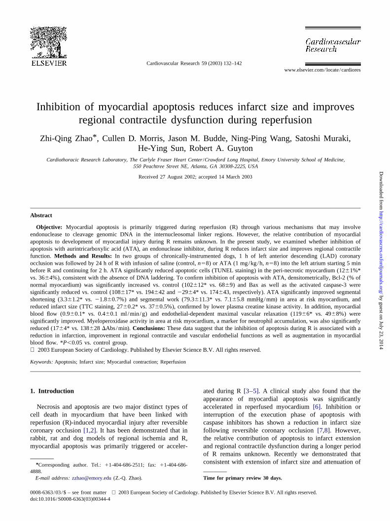
Cardiovascular Research 59 (2003) 132–142www.elsevier.com/ locate/cardiores
I nhibition of myocardial apoptosis reduces infarct size and improvesregional contractile dysfunction during reperfusion
*Zhi-Qing Zhao , Cullen D. Morris, Jason M. Budde, Ning-Ping Wang, Satoshi Muraki,He-Ying Sun, Robert A. Guyton
Cardiothoracic Research Laboratory, The Carlyle Fraser Heart Center /Crawford Long Hospital, Emory University School of Medicine,550 Peachtree Street NE, Atlanta, GA 30308-2225,USA
Received 27 August 2002; accepted 14 March 2003
Abstract
Objective: Myocardial apoptosis is primarily triggered during reperfusion (R) through various mechanisms that may involveendonuclease to cleavage genomic DNA in the internucleosomal linker regions. However, the relative contribution of myocardialapoptosis to development of myocardial injury during R remains unknown. In the present study, we examined whether inhibition ofapoptosis with aurintricarboxylic acid (ATA), an endonuclease inhibitor, during R reduces infarct size and improves regional contractilefunction. Methods and Results: In two groups of chronically-instrumented dogs, 1 h of left anterior descending (LAD) coronaryocclusion was followed by 24 h of R with infusion of saline (control,n58) or ATA (1 mg/kg/h,n58) into the left atrium starting 5 minbefore R and continuing for 2 h. ATA significantly reduced apoptotic cells (TUNEL staining) in the peri-necrotic myocardium (1261%*vs. 3664%), consistent with the absence of DNA laddering. To confirm inhibition of apoptosis with ATA, densitometrically, Bcl-2 (% ofnormal myocardium) was significantly increased vs. control (102612* vs. 6869) and Bax as well as the activated caspase-3 weresignificantly reduced vs. control (108617* vs. 194642 and22964* vs. 174643, respectively). ATA significantly improved segmentalshortening (3.361.2* vs. 21.860.7%) and segmental work (79.3611.3* vs. 7.165.8 mmHg/mm) in area at risk myocardium, andreduced infarct size (TTC staining, 2760.2* vs. 3760.5%), confirmed by lower plasma creatine kinase activity. In addition, myocardialblood flow (0.960.1* vs. 0.460.1 ml /min/g) and endothelial-dependent maximal vascular relaxation (11966* vs. 4968%) weresignificantly improved. Myeloperoxidase activity in area at risk myocardium, a marker for neutrophil accumulation, was also significantlyreduced (1764* vs. 138628 DAbs/min).Conclusions: These data suggest that the inhibition of apoptosis during R is associated with areduction in infarction, improvement in regional contractile and vascular endothelial functions as well as augmentation in myocardialblood flow. *P,0.05 vs. control group. 2003 European Society of Cardiology. Published by Elsevier Science B.V. All rights reserved.
Keywords: Apoptosis; Infarct size; Myocardial contraction; Reperfusion
1 . Introduction ated during R[3–5]. A clinical study also found that theappearance of myocardial apoptosis was significantly
Necrosis and apoptosis are two major distinct types of accelerated in reperfused myocardium[6]. Inhibition orcell death in myocardium that have been linked with interruption of the execution phase of apoptosis withreperfusion (R)-induced myocardial injury after reversible caspase inhibitors has shown a reduction in infarct sizecoronary occlusion[1,2]. It has been demonstrated that in following reversible coronary occlusion[7,8]. However,rabbit, rat and dog models of regional ischemia and R, the relative contribution of apoptosis to infarct extensionmyocardial apoptosis was primarily triggered or acceler- and regional contractile dysfunction during a longer period
of R remains unknown. Recently we demonstrated thatconsistent with extension of infarct size and attenuation of*Corresponding author. Tel.:11-404-686-2511; fax:11-404-686-
4888.E-mail address: [email protected](Z.-Q. Zhao). Time for primary review 30 days.
0008-6363/03/$ – see front matter 2003 European Society of Cardiology. Published by Elsevier Science B.V. All rights reserved.doi:10.1016/S0008-6363(03)00344-4
by guest on July 23, 2014http://cardiovascres.oxfordjournals.org/
Dow
nloaded from
133Z.-Q. Zhao et al. / Cardiovascular Research 59 (2003) 132–142
regional contractile function after coronary occlusion[9], sure regional myocardial blood flow. A plastic snare wasmyocardial apoptosis in the peri-necrotic myocardium placed below the first diagonal branch of LAD to reversib-progressively developed from early to late phase of R, ly produce a zone of regional ischemia in the left ventricle.suggesting that apoptosis may participate in exacerbation After 2 h of R, the thoracotomy incision was closed inof myocardial injury[10]. It is conceivable, therefore, that layers and a regimen of broad spectrum antibiotics andapoptotic cells detected from the peri-necrotic myocardium analgesia was initiated throughout the R period.may undergo secondary necrosis, and that the extension ofinfarction may in part be elicited from the delayed 2 .2. General experimental protocolapoptosis during R. If the end stage of R injury ismodulated by apoptosis, is there any ‘window of oppor- Dogs were randomly divided into two groups. Eithertunity’ that intervention can stop this suicide program? saline (control,n58) or aurintricarboxylic acid (ATA fromDoes the inhibition of apoptosis during R lessen extension Sigma, 1 mg/kg/h,n58) was infused through the leftof infarction and improve regional contractile function atrium at a rate of 1 ml /min beginning 5 min before R andafter myocardial ischemia? Since apoptosis represents a ending 2 h after R. All dogs underwent 1 h of LADpotentially preventable form of cell death due to its active coronary occlusion and 24 h of R. In both groups,and regulated natures, it is likely that inhibition of apop- hemodynamic and regional contractile function were mea-tosis may help to reduce myocardial injury during R. sured at baseline, ischemia, 2 h and 24 h of R. At the end
Aurintricarboxylic acid (ATA), an endonuclease inhib- of the experiment, segments of the LAD and left circum-itor known to inhibit apoptosis from in vivo and in vitro flex (LCX) coronary arteries were isolated and used tostudies[11–14], has shown a profound effect on infarct evaluate agonist-stimulated vascular endothelial response.size in gerbils when this compound was intraventricularly Tissue samples from the non-ischemic and ischemic zonesadministered before transient brain ischemia[12]. The were used to quantify myocardial apoptosis, infarct size,purpose of this study, therefore, was to use ATA as a tool myocardial blood flow and neutrophil accumulation (seeby its potent anti-apoptotic character to test whether below). An additional six dogs were used as sham controlexogenous ATA limits the extension of infarct size and to observe the effect of ATA on the heart after surgery. Inimproves regional contractile and endothelial function in a these animals, ATA was administered at a dose of 1dog model following extended R. mg/kg/h for 2 h and the chest was closed for 24 h after
completing surgical procedures without coronary occlu-sion.
2 . Methods2 .3. Tissue preparation
2 .1. Surgical preparation of animalsMyocardial tissues (150–200 mg for each) from non-
Studies were conducted according to the guidelines of ischemic and ischemic zones were kept in tubes for DNAthe committee on the Animal Welfare Act and Emory isolation and Western blot assay. For detection of apop-University Veterinary policies and theGuide for the Care tosis, the transmural ischemic tissue was embedded inand Use of Laboratory Animals of the Institute of Labora- optimal cutting temperature compound (O.C.T., Sakuratory Animal Resources, National Council Department of Finetek), frozen in liquid nitrogen and then stored atHealth and Human Services publication no. [NIH] 85-23, 270 8C. Cryosections (7mm thick) from frozen tissuerevised 1985. were then obtained using a Hacker–Bright cryostat and
Dogs of either sex were used in the study. All animals thaw-mounted onto Vectabond (Vector Laboratories, Burl-were initially anesthetized with intramuscular morphine ingame, CA, USA) coated slides or Fisher-Plus (Fishersulfate (4 mg/kg). A bolus injection of pentothal (20 Scientific) slides, refrozen, and stored at270 8C withmg/kg) was given followed by continuous inhalation of desiccant until use.isoflurane (0.5–2% in oxygen) after endotracheal intuba-tion. A left lateral thoracotomy was performed, and 2 .4. The detection of myocardial apoptosis by terminalmicromanometer pressure transducers were inserted intodeoxynucleotidyl transferase (TdT)-mediated dUTP-Xthe left internal mammalian artery and ventricle to monitor nick end labeling (TUNEL)aortic and ventricular pressure. A pair of ultrasonic crystalswas implanted in the anterior-midmyocardium to measure The TUNEL signal in the tissue sections were de-regional contractile function. A cuff-type flow probe termined using an in situ cell death detection kit (Boeh-(Pulsed Doppler Flowmeter, Triton Technology, San ringer Mannheim, Ridgefield, CT, USA) as describedDiego, CA, USA) was placed below the first diagonal previously[4]. Briefly, DNA fragments were detected bybranch of LAD and the ligating snare for measurement of an anti-fluorescein antibody conjugated with alkaline phos-coronary artery flow[15]. A catheter was inserted into the phatase, a reporter enzyme that catalytically generates aleft atrium for injection of colored microspheres to mea- red-colored product from Vector Red substrate. The slides
by guest on July 23, 2014http://cardiovascres.oxfordjournals.org/
Dow
nloaded from
Z.-Q. Zhao et al. / Cardiovascular Research 59 (2003) 132–142134
were counterstained with hematoxylin, dehydrated in area at risk tissue. The gravimetric method was used tograded alcohols and covered with Accumount medium. For quantify infarct size[3].each slide, color video images of 10 separate fields werecaptured randomly and digitized by using a320 objective 2 .8. Plasma creatine kinase (CK) activityon an Olympus IX50 microscope connected to an IBMcomputer. The apoptotic cells were calculated as per- Arterial blood samples were withdrawn at baseline, atcentage of TUNEL-positive cells from total cell nuclei[3]. the end of ischemia, 2 and 24 h of R to measure CK
activity. The plasma was drawn off after centrifugation andCK activity was analyzed spectrophotometrically accord-2 .5. The presence of DNA ladder in agarose geling to the method of Rosalki (Sigma Diagnostics). PlasmaelectrophoresisCK activity was expressed as IU/mg protein[3].
Appearance of a DNA ladder was detected usingagarose gel electrophoresis as described previously[3]. 2 .9. Determination of regional myocardial blood flowBriefly, frozen tissue samples from non-ischemic andischemic zones were minced in lysis buffer and were Colored microspheres were used to quantify myocardialquickly homogenized with a microfuge tube pestle. The blood flow. Samples of non-ischemic, ischemic subepicar-tissue was digested with proteinase K and incubated with dial and subendocardial myocardium were placed in taredRNase A. Supernatants containing DNA were precipitated vials marked according to their anatomical location. Tis-with isopropanol and the resulting DNA pellets after sues and reference blood samples were analyzed in acentrifugation were washed with ethanol and dissolved in Spectra Max Spectrophotometer (Molecular Devices) asDNA hydration solution. DNA electrophoresis was carried reported previously[9]. Results are expressed as ml /min/gout and DNA ladders were visualized under ultraviolet tissue.light.
2 .9.1. Postischemic vascular ring reactivityLAD and LCX coronary artery segments were isolated2 .6. Western blot analysis of Bcl-2, Bax and caspase-3
and placed into Radnoti tissue baths. After the coronaryproteinsrings were preconstricted with thromboxane A -mimetic2
U46619, and subsequently dilated in concentration–re-Determination of Bcl-2, Bax and caspase-3 proteins bysponse fashion with the endothelium-dependent vasodilat-Western blot assay was primarily described previously[3].or, acetylcholine, and the endothelium-independent vasodi-Briefly, tissue samples from non-ischemic and ischemiclator, sodium nitroprusside (SNP) in incremental con-zones were homogenized with lysis buffer and proteincentrations. Responses to vasodilators were analyzed usingconcentration was measured by the DC Protein Assay (Bioa videographics program developed in our laboratory[9].Rad). Proteins (80mg) for each lane were mixed with
loading buffer, and loaded onto gradient SDS–poly-acrylamide gel. Proteins kept in the nitrocellulose filter 2 .9.2. Measurement of myeloperoxidase activity (MPO)after transfer were blocked with milk and the membranes in cardiac tissuewere then exposed to antibodies (Santa Cruz, CA, USA) Tissue samples from non-ischemic and ischemic zonesincluding rabbit polyclonal anti-human Bcl-2 (DC21), were homogenized. After centrifugation, the supernatantsrabbit polyclonal anti-human Bax (P19) and rabbit anti- were decanted and mixed with O-dianisodine dihydroch-human caspase-3 (H277), respectively. Bound antibody loride and H O in phosphate buffer. The change in2 2was detected by horseradish peroxidase conjugated anti-absorbance was measured spectrophotometrically at 460rabbit IgG. Finally, ECL detection reagents were employed nm. MPO activity was expressed asDabsorbance units /to visualize peroxidase reaction product. The density at min [9].specific molecular weights was measured by NIH imageanalysis software on a Power Macintosh.
2 .9.3. Statistical analysisConcentration response curves of vascular relaxation
2 .7. Determination of area at risk and infarct size were calculated as a percentage of U46619-induced in-crease in isometric force. Student’st-test was used to
At the end of the experiment, Unisperse blue dye was analyze differences between such parameters as vascularinjected into the aortic root to stain the normally perfused responses, MPO, myocardial blood flow and infarct sizeregion blue and outline the area at risk. The area at risk data. Hemodynamic, regional contractile function andwas then separated from the non-ischemic zone and other time-dependent determinations were analyzed byincubated in a solution of 1% triphenyltetrazolium chloride repeated analysis of variance. AP value less than 0.05 was(TTC) to differentiate necrotic (pale) from non-necrotic accepted as statistically significant.
by guest on July 23, 2014http://cardiovascres.oxfordjournals.org/
Dow
nloaded from
135Z.-Q. Zhao et al. / Cardiovascular Research 59 (2003) 132–142
3 . Results
3 .1. Time course of hemodynamic changes
Hemodynamic data for heart rate (HR), mean aorticpressure (MAP), left ventricular systolic pressure (LVSP),dP/dt , left ventricular end-diastolic pressure (LVEDP),max
and LAD coronary artery blood flow (CBF) in all groupsare shown inTable 1.There were no differences betweengroups for any of the hemodynamic parameters at baseline.Coronary occlusion caused a significant increase in HR. At24 h of R, HR tended to be less in the ATA group, but itdid not reach a significant difference compared with thecontrol group. MAP in the control group at 24 h of R wassignificantly reduced, however, it remained consistentthroughout the procedure in the ATA group. There were nogroup differences in LVSP, LVEDP, positive and negativedp/dt during the course of experiment. ATA treatmentsignificantly increased LAD blood flow compared with thecontrol group during 2 h of R. Surgical operation withoutcoronary occlusion did not induce hemodynamic change inthe sham group.
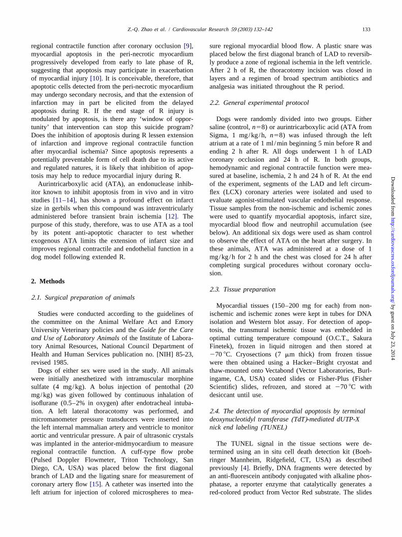
3 .2. Regional contractile function Fig. 1. Examples of instantaneous LV pressure–segment length loops fora representative experiment in control (A) and ATA (B) groups. ATAtreatment significantly improved systolic shortening at the end of reperfu-Regional contractile function data for systolic shorteningsion (R). Segmental work was calculated as the integral of each pressure–(SS) and segmental work (SW) in the area at risk arelength loop. CNTL, baseline time point; ISCH, end of ischemia; REP, 24shown inTable 1and Fig. 1. There was no difference onh of R.
any measured parameters at baseline. During ischemia,paradoxical systolic expansion was exhibited in bothgroups with a rightward shift in the pressure–length the entire period of R in the control group. However,relationship. There was a significant decline in SS and SW. significant recovery in SS and SW was detected in theAt 2 h of R, SS and SW in the ATA group tended to be ATA group at 24 h of R, suggesting an improvement ingreater than that in the control group, but it did not reach regional contractile function. No change in regional con-significance. Wall motion remained dyskinetic throughout tractile function was found in the sham group.
T able 1Hemodynamics and regional contractile function during the course of the experiment
Time Group Parameters
HR MAP LVSP LVEDP 1dp/dt 2dp/dt CBF SS SW
Baseline Control 8768 9466 10166 862 1473682 21413684 1661 15.861.2 148618ATA 8663 10564 10963 1061 15356186 21459650 1361 18.261.1 18669Sham 8766 9766 10166 961 1452668 21405654 1462 15.161.2 139614
Ischemia Control 10968* 10165 10366 1362 1443661 21432691 – 22.360.6* 32.669.6*ATA 10464* 10064 10563 1661 1459626 21367651 – 20.260.4* 4165.7Sham 8667 9867 10265 962 1443659 21402682 1463 15.261.1 139613
R2h Control 11466* 8463 9064 962 1369666 21280693 1461 21.560.4* 1664.6*†ATA 11164* 9563 9563 1362 1374660 21300662 2261* 1.360.9* 6369*
Sham 8968 9967 10165 1062 1449649 21414667 1561 15.660.8 140611
R24h Control 13167* 7762* 8464 962 1176104 210886104 761* 21.860.8* 7.3165.8*† † † †ATA 11966* 9563 9865 1261 1242692 212596105 1261 3.361.2* 79.3611.3*
Sham 9969 10167 10366 962 1467661 21441668 1662 14.660.8 135.12
HR, heart rate (beats /min); MAP, mean aortic pressure (mmHg); LVSP, left ventricular peak systolic pressure (mmHg); LVEDP, left ventricular enddiastolic pressure (mmHg); positive dp/dt (mmHg/s); negative dp/dt (mmHg/s); CBF, mean LAD flow (ml /min); SS, systolic shortening (%); SW,segmental work (mmHg/mm); R2h and R6h, 2 and 6 h of reperfusion, respectively. Sham (n56): the chest was closed for 24 h after surgical procedures
†with coronary occlusion. All values are expressed as mean6S.E.M. *P,0.05 vs. baseline value;P,0.05 vs. control group.
by guest on July 23, 2014http://cardiovascres.oxfordjournals.org/
Dow
nloaded from
Z.-Q. Zhao et al. / Cardiovascular Research 59 (2003) 132–142136
4064, respectively, in the control group, suggesting anextension of infarct size during R. However, CK activitywas significantly attenuated in the ATA group at thesetime points with 1061 and 1962,P,0.01, respectively.There was no difference in plasma CK activity after thechest was closed (1.263) for 24 h compared with baselinevalue (0.862) in the sham group.
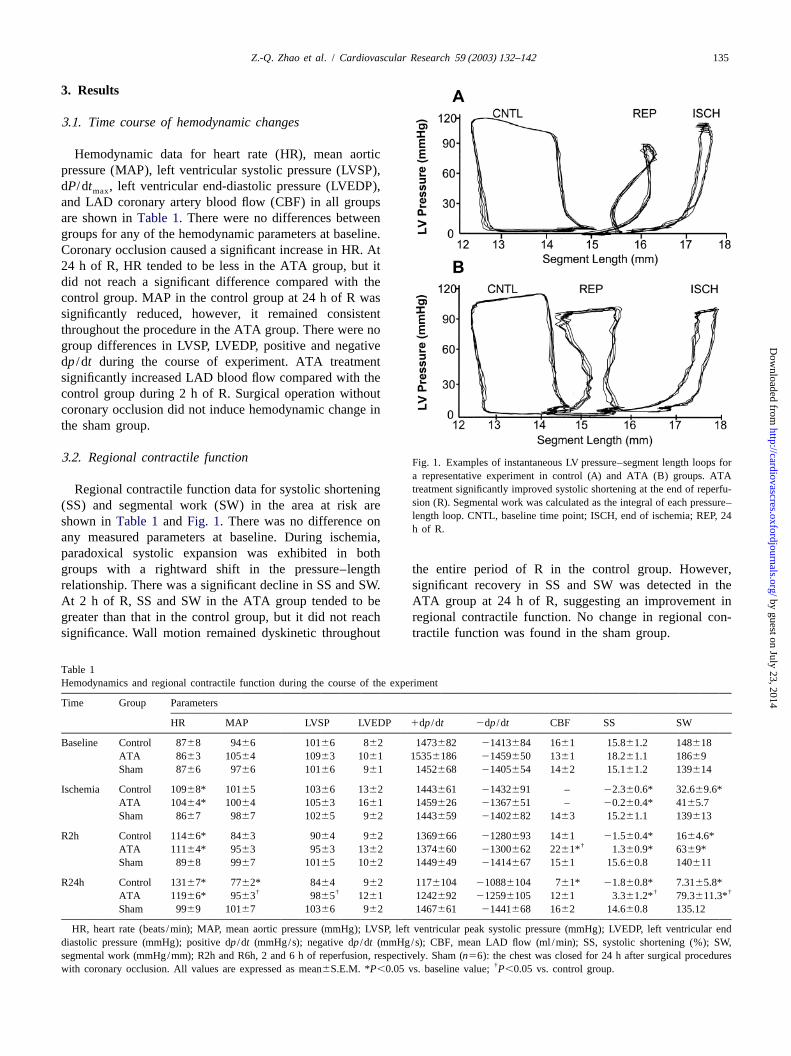
3 .5. Detection of TUNEL-positive cells in the peri-necrotic myocardium
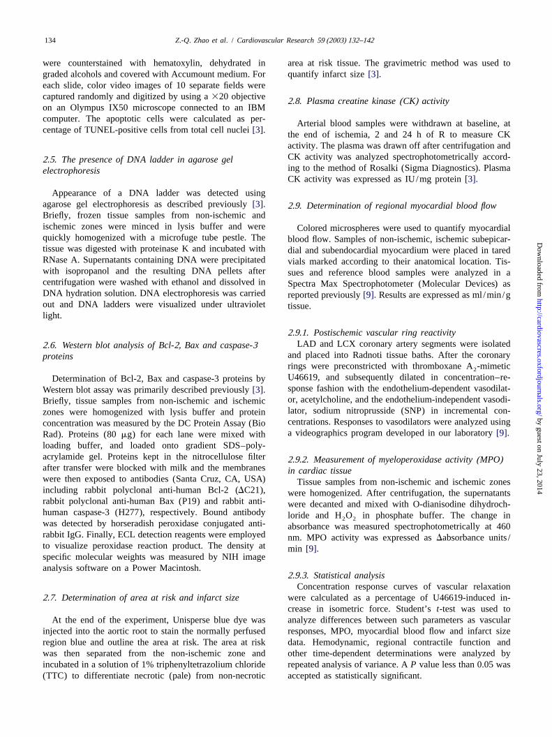
No TUNEL-positive cells were found in the non-is-Fig. 2. The area at risk relative to the left ventricle (Ar /LV) and infarctsize relative to the area at risk (An/Ar) after 1 h ischemia followed by 24 chemic zone in either group. As shown inFig. 3, however,h of R. ATA treatment significantly reduced infarct size after 24 h of R the number of TUNEL-positive cells expressed as acompared with the control group. Bars represent group mean; bracketspercent of total normal nuclei was significantly increasedindicate S.E.M. *P,0.01 vs. control.
in the peri-necrotic zone in the control group (3664%,Fig. 3B) relative to the non-ischemic zone, which was
3 .3. Reduction in extension of necrosis consistent with the appearance and increased intensity ofDNA ladders in this group (see below). ATA administered
The area placed at risk by coronary occlusion expressed at R significantly reduced TUNEL-positive cells in theas a percent of the left ventricular mass (Ar /LV), and the peri-necrotic zone (1261%) versus the control group (P,area of necrosis expressed as a percent of the area at risk 0.01), consistent with disappearance of DNA laddering in(An/Ar) are shown inFig. 2.Ar /LV was comparable in the this group (see below). No clear TUNEL-positive stainingtwo groups ranging between 25–28% in the control group was detected in the sham group.and 26–29% in the ATA group. Administration of ATA atR significantly reduced An/Ar by 33% compared with that 3 .6. DNA fragmentationin the control group at 24 h of R. No necrosis was detectedin the sham group. DNA nucleosomal fragmentation of myocyte in non-
ischemic and peri-necrotic zones in both groups is shown3 .4. Creatine kinase activity during the course of the in Fig. 4. No visible DNA ‘ladders’ were found in theexperiment non-ischemic zone in either group. In contrast, genomic
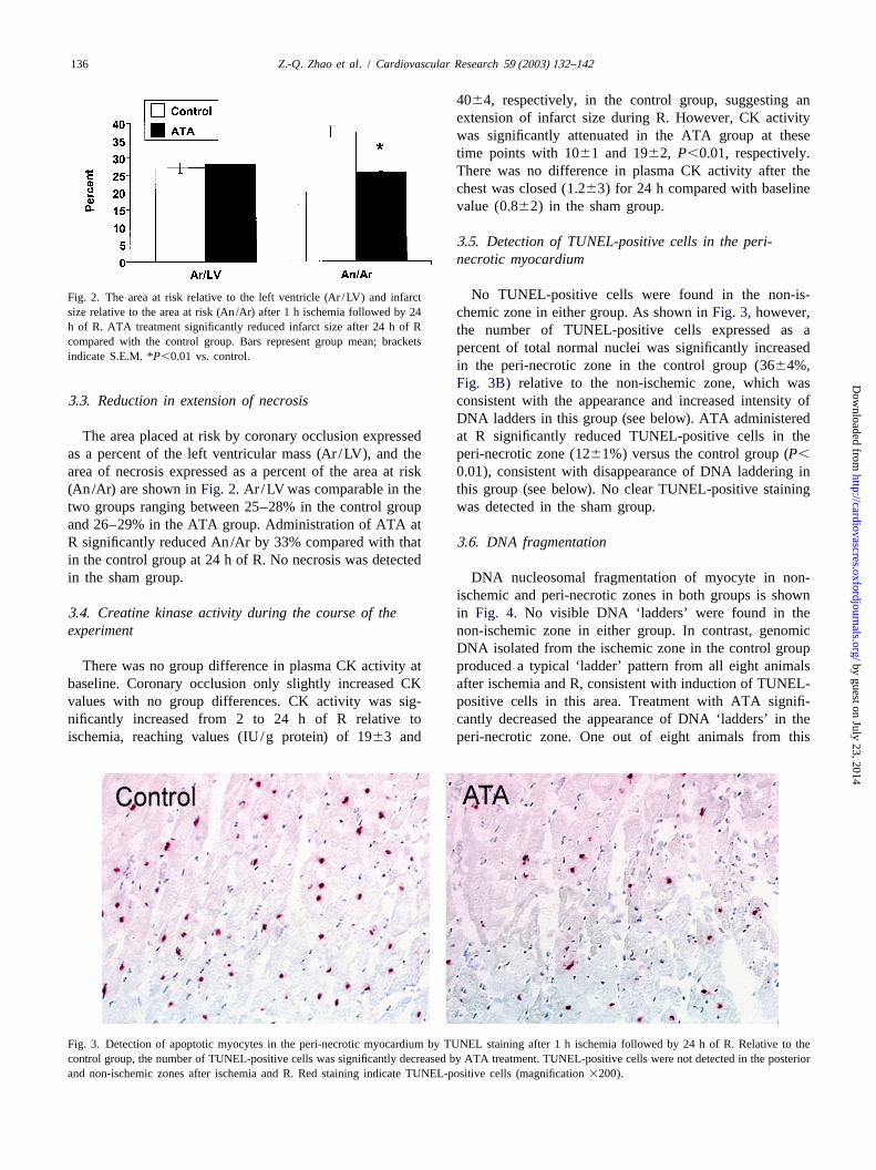
DNA isolated from the ischemic zone in the control groupThere was no group difference in plasma CK activity at produced a typical ‘ladder’ pattern from all eight animals
baseline. Coronary occlusion only slightly increased CK after ischemia and R, consistent with induction of TUNEL-values with no group differences. CK activity was sig- positive cells in this area. Treatment with ATA signifi-nificantly increased from 2 to 24 h of R relative to cantly decreased the appearance of DNA ‘ladders’ in theischemia, reaching values (IU/g protein) of 1963 and peri-necrotic zone. One out of eight animals from this
Fig. 3. Detection of apoptotic myocytes in the peri-necrotic myocardium by TUNEL staining after 1 h ischemia followed by 24 h of R. Relative to thecontrol group, the number of TUNEL-positive cells was significantly decreased by ATA treatment. TUNEL-positive cells were not detected in the posteriorand non-ischemic zones after ischemia and R. Red staining indicate TUNEL-positive cells (magnification3200).
by guest on July 23, 2014http://cardiovascres.oxfordjournals.org/
Dow
nloaded from
137Z.-Q. Zhao et al. / Cardiovascular Research 59 (2003) 132–142
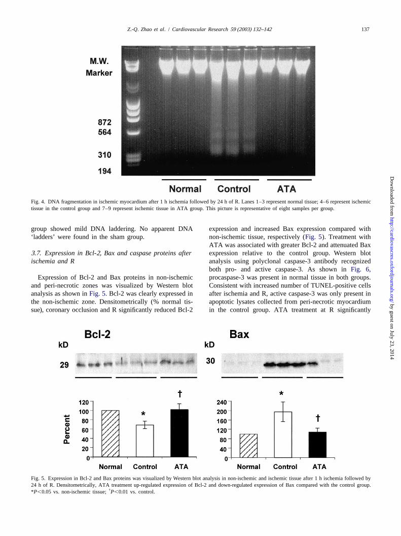
Fig. 4. DNA fragmentation in ischemic myocardium after 1 h ischemia followed by 24 h of R. Lanes 1–3 represent normal tissue; 4–6 represent ischemictissue in the control group and 7–9 represent ischemic tissue in ATA group. This picture is representative of eight samples per group.
group showed mild DNA laddering. No apparent DNA expression and increased Bax expression compared with‘ladders’ were found in the sham group. non-ischemic tissue, respectively (Fig. 5). Treatment with
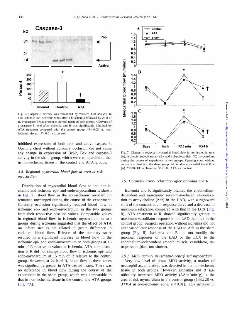
ATA was associated with greater Bcl-2 and attenuated Bax3 .7. Expression in Bcl-2, Bax and caspase proteins after expression relative to the control group. Western blotischemia and R analysis using polyclonal caspase-3 antibody recognized
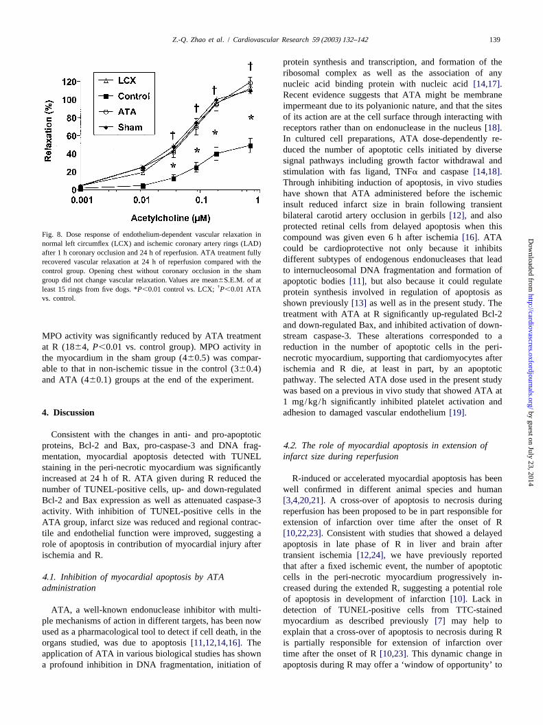
both pro- and active caspase-3. As shown inFig. 6,Expression of Bcl-2 and Bax proteins in non-ischemic procaspase-3 was present in normal tissue in both groups.
and peri-necrotic zones was visualized by Western blot Consistent with increased number of TUNEL-positive cellsanalysis as shown inFig. 5.Bcl-2 was clearly expressed in after ischemia and R, active caspase-3 was only present inthe non-ischemic zone. Densitometrically (% normal tis- apoptotic lysates collected from peri-necrotic myocardiumsue), coronary occlusion and R significantly reduced Bcl-2 in the control group. ATA treatment at R significantly
Fig. 5. Expression in Bcl-2 and Bax proteins was visualized by Western blot analysis in non-ischemic and ischemic tissue after 1 h ischemia followed by24 h of R. Densitometrically, ATA treatment up-regulated expression of Bcl-2 and down-regulated expression of Bax compared with the control group.
†*P,0.05 vs. non-ischemic tissue;P,0.01 vs. control.
by guest on July 23, 2014http://cardiovascres.oxfordjournals.org/
Dow
nloaded from
Z.-Q. Zhao et al. / Cardiovascular Research 59 (2003) 132–142138
Fig. 6. Caspase-3 activity was visualized by Western blot analysis innon-ischemic and ischemic zones after 1 h ischemia followed by 24 h ofR. Procaspase-3 was present in normal tissue in both groups. Cleavage ofprocaspase-3 level after ischemia and R was significantly inhibited byATA treatment compared with the control group. *P,0.05 vs. non-
†ischemic tissue;P,0.01 vs. control.
inhibited expression of both pro- and active caspase-3.Opening chest without coronary occlusion did not cause
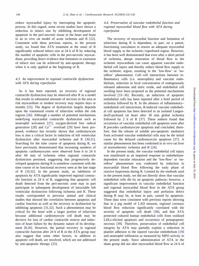
Fig. 7. Change in regional myocardial blood flow in non-ischemic zoneany change in expression of Bcl-2, Bax and caspase-3(A), ischemic subepicardial- (B) and subendocardial- (C) myocardiumactivity in the sham group, which were comparable to thatduring the course of experiment in two groups. Opening chest without
in non-ischemic tissue in the control and ATA groups. coronary occlusion in the sham group did not alter myocardial blood flow†(A). *P,0.001 vs. baseline.P,0.05 ATA vs. control.
3 .8. Regional myocardial blood flow at area at riskmyocardium
3 .9. Coronary artery relaxation after ischemia and RDistribution of myocardial blood flow to the non-is-
chemic and ischemic epi- and endo-myocardium is shown Ischemia and R significantly blunted the endothelium-in Fig. 7. Blood flow in the non-ischemic myocardium dependent and muscarinic receptor-mediated vasorelaxa-remained unchanged during the course of the experiment. tion to acetylcholine (Ach) in the LAD, with a rightwardCoronary occlusion significantly reduced blood flow in shift of the concentration–response curve and a decrease inischemic epi- and endo-myocardium in the two groups maximum relaxation compared with that in the LCX (Fig.from their respective baseline values. Comparable values8). ATA treatment at R showed significantly greater inin regional blood flow in ischemic myocardium in two maximum vasodilator response in the LAD than that in thegroups during ischemia suggested that the effect of ATA control group. Surgical operation without ischemia did noton infarct size is not related to group difference in alter vasodilator response of the LAD to Ach in the shamcollateral blood flow. Release of the coronary snare group (Fig. 8). Ischemia and R did not modify theresulted in a significant increase in blood flow in the maximal responses of the LAD or the LCX to theischemic epi- and endo-myocardium in both groups at 15 endothelium-independent smooth muscle vasodilator, ni-min of R relative to values at ischemia. ATA administra- troprusside (data not shown).tion at R did not change blood flow in ischemic epi- andendo-myocardium at 15 min of R relative to the control 3 .9.1. MPO activity in ischemic /reperfused myocardiumgroup. However, at 24 h of R, blood flow in these zones Very low level of tissue MPO activity, a marker ofwas significantly greater in ATA-treated hearts. There was neutrophil accumulation, was detected in the non-ischemicno difference in blood flow during the course of the tissue in both groups. However, ischemia and R sig-experiment in the sham group, which was comparable to nificantly increased MPO activity (DAbs/min/g) in thethat in non-ischemic tissue in the control and ATA groups area at risk myocardium in the control group (138628 vs.(Fig. 7A). 360.4 in non-ischemic zone,P,0.01). This increase in
by guest on July 23, 2014http://cardiovascres.oxfordjournals.org/
Dow
nloaded from
139Z.-Q. Zhao et al. / Cardiovascular Research 59 (2003) 132–142
protein synthesis and transcription, and formation of theribosomal complex as well as the association of anynucleic acid binding protein with nucleic acid[14,17].Recent evidence suggests that ATA might be membraneimpermeant due to its polyanionic nature, and that the sitesof its action are at the cell surface through interacting withreceptors rather than on endonuclease in the nucleus[18].In cultured cell preparations, ATA dose-dependently re-duced the number of apoptotic cells initiated by diversesignal pathways including growth factor withdrawal andstimulation with fas ligand, TNFa and caspase[14,18].Through inhibiting induction of apoptosis, in vivo studieshave shown that ATA administered before the ischemicinsult reduced infarct size in brain following transientbilateral carotid artery occlusion in gerbils[12], and alsoprotected retinal cells from delayed apoptosis when this
Fig. 8. Dose response of endothelium-dependent vascular relaxation incompound was given even 6 h after ischemia[16]. ATAnormal left circumflex (LCX) and ischemic coronary artery rings (LAD)
could be cardioprotective not only because it inhibitsafter 1 h coronary occlusion and 24 h of reperfusion. ATA treatment fullydifferent subtypes of endogenous endonucleases that leadrecovered vascular relaxation at 24 h of reperfusion compared with theto internucleosomal DNA fragmentation and formation ofcontrol group. Opening chest without coronary occlusion in the sham
group did not change vascular relaxation. Values are mean6S.E.M. of at apoptotic bodies[11], but also because it could regulate†least 15 rings from five dogs. *P,0.01 control vs. LCX; P,0.01 ATA protein synthesis involved in regulation of apoptosis as
vs. control.shown previously[13] as well as in the present study. Thetreatment with ATA at R significantly up-regulated Bcl-2and down-regulated Bax, and inhibited activation of down-
MPO activity was significantly reduced by ATA treatment stream caspase-3. These alterations corresponded to aat R (1864, P,0.01 vs. control group). MPO activity in reduction in the number of apoptotic cells in the peri-the myocardium in the sham group (460.5) was compar- necrotic myocardium, supporting that cardiomyocytes afterable to that in non-ischemic tissue in the control (360.4) ischemia and R die, at least in part, by an apoptoticand ATA (460.1) groups at the end of the experiment. pathway. The selected ATA dose used in the present study
was based on a previous in vivo study that showed ATA at1 mg/kg/h significantly inhibited platelet activation and
4 . Discussion adhesion to damaged vascular endothelium[19].
Consistent with the changes in anti- and pro-apoptoticproteins, Bcl-2 and Bax, pro-caspase-3 and DNA frag- 4 .2. The role of myocardial apoptosis in extension ofmentation, myocardial apoptosis detected with TUNEL infarct size during reperfusionstaining in the peri-necrotic myocardium was significantlyincreased at 24 h of R. ATA given during R reduced the R-induced or accelerated myocardial apoptosis has beennumber of TUNEL-positive cells, up- and down-regulated well confirmed in different animal species and humanBcl-2 and Bax expression as well as attenuated caspase-3[3,4,20,21].A cross-over of apoptosis to necrosis duringactivity. With inhibition of TUNEL-positive cells in the reperfusion has been proposed to be in part responsible forATA group, infarct size was reduced and regional contrac- extension of infarction over time after the onset of Rtile and endothelial function were improved, suggesting a [10,22,23].Consistent with studies that showed a delayedrole of apoptosis in contribution of myocardial injury after apoptosis in late phase of R in liver and brain afterischemia and R. transient ischemia[12,24], we have previously reported
that after a fixed ischemic event, the number of apoptotic4 .1. Inhibition of myocardial apoptosis by ATA cells in the peri-necrotic myocardium progressively in-administration creased during the extended R, suggesting a potential role
of apoptosis in development of infarction[10]. Lack inATA, a well-known endonuclease inhibitor with multi- detection of TUNEL-positive cells from TTC-stained
ple mechanisms of action in different targets, has been now myocardium as described previously[7] may help toused as a pharmacological tool to detect if cell death, in the explain that a cross-over of apoptosis to necrosis during Rorgans studied, was due to apoptosis[11,12,14,16].The is partially responsible for extension of infarction overapplication of ATA in various biological studies has shown time after the onset of R[10,23]. This dynamic change ina profound inhibition in DNA fragmentation, initiation of apoptosis during R may offer a ‘window of opportunity’ to
by guest on July 23, 2014http://cardiovascres.oxfordjournals.org/
Dow
nloaded from
Z.-Q. Zhao et al. / Cardiovascular Research 59 (2003) 132–142140
reduce myocardial injury by interrupting the apoptotic 4 .4. Preservation of vascular endothelial function andprocess. In this regard, some recent studies have shown aregional myocardial blood flow with ATA duringreduction in infarct size by inhibiting development of reperfusionapoptosis in the peri-necrotic tissue in the heart and brainin an in vivo rat model of acute ischemia and R[22]. The recovery of myocardial function and limitation ofConsistent with these previous reports, in the present infarction during R is dependent, in part, on a patent,study, we found that ATA treatment at the onset of R functioning vasculature to restore an adequate myocardialsignificantly reduced infarct size at 24 h of R by reducing blood supply to the ischemic/ reperfused region. However,the number of apoptotic cells in the peri-necrotic myocar- it has been well demonstrated that even after a short perioddium, providing direct evidence that limitation in extension of ischemia, abrupt restoration of blood flow to theof infarct size can be achieved by anti-apoptotic therapy ischemic myocardium can cause apparent vascular endo-when it is only applied at the beginning of R. thelial cell injury and thereby reduce blood flow supply to
the ischemic region, resulting in the ‘low-flow’ or ‘no-reflow’ phenomenon. Cell–cell interactions between in-
4 .3. An improvement in regional contractile dysfunction flammatory cells (i.e. neutrophils) and vascular endo-with ATA during reperfusion thelium, reduction in local concentration of endogenously
released adenosine and nitric oxide, and endothelial cellAs it has been reported, no recovery of regional swelling have been proposed as the potential mechanisms
contractile dysfunction may be observed after R in a model involved [33–36]. Recently, an induction of apoptoticthat results in substantial amounts of necrosis in the area atendothelial cells has been reported after a brief period ofrisk myocardium or modest recovery may require days or ischemia followed by R. In the absence of inflammatory–months [25]. The degree of dysfunction largely depends endothelial cell interactions, R-induced vascular endotheli-upon the transmural extent of infarction within the risk al cell apoptosis has been detected in an isolated Langen-regions[26]. Although a number of potential mechanisms dorff-perfused rat heart after 30 min global ischemiaunderlying myocardial contractile dysfunction such as followed by 2 h of R [37]. These authors found thatneutrophil activation [27], oxygen-derived free radical occurrence of vascular endothelial cell apoptosis soon after
21generation[28] and Ca overload[29] have been pro- R precedes cardiomyocyte apoptosis and concluded, there-posed, evidence has recently shown that cardiomyocyte fore, that the release of soluble pro-apoptotic mediatorsloss is also a critical factor in induction of left ventricular from activated vascular endothelial cells may be the initialdysfunction after myocardial ischemia and R[30,31]. cause for the delayed cardiomyocyte apoptosis[37]. ASearching for the time course of apoptosis during R, we similar phenomenon has been confirmed in in vivo rat liverhave previously demonstrated that increasing numbers of of normothermic ischemia and R[24].apoptotic cardiomyocytes were observed from 6 to 24 h In the present study, the vascular endothelial cell injuryafter 60 min of ischemia when regional contractile was manifested as an impaired response to endothelium-dysfunction persisted, suggesting that progressively de- dependent vascular relaxation and the ‘low-flow’ or ‘no-veloped apoptosis during R is somehow consistent with the reflow’ phenomenon was confirmed by reduction intime course of no functional recovery seen at the late stage myocardial blood flow following the early phase ofof R [10,32]. In the present study, an inhibition of reactive hyperemia during R. Limited by the methods usedapoptosis by ATA significantly improved regional contrac- in the present study, we did not directly show that vasculartile function at 24 h of R, suggesting that apoptotic cell endothelial cells die by an apoptotic pathway; however, adeath detected from the peri-necrotic zone may in part significant improvement in vascular endothelial functionparticipate in subsequent development of intractable left and regional myocardial blood flow in the ATA groupventricular dysfunction following ischemia and R. These suggested that endothelial injury and perfusion defectresults corresponded to previous animal and clinical during R may be, at least in part, caused by apoptosis.studies that showed the correlation between apoptosis and These data were consistent with previous reports showingcardiac function as well as the recovery in dysfunction by that in a pig model of LAD stenosis, regional coronaryinhibiting apoptosis[31,32]. Such a phenomenon may be blood flow reduction significantly correlated with thecritical for the heart with a larger portion of infarction severity of apoptotic cell death[38], and that ATAbecause additional cardiomyocyte cell death may be protected cultured human endothelial cells from oxidizeddecisive for loss of cardiac contractile reserve and induc- LDLs-elicited apoptosis and occurrence of postapoptotiction of heart failure by the dynamic nature of its develop- necrosis[39]. Therefore, preservation of endothelial cellment [8,26]. However, the partial recovery in regional integrity by ATA may partially explain a reduction incontractile function after 24 h of R in the ATA group may platelet adhesion to the injured vascular endothelium[19]also suggest that some other factors, in addition to and neutrophil infiltration as measured by MPO activity inapoptotic cell death, are involved, which are not addressed the present study. Since administration of ATA in theby anti-apoptotic therapy[25]. sham group did not alter myocardial blood flow at 24 h of
by guest on July 23, 2014http://cardiovascres.oxfordjournals.org/
Dow
nloaded from
141Z.-Q. Zhao et al. / Cardiovascular Research 59 (2003) 132–142
Attenuation of ischemia/ reperfusion injury in rats by a caspaseR, we could exclude a possibility that an improvement ininhibitor. Circulation 1998;97:276–281.myocardial blood flow at this time point by ATA was
[8] M ocanu MM, Baxter GF, Yellon DM. Caspase inhibition andinduced by a direct vasodilatory effect. However, future limitation of myocardial infarct size: protection against lethalstudies to elucidate potential mechanisms underlying an reperfusion injury. Br J Pharmacol 2000;130:197–200.inhibition of inflammatory and endothelial cell–cell inter- [9] Z hao Z-Q, Nakamura M, Wang N-P et al. Dynamic progression of
contractile and endothelial dysfunction and infarct extension in theactions as well as a correlation of improvement in myocar-late phase of reperfusion. J Surg Res 2000;94:1–12.dial blood flow with recovery in regional contractile
[10] Z hao Z-Q, Velez DA, Wang N-P et al. Progressively developedfunction and reduction in infarct size at the late stage of Rmyocardial apoptotic cell death during late phase of reperfusion.
by ATA are needed. Apoptosis 2001;6:279–290.[11] L am TT, Fu J, Hrynewycz M et al. The effect of aurintricarboxylic
acid, an endonuclease inhibitor, on ischemia/ reperfusion damage inrat retina. J Ocul Pharmacol Ther 1995;11:253–259.
5 . Conclusion [12] R osenbaum DM, D’Amore J, Llena J et al. Pretreatment withintraventricular aurintricarboxylic acid decreases infarct size byinhibiting apoptosis following transient global ischemia in gerbils.In summary, this study demonstrated that treatment withAnn Neurol 1998;43:654–660.ATA at R significantly reduced infarct size, preserved
[13] M esner Jr. PW, Bible KC, Martins LM et al. Characterization ofregional contractile and vascular endothelial cell function, caspase processing and activation in HL-60 cell cytosol underand improved supply of myocardial blood flow to the cell-free conditions. J Biol Chem 1999;274:22635–22645.ischemic myocardium after coronary occlusion, supporting [14] A ndrew DJ, Hay AWM, Evans SW. Aurintricarboxylic acid inhibits
apoptosis and supports proliferation in a haemopoietic growth-factorthat apoptotic cell death may be involved in exacerbationdependent myeloid cell line. Immunopharmacology 1999;41:1–10.of myocardial injury after ischemia and R. These results
[15] L axson DD, Homans DC, Dai X-Z et al. Oxygen consumption andalso provide evidence that in clinical practice, the treat-coronary reactivity in postischemic myocardium. Circ Res
ment for patients with thrombolytic therapy, percutaneous 1989;64:9–20.transluminal coronary angioplasty or coronary artery by- [16] R osenbaum DM, Rosenbaum PS, Gupta A et al. Retinal ischemia
leads to apoptosis which is ameliorated by aurintricarboxylic acid.pass graft surgery may thus need to consider an applicationVision Res 1997;37:3445–3451.of anti-apoptotic therapy in order to minimize total cell
[17] H eiduschka P, Thanos S. Aurintricarboxylic acid promotes survivalloss as a consequence of R.and regeneration of axotomised retinal ganglion cells in vivo.Neuropharmacology 2000;39:889–902.
[18] B erry R, Haimsohn M, Wertheim N et al. Activation of the insulin-like growth factor 1 signaling pathway by the antiapoptotic agents
A cknowledgements aurintricarboxylic acid and Evans blue. Endocrinology2001;142:3098–3107.
[19] M atsuno H, Kozawa O, Niwa M et al. Multiple inhibition of plateletThe authors are grateful for the technical contributionsactivation by aurintricarboxylic acid prevents vascular stenosis afterof Sara Katzmark, Susan Schmarkey and Jill Robinson inendothelial injury in hamster carotid artery. Thromb Haemostperforming this study, and for the assistance of Laurie1998;79:865–871.
Berley in preparing the manuscript. This work was sup- [20] U mansky SR, Pisarenko OI, Serebryakova LI et al. Dog car-ported by grants from the National Institute of Health diomyocyte death induced in vivo by ischemia and reperfusion.
BAM 1996;6:227–235.(HL64886) and the National American Heart Association[21] F liss H, Gattinger D. Apoptosis in ischemic and reperfused rat(Scientist Development Award) as well as the Carlyle
myocardium. Circ Res 1996;79:949–956.Fraser Heart Center of Emory University.[22] O livetti G, Quaini F, Sala R et al. Acute myocardial infarction in
humans is associated with activation of programmed myocyte celldeath in the surviving portion of the heart. J Mol Cell Cardiol1994;28:2005–2016.R eferences [23] U mansky SR, Tomei LD. Apoptosis in the heart. In: Kaufmann SH,editor, Apoptosis. Pharmacological implications and therapeutic
[1] H aunstetter A, Izumo S. Toward antiapoptosis as a new treatment opportunities, London: Academic Press, 1997, pp. 383–407.modality. Circ Res 2000;86:371–376. [24] K ohli V, Selzner M, Madden JF et al. Endothelial cell and
[2] B uja LM, Entman ML. Modes of myocardial cell injury and cell hepatocyte deaths occur by apoptosis after ischemia–reperfusiondeath in ischemic heart disease. Circulation 1998;98:1355–1357. injury in the rat liver. Transplantation 1999;67:1099–1105.
[3] Z hao Z-Q, Nakamura M, Wang N-P et al. Reperfusion induces [25] K loner RA, Jennings RB. Consequences of brief ischemia: stunning,myocardial apoptotic cell death. Cardiovasc Res 2000;45:651–660. preconditioning, and their clinical implications: part 1. Circulation
[4] G ottlieb RA, Burleson KO, Kloner RA et al. Reperfusion injury 2001;104:2981–2989.induces apoptosis in rabbit cardiomyocytes. J Clin Invest [26] I nserte J, Garcia-Dorado D, Ruiz-Meana M et al. Effect of inhibition
1 211994;94:1621–1628. of Na /Ca exchanger at the time of myocardial reperfusion on[5] F reude B, Master TN, Robicsek F et al. Apoptosis is initiated by hypercontracture and cell death. Cardiovasc Res 2002;55:739–748.
myocardial ischemia and executed during reperfusion. J Am Coll [27] H eusch G. The relationship between regional blood flow andCardiol 2000;32:197–208. contractile function in normal, ischemic, and reperfused myocar-
[6] V einot JP, Gattinger DA, Fliss H. Early apoptosis in human dium. Basic Res Cardiol 1991;86:197–218.myocardial infarcts. Hum Pathol 1997;28:485–492. [28] B uckberg GD. Protean causes of myocardial stunning in infants and
[7] Y aoita H, Ogawa K, Maehara K et al. Basic science reports. adults. J Cardiol Surg 1993;8(Suppl):214–219, Review.
by guest on July 23, 2014http://cardiovascres.oxfordjournals.org/
Dow
nloaded from
Z.-Q. Zhao et al. / Cardiovascular Research 59 (2003) 132–142142
¨[29] P iper HM, Garcia-Dorado D, Ovize M. A fresh look at reperfusion [35] M azzoni MC, Schmid-Schonbein GW. Mechanisms and conse-injury. Cardiovasc Res 1998;38:291–300. quences of cell activation in the microcirculation. Cardiovasc Res
[30] L augwitz KL, Moretti A, Weig HJ et al. Blocking caspase-activated 1996;32:709–719.apoptosis improves contractility in failing myocardium. Hum Gene [36] R ochitte CE, Lima JAC, Bluemke DA et al. Magnitude and timeTher 2001;12:2051–2063. course of microvascular obstruction and tissue injury after acute
[31] S abbah HN, Sharov VG, Gupta RC et al. Chronic therapy with myocardial infarction. Circulation 1998;98:1006–1014.metoprolol attenuates cardiomyocyte apoptosis in dogs with heart [37] S carabelli T, Stephanou A, Rayment N et al. Apoptosis of endotheli-failure. J Am Coll Cardiol 2000;36:1698–1705. al cells precedes myocyte cell apoptosis in ischemia/ reperfusion
¨[32] S chmitt JP, Schroder J, Schunkert H et al. Role of apoptosis in injury. Circulation 2001;104:253–256.myocardial stunning after open heart surgery. Ann Thorac Surg [38] C hen C, Ma L, Linfert DR et al. Myocardial cell death and apoptosis2002;73:1229–1235. in hibernating myocardium. J Am Coll Cardiol 1997;30:1407–1412.
[33] H earse DJ, Bolli R. Reperfusion induced injury: manifestations, [39] E scargueil-Blanc I, Meilhac O, Pieraggi M-T et al. Oxidized LDLsmechanisms, and clinical relevance. Cardiovasc Res 1992;26:101– induce massive apoptosis of cultured human endothelial cells108, Review. through a calcium-dependent pathway. Prevention by aurintricarbox-
[34] K ukielka GL, Youker KA, Michael LH et al. Role of early ylic acid. Arterioscler Thromb Biol 1997;17:331–339.reperfusion in the induction of adhesion molecules and cytokines inpreviously ischemic myocardium. Mol Cell Biochem 1995;147:5–12.
by guest on July 23, 2014http://cardiovascres.oxfordjournals.org/
Dow
nloaded from
Copyright © 2022 FDOKUMEN