Conditioned, quasi-stationary, restricted measures and escape from metastable states
Upload
sabanciunivCategory
view
3download
0
ava i l ab l e a t www.sc i enced i rec t . com
www.e l sev i e r. com/ loca te / sc r
Stem Cell Research (2008) 1, 129–137
Reduction of myocardial infarct size by humanmesenchymal stem cell conditioned mediumLeo Timmers a, Sai Kiang Limb,⁎, Fatih Arslan a, Jeffrey S. Armstrong c,Imo E. Hoefer a, Pieter A. Doevendans a, Jan J. Piek d,Reida Menshawe El Oakley e, Andre Choo f, Chuen Neng Lee g,Gerard Pasterkamp a, Dominique P.V. de Kleijn a,h,⁎
a Department of Cardiology, University Medical Center Utrecht, 3584 CX Utrecht, the Netherlandsb Institute of Medical Biology, Singapore 138667c Department of Biochemistry, NUS, Singapored Department of Cardiology, AMC Amsterdam, the Netherlandse Departments of Cardiac, Thoracic, and Vascular Surgery, NUS, Singaporef Biotechnology Institute, Singaporeg Department of Surgery, NUS, Singaporeh Interuniversity Cardiology Institute of the Netherlands, Utrecht, the Netherlands
Received 27 January 2008; received in revised form 23 February 2008; accepted 26 February 2008
Abstract Although paracrine effects of mesenchymal stem cells (MSCs) have been suggested previously, cardioprotection byhuman MSC secretions has never been demonstrated. Human MSC-conditioned medium (CM) was collected by following aclinically compliant protocol. In a porcine model of ischemia and reperfusion injury, intravenous and intracoronary MSC-CMtreatment significantly reduced myocardial nuclear oxidative stress as determined by immunostaining for 8-hydroxy-2′-deoxyguanosine. In addition, expression levels of phospho-SMAD2 and active caspase 3 were diminished following CMtreatment, suggesting that TGF-β signaling and apoptosis were reduced. This was associatedwith a 60% reduction in infarct sizeandmarked improvement of systolic and diastolic cardiac performance as assessedwith echocardiography and pressure volumeloops. Fractionation studies revealed that only the fraction of the CM containing products N1000 kDa (100–220 nm) providedcardioprotection in a mouse model of ischemia and reperfusion injury. This indicates that the responsible paracrine factor ofhuman MSCs is likely a large complex rather than a single small molecule. These data identify human MSC-CM as a promisingtherapeutic option to reduce myocardial infarct size in patients with acute MI and suggest that the use of stem cell secretionscould extend the applicability of stem cells for therapeutic purposes.
© 2008 Elsevier B.V. All rights reserved.⁎ Corresponding authors. de Kleijn is to be contacted at Depart-ment of Cardiology, University Medical Center Utrecht, 3584 CXUtrecht, the Netherlands. Fax: +31 30 2522693.
E-mail addresses: [email protected] (S.K. Lim),[email protected] (D.P.V. de Kleijn).
1873-5061/$ – see front matter © 2008 Elsevier B.V. All rights reserveddoi:10.1016/j.scr.2008.02.002
Introduction
Due to limited regenerative capacity of the heart, myocardialinfarction (MI) results in irreversible myocardial cell loss andfunctional impairment, eventually leading to heart failureand death (Lewis et al., 2003). Restoration of antegrade
.
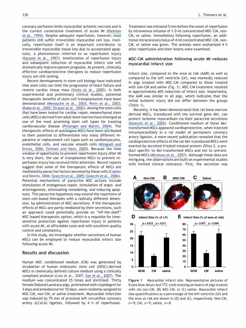
Figure 1 Myocardial infarct size. Representative pictures ofEvans blue (blue) and TTC (red) staining on hearts of pigs treatedwith (A) non-CM, (B) MSC-CM, or (C) saline. Myocardial infarctsize quantifications as a percentage of the left ventricle (LV) andthe area at risk are shown in (D) and (E), respectively. Non-CM,n=9; CM, n=9; saline, n=8.
130 L. Timmers et al.
coronary perfusion limits myocardial ischemic necrosis and isthe current cornerstone treatment of acute MI (Kleimanet al., 1994). Despite adequate reperfusion, however, mostpatients still suffer irreversible myocardial cell loss. Ironi-cally, reperfusion itself is an important contributor toirreversible myocardial tissue loss due to accelerated apop-tosis, a phenomenon referred to as reperfusion injury(Saraste et al., 1997). Amelioration of reperfusion injuryand subsequent reduction of myocardial infarct size willdramatically improve patient prognosis. At present, clinicallyeffective cardioprotective therapies to reduce reperfusioninjury are still lacking.
Recent developments in stem cell biology have indicatedthat stem cells can limit the progression of heart failure andrestore cardiac tissue mass (Smits et al., 2005). In bothexperimental and preliminary clinical studies, potentialtherapeutic benefits of stem cell transplantation have beendemonstrated (Menasche et al., 2003; Perin et al., 2003;Shake et al., 2002; Strauer et al., 2002). Among the stem cellsthat have been tested for cardiac repair, mesenchymal stemcells (MSCs) derived fromadult bonemarrow have emerged asone of the most promising stem cell types for treatingcardiovascular disease (Pittenger and Martin, 2004). Thetherapeutic effects of autologous MSCs have been attributedto their potential to differentiate into many different re-parative or replacement cell types such as cardiomyocytes,endothelial cells, and vascular smooth cells (Minguell andErices, 2006; Zimmet and Hare, 2005). Because the timewindow of opportunity to prevent reperfusion injury after MIis very short, the use of transplanted MSCs to prevent re-perfusion injury has received little attention. Recent reportssuggest that some of the therapeutic effects of MSCs aremediated by paracrine factors secreted by these cells (Caplanand Dennis, 2006; Gnecchi et al., 2005; Gnecchi et al., 2006).Potential mechanisms of paracrine MSC actions includestimulation of endogenous repair, stimulation of angio- andarteriogenesis, attenuating remodeling, and reducing apop-tosis. This paracrine hypothesis may extend the repertoire ofstem cell-based therapies with a radically different dimen-sion, by administration of MSC secretions. If the therapeuticeffects of MSCs are partly mediated by their secretions, suchan approach could potentially provide an “off-the-shelf”MSC-based therapeutic option, which is a requisite for time-sensitive protection against reperfusion injury in patientswith acute MI, at affordable costs and with excellent qualitycontrol and consistency.
In this study, we investigate whether secretions of humanMSCs can be employed to reduce myocardial infarct sizefollowing acute MI.
Results and discussion
Human MSC conditioned medium (CM) was generated byincubation of human embryonic stem cell (hESC)-derivedMSCs in chemically defined culture medium using a clinicallycompliant protocol (Lian et al., 2007; Sze et al., 2007). Themedium was concentrated 25 times and sterilized. Thirtyfemale Dalland Landrace pigs, pretreatedwith clopidogrel for3 days and amiodarone for 10 days,were randomly assigned toMSC-CM, non-CM, or saline treatment. Myocardial infarctionwas induced by 75 min of proximal left circumflex coronaryartery (LCxCA) ligation, followed by 4 h of reperfusion.
Treatment was initiated 5min before the onset of reperfusionby intravenous infusion of 1.0 ml concentrated MSC-CM, non-CM, or saline. Immediately following reperfusion, an addi-tional intracoronary bolus of 4 ml concentrated MSC-CM, non-CM, or saline was given. The animals were euthanized 4 hafter reperfusion and their hearts were examined.
MSC-CM administration following acute MI reducesmyocardial infarct size
Infarct size, compared to the area at risk (AAR) as well ascompared to the left ventricle (LV), was markedly reducedin pigs treated with MSC-CM compared to those treatedwith non-CM and saline (Fig. 1). MSC-CM treatment resultedin approximately 60% reduction of infarct size. Importantly,the AAR was similar in all pigs, which indicates that theinitial ischemic injury did not differ between the groups(Table 1).
Recently, it has been demonstrated that rat bone marrowderived MSCs, transduced with the survival gene Akt, canprotect ischemic myocardium via their paracrine secretions(Gnecchi et al., 2005). Conditioned medium of these Akt-transformed MSCs appeared cardioprotective, when injectedintramyocardially in a rat model of permanent coronaryartery ligation. A more recent publication revealed that thecardioprotective effects of the rat Akt-transduced MSCs wereexerted by secreted frizzled-related protein (Sfrp) 2, a pro-duct specific to Akt-transformed MSCs and not to untrans-formed MSCs (Mirotsou et al., 2007). Although these data areintriguing, the observations are built on experimental studieswith limited clinical relevance. First, the secretion was
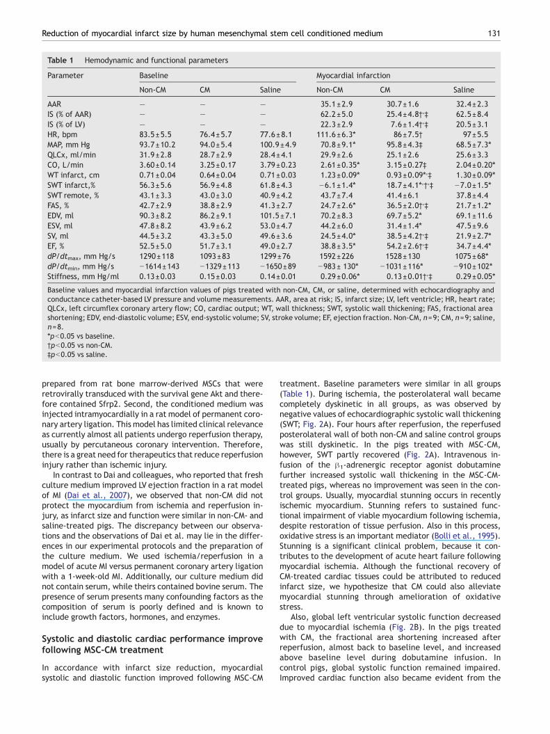
Table 1 Hemodynamic and functional parameters
Parameter Baseline Myocardial infarction
Non-CM CM Saline Non-CM CM Saline
AAR — — — 35.1±2.9 30.7±1.6 32.4±2.3IS (% of AAR) — — — 62.2±5.0 25.4±4.8†,‡ 62.5±8.4IS (% of LV) — — — 22.3±2.9 7.6±1.4†,‡ 20.5±3.1HR, bpm 83.5±5.5 76.4±5.7 77.6±8.1 111.6±6.3⁎ 86±7.5† 97±5.5MAP, mm Hg 93.7±10.2 94.0±5.4 100.9±4.9 70.8±9.1⁎ 95.8±4.3‡ 68.5±7.3⁎QLCx, ml/min 31.9±2.8 28.7±2.9 28.4±4.1 29.9±2.6 25.1±2.6 25.6±3.3CO, L/min 3.60±0.14 3.25±0.17 3.79±0.23 2.61±0.35⁎ 3.15±0.27‡ 2.04±0.20⁎WT infarct, cm 0.71±0.04 0.64±0.04 0.71±0.03 1.23±0.09⁎ 0.93±0.09⁎,‡ 1.30±0.09⁎SWT infarct,% 56.3±5.6 56.9±4.8 61.8±4.3 −6.1±1.4⁎ 18.7±4.1⁎,†,‡ −7.0±1.5⁎SWT remote, % 43.1±3.3 43.0±3.0 40.9±4.2 43.7±7.4 41.4±6.1 37.8±4.4FAS, % 42.7±2.9 38.8±2.9 41.3±2.7 24.7±2.6⁎ 36.5±2.0†,‡ 21.7±1.2⁎EDV, ml 90.3±8.2 86.2±9.1 101.5±7.1 70.2±8.3 69.7±5.2⁎ 69.1±11.6ESV, ml 47.8±8.2 43.9±6.2 53.0±4.7 44.2±6.0 31.4±1.4⁎ 47.5±9.6SV, ml 44.5±3.2 43.3±5.0 49.6±3.6 24.5±4.0⁎ 38.5±4.2†,‡ 21.9±2.7⁎EF, % 52.5±5.0 51.7±3.1 49.0±2.7 38.8±3.5⁎ 54.2±2.6†,‡ 34.7±4.4⁎dP/dtmax, mm Hg/s 1290±118 1093±83 1299±76 1592±226 1528±130 1075±68⁎dP/dtmin, mm Hg/s −1614±143 −1329±113 −1650±89 −983± 130⁎ −1031±116⁎ −910±102⁎Stiffness, mm Hg/ml 0.13±0.03 0.15±0.03 0.14±0.01 0.29±0.06⁎ 0.13±0.01†,‡ 0.29±0.05⁎
Baseline values and myocardial infarction values of pigs treated with non-CM, CM, or saline, determined with echocardiography andconductance catheter-based LV pressure and volume measurements. AAR, area at risk; IS, infarct size; LV, left ventricle; HR, heart rate;QLCx, left circumflex coronary artery flow; CO, cardiac output; WT, wall thickness; SWT, systolic wall thickening; FAS, fractional areashortening; EDV, end-diastolic volume; ESV, end-systolic volume; SV, stroke volume; EF, ejection fraction. Non-CM, n=9; CM, n=9; saline,n=8.⁎pb0.05 vs baseline.†pb0.05 vs non-CM.‡pb0.05 vs saline.
131Reduction of myocardial infarct size by human mesenchymal stem cell conditioned medium
prepared from rat bone marrow-derived MSCs that wereretrovirally transduced with the survival gene Akt and there-fore contained Sfrp2. Second, the conditioned medium wasinjected intramyocardially in a rat model of permanent coro-nary artery ligation. This model has limited clinical relevanceas currently almost all patients undergo reperfusion therapy,usually by percutaneous coronary intervention. Therefore,there is a great need for therapeutics that reduce reperfusioninjury rather than ischemic injury.
In contrast to Dai and colleagues, who reported that freshculture medium improved LV ejection fraction in a rat modelof MI (Dai et al., 2007), we observed that non-CM did notprotect the myocardium from ischemia and reperfusion in-jury, as infarct size and function were similar in non-CM- andsaline-treated pigs. The discrepancy between our observa-tions and the observations of Dai et al. may lie in the differ-ences in our experimental protocols and the preparation ofthe culture medium. We used ischemia/reperfusion in amodel of acute MI versus permanent coronary artery ligationwith a 1-week-old MI. Additionally, our culture medium didnot contain serum, while theirs contained bovine serum. Thepresence of serum presents many confounding factors as thecomposition of serum is poorly defined and is known toinclude growth factors, hormones, and enzymes.
Systolic and diastolic cardiac performance improvefollowing MSC-CM treatment
In accordance with infarct size reduction, myocardialsystolic and diastolic function improved following MSC-CM
treatment. Baseline parameters were similar in all groups(Table 1). During ischemia, the posterolateral wall becamecompletely dyskinetic in all groups, as was observed bynegative values of echocardiographic systolic wall thickening(SWT; Fig. 2A). Four hours after reperfusion, the reperfusedposterolateral wall of both non-CM and saline control groupswas still dyskinetic. In the pigs treated with MSC-CM,however, SWT partly recovered (Fig. 2A). Intravenous in-fusion of the β1-adrenergic receptor agonist dobutaminefurther increased systolic wall thickening in the MSC-CM-treated pigs, whereas no improvement was seen in the con-trol groups. Usually, myocardial stunning occurs in recentlyischemic myocardium. Stunning refers to sustained func-tional impairment of viable myocardium following ischemia,despite restoration of tissue perfusion. Also in this process,oxidative stress is an important mediator (Bolli et al., 1995).Stunning is a significant clinical problem, because it con-tributes to the development of acute heart failure followingmyocardial ischemia. Although the functional recovery ofCM-treated cardiac tissues could be attributed to reducedinfarct size, we hypothesize that CM could also alleviatemyocardial stunning through amelioration of oxidativestress.
Also, global left ventricular systolic function decreaseddue to myocardial ischemia (Fig. 2B). In the pigs treatedwith CM, the fractional area shortening increased afterreperfusion, almost back to baseline level, and increasedabove baseline level during dobutamine infusion. Incontrol pigs, global systolic function remained impaired.Improved cardiac function also became evident from the

132 L. Timmers et al.
PV-loop-derived indices (Table 1). Left ventricular ejectionfraction and stroke volume were significantly higher in CM-treated pigs. This translated into improved hemodynamicparameters such as cardiac output, mean arterial pres-sure, and heart rate. Diastolic function decreased in con-trol groups following ischemia and reperfusion injury, asobserved by increased end-diastolic myocardial stiffness.
Figure 2 Local and global systolic function. (A) Local systolicwall thickening as assessed with echocardiography of the infarctarea in pigs treated with non-CM, MSC-CM, or saline. (B) Globalsystolic function, measured as echocardiographic fractional areashortening. Non-CM, n=9; MSC-CM, n=9; saline, n=8.⁎pb0.05vs non-CM and saline.
In the CM-treated pigs, however, diastolic function was notimpaired.
Reduced myocardial oxidative stress and reducedapoptosis by MSC-CM treatment
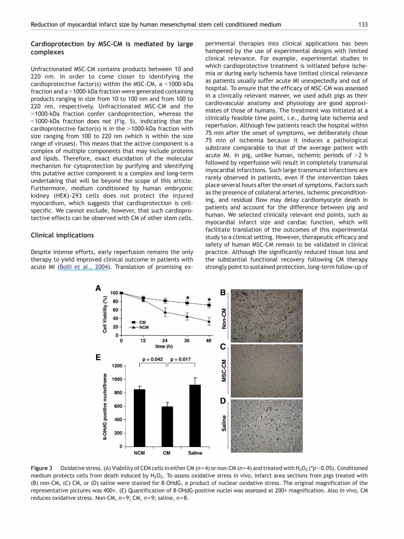
Despite using large animal models that are notoriously intrac-table to mechanistic investigation due to limited researchreagents, we were nonetheless able to identify mechanismsfor CM-elicited cardioprotection against ischemia/reperfusioninjury and subsequent functional recovery. Having demon-strated the reduction in infarct size and improvement offunction, we first used an in vitro assay of hydrogen peroxide(H2O2)-induced cell death to determine the effects of CM andnon-CM on oxidative stress, a major cause of ischemia/re-perfusion injury.We inducedH2O2-mediated oxidative stress inhuman leukemic CEM cells in the presence of either CM or non-CM and monitored cell viability by trypan blue exclusion.Results showed that CM significantly protected against H2O2-induced loss of cell viability compared to non-CM (Fig. 3A). Todetermine if CM also reduces oxidative stress in the hearts ofCM-treated pigs, nuclear oxidative stress in tissue sections ofpigs treated with CM, non-CM, or saline was quantified by 8-hydroxy-2′-deoxyguanosine (8-OHdG) immunostaining for oxi-dizedDNA. Intense nuclear staining indicative of DNAoxidationwas observed in sections of non-CM- and saline-treated pigscompared with CM-treated pigs (Figs. 3B–D). There weresignificantly more positive nuclei in non-CM- and saline-treated pigs (Fig. 3E). Therefore, CM can confer cytoprotec-tion against oxidative stress in vitro and in vivo.
The secretions of the MSCs contained many proteinsinvolved in TGF-β signaling (Sze et al., 2007). To assessthe influence of CM treatment on TGF-β signaling in vivo, wequantified phosphorylated SMAD2 in the myocardial tissuesamples of CM-treated pigs and control pigs by Westernblotting. CM treatment resulted in reduced pSMAD2 expres-sion, indicating that TGF-β signaling via ALK-5 was reduced(Figs. 4A and B). This suggests that altered TGF-β signalingmay be involved in CM-mediated cardioprotection.
Reperfusion injury causes cell death through apoptosisrather than necrosis (Dumont et al., 2001; Eefting et al.,2004; Hausenloy and Yellon, 2004).We established that in CM-treated hearts, apoptotic activity as measured by the level ofactivated caspase-3, a central mediator of apoptosis, wasreduced, indicating a CM-associated attenuation of apopto-sis. In the pigs treated with MSC-CM, active caspase-3 levelswere lower compared to both non-CM control and saline con-trol, suggesting that CM inhibits myocardial apoptosis(Figs. 4C and D).
Taking the results together, this study demonstrated thathuman MSC-CM protected cardiac tissue from apoptotic celldeath by attenuation of oxidative damage and modulationof TGF-β signaling. It is not likely that CM protected againstthrombosis in the epicardial coronary artery, since coronaryflow recovered rapidly to normal flow rates after an initialhyperemic reaction following reperfusion (Table 1). In addi-tion, all the animals in both test and control groups wereheparinized and treated with clopidogrel to preventthrombosis. Furthermore, the CM has no anticoagulativeeffect as assayed by activated partial thromboplastin timeand prothrombin time assays (data not shown).
133Reduction of myocardial infarct size by human mesenchymal stem cell conditioned medium
Cardioprotection by MSC-CM is mediated by largecomplexes
Unfractionated MSC-CM contains products between 10 and220 nm. In order to come closer to identifying thecardioprotective factor(s) within the MSC-CM, a b1000-kDafraction and a N1000-kDa fraction were generated containingproducts ranging in size from 10 to 100 nm and from 100 to220 nm, respectively. Unfractionated MSC-CM and theN1000-kDa fraction confer cardioprotection, whereas theb1000-kDa fraction does not (Fig. 5), indicating that thecardioprotective factor(s) is in the N1000-kDa fraction withsize ranging from 100 to 220 nm (which is within the sizerange of viruses). This means that the active component is acomplex of multiple components that may include proteinsand lipids. Therefore, exact elucidation of the molecularmechanism for cytoprotection by purifying and identifyingthis putative active component is a complex and long-termundertaking that will be beyond the scope of this article.Furthermore, medium conditioned by human embryonickidney (HEK)-293 cells does not protect the injuredmyocardium, which suggests that cardioprotection is cell-specific. We cannot exclude, however, that such cardiopro-tective effects can be observed with CM of other stem cells.
Clinical implications
Despite intense efforts, early reperfusion remains the onlytherapy to yield improved clinical outcome in patients withacute MI (Bolli et al., 2004). Translation of promising ex-
Figure 3 Oxidative stress. (A) Viability of CEM cells in either CM (n=medium protects cells from death induced by H2O2. To assess oxida(B) non-CM, (C) CM, or (D) saline were stained for 8-OHdG, a produrepresentative pictures was 400×. (E) Quantification of 8-OHdG-posireduces oxidative stress. Non-CM, n=9; CM, n=9; saline, n=8.
perimental therapies into clinical applications has beenhampered by the use of experimental designs with limitedclinical relevance. For example, experimental studies inwhich cardioprotective treatment is initiated before ische-mia or during early ischemia have limited clinical relevanceas patients usually suffer acute MI unexpectedly and out ofhospital. To ensure that the efficacy of MSC-CM was assessedin a clinically relevant manner, we used adult pigs as theircardiovascular anatomy and physiology are good approxi-mates of those of humans. The treatment was initiated at aclinically feasible time point, i.e., during late ischemia andreperfusion. Although few patients reach the hospital within75 min after the onset of symptoms, we deliberately chose75 min of ischemia because it induces a pathologicalsubstrate comparable to that of the average patient withacute MI. In pig, unlike human, ischemic periods of N2 hfollowed by reperfusion will result in completely transmuralmyocardial infarctions. Such large transmural infarctions arerarely observed in patients, even if the intervention takesplace several hours after the onset of symptoms. Factors suchas the presence of collateral arteries, ischemic precondition-ing, and residual flow may delay cardiomyocyte death inpatients and account for the difference between pig andhuman. We selected clinically relevant end points, such asmyocardial infarct size and cardiac function, which willfacilitate translation of the outcomes of this experimentalstudy to a clinical setting. However, therapeutic efficacy andsafety of human MSC-CM remain to be validated in clinicalpractice. Although the significantly reduced tissue loss andthe substantial functional recovery following CM therapystrongly point to sustained protection, long-term follow-up of
4) or non-CM (n=4) and treatedwith H2O2 (⁎pb0.05). Conditionedtive stress in vivo, infarct area sections from pigs treated withct of nuclear oxidative stress. The original magnification of thetive nuclei was assessed at 200× magnification. Also in vivo, CM
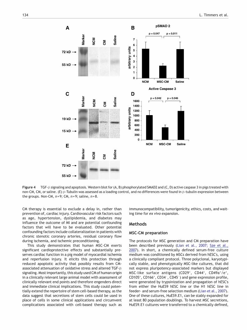
Figure 4 TGF-β signaling and apoptosis. Western blot for (A, B) phosphorylated SMAD2 and (C, D) active caspase 3 in pigs treatedwithnon-CM, CM, or saline. (E) β-Tubulin was assessed as a loading control, and no differences were found in β-tubulin expression betweenthe groups. Non-CM, n=9; CM, n=9; saline, n=8.
134 L. Timmers et al.
CM therapy is essential to exclude a delay in, rather thanprevention of, cardiac injury. Cardiovascular risk factors suchas age, hypertension, dyslipidemia, and diabetes mayinfluence the outcome of MI and are potential confoundingfactors that will have to be evaluated. Other potentialconfounding factors include collateralization in patients withchronic stenotic coronary arteries, residual coronary flowduring ischemia, and ischemic preconditioning.
This study demonstrates that human MSC-CM exertssignificant cardioprotective effects and substantially pre-serves cardiac function in a pig model of myocardial ischemiaand reperfusion injury. It elicits this protection throughreduced apoptotic activity that possibly results from CM-associated attenuation of oxidative stress and altered TGF-βsignaling.Most importantly, this study usedCMof humanoriginin a clinically relevant large animal model with assessment ofclinically relevant end points and therefore engenders directand immediate clinical implications. This study could poten-tially extend the repertoire of stem cell-based therapy, as thedata suggest that secretions of stem cells could be used inplace of cells in some clinical applications and circumventcomplications associated with cell-based therapy such as
immunocompatibility, tumorigenicity, ethics, costs, and wait-ing time for ex vivo expansion.
Methods
MSC-CM preparation
The protocols for MSC generation and CM preparation havebeen described previously (Lian et al., 2007; Sze et al.,2007). In short, a chemically defined serum-free culturemedium was conditioned by MSCs derived from hESC's, usinga clinically compliant protocol. Three polyclonal, karyotypi-cally stable, and phenotypically MSC-like cultures, that didnot express pluripotency-associated markers but displayedMSC-like surface antigens (CD29+, CD44+, CD49a+/e+,CD105+, CD166+, CD34−, CD45−) and gene expression profile,were generated by trypsinization and propagation of hESC'sfrom either the HuES9 hESC line or the H1 hESC line infeeder- and serum-free selection medium (Lian et al., 2007).One of these cultures, HuES9.E1, can be stably expanded forat least 80 population doublings. To harvest MSC secretions,HuES9.E1 cultures were transferred to a chemically defined,

Figure 5 Cardioprotective properties of CM fractions. Myo-cardial infarct size quantification in mice treated with saline(n=10), unfractionatedMSC-CM (n=12), HEK-293-CM (n=10), theb1000-kDa fraction (n=8), and the N1000-kDa fraction (n=8).Unfractionated MSC-CM reduces myocardial infarct size com-pared to saline-treated animals, in contrast to HEK-293-CM. TheN1000-kDa fraction of MSC-CM reduces infarct size, whereas theb1000-kDa fraction does not, indicating that the cardioprotec-tive factor(s) is larger than 1000 kDa. ⁎pb0.05 vs saline.
135Reduction of myocardial infarct size by human mesenchymal stem cell conditioned medium
serum-free culture medium to condition the medium for3 days before the media containing MSC secretions werecollected, clarified by centrifugation, concentrated 25 timesusing 10-kDa MW cutoff ultrafiltration membranes, andsterilized by filtration through a 220-nm filter. The secretoryproteome was analyzed by multidimensional protein identi-fication technology and cytokine antibody array analysis andrevealed the presence of 201 unique gene products.Computational analyses disclosed that this CM holds poten-tial cytoprotective properties (Sze et al., 2007).
Animals
All experiments were performed in accordance with theGuide for the Care and Use of Laboratory Pigs prepared bythe Institute of Laboratory Animal Resources and with priorapproval by the Animal Experimentation Committee of theFaculty of Medicine, Utrecht University, the Netherlands.
Study design
Thirty female Dalland Landrace pigs (60–70 kg; IDDLO,Lelystad, The Netherlands), all pretreated with clopidogrel75 mg/day for 3 days and amiodarone 400 mg/day for10 days, were randomly assigned to MSC-CM, non-CM, orsaline treatment. The saline group was added to assess apotential effect of fresh, nonconditioned culture medium. Inall pigs, MI was induced by 75 min of proximal LCxCA ligationand 4 h of subsequent reperfusion. An ischemic period of75 min was selected to inflict severe myocardial injurywithout inducing completely transmural myocardial infarc-tion. The 4-h reperfusion period was used because infarctsize measurement using triphenyltetrazolium chloride (TTC)staining is most reliable after 3 h of reperfusion (Birnbaum
et al., 1997). After longer periods of reperfusion, it becomesmore difficult to assess oxidative stress status and apoptoticmechanisms. Treatment was initiated 5 min before the onsetof reperfusion by intravenous infusion of MSC-CM (1.0 ml,2.0 mg protein), non-CM, or saline. Immediately followingreperfusion, an additional intracoronary bolus of MSC-CM(4.0 ml, 8.0 mg protein), non-CM, or saline was given.
MI and operational procedure
During the entire operation, ECG, systemic arterial pressure,and capnogram were monitored continuously. Under generalanesthesia as described before (Timmers et al., 2007a), amedian sternotomy was performed and two introductionsheets were inserted in the carotid arteries for a 6-Fr guidingcatheter and an 8-Fr conductance catheter (CD Leycom,Zoetermeer, the Netherlands). The distal tip of a Swan Ganzcatheter was placed into the pulmonary artery via theinternal jugular vein. Transonic flow probes (TransonicSystems, Ithaca, NY, USA) were placed around the proximalaorta and LCxCA to measure cardiac output and coronaryflow, and a wire was placed around the inferior caval vein toenable functional measurements under varying loadingconditions for PV loops. After functional measurements,10,000 IU of heparin was administered intravenously andsutures were tightened to occlude the proximal LCxCA.Internal defibrillation with 50 J was used when ventricularfibrillation occurred. After 75min of ischemia, the LCxCAwasreopened by release of the suture. Immediately followingreperfusion, nitroglycerine (0.1mg to prevent no-reflow)wasinfused through the LCxCA via the guiding catheter, followedby intracoronary treatment with MSC-CM, non-CM, or saline.After 4 h of reperfusion, the final functional measurementswere performed and the heart was explanted for infarct sizeanalysis.
Functional measurements
Short axis epicardial ultrasound and LV pressure and volumewere measured using the conductance catheter method, asdescribed previously (Timmers et al., 2007b). LV pressureand volume signals derived from the conductance catheterwere displayed and acquired at a 250-Hz sampling rate with aLeycom CFL-512 (CD Leycom). Data were acquired duringsteady state and during temporal caval vein occlusion, allwith the ventilator turned off at end expiration. Analysis ofthe pressure–volume loops was performed with customsoftware as described previously (Steendijk et al., 1998). Inaddition, short-axis epicardial ultrasound images (ProsoundSSD-5000, 5-MHz probe UST-5280-5; Aloka Holding EuropeAG, Zug, Switzerland) were obtained at the midpapillarymuscle level. Wall thickness (WT) of the infarct area, remotearea (septum), and LV internal area (LVia) were measured atend diastole (ED) and end systole (ES). SWTwas calculated as[(WT(ES) − WT(ED))/WT(ED)]×100%, fractional area short-ening as [(LVia(ED) − LVia(ES))/LVia(ED)]×100%, and leftventricular ejection fraction as [(EDV − ESV)/EDV]×100%.The end-diastolic chamber stiffness was quantified by meansof linear regression of the end-diastolic pressure–volumerelationship. Echocardiography and PV loops were measuredbefore MI, 1 h after ischemia, and 4 h after reperfusion. To

136 L. Timmers et al.
challenge stunned myocardium, additional measurementswere performed during pharmaceutically induced stressby intravenous dobutamine infusion (2.5 μg/kg/min and5.0 μg/kg/min).
Infarct size
Just prior to excision of the heart, the LCxCA (pigs) or leftcoronary artery (LCA) (mice) was religated at exactly thesame spot as for the induction of the MI. Evans blue dye wasinfused through the coronary system to delineate the AAR.The heart was then excised, and the LV was isolated and cutinto five slices from apex to base. The slices were incubated in1% TTC (Sigma–Aldrich Chemicals, Zwijndrecht, the Nether-lands) in 37 °C Sörensen buffer (13.6 g/L KH2PO4 + 17.8 g/LNa2H PO4 · 2H2O, pH 7.4) for 15 min to discriminate infarcttissue from viable myocardium. All slices were scanned fromboth sides, and in each slide, the infarct area was comparedwith area at risk and the total area by use of digital planimetrysoftware (ImageJ). After correction for the weight of theslices, infarct size was calculated as a percentage of the AARand of the LV.
Oxidation-induced cell death assay
Human leukemic CEM cells were plated in six-well plates andincubated in either CM (unconcentrated, i.e., 80 μg/mlprotein) or non-CM and treated with 50 μM H2O2 to induceoxidative stress. Cell viability was assessed using trypan blueexclusion at 12, 24, 36, and 48 h after H2O2 treatment.
Immunohistochemistry
Nuclear oxidative stress in the ischemia and reperfusion areawas assessed by immunostaining for 8-OHdG, a product ofoxidative stress to DNA. Tissue samples were fixed in 4%formalin before being embedded in paraffin. Followingantigen retrieval in 10 mM citric acid, the tissue sectionswere incubated with 10% normal horse serum for 30 min,mouse anti-8-OHdG (OXIS International, Foster City, CA, USA)1:20 in 0.1% PBSA overnight at 4 °C, biotin-labeled horse anti-mouse (Vector Laboratories, Burlingame, CA, USA) 1:500 for1 h, and streptavidin–HRP 1:1000 for 1 h. Finally, the sectionswere incubated with H2O2–diaminobenzidine for 10 min. Theamount of 8-OHdG-positive nuclei was quantified in fourrandomly picked fields per section with the digital imagemicroscopy software Analysis (Olympus, Münster, Germany)at 200× magnification.
Western blotting
Protein was isolated from frozen tissue samples collectedfrom the ischemia/reperfusion area of pigs using 1 ml TripureIsolation Reagent (Boehringer, Mannheim, Germany) accord-ing to the manufacturer's protocol. Eight micrograms of totalprotein was separated on by 10% SDS–PAGE, transferred ontoa Nitrocellulose C membrane (Amersham, Buckinghamshire,UK), and blocked using phosphate-buffered saline–0.1%Tween–5% Protifar (Nutricia, Netherlands). The membranewas incubated with a rabbit antibody for phospho-SMAD21:1000 (Cell Signaling Technology), for active caspase-3 1:100
(Chemicon, Germany), or for β-tubulin 1:5000 (Abcam,Cambridge, UK) and subsequently with goat anti-rabbit HRP1:2000 (DAKO, Glostrup, Denmark). Chemiluminescencesubstrate (NENk Life Science Products) was used for detec-tion; the bands were analyzed using the Gel Doc 1000 system(Bio-Rad, Veenendaal, Netherlands).
MSC-CM fractionation
The MSC-CM was prepared by sterile filtration through a 220-nm filter and concentrated through a 10-nm filter andtherefore contains components between 10 and 220 nm. Sub-sequently, a b1000-kDa fraction and a N1000-kDa fractionwere prepared by filtering the MSC-CM through a 1000-kDa-MWcutoff membrane with a 100-nm nominal pore size (Pall Corp.,Singapore), generating a fraction containing products between10 and 100 nm and a fraction containing products between 100and 220 nm. To identify the fraction containing the factor(s)within the medium that confers cardioprotection (10–100 or100–220 nm), a mouse model of ischemia and reperfusioninjury was used. MI was induced by 30 min LCA occlusion andsubsequent reperfusion. Mice were treated with 20 μl unfrac-tionated MSC-CM (10–220 nm), b1000-kDa fraction (10–100 nm), N1000-kDa fraction, or saline intravenously via thetail vein, 5 min before reperfusion. Infarct size was assessed24 h later using Evans blue and TTC as described previously(Timmers et al., 2007a). An additional group of mice wasexamined with medium conditioned by HEK-293 cells, toinvestigate whether the cardioprotective effect is cell-specific.
Data analysis
Data are presented as means ± SEM. Values were collected ina blinded fashion and compared using one-way ANOVA withpost hoc Bonferroni tests in SPSS 15.0. Cardioprotectivepotential of the human MSC-CM fractions was compared tosaline using a one-way ANOVAwith Dunnett's post hoc tests. Ap value of b0.05 was considered significant.
Acknowledgments
We gratefully acknowledge Cees Verlaan, Maringa Emons,Merel Schurink, Mirella de Haan, Nimco Youssuf, andChaylendra Strijder for excellent technical assistance. Thiswork was supported by the Netherlands Heart Foundation,Grant 2005T022, and an Internationalization grant by UMCUtrecht, the Netherlands.
References
Birnbaum, Y., Hale, S.L., Kloner, R.A., 1997. Differences inreperfusion length following 30 minutes of ischemia in the rabbitinfluence infarct size, as measured by triphenyltetrazoliumchloride staining. J. Mol. Cell. Cardiol. 29, 657–666.
Bolli, R., Zughaib, M., Li, X.Y., Tang, X.L., Sun, J.Z., Triana, J.F.,McCay, P.B., 1995. Recurrent ischemia in the canine heart causesrecurrent bursts of free radical production that have acumulative effect on contractile function: a pathophysiologicalbasis for chronic myocardial “stunning”. J. Clin. Invest. 96,1066–1084.
137Reduction of myocardial infarct size by human mesenchymal stem cell conditioned medium
Bolli, R., Becker, L., Gross, G., Mentzer Jr., R., Balshaw, D., Lathrop,D.A., 2004. Myocardial protection at a crossroads: the need fortranslation into clinical therapy. Circ. Res. 95, 125–134.
Caplan, A.I., Dennis, J.E., 2006. Mesenchymal stem cells as trophicmediators. J. Cell. Biochem. 98, 1076–1084.
Dai, W., Hale, S.L., Kloner, R.A., 2007. Role of a paracrine action ofmesenchymal stem cells in the improvement of left ventricularfunction after coronary artery occlusion in rats. Regen. Med. 2,63–68.
Dumont, E.A., Reutelingsperger, C.P., Smits, J.F., Daemen, M.J.,Doevendans, P.A., Wellens, H.J., Hofstra, L., 2001. Real-timeimaging of apoptotic cell-membrane changes at the single-celllevel in the beating murine heart. Nat. Med. 7, 1352–1355.
Eefting, F., Rensing, B., Wigman, J., Pannekoek, W.J., Liu, W.M.,Cramer, M.J., Lips, D.J., Doevendans, P.A., 2004. Role ofapoptosis in reperfusion injury. Cardiovasc. Res. 61, 414–426.
Gnecchi, M., He, H., Liang, O.D., Melo, L.G., Morello, F., Mu, H.,Noiseux, N., Zhang, L., Pratt, R.E., Ingwall, J.S., Dzau, V.J., 2005.Paracrine action accounts for marked protection of ischemicheart by Akt-modified mesenchymal stem cells. Nat. Med. 11,367–368.
Gnecchi, M., He, H., Noiseux, N., Liang, O.D., Zhang, L., Morello, F.,Mu, H., Melo, L.G., Pratt, R.E., Ingwall, J.S., Dzau, V.J., 2006.Evidence supporting paracrine hypothesis for Akt-modifiedmesenchymal stem cell-mediated cardiac protection and func-tional improvement. FASEB J. 20, 661–669.
Hausenloy, D.J., Yellon, D.M., 2004. New directions for protectingthe heart against ischaemia–reperfusion injury: targeting thereperfusion injury salvage kinase (RISK)-pathway. Cardiovasc.Res. 61, 448–460.
Kleiman, N.S.,White, H.D., Ohman, E.M., Ross, A.M.,Woodlief, L.H.,Califf, R.M., Holmes Jr., D.R., Bates, E., Pfisterer, M., Vahanian,A., et al., 1994. Mortality within 24 hours of thrombolysis formyocardial infarction: the importance of early reperfusion. TheGUSTO Investigators, Global Utilization of Streptokinase andTissue Plasminogen Activator for Occluded Coronary Arteries.Circulation 90, 2658–2665.
Lewis, E.F., Moye, L.A., Rouleau, J.L., Sacks, F.M., Arnold, J.M.,Warnica, J.W., Flaker, G.C., Braunwald, E., Pfeffer, M.A., 2003.Predictors of late development of heart failure in stable survivorsof myocardial infarction: the CARE study. J. Am. Coll. Cardiol. 42,1446–1453.
Lian, Q., Lye, E., Suan Yeo, K., KhiaWay Tan, E., Salto-Tellez, M., Liu,T.M., Palanisamy, N., El Oakley, R.M., Lee, E.H., Lim, B., Lim,S.K., 2007. Derivation of clinically compliant MSCs from CD105+,CD24− differentiated human ESCs. Stem Cells 25, 425–436.
Menasche, P., Hagege, A.A., Vilquin, J.T., Desnos, M., Abergel, E.,Pouzet, B., Bel, A., Sarateanu, S., Scorsin, M., Schwartz, K., et al.,2003. Autologous skeletal myoblast transplantation for severepostinfarction left ventricular dysfunction. J. Am.Coll. Cardiol. 41,1078–1083.
Minguell, J.J., Erices, A., 2006. Mesenchymal stem cells and the treat-ment of cardiac disease. Exp. Biol. Med. (Maywood) 231, 39–49.
Mirotsou, M., Zhang, Z., Deb, A., Zhang, L., Gnecchi, M., Noiseux,N., Mu, H., Pachori, A., Dzau, V., 2007. Secreted frizzled relatedprotein 2 (Sfrp2) is the key Akt-mesenchymal stem cell-releasedparacrine factor mediating myocardial survival and repair.Proc. Natl. Acad. Sci. U. S. A. 104, 1643–1648.
Perin, E.C., Dohmann, H.F., Borojevic, R., Silva, S.A., Sousa, A.L.,Mesquita, C.T., Rossi, M.I., Carvalho, A.C., Dutra, H.S., Dohmann,H.J., et al., 2003. Transendocardial, autologous bonemarrow celltransplantation for severe, chronic ischemic heart failure.Circulation 107, 2294–2302.
Pittenger, M.F., Martin, B.J., 2004. Mesenchymal stem cells and theirpotential as cardiac therapeutics. Circ. Res. 95, 9–20.
Saraste, A., Pulkki, K., Kallajoki, M., Henriksen, K., Parvinen, M.,Voipio-Pulkki, L.M., 1997. Apoptosis in human acute myocardialinfarction. Circulation 95, 320–323.
Shake, J.G., Gruber, P.J., Baumgartner, W.A., Senechal, G., Meyers,J., Redmond, J.M., Pittenger, M.F., Martin, B.J., 2002. Mesench-ymal stem cell implantation in a swine myocardial infarct model:engraftment and functional effects. Ann. Thorac. Surg. 73,1919–1925 discussion 1926.
Smits, A.M., van Vliet, P., Hassink, R.J., Goumans, M.J., Doeven-dans, P.A., 2005. The role of stem cells in cardiac regeneration.J. Cell. Mol. Med. 9, 25–36.
Steendijk, P., Baan Jr., J., Van der Velde, E.T., Baan, J., 1998. Effectsof critical coronary stenosis on global systolic left ventricularfunction quantified by pressure–volume relations during dobuta-mine stress in the canine heart. J. Am. Coll. Cardiol. 32, 816–826.
Strauer, B.E., Brehm,M., Zeus, T., Kostering,M., Hernandez, A., Sorg,R.V., Kogler, G., Wernet, P., 2002. Repair of infarctedmyocardiumby autologous intracoronarymononuclear bonemarrow cell trans-plantation in humans. Circulation 106, 1913–1918.
Sze, S.K., de Kleijn, D.P., Lai, R.C., Khia Way Tan, E., Zhao, H., Yeo,K.S., Low, T.Y., Lian, Q., Lee, C.N., Mitchell, W., et al., 2007.Elucidating the secretion proteome of human embryonic stemcell-derived mesenchymal stem cells. Mol. Cell Proteomics 6,1680–1689.
Timmers, L., Sluijter, J.P., van Keulen, J.K., Hoefer, I.E., Nederhoff, M.G., Goumans, M.J., Doevendans, P.A., van Echteld, C.J., Joles, J.A.,Quax, P.H., et al., 2007a. Toll-like receptor 4 mediates maladaptiveleft ventricular remodeling and impairs cardiac function followingmyocardial infarction. Circ. Res. 102, 257–264.
Timmers, L., Sluijter, J.P., Verlaan, C.W., Steendijk, P., Cramer, M.J.,Emons, M., Strijder, C., Grundeman, P.F., Sze, S.K., Hua, L., et al.,2007b. Cyclooxygenase-2 inhibition increasesmortality, enhancesleft ventricular remodeling, and impairs systolic function aftermyocardial infarction in the pig. Circulation 115, 326–332.
Zimmet, J.M., Hare, J.M., 2005. Emerging role for bone marrowderived mesenchymal stem cells in myocardial regenerativetherapy. Basic Res. Cardiol. 100, 471–481.
Copyright © 2022 FDOKUMEN