
A Categorization Technique for Resolving Information System ...
Upload
independentCategory
view
1download
0
Rapid and Highly Resolving: Affective Evaluation ofOlfactorily Conditioned Faces
Christian Steinberg1, Christian Dobel1, Harald T. Schupp2,Johanna Kissler2, Ludger Elling1, Christo Pantev1,
and Markus Junghöfer1
Abstract
■ Evidence from hemodynamic and electrophysiological mea-sures suggests that the processing of emotionally relevant infor-mation occurs in a spatially and temporally distributed affectivenetwork. ERP studies of emotional stimulus processing fre-quently report differential responses to emotional stimuli start-ing around 120 msec. However, the involvement of structuresthat seem to become activated at earlier latencies (i.e., amyg-dala and OFC) would allow for more rapid modulations, evenin distant cortical areas. Consistent with this notion, recentERP studies investigating associative learning have provided
evidence for rapid modulations in sensory areas earlier than120 msec, but these studies either used simple and/or very fewstimuli. The present study used high-density whole-head magneto-encephalography to measure brain responses to a multitude ofneutral facial stimuli, which were associated with an aversive orneutral odor. Significant emotional modulations were observedat intervals of 50–80 and 130–190 msec in frontal and occipito-temporal regions, respectively. In the absence of contingencyawareness and with only two learning instances, a remarkablecapacity for emotional learning is observed. ■
INTRODUCTION
The preferential processing of high-priority stimuli in theenvironment is an essential function of selective attention.fMRI studies have revealed increased activations byemotional stimuli in occipital, parietal, and inferior tem-poral cortex (ITC; Junghöfer, Schupp, Stark, & Vaitl,2005; Sabatinelli, Bradley, Fitzsimmons, & Lang, 2005;Bradley et al., 2003; Vuilleumier, Armony, Driver, & Dolan,2001). ERP studies have shown that emotional stimuli mod-ulate distinct ERP components between 120–300 and 300–700msec (Kissler, Herbert, Peyk, & Junghöfer, 2007; Schupp,Flaisch, Stockburger, & Junghöfer, 2006; Junghöfer, Bradley,Elbert, & Lang, 2001). Thus, emotion modulates vision atmidlatency and late processing stages, allowing for re-entrant processing and widespread activation, conveyingstimulus information to many associative cortical regions.However, several recent studies indicate that emotion
may modulate visual processing through the rapid extrac-tion of relevant features at even earlier stages of process-ing for both simple geometric gratings (Keil, Stolarova,Moratti, & Ray, 2007; Stolarova, Keil, & Moratti, 2006)and perceptuallymore complex faces (Morel, Ponz,Mercier,Vuilleumier, &George, 2009; Pizzagalli, Regard, & Lehmann,1999). The patterns paired with the threatening stimulielicited an increased C1 visual event-related componentbetween 65- and 90-msec poststimulus (Stolarova et al.,
2006), and a recent fMRI study revealed that Gabor grat-ings paired with shock resulted in increased activity inthe primary visual cortex (Padmala & Pessoa, 2008). Duringaversive conditioning of faces, extrastriate visual cortex re-sponseswere also identified earlier than 120msec (Pizzagalli,Greischar, & Davidson, 2003). Thus, recent findings sug-gest that emotional relevance supports associative learningin early visual stimulus processing.
Previous experiments either investigated associativelearning of simple geometric patterns and/or made useof very few conditioned stimuli (CS; Pizzagalli et al., 2003;Gottfried, Deichmann, Winston, & Dolan, 2002). Thus, inthese studies, the salience tagging might have always beenon the basis of simple physical feature differences ratherthan individual visual perceptual features, necessary to dif-ferentiate socially relevant stimuli in real life. The presentmagnetoencephalography (MEG) study investigated thetiming and regional distribution of cortical responses todifferent neutral facial stimuli (n = 104) that were asso-ciated with either an aversive odor or humid air. Thus,the discrimination among the CS+ and CS− stimuli wasmuch more difficult than in previous studies, and the largenumber of faces challenged the systemʼs resolving powerand capacity limits. Furthermore, conditioning effects wererevealed by comparing identical postconditioning andpreconditioning phases, in which face images were alsoshown in a different viewing angle to examine the hypoth-esis that associative learning relies on image-based repre-sentations (Riesenhuber & Poggio, 2000).1University of Münster, 2University of Konstanz
© 2011 Massachusetts Institute of Technology Journal of Cognitive Neuroscience 24:1, pp. 17–27

One hypothesis is derived from the perspective of ahierarchically organized parallel visual processing system(Zeki, 1993), in which the flow of visual information isthought to follow a serial retinogeniculostriate pathway,before it reaches temporal visual and amygdaloid structures(Vuilleumier, 2005). As the discrimination of individualfaces is presumed to depend on distinct higher-order visualstructures implicated in face processing (Kanwisher &Yovel, 2006), associative learning effects may occur com-paratively late in the visual processing stream (>120msec).Alternatively, if the analysis of emotional information is dis-tributed to several interacting cortical (prefrontal regions)and subcortical (i.e., amygdala) structures where relevantinformation can reach the amygdala quickly via a retino-collicular-pulvinar-amygdala pathway, higher-order visualprocessing of aversively conditioned faces may be modu-lated at very early latencies ( Johnson, 2005; Vuilleumier,2005; Bullier, 2001). Anatomically, support for this pathwaycomes, for example, from fiber-tracking studies in macaquemonkeys, showing that the amygdala shares numerousreciprocal connections with virtually every stage of visualprocessing along the ventral visual stream (Freese& Amaral,2005). Functionally, this pathway might exist in humans aswell, as demonstrated by emotional effects in imaging data(Morris, Ohman, & Dolan, 1998) and blindsight vision(Hamm et al., 2003; De Gelder, Vroomen, Pourtois, &Weiskrantz, 1999). Additionally, lateral and OFCs havebeen suggested to guide recognition in inferotemporalcortex by providing initial guesses and hypotheses, par-ticularly under circumstances of reduced discriminability(Bar et al., 2006).
METHODS
Participants
Twenty-four right-handed adults (12 women and 12 men,mean age = 25.5 years, range = 22–34 years) with normalor corrected-to-normal vision participated in our study. Allparticipants had no recorded history of neurological orpsychiatric disorders, were nonsmokers, and providedwritten informed consent to the protocol approved bythe review board of the University of Münster.
Stimuli and Apparatus
Conditioned Stimuli
The CS comprised 208 pictures of 104 different neutralfaces (104 frontal and 104 lateral views, 45° angle; matchedleft and right lateral view and matched male and femalegender). Most images (N = 140, lateral and frontal viewof 70 individuals) were taken from the Karolinska DirectedEmotional Faces set (Lundqvist, Flykt, & Öhman, 1998),complemented by additional pictures from the face data-base of our own laboratory. Faces were cropped (includinghair), converted to grayscale, and equalized in size (6 cm in
height) using Adobe Photoshop® Software. Figure 1shows examples of the stimuli used in the study.
Unconditioned Stimuli (US)
Hydrosulfide (H2S: 0.1% w/v in water) and humid cleanair served as US and control stimulus, respectively. A“nonsmelling odor” was selected as control stimuli toassure that the smell is perceived as emotionally neutral,hence behaviorally irrelevant. Self-report measures, ob-tained at the end of the experiment, confirmed expecteddifferences in the valence of the odors. Specifically, each ofthe two odors was evaluated three times on valence, andarousal dimension was assessed by the Self-AssessmentManikin (SAM) Scale (Bradley & Lang, 1994), which wasscored on a 9-point scale, where the valence scale rangedfrom 1 (negative) through 5 (neutral) to 9 ( positive) andthe arousal scale ranged from 1 (relaxed) to 9 (exited).Hydrosulfide (valence: M = 1.94; SD = 0.77, arousal: M =5.01; SD = 2.03) compared with humid air (valence: M =5.41; SD = 0.86, arousal: M = 2.84; SD = 1.72) was per-ceived as significantly more unpleasant (t(23) = −17.26,p= .000) and arousing (t(23) = 4.74, p= .000). The orderof odor presentation did not affect ratings of valence andarousal as neither the main effect nor high-order interac-tions involving the Presentation factor reached significance.Odors were delivered using a custom-made nonferro-
magnetic stimulator, which generated a continuous streamof compressed air, adjusted to a pressure of 0.3 bar. Twoflexible tubes were attached to the stimulator and led totwo sealed test tubes containing the two odorants. Anothertwo tubes led from the test tubes to a retaining deviceplaced on the subjectʼs chest. Once activated, the odoremanated from the vents of the tubes, which were orientedtoward the nostrils. In several simulations, odor deliverywas calibrated such that no perceivable tactile, thermal,or acoustic stimulation occurred. In the condition phase,the ISI was 2.5 sec between offset and onset of two con-secutively presented odors. Pretesting in a separate sampleof subjects revealed that the aversive odor was eliminatedalmost immediately after its offset. Thus, the chosen ISIassured that there were no carryover effects of odorbetween conditioning trials. This is also supported by theresults in the SAM rating for the faces (see below).
Experimental Paradigm
The main experiment consisted of three phases, a pre-training, a conditioning, and a posttraining phase. Thepretraining phase comprised of two parts. First, partici-pants were asked to rate all CS faces on a computerizedversion of the aforementioned SAM scales (Bradley &Lang, 1994). Stimuli were presented for 1000 msec andevaluated on dimensions of emotional meaning andarousal. Second, after the rating, participants were placedin the MEG scanner. They were presented with tworepetitions of the CS stimuli (2 × 208 faces) in a fully
18 Journal of Cognitive Neuroscience Volume 24, Number 1
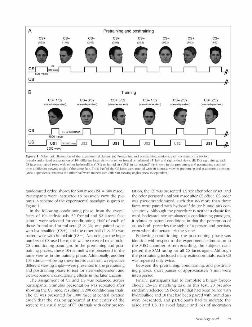
randomized order, shown for 500 msec (ISI = 500 msec).Participants were instructed to passively view the pic-tures. A scheme of the experimental paradigm is given inFigure 1.In the following conditioning phase, from the overall
faces of 104 individuals, 52 frontal and 52 lateral facestimuli were selected for conditioning. Half of each ofthese frontal and lateral sets (2 × 26) was paired twicewith hydrosulfide (CS+), and the other half (2 × 26) waspaired twice with humid air (CS−). According to the hugenumber of CS used here, this will be referred to as multi-CS conditioning paradigm. In the pretraining and post-training phases, these 104 stimuli were presented in thesame view as in the training phase. Additionally, another104 stimuli—showing these individuals from a respectivedifferent viewing angle—were presented in the pretrainingand posttraining phase to test for view-independent andview-dependent conditioning effects in the later analysis.The assignment of CS and US was balanced across
participants. Stimulus presentation was repeated aftershowing the CS once, resulting in 208 conditioning trials.The CS was presented for 1000 msec at central location(such that the nasion appeared at the center of thescreen) at a visual angle of 6°. On trials with odor presen-
tation, the CS was presented 1.5 sec after odor onset, andthe odor persisted until 500 msec after CS offset. CS orderwas pseudorandomized, such that no more than threefaces were paired with hydrosulfide (or humid air) con-secutively. Although the procedure is neither a classic for-ward, backward, nor simultaneous conditioning paradigm,it relates to natural conditions in that the perception ofodors both precedes the sight of a person and persists,even when the person left the scene.
Following conditioning, the posttraining phase wasidentical with respect to the experimental stimulation inthe MEG chamber. After recording, the subjects com-pleted the SAM rating for all CS faces again. Althoughthe posttraining included many extinction trials, each CSwas repeated only twice.
Between the pretraining, conditioning, and posttrain-ing phases, short pauses of approximately 5 min wereinterspersed.
Finally, participants had to complete a binary forced-choice CS–US matching task. In this test, 20 pseudo-randomly selected CS faces (10 that had been paired withhydrosulfide and 10 that had been paired with humid air)were presented, and participants had to indicate theassociated US. To avoid fatigue and loss of motivation
Figure 1. Schematic illustration of the experimental design. (A) Pretraining and posttraining sessions, each consisted of a twofoldpseudorandomized presentation of 104 different faces shown in either frontal or balanced 45° left- and right-sided views. (B) During training, eachCS face was paired twice with either hydrosulfide (US1) or humid air (US2) in its “original” (as shown in the pretraining and posttraining sessions)or in a different viewing angle of the same face. Thus, half of the CS faces were trained with an identical view in pretraining and posttraining sessions(view-dependent), whereas the other half were trained with different viewing angles (view-independent).
Steinberg et al. 19

because of unacceptable overall measurement time, onlya pseudorandomly selected subset of all CS was chosenfor the CS–US matching task. Overall, it took the partici-pants roughly about 60 min to complete the whole ex-periment. All behavioral tests were realized on a Dell®computer using Presentation® (Neurobehavioral Sys-tems, Albany, CA).
MEG Recording and Analysis
Visual-evoked magnetic fields were acquired using a 275-channel MEG whole-head sensor system (Omega 275,CTF, VSM Medtech Ltd., Coquitlam, British Columbia,Canada) with first-order axial SQUID gradiometers. Tomonitor the participantʼs position in the MEG scanner,landmark coils were attached to the two auditory canalsand the nasion. Furthermore, individual head shapeswere digitized using a Polhemus 3Space Fasttrack todetermine the individual head coordinate system. Con-tinuous MEG data in the frequency band between 0 and150 Hz were recorded with a sampling rate of 600 Hz.MEG data preprocessing, artifact rejection and correction,averaging, statistics, and visualization was conducted withthe Matlab-based EMEGS software (www.emegs.org;Peyk, De Cesarei, & Junghöfer, in press). Responses weredown-sampled to 300 Hz and filtered off-line between0.01 and 148 Hz. To optimize the detection of early andtransient brain responses in the higher-frequency range,further low-pass filtering was not applied. Data werealigned to stimulus onset, with an averaging epoch rangingfrom 200 msec before stimulus to 300 msec after stimulusand baseline-adjusted using a 150-msec pre-stimulus interval.The method for statistical control of artifacts in high-densityEEG/MEG data was used for single-trial data editing andartifact rejection ( Junghöfer, Elbert, Tucker, & Rockstroh,2000). This procedure (1) detects individual channelartifacts, (2) detects global artifacts, (3) replaces artifact-contaminated sensors with spline interpolation statisticallyweighted on the basis of all remaining sensors, and (4)computes the variance of the signal across trials to docu-ment the stability of the averaged waveform.1 The rejectionof artifact-contaminated trials and sensor epochs relies onthe calculation of statistical parameters for the absolutemeasured magnetic field amplitudes over time, their stan-dard deviation over time, the maximum of their gradientover time (first temporal derivative), and the determinationof boundaries for each of these three parameters. On aver-age, 5 of 275 sensors were interpolated and 9.6% of alltrials were rejected by the artifact rejection procedure.
After averaging, cortical sources of the event-relatedmagnetic fields were estimated using the L2 minimum-norm estimate method (Hämäläinen & Ilmoniemi, 1994).The L2 minimum-norm estimate method is an inversemodeling technique applied to reconstruct the topographyof the primary current underlying magnetic field distri-bution. It allows for the estimation of distributed neuralnetwork activity without a priori assumptions regarding the
location and/or number of current sources (Hauk, 2004). Inaddition, of all possible generator sources, only thoseexclusively determined by the measured magnetic fieldsare considered. A spherical shell with evenly distributed 2(azimuthal and polar direction, radial dipoles do not gener-ate magnetic fields outside of a sphere) × 350 dipoles wasused as source model. A source shell radius of 87% of theindividually fitted head radius was chosen, roughly corre-sponding to the gray matter depth. Although the distribu-ted source reconstruction inMEG does not give the preciselocation of cerebral generators, it allows for a fairly goodapproximation of cortical generators and correspondingassignment to larger cortical structures. Across all partici-pants and conditions, a Tikhonov regularization parameterk of 0.2 was applied.2 Topographies of source direction-independent neural activities—the vector length of the es-timated source activities at each position—were calculatedfor each individual participant, condition, and time pointon the basis of the averaged magnetic field distributionsand the individual sensor positions for each participantand run.Taking advantage of combining high-density MEG with
source localization methods, the analysis was based onthe neural generator activities obtained for every dipole,time point, and subject. To statistically test for condition-ing effects, these data were submitted to a repeated mea-sures ANOVA, including the factors Session (pretrainingvs. posttraining), Valence (negative vs. neutral), and View(frontal vs. lateral). As a result of this procedure, a spatio-temporal distribution of statistical values for each dipoleover time and across all subjects is obtained. Given a sig-nificance threshold of p < .05, only effects matching pre-defined spatio-temporal criteria survived this analysisstep. Accordingly, to avoid false-positive results, a statis-tical effect was considered meaningful only if it emergedin a region composed of at least 10 neighboring neuralsources (dipoles) and appeared within a time intervalcomprising at least 10 consecutive time points (33 msec).On the basis of the results of this spatio-temporal wave-form analysis, the identified regions showing a significantdifference were further analyzed using post hoc t tests tospecify the direction of effects. To assess conditioning ef-fects, brain activity measured in posttraining and pretrain-ing phases was compared with each other, thus two phaseswhere no odor was present at all. The pretraining andposttraining sessions were identical with respect to theirexperimental design.In advance of the Results section, it is important to
note that an additional analysis explored whether condi-tioning effects depended on an identical viewing angle inconditioning and testing phases (the identical image ofthe face) or if image-independent transfer of CS–US asso-ciations occurred (respective different viewing angle).This factor was labeled Image (same vs. different). How-ever, no significant conditioning effects were observed inthe posttraining period for faces shown in a different orien-tation than used during conditioning. This was true for
20 Journal of Cognitive Neuroscience Volume 24, Number 1

behavioral as well as MEG data. Further control analysis onthe basis of the mean activity in regions and time intervalsshowing conditioning effects also revealed no significantdifferences associated with faces in image-independentconditions. Accordingly, the data suggest that conditioningeffects were image-based and, for brevity, the Image factorwas not considered in the following analysis.
RESULTS
CS–US Matching Task
In the matching task, participants were asked to indicatewhich US (hydrosulfide or humid air) was delivered whileviewing the CS face. As expected and revealing an in-tended lack of contingency awareness, overall perfor-mance as measured by the sensitivity index d0 (M = 0.07,SD= 0.52)3 did not exceed chance (one-sample t test [testvalue = 0]: t(23) = 0.7, p = .49).
Valence Rating
To evaluate conditioning-induced changes in evaluativeratings of the CS faces, a repeated measures ANOVA in-cluding the factors Session, View, and US Valence wasconducted. Ratings of two participants were excludedfrom further analysis because of erroneous use of therating scales. As expected, a significant Session × USValence interaction was found, F(1, 21) = 5.2, p = .033,indicating a relatively more negative classification of CS+as compared with the CS− faces. Post hoc t tests forthe CSpost minus CSpre differences (CS+post − CS+pre vs.CS−post − CS−pre) revealed that CS+ faces (M = −.011,SD= .23) as compared with CS− faces (M= .12, SD= .28)were perceived to be more negative after conditioning(t(21) =−2.28, p= .033). Thus, despite the large numberof CS stimuli, evaluative conditioning effects were ob-served. Considering the results of the explicit CS–USmatching task, conditioning effects may occur implicitlyin the absence of awareness about CS–US pairings.Although the matching task probed only a randomly se-lected subset of CS, it is theoretically possible that, bychance, faces were primarily presented in the task, withmost pairings not explicitly learned by the subjects. How-ever, as the selection of this subset was completely randomand there was no tendency in any subject to reliably reportpreviously learned CS–US association, this issue does notappear to have biased the data. Conditioning had no effecton arousal ratings, hence results are not reported.
MEG
Waveform Analyses
Conditioning effects were explored by contrasting neuralactivity for CS+ (hydrosulfide) and CS− (humid air)
faces in the posttraining relative to the pretraining condi-tion. As expected, there were no significant differences inprocessing of the CS during the pretraining condition. Inthe posttraining condition, conditioning effects were re-vealed by the global power measure, showing a markedincrease in neural activity for faces paired with hydrosul-fide compared with faces paired with humid air in anearly (50–80 msec) and a midlatency (130–190 msec) timewindow (Figure 2). Between 50 and 80 msec, neuronalactivity associated with CS+ processing was increasedover frontal and right occipito-parieto-temporal regions.Later stages of processing (130–190 msec) revealedenhanced neural activity for CS+ processing in more dis-tributed frontal and occipito-parieto-temporal regions.
Area Score: “Early” Conditioning Effects (50–80 msec)
Each of the two source regions revealing conditioning ef-fects was submitted to an overall ANOVA analysis contain-ing the factors of Session (pretraining vs. posttraining),View (frontal vs. lateral), and US Valence (hydrosulfide vs.humid air).
Frontal region. A significant higher-order interaction ofSession × US Valence was observed, F(1, 23) = 14.89, p=.001. Although CS+ activity in this region significantlyincreased from pretraining to posttraining session (t(23) =−3.28, p = .003), CS− activity did not change (t(23) =0.58, p = .571; Figure 3A).
Right occipito-parieto-temporal region. Similar to thefrontal region, a significant interaction of Session × USValence was found, F(1, 23) = 9.86, p = .005. There wasa relative increase in CS+ processing as neural activity did
Figure 2. L2 minimum-norm difference. Global power of thedifference of estimated neural activity (posttraining minuspretraining) for CS+ (red line) and CS− (gray line) faces. CS+ facesevoke stronger neural activity after learning in an early andmidlatency time interval.
Steinberg et al. 21

not change between pretraining and posttraining (t(23) =−1.15, p = .264), whereas activity in the CS− (t(23) =2.53, p = .019) condition was significantly decreased(Figure 3B).
To identify the onset of effects found in both regions, a10-msec sliding window analysis for an interval rangingfrom 30 to 100 msec was performed. For each interval,the above-described ANOVA was calculated. In the frontalregion, the first significant difference between CS+ andCS− processing occurred in the 40- to 50-msec interval,F(1, 23) = 7.87, p= .01, whereas in the occipito-parieto-temporal region, the first significant effect was found inthe 50- to 60-msec interval, F(1, 23) = 4.83, p = .038).Thus, effects in the frontal region seem to precede effectsin the right occipito-parieto-temporal region.
Area Score: “Midlatency” Conditioning Effects(130–190 msec)
Three source regions showing conditioning effects wereanalyzed using the above-mentioned ANOVA.
Frontal region. Similar to the effects observed in the ear-lier interval, a highly significant interaction of Session ×
US Valence was found in a more lateral frontal region,F(1, 23) = 8.89, p = .007. Within this region, CS+ faces(t(23) = −3.24, p = .004) evoked significantly strongerresponses than CS− (t(23) = −0.072, p = .943) faces(Figure 4A).
Right and left occipito-parieto-temporal regions. As ex-pected, in these bilateral and more distributed regions,significant differences in the processing of the CS be-tween the pretraining and posttraining conditions werefound (right: F(1, 23) = 4.56, p = .044; left: F(1, 23) =10.04, p = .004).Within both areas, significant effects were characterized
by decreased neural activity in the CS− (right: t(23) =3.05, p = .006; left: t(23) = 3.5, p = .002) as comparedwith the CS+ (right: t(23) = 1.06, p = .299; left: t(23) =0.35, p = .729) condition. Finally, a left superior frontalregion failed to reach statistical significance ( p > .05) inthe second level analysis (Figure 4B).
DISCUSSION
Aversive associative learning was used to examine facialemotional processing. Using a much larger number of
Figure 3. Effects in the earlytime interval. (A) Top:Spherical projection ofsignificant ( p < .05).Session × US Valenceinteraction effects withinthe early time interval ontoa 3-D brain model (statisticalparametric map in frontview). Bottom: Mean neuralactivity within the frontal ROIfor the early interval.Bar graphs represent thedifference of activitybetween pretraining andposttraining (posttrainingminus pretraining). Thus,positive values indicatestronger activation whereasnegative values indicate lessactivity in the posttrainingphase. (B) Same as in (A)but shown for the rightoccipito-parieto-temporalregion. Both ROIs revealstronger neural CS+compared with CS−activity. Error bars indicate95% confidence interval.Please note that a directstatistical comparisonbetween the differencevalues for CS+ and CS−is not necessary, as thiswould result in the samep value as for the interactionof the corresponding ANOVA.
22 Journal of Cognitive Neuroscience Volume 24, Number 1

CS stimuli than previous studies, highly significant con-ditioning effects were observed, indicating that aversiveconditioning, unbeknownst to the individual, affects (emo-tional) face processing to a much greater extent thanpreviously thought. The main finding relates to the roleof PFC activation. CS+ compared with CS− processingwas associated with enhanced neural activity in PFC en-compassing lateral and orbital regions. This PFC activationoccurred rather early in the processing stream (∼50 msec),even preceding enhanced CS+ processing in infero-temporal regions implicated in object recognition. Overall,aversive associative learning efficiently alters the process-ing of socially relevant stimuli and induces short-latencyresponses in cortical structures related to object recogni-tion, categorization, and emotion processing.In both timewindows, between 50–80 and 130–190msec,
faces associated with hydrosulfide elicited more neuralactivity in the OFC than did faces associated with humidair. TheOFChas been implicated in stimulus–reinforcementlearning inmacaques, where neurons responded to learningnew associations between a neutral and a primary reinforcer(Kringelbach & Rolls, 2004; Thorpe, Rolls, & Maddison,1983). Furthermore, single-cell recordings in macaquesrevealed neurons in the OFC that selectively responded tofaces, providing a possible neuronal basis for the rapidlearning of face–reinforcement associations (Rolls, 2004).Human studies investigating associative learning are con-sistent with this notion. For instance, in a visuo-olfactoryconditioning study,OFC regionswere implicated in the stor-age of previously learned CS–US associations presumablymediated by an OFC-amygdala network (Gottfried &
Dolan, 2004). Overall, the present findings are in generalagreement with the notion that the OFC is involved inlearning new associations, and given its reciprocal connec-tions with the amygdale (Ghashghaei & Barbas, 2002), anearly engagement in emotion discrimination is likely.
In addition, faces associated with hydrosulfide relativeto humid air also elicited increased activity on the lateralsurface of PFC (Figure 3A). Previous animal studies haveshown that neuronal activity in lateral PFC and ITC reflectsthe category of visual stimuli. The ITC seems to be moreinvolved in the visual analysis of currently viewed images,whereas lateral PFC regions rather encode for behaviorallyrelevant factors, such as category membership (Freedman,Riesenhuber, Poggio, & Miller, 2003). A hallmark functionof visual categorization is that perceptually similar objectsmay be treated as different when they belong to differentcategories, whereas perceptually dissimilar objects aretreated alike when they belong to the same category(Wyttenbach, May, & Hoy, 1996). The present findingsimply that the emotional value can serve as a cardinal basisof categorization. Functionally, treating themany faces con-ditioned with an aversive odor in a similar way seemshighly adaptive to activate a motivational orientation ofavoidance (Lang, Bradley, & Cuthbert, 1997). Accordingly,it is proposed that lateral prefrontal activation may reflecta fundamental and basic categorization of human expe-rience: the discrimination of “bad” from “good” (Langet al., 1997; Cacioppo, Crites, Berntson, & Coles, 1993).
In addition, activity in both time windows extends tomotor and/or premotor areas (see Figures 3 and 4) ofthe right hemisphere, although no explicit task was given
Figure 4. Effects in the midlatency time interval. Procedure as in Figure 3. Again, right prefrontal and bilateral occipito-parieto-temporal regionsshow relatively stronger neural activity for CS+ compared with CS− processing.
Steinberg et al. 23
to the subjects requiring motor reactions, for example, but-ton presses. However, there are studies showing that thesupramarginal gyrus is involved in emotional processingas lesions of the right somatosensory areas (includingsupramarginal gyrus) can lead to deficits in judging emo-tions from faces (e.g., Adolphs, Damasio, Tranel, Cooper,& Damasio, 2000). Accordingly, it might be that somato-sensory areas are activated to increase sensitivity to bodilyresponses and encoded emotional associations. Alterna-tively, the activation along the parietal and frontal axis seenin Figures 3 and 4 might reflect activation in attentionalnetworks, that is, directing visuospatial attentional re-sources toward salient emotional features such as theeyes (Adolphs & Spezio, 2006).
Lateral PFC, orbital PFC, and ITC are reciprocally con-nected, providing a neural basis for the categorization ofvisual information (Bar, 2003; Freedman et al., 2003).Furthermore, the OFC is part of an OFC–amygdala–ITCtriad, which has extensive reciprocal connections toother limbic areas, premotor areas, and the autonomicmotor system (Ghashghaei & Barbas, 2002). Thus, PFCactivation may facilitate the detection of danger to avoidharm. It has been suggested that the early activation ofPFC may guide visual information processing by providingtop–down expectations to the ventral processing stream(Adolphs, 2002). A critical issue in this regard is thatPFC activation may precede activation in object-relatedregions in the temporal cortex, that is, lateral occipitalcortex and fusiform gyrus (Bar et al., 2006). The currentfindings support this notion, as PFC activation precededtemporal cortex activation. However, although latenciesof PFC and temporal cortex activations in the later timewindow (130–190 msec; Figure 4) nicely fit to previouslyreported latencies for PFC-driven modulation of sensoryprocessing (>120 msec; e.g., Rudrauf et al., 2008; Bar,2003; Adolphs, 2002), activation in the earlier time window(50–80 msec; Figure 3) indicates PFC-driven sensory mod-ulations already in the initial wave of processing. With re-gard to existing anatomical pathways, several alternativeexplanations of this result appear feasible:
First, rapid modulations mediated by limbic structurescould contribute to the early effects found here. It hasbeen shown that affect-related information can be con-veyed to emotional structures like the amygdala via directprojections from the sensory thalamus with response laten-cies around 20–30 msec (LeDoux, 2000) and 20–50 msec(Luo et al., 2009; Luo, Holroyd, Jones, Hendler, & Blair,2007) in the auditory and visual domain, respectively.The amygdala, as part of a widely distributed emotionalnetwork, shares numerous, mainly reciprocal connectionswith, for example, OFC, medial PFC, and the entire ventralvisual stream (Freese & Amaral, 2005; Ghashghaei &Barbas, 2002), perhaps facilitating processing in theseareas. Thus, initial stimulus categorization could be domi-nated by evaluation of emotional significance, allowing forrapid preparation of basic motivational systems (LeDoux,2000) and may even precede full (e.g., view-independent)
object recognition. However, in a recent review, Pessoaand Adolphs (2010) challenge the model of a fast thalamo-amygdala pathway in humans for the rapid transmission ofemotion-related information. Thus, this pathway might beless likely to produce the early effects seen in the presentstudy.Second, it is possible that fast geniculo-cortico-cortical
pathways are involved. Earliest response latencies in occip-ital (V1–V4), parietal (MT, MST), and prefrontal (FEF) areasin the macaque can be seen before 65 msec (Bullier, 2001;Lamme& Roelfsema, 2000). In humans, Kirchner, Barbeau,Thorpe, Régis, & Liégeois-Chauvel (2009) reported earliestV1 and FEF responses around 25 and 45msec, respectively.Thus, sensory information can reach frontal areas ex-tremely fast (<50 msec) through rapid cortico-corticalroutes (e.g., long-range association fibers such as longitu-dinal fasciculus). Our finding further accords with ultra-rapid saccadic eye movements (∼120 msec) in an objectdetection task, suggesting that a first analysis of even com-plex visual stimuli is provided within less than 100 msec(Kirchner & Thorpe, 2006).Third, it has been suggested that the remaining noncon-
scious visual capacities in blindsight patients might bemediated by an extrageniculostriate pathway, “bypassing”V1, involving the superior colliculus and the posteriorvisual thalamus (Morris, DeGelder, Weiskrantz, & Dolan,2001). Consistent with the current findings, a recentMEG study revealed increased activity to facial stimuli withemotional expressions at midtemporal visual areas alreadyaround 70 msec in a patient with “affective blindsight”(Andino, Menendez, Khateb, Landis, & Pegna, 2009).Currently, we cannot provide clear evidence in favor of
one main pathway or even a network combination of thesethree candidate pathways accounting for the early effectsfound in the present study. However, our findings—at leastfor the early interval—seem difficult to reconcile with astrict hierarchically organized model of visual processing.In such a model, earliest differential CS+ activity at PFCregions should have been observed in the midlatency timeinterval (>120msec). The finding of differential CS process-ing already between 50 and 80msec suggests that emotion-ally significant information is distributed to cortical(prefrontal regions) and subcortical structures early inthe processing stream (cf. Vuilleumier, 2005; Bullier, 2001).The finding in the midlatency time window (130–
190 msec) is consistent with previous EEG and MEGstudies reporting the amplified processing of aversivelyconditioned faces in this time region (Dolan, Heinze,Hurlemann, & Hinrichs, 2006; Pizzagalli et al., 2003).However, compared with these studies, which relied ona posttraining comparison of CS processing only, the pre-training condition of the present study provided addi-tional information regarding aversive conditioningeffects. Specifically, the amplified processing of CS+ ascompared with CS− processing can reflect either thatthe activation for the CS+ is stronger in the posttrainingsession whereas responses to the CS− seem to be
24 Journal of Cognitive Neuroscience Volume 24, Number 1

unaffected by conditioning across sessions (CS+post >CS+pre vs. CS−post = CS−pre), or the activation for theCS+ does not change from pretraining to posttrainingsessions whereas the responses to the CS− significantlydecrease or habituate, respectively (CS+post = CS+pre vs.CS−post < CS−pre). In both cases, activations in the post-training phase would be stronger for the CS+ comparedwith the CS− stimuli. Inspection of Figures 3 and 4 re-veals evidence for both alternatives: Over anterior frontalregions, CS+ processing seems to be facilitated as com-pared with pretraining, whereas CS− processing appearsattenuated at posterior occipito-temporal regions. Over-all, the occurrence of these response patterns might sug-gest different modes of biasing processing toward themore relevant CS+ in the posttraining phase, possiblyreflecting functional differences among brain regions.Previous studies provided evidence for PFC-driven
modulation of sensory processing (e.g., Rudrauf et al.,2008; Bar et al., 2006; Adolphs, 2002). It has been sug-gested that PFC regions can influence even early percep-tual processing in a top–down manner within the first120msec (Adolphs, 2002), possibly starting around 80msecafter stimulus onset (Bar et al., 2006) or even earlier, assuggested here. Additionally, lateral prefrontal regionshave been implicated in stimulus categorization and theencoding of behaviorally relevant information (Freedmanet al., 2003). Furthermore, neurons in the right PFC areselectively elicited by aversive events in a time range be-tween 120 and 160 msec (Kawasaki et al., 2001). Otherstudies regarding basic face perception have shown thatresponses to faces habituate over time if they are nottask-relevant (Maurer, Rossion, & McCandliss, 2008). Giventhat frontal areas are supposed to represent the emotionalvalue of sensory stimuli, we suggest that the activity foundin frontal areas in the present study guides the activityin more occipito-temporal areas by providing initial top–down guesses (Bar et al., 2006) and, furthermore, preventshabituation to the CS+ (because they are emotionally rel-evant) which, in turn, is evident for the CS−, leading torelative decrease of occipito-temporal CS− activation inthe pre–post comparison.The present findings also parallel previous results re-
garding enhanced CS+ sensory processing in the earlytime interval (<100 ms). For instance, previous researchusing gratings as CS+ stimuli revealed early differentialprocessing of CS+ compared with CS− stimuli in theC1 time window between 65 and 90 msec (Keil et al.,2007; Stolarova et al., 2006). However, whereas theserather simple CS+ stimuli primarily enhanced primaryvisual cortex activation, the more complex face CS+evoked more widespread preferential processing in sec-ondary occipito-temporal regions in the present study.Furthermore and as predicted by current models ofobject recognition (Riesenhuber & Poggio, 2000), en-hanced processing of CS+ faces within the early and mid-latency intervals appears to be based on view-dependentrepresentations.
Taken together, our results reveal a remarkable capacityfor emotional categorization. Pairing each of the 52 CS+faces with an aversive odor only twice was sufficient to in-duce associative learning. The large number of CS stimulieffectively prevented contingency awareness. When explic-itly asked, participants were at chance level to indicate CS–US relationships. In contrast, emotional valence ratingsclearly differed between CS conditions, as CS+ faces wererated as more unpleasant than CS− faces. Thus, there is adissociation between explicit representation of CS–US con-tingencies and implicit CS–US effects revealed by MEGfindings and self-report measures. This result is consistentwith numerous data demonstrating associative implicitlearning without necessarily requiring the explicit knowl-edge of contingencies (see Ohman & Mineka, 2001).
Overall, this study revealed associative learning effectsin extrastriate visual as well as prefrontal areas, which oc-curred in the absence of contingency awareness, with astimulus set overwhelmingly exceeding working memorycapacity and with only two learning instances. The ratherearly onset of the prefrontal effects (∼40–50 msec) isconsistent with nonsequential models of visual processingin which emotionally significant information leads toamplified processing in a distributed cortico-subcorticalnetworks early in the processing stream preceding fullvisual analysis.
Acknowledgments
This work was supported by the German Research Association(DFG; SFB-TRR58-C1) and the Academy of Science, Heidelberg.
Reprint requests should be sent toDr. Markus Junghöfer, Institutefor Biomagnetism and Biosignalanalysis, University of Münster,Malmedyweg 15, 48149 Münster, Germany, or via e-mail: [email protected].
Notes
1. The artifact detection process in EMEGS tests the good-ness of interpolation by interpolating a multitude (number ofsensors = 275) of test topographies. If many to-be-interpolatedsensors fall into one region or if many noisy sensors are positionedat the edge of the sensor coverage, many test topographies arenot interpolated with a sufficient accuracy and the correspondingtrials get rejected from further analysis.2. The degrees of freedom of the distributed source modelexceed the number of measured MEG sensors. The inversemodeling, thus, poses an underdetermined problem and theleadfield matrix inversion needs some regularization. The Tikhonovregularization is the most commonly used method of regulariza-tion of ill-posed problems.3. Formula used for calculating d0: d0 = z(hits) − z(falsealarms). In this formula, z calculation is based on the inverseof the normal distribution (see Green & Swets, 1966).
REFERENCES
Adolphs, R. (2002). Neural systems for recognizing emotion.Current Opinion in Neurobiology, 12, 169–177.
Steinberg et al. 25

Adolphs, R., Damasio, H., Tranel, D., Cooper, G., & Damasio,A. R. (2000). A role for somatosensory cortices in the visualrecognition of emotion as revealed by three-dimensionallesion mapping. Journal of Neuroscience, 20, 2683–2690.
Adolphs, R., & Spezio, M. (2006). Role of the amygdala inprocessing visual social stimuli. Progress in Brain Research,156, 363–378.
Andino, S. L., Menendez, R. G., Khateb, A., Landis, T., & Pegna,A. J. (2009). Electrophysiological correlates of affectiveblindsight. Neuroimage, 44, 581–589.
Bar, M. (2003). A cortical mechanism for triggering top–downfacilitation in visual object recognition. Journal of CognitiveNeuroscience, 15, 600–609.
Bar, M., Kassam, K. S., Ghuman, A. S., Boshyan, J., Schmid,A. M., Dale, A. M., et al. (2006). Top–down facilitation ofvisual recognition. Proceedings of the National Academyof Sciences, U.S.A., 103, 449–454.
Bradley, M. M., & Lang, P. J. (1994). Measuring emotion: Theself-assessment manikin and the semantic differential. Journalof Behavior Therapy and Experimental Psychiatry, 25, 49–59.
Bradley, M. M., Sabatinelli, D., Lang, P. J., Fitzsimmons, J. R.,King, W., & Desai, P. (2003). Activation of the visual cortex inmotivated attention. Behavioral Neuroscience, 117, 369–380.
Bullier, J. (2001). Integrated model of visual processing. BrainResearch, Brain Research Reviews, 36, 96–107.
Cacioppo, J. T., Crites, S. L., Jr., Berntson, G. G., & Coles, M. G.(1993). If attitudes affect how stimuli are processed shouldthey not affect the event-related brain potential?Psychological Science, 4, 108–112.
De Gelder, B., Vroomen, J., Pourtois, G., & Weiskrantz, L.(1999). Non-conscious recognition of affect in the absence ofstriate cortex. NeuroReport, 10, 3759–3763.
Dolan, R. J., Heinze, H. J., Hurlemann, R., & Hinrichs, H. (2006).Magnetoencephalography (MEG) determined temporalmodulation of visual and auditory sensory processing in thecontext of classical conditioning to faces. Neuroimage, 32,778–789.
Freedman, D. J., Riesenhuber, M., Poggio, T., & Miller, E. K.(2003). A comparison of primate prefrontal and inferiortemporal cortices during visual categorization. Journal ofNeuroscience, 23, 5235–5246.
Freese, J. L., & Amaral, D. G. (2005). The organization ofprojections from the amygdala to visual cortical areas TE andV1 in the macaque monkey. The Journal of ComparativeNeurology, 486, 295–317.
Ghashghaei, H. T., & Barbas, H. (2002). Pathways for emotion:Interactions of prefrontal and anterior temporal pathways inthe amygdala of the rhesus monkey. Neuroscience, 115,1261–1279.
Gottfried, J. A., Deichmann, R., Winston, J. S., & Dolan, R. J.(2002). Functional heterogeneity in human olfactory cortex:An event-related functional magnetic resonance imagingstudy. Journal of Neuroscience, 22, 10819–10828.
Gottfried, J. A., & Dolan, R. J. (2004). Human orbitofrontal cortexmediates extinction learning while accessing conditionedrepresentations of value. Nature Neuroscience, 7, 1144–1152.
Green, D. M., & Swets, J. A. (1966). Signal detection theory andpsychophysics. New York: Wiley.
Hämäläinen, M. S., & Ilmoniemi, R. J. (1994). Interpretingmagnetic fields of the brain: Minimum norm estimates.Medical & Biological Engineering & Computing, 32, 35–42.
Hamm, A. O., Weike, A. I., Schupp, H. T., Treig, T., Dressel, A.,& Kessler, C. (2003). Affective blindsight: Intact fearconditioning to a visual cue in a cortically blind patient.Brain, 126, 267–275.
Hauk, O. (2004). Keep it simple: A case for using classicalminimum norm estimation in the analysis of EEG and MEGdata. Neuroimage, 21, 1612–1621.
Johnson, M. H. (2005). Subcortical face processing. NatureReviews Neuroscience, 6, 766–774.
Junghöfer, M., Bradley, M. M., Elbert, T. R., & Lang, P. J. (2001).Fleeting images: A new look at early emotion discrimination.Psychophysiology, 38, 175–178.
Junghöfer, M., Elbert, T., Tucker, D., & Rockstroh, B. (2000).Statistical control of artifacts in dense array EEG/MEGstudies. Psychophysiology, 37, 523–532.
Junghöfer, M., Schupp, H., Stark, R., & Vaitl, D. (2005).Neuroimaging of emotion: Empirical effects of proportionalglobal signal scaling in fMRI data analysis. Neuroimage, 25,520–526.
Kanwisher, N., & Yovel, G. (2006). The fusiform face area: Acortical region specialized for the perception of faces.Philosophical Transactions of the Royal Society of London,B, 361, 2109–2128.
Kawasaki, H., Kaufman, O., Damasio, H., Damasio, A. R.,Granner, M., Bakken, H., et al. (2001). Single-neuronresponses to emotional visual stimuli recorded in humanventral prefrontal cortex. Nature Neuroscience, 4, 15–16.
Keil, A., Stolarova, M., Moratti, S., & Ray, W. J. (2007).Adaptation in human visual cortex as a mechanism for rapiddiscrimination of aversive stimuli. Neuroimage, 36, 472–479.
Kirchner, H., Barbeau, E. J., Thorpe, S. J., Régis, J., & Liégeois-Chauvel, C. (2009). Ultra-rapid sensory responses in thehuman frontal eye field region. Journal of Neuroscience, 29,7599–7606.
Kirchner, H., & Thorpe, S. J. (2006). Ultra-rapid object detectionwith saccadic eye movements: Visual processing speedrevisited. Vision Research, 46, 1762–1776.
Kissler, J., Herbert, C., Peyk, P., & Junghöfer, M. (2007).“Buzzwords”—Spontaneous cortical responses to emotionalwords. Psychological Science, 18, 475–480.
Kringelbach, M. L., & Rolls, E. T. (2004). The functionalneuroanatomy of the human orbitofrontal cortex: Evidencefrom neuroimaging and neuropsychology. Progress inNeurobiology, 72, 341–372.
Lamme, V. A., & Roelfsema, P. R. (2000). The distinct modes ofvision offered by feedforward and recurrent processing.Trends in Neurosciences, 23, 571–579.
Lang, P. J., Bradley, M. M., & Cuthbert, B. N. (1997). Motivatedattention: Affect, activation, and action. In P. J. Lang, R. F.Simons, & M. Balaban (Eds.), Attention and emotion:Sensory and motivational processes (pp. 97–135). New York:Erlbaum.
LeDoux, J. E. (2000). Emotion circuits in the brain. AnnualReview of Neuroscience, 23, 155–184.
Lundqvist, D., Flykt, A., & Öhman, A. (1998). The KarolinskaDirected Emotional Faces–KDEF, CD ROM from Departmentof Clinical Neuroscience, Psychology Section, KarolinskaInstitute, ISBN 91-630-7164-9.
Luo, Q., Holroyd, T., Jones, M., Hendler, T., & Blair, J. (2007).Neural dynamics for facial threat processing as revealed bygamma band synchronization using MEG. Neuroimage, 34,839–847.
Luo, Q., Mitchell, D., Cheng, X., Mondillo, K., Mccaffrey, D.,Holroyd, T., et al. (2009). Visual awareness, emotion, andgamma band synchronization. Cerebral Cortex, 19, 1896–1904.
Maurer, U., Rossion, B., & McCandliss, B. D. (2008). Categoryspecificity in early perception: Face and word n170 responsesdiffer in both lateralization and habituation properties.Frontiers in Human Neuroscience, 2, 1–7.
Morel, S., Ponz, A., Mercier, M., Vuilleumier, P., & George, N.(2009). EEG-MEG evidence for early differential repetitioneffects for fearful, happy and neutral faces. Brain Research,1254, 84–98.
Morris, J. S., DeGelder, B., Weiskrantz, L., & Dolan, R. J. (2001).Differential extrageniculostriate and amygdala responses to
26 Journal of Cognitive Neuroscience Volume 24, Number 1

presentation of emotional faces in a cortically blind field.Brain, 124, 1241–1252.
Morris, J. S., Ohman, A., & Dolan, R. J. (1998). Conscious andunconscious emotional learning in the human amygdala.Nature, 393, 467–470.
Ohman, A., & Mineka, S. (2001). Fears, phobias, andpreparedness: Toward an evolved module of fear and fearlearning. Psychological Review, 108, 483–522.
Padmala, S., & Pessoa, L. (2008). Affective learning enhancesvisual detection and responses in primary visual cortex.Journal of Neuroscience, 28, 6202–6210.
Pessoa, L., & Adolphs, R. (2010). Emotion processing and theamygdala: From a “low road” to “many roads” of evaluatingbiological significance. Nature Reviews Neuroscience, 11,773–783.
Peyk, P., De Cesarei, A., & Junghöfer, M. (in press).ElectroMagnetoEncephaloGraphy Software (EMEGS):Overview and integration with other EEG/MEG toolboxes.Computational Intelligence and Neuroscience, Specialissue: Academic software applications for electromagneticbrain mapping using MEG and EEG. doi: 10.1155/2011/861705.
Pizzagalli, D., Regard, M., & Lehmann, D. (1999). Rapidemotional face processing in the human right and left brainhemispheres: An ERP study. NeuroReport, 10, 2691–2698.
Pizzagalli, D. A., Greischar, L. L., & Davidson, R. J. (2003).Spatio-temporal dynamics of brain mechanisms in aversiveclassical conditioning: High-density event-related potentialand brain electrical tomography analyses. Neuropsychologia,41, 184–194.
Riesenhuber, M., & Poggio, T. (2000). Models of objectrecognition. Nature Neuroscience, 3, 1199–1204.
Rolls, E. T. (2004). The functions of the orbitofrontal cortex.Brain and Cognition, 55, 11–29.
Rudrauf, D., David, O., Lachaux, J. P., Kovach, C. K., Martinerie,J., Renault, B., et al. (2008). Rapid interactions between theventral visual stream and emotion-related structures rely on atwo-pathway architecture. Journal of Neuroscience, 28,2793–2803.
Sabatinelli, D., Bradley, M. M., Fitzsimmons, J. R., & Lang, P. J.(2005). Parallel amygdala and inferotemporal activationreflect emotional intensity and fear relevance. Neuroimage,24, 1265–1270.
Schupp, H., Flaisch, T., Stockburger, J., & Junghöfer, M. (2006).Emotion and attention: Event-related brain potential studies.Progress in Brain Research, 156, 31–51.
Stolarova, M., Keil, A., & Moratti, S. (2006). Modulation of theC1 visual event-related component by conditioned stimuli:Evidence for sensory plasticity in early affective perception.Cerebral Cortex, 16, 876–887.
Thorpe, S. J., Rolls, E. T., & Maddison, S. (1983). Theorbitofrontal cortex: Neuronal activity in the behavingmonkey. Experimental Brain Research, 49, 93–115.
Vuilleumier, P. (2005). How brains beware: Neural mechanisms ofemotional attention. Trends in Cognitive Sciences, 9, 585–594.
Vuilleumier, P., Armony, J. L., Driver, J., & Dolan, R. J. (2001).Effects of attention and emotion on face processing in thehuman brain: An event-related fMRI study.Neuron, 30, 829–841.
Wyttenbach, R. A., May, M. L., & Hoy, R. R. (1996). Categoricalperception of sound frequency by crickets. Science, 273,1542–1544.
Zeki, S. (1993). A vision of the brain. Oxford, UK: BlackwellScientific Publishing.
Steinberg et al. 27
Copyright © 2022 FDOKUMEN