Transcriptional coactivator PGC1α controls the energy state and contractile function of cardiac...
13
ARTICLE Transcriptional coactivator PGC-1α controls the energy state and contractile function of cardiac muscle Zoltan Arany, 1,7 Huamei He, 2,7 Jiandie Lin, 1,7 Kirsten Hoyer, 2 Christoph Handschin, 1 Okan Toka, 3 Ferhaan Ahmad, 3 Takashi Matsui, 6 Sherry Chin, 1 Pei-Hsuan Wu, 1 Igor I. Rybkin, 4 John M. Shelton, 4 Monia Manieri, 5 Saverio Cinti, 5 Frederick J. Schoen, 2 Rhonda Bassel-Duby, 4 Anthony Rosenzweig, 6 Joanne S. Ingwall, 2 and Bruce M. Spiegelman 1, * 1 Dana Farber Cancer Institute and the Department of Cell Biology, Harvard Medical School, Boston, Massachusetts 02115 2 NMR Laboratory for Physiological Chemistry, Department of Medicine, Brigham and Women’s Hospital and Harvard Medical School, Boston, Massachusetts 02115 3 Brigham and Women’s Hospital and Harvard Medical School, Boston, Massachusetts 02115 4 Departments of Molecular Biology and Internal Medicine, University of Texas Southwestern Medical Center, Dallas, Texas 75390 5 Institute of Normal Human Morphology, Faculty of Medicine, University of Ancona, Ancona 60020, Italy 6 Program in Cardiovascular Gene Therapy, CVRC, and Harvard Medical School, Boston, Massachusetts 02129 7 These authors contributed equally to this work. *Correspondence: [email protected] Summary Skeletal and cardiac muscle depend on high turnover of ATP made by mitochondria in order to contract efficiently. The transcriptional coactivator PGC-1α has been shown to function as a major regulator of mitochondrial biogenesis and respiration in both skeletal and cardiac muscle, but this has been based only on gain-of-function studies. Using genetic knockout mice, we show here that, while PGC-1α KO mice appear to retain normal mitochondrial volume in both muscle beds, expression of genes of oxidative phosphorylation is markedly blunted. Hearts from these mice have reduced mito- chondrial enzymatic activities and decreased levels of ATP. Importantly, isolated hearts lacking PGC-1α have a diminished ability to increase work output in response to chemical or electrical stimulation. As mice lacking PGC-1α age, cardiac dysfunction becomes evident in vivo. These data indicate that PGC-1α is vital for the heart to meet increased demands for ATP and work in response to physiological stimuli. Introduction numerous DNA binding transcription factors and then coordi- nating several biochemical events, including recruitment of Living organisms must convert chemical energy into mechani- chromatin modifying enzymes such as p300/CBP and SRC-1 cal work. Muscle is a specialized tissue devoted primarily to (Puigserver et al., 1999), interaction with the basal transcription this task. The energy needs in muscle are quite high and must machinery (Wallberg et al., 2003), and linking of transcription be precisely regulated. Nowhere is this more true than in the to RNA splicing (Monsalve et al., 2000). The outcome is robust heart, where work is generated unfailingly for decades and activation of gene expression in coherent metabolic pathways. where energy consumption is higher than in any other organ. PGC-1β, identified by virtue of sequence homology to PGC- The availability of energy in skeletal and cardiac muscle can be 1α, has a similar tissue distribution as PGC-1α and coactivates significantly altered in both health and disease. Chronic exer- an overlapping but not identical repertoire of transcription cise in skeletal muscle, for example, stimulates a switching factors (Kressler et al., 2002; Lin et al., 2002a; Lin et al., 2003). from predominantly glycolytic to more oxidative fibers, which In the liver, for instance, PGC-1α docks HNF4α and FOXO1 contain more mitochondria and are resistant to fatigue (Booth (Lin et al., 2003) and activates gluconeogenesis, while PGC-1β and Thomason, 1991). Conversely, aberrations in energy pro- activates lipid biosynthesis by docking SREBP (Lin et al., duction are seen in such diverse muscular diseases as muscu- 2005). lar dystrophies, mitochondrial myopathies (Kelly and Strauss, One of the primary effects of PGC-1α is the activation of 1994; Wallace, 1992), and chronic congestive heart failure mitochondrial biogenesis and oxidative phosphorylation. In (Ingwall, 2002; Ingwall and Weiss, 2004). How these defects brown fat, cold exposure induces PGC-1α expression, which come about remains incompletely understood, in particular at leads to mitochondrial proliferation, uncoupling of oxidative the level of gene expression control. phosphorylation through increased expression of UCP-1, and PPARγ coactivator-1α (PGC-1α) and -β are potent transcrip- energy dissipation in the form of heat (Lin et al., 2004; Puig- tional coactivators of nuclear receptors and other transcription server et al., 1998). In skeletal and cardiac muscle cells, forced factors (Knutti and Kralli, 2001; Puigserver and Spiegelman, expression of PGC-1α in vitro activates mitochondrial biogene- 2003; Scarpulla, 2002). They can control specific metabolic sis, oxidative phosphorylation, and respiration (Lehman et al., pathways, especially oxidative metabolism, in a variety of tis- 2000; St-Pierre et al., 2003). Forced expression of PGC-1α in type II skeletal muscle fibers in vivo leads to mitochondrial pro- sues. PGC-1α targets promoters by interacting directly with CELL METABOLISM : APRIL 2005 · VOL. 1 · COPYRIGHT © 2005 ELSEVIER INC. DOI 10.1016/j.cmet.2005.03.002 259
-
Upload
independent -
Category
Documents
-
view
0 -
download
0
Transcript of Transcriptional coactivator PGC1α controls the energy state and contractile function of cardiac...

A R T I C L E
Transcriptional coactivator PGC-1α controls the energy stateand contractile function of cardiac muscle
Zoltan Arany,1,7 Huamei He,2,7 Jiandie Lin,1,7 Kirsten Hoyer,2 Christoph Handschin,1 Okan Toka,3 Ferhaan Ahmad,3
Takashi Matsui,6 Sherry Chin,1 Pei-Hsuan Wu,1 Igor I. Rybkin,4 John M. Shelton,4 Monia Manieri,5 Saverio Cinti,5
Frederick J. Schoen,2 Rhonda Bassel-Duby,4 Anthony Rosenzweig,6 Joanne S. Ingwall,2
and Bruce M. Spiegelman1,*
1Dana Farber Cancer Institute and the Department of Cell Biology, Harvard Medical School, Boston, Massachusetts 021152 NMR Laboratory for Physiological Chemistry, Department of Medicine, Brigham and Women’s Hospital and Harvard Medical School, Boston,
Massachusetts 021153 Brigham and Women’s Hospital and Harvard Medical School, Boston, Massachusetts 021154 Departments of Molecular Biology and Internal Medicine, University of Texas Southwestern Medical Center, Dallas, Texas 753905 Institute of Normal Human Morphology, Faculty of Medicine, University of Ancona, Ancona 60020, Italy6 Program in Cardiovascular Gene Therapy, CVRC, and Harvard Medical School, Boston, Massachusetts 021297 These authors contributed equally to this work.*Correspondence: [email protected]
Summary
Skeletal and cardiac muscle depend on high turnover of ATP made by mitochondria in order to contract efficiently. Thetranscriptional coactivator PGC-1α has been shown to function as a major regulator of mitochondrial biogenesis andrespiration in both skeletal and cardiac muscle, but this has been based only on gain-of-function studies. Using geneticknockout mice, we show here that, while PGC-1α KO mice appear to retain normal mitochondrial volume in both musclebeds, expression of genes of oxidative phosphorylation is markedly blunted. Hearts from these mice have reduced mito-chondrial enzymatic activities and decreased levels of ATP. Importantly, isolated hearts lacking PGC-1α have a diminishedability to increase work output in response to chemical or electrical stimulation. As mice lacking PGC-1α age, cardiacdysfunction becomes evident in vivo. These data indicate that PGC-1α is vital for the heart to meet increased demandsfor ATP and work in response to physiological stimuli.
Introduction
Living organisms must convert chemical energy into mechani-cal work. Muscle is a specialized tissue devoted primarily tothis task. The energy needs in muscle are quite high and mustbe precisely regulated. Nowhere is this more true than in theheart, where work is generated unfailingly for decades andwhere energy consumption is higher than in any other organ.The availability of energy in skeletal and cardiac muscle can besignificantly altered in both health and disease. Chronic exer-cise in skeletal muscle, for example, stimulates a switchingfrom predominantly glycolytic to more oxidative fibers, whichcontain more mitochondria and are resistant to fatigue (Boothand Thomason, 1991). Conversely, aberrations in energy pro-duction are seen in such diverse muscular diseases as muscu-lar dystrophies, mitochondrial myopathies (Kelly and Strauss,1994; Wallace, 1992), and chronic congestive heart failure(Ingwall, 2002; Ingwall and Weiss, 2004). How these defectscome about remains incompletely understood, in particular atthe level of gene expression control.
PPARγ coactivator-1α (PGC-1α) and -β are potent transcrip-tional coactivators of nuclear receptors and other transcriptionfactors (Knutti and Kralli, 2001; Puigserver and Spiegelman,2003; Scarpulla, 2002). They can control specific metabolicpathways, especially oxidative metabolism, in a variety of tis-sues. PGC-1α targets promoters by interacting directly with
CELL METABOLISM : APRIL 2005 · VOL. 1 · COPYRIGHT © 2005 ELSEV
numerous DNA binding transcription factors and then coordi-nating several biochemical events, including recruitment ofchromatin modifying enzymes such as p300/CBP and SRC-1(Puigserver et al., 1999), interaction with the basal transcriptionmachinery (Wallberg et al., 2003), and linking of transcriptionto RNA splicing (Monsalve et al., 2000). The outcome is robustactivation of gene expression in coherent metabolic pathways.PGC-1β, identified by virtue of sequence homology to PGC-1α, has a similar tissue distribution as PGC-1α and coactivatesan overlapping but not identical repertoire of transcriptionfactors (Kressler et al., 2002; Lin et al., 2002a; Lin et al., 2003).In the liver, for instance, PGC-1α docks HNF4α and FOXO1(Lin et al., 2003) and activates gluconeogenesis, while PGC-1βactivates lipid biosynthesis by docking SREBP (Lin et al.,2005).
One of the primary effects of PGC-1α is the activation ofmitochondrial biogenesis and oxidative phosphorylation. Inbrown fat, cold exposure induces PGC-1α expression, whichleads to mitochondrial proliferation, uncoupling of oxidativephosphorylation through increased expression of UCP-1, andenergy dissipation in the form of heat (Lin et al., 2004; Puig-server et al., 1998). In skeletal and cardiac muscle cells, forcedexpression of PGC-1α in vitro activates mitochondrial biogene-sis, oxidative phosphorylation, and respiration (Lehman et al.,2000; St-Pierre et al., 2003). Forced expression of PGC-1α intype II skeletal muscle fibers in vivo leads to mitochondrial pro-
IER INC. DOI 10.1016/j.cmet.2005.03.002 259
A R T I C L E
liferation and switching to more oxidative (type IIa and I) fibersthat resist fatigue from repeated electrical stimulation (Lin etal., 2002b). Marked overexpression of PGC-1α in cardiac mus-cle in vivo leads to mitochondrial proliferation to such an extentthat the myofibrillar contractile apparatus is displaced, leadingto cardiomyopathy and congestive heart failure (Lehman et al.,2000). Temporally limiting PGC-1α overexpression to the adultheart results in reversible contractile dysfunction through un-clear mechanisms (Russell et al., 2004). Taken together, thesegain-of-function experiments have demonstrated the capacityof PGC-1α to activate the full programs of mitochondrial bio-genesis and oxidative phosphorylation in both skeletal andcardiac muscle beds.
Expression of PGC-1α in muscle beds is itself highly modu-lated, consistent with a role in cellular adaptation to environ-mental stimuli. Chronic exercise leads to increased PGC-1αmRNA in skeletal muscle, followed by an increased mitochon-drial content, resistance to fatigue, and presence of more oxi-dative fibers (Baar et al., 2002; Russell et al., 2003; Terada etal., 2002; Wu et al., 2002). Conversely, levels of PGC-1α andgenes of mitochondrial metabolism are decreased in skeletalmuscle of diabetic patients, as well as prediabetic family mem-bers, arguing for an early and perhaps causal role of this path-way in the pathogenesis of type 2 diabetes (Mootha et al.,2003; Patti et al., 2003). In the heart, PGC-1α expression isinduced perinatally and then in the adult heart, which has ledto the hypothesis that PGC-1α regulates the burst of mito-chondrial biogenesis also observed perinatally (Lehman andKelly, 2002b). Pathologically hypertrophied hearts, modeled bysurgical constriction of the aorta, have decreased expressionof both PGC-1α and its target genes of fatty acid oxidation andoxidative phosphorylation (Barger et al., 2000; Garnier et al.,2003; Lehman and Kelly, 2002b). Similarly, overexpression ofeither HDAC5 (Czubryt et al., 2003) or cyclinT1 (Sano et al.,2004) in the heart leads to PGC-1α downregulation, mitochon-drial dysfunction, and profound heart failure, suggesting a rolefor diminished PGC-1α in the development of heart failure.These experiments have suggested that PGC-1α in differentmuscle beds can coordinate levels of energy production withmechanical energy needs and that aberration of that balancecan lead to dysfunction and disease.
Here, we investigate whether PGC-1α is required for propermitochondrial function, energy generation, and contractile func-tion in skeletal and cardiac muscle. Using PGC-1α knockout(KO) mice, we show that, while PGC-1α is required neither formitochondrial biogenesis per se nor for differentiation of oxida-tive skeletal muscle fibers, the absence of PGC-1α leads toreduced mitochondrial function and profound defects in theability of the heart to respond to increased demand.
Results
Normal tissue structure and mitochondrial biogenesisin the absence of PGC-1αPGC-1α KO mice were generated by homologous recombina-tion (Lin et al., 2004). The mice are very cold sensitive, consis-tent with a role for PGC-1α in adaptive thermogenesis. Theyare also lean and hyperactive, associated with defects in thestriatal part of the brain (Lin et al., 2004).
At 3 months, ratios of heart weight to tibial length, often in-creased in the presence of cardiac structural abnormality, were
260
slightly lower in KO than in wt mice (Figure 1A). Hearts fromKO mice showed no histological evidence of hypertrophy ordilatation on low-power views, nor of increased fibrosis on highmagnification views of Trichrome stainings (Figures 1B and1C), both common cardiac mechanisms of compensation indisease. Isolated cardiomyocytes also did not show evidenceof hypertrophy (see Figure S1A in the Supplemental Data avail-able with this article online). In skeletal muscle, fibers were ofnormal size and revealed no gross structural abnormalities(Figure 2A). Hence, at 3 months, PGC-1α KO mice show noovert histological signs of abnormalities in these muscle beds.
PGC-1α can activate the full program of mitochondrial bio-genesis in vitro and in vivo (Lehman et al., 2000; Puigserver etal., 1998; St-Pierre et al., 2003; Wu et al., 1999). Moreover,PGC-1α gene expression in the heart is sharply increased atthe time of birth, coinciding with a sudden increase in mito-chondrial biogenesis (Lehman et al., 2000). This has led to thehypothesis that PGC-1α dictates, at least in part, mitochondrialbiogenesis in vivo. We tested this by examining mitochondriain cardiac and skeletal muscle by electron microscopy (EM).Surprisingly, only subtle differences were notable betweenmitochondria from KO and wt mice in either cardiac or skeletalmuscle (Figures 1D and 1E, respectively). In the heart, the mito-chondria appeared slightly less well packed between myofi-bers, and slight dilation of cristae was sometimes noted (Figure1D and data not shown). Importantly, however, the overall vol-ume content of mitochondria appeared unchanged in KO mice.Morphometric quantification of mitochondrial volume con-firmed that approximately one third of the heart is taken up bymitochondria, and no difference was seen between wt and KOmice (Figure 1D). Thus, despite the ability of PGC-1α to acti-vate the full program of mitochondrial biogenesis in vitro andin vivo, PGC-1α is not required for mitochondrial biogenesisin vivo in cardiac and skeletal muscle.
Skeletal muscle fiber types in the absence of PGC-1αMammals adjust to chronically increased mechanical energyneeds (e.g., exercise) by converting some glycolytic skeletalmuscle fibers into more oxidative fibers (Berchtold et al., 2000).The latter are rich in mitochondria and contain myofibrillar pro-teins that are more conducive to sustained work output (Hood,2001). The mechanism by which this conversion occurs re-mains incompletely understood, but the current evidence sug-gests that it involves, at least in part, the induction by cAMPand Ca2+ of PGC-1α, which then activates transcriptional pro-grams of fiber type switching and mitochondrial biogenesis(Czubryt et al., 2003; Handschin et al., 2003; Lin et al., 2002b;Wu et al., 2002). Ectopic expression of PGC-1α in vivo in gly-colytic (type IIb) skeletal myofibers is sufficient to drive fiberswitching to a phenotype with morphology and functional char-acteristics of more oxidative types (I and IIa) (Lin et al., 2002b).Whether PGC-1α is necessary for this process in vivo, how-ever, is not known.
Compared to wt mice, PGC-1α KO mice are hyperactive andmove more throughout the day, in effect constantly “exercis-ing” (Lin et al., 2004). From this aspect, one might expect thatskeletal muscle from KO mice would contain more oxidativefibers in response to the increased energy demands and neu-ronal stimulation. On the other hand, if PGC-1α were indeednecessary for differentiation to more oxidative fibers, onewould predict fewer (if any) type I and IIa fibers in the KO mice.
CELL METABOLISM : APRIL 2005

PGC-1α controls the energy state of cardiac muscle
Figure 1. Normal tissue structure and mitochondrial biogenesis in the absence of PGC-1αPGC-1α wt and KO mice were harvested at 3 months.A) Body weight (BW), heart weight (HW), and ratios of heart weight to body weight (HW/BW) and tibial length (HW/TL). Error bars are ±SEM.B) Low-power views after H&E staining of transverse sections from wt and KO hearts.C) High magnification views of H&E (left panels) and Trichrome (right panels) stainings from the same sections as in (B).D) Electron microscopy (8000×) photographs of longitudinal anterior sections from wt (top panels) and KO (lower panels) hearts. Morphometric quantification ofmitochondrial volume from 10 wt and 15 KO electron micrographs is shown on the right.E) Low-power electron microscopy photographs of transverse sections form wt and KO quadriceps muscle. Morphometric quantification of mitochondrial volume fromsamples of 11 wt and 8 KO electron micrographs is shown on the right.
To test these possibilities, fiber type composition in skeletalmuscle was evaluated in PGC-1α KO mice by metachromaticstaining and immunocytochemistry, which allows for the dis-tinction of type I, IIa, and IIb fibers. As seen in Figure 2, therewere no obvious differences between wt and KO mice in fibertype composition: the soleus muscle, a type I-rich (red) muscle,was of equal size (data not shown) and had equal numbers oftype I, IIa, and IIb fibers in KO and wt mice. Consistent withthis, expression of genes normally enriched in oxidative fibers(such as troponin I slow, SERCA2, and sarcolipin) was similarin soleus muscle from wt and KO mice (data not shown). Inter-estingly, expression of these genes was slightly but signifi-cantly (about 2-fold) elevated in KO quadriceps, a type IIb-richmuscle (Figure 2C). This is consistent with the notion that hy-peractivity in KO mice leads to a slight induction of oxidative
CELL METABOLISM : APRIL 2005
fiber type genes. Hence, despite its ability to induce type I andIIa fiber formation when expressed ectopically, PGC-1α is notabsolutely necessary for oxidative fiber formation in vivo.
Deficient mitochondria in skeletal and cardiac musclefrom PGC-1α KO miceHaving normal mitochondrial number does not necessarily in-dicate normal mitochondrial function. To test if there is an ab-normal pattern of gene expression in KO mice, we used RNAfrom skeletal and cardiac muscle, combined with microarrayanalyses and quantitative real-time PCR. These data (Figure 3Aand Figure S1B) revealed that the majority of the most highlyexpressed genes necessary for mitochondrial function, suchas subunits of ATP synthase (e.g., atp5i and atp5c1) and cyto-chrome c oxidase (e.g., cox5b and cox7a2), are reduced by
261

A R T I C L E
Figure 2. Skeletal muscle fiber types in the absence of PGC-1αA) Immunocytochemical staining of type I β-myosin heavy chain (MHC, top panel), metachromatic ATPase staining (middle panel), and standard H&E staining (bottompanel) of transverse sections from wt and KO soleus muscle. Asterisks indicate examples of type I fibers.B) Fraction of type I and IIa fibers in WT and KO soleus muscle, as determined by counting fibers in complete transverse sections from both sides of two WT and twoKO mice. Error bars are ±SEM.C) mRNA expression of genes, normally enriched in type I fibers, in quadriceps (type I-poor) muscle from three wt and three KO mice, as assayed by real-timequantitative PCR. *p < 0.05. Error bars are ±SEM.
20%–50% in skeletal muscle from KO mice. A comparison withcultured muscle cells ectopically expressing PGC-1α indicatedthat these same genes are induced by PGC-1α (Figure 3A, leftcolumn). Importantly, these changes in gene expression in theKO mice were observed in the absence of significant changesin proportion of oxidative fiber types (Figure 2).
PGC-1α KO mice have many aberrant characteristics, in-cluding hyperactivity and increased total body MVO2 (Lin etal., 2004), which might affect the changes in gene expressionobserved. To test this, we examined cultured muscle cells iso-lated from the whole animal, thereby removing the confoundingvariables inherent to the in vivo setting. As shown in Figure 3B,the reduced expression of genes necessary for mitochondrialfunction, such as cytochrome c (cycs) or cytochrome c oxidasesubunit 5b (cox5b), is still apparent in the absence of PGC-1α, even though these cells were isolated as satellite cells anddifferentiated in culture many days. These data strongly sug-gest that these decreases in gene expression in muscle are
262
cell autonomous, i.e., do not depend on the whole animal KOenvironment. In contrast, the type I fiber-specific genes thatwere induced in whole animal skeletal muscle, such as tro-ponin I slow (tnis) and calsarcin (Figure 2C), were not inducedin primary cells from wt and KO mice (Figure 3B), consistentwith the notion that the induction of these genes in vivo reflectsa neurohormonal response rather than a cell-autonomous phe-nomenon.
Decreases in expression of genes relating to mitochondrialbiology were even more marked in the heart (Figure 3C). Quan-titative real-time PCR demonstrated a 30%–50% reduction inthe expression of genes of oxidative phosphorylation, fatty acidoxidation, and ATP synthesis (Table 1 and data not shown). West-ern blotting with antibodies against products of two of thesegenes (cytochrome c and ND4L, a member of the NADH dehy-drogenase complex) showed significant reductions in proteinlevels (Figure S1D). The expression of many of these samegenes, such as cycs or cox5b, is reduced to a similar extent in
CELL METABOLISM : APRIL 2005

PGC-1α controls the energy state of cardiac muscle
Figure 3. Deficient mitochondria in skeletal muscle from PGC-1α KO mice
A) Representative Affymetrix microarray analysis of mRNA from wt and KO quadriceps muscle (left panel) and from C2C12 cells infected with adenovirus encoding forPGC-1α or GFP (right panel). The 25 mitochondrial genes that are most highly expressed in quadriceps muscle are shown. Red and green colors indicate increasedand decreased expression, respectively.B) mRNA expression of representative genes of mitochondrial biology (cycs, ndufc1, cox5b, atp5i) and genes normally enriched in type I fibers (troponin I slow,calsarcan) in differentiated primary skeletal cells harvested from quadriceps (type I-poor) muscle from wt and KO mice. *p < 0.05. Error bars are ±SEM.C) Representative Affymetrix microarray analysis of total RNA from wt and KO hearts. The 20 most highly expressed and decreased genes are shown. Color coding isas in (A). Statistical analysis of mitochondrial gene expression in the entire microarray is presented in Figure S1C.D) Enzymatic activity in extracts from wt and KO hearts (n = 5 and 5). CS, citrate synthase. COX, cytochrome c oxidase. CII, electron transport chain complex II. LDH,lactate dehydrogenase. Error bars are ±SEM.
a well-established model of cardiac hypertrophy generated byconstriction of the transverse aorta (Table 1), as has beenshown before (Barger and Kelly, 2000; Lehman and Kelly,2002a; Weinberg et al., 2003). In addition, the expression of anumber of transcription factors known to act in a positive feed-back loop with PGC-1α, including PPAR-α and ERR-α (Husset al., 2004; Mootha et al., 2004), was also decreased (Table 1)in KO mice, likely contributing to the reduction of target geneexpression. PGC-1α has also been shown to increase the ex-pression of the transcription factor TFAM (Wu et al., 1999),which in turn translocates to the mitochondria and mediatesboth transcription and replication of the mitochondrial genome(Kelly and Scarpulla, 2004). As shown in Table 1, the expres-sion of TFAM is reduced by 50% in PGC-1α KO hearts. Con-sistent with this, expression of genes encoded by the mito-
CELL METABOLISM : APRIL 2005
chondrial DNA (such as cybmm and ND5, Table 1) and theamount of mitochondrial DNA itself (Figure S1E) are both alsoreduced in KO hearts. Taken together, these data demonstratethat PGC-1α is required for normal expression of genes relatingto mitochondrial biology in skeletal and cardiac muscle in vivo.
To test if these changes in gene expression result in corre-sponding functional deficiencies, select enzymatic activities incardiac extracts were assayed. Cytochrome c oxidase (COX),a key enzyme in electron transport and oxidative phosphoryla-tion, and citrate synthase (CS), the rate-limiting enzyme in theKrebs cycle, both showed more than 30% reduced activity inhearts from KO mice (Figure 3D). Interestingly, complex II ofthe electron transport chain (CII), which is made up of subunitsencoded only by the nuclear genome, was not diminished inthe KO hearts. As comparison, the activity of the cytoplasmic
263

A R T I C L E
Table 1. Gene expression in wt and PGC-1α KO hearts
PGC-1α KO versus wt TAC versus sham
Fold ind. p value Fold ind. p value
cycs 0.55 0.01 0.81 0.14 Oxidative phosphorylation (encoded by nucleus)cox5b 0.59 0.005 0.61 0.02atp5o 0.48 0.005 0.64 0.06ndufb5 0.44 0.002 0.78 0.003cybmm 0.50 0.017 nd nd Oxidative phosphorylation (encoded by mitochondria)ND1 0.70 0.11 nd ndND5 0.50 0.021 nd ndMCAD 0.48 0.06 0.76 0.10 Fatty acid oxydationCPT-1 0.65 0.22 0.69 0.07CPT-2 0.67 0.03 nd ndCD36 0.71 0.20 nd ndTFAM 0.55 0.047 nd nd Transcription regulationERR-α 0.72 0.07 nd ndPPAR-α 0.60 0.11 nd ndPPAR-γ 1.03 0.98 nd ndNRF-1 0.87 0.35 nd ndNRF-2a 0.90 0.34 nd ndANP 7.5 0.005 18.0 0.0002 Cardiac stressBNP 2.1 0.06 2.2 0.23α-MHC 1.6 0.009 0.7 0.03β-MHC 2.6 0.06 11.9 0.001sk α-actin 13.5 0.004 7.2 0.008hsp70 2.8 0.58 2.6 0.09SERCA 2a 1.1 0.37 0.7 0.002
Fold induction of gene expression in KO hearts (n = 3) over that in wt hearts (n = 3) from 3-month-old mice is shown in the left columns. For comparison, fold inductionof the same genes in hearts from same-age mice subjected to 1 month of transverse thoracic aortic banding (TAC, n = 5) over that in sham-treated hearts (n = 5) isshown in the right columns. TAC hearts were kindly provided by Dr. Richard Lee.
enzyme lactate dehydrogenase (LDH) was slightly increased(Figure 4C), consistent with changes in LDH gene expression(data not shown).
Defective function in isolated heartsfrom PGC-1α KO miceGiven the high requirement of the heart for ATP and the defici-encies in gene expression and mitochondrial enzymatic activi-ties observed, we asked whether PGC-1α KO mice have al-tered contractile function. To do this, isolated and perfusedhearts (Langendorff preparations) were used. For these experi-ments, hearts are explanted and perfused ex vivo, allowingregular contractions to continue. A balloon is inserted into theleft ventricle and expanded to fill the cavity, and the pressurewithin the balloon is continuously measured. This allows pre-cise evaluation of pressures and rates of pressure change dur-ing each (now isovolumic) contraction. As stated earlier, KOmice are lean, hyperactive, and have a highly altered glucose/insulin axis (Lin et al., 2004). Using the well-established Lan-gendorff preparations therefore allowed us to define the con-tractile properties of the hearts independent of confoundingvariables present in the whole-body setting, such as availablesubstrates or neurohormonal environment.
Using this approach, hearts from 3-month-old PGC-1α KOmice revealed small but significant contractile deficiencieswhen allowed to spontaneously pump without further stimulus(Table S1A): both the rate of change of pressure during con-traction (+dP/dt) and the maximal achieved pressure (LVSP)were reduced about 10% (p < 0.05). These deficiencies, how-ever, became more pronounced under stimulation (Table S1Aand Figure 4A). For example, isolated hearts can be made to
264
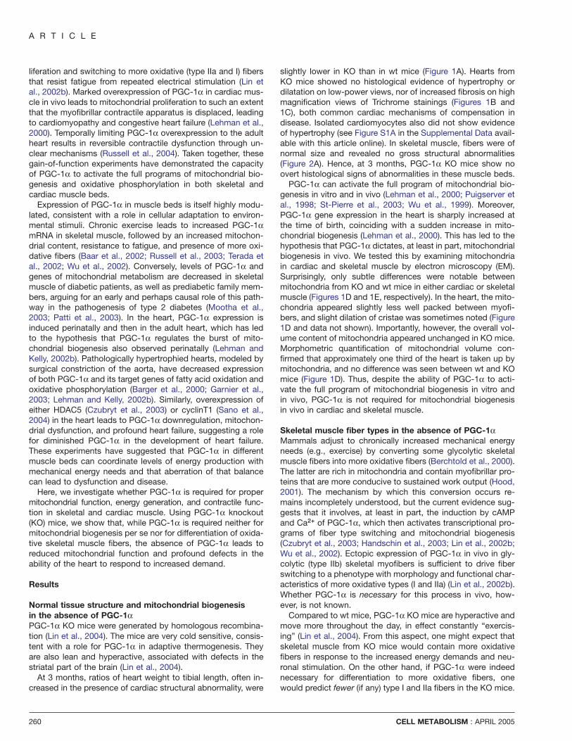
beat at higher rates by apposing an electrode directly onto theheart and pacing at an increased frequency. This purely chro-notropic challenge in KO hearts led to 20% less forceful con-tractions (+dP/dt) than in wt hearts (Table S1A). When thehearts were forced to abruptly increase work by infusion withdobutamine (a β-adrenergic agonist that leads to large in-creases in both heart rate and force of contraction, i.e., botha chronotropic and inotropic challenge), +dP/dt in wt heartsincreased by 30% more than in KO hearts (Figure 4A and TableS1A). A similar pattern of mild baseline deficiency and worsen-ing under stimulation was noted for the parameter that reflectsmeasured relaxation (−dP/dt), a process that also requires en-ergy (restoring the Ca2+ gradient). Interestingly, KO hearts alsoshowed a significant inability to increase heart rate in responseto dobutamine (Table S1A). Lower heart rate can increase +dP/dt; hence, the deficiency in +dP/dt seen here probably under-estimates the true magnitude of deficiency in KO hearts. A bet-ter index of cardiac work, therefore, is the rate pressure prod-uct (RPP), equal to heart rate times the increase in pressuredeveloped by each contraction (DevP). As seen in Figure 4B,wt hearts can increase RPP by nearly twice as much as canKO hearts in response to dobutamine.
In the experiments described above, hearts were perfusedwith modified Krebs bicarbonate buffer, which contains glu-cose and pyruvate as energy sources. However, the heart usesprimarily free fatty acids as a carbon source in vivo (Ingwall,2003). Moreover, PGC-1α is well-known to activate genes offatty acid import and oxidation, and expression of these genesis decreased in PGC-1α KO hearts (Table 1). We therefore alsomeasured the contractile function of wt and KO hearts per-fused with a buffer containing physiological concentrations of
CELL METABOLISM : APRIL 2005
PGC-1α controls the energy state of cardiac muscle
Figure 4. Defective cardiac function in PGC-1α KO mice
A) Contractile performance of hearts from PGC-1α KO (red, n = 5) compared to wt (blue, n = 5) hearts measured in Langendorff preparations. Hearts were electricallypaced at 7.0 Hz. After 10 min, hearts were challenged with 300 nM dobutamine for 12 min and then allowed to recover for another 15 min. +dP/dt, positive change inpressure over time during isovolumic contraction. −dP/dt, negative change in pressure over time during isovolumic relaxation. RPP, rate pressure product = heart ratetimes pressure differential between the fully contracted and relaxed states. RPP models the work done by the heart in vivo.B) Rate pressure product in wt and KO hearts at baseline and in response to dobutamine. In the top panel, hearts from wt (n = 5) and KO (n = 5) mice were perfusedwith modified Krebs bicarbonate buffer containing glucose and pyruvate. In the bottom panel, hearts from wt (n = 4) and KO (n = 4) mice were perfused with buffercontaining physiological concentrations of free fatty acids, glucose, lactate, and ketones, as described in Experimental Procedures. Error bars are ±SEM.
all carbon sources normally used by the heart: free fatty acids,glucose, ketones, and lactate. Under these conditions, thecontractile deficiencies of the KO hearts became even morepronounced (Table S1B). In response to dobutamine, for in-stance, the RPP increased only 30% as much in KO hearts asit did in wt hearts (Figure 4C and Table S1B). Together, thesedata show that hearts from PGC-1α KO mice have a profounddefect in contractile reserve, suggesting that PGC-1α is vitalto increasing cardiac work output in response to increaseddemand.
Hearts from PGC-1α KO mice are energy deficientThe KO hearts were next studied for their ability to maintainreadily available sources of energy for mechanical work. TheATP content of wt and KO hearts was measured by 31P nuclearmagnetic resonance (NMR) using the Langendorff heart prepa-rations (Figure 5A). From these measurements, concentrationsof ATP, inorganic phosphate (Pi), and phosphocreatine (PCr)
CELL METABOLISM : APRIL 2005
can be calculated from the area under each peak. PCr, theprimary energy reserve compound in excitable tissues, is nor-mally present in cardiac muscle at twice the concentration ofATP and is in rapid exchange with ATP; an accurate measureof available high-energy phosphate bonds thus requires eval-uation of both ATP and PCr concentrations. The most powerfuladvantages of this NMR technique stem from its ability to non-invasively measure ATP and PCr contents in intact, beatinghearts under differing conditions and while simultaneouslymeasuring contractile performance.
Using this approach, we found that ATP levels in unstimu-lated PGC-1α KO hearts were reduced by approximately 20%compared with wt hearts (Figures 5A and 5B and Figure S2A).The concentration of ATP in cardiac tissue is normally tightlycontrolled and, in cases of terminal heart failure, is reduced byonly approximately 30%. Hence, the absence of PGC-1α re-sults in a profound energetic defect in the heart. Similarly, theconcentration of PCr was also reduced by w20% (Figures 5A
265

A R T I C L E
Figure 5. Hearts from PGC-1α KO mice are energy deficient
A) Representative spectra acquired from wt and KO hearts during active, unstimulated contraction. Peaks corresponding to signals from the phosphorus atoms ineach of the phosphates of ATP and phosphocreatine (PCr), and free inorganic phosphate (Pi) are indicated.B) Average ATP and PCr concentrations in wt and KO hearts that are either unstimulated or maximally stimulated with 300 nM dobutamine infusion, as assayed fromNMR spectra as in (A). N = 5 and 5. Error bars are ±SEM.C) Relationship, in wt and KO hearts, between the rate pressure product (RPP, a measure of cardiac work) and �GwATP (representing the driving force available fromATP hydrolysis). Note that, at baseline, both KO and wt hearts generate similar amounts of work for a similar �GwATP, while, under dobutamine stimulation, KO heartshave markedly blunted work output for a similar drop in �GwATP. Note also that both KO and wt hearts are maximally stimulated by dobutamine to the theoretical limitfor �GwATP, i.e., the chemical driving force needed to drive Ca2+ into the sarcoplasmic reticulum by the SERCA channel (indicated with an arrow). Error bars are ±SEM.
and 5B and Figure S2A). Interestingly, inotropic and chrono-tropic stimulation of the hearts by dobutamine infusion resultedin PCr depletion down to similar concentrations in both wt andKO hearts (Figure 5B and Figure S2A).
In order to perform mechanical work, the heart requires asufficiently elevated chemical driving force (�GwATP ), calcu-lated from the ratio of concentrations of ATP to ADP and Pi,the so-called phosphorylation potential ([ATP]/[ADP]*[Pi]). Thisratio reflects the free energy released from ATP hydrolysis,which can then be used to overcome a coupled and unfavor-able reaction. Without a sufficiently large phosphorylation po-tential, ATP-dependent reactions such as the actomyosinATPase reaction and Ca2+ sequestration cannot occur. Thus,the heart can only increase work output until �GwATP approachesthe level needed by the most unfavorable coupled reaction.In wild-type hearts, it has been estimated that this thresholdpotential is that needed to drive the sarcoplasmic reticulum
266
Ca++ ATPase (SERCA), at a �GwATP of approximately 53 kJ/mol (Ingwall, 2002). As seen in Figure 5D, that threshold isreached in PGC-1α KO hearts with much less work output thanin wt hearts. Thus, for a similar change in �GwATP (equivalentto about 5 kJ/mol), hearts from PGC-1α KO mice generatemuch less work than do wt hearts.
Cardiac dysfunction in PGC-1α KO mice in vivoTo determine if the multiple defects described above result incardiac insufficiency in vivo, echocardiography was performed.At 3 months of age, no differences between wt and KO micewere apparent (Figure S2B). Cardiac dysfunction, however,often becomes apparent only with age, both in mice and hu-mans. Older mice were therefore also examined. As shown inFigure 6A, by 7–8 months of age, PGC-1α KO mice displayedclear features of cardiac dysfunction in vivo. Left ventricularchamber size in KO mice was slightly more dilated, both at end
CELL METABOLISM : APRIL 2005

PGC-1α controls the energy state of cardiac muscle
Figure 6. Cardiac dysfunction in PGC-1α KO mice in vivo
A) Cardiac echocardiograms were performed on 7- to 8-month-old wt (n = 4), heterozygote (n = 3), and KO (n = 3) female mice. The top panel shows representativeM mode echocardiograms (in which a one-dimensional signal across the center of a transverse section of the heart is visualized over time) of wt and KO mice. Thebottom panel shows in bar graph format the normalized left ventricular anterior wall (LVAW) and posterior wall (LVPW) thicknesses, end diastolic (LVEDD) and endsystolic (LVESD) diameters, and fractional shortening (FS, calculated as LVEDD − LVESD/LVEDD). Error bars are ±SEM.B) mRNA expression, in the same hearts as in (A), of the indicated genes as assayed by real-time quantitative PCR (top panel) and Northern blotting (bottom panel).EtBr, ethidium bromide stain of gel prior to transfer. Error bars are ±SEM.
diastole and end systole (Figure 6A). Most notably, KO miceshowed significantly reduced fractional shortening (the differ-ence in internal diameter between the fully dilated and fullycontracted state, Figure 6A), demonstrating poor cardiac con-traction in vivo in the absence of PGC-1α.
The 7- to 8-month-old KO mice also displayed molecular evi-dence of cardiac stress. Hearts faced with prolonged duressoften undergo partial genetic reprogramming, with the induc-tion of a number of so-called “fetal genes,” such as β-MHCand skeletal α-actin (Friddle et al., 2000; Olson and Schneider,2003). In addition, cardiac myocytes respond to stretch by in-ducing the genes for and secretion of atrial and brain natriureticpeptides (ANP and BNP), with the goal of reducing intravascu-lar volume by stimulating diuresis (Cameron and Ellmers,2003). As shown in Figure 6B, all these genes are also stronglyinduced in hearts of 7- to 8-month-old PGC-1α KO mice. To-gether, these data show that, by this later age, the defects re-sulting from the absence of PGC-1α lead to frank cardiac dys-function in vivo.
Interestingly, hearts from 3-month-old mice also revealedsimilar molecular signs of cardiac stress, although less pro-nounced (Table 1 and Figure S2C). In fact, circulating levels of
CELL METABOLISM : APRIL 2005
pro-ANP hormone were induced 2-fold in 3-month-old KOmice (Figure S2D), consistent with the induction of the ANPgene in the hearts. Hence, even at 3 months of age, PGC-1αKO hearts show clear signs of cardiac stress in vivo, consistentwith the significant energetic and contractile defects observedin isolated heart preparations.
Discussion
The heart must maintain an unfailing blood circulation in statesof both rest and high demand, such as that triggered by exer-cise. A trained athlete, for example, can rapidly increasecardiac output as much as 6-fold (Baim and Grossman, 2000).The amount of work done by the heart, therefore, must be effi-cient, dependable, and highly adaptable. Generating this workdepends on a copious and equally adaptable supply of ATP. Ina given day, the heart can consume approximately ten timesits own weight in ATP (Ingwall, 2003; Ingwall and Weiss, 2004).The heart therefore needs an abundant and ongoing generationof ATP that can adapt to changing external or internal de-mands. The bulk of this occurs through efficient oxidativephosphorylation in mitochondria.
267

A R T I C L E
Recent data have suggested an important role for PGC-1αin the control of mitochondrial metabolism of the heart, butthis has been based only on gain-of-function studies. What hasremained unclear to date is the requirement for endogenousPGC-1α in the function of the intact heart. In this study, wehave addressed this question by studying hearts from micelacking PGC-1α. Measurements of cardiac gene expressionand echocardiographic indices of cardiac function in vivo werecombined with functional analyses of isolated heart prepara-tions. The use of these Langendorff preparations allowed us toisolate the hearts from their nutrient and hormonal milieu,which was crucial because mice lacking PGC-1α demonstratea profound hyperactivity and an altered glucose/insulin axis(Lin et al., 2004). We show here that the absence of PGC-1αleads to blunted mitochondrial enzymatic activity and franklydecreased levels of ATP in the heart. In the face of this ener-getic defect, hearts from PGC-1α KO mice are unable to nor-mally perform the work of contraction, and KO mice eventuallydevelop cardiac dysfunction in vivo.
The contractile defects observed in isolated heart prepara-tions became most pronounced when hearts were forced toaccomplish more work, such as that needed for exercise or“fight or flight” responses. Such situations are typically trig-gered by catecholamine surges and can be modeled ex vivoin Langendorff preparations by stimulation with saturatingamounts of dobutamine, a potent β-adrenergic agonist. Whenthis was done, the synthesis of ATP in PGC-1α KO hearts wasinsufficient to meet increased demand, and the energetic limitwas reached with far less physical work achieved than in wthearts. These data indicate that functional PGC-1α is criticalfor normal ATP generation and cardiac work. Moreover, thedata suggest that the primary function of PGC-1α may be toserve an adaptive role, equipping the heart with the enhancedability to respond to sudden increases in demand. This is con-sistent with the observation that PGC-1α expression in cardiacmuscle is low before birth, when such responses are unneces-sary, and then induced during the first few weeks of life (Leh-man et al., 2000; Lehman and Kelly, 2002b).
It is important to note that hearts devoid of PGC-1α haveblunted responses of both contraction and heart rate in re-sponse to dobutamine challenge. In fact, cardiac force is typi-cally higher in the context of lower heart rates (Katz, 2001), sothe force-generating defect in KO mice may be underesti-mated. Altered β-adrenergic signaling could be an obviouscause for these blunted responses, but this is unlikely for sev-eral reasons: first, dobutamine stimulation causes similarchange in the chemical driving force for ATPase reaction, i.e.,energy drain, in both KO and wt hearts; second, the functionaldefects of KO hearts are also exacerbated by pacing at ele-vated heart rates; third, expression of genes of β-adrenergicsignaling such as the β receptors or βARK are not altered in KOmice (Figure S2E). Why the response of heart rate is blunted inPGC-1α KO hearts is unclear. Much of the regulation of heartrate in vivo is neurohormonal (Katz, 2001), but, in Langendorffpreparations, the hearts are no longer under this control.Hence, PGC-1α appears to be required for maximal automatic-ity of pacemaker cells. This may occur due to bioenergetic in-sufficiency, but it is also possible that the absence of PGC-1α leads to defects in heart rate (and/or contraction) throughmechanisms not involving the mitochondria. Further experi-mentation on this point will be needed.
268
That the cardiac defects observed in the Langendorff prepa-rations have physiological relevance in vivo is indicated by themolecular changes observed in 3-month-old hearts, includinginduction of ANP, β-MHC, and skeletal α-actin (Table 1). More-over, mice lacking PGC-1α go on, by 8 months of age, to de-velop enlargement of left ventricular chamber size and dimin-ished contractile function (Figure 6). At the same time, noevidence of cardiac hypertrophy is seen (Figures 1 and 6, Fig-ure S1A), suggesting that this phenotype is more akin to thatof primary dilated cardiomyopathy than to dilation secondaryto hypertrophic cardiomyopathy. It is likely that the phenotypewill deteriorate with age. It will also be interesting to determineif the lack of PGC-1α will impair cardiac adaptation to specificlong-term stressors, such as exercise or surgical constrictionof the aorta. Unfortunately, PGC-1α KO mice have other phe-notypes, including hyperactivity and an aberrant insulin/glu-cose axis (Lin et al., 2004), making it difficult to evaluate in awell-controlled fashion the cardiac response to exercise inwhole mice. A critical evaluation of the role of PGC-1α incardiac function in intact animals will thus require the cardiac-specific knockout of PGC-1α.
Physical activity induces PGC-1α expression in skeletalmuscle tissue (Baar et al., 2002; Terada et al., 2002; Wu et al.,2002), and PGC-1α induces muscle fiber type switching tomore oxidative fibers when expressed in transgenic mice (Linet al., 2002b). On this basis, one might have expected a de-crease of such fibers in PGC-1α KO mice. We show here, how-ever, that in the absence of PGC-1α there is no obvious de-crease in oxidative fibers in skeletal muscle (Figure 2). In fact,in quadriceps muscle, which is primarily glycolytic in nature,there is a 2- to 3-fold induction of genes specific to oxidativefibers (Figure 3B), although no frank increase in oxidative fiberswas noted in primarily glycolytic muscle such as extensor digi-torus longus (data not shown). This increase is not observed inisolated skeletal muscle cells from KO versus wt mice, sug-gesting that the difference is not cell autonomous (Figure 3B).These observations are most consistent with the notion thatthe hyperactivity of KO mice causes an increased motor nerveinput to skeletal muscle that is sufficient to activate the expres-sion of genes characteristic of oxidative fibers. Importantly,these data also demonstrate that mitochondria and oxidativefibers can form without the presence of PGC-1α. Since PGC-1β can also stimulate mitochondrial biogenesis and respirationwhen expressed ectopically (St-Pierre et al., 2003), it is plausi-ble that PGC-1β may be compensating for the absence ofPGC-1α in both mitochondrial and oxidative fiber formationin vivo. It is also possible that PGC-1α plays a more prominentrole in adaptive responses to physical exercise, rather than thebasal profiles of mitochondria or fiber types. Clearly, an under-standing of how the absence of PGC-1α affects muscle fibertype formation in mice with comparable levels of physicalmovement will require skeletal muscle-specific KO of PGC-1α.
The notion that energy “starvation” is a significant contrib-utor to the pathogenesis of cardiac failure has long been enter-tained (Ingwall and Weiss, 2004). The work described herelends significant support to this hypothesis: the absence ofPGC-1α causes a decrease in available ATP supply and leadsto significant contractile defects. Indeed, significant drops inPGC-1α transcript levels have been observed in various rodentmodels of cardiac failure (Czubryt et al., 2003; Feingold et al.,2004; Garnier et al., 2003; Lehman and Kelly, 2002b; Sano et
CELL METABOLISM : APRIL 2005

PGC-1α controls the energy state of cardiac muscle
al., 2004). The results presented here thus support the ideathat stimulation of PGC-1α activity represents a potential newtherapeutic modality. Indeed, ectopic expression of PGC-1α inskeletal muscle can improve indices of fatiguability (Lin et al.,2002b). While massive overexpression of PGC-1α in normalhearts leads to cardiomyopathy (Lehman et al., 2000; Russellet al., 2004), increasing the expression of active PGC-1α atmore physiological levels might improve contractile function infailing hearts.
Experimental procedures
AnimalsAll animal experiments were performed according to procedures approvedby the Institutional Animal Care and Use Committee. Mice were maintainedon a standard rodent chow diet with 12 hr light and dark cycles. Frozenheart samples from 3-month-old mice 1 month after sustaining banding oftheir aorta were kindly provided by Dr. Richard Lee (Weinberg et al., 2003).
Cells and reagentsC2C12 cells were from ATCC and were grown in DMEM 10% FBS anddifferentiated for 5 days in DMEM 4% HS prior to adenoviral infection. Pri-mary skeletal muscle cells were harvested as published (Sabourin et al.,1999) and grown and differentiated as above. Left ventricular cardiomyo-cytes were isolated as described (Matsui et al., 2002; Nagata et al., 2000).Cell surface was determined using NIH image. Adenovirus-expressingPGC-1α has been published (Puigserver et al., 1998). Pro-ANP assays wereperformed using the kit from ALPCO Diagnostics following the includedprotocol. Antibodies against cytochrome c and ND4L were from Phar-mingen and Santa Cruz, respectively.
Histological analysesTissues for regular histology were frozen in liquid nitrogen immediately uponmouse sacrifice. Cardiac H&E and Trichrome stains were performed byAmerican HistoLabs. Tissues for fiber type evaluation were embedded ingum tragacanth and OCT freezing matrix and quickly frozen in isopentanecooled in liquid nitrogen. Fiber typing was performed with cryostat sectionsusing metachromatic dye-ATPase methods as described (Ogilvie and Fee-back, 1990). Immunohistochemical staining was performed with a monoclo-nal antibody against skeletal slow myosin (NOQ7.5.4D, Sigma).
Genetic expression studiesTotal RNAs were isolated from mouse tissue or cultured cells using theTRIzol method (Invitrogen). For microarray analyses, skeletal muscle andcardiac RNAs were evaluated with Affymetrix U74v2 and 430A chips,respectively, using the DFCI core facility. All subsequent analyses were per-formed with dCHIP software. For Northern analyses, 10 µg of RNA wereseparated on a formaldehyde gel, transferred to nylon membrane, and hy-bridized with probes amplified by PCR from genomic DNA. Samples forreal-time PCR analyses were reverse transcribed (Invitrogen), and quantita-tive real-time PCR reactions were performed on the cDNAs in the presenceof fluorescent dye (Cybergreen, Bio-Rad). DNA product of the expected sizewas confirmed for each primer pair.
Langendorff preparationsMice were heparinized (100 units, IP) 15 min before being sacrificed. Heartswere isolated and perfused in the Langendorff mode as described (Chu etal., 1996). Hearts were perfused either with modified Krebs bicarbonatebuffer containing pyruvate (0.5 mM) and glucose (10 mM) or with buffercontaining physiological concentrations of mixed free fatty acid (0.4 mM)(palmitate, palmitoleic, linoleic, and oleic) carried in 1% BSA, glucose (5.5mM), β-hydroxybutarate (0.19 mM), and lactate (1.0 mM). The compositionof this mixture was chosen based on literature reports (Previs et al., 1999;Rofe et al., 1985) and direct measurements of substrate concentrations inthe plasma of mice (data not shown). A water-filled balloon was then in-serted into the left ventricle (LV) and inflated to LV end diastolic pressure of10 mmHg. Isovolumic contractile performance data were collected onlineat a sampling rate of 200 Hz. Baseline measurements were made after a 20min stabilization period (equivalent for all experiments and both genotypes).
CELL METABOLISM : APRIL 2005
The hearts were then paced at 7.0 Hz using monophasic square wavepulses delivered from a stimulator (model S88; Grass Instrument Co.,Quincy, MA) through salt bridge pacing wires consisting of PE-90 tubingfilled with 4 M KCl in 2% agarose. The hearts were then challenged with300 nM of dobutamine infused through a side tubing driven by a digitalconsole driver (Cole-Parmer Instrument Co.) at 2% of coronary flow rate.
31P NMR spectroscopy31P NMR spectra were obtained at 161.94 MHz using a GE-400 wide-borespectrometer (Omega, General Electric, Fremont, CA). The isolated per-fused hearts, in a 10 mm NMR sample tube, were inserted into a 1H/31Pdouble-tuned probe situated in a 89 cm bore, 9.4 T superconducting mag-net. Spectra were collected during 8 min periods and consisted of dataaveraged from 208 free induction decays as described (Chu et al., 1996;Spindler et al., 1998). 31P NMR spectra were analyzed using 20 Hz expo-nential multiplication and zero and first-order phase corrections. The reso-nance areas corresponding to ATP, PCr, and Pi were fitted to Lorentzianfunctions and corrected for saturation (ATP [1.0], PCr [1.2], and Pi [1.15]).The changes in concentration of ATP, PCr, and Pi during the protocols werecalculated as described (Spindler et al., 1998). The free energy released byATP hydrolysis (�GwATP) is calculated as �GwATP = �G° + RT ln([ADP][Pi]/[ATP]).
Biochemical assaysCardiac ventricular tissue was homogenized in potassium phosphate buffercontaining 1 mM EDTA and 1 mM β-mercaptoethanol (pH 7.4). Aliquotswere removed for assays of protein (Lowry et al., 1951) and total creatinecontent (Kammermeier, 1973). Triton X-100 was then added to the homoge-nate at a final concentration of 0.1%, and the activities of citrate synthase(Srere et al., 1963), cytochrome C oxidase (Stieglerova et al., 2000), andelectron transport chain complex II (Makita and Sagara, 1990) were mea-sured as described.
Echocardiographic analysesTransthoracic echocardiography was performed using a 15 Mhz linear-arrayprobe and a Sonos 4500 ultrasonograph (Hewlett Packard). The imageswere taken in 2K (left parasternal long and short axes) and M mode (leftparasternal short axis). All echocardiograms were performed blinded to themouse genotype. LV fractional shortening was calculated using the formulaLVEDD − LVESD/LVEDD.
Statistical analysisAll results are expressed as means ±SEM. Two-tailed independent Stu-dent’s t tests were used to determine all p values.
Supplemental dataSupplemental data include two figures and one table and can be found withthis article online at http://www.cellmetabolism.org/cgi/content/full/1/4/259/DC1/.
Acknowledgments
We thank Dr. Mukesh Jain for ongoing discussions and mentoring. We alsothank Dr. Richard T. Lee for samples from TAC hearts. This work is sup-ported by grants from the N.I.H., DK54477 and DK61562 (B.M.S.),DK065584 (J.L.), HL052320 and HL063985 (J.S.I.), HL06296 (R.B.-D.), andHL07604 (Z.A.); from the MDA (C.H.); from the Deutsche Forschungsgem-einschaft (O.T.); and from the Donald W. Reynolds Cardiovascular ClinicalResearch Center at the University of Texas Southwestern Medical Centerin Dallas, Texas (R.B.-D.).
Received: November 23, 2004Revised: February 25, 2005Accepted: March 16, 2005Published: April 12, 2005
References
Baar, K., Wende, A.R., Jones, T.E., Marison, M., Nolte, L.A., Chen, M., Kelly,D.P., and Holloszy, J.O. (2002). Adaptations of skeletal muscle to exercise:
269

A R T I C L E
rapid increase in the transcriptional coactivator PGC-1. FASEB J. 16,1879–1886.
Baim, D.S., and Grossman, W. (2000). Grossman’s Cardiac Catheterization,Angiography, and Intervention (Philadelphia: Lippincott, Williams & Wilkins).
Barger, P.M., and Kelly, D.P. (2000). PPAR signaling in the control of cardiacenergy metabolism. Trends Cardiovasc. Med. 10, 238–245.
Barger, P.M., Brandt, J.M., Leone, T.C., Weinheimer, C.J., and Kelly, D.P.(2000). Deactivation of peroxisome proliferator-activated receptor-alphaduring cardiac hypertrophic growth. J. Clin. Invest. 105, 1723–1730.
Berchtold, M.W., Brinkmeier, H., and Muntener, M. (2000). Calcium ion inskeletal muscle: its crucial role for muscle function, plasticity, and disease.Physiol. Rev. 80, 1215–1265.
Booth, F.W., and Thomason, D.B. (1991). Molecular and cellular adaptationof muscle in response to exercise: perspectives of various models. Physiol.Rev. 71, 541–585.
Cameron, V.A., and Ellmers, L.J. (2003). Minireview: natriuretic peptidesduring development of the fetal heart and circulation. Endocrinology 144,2191–2194.
Chu, G., Luo, W., Slack, J.P., Tilgmann, C., Sweet, W.E., Spindler, M.,Saupe, K.W., Boivin, G.P., Moravec, C.S., Matlib, M.A., et al. (1996). Com-pensatory mechanisms associated with the hyperdynamic function of phos-pholamban-deficient mouse hearts. Circ. Res. 79, 1064–1076.
Czubryt, M.P., McAnally, J., Fishman, G.I., and Olson, E.N. (2003). Regula-tion of peroxisome proliferator-activated receptor gamma coactivator 1 al-pha (PGC-1 alpha) and mitochondrial function by MEF2 and HDAC5. Proc.Natl. Acad. Sci. USA 100, 1711–1716.
Feingold, K., Kim, M.S., Shigenaga, J., Moser, A., and Grunfeld, C. (2004).Altered expression of nuclear hormone receptors and coactivators in mouseheart during the acute-phase response. Am. J. Physiol. Endocrinol. Metab.286, E201–E207.
Friddle, C.J., Koga, T., Rubin, E.M., and Bristow, J. (2000). Expression profil-ing reveals distinct sets of genes altered during induction and regression ofcardiac hypertrophy. Proc. Natl. Acad. Sci. USA 97, 6745–6750.
Garnier, A., Fortin, D., Delomenie, C., Momken, I., Veksler, V., and Ventura-Clapier, R. (2003). Depressed mitochondrial transcription factors and oxida-tive capacity in rat failing cardiac and skeletal muscles. J. Physiol. 551,491–501.
Handschin, C., Rhee, J., Lin, J., Tarr, P.T., and Spiegelman, B.M. (2003).An autoregulatory loop controls peroxisome proliferator-activated receptorgamma coactivator 1alpha expression in muscle. Proc. Natl. Acad. Sci. USA100, 7111–7116.
Hood, D.A. (2001). Invited review: contractile activity-induced mitochondrialbiogenesis in skeletal muscle. J. Appl. Physiol. 90, 1137–1157.
Huss, J.M., Torra, I.P., Staels, B., Giguere, V., and Kelly, D.P. (2004). Estro-gen-related receptor α directs peroxisome proliferator-activated receptor αsignaling in the transcriptional control of energy metabolism in cardiac andskeletal muscle. Mol. Cell. Biol. 24, 9079–9091.
Ingwall, J.S. (2002). Energetic basis for heart failure. In Heart Failure: ACompanion to Braunwald’s Heart Disease, D. Mann, ed. (London: KluwerAcademic Publishers).
Ingwall, J.S. (2003). ATP and the Heart (Boston: Kluwer Academic Pub-lishers).
Ingwall, J.S., and Weiss, R.G. (2004). Is the failing heart energy starved? Onusing chemical energy to support cardiac function. Circ. Res. 95, 135–145.
Kammermeier, H. (1973). Microassay of free and total creatine from tissueextracts by combination of chromatographic and fluorometric methods.Anal. Biochem. 56, 341–345.
Katz, A.M. (2001). Physiology of the Heart, Third Edition (Philadelphia: Lip-pincott Williams & Wilkins).
Kelly, D.P., and Scarpulla, R.C. (2004). Transcriptional regulatory circuitscontrolling mitochondrial biogenesis and function. Genes Dev. 18, 357–368.
270
Kelly, D.P., and Strauss, A.W. (1994). Inherited cardiomyopathies. N. Engl.J. Med. 330, 913–919.
Knutti, D., and Kralli, A. (2001). PGC-1, a versatile coactivator. Trends En-docrinol. Metab. 12, 360–365.
Kressler, D., Schreiber, S.N., Knutti, D., and Kralli, A. (2002). The PGC-1-related protein PERC is a selective coactivator of estrogen receptor alpha.J. Biol. Chem. 277, 13918–13925.
Lehman, J.J., and Kelly, D.P. (2002a). Gene regulatory mechanisms govern-ing energy metabolism during cardiac hypertrophic growth. Heart Fail. Rev.7, 175–185.
Lehman, J.J., and Kelly, D.P. (2002b). Transcriptional activation of energymetabolic switches in the developing and hypertrophied heart. Clin. Exp.Pharmacol. Physiol. 29, 339–345.
Lehman, J.J., Barger, P.M., Kovacs, A., Saffitz, J.E., Medeiros, D.M., andKelly, D.P. (2000). Peroxisome proliferator-activated receptor gamma coacti-vator-1 promotes cardiac mitochondrial biogenesis. J. Clin. Invest. 106,847–856.
Lin, J., Puigserver, P., Donovan, J., Tarr, P., and Spiegelman, B.M. (2002a).Peroxisome proliferator-activated receptor gamma coactivator 1beta (PGC-1beta), a novel PGC-1-related transcription coactivator associated withhost cell factor. J. Biol. Chem. 277, 1645–1648.
Lin, J., Wu, H., Tarr, P.T., Zhang, C.Y., Wu, Z., Boss, O., Michael, L.F., Puig-server, P., Isotani, E., Olson, E.N., et al. (2002b). Transcriptional co-activatorPGC-1 alpha drives the formation of slow-twitch muscle fibres. Nature 418,797–801.
Lin, J., Tarr, P.T., Yang, R., Rhee, J., Puigserver, P., Newgard, C.B., andSpiegelman, B.M. (2003). PGC-1beta in the regulation of hepatic glucoseand energy metabolism. J. Biol. Chem. 278, 30843–30848.
Lin, J., Wu, P.H., Tarr, P.T., Lindenberg, K.S., St-Pierre, J., Zhang, C.Y., Moo-tha, V.K., Jager, S., Vianna, C.R., Reznick, R.M., et al. (2004). Defects inadaptive energy metabolism with CNS-linked hyperactivity in PGC-1alphanull mice. Cell 119, 121–135.
Lin, J., Yang, R., Tarr, P.T., Wu, P.H., Handschin, C., Li, S., Yang, W., Pei, L.,Uldry, M., Tontonoz, P., et al. (2005). Hyperlipidemic effects of dietary satu-rated fats mediated through PGC-1beta coactivation of SREBP. Cell 120,261–273.
Lowry, O.H., Rosebrough, N.J., Farr, A.L., and Randall, R.J. (1951). Proteinmeasurement with the Folin phenol reagent. J. Biol. Chem. 193, 265–275.
Makita, T., and Sagara, E. (1990). Post-mortem changes in cytochemicallocalization and enzymological measurement of marker enzymes of themitochondria, SDH and Mg-ATPase, of porcine muscle stored at 4 degreesC, −18 degrees C, or −80 degrees C. Cell Biol. Int. Rep. 14, 123–131.
Matsui, T., Li, L., Wu, J.C., Cook, S.A., Nagoshi, T., Picard, M.H., Liao, R.,and Rosenzweig, A. (2002). Phenotypic spectrum caused by transgenicoverexpression of activated Akt in the heart. J. Biol. Chem. 277, 22896–22901.
Monsalve, M., Wu, Z., Adelmant, G., Puigserver, P., Fan, M., and Spiegel-man, B.M. (2000). Direct coupling of transcription and mRNA processingthrough the thermogenic coactivator PGC-1. Mol. Cell 6, 307–316.
Mootha, V.K., Lindgren, C.M., Eriksson, K.F., Subramanian, A., Sihag, S.,Lehar, J., Puigserver, P., Carlsson, E., Ridderstrale, M., Laurila, E., et al.(2003). PGC-1alpha-responsive genes involved in oxidative phosphorylationare coordinately downregulated in human diabetes. Nat. Genet. 34, 267–273.
Mootha, V.K., Handschin, C., Arlow, D., Xie, X., St Pierre, J., Sihag, S., Yang,W., Altshuler, D., Puigserver, P., Patterson, N., et al. (2004). Erralpha andGabpa/b specify PGC-1alpha-dependent oxidative phosphorylation geneexpression that is altered in diabetic muscle. Proc. Natl. Acad. Sci. USA101, 6570–6575.
Nagata, K., Ye, C., Jain, M., Milstone, D.S., Liao, R., and Mortensen, R.M.(2000). Galpha(i2) but not Galpha(i3) is required for muscarinic inhibition ofcontractility and calcium currents in adult cardiomyocytes. Circ. Res. 87,903–909.
CELL METABOLISM : APRIL 2005

PGC-1α controls the energy state of cardiac muscle
Ogilvie, R.W., and Feeback, D.L. (1990). A metachromatic dye-ATPasemethod for the simultaneous identification of skeletal muscle fiber types I,IIA, IIB and IIC. Stain Technol. 65, 231–241.
Olson, E.N., and Schneider, M.D. (2003). Sizing up the heart: developmentredux in disease. Genes Dev. 17, 1937–1956.
Patti, M.E., Butte, A.J., Crunkhorn, S., Cusi, K., Berria, R., Kashyap, S.,Miyazaki, Y., Kohane, I., Costello, M., Saccone, R., et al. (2003). Coordi-nated reduction of genes of oxidative metabolism in humans with insulinresistance and diabetes: Potential role of PGC1 and NRF1. Proc. Natl.Acad. Sci. USA 100, 8466–8471.
Previs, S.F., Cline, G.W., and Shulman, G.I. (1999). A critical evaluation ofmass isotopomer distribution analysis of gluconeogenesis in vivo. Am. J.Physiol. 277, E154–E160.
Puigserver, P., and Spiegelman, B.M. (2003). Peroxisome proliferator-acti-vated receptor-gamma coactivator 1 alpha (PGC-1 alpha): transcriptionalcoactivator and metabolic regulator. Endocr. Rev. 24, 78–90.
Puigserver, P., Wu, Z., Park, C.W., Graves, R., Wright, M., and Spiegelman,B.M. (1998). A cold-inducible coactivator of nuclear receptors linked toadaptive thermogenesis. Cell 92, 829–839.
Puigserver, P., Adelmant, G., Wu, Z., Fan, M., Xu, J., O’Malley, B., and Spie-gelman, B.M. (1999). Activation of PPARgamma coactivator-1 through tran-scription factor docking. Science 286, 1368–1371.
Rofe, A.M., Porter, S.J., Bais, R., and Conyers, R.A. (1985). The metabolicresponse of tumour-bearing mice to fasting. Br. J. Cancer 52, 619–623.
Russell, A.P., Feilchenfeldt, J., Schreiber, S., Praz, M., Crettenand, A., Gobe-let, C., Meier, C.A., Bell, D.R., Kralli, A., Giacobino, J.P., and Deriaz, O.(2003). Endurance training in humans leads to fiber type-specific increasesin levels of peroxisome proliferator-activated receptor-gamma coactiva-tor-1 and peroxisome proliferator-activated receptor-alpha in skeletal mus-cle. Diabetes 52, 2874–2881.
Russell, L.K., Mansfield, C.M., Lehman, J.J., Kovacs, A., Courtois, M., Saf-fitz, J.E., Medeiros, D.M., Valencik, M.L., McDonald, J.A., and Kelly, D.P.(2004). Cardiac-specific induction of the transcriptional coactivator peroxi-some proliferator-activated receptor gamma coactivator-1alpha promotesmitochondrial biogenesis and reversible cardiomyopathy in a develop-mental stage-dependent manner. Circ. Res. 94, 525–533.
Sabourin, L.A., Girgis-Gabardo, A., Seale, P., Asakura, A., and Rudnicki,M.A. (1999). Reduced differentiation potential of primary MyoD−/− myo-genic cells derived from adult skeletal muscle. J. Cell Biol. 144, 631–643.
Sano, M., Wang, S.C., Shirai, M., Scaglia, F., Xie, M., Sakai, S., Tanaka, T.,
CELL METABOLISM : APRIL 2005
Kulkarni, P.A., Barger, P.M., Youker, K.A., et al. (2004). Activation of cardiacCdk9 represses PGC-1 and confers a predisposition to heart failure. EMBOJ. 23, 3559–3569.
Scarpulla, R.C. (2002). Nuclear activators and coactivators in mammalianmitochondrial biogenesis. Biochim. Biophys. Acta 1576, 1–14.
Spindler, M., Saupe, K.W., Christe, M.E., Sweeney, H.L., Seidman, C.E.,Seidman, J.G., and Ingwall, J.S. (1998). Diastolic dysfunction and alteredenergetics in the alphaMHC403/+ mouse model of familial hypertrophic car-diomyopathy. J. Clin. Invest. 101, 1775–1783.
Srere, P., Brazil, H., and Gowen, L. (1963). The citrate condensing enzymeof pigeon breast muscle and moth flight muscle. Acta Chem. Scand. A 17,S129–S134.
Stieglerova, A., Drahota, Z., Ostadal, B., and Houstek, J. (2000). Optimalconditions for determination of cytochrome c oxidase activity in the ratheart. Physiol. Res. 49, 245–250.
St-Pierre, J., Lin, J., Krauss, S., Tarr, P.T., Yang, R., Newgard, C.B., andSpiegelman, B.M. (2003). Bioenergetic analysis of peroxisome proliferator-activated receptor gamma coactivators 1alpha and 1beta (PGC-1alpha andPGC-1beta) in muscle cells. J. Biol. Chem. 278, 26597–26603.
Terada, S., Goto, M., Kato, M., Kawanaka, K., Shimokawa, T., and Tabata,I. (2002). Effects of low-intensity prolonged exercise on PGC-1 mRNA ex-pression in rat epitrochlearis muscle. Biochem. Biophys. Res. Commun.296, 350–354.
Wallace, D.C. (1992). Diseases of the mitochondrial DNA. Annu. Rev. Bio-chem. 61, 1175–1212.
Wallberg, A.E., Yamamura, S., Malik, S., Spiegelman, B.M., and Roeder,R.G. (2003). Coordination of p300-mediated chromatin remodeling andTRAP/mediator function through coactivator PGC-1alpha. Mol. Cell 12,1137–1149.
Weinberg, E.O., Mirotsou, M., Gannon, J., Dzau, V.J., Lee, R.T., and Pratt,R.E. (2003). Sex dependence and temporal dependence of the left ventricu-lar genomic response to pressure overload. Physiol. Genomics 12, 113–127.
Wu, Z., Puigserver, P., Andersson, U., Zhang, C., Adelmant, G., Mootha, V.,Troy, A., Cinti, S., Lowell, B., Scarpulla, R.C., and Spiegelman, B.M. (1999).Mechanisms controlling mitochondrial biogenesis and respiration throughthe thermogenic coactivator PGC-1. Cell 98, 115–124.
Wu, H., Kanatous, S.B., Thurmond, F.A., Gallardo, T., Isotani, E., Bassel-Duby, R., and Williams, R.S. (2002). Regulation of mitochondrial biogenesisin skeletal muscle by CaMK. Science 296, 349–352.
271