HIF and reactive oxygen species regulate oxidative phosphorylation in cancer
RESEARCH ARTICLE Open Access
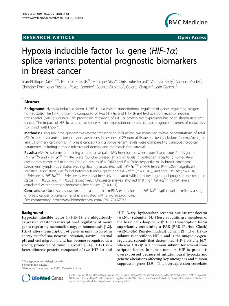
Hypoxia inducible factor 1a gene (HIF-1a)splice variants: potential prognostic biomarkersin breast cancerJean-Philippe Dales1,2*†, Nathalie Beaufils3†, Monique Silvy4, Christophe Picard4, Vanessa Pauly5, Vincent Pradel5,Christine Formisano-Tréziny1, Pascal Bonnier6, Sophie Giusiano2, Colette Charpin2, Jean Gabert1,3
Abstract
Background: Hypoxia-inducible factor 1 (HIF-1) is a master transcriptional regulator of genes regulating oxygenhomeostasis. The HIF-1 protein is composed of two HIF-1a and HIF-1b/aryl hydrocarbon receptor nucleartranslocator (ARNT) subunits. The prognostic relevance of HIF-1a protein overexpression has been shown in breastcancer. The impact of HIF-1a alternative splice variant expression on breast cancer prognosis in terms of metastasisrisk is not well known.
Methods: Using real-time quantitative reverse transcription PCR assays, we measured mRNA concentrations of totalHIF-1a and 4 variants in breast tissue specimens in a series of 29 normal tissues or benign lesions (normal/benign)and 53 primary carcinomas. In breast cancers HIF-1a splice variant levels were compared to clinicopathologicalparameters including tumour microvessel density and metastasis-free survival.
Results: HIF-1a isoforms containing a three base pairs TAG insertion between exon 1 and exon 2 (designatedHIF-1aTAG) and HIF-1a736 mRNAs were found expressed at higher levels in oestrogen receptor (OR)-negativecarcinomas compared to normal/benign tissues (P = 0.009 and P = 0.004 respectively). In breast carcinomaspecimens, lymph node status was significantly associated with HIF-1aTAG mRNA levels (P = 0.037). Significantstatistical association was found between tumour grade and HIF-1aTAG (P = 0.048), and total HIF-1a (P = 0.048)mRNA levels. HIF-1aTAG mRNA levels were also inversely correlated with both oestrogen and progesterone receptorstatus (P = 0.005 and P = 0.033 respectively). Univariate analysis showed that high HIF-1aTAG mRNA levelscorrelated with shortened metastasis free survival (P = 0.01).
Conclusions: Our results show for the first time that mRNA expression of a HIF-1aTAG splice variant reflects a stageof breast cancer progression and is associated with a worse prognosis.See commentary: http://www.biomedcentral.com/1741-7015/8/45
BackgroundHypoxia-inducible factor 1 (HIF-1) is a ubiquitouslyexpressed master transcriptional regulator of manygenes regulating mammalian oxygen homeostasis [1,2].HIF-1 alters transcription of genes mainly involved inenergy metabolism, neovascularisation, survival, internalpH and cell migration, and has become recognised as astrong promoter of tumour growth [3,4]. HIF-1 is aheterodimeric protein composed of two HIF-1a and
HIF-1b/aryl hydrocarbon receptor nuclear translocator(ARNT) subunits [5]. These subunits are members ofthe basic helix-loop-helix (bHLH) transcription factorsuperfamily containing a PAS [PER (Period Clock)-ARNT-SIM (Single-minded)] domain [2]. The HIF-1asubunit is specific to HIF-1 and is the unique oxygen-regulated subunit that determines HIF-1 activity [6,7]whereas HIF-1b is a common subunit for several tran-scription factors. In human tumours, HIF-1a protein isoverexpressed because of intratumoural hypoxia andgenetic alterations affecting key oncogenes and tumoursuppressor genes [8,9]. This overexpression correlates
* Correspondence: [email protected]† Contributed equally1Plateforme Transcriptome, CRO2, Marseille, France
Dales et al. BMC Medicine 2010, 8:44http://www.biomedcentral.com/1741-7015/8/44
© 2010 Dales et al; licensee BioMed Central Ltd. This is an Open Access article distributed under the terms of the Creative CommonsAttribution License (http://creativecommons.org/licenses/by/2.0), which permits unrestricted use, distribution, and reproduction inany medium, provided the original work is properly cited.

with tumour angiogenesis, treatment failure and patientmortality [3].Our group and others have previously reported that
HIF-1a protein overexpression is a marker of poorprognosis in primary breast cancer patients [10,11].HIF-1a is known to be mainly post-transcriptionallyregulated by protein ubiquitination and interaction withthe Von Hippel-Lindau tumour suppressor protein, andthen degraded by the proteasome. Hypoxia stabilisesHIF-1a protein by relaxing its ubiquitin-proteasomedegradation [12] and affects subcellular localisation,DNA binding capacity and transcriptional activationfunction of the HIF-1 complex. HIF-1a protein iscomposed of 826 amino acids [5]. Its N-terminal regioncontains the bHLH and the PAS domains, which areessential for DNA binding and dimerisation [13]. ItsC-terminal region contains two transactivation domainsand a nuclear localisation signal [14]. In the middle sec-tion of HIF-1a lies a Pro-Ser-Thr oxygen-dependentdegradation domain (ODDD, amino acids 401 to 603),which is responsible for the stability of the HIF-1a pro-tein [12].Although HIF-1a protein regulation has been well
studied, HIF-1a messenger regulation has only recentlybegun to be documented [15]. HIF-1a is encoded by theHIF1A locus, which is located on human chromosome14q21-q24 and encodes for 15 exons, resulting in aprincipal transcript of about 8 kb [16]. Different func-tional domains of the HIF-1a protein are encoded byseparate exons. In addition to the originally describedwild type of HIF-1a (HIF-1aWT) [1], eight alternativesplice variants of HIF-1a (HIF-1a827, HIF-1a736,HIF-1a557, HIF-1a516, HIF-1a785, HIF-1a417, HIF-1aTE
and an isoform with alternative exon 1 (I.2 isoform))with different activity have been reported in human celllines [17-23]. All HIF-1a splice variants have beenshown to be translated, to dimerise with HIF-1b and tobe active in human cells. Only one isoform (HIF-1a736)has been documented in human breast cancer [24]. Theaim of this study was to analyse HIF-1a splice variantexpression in human breast cancer and to evaluate itsclinical relevance in terms of prognosis. In this study wequantified the mRNA levels of 3 isoforms (HIF-1a736,HIF-1a557 and HIF-1a516) in breast specimens of 53primary cancers and 29 normal tissues or benign lesionsby real-time quantitative reverse transcription PCR(RT-qPCR) assays. HIF-1a827 is similar to wild typeexcept for an additional three base pairs TAG insertionat the exon 1 to 2 junction involving a difference of twoamino acids upstream of the bHLH domain [17].HIF-1a736 is also characterised by an additional threebase pairs TAG insertion at the exon 1 to 2 junction,and the lack of exon 14, which produces a frame shiftand introduces a stop codon in the coding sequence
after the Ile735 [17]. HIF-1a557 loses exon 12 andHIF-1a516 lacks exons 11 and 12 [18,19]. HIF-1a557 hasbeen shown to be induced by zinc ions. HIF-1a557 andHIF-1a516 have been shown to function as dominantnegative isoforms of HIF-1a in vitro. HIF-1a785 isdeprived of exon 11 and was described as being inducedin vitro by phorbol-12-myristate-13-acetate [20].HIF-1a417 isoform is deprived of exon 10 [21]. TheHIF-1aTE variant with alternative exon 1 (I.1 isoform)was previously found to be expressed in the testis [22].For these reasons, both these isoforms were thought tobe out of the scope of this study on breast tissues sam-ples. We also developed tools to quantify expression ofHIF-1a splice transcripts characterised by insertion of athree base pairs TAG at the exon 1 to 2 junction (whichwe designated HIF-1aTAG) involving a difference of twoamino acids upstream of the bHLH domain (HIF-1a827
and HIF-1a736 isoforms) [17]. The last known isoformwas not quantified because it was described in humansafter the end of our study [23]. Expression levels ofthese isoforms were also compared to standard prognos-tic clinicopathological parameters, microvessel density oftumours and clinical outcome.
MethodsPatients and breast tissue samplesA series of 29 normal tissues or benign lesions (normal/benign) of breast tissue and 53 primary invasive breastcarcinomas tissue specimens were selected. All frozentissues were available in the tumour library of theDepartment of Pathology (Hôpital Nord, Assitance Publi-que-Hôpitaux de Marseille, France). Each patient gaveinformed consent and the study was approved by ourinstitutional review board. Within 15 min of excisionfresh tissue samples were frozen in liquid nitrogen by thepathologist (freezing delay shorter than 15 min). All tis-sue specimens were then stored at -80°C and were neverthawed (freezers under temperature control 24 h a day).Specimens of normal breast tissues were obtained fromadjacent tumour-free tissue taken from 12 breast cancerpatients. Benign lesions were obtained from womenundergoing surgery for fibroadenoma (n = 13) or adeno-sis (n = 4). Normal/benign tissues were obtained from 29patients aged from 12 to 79 years (mean 44.7 years, SD16.9). Malignant tumour specimens represented 53 earlyinvasive breast adenocarcinomas diagnosed between9 September 1989 and 1 May 1996. Tumour tissue speci-mens were obtained from patients aged from 30 to84 years (mean 54 years, SD 11.4). Surgery was in allcases the first treatment. The patients underwent axillarynode excision combined with wide local excision withmargin clearance or mastectomy according to currentEuropean recommendations in the department of Onco-logic Gynaecology in Hôpital de la Conception, Marseille.
Dales et al. BMC Medicine 2010, 8:44http://www.biomedcentral.com/1741-7015/8/44
Page 2 of 11
For this first step of treatment, patient management washandled by the same group of surgeons (PB). Likewise,radiotherapy, chemotherapy and hormone therapy wereapplied according to criteria currently used at that time.Each of the respective areas was identified microscopi-cally and dissected immediately upon receipt of thebreast specimen by the pathologist. The tumour contentof each specimen was verified by histological examinationof frozen section by haematoxylin staining. Histologicalexamination of surgical specimens was also performedon paraffin embedded sections stained with haematoxy-lin, eosin and saffronin. Tumour grading, initiallyassessed by using the grading methods of Bloomand Richardson [25], was re-evaluated according toElston and Ellis [26]. Duration of follow-up ranged from9 to 162 months (mean 80 months, SD 43). For statisticalpurposes, we selected approximately equal numbers oflong (n = 31) and short metastasis-free survival patients(n = 22).
Clinical and pathological parametersLymph node status, tumour size, peritumoural vascularinvasion, histological type and histological grade oftumours were obtained from pathological reports. Theoestrogen receptor (OR) and progesterone receptor(PgR) status was determined by immunohistochemistryon paraffin-embedded sections as previously described[27,28].
Tumour microvascular densityImmunodetection studies were performed on frozensections 5 ?m thick and automated immunoperoxidaseprocedures as previously reported [29].
RNA extraction and reverse transcriptionHomogenising Trizol procedures have been performed inaccordance with current protocols. Frozen tissue (25 to 50mg) was homogenised in 0.5 ml Trizol reagent (Life Tech-nologies, Carlsbad, California) with lysing matrix D in aFastPrep machine (Q-BIOgene, MP Biomedicals, Illkirch,France) for six cycles of 30 s at a setting of 6 m/s asrecommended by the manufacturer. Chloroform (100 μl)was added to each sample, which were mixed vigorouslyfor 15 s and incubated for 3 min at room temperature.Phases were separated by centrifugation (12,000 g for15 min at 4°C) and the aqueous phase was recovered. Iso-propanol (250 μl) was added to each of the samples, whichwere then mixed by inversion and incubated for 10 min atroom temperature. Total RNA was pelleted by centrifuga-tion (12,000 g for 10 min at 4°C), washed with 1 ml of ice-cold 75% ethanol, air dried and resuspended in 15 μlRnase-free distilled water. Then, 1 μg of RNA was reversetranscribed with 200 U MMLV Reverse Transcriptasefollowing the Europe Against Cancer (EAC) protocol [30].
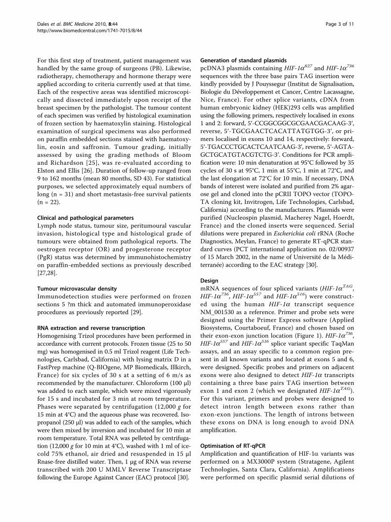
Generation of standard plasmidspcDNA3 plasmids containing HIF-1a827 and HIF-1a736
sequences with the three base pairs TAG insertion werekindly provided by J Pouyssegur (Institut de Signalisation,Biologie du Développement et Cancer, Centre Lacassagne,Nice, France). For other splice variants, cDNA fromhuman embryonic kidney (HEK)293 cells was amplifiedusing the following primers, respectively localised in exons1 and 2: forward, 5′-CCGGCGGCGCGAACGACAAG-3′,reverse, 5′-TGCGAACTCACATTATGTGG-3′, or pri-mers localised in exons 10 and 14, respectively: forward,5′-TGACCCTGCACTCAATCAAG-3′, reverse, 5′-AGTA-GCTGCATGTACGTCTG-3′. Conditions for PCR ampli-fication were: 10 min denaturation at 95°C followed by 35cycles of 30 s at 95°C, 1 min at 55°C, 1 min at 72°C, andthe last elongation at 72°C for 10 min. If necessary, DNAbands of interest were isolated and purified from 2% agar-ose gel and cloned into the pCRII TOPO vector (TOPO-TA cloning kit, Invitrogen, Life Technologies, Carlsbad,California) according to the manufacturers. Plasmids werepurified (Nucleospin plasmid, Macherey Nagel, Hoerdt,France) and the cloned inserts were sequenced. Serialdilutions were prepared in Escherichia coli rRNA (RocheDiagnostics, Meylan, France) to generate RT-qPCR stan-dard curves (PCT international application no. 02/00937of 15 March 2002, in the name of Université de la Médi-terranée) according to the EAC strategy [30].
DesignmRNA sequences of four spliced variants (HIF-1aTAG,HIF-1a736, HIF-1a557 and HIF-1a516) were construct-ed using the human HIF-1a transcript sequenceNM_001530 as a reference. Primer and probe sets weredesigned using the Primer Express software (AppliedBiosystems, Courtaboeuf, France) and chosen based ontheir exon-exon junction location (Figure 1). HIF-1a736,HIF-1a557 and HIF-1a516 splice variant specific TaqManassays, and an assay specific to a common region pre-sent in all known variants and located at exons 5 and 6,were designed. Specific probes and primers on adjacentexons were also designed to detect HIF-1a transcriptscontaining a three base pairs TAG insertion betweenexon 1 and exon 2 (which we designated HIF-1aTAG).For this variant, primers and probes were designed todetect intron length between exons rather thanexon-exon junctions. The length of introns betweenthese exons on DNA is long enough to avoid DNAamplification.
Optimisation of RT-qPCRAmplification and quantification of HIF-1a variants wasperformed on a MX3000P system (Stratagene, AgilentTechnologies, Santa Clara, California). Amplificationswere performed on specific plasmid serial dilutions of
Dales et al. BMC Medicine 2010, 8:44http://www.biomedcentral.com/1741-7015/8/44
Page 3 of 11
each variant in a total volume of 25 μl containing 1 ×TaqMan universal PCR MasterMix (Applied Biosys-tems, Courtaboeuf, France) and both forward andreverse primers and probes. Different ranges of primerconcentrations were tested and optimised for eachamplicon to obtain the most specific, sensitive and effi-cient RT-PCR amplification. Selected primer and probeconcentrations are shown in Table 1. OptimisedRT-qPCR reaction conditions for gene amplificationwere as follows: 50°C for 2 min, denaturation for10 min at 95°C followed by 50 cycles of 30 s at 95°C,1 min annealing and extension at 60°C for all tran-scripts except for HIF-1aTAG with 1 min at 62°C. Forvalidation of assays specificity, the primers and probesused to detect each splice variant were tested by RT-PCR with 1 μg RNA of the HEK293 cell line (DSMZ),1 μg genomic DNA of peripheral blood mononuclearcells (PBMCs) and standards (106 copies of plasmids)specific for another splice variant. No template control(NTC) was also performed. RT-qPCR products wereanalysed by Agilent 2100 Electrophoresis Bioanalyzer(Agilent Technologies, Santa Clara, California). Serialdilutions of specific standard plasmids were used tovalidate sensitivity, linearity and detection limits of
RT-qPCR assays. Intrareproducibility and inter-reprodu-cibility assays were performed by four RT-qPCR experi-ments, each time in triplicate.
RT-qPCR assaysAmplifications were performed with 5 μl of cDNA ofeach sample using RT-qPCR optimised conditions aspreviously described. mRNA from HEK293 cells wasused as positive control throughout the study. Standard(106 copies of plasmid) specifics of another splice variantwere amplified as negative control throughout the studyand NTC was performed. Standard curves were gener-ated by serial dilutions of HIF-1a splice variant plasmidsranging from 1 to 106 copies and used for calculation ofcopy numbers (CN) for each transcript. Transcripts ofthe gene coding for the TATA box-binding protein(TPB) were also quantified as endogenous gene controls,and each sample was normalised on the basis of its TBPcontent as previously described [31]. All experimentswere carried out with duplicates for each data point. Foreach experiment sample, the mRNA copy number ofHIF-1a gene (CNHIF-1a) and endogenous reference TBPgene (CNTBP) were quantified from standard curves.Final HIF-1a mRNA concentrations were expressed in
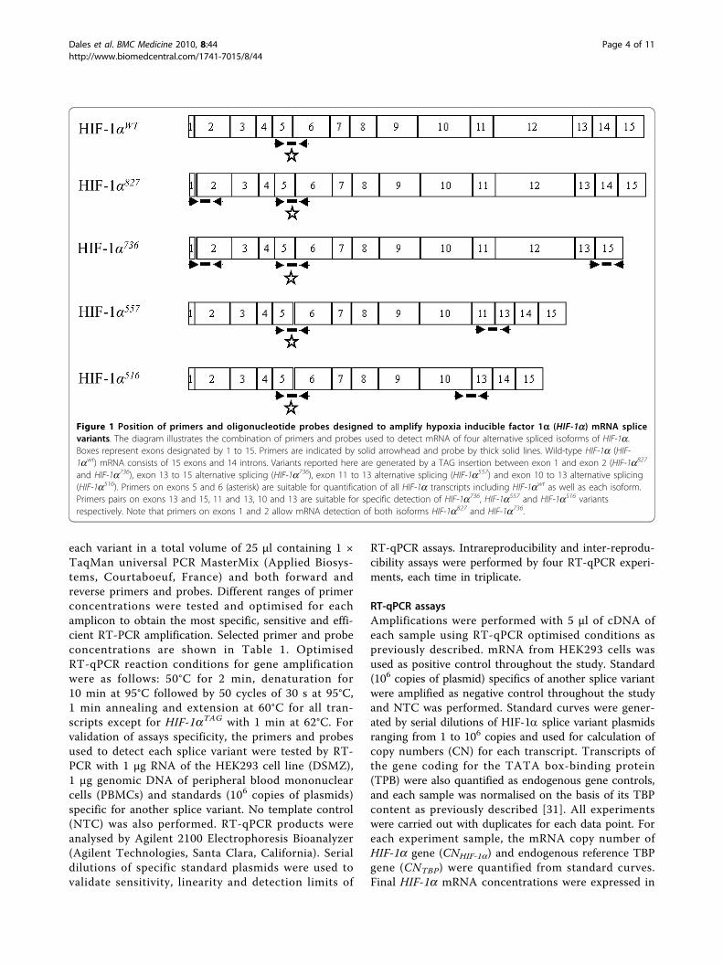
Figure 1 Position of primers and oligonucleotide probes designed to amplify hypoxia inducible factor 1a (HIF-1a) mRNA splicevariants. The diagram illustrates the combination of primers and probes used to detect mRNA of four alternative spliced isoforms of HIF-1a.Boxes represent exons designated by 1 to 15. Primers are indicated by solid arrowhead and probe by thick solid lines. Wild-type HIF-1a (HIF-1awt) mRNA consists of 15 exons and 14 introns. Variants reported here are generated by a TAG insertion between exon 1 and exon 2 (HIF-1a827
and HIF-1a736), exon 13 to 15 alternative splicing (HIF-1a736), exon 11 to 13 alternative splicing (HIF-1a557) and exon 10 to 13 alternative splicing(HIF-1a516). Primers on exons 5 and 6 (asterisk) are suitable for quantification of all HIF-1a transcripts including HIF-1awt as well as each isoform.Primers pairs on exons 13 and 15, 11 and 13, 10 and 13 are suitable for specific detection of HIF-1a736, HIF-1a557 and HIF-1a516 variantsrespectively. Note that primers on exons 1 and 2 allow mRNA detection of both isoforms HIF-1a827 and HIF-1a736.
Dales et al. BMC Medicine 2010, 8:44http://www.biomedcentral.com/1741-7015/8/44
Page 4 of 11
normalised copy numbers (NCNHIF-1a), as follows:NCNHIF-1a = CNHIF-1a/CNTBP.
Statistical analysisDifferences among groups of breast tissue specimens(normal/benign tissues vs carcinomas) in terms of theirHIF-1a mRNA levels were assessed using Kruskal-Wallisand Mann-Whitney tests with Bonferroni correction.Since levels of expression in breast cancer specimensshowed non-Gaussian distribution, non-parametric testswere used for the analysis of correlation with clinico-pathological parameters. Metastasis-free survival (MFS)time, defined as the time from surgery until diagnosis ofmetastasis, was used as a follow-up endpoint. Regressionmodels (univariate and multivariate) were used to com-pare survival and identify predictors of survival. All pre-operative predictors (clinicopathological and biologicalparameters) were included in the analysis. The predic-tors with P value < 0.20 in the univariate model weretested in the multivariate regression model. Then back-ward conditional method was used for variable selectionby the Cox multivariate regression model. Finally, vari-ables with adjusted P values < 0.05 were kept into thefinal model. All statistical analyses were performed withSPSS V.13.0.1 software (SPSS, Chicago, IL, USA).
ResultsClinicopathological parametersTumour characteristics are summarised in Table 2.Tumours corresponded to ductal carcinomas (n = 41)
and lobular carcinomas (n = 12). Tumours were grade 1in 9 cases, grade 2 in 26 cases and grade 3 in 17 cases.A mean of 16.9 (SD ± 5.1) lymph nodes were found inaxillary node excision, and 22 patients were node posi-tive. All patients underwent axillary node excision com-bined with wide local excision with margins clearanceor mastectomy in the same department. Treatment postsurgery consisted of radiotherapy, chemotherapy andhormone therapy performed by the same group ofoncologists. Duration of follow-up ranged from 9 to162 months. The 2004 records showed that among53 patients, 22 (41.5%) patients relapsed, among whom18 died.
Optimisation and validation of RT-PCR assaysPrimers and probes sets for HIF-1a variants were testedusing RT-qPCR on the HEK293 RNA and specific stan-dard plasmids. Primer concentration optimisation assayswere carried out to determine the optimal concentra-tions for each primer and probe set. To test the specifi-city and crossreaction of the primers used for RT-qPCR,experiments were performed using genomic DNA, stan-dard plasmids specific of another splice variant, andnNTC. No amplification was observed with genomicDNA of PBMCs, specific standard plasmids of othersplice variants and NTC. Serial dilutions of HIF-1a var-iants specific standard plasmid were used to generate astandard curve to assess the intra-assay and interassayreproducibility and sensitivity. For each variant, excel-lent reproducibility and linearity of standard curves
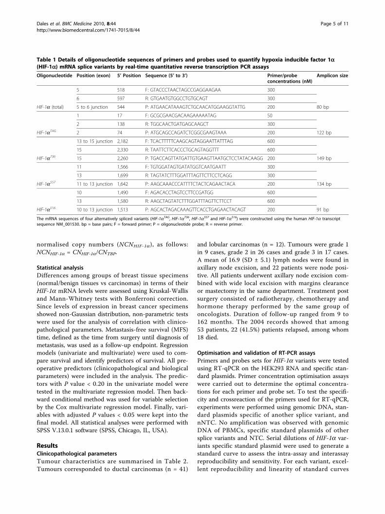
Table 1 Details of oligonucleotide sequences of primers and probes used to quantify hypoxia inducible factor 1a(HIF-1a) mRNA splice variants by real-time quantitative reverse transcription PCR assays
Oligonucleotide Position (exon) 5’ Position Sequence (5’ to 3’) Primer/probeconcentrations (nM)
Amplicon size
5 518 F: GTACCCTAACTAGCCGAGGAAGAA 300
6 597 R: GTGAATGTGGCCTGTGCAGT 300
HIF-1a (total) 5 to 6 junction 544 P: ATGAACATAAAGTCTGCAACATGGAAGGTATTG 200 80 bp
1 17 F: GCGCGAACGACAAGAAAAATAG 50
2 138 R: TGGCAACTGATGAGCAAGCT 300
HIF-1aTAG 2 74 P: ATGCAGCCAGATCTCGGCGAAGTAAA 200 122 bp
13 to 15 junction 2,182 F: TCACTTTTTCAAGCAGTAGGAATTATTTAG 600
15 2,330 R: TAATTCTTCACCCTGCAGTAGGTTT 600
HIF-1a736 15 2,260 P: TGACCAGTTATGATTGTGAAGTTAATGCTCCTATACAAGG 200 149 bp
11 1,566 F: TGTGGATAGTGATATGGTCAATGAATT 300
13 1,699 R: TAGTATCTTTGGATTTAGTTCTTCCTCAGG 300
HIF-1a557 11 to 13 junction 1,642 P: AAGCAAACCCATTTTCTACTCAGAACTACA 200 134 bp
10 1,490 F: AGACACCTAGTCCTTCCGATGG 600
13 1,580 R: AAGCTAGTATCTTTGGATTTAGTTCTTCCT 600
HIF-1a516 10 to 13 junction 1,513 P: AGCACTAGACAAAGTTCACCTGAGAACTACAGT 200 91 bp
The mRNA sequences of four alternatively spliced variants (HIF-1aTAG, HIF-1a736, HIF-1a557 and HIF-1a516) were constructed using the human HIF-1a transcriptsequence NM_001530. bp = base pairs; F = forward primer; P = oligonucleotide probe; R = reverse primer.
Dales et al. BMC Medicine 2010, 8:44http://www.biomedcentral.com/1741-7015/8/44
Page 5 of 11
were found. All runs exhibited amplification slopesbetween -3.31 and -3.55, which correspond to efficien-cies higher than 92% and coefficients of correlationbetween 0.993 and 1. The assays sensitivity was found at10 copies for all variants. We did not succeed in quanti-fying the HIF-1a417 isoform.
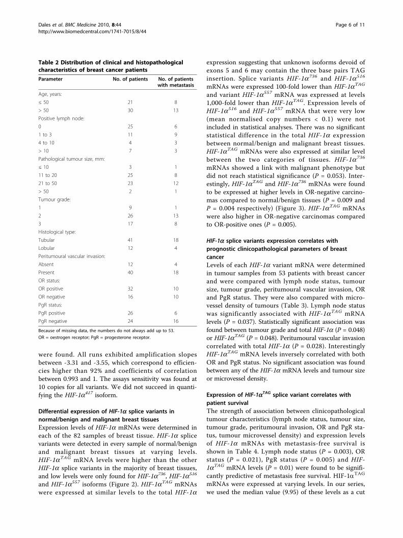
Differential expression of HIF-1a splice variants innormal/benign and malignant breast tissuesExpression levels of HIF-1a mRNAs were determined ineach of the 82 samples of breast tissue. HIF-1a splicevariants were detected in every sample of normal/benignand malignant breast tissues at varying levels.HIF-1aTAG mRNA levels were higher than the otherHIF-1a splice variants in the majority of breast tissues,and low levels were only found for HIF-1a736, HIF-1a516
and HIF-1a557 isoforms (Figure 2). HIF-1aTAG mRNAswere expressed at similar levels to the total HIF-1a
expression suggesting that unknown isoforms devoid ofexons 5 and 6 may contain the three base pairs TAGinsertion. Splice variants HIF-1a736 and HIF-1a516
mRNAs were expressed 100-fold lower than HIF-1aTAG
and variant HIF-1a557 mRNA was expressed at levels1,000-fold lower than HIF-1aTAG. Expression levels ofHIF-1a516 and HIF-1a557 mRNA that were very low(mean normalised copy numbers < 0.1) were notincluded in statistical analyses. There was no significantstatistical difference in the total HIF-1a expressionbetween normal/benign and malignant breast tissues.HIF-1aTAG mRNAs were also expressed at similar levelbetween the two categories of tissues. HIF-1a736
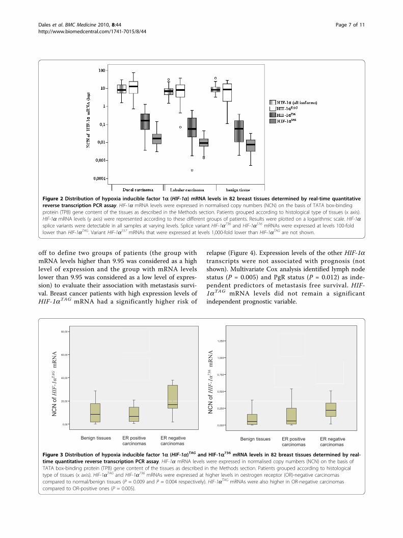
mRNAs showed a link with malignant phenotype butdid not reach statistical significance (P = 0.053). Inter-estingly, HIF-1aTAG and HIF-1a736 mRNAs were foundto be expressed at higher levels in OR-negative carcino-mas compared to normal/benign tissues (P = 0.009 andP = 0.004 respectively) (Figure 3). HIF-1aTAG mRNAswere also higher in OR-negative carcinomas comparedto OR-positive ones (P = 0.005).
HIF-1a splice variants expression correlates withprognostic clinicopathological parameters of breastcancerLevels of each HIF-1a variant mRNA were determinedin tumour samples from 53 patients with breast cancerand were compared with lymph node status, tumoursize, tumour grade, peritumoural vascular invasion, ORand PgR status. They were also compared with micro-vessel density of tumours (Table 3). Lymph node statuswas significantly associated with HIF-1aTAG mRNAlevels (P = 0.037). Statistically significant association wasfound between tumour grade and total HIF-1a (P = 0.048)or HIF-1aTAG (P = 0.048). Peritumoural vascular invasioncorrelated with total HIF-1a (P = 0.028). InterestinglyHIF-1aTAG mRNA levels inversely correlated with bothOR and PgR status. No significant association was foundbetween any of the HIF-1a mRNA levels and tumour sizeor microvessel density.
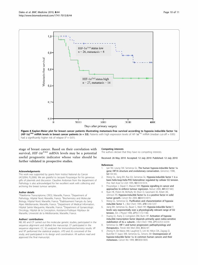
Expression of HIF-1aTAG splice variant correlates withpatient survivalThe strength of association between clinicopathologicaltumour characteristics (lymph node status, tumour size,tumour grade, peritumoural invasion, OR and PgR sta-tus, tumour microvessel density) and expression levelsof HIF-1a mRNAs with metastasis-free survival isshown in Table 4. Lymph node status (P = 0.003), ORstatus (P = 0.021), PgR status (P = 0.005) and HIF-1aTAG mRNA levels (P = 0.01) were found to be signifi-cantly predictive of metastasis free survival. HIF-1aTAG
mRNAs were expressed at varying levels. In our series,we used the median value (9.95) of these levels as a cut
Table 2 Distribution of clinical and histopathologicalcharacteristics of breast cancer patients
Parameter No. of patients No. of patientswith metastasis
Age, years:
≤ 50 21 8
> 50 30 13
Positive lymph node:
0 25 6
1 to 3 11 9
4 to 10 4 3
> 10 7 3
Pathological tumour size, mm:
≤ 10 3 1
11 to 20 25 8
21 to 50 23 12
> 50 2 1
Tumour grade:
1 9 1
2 26 13
3 17 8
Histological type:
Tubular 41 18
Lobular 12 4
Peritumoural vascular invasion:
Absent 12 4
Present 40 18
OR status:
OR positive 32 10
OR negative 16 10
PgR status:
PgR positive 26 6
PgR negative 24 16
Because of missing data, the numbers do not always add up to 53.
OR = oestrogen receptor; PgR = progesterone receptor.
Dales et al. BMC Medicine 2010, 8:44http://www.biomedcentral.com/1741-7015/8/44
Page 6 of 11
off to define two groups of patients (the group withmRNA levels higher than 9.95 was considered as a highlevel of expression and the group with mRNA levelslower than 9.95 was considered as a low level of expres-sion) to evaluate their association with metastasis survi-val. Breast cancer patients with high expression levels ofHIF-1aTAG mRNA had a significantly higher risk of
relapse (Figure 4). Expression levels of the other HIF-1atranscripts were not associated with prognosis (notshown). Multivariate Cox analysis identified lymph nodestatus (P = 0.005) and PgR status (P = 0.012) as inde-pendent predictors of metastasis free survival. HIF-1aTAG mRNA levels did not remain a significantindependent prognostic variable.
Figure 2 Distribution of hypoxia inducible factor 1a (HIF-1a) mRNA levels in 82 breast tissues determined by real-time quantitativereverse transcription PCR assay. HIF-1a mRNA levels were expressed in normalised copy numbers (NCN) on the basis of TATA box-bindingprotein (TPB) gene content of the tissues as described in the Methods section. Patients grouped according to histological type of tissues (x axis).HIF-1a mRNA levels (y axis) were represented according to these different groups of patients. Results were plotted on a logarithmic scale. HIF-1asplice variants were detectable in all samples at varying levels. Splice variant HIF-1a736 and HIF-1a516 mRNAs were expressed at levels 100-foldlower than HIF-1aTAG. Variant HIF-1a557 mRNAs that were expressed at levels 1,000-fold lower than HIF-1aTAG are not shown.
80,00
60,00
34
1,250
7
26
NA NA
E1E2/TBP
40,00
20 00
57
26
8
E13E15/TBP
1,000
0,750
0,500
30
39
ofHIF-1αTAGmRN
ofHIF-1α736mRN
Groupe_RERE2cat0 AbsRE2cat1 PrésBénin
20,00
0,00
Groupe_RERE2cat0 AbsRE2cat1 PrésBénin
0,250
0,000
NC
No
NC
N
Benign tissues ER positive carcinomas
ER negative carcinomas
Benign tissues ER positive carcinomas
ER negative carcinomascarcinomas carcinomas
Figure 3 Distribution of hypoxia inducible factor 1a (HIF-1a)TAG and HIF-1a736 mRNA levels in 82 breast tissues determined by real-time quantitative reverse transcription PCR assay. HIF-1a mRNA levels were expressed in normalised copy numbers (NCN) on the basis ofTATA box-binding protein (TPB) gene content of the tissues as described in the Methods section. Patients grouped according to histologicaltype of tissues (x axis). HIF-1aTAG and HIF-1a736 mRNAs were expressed at higher levels in oestrogen receptor (OR)-negative carcinomascompared to normal/benign tissues (P = 0.009 and P = 0.004 respectively). HIF-1aTAG mRNAs were also higher in OR-negative carcinomascompared to OR-positive ones (P = 0.005).
Dales et al. BMC Medicine 2010, 8:44http://www.biomedcentral.com/1741-7015/8/44
Page 7 of 11

DiscussionSeveral isoforms of HIF-1a resulting from alternativesplicing have been described in various human tumourcell lines or tissues. Only one isoform (HIF-1a736) hasbeen documented in human breast cancer [24]. More-over, only a few studies have been performed to linkHIF-1a mRNA expression and tumour-specific para-meters [32,33]. Specific antibodies allowing HIF-1asplice variant detection at the protein level are not cur-rently available. We used a real-time quantitative reversetranscription PCR assay to measure mRNA expressionlevels of 4 alternatively spliced transcripts of HIF-1a inbreast specimens of 53 primary cancers and 29 normaltissues or benign lesions.Interestingly, this work shows for the first time higher
expression levels of two HIF-1a splice variants (HIF-1aTAG and HIF-1a736) in OR-negative carcinomas com-pared to normal/benign tissues. We also investigated theprognostic value of HIF-1a transcript expression levelsin breast cancer and found a significant relationshipbetween either clinicopathological characteristics orpatient metastasis-free survival. First, we found thatHIF-1aTAG mRNAs levels were substantially higher inhigh grade and steroid hormone receptor-negativetumours. The second and most striking observation was
that HIF-1aTAG mRNA levels were indicative of shortermetastasis-free survival, and that this correlated withlymph node status. Contrary to the Cayre et al. report[24], we did not find significant correlation betweentotal HIF-1a mRNA expression and lymph node statusbut we observed significant association with tumourgrade. This could be because our series was smallerthan series of Cayre et al. or because the technologyused in our study was more sensitive. In our series, totalHIF-1a mRNA expression negatively correlated with ORstatus. Similar to Cayre et al. we did not find any corre-lation between total HIF-1a mRNA expression and out-come. Our results showing that HIF-1a736 mRNAexpression does not correlate with clinicopathologicalcharacteristics of tumours also concur with earlier find-ings of the same group [24]. In this study, effect of adju-vant (none, chemotherapy or hormone) treatment onsurvival and interactions with expression levels of HIF-1a splice variants were not checked because of the lim-ited number of patients in each subgroup. Furtherexperiments in a larger series are required to answerthis question.Alternative splicing is known to play an important role
in gene expression regulation by modulating the func-tional properties of transcription factors [34]. In this
Table 3 Relationship between the hypoxia inducible factor 1a (HIF-1a) splice variant’s expression levels (normalisedcopy numbers) and clinicopathological factors or microvessel density in tumour tissue specimens from 53 breastcancer patients
Variable N Total HIF-1a,mean (SD)
P value HIF-1aTAG,mean (SD)
P value HIF-1a736,mean (SD)
P value
Lymph node status:
Negative 25 10.0 (9.0) NS 10.9 (10.9) 0.037 0.19 (0.29) NS
Positive 22 12.8 (7.8) 21.1 (19.2) 0.26 (0.28)
Pathological tumour size:
< 20 mm 28 10.2 (8.3) NS 12.4 (13.2) NS 0.17 (0.26) NS
≥ 20 mm 25 11.8 (8.3) 17.9 (17.4) 0.25 (0.28)
Tumour grade:
1/2 vs 35 9.2 (7.7) 0.048 11.8 (12.3) 0.048 0.19 (0.30) NS
3 17 14.6 (8.7) 21.5 (19.4) 0.23 (0.20)
Peritumoural vascular invasion:
Absent 12 8 (9) 0.028 9.6 (10.1) NS 0.19 (0.36) NS
Present 40 11.9 (8.1) 16.6 (16.6) 0.20 (0.24)
OR status:
Negative 16 14.2 (7.3) 0.024 20.8 (11.9) 0.005 0.25 (0.19) 0.06
Positive 32 10 (8.8) 12.7 (17.1) 0.20 (0.31)
PgR status:
Negative 24 11.8 (7.3) NS 20.08 (17.8) 0.033 0.23 (0.19) 0.077
Positive 26 10.8 (9.4) 11.3 (12.3) 0.2 (0.34)
Tumour microvessel density (low vs high) NS NS NS
Because of missing data, the numbers do not always add up to 53. Expression levels of HIF-1a516 and HIF-1a557 mRNA were not included in statistical analysesbecause of their very low levels (mean normalised copy numbers < 0.1).
NS = non-significant; OR = oestrogen receptor; PgR = progesterone receptor.
Dales et al. BMC Medicine 2010, 8:44http://www.biomedcentral.com/1741-7015/8/44
Page 8 of 11

regard, alternative splicing can modify DNA-bindingproperties of transcription factors [35], introduce oreliminate activating domains or increase the in vivo sta-bility of a given isoform [36]. Moreover, the abundanceof specific isoforms is likely to result from differentialexpression, RNA stability and selective splicing processleading to an increase of some mRNA species. Recentevidence indicates that in several cancers the ratio ofsplice variants is dramatically altered and that differen-tial expression of alternatively spliced isoforms in cancerpatients can have severe implications for clinical out-come [37]. Remarkably, statistical association has pre-viously been reported between HIF-1b splicing variantexpression, oestrogen receptors and breast cancer survi-val [38]. It should be noted that the primers designedon exons 1 and 2 in our study allowed quantification ofthe HIF-1aTAG sequence present in both HIF-1a827 andHIF-1a736 splice variants [17]. The HIF-1aTAG transcriptis characterised by insertion of a three base pairs TAG
insertion that may be generated by the use of twopotential splice acceptor dinucleotides (AG) of intron 1at a splice junction site as previously described [39,1].This splicing results in the replacement of Lys12 byAsn12 and the addition of Arg13 residue locatedupstream from the bHLH domain of the protein. Thisreplacement may modify the DNA binding affinity ofthe protein complex as previously shown for Arg14 andArg15 residues in aryl hydrocarbon receptor (AHR)/arylhydrocarbon receptor nuclear translocator (ARNT) het-erodimer [40]. Further biochemical and structural stu-dies are required for a better understanding of thefunctional properties of this variant.
ConclusionsTo our knowledge, our results suggest for the first timethat at least one HIF-1a splice a variant may be a mar-ker for the advanced clinical and oestrogen-resistant
Table 4 Univariate and multivariate (backward conditional selection) Cox regression analysis of metastasis-freesurvival (MFS) in breast cancer patients considering clinicopathological characteristics, tumour microvessel densityand hypoxia inducible factor 1a (HIF-1a)TAG mRNA levels
Characteristics Univariate analysis of MFS,Exp B (95% CI)
P value Multivariate analysis of MFS,Exp B (95% CI)
P value
Age, years:
< 50 1 0.561
≥ 50 1.32 (0.52 to 3.37)
Positive lymph node:
0 1 0.003 1 0.005
1 to 3 8.00 (2.56 to 24.81) 5.56 (1.75 to 17.63)
4 to 9 7.50 (1.70 to 30.00) 6.68 (1.48 to 30.14)
> 10 1.20 (0.14 to 10.00) 0.48 (0.05 to 4.25)
Pathological tumour size, mm:
< 10 1 0.39
10 to 20 1.10 (0.10 to 8.70)
21 to 49 2.40 (0.30 to 18.90)
> 50 2.70 (0.20 to 42.90)
Tumour grade:
1/2 vs 1 0.24
3 1.70 (0.70 to 4.40)
Peritumoural vascular invasion:
Absent 1 0.104
Present 3.40 (0.80 to 14.80)
OR status:
Positive
Negative 0.33 (0.13 to 0.85) 0.021
PgR status:
Positive
Negative 0.20 (0.07 to 0.60) 0.005 0.23 (0.07 to 0.72) 0.012
Tumour microvessel density 1.02 (0.07 to 0.60) 0.22
HIF-1aTAG mRNA levels 1.03 (1.00 to 1.05) 0.01
OR = oestrogen receptor; PgR = progesterone receptor, Exp B = exponential of beta
Dales et al. BMC Medicine 2010, 8:44http://www.biomedcentral.com/1741-7015/8/44
Page 9 of 11
stage of breast cancer. Based on their correlation withsurvival, HIF-1aTAG mRNA levels may be a potentialuseful prognostic indicator whose value should befurther validated in prospective studies.
AcknowledgementsThis work was supported by grants from Institut National du Cancer(ACI2004, PL2006). We are grateful to Jacques Pouyssegur for his generousgifts of plasmids and discussion. Claudine Andonian from the department ofPathology is also acknowledged for her excellent work with collecting andarchiving the breast tumour samples.
Author details1Plateforme Transcriptome, CRO2, Marseille, France. 2Department ofPathology, Hôpital Nord, Marseille, France. 3Biochemistry and MolecularBiology, Hôpital Nord, Marseille, France. 4Etablissement Français du SangAlpes Méditerranée, Marseille, France. 5Department of Medical Information,Hôpital Sainte Marguerite, Marseille, France. 6Department of GynaecologicOncology, Hôpital de la Conception, Assistance-Publique Hôpitaux deMarseille, Université de la Méditerranée, Marseille, France.
Authors’ contributionsNB, MS and CP carried out the molecular genetic studies, participated in thesequence alignment and drafted the manuscript. CF participated in thesequence alignment. CC, SG analysed the immunohistochemistry results. VPand VP performed the statistical analysis. J-PD and JG conceived of thestudy, and participated in its design and coordination. All authors read andapproved the final manuscript.
Competing interestsThe authors declare that they have no competing interests.
Received: 26 May 2010 Accepted: 12 July 2010 Published: 12 July 2010
References1. Iyer NV, Leung SW, Semenza GL: The human hypoxia-inducible factor 1a
gene: HIF1A structure and evolutionary conservation. Genomics 1998,52:159-165.
2. Wang GL, Jiang BH, Rue EA, Semenza GL: Hypoxia-inducible factor 1 is abasic-helix-loop-helix-PAS heterodimer regulated by cellular O2 tension.Proc Natl Acad Sci USA 1995, 92:5510-5514.
3. Pouysségur J, Dayan F, Mazure NM: Hypoxia signalling in cancer andapproaches to enforce tumour regression. Nature 2006, 441:437-443.
4. Ryan HE, Poloni M, McNulty W, Elson D, Gassmann M, Arbeit JM,Johnson RS: Hypoxia-inducible factor-1a is a positive factor in solidtumor growth. Cancer Res 2000, 60:4010-4015.
5. Wang GL, Semenza GL: Purification and characterization of hypoxia-inducible factor 1. J Biol Chem 1995, 270:1230-1237.
6. Jiang BH, Semenza GL, Bauer C, Marti HH: Hypoxia-inducible factor 1levels vary exponentially over a physiologically relevant range of O2tension. Am J Physiol 1996, 271:C1172-1180.
7. Huang LE, Arany Z, Livingston DM, Bunn HF: Activation of hypoxia-inducible transcription factor depends primarily upon redox-sensitivestabilization of its a subunit. J Biol Chem 1996, 271:32253-32259.
8. Semenza GL: HIF-1 and tumor progression: pathophysiology andtherapeutics. Trends Mol Med 2002, 8:S62-67.
9. Zhong H, De Marzo AM, Laughner E, Lim M, Hilton DA, Zagzag D,Buechler P, Isaacs WB, Semenza GL, Simons JW: Overexpression ofhypoxia-inducible factor 1a in common human cancers and theirmetastases. Cancer Res 1999, 59:5830-5835.
Figure 4 Kaplan-Meier plot for breast cancer patients illustrating metastasis-free survival according to hypoxia inducible factor 1a(HIF-1a)TAG mRNA levels in breast cancer patients (n = 53). Patients with high expression levels of HIF-1aTAG mRNA (median cut-off = 9.95)had a significantly higher risk of relapse (P = 0.01).
Dales et al. BMC Medicine 2010, 8:44http://www.biomedcentral.com/1741-7015/8/44
Page 10 of 11
10. Dales JP, Garcia S, Meunier-Carpentier S, Andrac-Meyer L, Haddad O,Lavaut MN, Allasia C, Bonnier P, Charpin C: Overexpression of hypoxia-inducible factor HIF-1a predicts early relapse in breast cancer:retrospective study in a series of 745 patients. Int J Cancer 2005,116:734-739.
11. Schindl M, Schoppmann SF, Samonigg H, Hausmaninger H, Kwasny W,Gnant M, Jakesz R, Kubista E, Birner P, Oberhuber G: Overexpression ofhypoxia-inducible factor 1a is associated with an unfavorable prognosisin lymph node-positive breast cancer. Clin Cancer Res 2002, 8:1831-1837.
12. Huang LE, Gu J, Schau M, Bunn HF: Regulation of hypoxia-inducible factor1a is mediated by an O2-dependent degradation domain via theubiquitin-proteasome pathway. Proc Natl Acad Sci USA 1998, 95:7987-7992.
13. Jiang BH, Rue E, Wang GL, Roe R, Semenza GL: Dimerization, DNA binding,and transactivation properties of hypoxia-inducible factor 1. J Biol Chem1996, 271:17771-17778.
14. Jiang BH, Zheng JZ, Leung SW, Roe R, Semenza GL: Transactivation andinhibitory domains of hypoxia-inducible factor 1a. Modulation oftranscriptional activity by oxygen tension. J Biol Chem 1997,272:19253-19260.
15. Pagé EL, Robitaille GA, Pouysségur J, Richard DE: Induction of hypoxia-inducible factor-1a by transcriptional and translational mechanisms. JBiol Chem 2002, 277:48403-48409.
16. Semenza GL, Rue EA, Iyer NV, Pang MG, Kearns WG: Assignment of thehypoxia-inducible factor 1a gene to a region of conserved synteny onmouse chromosome 12 and human chromosome 14q. Genomics 1996,34:437-439.
17. Gothié E, Richard DE, Berra E, Pagès G, Pouysségur J: Identification ofalternative spliced variants of human hypoxia-inducible factor-1a. J BiolChem 2000, 275:6922-6927.
18. Chun YS, Choi E, Yeo EJ, Lee JH, Kim MS, Park JW: A new HIF-1 a variantinduced by zinc ion suppresses HIF-1-mediated hypoxic responses. J CellSci 2001, 114:4051-4061.
19. Chun YS, Choi E, Kim TY, Kim MS, Park JW: A dominant-negative isoformlacking exons 11 and 12 of the human hypoxia-inducible factor-1agene. Biochem J 2002, 362:71-79.
20. Chun YS, Lee KH, Choi E, Bae SY, Yeo EJ, Huang LE, Kim MS, Park JW:Phorbol ester stimulates the nonhypoxic induction of a novel hypoxia-inducible factor 1a isoform: implications for tumor promotion. CancerRes 2003, 63:8700-8707.
21. Lee KH, Park JW, Chun YS: Non-hypoxic transcriptional activation of thearyl hydrocarbon receptor nuclear translocator in concert with a novelhypoxia-inducible factor-1a isoform. Nucleic Acids Res 2004, 32:5499-5511.
22. Depping R, Hägele S, Wagner KF, Wiesner RJ, Camenisch G, Wenger RH,Katschinski DM: A dominant-negative isoform of hypoxia-induciblefactor-1 a specifically expressed in human testis. Biol Reprod 2004,71:331-339.
23. Lukashev D, Sitkovsky M: Preferential expression of the novel alternativeisoform I.3 of hypoxia-inducible factor 1a in activated human Tlymphocytes. Hum Immunol 2008, 69:421-425.
24. Cayre A, Rossignol F, Clottes E, Penault-Llorca F: aHIF but not HIF-1atranscript is a poor prognostic marker in human breast cancer. BreastCancer Res 2003, 5:R223-230.
25. Bloom HJ, Richardson WW: Histological grading and prognosis in breastcancer; a study of 1409 cases of which 359 have been followed for 15years. Br J Cancer 1957, 11:359-377.
26. Elston CW, Ellis IO: Pathological prognostic factors in breast cancer. I. Thevalue of histological grade in breast cancer: experience from a largestudy with long-term follow-up. Histopathology 1991, 19:403-410.
27. Charpin C, Martin PM, De Victor B, Lavaut MN, Habib MC, Andrac L, Toga M:Multiparametric study (SAMBA 200) of estrogen receptorimmunocytochemical assay in 400 human breast carcinomas: analysis ofestrogen receptor distribution heterogeneity in tissues and correlationswith dextran coated charcoal assays and morphological data. Cancer Res1988, 48:1578-1586.
28. Charpin C, Jacquemier J, Andrac L, Vacheret H, Habib MC, Devictor B,Lavaut MN, Toga M: Multiparametric analysis (SAMBA 200) of theprogesterone receptor immunocytochemical assay in nonmalignant andmalignant breast disorders. Am J Pathol 1988, 132:199-211.
29. Dales JP, Garcia S, Andrac L, Carpentier S, Ramuz O, Lavaut MN, Allasia C,Bonnier P, Charpin C: Prognostic significance of angiogenesis evaluatedby CD105 expression compared to CD31 in 905 breast carcinomas:
correlation with long-term patient outcome. Int J Oncol 2004,24:1197-1204.
30. Gabert J, Beillard E, van der Velden VH, Bi W, Grimwade D, Pallisgaard N,Barbany G, Cazzaniga G, Cayuela JM, Cavé H, Pane F, Aerts JL, De Micheli D,Thirion X, Pradel V, González M, Viehmann S, Malec M, Saglio G, vanDongen JJ: Standardization and quality control studies of ‘real-time’quantitative reverse transcriptase polymerase chain reaction of fusiongene transcripts for residual disease detection in leukemia - a EuropeAgainst Cancer program. Leukemia 2003, 17:2318-2357.
31. Bièche I, Laurendeau I, Tozlu S, Olivi M, Vidaud D, Lidereau R, Vidaud M:Quantitation of MYC gene expression in sporadic breast tumors with areal-time reverse transcription-PCR assay. Cancer Res 1999, 59:2759-2765.
32. Nakayama K, Kanzaki A, Hata K, Katabuchi H, Okamura H, Miyazaki K,Fukumoto M, Takebayashi Y: Hypoxia-inducible factor 1a (HIF-1 a) geneexpression in human ovarian carcinoma. Cancer Lett 2002, 176:215-223.
33. Søndergaard KL, Hilton DA, Penney M, Ollerenshaw M, Demaine AG:Expression of hypoxia-inducible factor 1a in tumors of patients withglioblastoma. Neuropathol Appl Neurobiol 2002, 28:210-217.
34. López AJ: Developmental role of transcription factor isoforms generatedby alternative splicing. Dev Biol 1995, 172:396-411.
35. Kozmik Z, Czerny T, Busslinger M: Alternatively spliced insertions in thepaired domain restrict the DNA sequence specificity of Pax6 and Pax8.EMBO J 1997, 16:6793-6803.
36. Foulkes NS, Mellström B, Benusiglio E, Sassone-Corsi P: Developmentalswitch of CREM function during spermatogenesis: from antagonist toactivator. Nature 1992, 355:80-84.
37. Venables JP: Unbalanced alternative splicing and its significance incancer. Bioessays 2006, 28:378-386.
38. Qin C, Wilson C, Blancher C, Taylor M, Safe S, Harris AL: Association ofARNT splice variants with estrogen receptor-negative breast cancer,poor induction of vascular endothelial growth factor under hypoxia, andpoor prognosis. Clin Cancer Res 2001, 7:818-823.
39. Wenger RH, Rolfs A, Spielmann P, Zimmermann DR, Gassmann M: Mousehypoxia-inducible factor-1a is encoded by two different mRNA isoforms:expression from a tissue-specific and a housekeeping-type promoter.Blood 1998, 91:3471-3480.
40. Wache SC, Hoagland EM, Zeigler G, Swanson HI: Role of arginine residues14 and 15 in dictating DNA binding stability and transactivation of thearyl hydrocarbon receptor/aryl hydrocarbon receptor nucleartranslocator heterodimer. Gene Expr 2005, 12:231-243.
Pre-publication historyThe pre-publication history for this paper can be accessed here:http://www.biomedcentral.com/1741-7015/8/44/prepub
doi:10.1186/1741-7015-8-44Cite this article as: Dales et al.: Hypoxia inducible factor 1a gene (HIF-1a) splice variants: potential prognostic biomarkers in breast cancer.BMC Medicine 2010 8:44.
Submit your next manuscript to BioMed Centraland take full advantage of:
• Convenient online submission
• Thorough peer review
• No space constraints or color figure charges
• Immediate publication on acceptance
• Inclusion in PubMed, CAS, Scopus and Google Scholar
• Research which is freely available for redistribution
Submit your manuscript at www.biomedcentral.com/submit
Dales et al. BMC Medicine 2010, 8:44http://www.biomedcentral.com/1741-7015/8/44
Page 11 of 11
Copyright © 2022 FDOKUMEN