HIF and reactive oxygen species regulate oxidative phosphorylation in cancer

Effects of TLR Agonists on the Hypoxia-RegulatedTranscription Factor HIF-1a and Dendritic CellMaturation under Normoxic ConditionsRolf Spirig1., Siamak Djafarzadeh2,5., Tomas Regueira2,5, Sidney G. Shaw3, Christophe von Garnier4,
Jukka Takala2,5, Stephan M. Jakob2,5, Robert Rieben1, Philipp M. Lepper2,5*
1 Laboratory of Cardiovascular Research, Department of Clinical Research, University of Bern, Bern, Switzerland, 2 Laboratory of Intensive Care, Department of Clinical
Research, University of Bern, Bern, Switzerland, 3 Laboratory of Vasoactive Peptides, Department of Clinical Research, University of Bern, Bern, Switzerland, 4 Department
of Pneumology, Bern University Hospital, Bern, Switzerland, 5 Department of Intensive Care Medicine, Bern University Hospital, Bern, Switzerland
Abstract
Dendritic cells (DC) are professional antigen presenting cells that represent an important link between innate and adaptiveimmunity. Danger signals such as toll-like receptor (TLR) agonists induce maturation of DC leading to a T-cell mediatedadaptive immune response. In this study, we show that exogenous as well as endogenous inflammatory stimuli for TLR4and TLR2 induce the expression of HIF-1a in human monocyte-derived DC under normoxic conditions. On the functionallevel, inhibition of HIF-1a using chetomin (CTM), YC-1 and digoxin lead to no consistent effect on MoDC maturation, orcytokine secretion despite having the common effect of blocking HIF-1a stabilization or activity through differentmechanisms. Stabilization of HIF-1a protein by hypoxia or CoCl2 did not result in maturation of human DC. In addition, wecould show that TLR stimulation resulted in an increase of HIF-1a controlled VEGF secretion. These results show thatstimulation of human MoDC with exogenous as well as endogenous TLR agonists induces the expression of HIF-1a in atime-dependent manner. Hypoxia alone does not induce maturation of DC, but is able to augment maturation after TLRligation. Current evidence suggests that different target genes may be affected by HIF-1a under normoxic conditions withphysiological roles that differ from those induced by hypoxia.
Citation: Spirig R, Djafarzadeh S, Regueira T, Shaw SG, von Garnier C, et al. (2010) Effects of TLR Agonists on the Hypoxia-Regulated Transcription Factor HIF-1aand Dendritic Cell Maturation under Normoxic Conditions. PLoS ONE 5(6): e10983. doi:10.1371/journal.pone.0010983
Editor: Patricia T. Bozza, Fundacao Oswaldo Cruz, Brazil
Received August 17, 2009; Accepted May 9, 2010; Published June 7, 2010
Copyright: � 2010 Spirig et al. This is an open-access article distributed under the terms of the Creative Commons Attribution License, which permitsunrestricted use, distribution, and reproduction in any medium, provided the original author and source are credited.
Funding: This work was supported by the Foundation for Research in Anaesthesia and Intensive Care Medicine (to PL, to SD), the EU 6th Framework IntegratedResearch Project ‘‘Reprogramming the Immune System for the Establishment of Tolerance’’ (RISET) and the Swiss National Science Foundation grant no. 3200B0-116618 (to RR). The funders had no role in study design, data collection and analysis, decision to publish, or preparation of the manuscript.
Competing Interests: The authors have declared that no competing interests exist.
* E-mail: [email protected]
. These authors contributed equally to this work.
Introduction
DC are a unique leukocyte population of professional antigen
presenting cells (APC) that play an important role in bridging
innate and adaptive immunity [1]. They are crucial for inducing
T-cell mediated immune responses, as seen in infection, allograft
rejection, as well as the induction of peripheral tolerance [2,3]. DC
continuously scan their environment. For this purpose, they
express pattern recognition receptors (PRR) including Toll-like
receptors (TLR), nucleotide-binding oligomerization domain
(NOD)-like receptors, C-type lectin receptors and others. Activat-
ing signals such as pathogen-derived molecules, e.g. LPS or
lipoteichoic acid (LTA), induce maturation of the cells. Recent
studies have shown that some of these PRR can also sense the
presence of endogenous danger molecules, which are released in
the context of tissue injury [4]. Ischemia/reperfusion (I/R) results
in shedding and degradation of the glycosaminoglycans heparan
sulfate (HS) [5–8] and hyaluronic acid (HA) [9,10] from the
endothelial cell surface. It has been shown that HS and HA induce
maturation of DC via TLR4 in vitro [11,12]. Furthermore, it has
been demonstrated that TLR2 and TLR4 are crucially involved in
I/R injury in vivo [13–17]. Additionally, maturation of DC is
influenced by their microenvironment. Recent evidence suggests,
that Toll-like receptor ligation can lead to stabilization of the
transcription factor hypoxia-inducible factor 1a (HIF-1a) under
normoxic conditions, most likely via NFkB [18,19]. HIF-1a has
been described as a key regulator of a broad range of cellular and
systemic responses to hypoxic conditions. HIF-1a is also involved
in myeloid cell-mediated inflammation [20]. Furthermore, HIF-1ais a master regulator of the bactericidal capacity of phagocytes
[21]. Lipopolysaccharides (LPS) induce the expression of HIF-1ain murine macrophages [22] and dendritic cells (DC) [23].
Additionally, HIF-1a knock-out mice develop less clinical signs of
sepsis [24]. These findings suggest the hypothesis that HIF-1a may
also be an important mediator of inflammatory responses in the
absence of hypoxia and that HIF-1a activation by TLR ligands
under normoxic conditions might have functional consequences
for human DC maturation and cytokine production. Thus, we
investigated the effect of TLR2 and TLR4 activation by
endogenous and exogenous ligands on the upregulation of the
transcription factor HIF-1a, dendritic cell maturation and
cytokine production in human monocyte-derived DC (MoDC)
PLoS ONE | www.plosone.org 1 June 2010 | Volume 5 | Issue 6 | e10983

under normoxia. Results show that stimulation of human MoDC
with exogenous as well as endogenous TLR agonists induces the
expression of HIF-1a in a time-dependent manner. Interestingly,
hypoxia alone does not induce maturation of DC, but is able to
augment maturation after TLR ligation. Furthermore, we have
investigated the effect of three previously described HIF-1ainhibitors (chetomin [CTM], digoxin and an inhibitor of both,
NFkB and HIF-1a signaling, YC-1 on MoDC maturation and
function.
Results
Upregulation of HIF-1a by exogenous and endogenousTLR4 agonists in human MoDC under normoxia
To investigate if HIF-1a is upregulated by TLR stimulation
under normoxic conditions, MoDC were incubated with selective
TLR ligands for the indicated time periods in a normal humidified
cell incubator. We used LPS (1 mg/ml, Fig. 1A) and HA (20 mg/
ml, Fig. 1B) as exogenous and endogenous TLR4 agonists,
respectively. HIF-1a expression at the protein level was deter-
mined by Western blot analyses. As shown in Fig. 1A and B, both
TLR4 agonists induced the expression of HIF-1a in a time-
dependent manner. In parallel, cells were stained for the
expression of the co-stimulatory molecules CD80 and CD86 as
markers of cell maturation to confirm activation of the cells
(Fig. 2A). To rule out contamination of HA with LPS, we
incubated the cells with polymyxin B, an inhibitor of the biological
activities of endotoxin. As shown in Fig. 2B, polymyxin B did not
affect maturation of MoDC induced by HA whereas the effect of
LPS was abolished.
HIF-1a expression is induced by TLR2To investigate, if stabilization of HIF-1a protein is restricted to
TLR4 signaling, we tested the effect of the TLR2 agonist LTA on
the expression of HIF-1a. As shown in Fig. 1C, LTA also induced
the stabilization of HIF-1a protein in a time dependent manner
under normoxic conditions.
Hypoxia and stabilization of HIF-1a by the agonist CoCl2does not induce maturation of MoDC
We further analyzed, if stabilization of HIF-1a protein by
CoCl2 alone, without stimulation of TLR, leads to an activation of
MoDC. As shown in Fig. 1D, treatment with CoCl2 led to the
stabilization of HIF-1a but did not lead to a significant increase of
the expression of the co-stimulatory molecules CD80 and CD86
(Fig. 2A). In addition we investigated the effect of hypoxic
conditions (1.5% oxygen) on the maturation of MoDC. Hypoxia
alone did not lead to phenotypic maturation of human MoDC
(Fig. 2A), whereas the cells still matured if they were incubated
together with LPS (Fig. 2A). MoDC stimulated with LPS under
hypoxic conditions showed a synergistic response with a significant
increase of double positive CD80+/CD86+ cells (Fig. 2A) com-
pared to LPS stimulation under normoxia.
Analysis of commonly used inhibitors of HIF-1a/NFkB(CTM, digoxin and YC-1) on phenotypic maturation ofhuman MoDC
To further characterize the potential involvement of the
transcription factor HIF-1a and NFkB in the maturation process
of human MoDC, we used CTM (Chetomin), YC-1 and digoxin
to attenuate HIF-1a and/or NFkB responses. CTM has been
shown to block the interaction of HIF1a with transcriptional co-
activators p300 and cAMP response element binding protein
(CREB), thereby attenuating hypoxia-inducible transcription [25].
CTM, however, did not prevent the increase in NFkB p65 subunit
nuclear levels or NFkB transactivation activity in response to LPS-
IFNc challenge [19].
YC-1 in contrast has been reported to abolish constitutive
nuclear translocation and activation of NFkB/p65 [26] an
upstream regulator of HIF-1a. However, YC-1 might also act
on the PI3K/Akt/mTOR pathway, which can also regulate HIF-
1a expression at the translational step [27]. Digoxin is thought to
block accumulation of HIF-1a protein [28].
MoDC were pretreated with CTM for 3 hours and then
stimulated with LPS, LTA or HA for 24 hours. Afterwards, cells
were analyzed for expression of CD40, CD80, CD86 and ICAM-1
(CD54) by flow cytometry. CTM consistently inhibited the up-
regulation of all markers compared to the positive controls in
response to all TLR ligands tested (Fig. 3A-D). In contrast,
pretreatment of MoDC with YC-1 for 5 minutes before exposure
to LPS, LTA or HA for 24 hours significantly inhibited only the
up-regulation of CD80, CD86 and ICAM-1 primarily in response
to LPS. No significant inhibitory response of YC-1 was observed
for HA or LTA except for ICAM-1 induction by LTA (Fig. 4A-D).
In a second series of experiments digoxin was used as an additional
inhibitor of HIF-1a in an attempt to confirm the above
observations. MoDC were pretreated with Digoxin (50 nM,
100nM) for one hour then stimulated with LPS, LTA or HA for
24 hours. As before, cells were analyzed for expression of CD40,
CD80, CD86 and ICAM-1 by flow cytometry. Under these
conditions, there was no significant effect of digoxin on
the expression of any of the maturation markers analysed.
(Figure 5A-D).
In all studies, exposure of MoDC to the used concentrations of
CTM, CoCl2, LPS or YC-1 for 24hr did not affect viability of the
cells as determined by flow cytometry (Figure 6). As a general rule
any dead cells were always excluded from flow cytometric analysis
by PI staining and there was no indication of synergistic loss of cell
viability in co-incubation studies.
Modification of VEGF secretion by MoDC by CTM, YC-1and digoxin
Under hypoxic conditions, the vegf-gene is known to be at least
partly under control of the transcription factor HIF-1a. To
analyze if a similar dependency could be observed in response to
TLR agonists under normoxic conditions, VEGF stimulation by
selective TLR ligands was measured in the supernatants of
MoDC. As shown in Fig. 7A, treatment with CTM significantly
inhibited the TLR-induced production of VEGF whereas no
inhibition was observed in the presence of YC-1 or digoxin. In
fact, responses to HA and LTA in the presence of YC-1 were
enhanced (Fig. 7B and C). As expected hypoxia-induced secretion
of VEGF by MoDC was increased by incubation for 24 hours
under hypoxic conditions (data not shown).
Digoxin inhibits LPS-, HA- and LTA-induced increases inHIF-1a but does not alter TLR4 expression
Given the lack of effect of digoxin on expression of DC
maturation markers or VEGF production in response to TLR
ligands we determined the effect of digoxin on cellular HIF-1aprotein levels. Cells were exposed to various concentrations of
digoxin and thereafter stimulated with TLR ligands. As shown in
Fig. 8A, pretreatment of cells with 50 and 100 nM digoxin dose
dependently inhibited TLR agonist stimulated cellular HIF-1aprotein increases whereas TLR4 levels were unaffected
(Fig. 8B).
TLR and HIF in Human DCs
PLoS ONE | www.plosone.org 2 June 2010 | Volume 5 | Issue 6 | e10983

Figure 1. Time dependent stabilization of HIF-1a protein by TLR agonists, CoCl2 and Hypoxia. Monocytes isolated from buffy coat werecultured in presence of GM-CSF and IL-4 for 6 days. Cell lysates from MoDC that had been incubated with LPS (1 mg/ml, A), HA (20 mg/ml, B), LTA(5 mg/ml, C), CoCl2 (200 mM, D) or cultured under hypoxic conditions (1.5% oxygen, E) for the indicated periods were prepared. Equal amounts ofprotein were loaded and separated by SDS page, transferred onto membranes and probed for HIF-1a and actin. Relative Intensity Values areindicated on the right side of each blot. Protein Densiometry was quantified using Adobe Photoshop CS3. Values are representative for 3–4experiments with cells from different donors.doi:10.1371/journal.pone.0010983.g001
TLR and HIF in Human DCs
PLoS ONE | www.plosone.org 3 June 2010 | Volume 5 | Issue 6 | e10983

Treatment of MoDC with CTM, YC-1 or digoxindifferentially modulates secretion of proinflammatorycytokines and IL-10
The effect of HIF-1a/NFkB inhibitors on the secretion of cytokines
by MoDC was evaluated using a Luminex multiplex array system.
Supernatants of LPS, HA and LTA stimulated cells were assayed for
IL-1b, IL-6, IL-10 and TNF-a (for digoxin IL-8 was also analyzed).
Stimulation of MoDC with LPS resulted in a marked increase in
the proinflammatory cytokine IL-6. which was inhibited approx.
75% by CTM treatment after 24 hours of stimulation. The trend
towards a similar attenuated response to HA did not reach
statistical significance (Fig. 9A). None of the TLR agonists
significantly affected TNF-a or IL-1b production (Fig 9B and C)
but treatment with CTM reduced IL-10 secretion in response to
stimulation with the endogenous TLR4 ligands LPS and HA
(Fig. 9D). In contrast no significant inhibitory effects of YC-1
or digoxin were observed for any of the cytokines analyzed
(Fig. 10, 11).
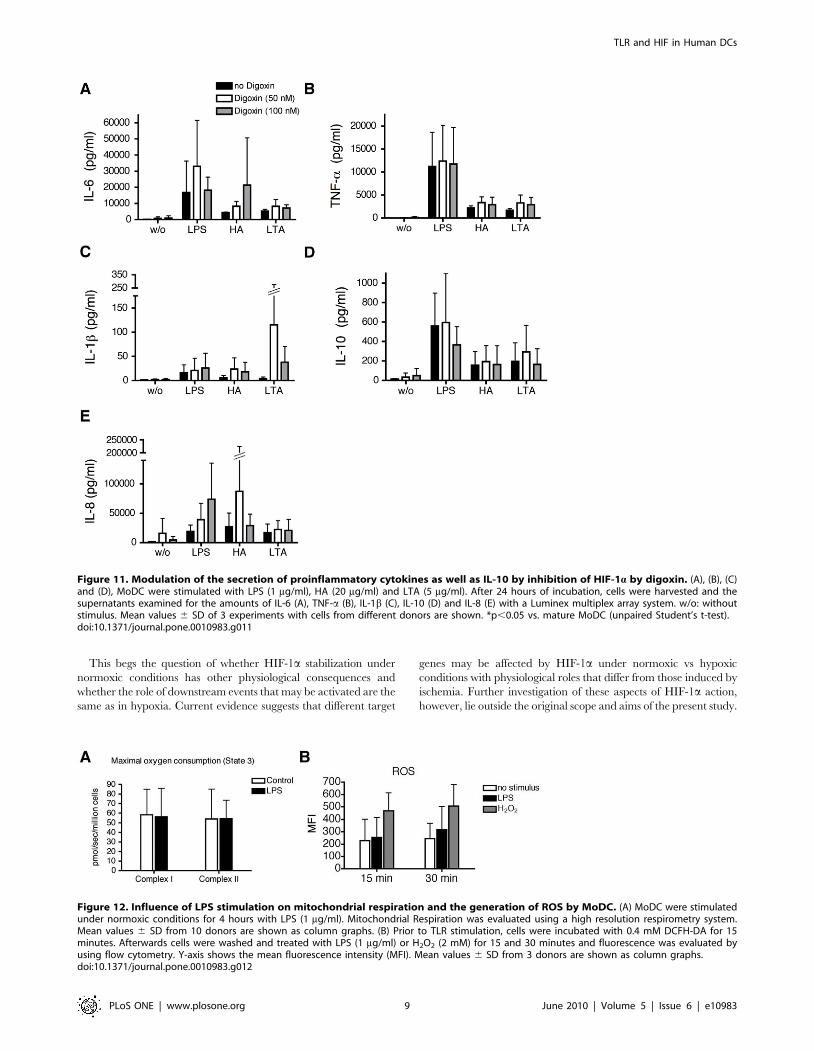
Effect of LPS stimulation on mitochondrial respiration ofMoDC
As maturation is an energy dependent process, we analyzed the
functional effect of TLR receptor stimulation on energy
metabolism, and measured the mitochondrial respiration of
MoDC, which leads to generation of ATP. DC were incubated
with 1 mg/ml of LPS under normoxic conditions. After 4 hours of
incubation no significant changes in maximal oxygen consumption
(state 3) was observed (Fig. 12A)
Influence of LPS on the generation of ROS in humanMoDC
Since LPS can induce generation of ROS in human primary
MoDC, which in turn might play a role in the maturation process
we analyzed the generation of ROS using flow cytometry. We
could not observe an increase in ROS after 15 and 30 minutes
stimulation with LPS, whereas levels of ROS treated with the
positive control hydrogen peroxide were increased (Fig. 12B).
Discussion
The present study addressed the question of whether HIF-1acould be induced by different TLR agonists under normoxia and if
stabilization is responsible for the subsequent MoDC maturation
and TLR ligand-induced cytokine production under these
conditions. The results demonstrate that under normoxic
conditions, endogenous and exogenous TLR2- and TLR4-ligands
induce maturation of human MoDC, which is associated with cell
surface expression of CD40, CD80, CD86 and ICAM-1 in a time
dependent manner together with secretion of VEGF and
proinflammatory cytokines, and stabilization of the transcription
factor HIF-1a.
Recent evidence suggests that endogenous TLR agonists play an
important role in human disease states such as I/R injury which
occurs in solid organ transplantation, myocardial infarction,
autoimmunity or trauma. HA, a non-sulfated glycosaminoglycan,
is a major constituent of the extracellular matrix (ECM). During
tissue injury or inflammation, HA is degraded and shed from the
ECM and cell surface and serves as an endogenous danger signal
[9,29]. Subsequent DC maturation and activation, with induction
of proinflammatory cytokines, then contributes to a prolongation
and exacerbation of tissue damage. Inflammatory responses of
mouse macrophages to endogenous and exogenous TLR2 and
TLR4 ligands [30] have previously been reported [31]. A potential
role, however, or involvement of HIF-1a in these processes has not
been considered. Untill now, HIF-1a protein stabilization under
normoxic conditions by TLR ligands has been shown only for
stimulation with LPS in murine bone-marrow derived DC, [23]
and several studies have highlighted distinct differences between
mouse and human immunology [32]. Hence the relevance of these
earlier observations for human disease has largely remained
unknown. In the present experiments, LTA from the gram-
Figure 2. Evaluation of phenotypic maturation of MoDC. (A)Percentages of CD80+ CD86+ double-positive cells stimulated with LPS(1 mg/ml), HA (20 mg/ml), LTA (5 mg/ml), CoCl2 (200 mM) or culturedunder hypoxia 6 LPS are shown as mean values 6 SD of 3–5experiments with cells from different donors. N: Normoxia, H: Hypoxia.(B) Polymyxin B does not affect stimulatory activity of HA. MoDC wereincubated with polymyxin B (1 or 10 mg/ml) and then stimulated withHA (20 mg/ml) or LPS (1 mg/ml) for 24 hours. Afterwards, cells werewashed and analyzed for the expression of CD86 as a maturationmarker. Y-axis shows the median fluorescence intensity (MFI). Thedotted line indicates the MFI of non-stimulated MoDC. Data arerepresentative of three independent experiments.doi:10.1371/journal.pone.0010983.g002
TLR and HIF in Human DCs
PLoS ONE | www.plosone.org 4 June 2010 | Volume 5 | Issue 6 | e10983

positive cocci S. aureus, a TLR2 ligand [33], induced DC
maturatuion and HIF-1a protein stabilization, comparable to
the stimulation induced by LPS from the gram-negative rod E. coli.
Similar effects were induced by the endogenous TLR4 ligand HA
although some differences were observed in the time course. A
possible explanation may relate to the known differences in
Figure 3. Effect of CTM on phenotypic maturation of MoDC. Monocytes isolated from buffy coat were cultured in the presence of GM-CSFand IL-4 for 6 days. Inhibition of HIF-1a expression by CTM inhibited maturation of MoDC in response to LPS. MoDC were incubated with CTM(200 nM) for 3 hours, afterwards cells were stimulated for 24 hours with LPS (1 mg/ml), HA (20 mg/ml) and LTA (5 mg/ml). Cells were washed andanalyzed for the expression of CD40 (A), CD80 (B), CD86 (C) and ICAM-1 (D). Y-axis shows the median fluorescence intensity (MFI). Mean values 6 SDof 6 experiments with cells from different donors are shown. *p,0.05; **p,0.01; ***p,0.001 vs. mature MoDC (unpaired Student’s t-test).doi:10.1371/journal.pone.0010983.g003
Figure 4. Effect of YC-1 on phenotypic maturation of MoDC. Monocytes isolated from buffy coat were cultured in the presence of GM-CSFand IL-4 for 6 days. MoDC were incubated with YC-1 (100 mM) for 5 min, afterwards cells were stimulated for 24 hours with LPS (1 mg/ml), HA m20 (g/ml) and LTA (5 mg/ml). Cells were washed and analyzed for the expression of CD40 (A), CD80 (B), CD86 (C) and ICAM-1 (D). Y-axis shows the medianfluorescence intensity (MFI). Mean values 6 SD of 4 experiments with cells from different donors are shown. *p,0.05; **p,0.01 vs. mature MoDC(unpaired Student’s t-test).doi:10.1371/journal.pone.0010983.g004
TLR and HIF in Human DCs
PLoS ONE | www.plosone.org 5 June 2010 | Volume 5 | Issue 6 | e10983

requirement of HA and LPS for distinct accessory molecules
leading to alternate downstream signaling pathway activation
(Taylor 2007).
In contrast, neither hypoxia nor CoCl2 induced HIF-1aaccumulation lead to phenotypic maturation of MoDC examined
after 24 hours suggesting that changes in HIF-1a per se do not
necessarily lead to maturation. At first sight this may suggest
methodological differences as an underlying factor. However, at
the dose used, CoCl2 has clearly been shown to be an inducer of
HIF-1a activation [34–36]. Furthermore, different ranges of
oxygen levels have been considered as hypoxic conditions, starting
from almost anoxic conditions of 0.1% up to 2% O2, oxygen levels
in the body vary from around 16% in the pulmonary alveoli down
to approximately 6% in other tissues [37]. Although there is no
current consensus regarding a critical threshold level we used the
commonly quoted value of 1.5% [38,39]. While this level of
hypoxia did not directly lead to MoDC maturation we did observe
a synergistic effect on LPS induced phenotypic maturation of
MoDC in the presence of hypoxia. Consequently, hypoxia may
augment an inflammatory response elicited in the presence of a
danger signal associated with infection or ischemia. In contrast,
hypoxia without pro-inflammatory stimuli does not induce
activation, which may serve as a protection mechanism of the
host preventing development of an autoimmune response. These
data are consistent with the finding of Jantsch et al. in murine DC
for hypoxia [23]. In contrast, a recent study has shown that human
MoDC differentiated from monocytes under permanent hypoxic
(1% O2) conditions exhibit a reduced up-regulation of CD40,
CD80, CD83 and CD86 in response to LPS, whereas the secretion
of TNF-a, CCL22 and IL-1b was increased [40]. Another study
demonstrated, that MoDC generated under hypoxic conditions
(1% O2) change their chemokine releasing profile and exhibit a
reduced Ag-uptake capacity [41]. These differences may relate to
Figure 5. Effect of Digoxin (50 nM or 100 nM) on phenotypic maturation of MoDC. Monocytes isolated from buffy coat were cultured inthe presence of GM-CSF and IL-4 for 6 days. MoDC were incubated with Digoxin for one hour, afterwards cells were stimulated for 24 hours with LPS(1 mg/ml), HA (20 mg/ml) and LTA (5 mg/ml). Cells were washed and analyzed for the expression of CD40 (A), CD80 (B), CD86 (C) and ICAM-1 (D). Y-axisshows the median fluorescence intensity (MFI). Mean values 6 SD of 3 experiments with cells from different donors are shown.doi:10.1371/journal.pone.0010983.g005
Figure 6. MoDC viability in response to CoCl2, CTM and LPS.Cells were treated with CoCl2, CTM and LPS for 24 hours. Thereafter cellswere harvested and stained with PI (5 mg/ml). Unstained viable cells(PIneg) were enumerated by flow cytometry. Mean values 6 SD of 3experiments with cells from different donors are shown.doi:10.1371/journal.pone.0010983.g006
TLR and HIF in Human DCs
PLoS ONE | www.plosone.org 6 June 2010 | Volume 5 | Issue 6 | e10983

the fact that hypoxia during the differentiation process of
monocytes into DC has additional effects on cells that differ from
hypoxic exposure of already fully differentiated DC.
In view of the complex interplay between these factors we
subsequently evaluated a potential role of HIF-1a stabilization in
the processes of MoDC maturation and cytokine production using
three inhibitors of HIF-1a and/or NFkB, namely CTM [25], YC-
1 [27] and digoxin [28] (see Fig. 13).
Taken together the data showed that there is no consistent effect
of the three inhibitors on MoDC maturation, or cytokine secretion
despite having the common effect of blocking HIF-1a stabilization
or activity through different mechanisms. Thus the data of YC-1
and digoxin did not support the original hypothesis that HIF-1aactivation by TLR ligands under normoxic conditions may have
functional consequences for human MoDC maturation, VEGF
and cytokine production. These events appear to occur indepen-
dently of the observed changes in HIF-1a stabilization. This would
suggest that other, as yet unknown effects of these inhibitors are
responsible for differential responses to the individual inhibitors in
relation to cell maturation (cell surface marker expression, or
VEGF and cytokine production). This is reinforced by the
observation that digoxin was without effect on any of the
parameters measured despite preventing TLR induced stabiliza-
tion of HIF-1a in response to LPS, LTA and HA.
Similarly variable responses of the antagonists were seen for
VEGF production by TLR ligands where only CTM showed any
inhibition and responses to HA and LTA in the presence of YC-1
were even enhanced.
In this regard, non-HIF-1a dependent stimuli including insulin-
like growth factor-1 (IGF-1), also stimulate VEGF secretion which
is phosphatidyl-inositol 3-kinase (PI3K)/Akt/mTOR (mammalian
target of rapamycin)-dependent. Other regulators include angio-
tensin, nitric oxide and guanyl cyclases [42,43]. Interestingly YC-1
was originally characterized as a cGMP inducer because it
stimulated soluble guanylyl cyclase activation in response to nitric
oxide or carbon monoxide [44] in some cells, only low
concentrations of YC-1 (1–20 mmol/L) are required for anti-
HIF-1a activity, whereas cGMP elevation requires higher
concentrations (.50 mmol/L; [44] as used in the present study.
Whether and to what extent this may play a role in the differential
responses of MoDC to YC-1 in the presence of different TLR
ligands, particularly in relation to the observed stimulatory effects
on VEGF remain to be determined.
Figure 7. Effect of HIF-1a inhibition by CTM, YC-1 and Digoxinon VEGF production of MoDC. MoDC were incubated with CTM(200 nM) for 3 hours, YC-1 (100 mM) for 5 minutes and Digoxin (50 nM or100 nM) for one hour, then cells were stimulated with LPS (1 mg/ml), HA(20 mg/ml) and LTA (5 mg/ml). After 24 hours cells were harvested and thesupernatant assayed for VEGF with a Luminex multiplex array system. w/o: without stimulus. Mean values 6 standard deviation of 6 experiments(4 for YC-1 and 3 for Digoxin) with cells from different donors are shown.*p,0.05; **p,0.01 vs. mature MoDC (unpaired Student’s t-test).doi:10.1371/journal.pone.0010983.g007
Figure 8. Digoxin inhibits LPS-, HA- and LTA-induced upregu-lation of HIF-1a (A) and does not modify TLR-4 expression (B).Cell lysates from MoDC that were incubated with digoxin (50 nM [D50]or 100 nM [D100]) and then stimulated with LPS (1 mg/ml), HA (20 mg/ml) or LTA (20 mg/ml) for 24 hours. Equal amounts of protein wereloaded and separated by SDS page, transferred onto membranes andprobed for HIF-1a and TLR-4.doi:10.1371/journal.pone.0010983.g008
TLR and HIF in Human DCs
PLoS ONE | www.plosone.org 7 June 2010 | Volume 5 | Issue 6 | e10983

In general, different pharmacokinetic properties (accessibility,
excretion, or metabolism) and characters of target molecules
(expression, activation, or isoforms) are considered major factors
determining drug sensitivity. However, these aspects in relation to
CTM, YC-1 and digoxin have not been fully determined to date;
thus, the underlying basis for the varying sensitivity and respon-
siveness of the maturation process, cytokine production and VEGF
secretion to the different inhibitors remains to be investigated.
Figure 9. Modulation of proinflammatory cytokine secretion and IL-10 by inhibition of HIF-1a by CTM. (A), (B), (C) and (D), MoDC werestimulated with LPS (1 mg/ml), HA (20 mg/ml) and LTA (5 mg/ml). After 24 hours of incubation, cells were harvested and the supernatants examinedfor the amounts of IL-6 (A), TNF-a (B), IL-1b (C) and IL-10 (D) with a Luminex multiplex array system. w/o: without stimulus. Mean values 6 SD of 6experiments with cells from different donors are shown. *p,0.05 vs. mature MoDC (unpaired Student’s t-test).doi:10.1371/journal.pone.0010983.g009
Figure 10. Modulation of proinflammatory cytokine secretion and IL-10 by YC-1. (A), (B), (C) and (D), MoDC were stimulated with LPS(1 mg/ml), HA (20 mg/ml) and LTA (5 mg/ml). After 24 hours of incubation, cells were harvested and the supernatants examined for the amounts of IL-6 (A), TNF-a (B), IL-1b (C) and IL-10 (D) with a Luminex multiplex array system. w/o: without stimulus. Mean values 6 SD of 4 experiments with cellsfrom different donors are shown.doi:10.1371/journal.pone.0010983.g010
TLR and HIF in Human DCs
PLoS ONE | www.plosone.org 8 June 2010 | Volume 5 | Issue 6 | e10983
This begs the question of whether HIF-1a stabilization under
normoxic conditions has other physiological consequences and
whether the role of downstream events that may be activated are the
same as in hypoxia. Current evidence suggests that different target
genes may be affected by HIF-1a under normoxic vs hypoxic
conditions with physiological roles that differ from those induced by
ischemia. Further investigation of these aspects of HIF-1a action,
however, lie outside the original scope and aims of the present study.
Figure 11. Modulation of the secretion of proinflammatory cytokines as well as IL-10 by inhibition of HIF-1a by digoxin. (A), (B), (C)and (D), MoDC were stimulated with LPS (1 mg/ml), HA (20 mg/ml) and LTA (5 mg/ml). After 24 hours of incubation, cells were harvested and thesupernatants examined for the amounts of IL-6 (A), TNF-a (B), IL-1b (C), IL-10 (D) and IL-8 (E) with a Luminex multiplex array system. w/o: withoutstimulus. Mean values 6 SD of 3 experiments with cells from different donors are shown. *p,0.05 vs. mature MoDC (unpaired Student’s t-test).doi:10.1371/journal.pone.0010983.g011
Figure 12. Influence of LPS stimulation on mitochondrial respiration and the generation of ROS by MoDC. (A) MoDC were stimulatedunder normoxic conditions for 4 hours with LPS (1 mg/ml). Mitochondrial Respiration was evaluated using a high resolution respirometry system.Mean values 6 SD from 10 donors are shown as column graphs. (B) Prior to TLR stimulation, cells were incubated with 0.4 mM DCFH-DA for 15minutes. Afterwards cells were washed and treated with LPS (1 mg/ml) or H2O2 (2 mM) for 15 and 30 minutes and fluorescence was evaluated byusing flow cytometry. Y-axis shows the mean fluorescence intensity (MFI). Mean values 6 SD from 3 donors are shown as column graphs.doi:10.1371/journal.pone.0010983.g012
TLR and HIF in Human DCs
PLoS ONE | www.plosone.org 9 June 2010 | Volume 5 | Issue 6 | e10983

Materials and Methods
Generation and stimulation of human monocyte-derivedDC (MoDC)
Human peripheral blood mononuclear cells (PBMC) were
isolated from buffy coats obtained from healthy blood donors
(Regional Red Cross Blood Donation Center, Bern, Switzerland) by
density gradient centrifugation over Ficoll-Paque (Amersham,
Uppsala, Sweden). Monocytes were isolated from PBMC as
previously described [45–47] or by CD14 positive selection by
MACS (Miltenyi Biotec GmbH, Bergisch Gladbach, Germany)
according the manufacturer’s protocol (for experiments with
digoxin). Purity of isolated monocytes was characterized by high
expression of CD14 (.96% positive cells) and absent expression of
CD1a and DC-SIGN. Monocytes were incubated for 6 days in
RPMI 1640 medium (Invitrogen Life Technologies, Basel,
Switzerland) containing 10% fetal calf serum ((FCS, Amimed /
BioConcept, Allschwil, Switzerland), 1% [2 mM] L-Glutamine
(Invitrogen), 1% [100 U/ml] Penicillin/Streptomycin (Invitrogen),
10 ng/ml GM-CSF (R&D Systems Europe Ltd, Abingdon, Oxon,
UK), and 10 ng/ml IL-4 (R&D) to generate MoDC as described
initially by Sallusto and Lanzavecchia [48]. Immature MoDC were
characterized by absent expression of CD14 and high expression of
CD1a, HLA-DR, DC-SIGN and phagocytic activity. Maturation of
these cells leads to a massive up-regulation of CD40, CD80, CD83,
CD86, ICAM-1, CCR7 and HLA-DR. Mature MoDC exhibit a
very potent T cell proliferation capacity and reduced Ag-uptake as
previously described [46,47,49]. Cells were kept at 37uC in a 5%
CO2 humidified atmosphere. On day 3, the culture medium was
replaced with fresh medium. For induction of maturation 1 mg/ml
LPS (Sigma Aldrich, Buchs, Switzerland), 5 mg/ml LTA or 20 mg/
ml HA (Sigma Aldrich, Buchs, Switzerland) were added to the
MoDC cultures for the indicated periods.
FACS analysis and cell viabilityCells were incubated with FITC- or PE-labeled monoclonal
antibody (mAb) against CD80, CD86 (BD, Franklin Lakes, NJ,
USA), isotype control IgG1 (BD), or unlabeled mAb against
CD40, ICAM-1 (Diaclone, Besancon, France) followed by a
FITC-labeled polyclonal goat anti-mouse IgG (Sigma).
For determination of viability, the cells were stained with 5 mg/
ml of propidium iodide (PI; Invitrogen) and analyzed by flow
cytometry. As positive control for PI staining, cells were treated
with PBS containing 0.1% BSA and 0.1% saponin (Sigma Aldrich,
Buchs, Switzerland). Measurements were performed with a BD
FACScan flow cytometer and analyzed using the FlowJo software
(Tree Star Inc., Ashland, OR, USA).
Hypoxic conditionsCells were incubated in a hypoxia chamber (1.5% oxygen,
37uC) for the indicated time-periods. In some experiments (as
indicated), LPS (1 mg/ml) was given in the hypoxia-chamber one
hour after putting the cells under hypoxic conditions.
SDS-PAGE and Western blottingDC were lysed in 60 mM Tris-HCl, 8.5% glycerol, and 2%
SDS. Protein concentration was determined with the Quanti-IT
assay kit and read with the Qubit fluorometer (Invitrogen). Equal
amounts of protein (20 mg per lane) were loaded and separated by
4–12% SDS-PAGE. Gels were then transferred to nitrocellulose
membranes with the iBlot dry blotting system (Invitrogen). Equal
loading was verified by staining the extracted gel with SimplyBlue
SafeStain (Invitrogen). Afterwards, the membranes were blocked
for 30 min with incubation buffer (10 mM Tris-HCl pH 7.5, 100
mM NaCl and 0.1% w/v Tween 20) supplemented with 5% non-
fat dry skim milk, and incubated overnight with the primary
antibodies against HIF-1a (Novus Biologicals, Littleton, CO,
USA; dilution 1:2000) or primary antibodies against TLR-4
(Invitrogen, Basel, Switzerland; dilution 1:1000) and actin (Sigma
Aldrich, Buchs, Switzerland; dilution 1:3000). Membranes were
washed with incubation buffer and incubated for 1 hour with
horseradish peroxidase-coupled goat polyclonal anti-rabbit IgG
(dilution 1:3000). Finally, the membranes were developed with a
chemiluminescence detection kit (Pierce, Rockford, IL, USA). All
Western blotting experiments were performed in triplicates.
Protein Densiometry was quantified using Adobe Photoshop CS3.
Cytokine assaysMoDC (106 cells/ml) were treated with LPS, LTA or HA for 24
hours. Cell culture supernatants were analyzed using a Luminex
multiplex suspension array system from Bio-Rad (Bio-Rad,
Hercules, CA, USA) for IL-1b, IL-6, IL-8, IL-10, TNF-a and
VEGF (all kits from BioSource, Invitrogen, Carlsbad, CA, USA)
according the manufacturer’s instructions.
Inhibitors and agonists of HIF-1a and/or NFkBBlocking of HIF-1a was achieved using the commercially
available inhibitors CTM (Chetomin, NSC 289491, Invitrogen),
and digoxin (Sigma-Aldrich, Buchs, Switzerland). Chetomin is a
natural metabolite produced by several species of the genus
Chaetomium. To inhibit NFkB/p65 nuclear translocation, YC-1 (3-
(59-Hydroxymethyl-29-furyl)-1-benzyl indazole; Calbiochem-No-
vabiochem, Zug, Switzerland) was used. Stabilization of HIF-1aprotein in control experiments was achieved using CoCl2 (Sigma
Aldrich, Buchs, Switzerland), which is a potent inhibitor of post-
translational hydroxylation of proline residues in the oxygen-
dependent degradation (ODD) domain of HIF-1a and thus
inhibits HIF-1a degradation.
High-resolution respirometryDC were centrifuged for 5 min (350 g) and resuspended in
respiration buffer (110 mM sucrose, 0.5 mM EGTA, 3.0 mM
MgCl2, 80 mM KCl, 60 mM K-lactobionate, 10 mM KH2PO4,
Figure 13. Summary of the effects of NFkB and HIF-1a on DCmaturation. Ligation of TLRs leads to NFkB activation and subsequentupregulation of HIF-1a under normoxic conditions. HIF-1a contributesto NFkB driven phenotypic DC maturation and cytokine production.The effects were investigated using an inhibitor of NFkB/p65 nucleartranslocation (YC-1), inhibition of HIF-1a nuclear translocation (cheto-min), and inhibition of HIF-1a protein accumulation (digoxin). CoCl2and hypoxia served as controls.doi:10.1371/journal.pone.0010983.g013
TLR and HIF in Human DCs
PLoS ONE | www.plosone.org 10 June 2010 | Volume 5 | Issue 6 | e10983

20 mM taurine, 20 mM HEPES, 1.0 g/l BSA, pH 7.1) at a
concentration of 3-56106 cells/ml. Cells were incubated with
1 mg/mL LPS for 4 h. Respiration rates were measured at 37uC in
a high-resolution oxygraph (Oxygraph-2k, Oroboros Instruments,
Innsbruck, Austria). For assessment of mitochondrial complex
activity cells were first permeabilized with digitonin (8.1 mM) for 5
min. Afterwards, for complex I-dependent maximal respiration
stimulation, substrates added were glutamate (10 mM) and malate
(5 mM), followed by addition of adenosine diphosphate (ADP)
(0.25 mM) causing a sudden burst of oxygen uptake as ADP is
converted into ATP. After a stable signal was reached and marked,
rotenone (0.5 mM) was added to inhibit complex I, and then
complex II-dependent respiration was stimulated by adding
succinate (10 mM), while complex III was inhibited by antimycin
A (0.5 mM) (Sigma Aldrich, Buchs, Switzerland). Respiration rates
were calculated and recorded using DatLab software for data
acquisition and analysis; Oroboros Instruments, Innsbruck,
Austria.
Measurement of reactive oxygen species (ROS)generation by flow cytometry
MoDC (26105 cells) were plated in 96-well round bottom well
plate. Cells were pretreated with 0.4 mM of to dichlorofluorescin
diacetate (DCFH-DA [Sigma]) for 15 minutes. Afterwards, cells
were washed and stimulated with LPS (1 mg/ml) or hydrogen
peroxide (2 mM, Sigma) for 15 and 30 minutes and immediately
analyzed by flow cytometry.
Statistical analysisData are presented as mean 6 standard deviation (SD)
representing experiments with up to 10 different donors. Unpaired
Students t-tests were performed for evaluation of significance.
Cellular respiration was compared using the paired t-test.
Differences were considered as statistically significant at p-values
less than 0.05. Data were analyzed using GraphPad Prism
software 4.0 (GraphPad, San Diego, CA).
Acknowledgments
We would like to thank Prof. Daniel Candinas and Dr. Deborah Stroka,
Department of Clinical Research, Visceral and Transplantation Surgery,
and Prof. Thomas Geiser and Dr. Fabian Blank, Department of
Pneumology, University Hospital of Bern and University of Bern, for
their support. We would also like to thank Karin de Peyer, Sandra Nansoz
and Katja Matozan for expert technical assistance.
Author Contributions
Conceived and designed the experiments: RS SD PML. Performed the
experiments: RS SD TR PML. Analyzed the data: RS SD SGS JT SMJ
RR PML. Contributed reagents/materials/analysis tools: SGS CvG JT
SMJ RR. Wrote the paper: RS SD SGS PML. Obtained funding: SD JT
SMJ RR PML. Performed additional experiments for revision: CvG.
Critical revision of the paper: JT SMJ RR.
References
1. Banchereau J, Steinman RM (1998) Dendritic cells and the control of immunity.Nature 392: 245–252.
2. Thomson AW, Lu L (1999) Dendritic cells as regulators of immune reactivity:
implications for transplantation. Transplantation 68: 1–8.
3. Shlomchik WD, Couzens MS, Tang CB, McNiff J, Robert ME, et al. (1999)Prevention of graft versus host disease by inactivation of host antigen-presenting
cells. Science 285: 412–415.
4. Beg AA (2002) Endogenous ligands of Toll-like receptors: implications forregulating inflammatory and immune responses. Trends Immunol 23: 509–512.
5. Platt JL, Dalmasso AP, Lindman BJ, Ihrcke NS, Bach FH (1991) The role of
C5a and antibody in the release of heparan sulfate from endothelial cells.Eur J Immunol 21: 2887–2890.
6. Ihrcke NS, Platt JL (1996) Shedding of heparan sulfate proteoglycan by
stimulated endothelial cells: evidence for proteolysis of cell-surface molecules.J Cell Physiol 168: 625–637.
7. Platt JL, Vercellotti GM, Lindman BJ, Oegema TR, Jr., Bach, et al. (1990)
Release of heparan sulfate from endothelial cells. Implications for pathogenesisof hyperacute rejection. J Exp Med 171: 1363–1368.
8. Rehm M, Bruegger D, Christ F, Conzen P, Thiel M, et al. (2007) Shedding of
the endothelial glycocalyx in patients undergoing major vascular surgery withglobal and regional ischemia. Circulation 116: 1896–1906.
9. Nieuwdorp M, van Haeften TW, Gouverneur MC, Mooij HL, van
Lieshout MH, et al. (2006) Loss of endothelial glycocalyx during acutehyperglycemia coincides with endothelial dysfunction and coagulation activation
in vivo. Diabetes 55: 480–486.
10. Taylor KR, Trowbridge JM, Rudisill JA, Termeer CC, et al. (2004) Hyaluronanfragments stimulate endothelial recognition of injury through TLR4. J Biol
Chem 2004 279: 17079–17084.
11. Johnson GB, Brunn GJ, Kodaira Y, Platt JL (2002) Receptor-mediatedmonitoring of tissue well-being via detection of soluble heparan sulfate by
Toll-like receptor 4. J Immunol 168: 5233–5239.
12. Termeer C, Benedix F, Sleeman J, Fieber C, Voith U, et al. (2002)Oligosaccharides of Hyaluronan activate dendritic cells via toll-like receptor 4.
J Exp Med 195: 99–111.
13. Leemans JC, Stokman G, Claessen N, Rouschop KM, Teske GJ, et al. (2005)Renal-associated TLR2 mediates ischemia/reperfusion injury in the kidney.
J Clin Invest 115: 2894–2903.
14. Wu H, Chen G, Wyburn KR, Yin J, Bertolino P, et al. TLR4 activationmediates kidney ischemia/reperfusion injury J Clin Invest 117(10): 2847–2859.
15. Kaczorowski DJ, Nakao A, Mollen KP, Vallabhaneni R, Sugimoto R, et al.
(2007) Toll-like receptor 4 mediates the early inflammatory response after coldischemia/reperfusion. Transplantation 84: 1279–1287.
16. Hua F, Ma J, Ha T, Kelley J, Williams DL, et al. (2008) Preconditioning with a
TLR2 specific ligand increases resistance to cerebral ischemia/reperfusioninjury. J Neuroimmunol 199: 75–82.
17. Hua F, Ma J, Ha T, Xia Y, Kelley J, et al. (2007) Activation of Toll-like receptor4 signaling contributes to hippocampal neuronal death following global cerebral
ischemia/reperfusion. J Neuroimmunol 190: 101–111.
18. Rius J, Guma M, Schachtrup C, Akassoglou K, Zinkernagel AS, et al. (2008)
NF-kappaB links innate immunity to the hypoxic response through transcrip-tional regulation of HIF-1alpha. Nature 453: 807–811.
19. Tacchini L, Gammella E, De Ponti C, Recalcati S, Cairo G (2008) Role of HIF-
1 and NF-kappaB transcription factors in the modulation of transferrin receptorby inflammatory and anti-inflammatory signals. J Biol Chem 283: 20674–20686.
20. Cramer T, Yamanishi Y, Clausen BE, Forster I, Pawlinski R, et al. (2003) HIF-
1alpha is essential for myeloid cell-mediated inflammation. Cell 112: 645–657.
21. Peyssonnaux C, Datta V, Cramer T, Doedens A, Theodorakis EA, et al. (2005)
HIF-1alpha expression regulates the bactericidal capacity of phagocytes. J ClinInvest 115: 1806–1815.
22. Blouin CC, Page EL, Soucy GM, Richard DE (2004) Hypoxic gene activation
by lipopolysaccharide in macrophages: implication of hypoxia-inducible factor1alpha. Blood 103: 1124–1130.
23. Jantsch J, Chakravortty D, Turza N, Prechtel AT, Buchholz B, et al. (2008)
Hypoxia and hypoxia-inducible factor-1 alpha modulate lipopolysaccharide-
induced dendritic cell activation and function. J Immunol 180: 4697–4705.
24. Peyssonnaux C, Cejudo-Martin P, Doedens A, Zinkernagel AS, Johnson RS, etal. (2007) Cutting edge: Essential role of hypoxia inducible factor-1alpha in
development of lipopolysaccharide-induced sepsis. J Immunol 178: 7516–7519.
25. Kung AL, Zabludoff SD, France DS, Freedman SJ, Tanner EA, et al. (2004)Small molecule blockade of transcriptional coactivation of the hypoxia-inducible
factor pathway. Cancer Cell 6: 33–43.
26. Huang YT, Pan SL, Guh JH, Chang YL, Lee, et al. (2005) YC-1 suppressesconstitutive nuclear factor-kappaB activation and induces apoptosis in human
prostate cancer cells. Mol Cancer Ther 4: 1628–1635.
27. Sun HL, Liu YN, Huang YT, Pan SL, Huang DY, et al. (2007) YC-1 inhibits
HIF-1 expression in prostate cancer cells: contribution of Akt/NF-kappaBsignaling to HIF-1alpha accumulation during hypoxia. Oncogene 26:
3941–3951.
28. Zhang H, Qian DZ, Tan YS, Lee K, Gao P, et al. (2008) Digoxin and othercardiac glycosides inhibit HIF-1alpha synthesis and block tumor growth. Proc
Natl Acad Sci U S A 105: 19579–19586.
29. Luke HJ, Prehm P (1999) Synthesis and shedding of hyaluronan from plasma
membranes of human fibroblasts and metastatic and non-metastatic melanomacells. Biochem J 343 Pt1: 71–75.
30. Re F, Strominger JL (2001) Toll-like receptor 2 (TLR2) and TLR4 differentially
activate human dendritic cells. J Biol Chem 276: 37692–37699.
31. Taylor KR, Yamasaki K, Radek KA, Di Nardo A, Goodarzi H, et al. (2007)Recognition of hyaluronan released in sterile injury involves a unique receptor
complex dependent on Toll-like receptor 4, CD44, and MD-2. J Biol Chem 282:
18265–18275.
TLR and HIF in Human DCs
PLoS ONE | www.plosone.org 11 June 2010 | Volume 5 | Issue 6 | e10983

32. Mestas J, Hughes CC (2004) Of mice and not men: differences between mouse
and human immunology. J Immunol 172: 2731–2738.33. Schwandner R, Dziarski R, Wesche H, Rothe M, Kirschning CJ (1999)
Peptidoglycan- and lipoteichoic acid-induced cell activation is mediated by toll-
like receptor 2. J Biol Chem 274: 17406–17409.34. Treins C, Giorgetti-Peraldi S, Murdaca J, Monthouel-Kartmann MN, Van
Obberghen E (2005) Regulation of hypoxia-inducible factor (HIF)-1 activity andexpression of HIF hydroxylases in response to insulin-like growth factor I. Mol
Endocrinol 19: 1304–1317.
35. Wang GL, Jiang BH, Rue EA, Semenza GL (1995) Hypoxia-inducible factor 1 isa basic-helix-loop-helix-PAS heterodimer regulated by cellular O2 tension. Proc
Natl Acad Sci U S A 92: 5510–5514.36. Pugh CW, O’Rourke JF, Nagao M, Gleadle JM, Ratcliffe PJ (1997) Activation of
hypoxia-inducible factor-1; definition of regulatory domains within the alphasubunit. J Biol Chem 272: 11205–11214.
37. Semenza GL (2001) Hypoxia-inducible factor 1: oxygen homeostasis and disease
pathophysiology. Trends Mol Med 7: 345–350.38. Chandel NS, McClintock DS, Feliciano CE, Wood TM, Melendez, et al. (2000)
Reactive oxygen species generated at mitochondrial complex III stabilizehypoxia-inducible factor-1alpha during hypoxia: a mechanism of O2 sensing.
J Biol Chem 275: 25130–25138.
39. Mansfield KD, Simon MC, Keith B (2004) Hypoxic reduction in cellularglutathione levels requires mitochondrial reactive oxygen species. J Appl Physiol
2004 97: 1358–1366.40. Mancino A, Schioppa T, Larghi P, Pasqualini F, Nebuloni M, et al. Divergent
effects of hypoxia on dendritic cell functions. Blood 112(9): 3723–3734.41. Elia AR, Cappello P, Puppo M, Fraone T, Vanni C, et al. (2008) Human
dendritic cells differentiated in hypoxia down-modulate antigen uptake and
change their chemokine expression profile. J Leukoc Biol 84: 1472–1482.
42. Feliers D, Gorin Y, Ghosh-Choudhury G, Abboud HE, Kasinath BS (2006)
Angiotensin II stimulation of VEGF mRNA translation requires production of
reactive oxygen species. Am J Physiol Renal Physiol 290: F927–936.
43. Chin K, Kurashima Y, Ogura T, Tajiri H, Yoshida S, et al. (1997) Induction of
vascular endothelial growth factor by nitric oxide in human glioblastoma and
hepatocellular carcinoma cells. Oncogene 15: 437–442.
44. Slupski M, Szadujkis-Szadurski L, Grzesk G, Szadujkis-Szadurski R, Szadujkis-
Szadurska K, et al. (2007) Guanylate cyclase activators influence reactivity of
human mesenteric superior arteries retrieved and preserved in the same
conditions as transplanted kidneys. Transplant Proc 39: 1350–1353.
45. Mentzer SJ, Guyre PM, Burakoff SJ, Faller DV (1986) Spontaneous aggregation
as a mechanism for human monocyte purification. Cell Immunol 101: 312–319.
46. Obregon C, Rothen-Rutishauser B, Gitahi SK, Gehr P, Nicod LP (2006)
Exovesicles from human activated dendritic cells fuse with resting dendritic cells,
allowing them to present alloantigens. Am J Pathol 169: 2127–2136.
47. Spirig R, van Kooten C, Obregon C, Nicod L, Daha M, et al. (2008) The
complement inhibitor low molecular weight dextran sulfate prevents TLR4-
induced phenotypic and functional maturation of human dendritic cells.
J Immunol 181: 878–890.
48. Sallusto F, Lanzavecchia A (1994) Efficient presentation of soluble antigen by
cultured human dendritic cells is maintained by granulocyte/macrophage
colony-stimulating factor plus interleukin 4 and downregulated by tumor
necrosis factor alpha. J Exp Med 179: 1109–1118.
49. Regamey N, Obregon C, Ferrari-Lacraz S, van Leer C, Chanson M, et al.
(2007) Airway epithelial IL-15 transforms monocytes into dendritic cells.
Am J Respir Cell Mol Biol 37: 75–84.
TLR and HIF in Human DCs
PLoS ONE | www.plosone.org 12 June 2010 | Volume 5 | Issue 6 | e10983
Copyright © 2022 FDOKUMEN