HIF-1 Inhibits Mitochondrial Biogenesis and Cellular Respiration in VHL-Deficient Renal Cell...
14
Cancer Cell Article HIF-1 Inhibits Mitochondrial Biogenesis and Cellular Respiration in VHL-Deficient Renal Cell Carcinoma by Repression of C-MYC Activity Huafeng Zhang, 1,2 Ping Gao, 3 Ryo Fukuda, 1,2 Ganesh Kumar, 5 Balaji Krishnamachary, 1,2 Karen I. Zeller, 3 Chi V. Dang, 3 and Gregg L. Semenza 1,2,3,4, * 1 Vascular Biology Program, Institute for Cell Engineering 2 McKusick-Nathans Institute of Genetic Medicine 3 Departments of Medicine and Oncology 4 Departments of Pediatrics and Radiation Oncology The Johns Hopkins University School of Medicine, Baltimore, MD 21205, USA 5 Center for Systems Biology, Department of Medicine, University of Chicago, Chicago, IL 60637, USA *Correspondence: [email protected] DOI 10.1016/j.ccr.2007.04.001 SUMMARY Many cancer cells are characterized by increased glycolysis and decreased respiration, even under aerobic conditions. The molecular mechanisms underlying this metabolic reprogramming are un- clear. Here we show that hypoxia-inducible factor 1 (HIF-1) negatively regulates mitochondrial bio- genesis and O 2 consumption in renal carcinoma cells lacking the von Hippel-Lindau tumor suppres- sor (VHL). HIF-1 mediates these effects by inhibiting C-MYC activity via two mechanisms. First, HIF-1 binds to and activates transcription of the MXI1 gene, which encodes a repressor of C-MYC tran- scriptional activity. Second, HIF-1 promotes MXI-1-independent, proteasome-dependent degrada- tion of C-MYC. We demonstrate that transcription of the gene encoding the coactivator PGC-1b is C-MYC dependent and that loss of PGC-1b expression is a major factor contributing to reduced respiration in VHL-deficient renal carcinoma cells. INTRODUCTION The efficiency with which mitochondria synthesize ATP through oxidative phosphorylation is critical for the gener- ation and maintenance of the structural and functional complexity that characterizes multicellular organisms. Energy generation is required for cell survival, but the mitochondria also generate reactive oxygen species (ROS) that can oxidize nucleic acids and proteins, result- ing in cell dysfunction or death. In the presence of O 2 , non- transformed cells convert glucose to pyruvate, which enters mitochondria, is converted to acetyl coenzyme A, and metabolized via the tricarboxylic acid cycle, yielding reducing equivalents that are used for oxidative phos- phorylation to generate ATP. Under hypoxic conditions, lactate dehydrogenase A (LDH-A) catalyzes conversion of pyruvate to lactate. A characteristic feature of cancer cells is their increased metabolism of glucose to lactate even under aerobic conditions (the Warburg effect). Warburg proposed that respiration was impaired in cancer cells leading to increased glycolytic metabolism (Warburg, 1930). The increase in glucose uptake is such a reliable feature of cancer cells that it provides the basis for clinical identification of occult metastases by [ 18 F]-deoxyglucose positron-emission tomography (Gatenby and Gillies, 2004). SIGNIFICANCE In the presence of O 2 , cells convert glucose to pyruvate, which enters the mitochondria and is metabolized, yield- ing reducing equivalents that are used for oxidative phosphorylation to generate ATP. Under hypoxic conditions, pyruvate is shunted away from the mitochondria by conversion to lactate. Cancer cells are characterized by in- creased glucose uptake, increased lactate production, and decreased respiration, even under aerobic conditions. The molecular mechanisms underlying this metabolic reprogramming of cancer cells are unclear. Loss of function of the von Hippel-Lindau tumor suppressor protein in renal cell carcinoma leads to dramatically reduced mito- chondrial mass and O 2 consumption. We delineate a transcriptional network controlled by hypoxia-inducible fac- tor 1 that is responsible for the dramatic decrease in respiration observed in renal carcinoma cells. Cancer Cell 11, 407–420, May 2007 ª2007 Elsevier Inc. 407
-
Upload
independent -
Category
Documents
-
view
4 -
download
0
Transcript of HIF-1 Inhibits Mitochondrial Biogenesis and Cellular Respiration in VHL-Deficient Renal Cell...

Cancer Cell
Article
HIF-1 Inhibits Mitochondrial Biogenesisand Cellular Respiration in VHL-Deficient RenalCell Carcinoma by Repression of C-MYC ActivityHuafeng Zhang,1,2 Ping Gao,3 Ryo Fukuda,1,2 Ganesh Kumar,5 Balaji Krishnamachary,1,2 Karen I. Zeller,3
Chi V. Dang,3 and Gregg L. Semenza1,2,3,4,*1 Vascular Biology Program, Institute for Cell Engineering2 McKusick-Nathans Institute of Genetic Medicine3 Departments of Medicine and Oncology4 Departments of Pediatrics and Radiation Oncology
The Johns Hopkins University School of Medicine, Baltimore, MD 21205, USA5 Center for Systems Biology, Department of Medicine, University of Chicago, Chicago, IL 60637, USA*Correspondence: [email protected]
DOI 10.1016/j.ccr.2007.04.001
SUMMARY
Many cancer cells are characterized by increased glycolysis and decreased respiration, even underaerobic conditions. The molecular mechanisms underlying this metabolic reprogramming are un-clear. Here we show that hypoxia-inducible factor 1 (HIF-1) negatively regulates mitochondrial bio-genesis and O2 consumption in renal carcinoma cells lacking the von Hippel-Lindau tumor suppres-sor (VHL). HIF-1 mediates these effects by inhibiting C-MYC activity via two mechanisms. First, HIF-1binds to and activates transcription of the MXI1 gene, which encodes a repressor of C-MYC tran-scriptional activity. Second, HIF-1 promotes MXI-1-independent, proteasome-dependent degrada-tion of C-MYC. We demonstrate that transcription of the gene encoding the coactivator PGC-1b isC-MYC dependent and that loss of PGC-1b expression is a major factor contributing to reducedrespiration in VHL-deficient renal carcinoma cells.
INTRODUCTION
The efficiency with which mitochondria synthesize ATP
through oxidative phosphorylation is critical for the gener-
ation and maintenance of the structural and functional
complexity that characterizes multicellular organisms.
Energy generation is required for cell survival, but the
mitochondria also generate reactive oxygen species
(ROS) that can oxidize nucleic acids and proteins, result-
ing in cell dysfunction or death. In the presence of O2, non-
transformed cells convert glucose to pyruvate, which
enters mitochondria, is converted to acetyl coenzyme A,
and metabolized via the tricarboxylic acid cycle, yielding
reducing equivalents that are used for oxidative phos-
phorylation to generate ATP. Under hypoxic conditions,
lactate dehydrogenase A (LDH-A) catalyzes conversion
of pyruvate to lactate. A characteristic feature of cancer
cells is their increased metabolism of glucose to lactate
even under aerobic conditions (the Warburg effect).
Warburg proposed that respiration was impaired in cancer
cells leading to increased glycolytic metabolism (Warburg,
1930). The increase in glucose uptake is such a reliable
feature of cancer cells that it provides the basis for clinical
identification of occult metastases by [18F]-deoxyglucose
positron-emission tomography (Gatenby and Gillies,
2004).
SIGNIFICANCE
In the presence of O2, cells convert glucose to pyruvate, which enters the mitochondria and is metabolized, yield-ing reducing equivalents that are used for oxidative phosphorylation to generate ATP. Under hypoxic conditions,pyruvate is shunted away from the mitochondria by conversion to lactate. Cancer cells are characterized by in-creased glucose uptake, increased lactate production, and decreased respiration, even under aerobic conditions.The molecular mechanisms underlying this metabolic reprogramming of cancer cells are unclear. Loss of functionof the von Hippel-Lindau tumor suppressor protein in renal cell carcinoma leads to dramatically reduced mito-chondrial mass and O2 consumption. We delineate a transcriptional network controlled by hypoxia-inducible fac-tor 1 that is responsible for the dramatic decrease in respiration observed in renal carcinoma cells.
Cancer Cell 11, 407–420, May 2007 ª2007 Elsevier Inc. 407

Cancer Cell
HIF-1 Inhibits Mitochondrial Biogenesis
Studies of Rous sarcoma virus-transformed cells dem-
onstrated that one of the earliest effects of v-src expres-
sion was an increase in glucose uptake and lactate pro-
duction (Carroll et al., 1978). Inhibition of LDH-A activity
impairs tumor cell proliferation ex vivo and in vivo (Shim
et al., 1997; Fantin et al., 2006). Expression of two other
glycolytic enzymes, phosphoglycerate mutase and glu-
cose phosphate isomerase, was shown to immortalize pri-
mary human cells and attenuate the mitochondrial gener-
ation of ROS, which are known to cause cellular
senescence (Kondoh et al., 2005).
The increased transcription of genes encoding glucose
transporters and glycolytic enzymes, including LDH-A, in
response to intratumoral hypoxia, V-SRC, and other onco-
proteins is mediated by hypoxia-inducible factor 1 (HIF-1)
(Semenza et al., 1994, 1996; Firth et al., 1995; Jiang et al.,
1997; Iyer et al., 1998; Seagroves et al., 2001; Robey et al.,
2005). Other transcription factors, including C-MYC (Shim
et al., 1997; Osthus et al., 2000) and SP1/SP3 (Discher
et al., 1998), contribute to the regulated expression of
these genes. Glucose transport and aerobic glycolysis
are also stimulated by oncogenic alterations that increase
AKT kinase activity (Elstrom et al., 2004).
Several different transcription factors have been shown
to regulate mitochondrial biogenesis, including mitochon-
drial transcription factors A (TFAM) and B (TFB1M,
TFB2M), nuclear respiratory factor 1 (NRF-1), GA binding
proteins (GABPa, GABPb2), peroxisome proliferator-acti-
vated receptors (PPAR-a, PPAR-g), PPAR-g coactivators
(PGC-1a, PGC-1b), PGC-1-related coactivator 1 (PPRC1),
and C-MYC (Kelly and Scarpulla, 2004; Bogacka et al.,
2005; Li et al., 2005). Somatic mutations have been iden-
tified in the mitochondrial genome of cancer cells (Polyak
et al., 1998) and shown to promote tumor growth in xeno-
graft assays (Petros et al., 2005). Cellular content and ac-
tivity of mitochondria are reduced (Cuezva et al., 2002,
2004), lactate production is increased (Brizel et al., 2001)
in many cancers, and these metabolic alterations are as-
sociated with poor prognosis. However, the molecular
pathways underlying these observations have not been
fully delineated.
In the majority of cases of renal clear cell carcinoma, ac-
tivity of the von Hippel-Lindau tumor suppressor (VHL)
protein is lost as a result of genetic or epigenetic changes
in the cancer cell genome (Garcia, 2006). VHL loss of func-
tion, which leads to HIF-1 gain of function, is the earliest
detectable molecular event in the pathogenesis of this
cancer (Mandriota et al., 2002). HIF-1 is required for tu-
morigenesis in VHL-deficient renal carcinoma cells
(Kondo et al., 2002; Maranchie et al., 2002). HIF-1 is a het-
erodimer composed of a constitutively expressed HIF-1b
subunit and an O2-regulated HIF-1a or HIF-2a subunit
(Wang et al., 1995; Tian et al., 1997). HIF-1 has been
shown to play critical roles in tumor angiogenesis, glucose
metabolism, invasion/metastasis, and response to radia-
tion and chemotherapy (Semenza, 2003; Tang et al.,
2004; Moeller et al., 2005; Thomas et al., 2006 Esteban
et al., 2006; Krishnamachary et al., 2006; Melillo, 2006).
VHL binds to HIF-1a or HIF-2a and targets the protein
408 Cancer Cell 11, 407–420, May 2007 ª2007 Elsevier Inc.
for ubiquitination and proteasomal degradation in an O2-
dependent manner (Maxwell et al., 1999). VHL also has
important functions that are not HIF-1-dependent, such
as the regulation of p53 activity (Roe et al., 2006).
Reduced levels of mitochondrial DNA and respiratory
chain proteins as well as increased levels of glycolytic en-
zymes have been reported in renal cell carcinoma (Simon-
net et al., 2002; Unwin et al., 2003; Meierhofer et al., 2004;
Craven et al., 2006). Forced expression of wild-type VHL
protein in cell lines derived from VHL-deficient renal carci-
noma leads to increased mitochondrial electron transport
complex activity and increased levels of mitochondrial
DNA and respiratory chain proteins (Hervouet et al.,
2005), but the molecular pathways leading to these alter-
ations have not been delineated (Hervouet and Godinot,
2006). In addition to its role in promoting glucose uptake
and glycolysis, HIF-1 was recently shown to inhibit the
metabolism of pyruvate to acetyl CoA in the mitochondria
under hypoxic conditions by transactivation of the PDK1
gene encoding pyruvate dehydrogenase kinase (Kim
et al., 2006; Papandreou et al., 2006), suggesting that
HIF-1 also regulates mitochondrial respiration. In this
study, we tested the hypothesis that HIF-1 plays a broader
role in the control of glucose and energy metabolism by
negatively regulating mitochondrial biogenesis and O2
consumption in renal carcinoma cells.
RESULTS
Mitochondrial Mass and O2 Consumption
Are Positively Regulated by VHL
We first sought to confirm a recent report that VHL loss of
function is associated with a reduction in mitochondrial
DNA content in renal carcinoma cells (Hervouet et al.,
2005). RCC4 cells lack VHL activity and constitutively ex-
press HIF-1a and HIF-2a protein, whereas in the RCC4-
VHL subclone, which is stably transfected with a VHL
expression vector, the expression of HIF-1a and HIF-2a
protein is induced only under hypoxic culture conditions
(Figure 1A), as previously demonstrated (Maxwell et al.,
1999; Hu et al., 2003; Krishnamachary et al., 2006). DNA
extracted from cells cultured under nonhypoxic condi-
tions was analyzed by quantitative real-time PCR to deter-
mine the ratio of mitochondrial:nuclear DNA. Stable
transfection of a VHL expression vector into RCC4 cells
resulted in a 2.5-fold increase in mitochondrial DNA
content (Figure 1B).
To determine whether the reduction in mitochondrial
DNA associated with VHL loss of function reflected a re-
duction in mitochondrial mass, cells were stained with
nonyl acridine orange (NAO), a metachromatic dye that
binds to cardiolipin in mitochondria regardless of their en-
ergetic state or membrane potential. Compared to RCC4-
VHL cells, the distribution of mitochondrial mass in RCC4
cells was shifted toward reduced mitochondrial mass
(Figure 1C). The 2.5-fold reduction in mitochondrial mass
was associated with a >2.5-fold reduction in O2 consump-
tion by RCC4 as compared to RCC4-VHL cells (Figure 1D
and 1E). Measurement of electron transport complex III
Cancer Cell
HIF-1 Inhibits Mitochondrial Biogenesis
Figure 1. Analysis of VHL-Deficient and
VHL-Rescued RCC4 Subclones
(A) The expression of HIF-1 subunit proteins
under nonhypoxic (�) or hypoxic (+) conditions
was determined by immunoblot assay of VHL-
deficient parental RCC4 cells and a subclone
that was stably transfected with a VHL expres-
sion vector (RCC4-VHL).
(B) The ratio of mitochondrial:nuclear DNA was
determined by real-time PCR in RCC4-VHL
cells and normalized to the result obtained
from RCC4 cells.
(C) Equal numbers of RCC4 and RCC4-VHL
cells were stained with NAO and analyzed by
flow cytometry to measure mitochondrial
mass.
(D and E) RCC4 subclones were incubated with
a Clark-type electrode to measure O2 concen-
tration as a function of time (D) and to calculate
rates of O2 consumption (E).
(F) Electron transport complex (ETC) III activity
was determined in isolated mitochondria.
*Mean (±SEM) that is significantly different
from RCC4 (p < 0.05 by Student’s t test).
activity in isolated mitochondria revealed a 2.8-fold differ-
ence between RCC4 and RCC4-VHL cells (Figure 1F).
RCC10 is an independently derived VHL-deficient renal
carcinoma cell line (Krieg et al., 2000; Esteban et al., 2006)
in which HIF-1a and HIF-2a are also overexpressed, re-
sulting in increased expression of HIF-1 target genes (Fig-
ure S1 in the Supplemental Data available with this article
online). RCC10 cells also showed reduced mitochondrial
DNA content, mitochondrial mass, and O2 consumption
as compared to RCC10-VHL, a subclone that is stably
transfected with a VHL expression vector (Figure S2).
Mitochondrial Mass and O2 Consumption
Are Negatively Regulated by HIF-1
To determine whether HIF-1 gain of function played a role
in the reduced mitochondrial mass and O2 consumption
that was associated with VHL loss of function, RCC4 cells
were stably transfected with an expression vector encod-
ing HIF-1aDN, a dominant-negative form of HIF-1a that
forms heterodimers with HIF-1b that cannot bind to DNA
(Jiang et al., 1996), thus inhibiting the expression of HIF-
1 target genes (Figure S3). Mitochondrial DNA content
(Figure 2A), mitochondrial mass (Figure 2B), and O2 con-
sumption (Figure 2C) were all increased in the RCC4-DN
subclone as compared to the parental RCC4 cells. These
data indicate that HIF-1 transcriptional activity is neces-
sary for the reduced mitochondrial content and O2 con-
sumption associated with VHL loss of function.
HIF-1aDN competes with both HIF-1a and HIF-2a for
dimerization with HIF-1b. HIF-1 heterodimers containing
HIF-1a or HIF-2a regulate an overlapping but distinct set
of transcriptional target genes (Hu et al., 2003; Raval
et al., 2005). To determine whether HIF-1a was specifically
required for the loss of mitochondrial metabolism in RCC4
cells, we analyzed subclones stably transfected with an
empty vector (RCC4-EV) or vector encoding a short hair-
pin RNA (shRNA) that specifically targets HIF-1a mRNA
for degradation (RCC4-sh1). RCC4-sh1 cells expressed
reduced levels of HIF-1a protein under nonhypoxic and
hypoxic conditions, whereas the expression of HIF-1b
and HIF-2a was unaffected (Figure 2D). Reduced HIF-1a
expression resulted in a partial restoration of mitochon-
drial DNA content (Figure 2E) and O2 consumption
(Figure 2F) in RCC4-sh1 as compared to RCC4-EV cells.
We also analyzed stably transfected subclones ex-
pressing an shRNA that specifically targets HIF-2a
mRNA for degradation (RCC4-sh2 pools P1 and P4). The
HIF-2a protein level was inversely correlated with the
mitochondrial DNA content and rate of O2 consumption
in these pools (Figure 2G, H). Thus, both HIF-1a and
HIF-2a contribute to the negative regulation of mitochon-
drial metabolism in RCC4 cells.
The VHL/HIF-1 Pathway Regulates C-MYC
Transcriptional Activity
Recently, C-MYC was shown to promote mitochondrial
biogenesis (Li et al., 2005). To test the hypothesis that
HIF-1 negatively regulates C-MYC activity in RCC4 cells,
we performed quantitative real-time reverse transcription
PCR (qrtPCR) to analyze the expression of two known
Cancer Cell 11, 407–420, May 2007 ª2007 Elsevier Inc. 409

Cancer Cell
HIF-1 Inhibits Mitochondrial Biogenesis
Figure 2. Effect of HIF-1 Loss of Func-
tion on Mitochondrial Metabolism
Mitochondrial DNA content, mitochondrial
mass, and O2 consumption were measured in
RCC4 cells and subclones expressing HIF-
1aDN, a dominant negative form of HIF-1a
(A–C); short hairpin RNA directed against
HIF-1a (D–F); or short hairpin RNA directed
against HIF-2a ([G] and [H]; inset shows
HIF-2a protein levels). *Mean (±SEM) that is
significantly different from RCC4 or RCC4-EV.
C-MYC target genes, CAD and RCL (Boyd and Farnham,
1997; Lewis et al., 1997). In RCC4-VHL cells, expression
of CAD (Figure S4A) and RCL (Figure S4C) mRNA was
higher under nonhypoxic than under hypoxic conditions,
whereas in VHL-deficient RCC4 cells, mRNA levels were
not O2-regulated and were similar to the levels in hypoxic
RCC4-VHL cells. VHL-dependent CAD and RCL mRNA
expression was also observed in RCC10 subclones
(Figure S5). In VHL-deficient RCC4-sh1 cells, which ex-
press shRNA against HIF-1a, expression of the C-MYC
target genes CAD (Figure S4B) and RCL (Figure S4D)
was increased.
C-MYC Regulates Mitochondrial Mass
and O2 Consumption
To test the hypothesis that downregulation of C-MYC
transcriptional activity contributes to the reduction of mi-
tochondrial mass in RCC4 cells, a loss-of-function strat-
egy was employed. RCC4 and RCC4-VHL cells were
410 Cancer Cell 11, 407–420, May 2007 ª2007 Elsevier Inc.
transfected with a pool of four short interfering RNAs
(siRNAs), which target C-MYC mRNA for degradation, or
a control RISC-free siRNA. The siRNAs eliminated the
expression of C-MYC protein without affecting b-actin ex-
pression (Figure 3A) and decreased CAD and RCL mRNA
expression (Figure S6A). In RCC4 cells, transfection of
C-MYC siRNA did not affect mitochondrial DNA content
(Figure 3B, compare lanes 1 and 3). However, in RCC4-
VHL cells, which have high C-MYC levels, C-MYC siRNA
significantly reduced mitochondrial DNA content (Fig-
ure 3B, lanes 2 and 4). C-MYC siRNA also reduced mito-
chondrial mass, as determined by NAO staining (Fig-
ure 3C), and O2 consumption (Figure 3D) in RCC4-VHL
cells to levels similar to those observed in RCC4 cells. In
contrast, transfection of RISC-free control siRNA had no
effect on C-MYC expression or mitochondrial mass.
In a complementary gain-of-function strategy, RCC4
and RCC4-VHL cells were transfected with expression
vector encoding C-MYC, which resulted in increased

Cancer Cell
HIF-1 Inhibits Mitochondrial Biogenesis
Figure 3. Effect of C-MYC Loss and Gain
of Function on Mitochondrial Metabo-
lism
(A–D) C-MYC loss of function. VHL-deficient
and VHL-rescued RCC4 subclones were trans-
fected with a control siRNA (CTR) or siRNA tar-
geting C-MYC (siMYC) and analyzed for
C-MYC protein expression (A), mitochondrial
DNA content (B), mitochondrial mass (C), and
O2 consumption (D). *Mean (±SEM) that is sig-
nificantly different from RCC4.
(E–H) C-MYC gain of function. RCC4 and
RCC4-VHL cells were transfected with a retro-
virus encoding C-MYC or an empty vector (EV)
and analyzed for C-MYC protein expression
(E), mitochondrial DNA content (F), mitochon-
drial mass by Deep Red 633 staining (G), and
O2 consumption (H). *Mean (±SEM) that is
significantly different from RCC4-CTR or
RCC4-EV.
C-MYC levels (Figure 3E) and increased CAD and RCL
mRNA expression (Figure S6B). C-MYC overexpression
led toan increase inmitochondrialDNA content (Figure 3F),
mitochondrial mass (Figure 3G), and O2 consumption
(Figure 3H) in RCC4 and RCC4-VHL cells. Taken together,
the gain/loss-of-function studies demonstrate that the
effect of the VHL/HIF-1 pathway on mitochondrial metab-
olism is mediated through regulation of C-MYC activity.
HIF-1 Controls MXI-1 Expression in RCC4 Cells
MXI-1 competes with C-MYC for dimerization with MAX.
MYC:MAX heterodimers activate transcription of target
genes, whereas DNA binding of MXI-1:MAX heterodimers
leads to transcriptional repression. MXI-1 expression is in-
duced by hypoxia in wild-type, but not in HIF-1b-deficient,
mouse hepatoma cells (Corn et al., 2005) and in human en-
dothelial cells exposed to hypoxia or to adenovirus encod-
ing a constitutively-active form of HIF-1a (Manalo et al.,
2005). In VHL null RCC4 cells, MXI-1 mRNA (Figure 4A)
and protein (Figure 4B) levels were markedly increased rel-
ative to levels in the RCC4-VHL subclone. Similar results
were obtained using RCC10 subclones (Figures S5 and
S7). The differences in MXI-1 expression between RCC4
and RCC4-VHL were independent of C-MYC (Figure S8).
Among the other known members of the MYC/MAX/
MXI-1 family, only MAD3 showed increased mRNA levels
in RCC4 relative to RCC4-VHL cells (Figure S9).
Expression of shRNA against HIF-1a reduced MXI-1
mRNA and protein levels in the RCC4-sh1 cells as com-
pared to the RCC4-EV subclone (Figure S10). Expression
Cancer Cell 11, 407–420, May 2007 ª2007 Elsevier Inc. 411

Cancer Cell
HIF-1 Inhibits Mitochondrial Biogenesis
Figure 4. Analysis of MXI-1 Expression
and Function in RCC4 and RCC4-VHL
Cells
(A and B) Expression of MXI-1 mRNA (A) and
protein (B) was determined in RCC4 sub-
clones. b-actin was analyzed as a protein load-
ing control. *Mean (±SEM) that is significantly
different from RCC4.
(C) A 53-base-pair wild-type (WT) sequence
from the human MXI1 gene was identified
that contains two copies of the consensus
HIF-1 binding site 50-RCGTG-30 (red font) and
one copy of the accessory sequence 50-
CACAG-30 (green font). The putative hypoxia
response element (HRE) sequence was ampli-
fied by PCR and inserted into a reporter
plasmid, in which transcription of firefly lucifer-
ase coding sequences is driven by a basal
promoter from the SV40 genome. A mutant
sequence (MUT) containing nucleotide substi-
tutions in the putative HIF-1 binding sites was
also generated.
(D–F) 293 cells were transfected with a firefly
luciferase reporter gene that contained WT,
MUT, or no HRE. The cells were incubated un-
der nonhypoxic or hypoxic conditions (D) or
were cotransfected with HIF-1a, HIF-2a, or
VHL expression vector (E and F) and incubated
under nonhypoxic conditions for 24 hr. All cells
were cotransfected with a control reporter ex-
pressing Renilla luciferase from the SV40 pro-
moter. The ratio of firefly:Renilla luciferase
was determined and normalized to the result
obtained from cells incubated at 20% O2 in
the absence of HIF-1a. *Mean (±SEM) that is
significantly different from control sample in
first column of each panel.
(G) Chromatin from RCC4 or RCC4-VHL cells
was subjected to immunoprecipitation (IP)
with IgG or antibodies against HIF-1a or HIF-
2a. DNA from the IP or genomic DNA (gDNA)
was amplified by PCR using primers spanning
the MXI1 HRE.
(H–K) Cells were transfected with a control
siRNA (CTR) or siRNA directed against MXI-1
(siMxi1) and assayed for MXI-1 protein by im-
munoblot assay (H), mitochondrial DNA con-
tent by real time PCR (I), mitochondrial mass
by NAO staining and flow cytometry (J), and
O2 consumption using a Clark-type electrode
(K). Results were normalized to those obtained
from RCC4 cells transfected with CTR. *Mean
(±SEM) that is significantly different from
RCC4-CTR.
of shRNA against HIF-2a also reduced MXI-1 protein and
mRNA levels in the RCC4-sh2 pools (Figure S11). Thus,
both HIF-1a and HIF-2a contribute to MXI1 gene
expression in VHL-deficient renal carcinoma cells.
To demonstrate that MXI1 is a direct target of HIF-1, we
searched for a hypoxia response element (HRE). Known
HREs consist of sequences of <70 bp that either contain
412 Cancer Cell 11, 407–420, May 2007 ª2007 Elsevier Inc.
a core HIF-1 binding site (50-RCGTG-30) followed after
1–8 bp by the sequence 50-CACAG-30 or contain multiple
HIF-1 binding sites. We identified a sequence in the first
intron of the human MXI1 gene that fulfills these criteria
and inserted it into a reporter plasmid in which luciferase
coding sequences were driven by a minimal SV40 pro-
moter (Figure 4C). Human 293 cells were transfected

Cancer Cell
HIF-1 Inhibits Mitochondrial Biogenesis
with a reporter plasmid that contained the wild-type (WT)
HRE, an HRE in which the HIF-1 sites were mutated
(MUT), or no HRE. Luciferase activity was specifically in-
creased in cells that were transfected with the WT reporter
and cultured under hypoxic conditions (Figure 4D). Co-
transfection of an expression vector encoding HIF-1a
(Figure 4E-F) or HIF-2a (Figure 4F) increased WT reporter
expression under nonhypoxic conditions. In contrast, VHL
inhibited WT reporter expression induced by cotrans-
fected HIF-1a or HIF-2a vector (Figure 4F). Mutation of
the HIF-1 binding sites in the HRE resulted in loss of re-
porter gene response to hypoxia, HIF-1a, HIF-2a, or
VHL (Figures 4C–4D; Figure S12). Chromatin immunopre-
cipitation (ChIP) assay demonstrated increased binding of
HIF-1a and HIF-2a to the HRE in RCC4 cells as compared
to RCC4-VHL cells (Figure 4G).
If HIF-1 inhibits mitochondrial metabolism by MXI-1-
mediated repression of C-MYC, then MXI-1 loss of func-
tion should reverse the effects of VHL loss of function.
Transfection of RCC4 cells with siRNA against MXI-1 low-
ered the levels of MXI-1 protein, although it remained ele-
vated compared to RCC4-VHL cells (Figure 4H). MXI-1
loss of function was associated with recovery of mito-
chondrial DNA content, mitochondrial mass, and O2 con-
sumption (Figures 4I–4K).
HIF-1 Regulates C-MYC Protein Stability
Immunoblot assays revealed that C-MYC protein levels
were regulated by VHL and O2 in RCC4 cells (Figures
3A, 3E, 4H, and 5A). Expression of HIF-1aDN led to de-
creased MXI-1 and increased C-MYC protein levels
(Figure 5B). Both HIF-1a and HIF-2a contributed to loss
of C-MYC in RCC4 cells (Figures 5C and 5D). If the MXI-
1-dependent inhibition of C-MYC dimerization with MAX
led to C-MYC degradation, then MXI-1 knockdown should
rescue C-MYC levels in RCC4 cells, but this was not ob-
served (Figure 4H). The observed changes in C-MYC pro-
tein were not due to changes in C-MYC mRNA expression
(Figure S13). However, the loss of C-MYC induced by hyp-
oxia or VHL deficiency could be rescued by treatment with
MG132 (Figures 5E and 5F), indicating that HIF-1 pro-
motes C-MYC degradation through an MXI-1-indepen-
dent, proteasome-dependent mechanism.
PGC-1b mRNA Expression Is Regulated
by the VHL/HIF-1/C-MYC Pathway
To search for a factor that functions in the mitochondrial
biogenesis pathway downstream of C-MYC, we per-
formed qrtPCR to analyze the expression of mRNAs en-
coding mitochondrial RNA polymerase (POLRMT) and
11 transcription factors (GABPa, GABPb2, NRF1, PPARa,
PPARg, PGC-1a, PGC-1b, PPRC1, TFAM, TFB1M, and
TFB2M) that have been implicated in mitochondrial bio-
genesis (Kelly and Scarpulla, 2004; Bogacka et al., 2005;
Uldry et al., 2006). Of these 12 mRNA species, only
PGC-1b mRNA (Figure 6A and data not shown) and pro-
tein (Figure 6B) were increased in RCC4-VHL relative to
RCC4 cells and therefore correlated with increased
C-MYC activity and increased mitochondrial mass in
these cells. PGC-1b mRNA and protein expression was
also VHL dependent in RCC10 subclones (Figures S5
and S7). PGC-1b mRNA levels were increased in RCC4
subclones with loss of function for HIF-1a and/or HIF-2a
(Figures 6C and 6D), consistent with regulation through
the VHL/HIF-1 pathway.
PGC-1b mRNA and protein levels were increased in
subclones of RCC4 and RCC4-VHL cells transduced
with a vector encoding C-MYC (Figures 6A and 6B). Reg-
ulated expression of PGC-1b mRNA and protein was also
observed in Epstein-Barr virus (EBV)-transformed P493
human lymphoid cells stably transfected with a tetracy-
cline-repressible C-MYC expression vector (Figure 6E).
Global mapping studies reported binding of C-MYC to
the PPARGC1B gene encoding PGC-1b (Zeller et al.,
2006). Two copies of the C-MYC binding sequence (50-
CACGTG-30) were identified within intron 1 of PPARGC1B.
Using primers that span these sites, tetracycline-regu-
lated C-MYC binding to the PPARGC1B gene in P493
cells was demonstrated by ChIP (Figure 6F).
To demonstrate the critical role of PGC-1b as a down-
stream mediator of VHL and C-MYC, RCC4-VHL cells
were transduced with a retroviral vector encoding shRNA
against PGC-1b (Figure S14), which completely eliminated
the differences between the RCC4-VHL and RCC4
Figure 5. Regulation of C-MYC Protein Levels by HIF-1
(A–D) Immunoblot assays were performed with antibodies against the
indicated proteins using lysates prepared from: RCC4 and RCC4-VHL
cells that were incubated at 20% or 1% O2 for 48 hr (A); nonhypoxic
RCC4 and RCC4-DN cells (B); RCC4-EV and RCC4-sh1 cells that
were incubated at 20% or 1% O2 for 48 hr (C); and nonhypoxic
RCC4-EV and RCC4-sh2 pools (D).
(E and F) RCC4 (�) and RCC4-VHL (+) cells were treated with 10 mM
MG132 or vehicle (DMSO) for the last 4 hr of a 24 hr incubation at
20% O2 (E) or at the indicated O2 concentration (F).
Cancer Cell 11, 407–420, May 2007 ª2007 Elsevier Inc. 413

Cancer Cell
HIF-1 Inhibits Mitochondrial Biogenesis
Figure 6. PGC-1b-Mediated Mitochondrial Biogenesis Is Regulated by HIF-1 and C-MYC
(A, C, and D) qrtPCR was performed for PGC-1b mRNA relative to 18S rRNA. The results for each subclone were normalized to those obtained from
RCC4 cells transfected with empty vector (EV). (B) PGC-1b protein levels in RCC4 subclones were determined by immunoblot assay. (E and F) P493
cells transfected with a tetracycline (Tet)-repressible C-MYC expression vector were incubated for 72 hr in the absence (�) or presence (+) of Tet and
analyzed for: C-MYC, PGC-1b, and b-actin mRNA and protein (E); and binding of C-MYC to the PPARGC1B promoter by ChIP (F). (G–I) Mitochondrial
DNA content, mitochondrial mass, and O2 consumption were measured in RCC4 and RCC4-VHL cells that were transfected with EV or vector en-
coding shRNA against PGC-1b. (J–L) P493 cells were incubated in media containing estradiol in the absence (�) or presence (+) of Tet for 72 hr
and analyzed for C-MYC and PGC-1b protein expression (J), cell proliferation (K), and mitochondrial mass (L). *Mean (±SEM) that is significantly
different from RCC4-EV.
subclones with regard to mitochondrial DNA, mitochon-
drial mass, and O2 consumption (Figures 6G–6I). C-MYC
plays an important role in cell proliferation, and downregu-
lation of C-MYC activity may inhibit mitochondrial biogen-
esis indirectly through growth arrest. To rule out this possi-
bility, we analyzed P493 cells transduced with empty
vector or vector encoding shRNA against PGC-1b. The
414 Cancer Cell 11, 407–420, May 2007 ª2007 Elsevier Inc.
cells were grown in the presence of estradiol, which acti-
vates an EBV nuclear antigen-estrogen receptor fusion
protein that is expressed from sequences engineered
into the EBV genome. Estradiol treatment triggers an
EBV-mediated proliferative program that induces endoge-
nous C-MYC expression. Whereas treatment with Tet
alone induces growth arrest, treatment with estradiol and

Cancer Cell
HIF-1 Inhibits Mitochondrial Biogenesis
Figure 7. Analysis of ATP and Reactive
Oxygen Species in RCC4 Subclones
The indicated subclones of RCC4 cells were
analyzed for ATP concentration (A–C) and for
ROS levels by DCF (D–G) or amplex red (H)
fluorescence. *Mean (±SEM) that is signifi-
cantly different from RCC4-EV or RCC4-CTR.
Tet does not induce growth arrest, allowing analysis of the
effects of PGC-1b loss of function under conditions of ec-
topic (high) or endogenous (low) C-MYC expression in cells
treated with estradiol or estradiol and Tet, respectively.
Tetracycline treatment reduced C-MYC levels, cell prolif-
eration, and mitochondrial mass, whereas PGC-1b loss
of function reduced mitochondrial mass, especially in the
presence of C-MYC, without a significant effect on cell
proliferation (Figures 6J–6L). Taken together, the data in
Figure 6 indicate that by activating PPARGC1B gene
expression, C-MYC specifically promotes mitochondrial
biogenesis.
Functional Consequences of Reduced Mitochondrial
Metabolism in RCC4 Cells
We next investigated the effect of VHL loss of function on
cellular energetics. The reduction in ATP levels that was
observed in VHL-deficient RCC4 cells could be rescued
by VHL or C-MYC gain of function (Figure 7A) and by
HIF-1 (Figure 7B) or MXI-1 (Figure 7C) loss of function.
ATP levels were also VHL regulated in RCC10 subclones
(Figure S15A). These results indicate that the increased
glucose transport and glycolysis, which occur as a result
of HIF-1 gain of function in RCC4 cells, are not sufficient
to compensate for the markedly reduced efficiency of gly-
colysis as a means of generating ATP from glucose.
Because mitochondria are the major site of ROS pro-
duction, we next investigated the levels of ROS in RCC4
subclones. Cells were incubated with the nonfluorescent
compound dichlorodihydrofluorescein diacetate, which
in the presence of oxidants is converted to the highly
fluorescent dichlorofluorescein (DCF). Flow cytometry
was performed to quantify the DCF signal. Com-
pared to RCC4 cells, RCC4-VHL (Figure 7D), RCC4-DN
Cancer Cell 11, 407–420, May 2007 ª2007 Elsevier Inc. 415
Cancer Cell
HIF-1 Inhibits Mitochondrial Biogenesis
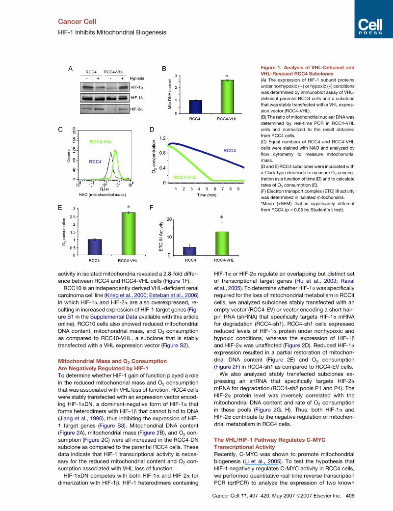
Figure 8. Analysis of Cells Lacking Both
HIF-1a and HIF-2a
(A–F) Parental RCC4 cells and subclones
transfected with empty vector (EV), vector en-
coding a scrambled negative control shRNA
(SNC), or vectors encoding shRNA against
HIF-1a (RCC4-sh1), HIF-2a (RCC4-sh2), or
HIF-1a and HIF-2a (RCC4-sh1/2) were ana-
lyzed for protein expression (A), mitochondrial
DNA (B), mitochondrial mass (C), O2 consump-
tion (D), and expression of MXI-1 (E) or PGC-1b
(F) mRNA. *Mean (±SEM) that is significantly
different from RCC4-EV.
(G) Molecular pathways regulating mitochon-
drial biogenesis, respiration, and ROS produc-
tion in renal carcinoma cells.
(Figure 7E), and RCC4-siMxi1 (Figure 7F) cells all mani-
fested increased levels of ROS, whereas PGC-1b loss of
function led to reduced ROS levels in RCC4-VHL-shPGC
cells (Figure 7G). Amplex red staining of RCC4-MYC
cells revealed increased ROS production compared to
RCC4-EV cells (Figure 7H). Levels of ROS were also in-
creased in RCC10-VHL as compared to RCC10 cells
(Figure S15B).
Analysis of RCC4 Subclones with Combined
Loss of HIF-1a and HIF-2a
RCC4 cells were cotransduced with retroviruses encoding
shRNAs against HIF-1a and HIF-2a, and two pools of cells
with very low levels of both HIF-1a and HIF-2a protein
were established (Figure 8). Compared to cells with
knockdown of either HIF-1a or HIF-2a (Figures 2, 5, and
416 Cancer Cell 11, 407–420, May 2007 ª2007 Elsevier Inc.
6), the double knockdown cells showed more dramatic in-
creases in mitochondrial DNA, mitochondrial mass, O2
consumption, C-MYC protein, CAD, RCL, and PGC-1b
mRNA and more dramatic reductions in MXI-1 mRNA
and protein levels (Figure 8; Figure S16). Taken together
with the results of the single subunit knockdown experi-
ments, these data indicate that mitochondrial mass and
respiration are inversely related to the total HIF-1 activity
(HIF-1a:HIF-1b and HIF-2a:HIF-1b heterodimers) in these
cells. In all of these assays, the results obtained with the
double knockdown cells were similar to those from
RCC4-VHL cells, indicating that the combined expression
of HIF-1a and HIF-2a is responsible for the loss of C-MYC
and PGC-1b activity that leads to loss of mitochondrial
mass and respiration in VHL-deficient renal carcinoma
cells.

Cancer Cell
HIF-1 Inhibits Mitochondrial Biogenesis
DISCUSSION
In this study we have delineated a molecular pathway by
which glucose/energy metabolism is reprogrammed in
VHL-deficient renal carcinoma cells such that mitochon-
drial DNA content, mitochondrial mass, cellular O2 con-
sumption, and ROS production are significantly reduced
(Figure 8G). This pathway is triggered by loss of VHL func-
tion and the consequent dysregulated activity of HIF-1,
which leads to the inhibition of C-MYC transcriptional ac-
tivity by MXI-1 expression and increased C-MYC degra-
dation by the proteasome, both of which are HIF-1 depen-
dent. The resulting loss of C-MYC-dependent PGC-1b
expression is responsible for the reduction in mitochon-
drial mass in VHL-deficient renal carcinoma cells. Recent
studies have demonstrated that VHL positively regulates
p53 activity (Roe et al., 2006) and p53 positively regulates
cytochrome c oxidase assembly (Matoba et al., 2006).
However, in RCC4 and RCC10 cells, it appears that in-
creased HIF-1 activity resulting from VHL loss of function
is sufficient to account for the repression of mitochondrial
metabolism and cellular respiration.
Previous studies have demonstrated that HIF-1 trans-
activates genes encoding glucose transporters and glyco-
lytic enzymes, including LDH-A, thus increasing flux
through the glycolytic pathway and the conversion of py-
ruvate to lactate (Semenza et al., 1994, 1996; Firth et al.,
1995; Iyer et al., 1998; Seagroves et al., 2001; Robey
et al., 2005). Recent studies have demonstrated that
HIF-1 actively shunts pyruvate away from the mitochon-
dria by activating expression of PDK1, a kinase that
inactivates pyruvate dehydrogenase and prevents the
conversion of pyruvate to acetyl CoA (Kim et al., 2006;
Papandreou et al., 2006). In the present study we demon-
strate that mitochondrial metabolism and biogenesis are
actively repressed in renal carcinoma cells with VHL loss
of function and consequent HIF-1 gain of function. Taken
together with previous studies, these results in VHL-defi-
cient renal carcinoma cells provide the most detailed de-
lineation of the molecular mechanisms underlying the triad
of increased glucose uptake, increased lactate produc-
tion, and decreased respiration that are the hallmarks of
cancer cell metabolism.
Our studies have provided insights into the cooperative
and antagonistic functional relationships between HIF-1
and C-MYC as they relate to the control of energy metab-
olism. Whereas HIF-1 and C-MYC both promote glucose
transport and glycolysis, HIF-1 counteracts C-MYC-medi-
ated mitochondrial biogenesis, highlighting functional
differences between HIF-1, which mediates adaptive re-
sponses to low O2, and C-MYC, which stimulates growth
and proliferation. Data from the present study indicate that
in renal carcinoma cells HIF-1 is upstream of C-MYC in the
transcriptional network regulating oxidative and glycolytic
metabolism. Previous studies demonstrated that TFAM is
a C-MYC target gene in lymphoid cells (Li et al., 2005), but
TFAM mRNA levels were not correlated with C-MYC activ-
ity or VHL status in RCC4 cells (data not shown). Instead,
loss of C-MYC-dependent PGC-1b expression plays
a critical role in the loss of mitochondrial mass in VHL-
deficient RCC4 cells (Figure 6). Although we have focused
on the consequences of VHL loss of function, MXI-1 and
C-MYC expression and activity were O2 regulated in
VHL-rescued cells (Figures 4 and 5; Figure S4), suggesting
that HIF-1 may mediate reduced mitochondrial biogenesis
and respiration in hypoxic regions of tumors that are not
VHL deficient. Although increased C-MYC degradation
in hypoxic cells has been observed (Corn et al., 2005),
the regulation of C-MYC protein levels by HIF-1 has not
been previously reported. Additional experimentation will
be required to identify domains within C-MYC that are
required for degradation and to determine whether HIF-
1-dependent degradation of C-MYC involves changes in
posttranslational modifications or protein-protein interac-
tions or both.
Does reprogramming cellular glucose and energy me-
tabolism provide a selective advantage to cells with VHL
loss of function? ATP levels in VHL-deficient RCC4 cells
are remarkably reduced and this deficiency can be cor-
rected by forced expression of VHL or C-MYC and by in-
hibition of HIF-1 or MXI-1 (Figure 7). These results indicate
that under nonhypoxic conditions, VHL loss of function
provides no selective advantage with respect to cellular
energetics. Instead, our studies have demonstrated that
decreased respiration is associated with a reduction in
ROS. Preliminary studies suggest that HIF-1-dependent
downregulation of mitochondrial metabolism may provide
a survival benefit by reducing the risk of apoptosis (data
not shown). Forced expression of HIF-1a in oral squa-
mous cell carcinoma lines leads to reduced ROS levels
and decreased hypoxia-induced apoptosis (Sasabe
et al., 2005). A correlation between levels of mitochondrial
respiration, ROS production, and apoptosis has been re-
ported in several cancer cell lines (Santamaria et al.,
2006). Another potentially important consequence of in-
creased glycolysis and reduced ROS generation is inhibi-
tion of cellular senescence (Kondoh et al., 2005). Thus,
proliferation and survival of renal carcinoma cells may be
promoted by the reprogramming of glucose and energy
metabolism that results from dysregulation of the VHL/
HIF-1/C-MYC pathway. High LDH levels and low mito-
chondrial respiratory chain content are each associated
with poor prognosis in advanced renal cell carcinoma
(Simonnet et al., 2002; Motzer et al., 2004). The surprising
finding that in RCC4 and RCC10 cells HIF-1 dominantly
represses the activity of C-MYC, which is upregulated
in many other cancers, suggests that metabolic reprog-
ramming may be a critical step during renal cell
carcinogenesis.
EXPERIMENTAL PROCEDURES
Establishment and Maintenance of Cell Lines
VHL-deficient lines RCC4 and RCC10 and subclones stably trans-
fected with an expression vector encoding VHL and neomycin resis-
tance were maintained in high glucose (4.5 mg/ml) DMEM with 10% fe-
tal bovine serum (FBS) and 1% penicillin-streptomycin. RCC4-sh1
cells were described previously (Krishnamachary et al., 2006). shRNA
targeting HIF-2a (nucleotides 1601–1619, GenBank NM_001430) was
Cancer Cell 11, 407–420, May 2007 ª2007 Elsevier Inc. 417
Cancer Cell
HIF-1 Inhibits Mitochondrial Biogenesis
inserted into pSR.retro.GFP.Neo (OligoEngine, Seattle, WA), and
RCC4-sh2 pools were established by retrovirus infection and antibiotic
selection. G418 (0.8 mg/ml) was added to the medium of RCC4-VHL,
RCC10-VHL, RCC4-sh1, and RCC4-sh2 cells. The PGC-1b shRNA
coding sequence (nucleotides 2834–2852, GenBank NM_133263)
was inserted into pSuper.retro.puro (OligoEngine). RCC4-sh1/sh2
was generated by retrovirus infection (Neo-sh1/Puro-sh2) and antibi-
otic selection with G418 (0.8 mg/ml) and puromycin (0.8 mg/ml).
RCC4-DN cells (Jiang et al., 1996) were maintained in hygromycin
(400 mg/ml). Nonhypoxic cells (20% O2) were maintained at 37�C in
a 5% CO2, 95% air incubator. Hypoxic cells (1% O2) were maintained
in a chamber flushed with a gas mixture containing 1% O2, 5% CO2,
and 94% N2 at 37�C. The pBabeMNiresGFP-C-Myc vector (Oster
et al., 2003) or pBabeMNiresGFP empty vector (provided by Dr. L.
Penn) was cotransfected with plasmids encoding group antigen/poly-
merase/envelope and vesicular stomatitis virus G proteins into 293T
packaging cells using Fugene-6 (Roche Applied Science), and trans-
duction was performed as described (Krishnamachary et al., 2006).
P493 cells were previously described (Pajic et al., 2001).
Immunoblot Assays
Equal amounts of protein extracted from cells with RIPA buffer were
fractionated by 10% SDS-PAGE. Polyclonal anti-HIF-2a antibody
(Novus Biologicals), monoclonal anti-HIF-1a (H1a67; Zhong et al.,
1999) and HIF-1b (H1b234; Zagzag et al., 2000) antibodies (Novus
Biologicals), monoclonal anti-C-MYC antibody (9E10; Santa Cruz),
and polyclonal anti-MXI-1 antibody (G-16; Santa Cruz) were used for
immunoblot assays. Blots were stripped and reprobed with a poly-
clonal antibody against b-actin (Santa Cruz) to confirm equal protein
loading and transfer.
Mitochondrial DNA Copy Measurement
Total DNA was isolated from cell lysates. The amount of mitochondrial
DNA relative to nuclear genomic DNA was determined by quantitative
real-time PCR using primers (listed in Table S1) for cytochrome b (mi-
tochondrial) and RPL13A (nuclear). Relative mitochondrial DNA levels
were calculated based on the threshold cycle (Ct) as 2�D(DCt), where
DCt = CtCytochrome b � CtRPL13A and D(DCt) = DCtRCC4 � DCtsubclone.
Flow Cytometry
Mitochondrial mass and intracellular ROS production were measured
by staining cells with 10 nM NAO or 25 nM deep red 633 and 1 mM di-
chlorodihydrofluorescein diacetate (Molecular Probes), respectively,
at 37�C for 15 min in PBS containing 5% FBS. Stained cells were fil-
tered and analyzed immediately in a FACScan flow cytometer (BD Bio-
science). Gain and amplifier settings were held constant during the
experiment.
Measurement of Total Cellular O2 Consumption
Cells were trypsinized and suspended at 3–8 3 106 per ml in DMEM
medium with 10% FBS and 25 mM HEPES buffer. For each experi-
ment, equal numbers of cells suspended in 0.4 ml were pipetted into
the chamber of an Oxytherm electrode unit (Hansatech Instrument
Ltd.), which uses a Clark-type electrode to monitor the dissolved oxy-
gen concentration in the sealed chamber over time. The data were ex-
ported to a computerized chart recorder (Oxygraph, Hansatech Instru-
ment Ltd.), which calculated the rate of O2 consumption. The
temperature was maintained at 37�C during the measurement. The
O2 concentration in 0.4 ml of DMEM medium without cells was also
measured over time to provide background values. Relative O2 con-
sumption rate was calculated after correcting for background.
Measurement of Electron Transport Complex III Activity
Ubiquinol-ferricytochrome c oxido-reductase activity of mitochondria
isolated from 3 3 106 cells was measured in the presence and absence
of antimycin A (10 mg/ml) as previously described (Yuan et al., 2003)
and the antimycin-inhibited activity was expressed as nmol of cyto-
chrome c reduced per min.
418 Cancer Cell 11, 407–420, May 2007 ª2007 Elsevier Inc.
qrtPCR
RNA was isolated using Trizol (Invitrogen) followed by DNase (Ambion)
treatment. Primers were designed using Beacon Designer software,
and cDNA was prepared using the iScript cDNA synthesis kit (Bio-
Rad). cDNA samples were diluted 1:10, and real-time PCR was per-
formed using iQ SYBR Green Supermix and the iCycler Real-time
PCR Detection System (Bio-Rad). For each primer pair (Table S1),
annealing temperature was optimized by gradient PCR. The fold
change in expression of each target mRNA relative to 18S rRNA
was calculated as 2�D(DCt), where DCt = Cttarget � Ct18S and D(DCt) =
DCtRCC4 � DCtsubclone.
siRNA Transfection
siRNAs targeting human C-MYC or MXI-1 mRNA (siGENOME SMART
pools M-003282-04 and M-009947-00, Dharmacon Research Inc.) or
RISC-free control siRNA (Dharmacon) were transfected into RCC4
subclones in the presence of Oligofectamine (Invitrogen). After 72 hr,
cells were harvested.
Reporter Gene Assay
A 53-bp sequence from human MXI1 gene was amplified by PCR and
inserted into the Bgl II site of pGL2-Promoter (Promega). HEK293 cells
(4 3 104 per well) were seeded in a 48-well plate. After overnight incu-
bation, cells were transfected either with 100 ng of firefly luciferase
reporter plasmid and 4 ng of pSV-Renilla plasmid, or further cotrans-
fected with expression vector encoding Flag-HIF-1a or HIF-2a. The
total amount of DNA transfected per well was held constant by addi-
tion of empty vector. Eighteen hours after transfection, cells were incu-
bated at 1% or 20% O2 for 24 hr. Luciferase activities were measured
using the Dual-Luciferase Reporter Assay System (Promega). For each
experiment, at least two independent transfections in triplicate were
performed.
ChIP Assay
ChIP was performed with the ChIP assay kit from Upstate-Cell Signal-
ing Solutions (Temecula, CA) with rabbit polyclonal antibodies against
HIF-1a, HIF-2a, or C-MYC (Santa Cruz Biotechnology).
Intracellular ATP
ATP levels were measured using an ATP assay kit (Sigma) according to
the manufacturer’s instructions. Luminescence was measured using
a Wallace microplate luminescence reader (Perkin Elmer) and normal-
ized to the protein concentration.
Supplemental Data
The Supplemental Data include 16 supplemental figures and one sup-
plemental table and can be found with this article online at http://www.
cancercell.org/cgi/content/full/11/5/407/DC1/.
ACKNOWLEDGMENTS
We thank Celeste Simon and Miguel Esteban for providing RCC4-VHL
and RCC10-VHL cell lines, respectively; Joseph Garcia for providing
HIF-2a expression vector; Bruce Spiegelman and Pere Puigserver
for anti-PGC-1b antibodies; Karen Padgett and Novus Biologicals for
anti-HIF-2a antibodies; Linda Penn for providing pBabeMNiresGFP-
C-Myc retroviral vector; and Linzhao Cheng for providing advice and
reagents for retrovirus production. This work was supported in part
by PHS grants P50-CA103175 and R37-CA51497 from the NIH.
Received: June 26, 2006
Revised: January 22, 2007
Accepted: April 2, 2007
Published: May 7, 2007

Cancer Cell
HIF-1 Inhibits Mitochondrial Biogenesis
REFERENCES
Bogacka, I., Xie, H., Bray, G.A., and Smith, S.R. (2005). Pioglitazone
induces mitochondrial biogenesis in human subcutaneous adipose
tissue in vivo. Diabetes 54, 1392–1399.
Boyd, K.E., and Farnham, P.J. (1997). Myc versus USF: Discrimination
at the cad gene is determined by core promoter elements. Mol. Cell.
Biol. 17, 2529–2537.
Brizel, D.M., Schroeder, T., Scher, R.L., Walenta, S., Clough, R.W.,
Dewhirst, M.W., and Mueller-Klieser, W. (2001). Elevated tumor lactate
concentrations predict for an increased risk of metastases in head-
and-neck cancer. Int. J. Radiat. Oncol. Biol. Phys. 51, 349–353.
Carroll, R.C., Ash, R.F., Vogt, P.K., and Singer, S.J. (1978). Reversion
of transformed glycolysis to normal by inhibition of protein synthesis in
rat kidney cells infected with temperature-sensitive mutant of Rous
sarcoma virus. Proc. Natl. Acad. Sci. USA 75, 5015–5019.
Corn, P.G., Ricci, M.S., Scata, K.A., Arsham, A.M., Simon, M.C.,
Dicker, D.T., and El-Deiry, W.S. (2005). Mxi1 is induced by hypoxia
in a HIF-1-dependent manner and protects cells from c-Myc-induced
apoptosis. Cancer Biol. Ther. 4, 1285–1294.
Craven, R.A., Hanrahan, S., Totty, N., Harnden, P., Stanley, A.J.,
Maher, E.R., Harris, A.L., Trimble, W.S., Selby, P.J., and Banks, R.E.
(2006). Proteomic identification of a role for the von Hippel Lindau tu-
mour suppressor in changes in the expression of mitochondrial pro-
teins and septin 2 in renal cell carcinoma. Proteomics 6, 3880–3893.
Cuezva, J.M., Chen, G., Alonso, A.M., Isidoro, A., Misek, D.E., Hanash,
S.M., and Beer, D.G. (2004). The bioenergetic signature of lung adeno-
carcinomas is a molecular marker of cancer diagnosis and prognosis.
Carcinogenesis 25, 1157–1163.
Cuezva, J.M., Krajewska, M., Lopez de Heredia, M., Krajewski, S.,
Santamaria, G., Kim, H., Zapata, J.M., Marusawa, H., Chamorrow,
M., and Reed, J.C. (2002). The bioenergetic signature of cancer: A
marker of tumor progression. Cancer Res. 62, 6674–6681.
Discher, D.J., Bishopric, N.H., Wu, X., Peterson, C.A., and Webster,
K.A. (1998). Hypoxia regulates beta-enolase and pyruvate kinase-M
promoters by modulating Sp1/Sp3 binding to a conserved GC ele-
ment. J. Biol. Chem. 273, 26087–26093.
Elstrom, R.L., Bauer, D.E., Buzzai, M., Karnauskas, R., Harris, M.H.,
Plas, D.R., Zhuang, H., Cinalli, R.M., Alavi, A., Rudin, C.M., and
Thompson, C.B. (2004). Akt stimulates aerobic glycolysis in cancer
cells. Cancer Res. 64, 3892–3899.
Esteban, M.A., Tran, M.G., Harten, S.K., Hill, P., Castellanos, M.C.,
Chandra, A., Raval, R., O’Brien, T.S., and Maxwell, P.H. (2006). Regu-
lation of E-cadherin expression by VHL and hypoxia-inducible factor.
Cancer Res. 66, 3567–3575.
Fantin, V.R., St-Pierre, J., and Leder, P. (2006). Attenuation of LDH-A
expression uncovers a link between glycolysis, mitochondrial physiol-
ogy, and tumor maintenance. Cancer Cell 9, 425–434.
Firth, J.D., Ebert, B.L., and Ratcliffe, P.J. (1995). Hypoxic regulation of
lactate dehydrogenase A: Interaction between hypoxia-inducible fac-
tor 1 and cAMP response elements. J. Biol. Chem. 270, 21021–21027.
Garcia, J.A. (2006). HIFing the brakes: Therapeutic opportunities for
treatment of human malignancies. Sci. STKE. 30, pe25.
Gatenby, R.A., and Gillies, R.J. (2004). Why do cancers have high aer-
obic glycolysis? Nat. Rev. Cancer 4, 891–899.
Hervouet, E., Demont, J., Pecina, P., Vojtiskova, A., Houstek, J.,
Simonnet, H., and Godinot, C. (2005). A new role for the von Hippel-
Lindau tumor suppressor protein: Stimulation of mitochondrial oxidative
phosphorylation complex biogenesis. Carcinogenesis 26, 531–539.
Hervouet, E., and Godinot, C. (2006). Mitochondrial disorders in renal
tumors. Mitochondrion 6, 105–117.
Hu, C.J., Wang, L.Y., Chodosh, L.A., Keith, B., and Simon, M.C. (2003).
Differential roles of hypoxia-inducible factor 1a (HIF-1a) and HIF-2a in
hypoxic gene regulation. Mol. Cell. Biol. 23, 9361–9374.
Iyer, N.V., Kotch, L.E., Agani, F., Leung, S.W., Laughner, E., Wenger,
R.H., Gassmann, M., Gearhart, J.D., Lawler, A.M., Yu, A.Y., and Se-
menza, G.L. (1998). Cellular and developmental control of O2 homeo-
stasis by hypoxia-inducible factor 1a. Genes Dev. 12, 149–162.
Jiang, B.-H., Agani, F., Passaniti, A., and Semenza, G.L. (1997). V-SRC
induces expression of hypoxia-inducible factor 1 (HIF-1) and transcrip-
tion of genes encoding vascular endothelial growth factor and enolase
1: Involvement of HIF-1 in tumor progression. Cancer Res. 57, 5328–
5335.
Jiang, B.H., Rue, E., Wang, G.L., Roe, R., and Semenza, G.L. (1996).
Dimerization, DNA binding, and transactivation properties of hyp-
oxia-inducible factor 1. J. Biol. Chem. 271, 17771–17778.
Kelly, D.P., and Scarpulla, R.C. (2004). Transcriptional regulatory cir-
cuits controlling mitochondrial biogenesis and function. Genes Dev.
18, 357–368.
Kim, J.-w., Tchernyshyov, I., Semenza, G.L., and Dang, C.V. (2006).
HIF-1-mediated expression of pyruvate dehydrogenase kinase: A met-
abolic switch required for cellular adaptation to hypoxia. Cell Metab. 3,
177–185.
Kondo, K., Klco, J., Nakamura, E., Lechpammer, M., and Kaelin, W.G.,
Jr. (2002). Inhibition of HIF is necessary for tumor suppression by the
von Hippel-Lindau protein. Cancer Cell 1, 237–246.
Kondoh, H., Lleonart, M.E., Gil, J., Wang, J., Degan, P., Peters, G.,
Martinez, D., Carnero, A., and Beach, D. (2005). Glycolytic enzymes
can modulate cellular life span. Cancer Res. 65, 177–185.
Krieg, M., Haas, R., Brauch, H., Acker, T., Flamme, I., and Plate, K.H.
(2000). Up-regulation of hypoxia-inducible factors HIF-1a and HIF-2a
under normoxic conditions in renal carcinoma cells by von Hippel-Lin-
dau tumor suppressor gene loss of function. Oncogene 19, 5435–
5443.
Krishnamachary, B., Zagzag, D., Nagasawa, H., Rainey, K., Okuyama,
H., Baek, J.H., and Semenza, G.L. (2006). Hypoxia-inducible factor-1-
dependent repression of E-cadherin in von Hippel-Lindau tumor sup-
pressor-null renal cell carcinoma mediated by TCF3, ZFHX1A, and
ZFHX1B. Cancer Res. 66, 2725–2731.
Lewis, B.C., Shim, H., Li, Q., Wu, C.S., Lee, L.A., Maity, A., and Dang,
C.V. (1997). Identification of putative c-Myc-responsive genes: Char-
acterization of rcl, a novel growth-related gene. Mol. Cell. Biol. 17,
4967–4978.
Li, F., Wang, Y., Zeller, K.I., Potter, J.J., Wonsey, D.R., O’Donnell, K.A.,
Kim, J.-w., Yustein, J.T., Lee, L.A., and Dang, C.V. (2005). Myc stimu-
lates nuclearly encoded mitochondrial genes and mitochondrial bio-
genesis. Mol. Cell. Biol. 25, 6225–6234.
Manalo, D.J., Rowan, A., Lavoie, T., Natarajan, L., Kelly, B.D., Ye, S.Q.,
Garcia, J.G., and Semenza, G.L. (2005). Transcriptional regulation of
vascular endothelial cell responses to hypoxia by HIF-1. Blood 105,
659–669.
Mandriota, S.J., Turner, K.J., Davies, D.R., Murray, P.G., Morgan, N.V.,
Sowter, H.M., Wykoff, C.C., Maher, E.R., Harris, A.L., Ratcliffe, P.J.,
and Maxwell, P.H. (2002). HIF activation identifies early lesions in
VHL kidneys: Evidence for site-specific tumor suppressor function in
the nephron. Cancer Cell 1, 459–468.
Maranchie, J.K., Vasselli, J.R., Riss, J., Bonifacino, J.S., Linehan,
W.M., and Klausner, R.D. (2002). The contribution of VHL substrate
binding and HIF-1a to the phenotype of VHL loss in renal cell carci-
noma. Cancer Cell 1, 247–255.
Matoba, S., Kang, J.-G., Patino, W.D., Wragg, A., Boehm, M., Gavri-
lova, O., Hurley, P.J., Bunz, F., and Hwang, P.M. (2006). p53 regulates
mitochondrial respiration. Science 312, 1650–1653.
Maxwell, P.H., Wiesener, M.S., Chang, G.W., Clifford, S.C., Vaux, E.C.,
Cockman, M.E., Wykoff, C.C., Pugh, C.W., Maher, E.R., and Ratcliffe,
P.J. (1999). The tumor suppressor protein VHL targets hypoxia-induc-
ible factors for oxygen-dependent proteolysis. Nature 399, 271–275.
Cancer Cell 11, 407–420, May 2007 ª2007 Elsevier Inc. 419

Cancer Cell
HIF-1 Inhibits Mitochondrial Biogenesis
Meierhofer, D., Mayr, J.A., Foetschl, U., Berger, A., Fink, K., Schmeller,
N., Hacker, G.W., Hauser-Kronberger, C., Kofler, B., and Sperl, W.
(2004). Decrease of mitochondrial DNA content and energy metabo-
lism in renal cell carcinoma. Carcinogenesis 25, 1005–1010.
Melillo, G. (2006). Inhibiting hypoxia-inducible factor 1 for cancer ther-
apy. Mol. Cancer Res. 4, 601–605.
Moeller, B.J., Dreher, M.R., Rabbani, Z.N., Schroeder, T., Cao, Y., Li,
C.Y., and Dewhirst, M.W. (2005). Pleiotropic effects of HIF-1 blockade
on tumor radiosensitivity. Cancer Cell 8, 99–110.
Motzer, R.J., Bacik, J., and Mazumdar, M. (2004). Prognostic factors
for survival of patients with stage IV renal cell carcinoma: Memorial
Sloan-Kettering Cancer Center experience. Clin. Cancer Res. 10,
6302S–6303S.
Oster, S.K., Mao, D.Y., Kennedy, J., and Penn, L.Z. (2003). Functional
analysis of the N-terminal domain of the Myc oncoprotein. Oncogene
22, 1998–2010.
Osthus, R.C., Shim, H., Kim, S., Li, Q., Reddy, R., Mukherjee, M., Xu,
Y., Wonsey, D., Lee, L.A., and Dang, C.V. (2000). Deregulation of glu-
cose transporter 1 and glycolytic gene expression by c-Myc. J. Biol.
Chem. 275, 21797–21800.
Pajic, A., Staege, M.S., Dudziak, D., Schuhmacher, M., Spitkovsky, D.,
Eissner, G., Brielmeier, M., Polack, A., and Bornkamm, G.W. (2001).
Antagonistic effects of c-myc and Epstein-Barr virus latent genes on
the phenotype of human B cells. Int. J. Cancer 93, 810–816.
Papandreou, I., Cairns, R.A., Fontana, L., Lim, A.L., and Denko, N.C.
(2006). HIF-1 mediates adaptation to hypoxia by actively downregulat-
ing mitochondrial oxygen consumption. Cell Metab. 3, 187–197.
Petros, J.A., Baumann, A.K., Ruiz-Pesini, E., Amin, M.B., Sun, C.Q.,
Hall, J., Lim, S., Issa, M.M., Flanders, W.D., Hosseini, S.H., et al.
(2005). mtDNA mutations increase tumorigenicity in prostate cancer.
Proc. Natl. Acad. Sci. USA 102, 719–724.
Polyak, K., Li, Y., Zhu, H., Lengauer, C., Willson, J.K., Markowitz, S.D.,
Trush, M.A., Kinzler, K.W., and Vogelstein, B. (1998). Somatic muta-
tions of the mitochondrial genome in human colorectal tumors. Nat.
Genet. 20, 291–293.
Raval, R.R., Lau, K.W., Tran, M.G., Sowter, H.M., Mandriota, S.J., Li,
J.L., Pugh, C.W., Maxwell, P.H., Harris, A.L., and Ratcliffe, P.J.
(2005). Contrasting properties of hypoxia-inducible factor 1 (HIF-1)
and HIF-2 in von Hippel-Lindau-associated renal cell carcinoma.
Mol. Cell. Biol. 25, 5675–5686.
Robey, I.F., Lien, A.D., Welsh, S.J., Baggett, B.K., and Gillies, R.J.
(2005). Hypoxia-inducible factor-1a and the glycolytic phenotype in
tumors. Neoplasia 7, 324–330.
Roe, J.S., Kim, H., Lee, S.M., Kim, S.T., Cho, E.J., and Youn, H.D.
(2006). p53 stabilization and transactivation by von Hippel-Lindau pro-
tein. Mol. Cell 22, 395–405.
Santamaria, G., Martinez-Diez, M., Fabregat, I., and Cuezva, J.M.
(2006). Efficient execution of cell death in non-glycolytic cells requires
the generation of ROS controlled by the activity of mitochondrial
H+-ATP synthase. Carcinogenesis 27, 925–935.
Sasabe, E., Tatemoto, Y., Li, D., Yamamoto, T., and Osaki, T. (2005).
Mechanism of HIF-1a-dependent suppression of hypoxia-induced ap-
optosis in squamous cell carcinoma cells. Cancer Sci. 96, 394–402.
Seagroves, T.N., Ryan, H.E., Lu, H., Wouters, B.G., Knapp, M., Thi-
bault, P., Laderoute, K., and Johnson, R.S. (2001). Transcription factor
HIF-1 is a necessary mediator of the Pasteur effect in mammalian cells.
Mol. Cell. Biol. 21, 3436–3444.
Semenza, G.L. (2003). Targeting HIF-1 for cancer therapy. Nat. Rev.
Cancer 3, 721–732.
420 Cancer Cell 11, 407–420, May 2007 ª2007 Elsevier Inc.
Semenza, G.L., Jiang, B.H., Leung, S.W., Passantino, R., Concordet,
J.P., Maire, P., and Giallongo, A. (1996). Hypoxia response elements
in the aldolase A, enolase 1, and lactate dehydrogenase A gene pro-
moters contain essential binding sites for hypoxia-inducible factor 1.
J. Biol. Chem. 271, 32529–32537.
Semenza, G.L., Roth, P.H., Fang, H.M., and Wang, G.L. (1994). Tran-
scriptional regulation of genes encoding glycolytic enzymes by hyp-
oxia-inducible factor 1. J. Biol. Chem. 269, 23757–23763.
Shim, H., Dolde, C., Lewis, B.C., Wu, C.S., Dang, G., Jungmann, R.A.,
Dalla-Favera, R., and Dang, C.V. (1997). c-Myc transactivation of LDH-
A: Implications for tumor metabolism and growth. Proc. Natl. Acad.
Sci. USA 94, 6658–6663.
Simonnet, H., Alazard, N., Pfeiffer, K., Gallou, C., Beroud, C., Demont,
J., Bouvier, R., Schagger, H., and Godinot, C. (2002). Low mitochon-
drial respiratory chain content correlates with tumor aggressiveness
in renal cell carcinoma. Carcinogenesis 23, 759–768.
Tang, N., Wang, L., Esko, J., Giordano, F.J., Huang, Y., Gerber, H.-P.,
Ferrara, N., and Johnson, R.S. (2004). Loss of HIF-1a in endothelial
cells disrupts a hypoxia-driven VEGF autocrine loop necessary for
tumorigenesis. Cancer Cell 6, 485–495.
Thomas, G.V., Tran, C., Mellinghoff, I.K., Welsbie, D.S., Chan, E.,
Fueger, B., Czernin, J., and Sawyers, C.L. (2006). Hypoxia-inducible
factor determines sensitivity to inhibitors of mTOR in kidney cancer.
Nat. Med. 12, 122–127. Published online December 11, 2005. 10.
1038/nm1337.
Tian, H., McKnight, S.L., and Russell, D.W. (1997). Endothelial PAS do-
main protein 1 (EPAS1), a transcription factor selectively expressed in
endothelial cells. Genes Dev. 11, 72–82.
Uldry, M., Yang, W., St-Pierre, J., Lin, J., Seale, P., and Spiegelman,
B.M. (2006). Complementary action of the PGC-1 coactivators in mito-
chondrial biogenesis and brown fat differentiation. Cell Metab. 3, 333–
341.
Unwin, R.D., Craven, R.A., Harnden, P., Hanrahan, S., Totty, N.,
Knowles, M., Eardley, I., Selby, P.J., and Banks, R.E. (2003). Proteomic
changes in renal cancer and co-ordinate demonstration of both the
glycolytic and mitochondrial aspects of the Warburg effect. Proteo-
mics 3, 1620–1632.
Wang, G.L., Jiang, B.-H., Rue, E.A., and Semenza, G.L. (1995). Hyp-
oxia-inducible factor 1 is a basic-helix-loop-helix-PAS heterodimer
regulated by cellular O2 tension. Proc. Natl. Acad. Sci. USA 92,
5510–5514.
Warburg, O. (1930). The Metabolism of Tumours (London: Arnold Con-
stable).
Yuan, G., Adhikary, G., McCormick, A.A., Holcroft, J.J., Kumar, G.K.,
and Prabhakar, N.R. (2003). Role of oxidative stress in intermittent hyp-
oxia-induced immediate early gene activation in rat PC12 cells.
J. Physiol. 557, 773–783.
Zagzag, D., Zhong, H., Scalzitti, J.M., Laughner, E., Simons, J.W., and
Semenza, G.L. (2000). Expression of hypoxia-inducible factor 1a in
brain tumors: Association with angiogenesis, invasion, and progres-
sion. Cancer 88, 2606–2618.
Zeller, K.I., Zhao, X.D., Lee, C.W.H., Chiu, K.P., Yao, F., Yustein, J.T.,
Ooi, H.S., Orlov, Y.L., Shahab, A., Yong, H.C., et al. (2006). Global
mapping of c-myc binding sites and target gene networks in human
B cells. Proc. Natl. Acad. Sci. USA 103, 17834–17839.
Zhong, H., De Marzo, A.M., Laughner, E., Lim, M., Hilton, D.A., Zagzag,
D., Buechler, P., Isaacs, W.B., Semenza, G.L., and Simons, J.W.
(1999). Overexpression of hypoxia-inducible factor 1a in common
human cancers and their metastases. Cancer Res. 59, 5830–5835.