A high HIF-1α expression genotype is associated with poor prognosis of upper aerodigestive tract...
6
A high HIF-1a expression genotype is associated with poor prognosis of upper aerodigestive tract carcinoma patients Carlos Alberto de Carvalho Fraga a , Marcos Vinícius Macedo de Oliveira a , Érica Silva de Oliveira a , Lucas Oliveira Barros a , Francis Balduino Guimarães Santos b , Ricardo Santiago Gomez c , Alfredo Maurício Batista De-Paula a , André Luiz Sena Guimarães a,⇑ a Department of Dentistry, Universidade Estadual de Montes Claros, Montes Claros, Brazil b Department of Medicine, Universidade Estadual de Montes Claros, Montes Claros, Brazil c Department of Clinical, Surgery and Oral Pathology, School of Dentistry, Universidade Federal de Minas Gerais, Belo Horizonte, Brazil article info Article history: Received 23 May 2011 Received in revised form 16 August 2011 Accepted 28 August 2011 Available online 25 September 2011 Keywords: Metastasis Angiogenesis Hypoxia HIF Polymorphism VEGFR1 Upper aerodigestive tract cancer summary The aim of the present study was to evaluate the role of HIF-1a genetic polymorphisms and protein expression in the development of metastasis in upper aerodigestive tract cancer (UADTC) patients. The expression of pro-angiogenic markers was also evaluated. Protein expression was analysed using immu- nohistochemistry, and RFLP analysis was used to investigate HIF-1a C1779T and G1790A polymorphisms in 52 patients with UADTC. Primary lesions were divided into 2 groups according to the absence or pres- ence of metastasis. Lymph node samples were divided into 3 groups: metastatic lymph nodes, non-met- astatic lymph nodes (both derived from patients with metastatic disease), and control lymph nodes, which were obtained from patients without any metastasis. The allele T was more frequently found in patients with metastatic disease. HIF-1a protein expression in the lymph nodes was increased in the presence of the T allele. Metastatic lymph nodes showed lower levels of HIF-1a, VEGFR1, and MMP-9 pro- teins compared to lymph nodes without metastasis, while VEGFR2 protein levels were increased. In agreement, HIF-1a expression was correlated with MMP-9. Cox regression analysis demonstrated that higher HIF-1a and MMP-9 protein expression levels and GA and GG genotypes were associated with poor survival. Our findings show that the C1772T and G1790A polymorphisms of the HIF-1a gene are associ- ated with increased expression of the HIF-1a protein in UADTC. The present data indicate that non-met- astatic tissues express higher levels of HIF-1a, VEGFR1, and MMP-9, while in metastatic lymph nodes, VEGFR2 protein expression is elevated. The present study also shows that the HIF-1a G1790A polymor- phism and its protein expression have an impact on the prognosis of UADTC patients. Ó 2011 Elsevier Ltd. All rights reserved. Introduction Upper aerodigestive tract cancer (UADTC) is one of the most common cancers worldwide. 1 The aetiology of UADTC is complex owing to the multigenic nature of the disease and the number of potential environmental agents to which affected individuals may have been exposed. It is clear that the major aetiological agents are tobacco and alcohol exposure. 2 However, other factors such as genetic predisposition have an important role in disease genesis and progression. 3,4 The role of genetic polymorphisms in altering protein levels/function and predisposing patients to a vari- ety of diseases has been demonstrated extensively. 5–10 Rapid tumour expansion leads to an oxygen demand that cannot be supported by the existing tumour vasculature. Poorly oxygenated regions within a tumour are the main characteristics of the hypoxic phenomenon. 11 Hypoxia has not only been associ- ated with resistance to treatment but also metastasis 12 and epithe- lial–mesenchymal transition. 13 The hypoxic response is regulated by hypoxia-inducible factor-1 (HIF-1), which is a basic helix- loop-helix transcription factor composed of 2 subunits, HIF-1a and HIF-1b. 14 HIF-1a is the main gene involved in the hypoxic phe- nomenon, and its polymorphisms have been associated with metastasis in several solid tumours. 15,16 In this context, non- neoplastic cells can modify specific sites for future metastatic adhesion; there is evidence that this cell population can portend a future metastatic site. This non-neoplastic cell population in- cludes fibroblasts, haematopoietic progenitors and other cells that express factors that promote the establishment of metastatic cells. The identification of this cell population in human tissues prior to 1368-8375/$ - see front matter Ó 2011 Elsevier Ltd. All rights reserved. doi:10.1016/j.oraloncology.2011.08.023 ⇑ Corresponding author. Address: Universidade Estadual de Montes Claros, Hospital Universitário Clemente de Faria, Laboratório de Pesquisa em Saúde, Avenida Cula Mangabeira, 562, Montes Claros, Minas Gerais, cep 39401-001, Brazil. E-mail address: [email protected] (A.L.S. Guimarães). Oral Oncology 48 (2012) 130–135 Contents lists available at SciVerse ScienceDirect Oral Oncology journal homepage: www.elsevier.com/locate/oraloncology
-
Upload
portalamericas -
Category
Documents
-
view
3 -
download
0
Transcript of A high HIF-1α expression genotype is associated with poor prognosis of upper aerodigestive tract...

Oral Oncology 48 (2012) 130–135
Contents lists available at SciVerse ScienceDirect
Oral Oncology
journal homepage: www.elsevier .com/locate /ora loncology
A high HIF-1a expression genotype is associated with poor prognosisof upper aerodigestive tract carcinoma patients
Carlos Alberto de Carvalho Fraga a, Marcos Vinícius Macedo de Oliveira a, Érica Silva de Oliveira a,Lucas Oliveira Barros a, Francis Balduino Guimarães Santos b, Ricardo Santiago Gomez c,Alfredo Maurício Batista De-Paula a, André Luiz Sena Guimarães a,⇑a Department of Dentistry, Universidade Estadual de Montes Claros, Montes Claros, Brazilb Department of Medicine, Universidade Estadual de Montes Claros, Montes Claros, Brazilc Department of Clinical, Surgery and Oral Pathology, School of Dentistry, Universidade Federal de Minas Gerais, Belo Horizonte, Brazil
a r t i c l e i n f o s u m m a r y
Article history:Received 23 May 2011Received in revised form 16 August 2011Accepted 28 August 2011Available online 25 September 2011
Keywords:MetastasisAngiogenesisHypoxiaHIFPolymorphismVEGFR1Upper aerodigestive tract cancer
1368-8375/$ - see front matter � 2011 Elsevier Ltd. Adoi:10.1016/j.oraloncology.2011.08.023
⇑ Corresponding author. Address: UniversidadeHospital Universitário Clemente de Faria, LaboratóAvenida Cula Mangabeira, 562, Montes Claros, Minas
E-mail address: [email protected] (A
The aim of the present study was to evaluate the role of HIF-1a genetic polymorphisms and proteinexpression in the development of metastasis in upper aerodigestive tract cancer (UADTC) patients. Theexpression of pro-angiogenic markers was also evaluated. Protein expression was analysed using immu-nohistochemistry, and RFLP analysis was used to investigate HIF-1a C1779T and G1790A polymorphismsin 52 patients with UADTC. Primary lesions were divided into 2 groups according to the absence or pres-ence of metastasis. Lymph node samples were divided into 3 groups: metastatic lymph nodes, non-met-astatic lymph nodes (both derived from patients with metastatic disease), and control lymph nodes,which were obtained from patients without any metastasis. The allele T was more frequently found inpatients with metastatic disease. HIF-1a protein expression in the lymph nodes was increased in thepresence of the T allele. Metastatic lymph nodes showed lower levels of HIF-1a, VEGFR1, and MMP-9 pro-teins compared to lymph nodes without metastasis, while VEGFR2 protein levels were increased. Inagreement, HIF-1a expression was correlated with MMP-9. Cox regression analysis demonstrated thathigher HIF-1a and MMP-9 protein expression levels and GA and GG genotypes were associated with poorsurvival. Our findings show that the C1772T and G1790A polymorphisms of the HIF-1a gene are associ-ated with increased expression of the HIF-1a protein in UADTC. The present data indicate that non-met-astatic tissues express higher levels of HIF-1a, VEGFR1, and MMP-9, while in metastatic lymph nodes,VEGFR2 protein expression is elevated. The present study also shows that the HIF-1a G1790A polymor-phism and its protein expression have an impact on the prognosis of UADTC patients.
� 2011 Elsevier Ltd. All rights reserved.
Introduction
Upper aerodigestive tract cancer (UADTC) is one of the mostcommon cancers worldwide.1 The aetiology of UADTC is complexowing to the multigenic nature of the disease and the number ofpotential environmental agents to which affected individualsmay have been exposed. It is clear that the major aetiologicalagents are tobacco and alcohol exposure.2 However, other factorssuch as genetic predisposition have an important role in diseasegenesis and progression.3,4 The role of genetic polymorphisms inaltering protein levels/function and predisposing patients to a vari-ety of diseases has been demonstrated extensively.5–10
ll rights reserved.
Estadual de Montes Claros,rio de Pesquisa em Saúde,
Gerais, cep 39401-001, Brazil..L.S. Guimarães).
Rapid tumour expansion leads to an oxygen demand thatcannot be supported by the existing tumour vasculature. Poorlyoxygenated regions within a tumour are the main characteristicsof the hypoxic phenomenon.11 Hypoxia has not only been associ-ated with resistance to treatment but also metastasis12 and epithe-lial–mesenchymal transition.13 The hypoxic response is regulatedby hypoxia-inducible factor-1 (HIF-1), which is a basic helix-loop-helix transcription factor composed of 2 subunits, HIF-1aand HIF-1b.14 HIF-1a is the main gene involved in the hypoxic phe-nomenon, and its polymorphisms have been associated withmetastasis in several solid tumours.15,16 In this context, non-neoplastic cells can modify specific sites for future metastaticadhesion; there is evidence that this cell population can portenda future metastatic site. This non-neoplastic cell population in-cludes fibroblasts, haematopoietic progenitors and other cells thatexpress factors that promote the establishment of metastatic cells.The identification of this cell population in human tissues prior to

C.A.C. Fraga et al. / Oral Oncology 48 (2012) 130–135 131
the spread of neoplastic cells supports the targeting of proteinsassociated with angiogenesis such as vascular endothelial growthfactor A (VEGF-A) and its receptors VEGFR1, VEGFR217, or extracel-lular matrix degradation proteins such as metallopeptidase-9(MMP-9).18 Based on these data, the present study aimed toevaluate the role of HIF-1a genetic polymorphisms and proteinexpression in the development of nodal metastases in UADTCpatients. The findings of previous studies15,19,16,12,20–22 led to thehypothesis that the HIF-1a C1779T and G1790A polymorphismscould enhance the HIF-1a pathway by up-regulating HIF-1a pro-tein expression and facilitate the spread of tumour cells and theirattachment to lymph nodes. Thus, we also evaluated angiogenesisby assessing the protein expression of VEGF-A, VEGFR1, VEGFR2,MMP-9, and CD105 at the primary tumour and metastatic andnon-metastatic nodes.
Patients and methods
Patients and ethical aspects
In this retrospective study, we analysed 52 patients who werediagnosed at a stomatological clinic and referred to the Head andNeck Service of the Universidade Estadual de Montes Claros (Mon-tes Claros, Minas Gerais State, Brazil) between 1998 and 2008.23,5
Only cases with available primary tumour and lymph node tissuesfor molecular and immunohistochemistry analysis were includedin the study. The health records of these patients were retrieved,and socio-demographic, clinical, and outcome data were obtained(Supplementary Table 1). All UADTC patients were classified onthe basis of the primary site according to the International UnionAgainst Cancer (UICC)-TNM classification of malignant tumours,as described in the International Classification of Diseases forOncology.24 One sample from the primary malignant tumour andat least 1 cervical lymph node were obtained from each UADTC pa-tient and included in the analysis. Ethical approval for this studywas obtained from the local ethics committees (Unimontes, CEP1852/2010). All patients underwent surgical resection and weretreated with postoperative radiotherapy.
Evaluation and classification of locoregional lymph nodes
All samples were fixed in formalin, embedded in paraffin, andserially sectioned (5-lm thickness). The sections were stained withhaematoxylin and eosin (H&E) and evaluated under a conventionallight microscope. Cervical lymph nodes were analysed usingmorphological and immunohistochemical methods to identify ametastatic focus. A high-molecular weight cytokeratin primaryantibody was used to identify micrometastases. Immunohistochem-istry staining, DNA isolation, and HIF-1a genotyping were per-formed as described in the Supplementary Materials.
Primary cancer lesion classifications
Primary lesions were divided into 2 groups according to thepresence (n = 26) or absence of metastasis (n = 26). Patients inthe 2 groups were matched by tumour size, WHO grade, anatomi-cal site, and age. Other clinical characteristics were also similar(Supplementary Table 1).
Lymph node classifications
Histological, immunohistochemical, and clinical examinationrevealed lymph node metastases in 26 patients. In this group, themetastatic and non-metastatic lymph nodes were analysed. Inaddition, the lymph nodes from 26 patients without metastasiswere analysed (control group).
Statistical analysis
Chi-square and Fisher’s exact statistical tests were used to eval-uate the association between HIF-1a polymorphic variants andmetastasis. In addition, a multivariate analysis using binary logisticregression was performed to build a model of variables to evaluatethe risk of locoregional metastasis.
Immunolocalization analysis of HIF-1a, VEGFA, VEGFR1, VEG-FR2, and MMP-9 proteins assumed non-parametrical distribution,and comparisons between groups were performed using theMann–Whitney test. The analysis of CD105 protein expression intissues assumed parametrical distribution, and comparisons be-tween groups were performed using the Student’s t-test. TheSpearman correlation test was used to evaluate the correlation be-tween the expression levels of different proteins. For survival anal-yses, the Kaplan–Meier test was performed, and the variables werecompared using the log-rank test. Variables with p 6 0.25 were in-cluded in the Cox proportional hazards regression to estimate pre-dictive factors of crude survival. All statistical analyses wereperformed with the statistical software package SPSS�, version13.0 for Windows�. P values <0.05 were considered significant.
Results
Immunohistochemical and molecular data associated with metastasis
The distribution of HIF-1a genotypes according to the presenceof metastasis is shown in Supplementary Table 2. The data revealan association between the C1772T polymorphism and the pres-ence of metastasis (p = 0.023). The frequency of the CT genotypewas higher in patients that developed metastasis. The allele Twas more frequently found in patients who had metastasis. How-ever, the G1790A polymorphism was not associated with metasta-sis (p = 0.172).
To further define the factors that contribute to metastasis, weperformed binary logistic regression for all lesions and lymphnodes. In primary lesions, there was no association between thepolymorphism findings and protein expression levels. The binarylogistic regression showed an association between the CT genotypeand the presence of metastasis (p = 0.009). In addition, low expres-sion of VEGFR1 was correlated with metastasis (p = 0.023) (Table 1).
Genotypes and protein expression in lymph nodes
We next investigated whether the C1772T and G1790A poly-morphisms could independently induce a change in proteinexpression levels. HIF-1a protein expression was higher in the TTgenotype than the CC genotype (79.575 ± 23.293 and22.625 ± 41.101, respectively; p = 0.027) in all lymph nodes groups.In metastatic lymph nodes, HIF-1a protein expression in lymphnodes increased in the presence of the T allele. Similarly, HIF-1aprotein expression was higher in the AA genotype than in the GGgenotype (63.025 ± 47.199 vs. 44.139 ± 41.948; p = 0.028).
Expression of angiogenesis-associated proteins in the different lymphnode groups
Increased expression levels of HIF-1a, VEGFR1, and MMP-9were observed in non-metastatic lymph nodes (p = 0.047,p = 0.003, and p = 0.022, respectively). VEGFR2 expression washigher in tissues from metastatic lymph nodes than in non-metastatic lymph nodes (Fig. 1). There was no difference in theexpression of any of the proteins studied in non-metastatic lymphnodes between patients with metastasis and those in the controlgroup. No difference in neo-vessels (CD105 staining) was observedbetween the different lymph node groups.

Table 1Lymph node parameters associated to risk of locoregional metastasis evaluated bybinary logistic regression in the UADTC patients.
Variables 95 CI
OR Lower Upper p Value
Polymorphism CTCC ReferentCT 5.481 1.532 19.606 0.009a
TT NA NA NA 0.999
Polymorphism GAGG ReferentGA 0.679 0.165 2.791 0.592AA 5.594 0.390 80.268 0.205
ImmunohistochemistryHIF-1 a 0.993 0.980 1.005 0.255VEGFA 0.989 0.965 1.014 0.394VEGFR1 0.962 0.930 0.995 0.023a
VEGFR2 1.009 0.985 1.035 0.457MMP9 0.998 0.970 1.026 0.884CD105 1.034 0.972 1.100 0.292
OR: odds ratio; CI: confidence interval; n: total number; NA: not applicable. Themodel was fitted to the best-fit model.
a Results statistically significant.
132 C.A.C. Fraga et al. / Oral Oncology 48 (2012) 130–135
Expression of angiogenesis-associated proteins in the primary cancerlesion
The expression pattern of HIF-1a, VEGF-A, VEGFR1, VEGFR2,MMP-9, and CD105 in primary lesions analysed by immunohisto-chemistry is shown in Fig. 2. CD105 was used to identify
Figure 1 Expression of HIF-1a, cytokeratin, VEGF-A, VEGFR1, VEGFR2, MMP-9, and CD10MMP-9, and CD105 protein expression levels in leukocyte cells of lymph node samples. Ametastatic and non-metastatic groups in the protein expression levels of HIF-1a (a) (pstatistically significant differences between metastatic and control groups in the prote(p = 0.006), and MMP-9 (d) (p = 0.022). There were no statistically significant differencesof HIF-1a (B), cytokeratin (C), VEGF-A (D), VEGFR1 (E), VEGFR2 (F), MMP-9 (G), and CD10and metastatic group (right panel). P values of HIF-1a, VEGF-A, VEGFR1, VEGFR2, and MMof CD105 protein expression was calculated by Student’s t test. ⁄Counting performed by
neo-vessels. The immunohistochemical expression of CD105 in pri-mary tumours with metastasis was significantly higher than in thegroup without metastasis (Fig. 2).
Protein expression correlations
To understand the effect of HIF-1a on the levels of the otherproteins, we next analysed the correlation between the expressionlevels of the different proteins studied in all groups. A positivecorrelation was found between HIF-1a and MMP-9 in both theprimary tumour and metastatic samples (r = + 0.374, p = 0.004and r = + 0.640, p = 0.001, respectively). In the neoplastic cells,the expression of VEGFA was correlated with VEGFR2 in themetastatic lymph nodes (r = + 0.418, p = 0.034). The expression ofVEGFR1 was correlated with the expression of MMP-9 in lymphnodes of patients without metastasis (r = + 0.304, p = 0.029). In alllymph node groups analysed, the expression of HIF-1a was corre-lated with the expression of the VEGFR1 protein (r = + 0.232,p = 0.042). In addition, VEGFR1 was correlated with MMP-9 whenall lymph node groups were analysed (r = + 0.343, p = 0.002).
Risk of death and molecular findings
The mean survival for all patients was 1106.2 days after diagno-sis. High MMP-9 expression in primary lesions was associated withpoor survival. GG and GA genotypes and elevated HIF-1a proteinexpression in lymph nodes had a negative impact on survival(Table 2).
5 proteins in lymph node samples. (A) Analyses of HIF-1a, VEGF-A, VEGFR1, VEGFR2,nalysis by Mann–Whitney test showed statistically significant differences between= 0.047), VEGFR1 (b) (p = 0.003), and VEGFR2 (c) (p = 0.003). Similarly, there werein expression levels of HIF-1a (a) (p = 0.029), VEGFR1 (b) (p < 0.001), VEGFR2 (c)in protein expression levels between non-metastatic and control groups. Expression5 (H) proteins in the control group (left panel), non-metastatic group (middle panel)P-9 protein expressions were calculated using the Mann–Whitney test. The P valuemicrovessel density. Scale bar, 20 lm.

Figure 2 Expression of HIF-1a, VEGF-A, VEGFR1, VEGFR2, MMP-9, and CD105proteins in primary lesions. (A) Counting was carried out in neoplastic cells ofprimary lesions. Note that there was a statistically significant increase in theexpression of the CD105 protein in primary lesions that present metastasis (a)(p < 0.001). The right panel shows the expression levels of HIF-1a (B), VEGF-A (C),VEGFR1 (D), VEGFR2 (E), MMP-9 (F), and CD105 (G) proteins in primary lesions ofpatients with metastasis. The left panel shows the expression of HIF-1a (B), VEGF-A(C), VEGFR1 (D), VEGFR2 (E), MMP-9 (F), and CD105 (G) proteins in primary lesionsof patients without any metastasis. P values of HIF-1a, VEGF-A, VEGFR1, VEGFR2,and MMP-9 protein expression levels were calculated using the Mann–Whitneytest. The p value for CD105 protein expression was calculated by Student’s t test.⁄Counting performed by microvessel density. Scale bar, 50 lm.
C.A.C. Fraga et al. / Oral Oncology 48 (2012) 130–135 133
Discussion
Metastasis is one of the hallmarks of malignant disease and thecause of death for the majority of cancer patients. The molecularmechanisms underlying the early onset of metastasis are complex,involving both genetic and epigenetic alterations in malignant cellsand the tumour environment. Tumour cells have many mecha-nisms to escape the host defence mechanisms, and metastasisdevelopment requires tumour cells to lose adhesion to surround-ing cells, cross the basement membrane, migrate through lympha-tic channels, and subsequently, extravasate into foreign nodaltissue.25,21 Hypoxia has been shown to promote regional metasta-sis in several solid tumours.26,27 To maintain cell survival in amicroenvironment that is low in oxygen, cells can alter their pro-tein expression.28 Under normal oxygen tension, HIF-1a is de-graded by ubiquitin dependent proteolysis, whereas HIF-1b isstable.29 Angiogenesis, glycolytic metabolism, and cell survivaland invasion are regulated through HIF-1a signalling.22 Duringearly tumourigenesis, HIF-1a expression is required for the induc-tion of angiogenesis although other factors can facilitate tumourangiogenesis. Liao et al.20 reported that HIF-1a expression acts asan accelerating factor in tumour progression and metastasis.20
Polymorphisms in the HIF-1a gene have been associated withan increased risk of developing certain solid tumours.30,15,16
However, the role of single-nucleotide polymorphisms in theoxygen-dependent degradation domain of the HIF-1a gene in car-cinogenesis appears complex and is not currently known. To date,no conclusive results have established a link between HIF-1a poly-morphism and carcinogenesis.19,18,31–33 Genetic polymorphisms atC1772T and G1790A of HIF-1a have been associated with in-creased transcriptional activity16,32 and high expression levels ofHIF-1a in oral cancer.32 Munoz-Guerra et al. demonstrated thatthe frequencies of the heterozygous GA and homozygous AA geno-types were high in patients with oral cancer.34 Our study showedthat the CT heterozygous genotype is associated with lymph nodemetastasis. Moreover, based on lymph node analyses, the homozy-gous TT genotype is associated with a higher number of HIF-1a+
leukocyte cells than the homozygous CC genotype, and HIF-1a+
leukocyte cell numbers are higher in homozygous AA genotypesthan in homozygous GG genotypes.
Although the precise molecular and cellular mechanisms thatcontrol tumour metastasis have not been defined, several studieshave demonstrated that tumours have a predilection for metastasisto specific organs. Kaplan et al.17 introduced the concept that tu-mour metastasis is initiated by a well-defined sequence of eventsthat depend on the expression patterns of fibronectin and VEG-FR1+VLA4+ clusters, which dictate organ-specific tumour spread.These clusters can modify the microenvironment by regulatingthe production of MMP-9, resulting in the attraction of tumourcells and their establishment in a niche.17 HIF-1a might enhancethis signalling by up-regulating MMP-918 and VEGFR1 expressionin these sites. In the present study, increased expression of HIF-1a, VEGFR1, and MMP-9 was detected in non-metastatic lymphnodes compared to metastatic samples. In addition, VEGFR1 pro-tein expression was correlated with HIF-1a and MMP-9 proteinlevels in non-metastatic lymph nodes. According to the pre-meta-static niche theory, non-metastatic lymph nodes prepare for tu-mour cell spreading by up-regulating some proteins. After theestablishment of the metastases, a down-regulation of the proteinsis observed.17,35 Notably, a decrease in protein levels after metasta-sis does not occur in some types of tumours.36 Therefore, distincttime-dependent events may or may not occur before, during, andafter the establishment of a metastatic niche.
Despite contradictory results on the clinical significance ofmicrovessel density in UADTC, this parameter has been regarded
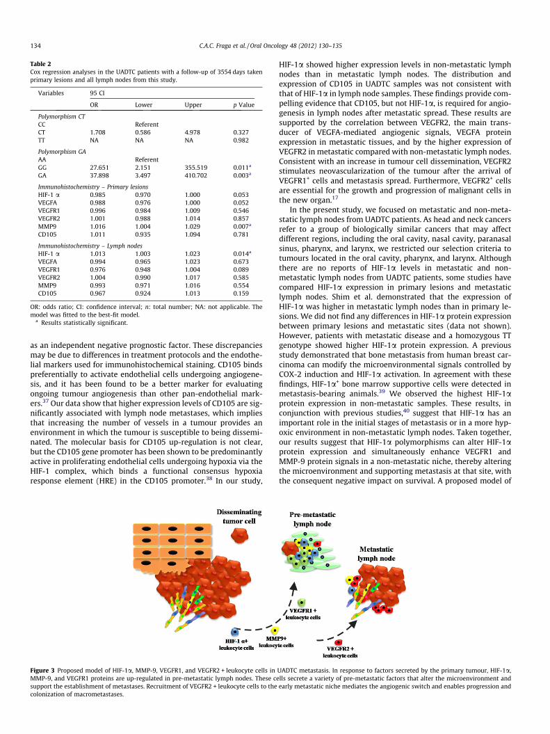
Table 2Cox regression analyses in the UADTC patients with a follow-up of 3554 days takenprimary lesions and all lymph nodes from this study.
Variables 95 CI
OR Lower Upper p Value
Polymorphism CTCC ReferentCT 1.708 0.586 4.978 0.327TT NA NA NA 0.982
Polymorphism GAAA ReferentGG 27.651 2.151 355.519 0.011a
GA 37.898 3.497 410.702 0.003a
Immunohistochemistry – Primary lesionsHIF-1 a 0.985 0.970 1.000 0.053VEGFA 0.988 0.976 1.000 0.052VEGFR1 0.996 0.984 1.009 0.546VEGFR2 1.001 0.988 1.014 0.857MMP9 1.016 1.004 1.029 0.007a
CD105 1.011 0.935 1.094 0.781
Immunohistochemistry – Lymph nodesHIF-1 a 1.013 1.003 1.023 0.014a
VEGFA 0.994 0.965 1.023 0.673VEGFR1 0.976 0.948 1.004 0.089VEGFR2 1.004 0.990 1.017 0.585MMP9 0.993 0.971 1.016 0.554CD105 0.967 0.924 1.013 0.159
OR: odds ratio; CI: confidence interval; n: total number; NA: not applicable. Themodel was fitted to the best-fit model.
a Results statistically significant.
134 C.A.C. Fraga et al. / Oral Oncology 48 (2012) 130–135
as an independent negative prognostic factor. These discrepanciesmay be due to differences in treatment protocols and the endothe-lial markers used for immunohistochemical staining. CD105 bindspreferentially to activate endothelial cells undergoing angiogene-sis, and it has been found to be a better marker for evaluatingongoing tumour angiogenesis than other pan-endothelial mark-ers.37 Our data show that higher expression levels of CD105 are sig-nificantly associated with lymph node metastases, which impliesthat increasing the number of vessels in a tumour provides anenvironment in which the tumour is susceptible to being dissemi-nated. The molecular basis for CD105 up-regulation is not clear,but the CD105 gene promoter has been shown to be predominantlyactive in proliferating endothelial cells undergoing hypoxia via theHIF-1 complex, which binds a functional consensus hypoxiaresponse element (HRE) in the CD105 promoter.38 In our study,
Figure 3 Proposed model of HIF-1a, MMP-9, VEGFR1, and VEGFR2 + leukocyte cells inMMP-9, and VEGFR1 proteins are up-regulated in pre-metastatic lymph nodes. These cesupport the establishment of metastases. Recruitment of VEGFR2 + leukocyte cells to thecolonization of macrometastases.
HIF-1a showed higher expression levels in non-metastatic lymphnodes than in metastatic lymph nodes. The distribution andexpression of CD105 in UADTC samples was not consistent withthat of HIF-1a in lymph node samples. These findings provide com-pelling evidence that CD105, but not HIF-1a, is required for angio-genesis in lymph nodes after metastatic spread. These results aresupported by the correlation between VEGFR2, the main trans-ducer of VEGFA-mediated angiogenic signals, VEGFA proteinexpression in metastatic tissues, and by the higher expression ofVEGFR2 in metastatic compared with non-metastatic lymph nodes.Consistent with an increase in tumour cell dissemination, VEGFR2stimulates neovascularization of the tumour after the arrival ofVEGFR1+ cells and metastasis spread. Furthermore, VEGFR2+ cellsare essential for the growth and progression of malignant cells inthe new organ.17
In the present study, we focused on metastatic and non-meta-static lymph nodes from UADTC patients. As head and neck cancersrefer to a group of biologically similar cancers that may affectdifferent regions, including the oral cavity, nasal cavity, paranasalsinus, pharynx, and larynx, we restricted our selection criteria totumours located in the oral cavity, pharynx, and larynx. Althoughthere are no reports of HIF-1a levels in metastatic and non-metastatic lymph nodes from UADTC patients, some studies havecompared HIF-1a expression in primary lesions and metastaticlymph nodes. Shim et al. demonstrated that the expression ofHIF-1a was higher in metastatic lymph nodes than in primary le-sions. We did not find any differences in HIF-1a protein expressionbetween primary lesions and metastatic sites (data not shown).However, patients with metastatic disease and a homozygous TTgenotype showed higher HIF-1a protein expression. A previousstudy demonstrated that bone metastasis from human breast car-cinoma can modify the microenvironmental signals controlled byCOX-2 induction and HIF-1a activation. In agreement with thesefindings, HIF-1a+ bone marrow supportive cells were detected inmetastasis-bearing animals.39 We observed the highest HIF-1aprotein expression in non-metastatic samples. These results, inconjunction with previous studies,40 suggest that HIF-1a has animportant role in the initial stages of metastasis or in a more hyp-oxic environment in non-metastatic lymph nodes. Taken together,our results suggest that HIF-1a polymorphisms can alter HIF-1aprotein expression and simultaneously enhance VEGFR1 andMMP-9 protein signals in a non-metastatic niche, thereby alteringthe microenvironment and supporting metastasis at that site, withthe consequent negative impact on survival. A proposed model of
UADTC metastasis. In response to factors secreted by the primary tumour, HIF-1a,lls secrete a variety of pre-metastatic factors that alter the microenvironment andearly metastatic niche mediates the angiogenic switch and enables progression and

C.A.C. Fraga et al. / Oral Oncology 48 (2012) 130–135 135
HIF-1a, MMP-9, VEGFR1, and VEGFR2 + leukocyte cells in UADTCmetastasis is shown in Fig. 3.
Conclusion
In conclusion, our study demonstrated that the C1772T andG1790A polymorphisms of the HIF-1a gene are associated with in-creased expression of the HIF-1a protein in UADTC. Our data indi-cate that the expressions of HIF-1a, VEGFR1, and MMP-9 increasein non-metastatic tissues, while the VEGFR2 protein is expressed athigher levels in metastatic lymph nodes. Finally, the present studyshows that the HIF-1a G1790A polymorphism and its proteinexpression have an impact on the prognosis of patients withUADTC. These findings suggest an additional role for HIF-1a inUADTC. Further studies are necessary to elucidate the HIF-1a path-way, which would facilitate the development of novel therapeuticstrategies for the prevention and treatment of metastases inUADTC and other solid tumours.
Acknowledgements
This study was supported by grants from the Conselho Nacionalde Desenvolvimento Científico e Tecnológico and Fundação deAmparo a Pesquisa do Estado de Minas Gerais. Dr Guimarães,and Dr Gomez are research fellows of CNPq. Dr De Paula is a re-search fellow of FAPEMIG.
Appendix. . Supplementary data
Supplementary data associated with this article can be found, inthe online version, at doi:10.1016/j.oraloncology.2011.08.023.
References
1. Curado MP, Hashibe M. Recent changes in the epidemiology of head and neckcancer. Curr Opin Oncol 2009;21(3):194–200.
2. Argiris A, Karamouzis MV, Raben D, Ferris RL. Head and neck cancer. Lancet2008;371(9625):1695–709.
3. Gomez RS, Pimenta FJ, Guimaraes AL, Souza LN, Salomao UE, de Almeida HC,et al. Effect of bone marrow transplantation on the immunolocalization of p53,hMSH2, and hMLH1 proteins on oral mucosa. Oral Dis 2004;10(4):207–11.
4. Scully C, Field JK, Tanzawa H. Genetic aberrations in oral or head and necksquamous cell carcinoma 2: chromosomal aberrations. Oral Oncol2000;36(4):311–27.
5. Farias LC, De Carvalho Fraga CA, DeOliveira MV, Silva TF, Marques-Silva L,Moreira PR, et al. Effect of age on the association between p16CDKN2Amethylation and DNMT3B polymorphism in head and neck carcinoma andpatient survival. Int J Oncol 2010;37(1):167–76.
6. Gomes CC, Drummond SN, Guimaraes AL, Andrade CI, Mesquita RA, Gomez RS.P21/WAF1 and cyclin D1 variants and oral squamous cell carcinoma. J OralPathol Med 2008;37(3):151–6.
7. Guimaraes AL, deSa AR, Victoria JM, Correia-Silva JF, Pessoa PS, Diniz MG, et al.Association of interleukin-1beta polymorphism with recurrent aphthousstomatitis in Brazilian individuals. Oral Dis 2006;12(6):580–3.
8. Guimaraes AL, Correia-Silva JF, Diniz MG, Xavier GM, Horta MC, Gomez RS.Investigation of functional gene polymorphisms: IL-1B, IL-6 and TNFA in benignmigratory glossitis in Brazilian individuals. J Oral Pathol Med 2007;36(9):533–7.
9. Guimaraes AL, Correia-Silva JF, Sa AR, Victoria JM, Diniz MG, Costa FO, et al.Investigation of functional gene polymorphisms IL-1beta, IL-6, IL-10 and TNF-alpha in individuals with recurrent aphthous stomatitis. Arch Oral Biol2007;52(3):268–72.
10. Xavier GM, deSa AR, Guimaraes AL, daSilva TA, Gomez RS. Investigation offunctional gene polymorphisms interleukin-1beta, interleukin-6, interleukin-10 and tumor necrosis factor in individuals with oral lichen planus. J Oral PatholMed 2007;36(8):476–81.
11. Brown JM, Giaccia AJ. The unique physiology of solid tumors: opportunities(and problems) for cancer therapy. Cancer Res 1998;58(7):1408–16.
12. Le QT, Denko NC, Giaccia AJ. Hypoxic gene expression and metastasis. CancerMetastasis Rev 2004;23(3–4):293–310.
13. Luo Y, He DL, Ning L, Shen SL, Li L, Li X. Hypoxia-inducible factor-1alphainduces the epithelial–mesenchymal transition of human prostatecancer cells.Chin Med J (Engl) 2006;119(9):713–8.
14. Wang GL, Jiang BH, Rue EA, Semenza GL. Hypoxia-inducible factor 1 is a basic-helix-loop-helix-PAS heterodimer regulated by cellular O2 tension. Proc NatlAcad Sci U S A 1995;92(12):5510–4.
15. Chau CH, Permenter MG, Steinberg SM, Retter AS, Dahut WL, Price DK, et al.Polymorphism in the hypoxia-inducible factor 1alpha gene may confersusceptibility to androgen-independent prostate cancer. Cancer Biol Ther2005;4(11):1222–5.
16. Fu XS, Choi E, Bubley GJ, Balk SP. Identification of hypoxia-inducible factor-1alpha (HIF-1alpha) polymorphism as a mutation in prostate cancer thatprevents normoxia-induced degradation. Prostate 2005;63(3):215–21.
17. Kaplan RN, Riba RD, Zacharoulis S, Bramley AH, Vincent L, Costa C, et al.VEGFR1-positive haematopoietic bone marrow progenitors initiate the pre-metastatic niche. Nature 2005;438(7069):820–7.
18. Kim SJ, Rabbani ZN, Dewhirst MW, Vujaskovic Z, Vollmer RT, Schreiber EG, et al.Expression of HIF-1alpha, CA IX, VEGF, and MMP-9 in surgically resected non-small cell lung cancer. Lung Cancer 2005;49(3):325–35.
19. Chen MK, Chiou HL, Su SC, Chung TT, Tseng HC, Tsai HT, et al. The associationbetween hypoxia inducible factor-1alpha gene polymorphisms and increasedsusceptibility to oral cancer. Oral Oncol 2009;45(12):e222–6.
20. Liao D, Corle C, Seagroves TN, Johnson RS. Hypoxia-inducible factor-1alpha is akey regulator of metastasis in a transgenic model of cancer initiation andprogression. Cancer Res 2007;67(2):563–72.
21. Rofstad EK, Mathiesen B, Henriksen K, Kindem K, Galappathi K. The tumor bedeffect: increased metastatic dissemination from hypoxia-inducedup-regulation of metastasis-promoting gene products. Cancer Res 2005;65(6):2387–96.
22. Semenza GL. Targeting HIF-1 for cancer therapy. Nat Rev Cancer 2003;3(10):721–32.
23. De Paula AM, Souza LR, Farias LC, Correa GT, Fraga CA, Eleuterio NB, et al.Analysis of 724 cases of primary head and neck squamous cell carcinoma(HNSCC) with a focus on young patients and p53 immunolocalization. OralOncol 2009;45(9):777–82.
24. Sobin LH. TNM: evolution and relation to other prognostic factors. Semin SurgOncol 2003;21(1):3–7.
25. Le QT, Harris J, Magliocco AM, Kong CS, Diaz R, Shin B, et al. Validation of lysyloxidase as a prognostic marker for metastasis and survival in head and necksquamous cell carcinoma: Radiation Therapy Oncology Group trial 90-03. J ClinOncol 2009;27(26):4281–6.
26. Cairns RA, Hill RP. Acute hypoxia enhances spontaneous lymph nodemetastasis in an orthotopic murine model of human cervical carcinoma.Cancer Res 2004;64(6):2054–61.
27. Rofstad EK, Galappathi K, Mathiesen B, Ruud EB. Fluctuating anddiffusion-limited hypoxia in hypoxia-induced metastasis. Clin Cancer Res2007;13(7):1971–8.
28. North S, Moenner M, Bikfalvi A. Recent developments in the regulation of theangiogenic switch by cellular stress factors in tumors. Cancer Lett 2005;218(1):1–14.
29. Semenza GL. Hydroxylation of HIF-1: oxygen sensing at the molecular level.Physiology (Bethesda) 2004;19:176–82.
30. Anastasiadis AG, Ghafar MA, Salomon L, Vacherot F, Benedit P, Chen MW, et al.Human hormone-refractory prostate cancers can harbor mutations in the O(2)-dependent degradation domain of hypoxia inducible factor-1alpha (HIF-1alpha). J Cancer Res Clin Oncol 2002;128(7):358–62.
31. Maxwell PH, Pugh CW, Ratcliffe PJ. Activation of the HIF pathway in cancer.Curr Opin Genet Dev 2001;11(3):293–9.
32. Tanimoto K, Yoshiga K, Eguchi H, Kaneyasu M, Ukon K, Kumazaki T, et al.Hypoxia-inducible factor-1alpha polymorphisms associated with enhancedtransactivation capacity, implying clinical significance. Carcinogenesis2003;24(11):1779–83.
33. Trastour C, Benizri E, Ettore F, Ramaioli A, Chamorey E, Pouyssegur J, et al. HIF-1alpha and CA IX staining in invasive breast carcinomas: prognosis andtreatment outcome. Int J Cancer 2007;120(7):1451–8.
34. Munoz-Guerra MF, Fernandez-Contreras ME, Moreno AL, Martin ID, Herraez B,Gamallo C. Polymorphisms in the hypoxia inducible factor 1-alpha and theimpact on the prognosis of early stages of oral cancer. Ann Surg Oncol2009;16(8):2351–8.
35. Kaplan RN, Rafii S, Lyden D. Preparing the ‘‘soil’’: the premetastatic niche.Cancer Res 2006;66(23):11089–93.
36. Karaca Z, Tanriverdi F, Elbuken G, Cakir I, Donmez H, Selcuklu A, et al.Comparison of primary octreotide-lar and surgical treatment in newlydiagnosed patients with acromegaly. Clin Endocrinol (Oxf) 2011.
37. Marioni G, Marino F, Giacomelli L, Staffieri C, Mariuzzi ML, Violino E, et al.Endoglin expression is associated with poor oncologic outcome in oral andoropharyngeal carcinoma. Acta Otolaryngol 2006;126(6):633–9.
38. Fonsatti E, Maio M. Highlights on endoglin (CD105): from basic findingstowards clinical applications in human cancer. J Transl Med 2004;2(1):18.
39. Maroni P, Matteucci E, Luzzati A, Perrucchini G, Bendinelli P, Desiderio MA.Nuclear co-localization and functional interaction of COX-2 and HIF-1alphacharacterize bone metastasis of human breast carcinoma. Breast Cancer ResTreat 2010.
40. Shim H, Lau SK, Devi S, Yoon Y, Cho HT, Liang Z. Lower expression of CXCR4 inlymph node metastases than in primary breast cancers: potential regulation byligand-dependent degradation and HIF-1alpha. Biochem Biophys Res Commun2006;346(1):252–8.