High levels of HIF-2α highlight an immature neural crest-like neuroblastoma cell cohort located in...
7
Journal of Pathology J Pathol 2008; 214: 482–488 Published online 11 December 2007 in Wiley InterScience (www.interscience.wiley.com) DOI: 10.1002/path.2304 Original Paper High levels of HIF-2α highlight an immature neural crest-like neuroblastoma cell cohort located in a perivascular niche A Pietras, 1 D Gisselsson, 2 I Øra, 1,3 R Noguera, 4 S Beckman, 1 S Navarro 4 and S P˚ ahlman 1 * 1 Center for Molecular Pathology, Department of Laboratory Medicine, CREATE Health, Lund University, University Hospital MAS, Malm¨ o, Sweden 2 Department of Clinical Genetics, Lund University Hospital, Lund, Sweden 3 Department of Pediatric Oncology and Hematology, Lund University Hospital, Lund, Sweden 4 Department of Pathology, University of Valencia, Medical School, Val` encia, Spain *Correspondence to: S P˚ ahlman, Center for Molecular Pathology, Department of Laboratory Medicine, University Hospital MAS, Entrance 78, SE-205 02 Malm¨ o, Sweden. E-mail: [email protected] No conflicts of interest were declared. Received: 7 June 2007 Revised: 25 October 2007 Accepted: 16 November 2007 Abstract High HIF-2α protein levels in the sympathetic nervous system-derived childhood tumour neuroblastoma as well as immature phenotype correlate to unfavourable outcome. Here we show that a small subset of perivascularly located, strongly HIF-2α-positive tumour cells (MYCN amplified) lacks expression of differentiation markers, but expresses neural crest and early sympathetic progenitor marker genes such as Notch-1, HES-1, c-Kit, dHAND, and vimentin. HIF-2α- and CD68-positive tumour-associated macrophages were frequently found close to the immature and HIF-2α-positive neuroblastoma cells and as VEGF levels are high in the perivascular niche, we hypothesize that neuroblastoma neural crest-like cells and macrophages cooperate to facilitate angiogenesis and thereby contribute to the aggressive neuroblastoma phenotype. Copyright 2008 Pathological Society of Great Britain and Ireland. Published by John Wiley & Sons, Ltd. Keywords: HIF-2α; hypoxia; neuroblastoma; neural crest; tumour stem cells; perivascular niche; tumour-associated macrophages Introduction Tumour cells as well as non-transformed cells have the capacity to adapt to hypoxia. The hypoxic response involves a shift towards anaerobic metabolism and increased glycolysis and a decrease in energy- consuming activities such as protein translation, DNA repair, and cell growth. The bottom line in this adap- tation process is to save and facilitate production of cellular energy during oxygen shortage. Much of the cellular adaptation to hypoxia is the result of a pheno- typical shift caused by the stabilization and activation of the hypoxia-inducible transcription factors HIF-1α and HIF-2α. Under physiological oxygen conditions, HIF-1α and HIF-2α are rapidly targeted for protea- somal degradation [1]. Due to poor vascularization, solid tumours frequently develop hypoxic areas where HIF-1α and HIF-2α become stabilized, and a corre- lation between low oxygenation grade and poor out- come has been demonstrated in many tumour forms [2]. An increasing number of reports have demon- strated that HIF proteins can also be active under normoxic conditions [3] but the underlying molecular mechanisms are not well understood. One interest- ing example is the high HIF-2α levels in develop- ing sympathetic nervous system (SNS) structures and in the SNS-derived paediatric tumour neuroblastoma [4]. HIF-2α is selectively and transiently expressed at mRNA and protein levels in embryonal/fetal murine and human SNS ganglia and paraganglia [5–7]. We recently showed that HIF-2α, but not HIF-1α, pro- tein is expressed in small subsets of cells located in restricted areas of well-vascularized neuroblastoma tis- sue [4]. Furthermore, high HIF-2α levels correlated to unfavourable outcome in a large neuroblastoma material and transient knock-down of HIF-2α in xeno- transplanted neuroblastoma cells gave rise to more slow-growing tumours compared with cells transfected with unspecific siRNA. In vitro, neuroblastoma cells accumulate HIF-2α at 5% O 2 , a near-physiological oxygen tension, and as demonstrated by direct in vivo binding to HIF-target genes such as vascular endothe- lial growth factor (VEGF ) and DEC1 by chromatin immunoprecipitation, HIF-2α is also active under non- hypoxic conditions. Global gene expression analysis further revealed an overall ‘hypoxic’ response at 5% O 2 . Among the genes driven by HIF-2α at 5% O 2 was VEGF and there was a correlation between HIF- 2α and VEGF expression in the clinical neuroblas- toma material. As the HIF-2α/VEGF-positive cells were located close to blood vessels, we hypothesized Copyright 2008 Pathological Society of Great Britain and Ireland. Published by John Wiley & Sons, Ltd. www.pathsoc.org.uk
-
Upload
independent -
Category
Documents
-
view
4 -
download
0
Transcript of High levels of HIF-2α highlight an immature neural crest-like neuroblastoma cell cohort located in...

Journal of PathologyJ Pathol 2008; 214: 482–488Published online 11 December 2007 in Wiley InterScience(www.interscience.wiley.com) DOI: 10.1002/path.2304
Original Paper
High levels of HIF-2α highlight an immature neuralcrest-like neuroblastoma cell cohort located in aperivascular niche
A Pietras,1 D Gisselsson,2 I Øra,1,3 R Noguera,4 S Beckman,1 S Navarro4 and S Pahlman1*1Center for Molecular Pathology, Department of Laboratory Medicine, CREATE Health, Lund University, University Hospital MAS, Malmo, Sweden2Department of Clinical Genetics, Lund University Hospital, Lund, Sweden3Department of Pediatric Oncology and Hematology, Lund University Hospital, Lund, Sweden4Department of Pathology, University of Valencia, Medical School, Valencia, Spain
*Correspondence to:S Pahlman, Center for MolecularPathology, Department ofLaboratory Medicine, UniversityHospital MAS, Entrance 78,SE-205 02 Malmo, Sweden.E-mail: [email protected]
No conflicts of interest weredeclared.
Received: 7 June 2007Revised: 25 October 2007Accepted: 16 November 2007
AbstractHigh HIF-2α protein levels in the sympathetic nervous system-derived childhood tumourneuroblastoma as well as immature phenotype correlate to unfavourable outcome. Here weshow that a small subset of perivascularly located, strongly HIF-2α-positive tumour cells(MYCN amplified) lacks expression of differentiation markers, but expresses neural crestand early sympathetic progenitor marker genes such as Notch-1, HES-1, c-Kit, dHAND,and vimentin. HIF-2α- and CD68-positive tumour-associated macrophages were frequentlyfound close to the immature and HIF-2α-positive neuroblastoma cells and as VEGF levelsare high in the perivascular niche, we hypothesize that neuroblastoma neural crest-likecells and macrophages cooperate to facilitate angiogenesis and thereby contribute to theaggressive neuroblastoma phenotype.Copyright 2008 Pathological Society of Great Britain and Ireland. Published by JohnWiley & Sons, Ltd.
Keywords: HIF-2α; hypoxia; neuroblastoma; neural crest; tumour stem cells; perivascularniche; tumour-associated macrophages
Introduction
Tumour cells as well as non-transformed cells have thecapacity to adapt to hypoxia. The hypoxic responseinvolves a shift towards anaerobic metabolism andincreased glycolysis and a decrease in energy-consuming activities such as protein translation, DNArepair, and cell growth. The bottom line in this adap-tation process is to save and facilitate production ofcellular energy during oxygen shortage. Much of thecellular adaptation to hypoxia is the result of a pheno-typical shift caused by the stabilization and activationof the hypoxia-inducible transcription factors HIF-1αand HIF-2α. Under physiological oxygen conditions,HIF-1α and HIF-2α are rapidly targeted for protea-somal degradation [1]. Due to poor vascularization,solid tumours frequently develop hypoxic areas whereHIF-1α and HIF-2α become stabilized, and a corre-lation between low oxygenation grade and poor out-come has been demonstrated in many tumour forms[2]. An increasing number of reports have demon-strated that HIF proteins can also be active undernormoxic conditions [3] but the underlying molecularmechanisms are not well understood. One interest-ing example is the high HIF-2α levels in develop-ing sympathetic nervous system (SNS) structures and
in the SNS-derived paediatric tumour neuroblastoma[4].
HIF-2α is selectively and transiently expressed atmRNA and protein levels in embryonal/fetal murineand human SNS ganglia and paraganglia [5–7]. Werecently showed that HIF-2α, but not HIF-1α, pro-tein is expressed in small subsets of cells located inrestricted areas of well-vascularized neuroblastoma tis-sue [4]. Furthermore, high HIF-2α levels correlatedto unfavourable outcome in a large neuroblastomamaterial and transient knock-down of HIF-2α in xeno-transplanted neuroblastoma cells gave rise to moreslow-growing tumours compared with cells transfectedwith unspecific siRNA. In vitro, neuroblastoma cellsaccumulate HIF-2α at 5% O2, a near-physiologicaloxygen tension, and as demonstrated by direct in vivobinding to HIF-target genes such as vascular endothe-lial growth factor (VEGF ) and DEC1 by chromatinimmunoprecipitation, HIF-2α is also active under non-hypoxic conditions. Global gene expression analysisfurther revealed an overall ‘hypoxic’ response at 5%O2. Among the genes driven by HIF-2α at 5% O2was VEGF and there was a correlation between HIF-2α and VEGF expression in the clinical neuroblas-toma material. As the HIF-2α/VEGF-positive cellswere located close to blood vessels, we hypothesized
Copyright 2008 Pathological Society of Great Britain and Ireland. Published by John Wiley & Sons, Ltd.www.pathsoc.org.uk
HIF-2α highlights neural crest-like neuroblastoma cells 483
that HIF-2α is driving vascularization in these tumourareas and that HIF-2α acts as an oncogene in neurob-lastoma [4].
Here we show that the neuroblastoma cells stain-ing intensely for HIF-2α (HIF-2α+) lack detectableexpression of SNS markers such as tyrosine hydrox-ylase (TH ) and neuron-specific enolase (NSE ). Asneuroblastomas are derived from SNS precursor cells[8], which in turn are derived from the neural crest, weexamined the expression of a set of neural crest mark-ers in this subset of HIF-2α+ neuroblastoma cells. Fur-thermore, as tumour-associated macrophages (TAMs)are known to express high HIF-2α levels and to belocated perivascularly [9], we stained tumours for themacrophage marker CD68. Based on data from thesestudies and the demonstration by MYCN-FISH anal-yses that the majority of HIF-2α+ cells are indeedtumour-derived, we conclude that the perivascularlylocated subsets of intensely HIF-2α staining neurob-lastoma cells have immature, neural crest-like charac-teristics. As these cells appear to express VEGF andare located close to blood vessels, we propose thatthey, together with the CD68+, perivascular TAMs,promote neuroblastoma vascularization and aggressivegrowth.
Materials and methods
Cell culture and quantitative real-time PCR
Human neuroblastoma cells [SK-N-BE(2)c and KCN-69n] were grown routinely as described previously[5]. For quantitative real-time PCR, cDNA synthe-sized using random hexamers and a superscript IIreverse-transcriptase (Invitrogen, Carlsbad, CA, USA)was used as a template with SYBR Green PCR MasterMix from Applied Biosystems, Foster City, CA, USA[4]. Relative gene expression levels were quantified byuse of the comparative Ct method. Three housekeepinggenes (UBC, YWHAZ, and SDHA) were used for nor-malization of expression levels. The primer sequenceswere as follows (5′-3′): IGF2 f: CCGTGCTTCCG-GACAACTT, r: CTGCTTCCAGGTGTCATATTGG;SDHA f: TGGGAACAAGAGGGCATCTG, r: CCAC-CACTGCATCAAATTCATG; TH f: GCGCAGGAA-GCTGATTGC, r: CAATCTCCTCGGCGGTGTAC;UBC f: ATTTGGGTCGCGGTTCTTG, r: TGCCTT-GACATTCTCGATGGT; YWHAZ f: ACTTTTGGTA-CATTGTGGCTTCAA, r: CCGCCAGGACAAAC-CAGTAT
Patient material, immunohistochemistry, in situhybridization, and fluorescence in situ hybridization(FISH)
Human neuroblastoma specimens were routinely fixedand embedded in paraffin before analysis: ethicalapproval for this study was obtained from both LundUniversity and the University of Valencia. Immuno-histochemistry was performed as previously described
[10] and immunoreactivity was detected after antigenretrieval using the Envision system and DAKO Tech-mate 500. HIF-2α staining was performed on freshlysectioned material from a single block of each tumouranalysed. The following antibodies were used: anti-CD31 (diluted 1/100; Dako, Glostrup, Denmark),CD68 (1/500; Dako), c-Kit (1/100; Dako), dHAND(1/600; Santa Cruz Biotechnology, Santa Cruz, CA,USA), HES-1 (1/1000 [11]), HIF-1α (1/10 000; NovusBiologicals, Littleton, CO, USA), HIF-2α (1/1000;Novus), Notch-1 (1/100; Santa Cruz), NSE (1/100;Dako), TH (1/250; Roche Molecular Biochemicals,Mannheim, Germany), VEGF (1/250; Santa Cruz),and vimentin (1/1000; Dako). Double immunohisto-chemistry was performed as above, using two anti-bodies from different species (mouse anti-HIF-2α, rab-bit anti-c-Kit, and goat anti-Notch-1) and two differ-ent chromogens. HIF-2α staining was visualized byEnhanced Fast Red A (red/pinkish colour), while c-Kitand Notch-1 staining was visualized by diaminoben-zidine (DAB; brown colour). IGF2 in situ hybridiza-tion was performed as previously described [8]. Com-bined immunofluorescence and FISH for MYCN intwo MYCN-amplified tumours were performed on4 µm tissue sections. After incubation with anti-HIF-2α antibodies, the tissue was fixed in 4% formalde-hyde for 1 min, washed in buffered saline, pretreatedwith protease (Digest-All 3;, Zymed Laboratories, SanFrancisco, CA, USA), and hybridized with a MYCNfluorescein-labelled single-copy probe (Vysis-AbbottInc, Chicago, IL, USA) for 24 h. Stringency wash-ing was in 0.5× SSC for 2 min, after which the tis-sue was incubated with Cy3-conjugated streptavidin(Amersham Biosciences, Amersham Place, UK) for30 min.
Results
HIF proteins in neuroblastoma specimens
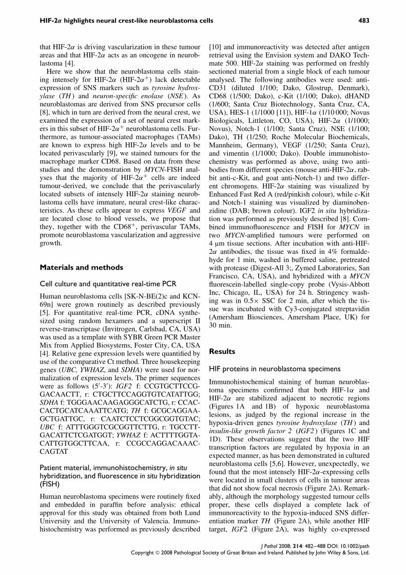
Immunohistochemical staining of human neuroblas-toma specimens confirmed that both HIF-1α andHIF-2α are stabilized adjacent to necrotic regions(Figures 1A and 1B) of hypoxic neuroblastomalesions, as judged by the regional increase in thehypoxia-driven genes tyrosine hydroxylase (TH ) andinsulin-like growth factor 2 (IGF2 ) (Figures 1C and1D). These observations suggest that the two HIFtranscription factors are regulated by hypoxia in anexpected manner, as has been demonstrated in culturedneuroblastoma cells [5,6]. However, unexpectedly, wefound that the most intensely HIF-2α-expressing cellswere located in small clusters of cells in tumour areasthat did not show focal necrosis (Figure 2A). Remark-ably, although the morphology suggested tumour cellsproper, these cells displayed a complete lack ofimmunoreactivity to the hypoxia-induced SNS differ-entiation marker TH (Figure 2A), while another HIFtarget, IGF2 (Figure 2A), was highly co-expressed
J Pathol 2008; 214: 482–488 DOI: 10.1002/pathCopyright 2008 Pathological Society of Great Britain and Ireland. Published by John Wiley & Sons, Ltd.

484 A Pietras et al
Figure 1. HIF-1α and HIF-2α are stabilized in hypoxic tumour areas. (A–D) Immunohistochemical staining of HIF-1α (A), HIF-2α(B), and TH (C), and in situ hybridization of IGF2 (D) in a necrotic region of a stage 4S neuroblastoma tumour. Scale bars = 100 µm;in insets, scale bars = 25 µm
Figure 2. HIF-2α is highly expressed in non-hypoxic tumour areas. (A–C) Immunohistochemical analysis of HIF-2α and TH, andin situ hybridization of IGF2 (A) showing HIF-2α+, IGF2+, and TH− cells of a stage 4S neuroblastoma tumour. Sections are notconsecutive, but close to each other. Scale bars = 100 µm; in insets, scale bars = 50 µm. (B, C) TH mRNA (B) and IGF2 mRNA(C) expression in two human neuroblastoma cell lines grown under normoxic (21% O2) and hypoxic (5% and 1% O2) conditionsfor 72 h. Data are expressed as mean ± SD
with HIF-2α. Quantitative real-time PCR of cDNAderived from two neuroblastoma cell lines [KCN-69nand SK-N-BE(2)c] grown at 21%, 5%, and 1% O2confirmed that both TH and IGF2 are up-regulated byhypoxia (Figure 2C). Thus, the lack of TH expressionand focal necrosis indicated sufficient oxygenationand suggested substantial non-hypoxic stabilization ofHIF-2α in these perivascular tumour cells.
HIF-2α+ cells express neural crest and early SNSprogenitor markers
Staining neuroblastomas for HIF-2α revealed a subsetof tumours with a striking pattern of perivascular cellson the border between tumour and stroma expressinghigh levels of HIF-2α, with less intensive or neg-ative staining in other parts of the tumours. While
J Pathol 2008; 214: 482–488 DOI: 10.1002/pathCopyright 2008 Pathological Society of Great Britain and Ireland. Published by John Wiley & Sons, Ltd.
HIF-2α highlights neural crest-like neuroblastoma cells 485
the vast majority of tumour cells expressed TH asexpected, the perivascular HIF-2α+ cells were com-pletely devoid of detectable TH (Figures 3A–3C). Ofthe 22 tumours studied, such cells were found in 11cases, including both high- and low-stage neuroblas-tomas. However, as we have not sectioned throughan entire tumour or a complete block of tumour tis-sue in the sections lacking these cells, we cannotexclude the possibility that they are found in all neu-roblastomas. TH is a marker of sympathetic differen-tiation and lack of or low expression might indicatean immature phenotype. In accordance, these HIF-2α+ TH− cells in well-vascularized areas, as con-firmed by CD31 staining (Figures 3C and 4A), consis-tently lacked detectable neuron-specific enolase (NSE)(Figure 4A). As HIF-2α is expressed during normalsympathetic development, we tested the hypothesisthat the HIF-2α+, TH−, and NSE− cells representedimmature cells by analysing neuroblastoma sectionsfor expression of neural crest and early SNS markergenes. We found elevated expression of early SNS-expressed genes such as dHAND and vimentin as wellas neural crest markers such as Notch-1, HES-1, andc-Kit (Figures 4 and 5). To verify that HIF-2α andneural crest markers (c-Kit and Notch-1) co-localizein the same cell, double immunohistochemical stainingwas performed (Figure 4B).
Neural crest-like tumour cells and TAMs co-existin the perivascular niche
Solid tumours are known to contain perivascularlylocated tumour-associated macrophages (TAMs)expressing HIF-2α and VEGF [9,12]. To check if afraction of the HIF-2α+ cells in neuroblastoma wasTAMs, we stained tumours for the macrophage markerCD68. As exemplified in Figure 5A, the perivascularniche of some neuroblastomas contained a large pro-portion of macrophages (CD68+) as well as CD68−,immature cells (c-Kit+) expressing high HIF-2α lev-els. Other neuroblastomas contained a markedly lowernumber of macrophages in close vicinity to immatureHIF-2α+ cells (Figure 5B). Interestingly, VEGF levelsare high in the perivascular niche, suggesting that bothTAMs and the immature neuroblastoma cells expressVEGF (Figure 5A).
HIF-2α+, TH− tumour cells harbour MYCNamplifications
To establish that the HIF-2α+, TH− cells wereindeed tumour-derived, we performed a combinedimmunofluorescence and FISH experiment by stain-ing tumours for HIF-2α before performing a MYCN-FISH in neuroblastomas carrying an amplified MYCNgene. As shown in Figure 5C, the most intensely HIF-2α+ cells surrounding blood vessels harboured MYCNamplifications. Thus, we conclude that the HIF-2α+,TH− and neural crest and early SNS marker+ cellswere tumour-derived and, for example, not TAMs orendothelial or stromal cells.
Discussion
The unexpected finding from previous studies thatHIF-2α protein levels were also high in some HIF-1α-negative tumour areas that did not show focal necrosis[4,6] led us to investigate the nature of these cells.Although we could demonstrate that HIF-2α, but notHIF-1α, is stabilized at near-physiological (5%) oxy-gen concentrations in vitro [4], this observation couldnot explain why discrete populations of perivascularlylocated tumour cells with high HIF-2α levels are sur-rounded by HIF-2α− tumour cells further away fromthe blood vessels. This, together with the puzzlingobservation that the HIF-2α staining intensity wasmore important for the prediction of prognosis thanthe number of HIF-2α+ cells, suggested that the rela-tionship between HIF-2α expression and aggressiveneuroblastoma behaviour is independent of hypoxia.The selective and transient expression of HIF-2α dur-ing SNS development supports this conclusion [6,7].The recent observation that the stem cell-associatedexpression of OCT4 is driven primarily by HIF-2α[13] indeed suggests that HIF-2α has a broader roleduring development. Furthermore, a direct role forHIF-2α in tumour growth by promoting MYC activitywas recently implicated [14].
Subsets of cells, frequently referred to as cancerstem cells (CSCs), with an exclusive ability to main-tain tumour growth have been described in severalcancers [15,16]. Recent studies have suggested thatCSCs, like normal stem cells, depend on a specific cel-lular micro-environment, called the stem cell niche, tomaintain stem cell characteristics. For example, likeneural stem cells, neural CSCs appear to rely on a‘perivascular niche’ [17]. In this study, we identifiedsubsets of neuroblastoma cells, strongly positive forHIF-2α with a neural crest-like phenotype, that arelocated in a perivascular niche. Putative neuroblastomastem cells can be expected to express high levels ofneural crest markers such as Notch-1, HES-1, and c-Kit, as well as early SNS-expressed proteins such asvimentin and dHAND. In addition to elevated expres-sion of these neural markers, the cells identified inthis study consistently lacked detectable expression ofthe sympathetic differentiation markers TH and NSE,while still carrying MYCN amplification found in thebulk of tumour cells. As the presence of intenselyHIF-2α-staining cells correlates to poor prognosis [4],we hypothesize that the Notch-1+, HES-1+, c-Kit+,vimentin+, dHAND+, and HIF-2α+ phenotype definesa subpopulation of neuroblastoma cells with tumourfounding/stem cell features. However, not all stronglyHIF-2α+ neuroblastoma cells have neural crest-likefeatures, but they still seem to provide prognosticinformation [4]. One explanation would be that theseHIF-2α+ cells are recently formed from tumour found-ing/stem cells, and thus reflect their number, and thatthey are on their way to differentiate based on theirhigher expression of neuronal markers such as NSEand TH.
J Pathol 2008; 214: 482–488 DOI: 10.1002/pathCopyright 2008 Pathological Society of Great Britain and Ireland. Published by John Wiley & Sons, Ltd.

486 A Pietras et al
Figure 3. Distinct subsets of highly HIF-2α+ cells lack the sympathetic differentiation marker TH. (A–C) Immunohistochemicalanalysis of HIF-2α and TH localization in one stage 3 (A) and two stage 4 (B, C) neuroblastoma tumours. CD31 staining showsvasculature in an area indicated by an asterisk with HIF-2α+ cells (C). Arrowheads indicate HIF-2α+ and TH− cells. Sections arenot consecutive, but close to each other. Scale bars = 100 µm
Figure 4. Perivascular cells with high HIF-2α protein levels show a neural crest-like expression pattern. (A) Immunohistochemicalanalysis of HIF-2α, TH, CD31, NSE, Notch-1, HES-1, and vimentin localization in a stage 3 neuroblastoma tumour. Sections arenot consecutive, but close to each other. Scale bars = 100 µm; in magnifications, scale bars = 25 µm (magnified areas indicatedby an asterisk). (B) Double immunohistochemical staining for HIF-2α/c-Kit and HIF-2α/Notch-1 in consecutive sections. Rightbottom panel shows HIF-2α staining alone, red/pinkish colour (Enhanced Fast Red A). Anti-c-Kit and anti-Notch-1 staining shownin brown (DAB). Scale bars = 10 µm
J Pathol 2008; 214: 482–488 DOI: 10.1002/pathCopyright 2008 Pathological Society of Great Britain and Ireland. Published by John Wiley & Sons, Ltd.

HIF-2α highlights neural crest-like neuroblastoma cells 487
Figure 5. Both tumour-associated macrophages and neural crest-like neuroblastoma cells in well-vascularized areas expresshigh HIF-2α levels. (A) Immunohistochemical analysis of HIF-2α, VEGF, CD68, and c-Kit localization in a well-vascularized areaof a stage 4 tumour with a high macrophage content. (B) Immunohistochemical analysis of HIF-2α, CD31, CD68, and dHANDexpression in a well-vascularized area of a stage 4 tumour with a low macrophage content. Sections are not consecutive, but closeto each other. (C) Combined immunofluorescence (HIF-2α) and FISH (MYCN) showing HIF-2α signal (red colour, highlighted bywhite arrowheads) in MYCN-amplified (light bluish green colour, homogeneously staining regions, highlighted by white arrows)perivascular neuroblastoma cells (blood vessel endothelial cells display green autofluorescence). Left panel shows MYCN FISHonly: please note that blood vessel autofluorescence is red in this panel
Recently, stem cell-like glioma cells were reportedto promote angiogenesis via high VEGFexpression [18]. HIF-2α is one of the most potentinducers of VEGF and we recently demonstrated thatHIF-2α is primarily responsible for VEGF expres-sion at prolonged hypoxia and at non-hypoxic, near-physiological oxygen tensions [4]. Considering thedata presented here, revealing high HIF-2α and VEGFlevels in perivascular subsets of neural crest-like neu-roblastoma cells, we suggest that HIF-2α plays animportant role in neuroblastoma angiogenesis as wellas in glioma CSC behaviour and angiogenesis.
Another cell type reported to promote angiogen-esis and tumour growth through HIF-2α expressionis TAMs [9,12]. Here we report that the perivascu-lar niche of neuroblastomas also commonly containsmacrophages, as defined by CD68 expression. Withthis finding and the knowledge that the occurrence ofTAMs in breast cancer lesions correlates with poorprognosis [19], it was important to verify the existenceof HIF-2α+, CD68− cells with a neural crest and SNSprogenitor phenotype. We therefore demonstrated that
HIF-2α+ cells were MYCN-amplified as the bulk oftumour cells. Because of the lack of SNS differenti-ation marker gene expression and the localization ofthese immature HIF-2α+ cells, they are easily over-looked and have until now probably been taken fornon-tumourous cells. It is therefore of interest to notethat neuroblastoma cells with multi-lineage pheno-types have been previously described [20,21]. Whetherthese cells have stem cell characteristics has not beenreported. One assumption would be that they indeedcorrespond to the cells identified here. However, thetumours that we have studied do not show vascularmimicry, as the vascular endothelial cells are HIF-2α−and lack MYCN amplification (Figure 5C).
In this study, we have identified immature, neu-ral crest-like neuroblastoma cells and TAMs thatboth express HIF-2α and reside in a perivascularniche in human neuroblastomas. As both cell typesappear to express VEGF, presumably in a HIF-2α-dependent fashion, we hypothesize that they coop-erate in promoting neuroblastoma angiogenesis. Ourdata further suggest a potential therapeutic value
J Pathol 2008; 214: 482–488 DOI: 10.1002/pathCopyright 2008 Pathological Society of Great Britain and Ireland. Published by John Wiley & Sons, Ltd.
488 A Pietras et al
in targeting neuroblastoma vasculature, as we havepreviously demonstrated a correlation between HIF-2α expression and poor clinical outcome. Disruptionof the stem cell niche normally leads to loss of stemcell characteristics and if the cells identified here areindeed neuroblastoma stem cells, disrupting the nicheon which their self-renewal capacity relies may be astrategy to treat high-stage neuroblastomas.
Acknowledgements
We thank Dr Linda Holmquist-Mengelbier for providing neu-roblastoma cDNA. This work was supported by the SwedishCancer Society, the Children’s Cancer Foundation of Swe-den, the Swedish Research Council, the SSF Strategic Centerfor Translational Cancer Research — CREATE Health, Hansvon Kantzows Stiftelse, Gunnar Nilsson’s Cancer Founda-tion, Malmo University Hospital research funds, and grantsPI06/5176 and RD06/0020/0102 from Instituto Carlos III,Madrid, Spain to SN and RN.
References
1. Metzen E, Ratcliffe PJ. HIF hydroxylation and cellular oxygensensing. Biol Chem 2004;385:223–230.
2. Brown JM, Wilson WR. Exploiting tumour hypoxia in cancertreatment. Nature Rev Cancer 2004;4:437–447.
3. Semenza GL. Targeting HIF-1 for cancer therapy. Nature RevCancer 2003;3:721–732.
4. Holmquist-Mengelbier L, Fredlund E, Lofstedt T, Noguera R,Navarro S, Nilsson H, et al. Recruitment of HIF-1alpha and HIF-2alpha to common target genes is differentially regulated inneuroblastoma: HIF-2alpha promotes an aggressive phenotype.Cancer Cell 2006;10:413–423.
5. Jogi A, Øra I, Nilsson H, Lindeheim A, Makino Y, Poellinger L,et al. Hypoxia alters gene expression in human neuroblastoma cellstoward an immature and neural crest-like phenotype. Proc NatlAcad Sci U S A 2002;99:7021–7026.
6. Nilsson H, Jogi A, Beckman S, Harris AL, Poellinger L,Pahlman S. HIF-2alpha expression in human fetal paragangliaand neuroblastoma: relation to sympathetic differentiation, glucosedeficiency, and hypoxia. Exp Cell Res 2005;303:447–456.
7. Tian H, Hammer RE, Matsumoto AM, Russell DW, McKnightSL. The hypoxia-responsive transcription factor EPAS1 is essentialfor catecholamine homeostasis and protection against heart failureduring embryonic development. Genes Dev 1998;12:3320–3324.
8. Hoehner JC, Gestblom C, Hedborg F, Sandstedt B, Olsen L,Pahlman S. A developmental model of neuroblastoma: differen-tiating stroma-poor tumors’ progress along an extra-adrenal chro-maffin lineage. Lab Invest 1996;75:659–675.
9. Talks KL, Turley H, Gatter KC, Maxwell PH, Pugh CW,Ratcliffe PJ, et al. The expression and distribution of the hypoxia-inducible factors HIF-1alpha and HIF-2alpha in normal humantissues, cancers, and tumor-associated macrophages. Am J Pathol2000;157:411–421.
10. Helczynska K, Kronblad A, Jogi A, Nilsson E, Beckman S, Land-berg G, et al. Hypoxia promotes a dedifferentiated phenotype inductal breast carcinoma in situ . Cancer Res 2003;63:1441–1444.
11. Kaneta M, Osawa M, Sudo K, Nakauchi H, Farr AG, Taka-hama Y. A role for pref-1 and HES-1 in thymocyte development.J Immunol 2000;164:256–264.
12. Leek RD, Hunt NC, Landers RJ, Lewis CE, Royds JA, HarrisAL. Macrophage infiltration is associated with VEGF and EGFRexpression in breast cancer. J Pathol 2000;190:430–436.
13. Covello KL, Kehler J, Yu H, Gordan JD, Arsham AM, Hu CJ,et al. HIF-2alpha regulates Oct-4: effects of hypoxia on stem cellfunction, embryonic development, and tumor growth. Genes Dev2006;20:557–570.
14. Gordan JD, Bertout JA, Hu CJ, Diehl JA, Simon MC. HIF-2alpha promotes hypoxic cell proliferation by enhancing c-Myctranscriptional activity. Cancer Cell 2007;11:335–347.
15. Reya T, Morrison SJ, Clarke MF, Weissman IL. Stem cells,cancer, and cancer stem cells. Nature 2001;414:105–111.
16. Wicha MS, Liu S, Dontu G. Cancer stem cells: an old idea — aparadigm shift. Cancer Res 2006;66:1883–1890; discussion1895–1896.
17. Calabrese C, Poppleton H, Kocak M, Hogg TL, Fuller C,Hamner B, et al. A perivascular niche for brain tumor stem cells.Cancer Cell 2007;11:69–82.
18. Bao S, Wu Q, Sathornsumetee S, Hao Y, Li Z, Hjelmeland AB,et al. Stem cell-like glioma cells promote tumor angiogenesisthrough vascular endothelial growth factor. Cancer Res2006;66:7843–7848.
19. Leek RD, Lewis CE, Whitehouse R, Greenall M, Clarke J, HarrisAL. Association of macrophage infiltration with angiogenesisand prognosis in invasive breast carcinoma. Cancer Res1996;56:4625–4629.
20. Mora J, Cheung NK, Juan G, Illei P, Cheung I, Akram M, et al.Neuroblastic and Schwannian stromal cells of neuroblastomaare derived from a tumoral progenitor cell. Cancer Res2001;61:6892–6898.
21. Pezzolo A, Parodi F, Corrias MV, Cinti R, Gambini C, Pistoia V.Tumor origin of endothelial cells in human neuroblastoma. J ClinOncol 2007;25:376–383.
J Pathol 2008; 214: 482–488 DOI: 10.1002/pathCopyright 2008 Pathological Society of Great Britain and Ireland. Published by John Wiley & Sons, Ltd.