Hylin a1, the first cytolytic peptide isolated from the arboreal South American frog Hypsiboas...
6
Hylin a1, the first cytolytic peptide isolated from the arboreal South American frog Hypsiboas albopunctatus (‘‘spotted treefrog’’) Mariana S. Castro a,b, *, Ta ˆnia Cristina G. Ferreira a , Eduardo M. Cilli c , Edson Crusca Jr. c , Maria Jose ´ Soares Mendes-Giannini d , Antonio Sebben b , Carlos Andre ´ O. Ricart a , Marcelo V. Sousa a , Wagner Fontes a a Brazilian Center for Protein Research, Department of Cell Biology, University of Brasilia, CEP 70.910-900, Brasilia/DF, Brazil b Toxinology Laboratory, Department of Physiological Sciences, University of Brasilia, CEP 70.910-900, Brasilia/DF, Brazil c Department of Biochemistry and Chemical Technology, Institute of Chemistry, UNESP, Sa˜o Paulo State University, CEP 14.800-900, Araraquara/SP, Brazil d Department of Clinical Analysis, School of Pharmaceutical Sciences, UNESP, Sa˜o Paulo State University, CEP 14.800-900, Araraquara/SP, Brazil 1. Introduction The emergence in recent years of bacteria that are resistant to multiple antibiotics [1,14] has led to a search for novel antibacterial agents. In this context, the antimicrobial peptides may represent an alternative for the antibiotic crisis [21]. The gene-encoded antimicrobial peptides (AMPs) are effectors of the innate defense immunity found in many living organisms, including microorganisms, plants, insects, spiders and vertebrates [47,10,23,7,19,15]. Among them, anurans (frogs and toads) granular glands are a remarkable source for antimicrobial peptides which are released in response to stress or injury. Most of them are cationic molecules (due to the high content in arginine and/or lysine amino acids residues and reinforced by the frequent presence of C-terminal amidation) comprising between 10 and 48 amino acid residues. AMPs often adopt an amphipathic a- helical structure in hydrophobic environments which facilitates its interaction and insertion within anionic membranes of micro- organisms, mainly Gram-negative and Gram-positive bacteria [45,40,32,37]. Several mechanisms have been proposed to explain the membrane permeabilization and cell death induced by AMPs [35]. In the ‘‘barrel-stave’’ model, the cell lysis is carried out by pore formation, being such pores composed by AMP units that expose the hydrophobic regions aligned with the lipid core region of the bilayer whilst the hydrophilic peptide regions form the interior face of the pore. A second model proposed is the ‘‘toroidal pore’’, observed in the mechanism proposed for magainins [26,28], where the peptides remain strongly bound to lipid polar groups throughout its extension, due to rearrangement of the lipid packing. Either way, the pores turn the cell unable to control solute and water fluxes, leading to its collapse. The antibiotic peptide dermaseptin isolated from frogs of the genus Phyllomedusa presents a cytolytic activity that is not pore-dependent. In the ‘‘carpet-like’’ model the peptides are electrostatically attracted to the anionic phospholipid head groups covering the membrane like a carpet. They remain parallel to the cellular surface and, when a threshold concentration of peptide is reached, the peptides rotate and reorient themselves toward the hydrophobic core of the membrane. Such rearrangement causes the cell permeabilization due to disintegration of the membrane by disrupting the bilayer curvature and consequent micellization [36,43]. Peptides 30 (2009) 291–296 ARTICLE INFO Article history: Received 29 August 2008 Received in revised form 29 October 2008 Accepted 3 November 2008 Available online 13 November 2008 Keywords: Amphibian Hypsiboas Skin secretion Hemolysis Antimicrobial peptide ABSTRACT RP-HPLC fractionation of the electrically stimulated skin secretion of the arboreal South American frog Hypsiboas albopunctatus (‘‘spotted treefrog’’) led to the isolation of a cytolytic C-terminally amidated peptide. This novel peptide, named hylin a1 (Hy-a1), consists of 18 amino acid residues (IFGAILPLALGALKNLIK-NH 2 ). The a-helical structure of the synthetic hylin a1 peptide was confirmed by CD spectroscopy in the presence of 60% (v/v) TFE. The synthetic peptide displayed broad-spectrum antimicrobial activity against Gram-negative and Gram-positive bacteria including Escherichia coli, Staphylococcus aureus, Enterococcus faecalis, Bacillus subtilis and Pseudomonas aeruginosa and also against fungi (Candida albicans, C. krusei, C. parapsilosis and Cryptococcus neoformans). Hylin a1 was also able to disrupt human erytrocytes (HC 50 = 18 mM). Similarity analysis using PSI-BLAST revealed 50–44% of identity to maximins Hv, H16, H15 and H10 from Bombina maxima and also to hylins b1 and b2 (Hy-b1 and Hy-b2) from Hypsiboas lundii (synonym: Hyla biobeba). ß 2008 Elsevier Inc. All rights reserved. * Corresponding author at: Toxinology Laboratory, Department of Physiological Sciences, University of Brasilia, CEP 70.910-900, Brasilia/DF, Brazil. Tel.: +55 61 3307 2160; fax: +55 61 3274 1251. E-mail address: [email protected] (M.S. Castro). Contents lists available at ScienceDirect Peptides journal homepage: www.elsevier.com/locate/peptides 0196-9781/$ – see front matter ß 2008 Elsevier Inc. All rights reserved. doi:10.1016/j.peptides.2008.11.003
Transcript of Hylin a1, the first cytolytic peptide isolated from the arboreal South American frog Hypsiboas...

Hylin a1, the first cytolytic peptide isolated from the arboreal South Americanfrog Hypsiboas albopunctatus (‘‘spotted treefrog’’)
Mariana S. Castro a,b,*, Tania Cristina G. Ferreira a, Eduardo M. Cilli c, Edson Crusca Jr.c,Maria Jose Soares Mendes-Giannini d, Antonio Sebben b, Carlos Andre O. Ricart a,Marcelo V. Sousa a, Wagner Fontes a
a Brazilian Center for Protein Research, Department of Cell Biology, University of Brasilia, CEP 70.910-900, Brasilia/DF, Brazilb Toxinology Laboratory, Department of Physiological Sciences, University of Brasilia, CEP 70.910-900, Brasilia/DF, Brazilc Department of Biochemistry and Chemical Technology, Institute of Chemistry, UNESP, Sao Paulo State University, CEP 14.800-900, Araraquara/SP, Brazild Department of Clinical Analysis, School of Pharmaceutical Sciences, UNESP, Sao Paulo State University, CEP 14.800-900, Araraquara/SP, Brazil
Peptides 30 (2009) 291–296
A R T I C L E I N F O
Article history:
Received 29 August 2008
Received in revised form 29 October 2008
Accepted 3 November 2008
Available online 13 November 2008
Keywords:
Amphibian
Hypsiboas
Skin secretion
Hemolysis
Antimicrobial peptide
A B S T R A C T
RP-HPLC fractionation of the electrically stimulated skin secretion of the arboreal South American frog
Hypsiboas albopunctatus (‘‘spotted treefrog’’) led to the isolation of a cytolytic C-terminally amidated
peptide. This novel peptide, named hylin a1 (Hy-a1), consists of 18 amino acid residues
(IFGAILPLALGALKNLIK-NH2). The a-helical structure of the synthetic hylin a1 peptide was confirmed
by CD spectroscopy in the presence of 60% (v/v) TFE. The synthetic peptide displayed broad-spectrum
antimicrobial activity against Gram-negative and Gram-positive bacteria including Escherichia coli,
Staphylococcus aureus, Enterococcus faecalis, Bacillus subtilis and Pseudomonas aeruginosa and also against
fungi (Candida albicans, C. krusei, C. parapsilosis and Cryptococcus neoformans). Hylin a1 was also able to
disrupt human erytrocytes (HC50 = 18 mM). Similarity analysis using PSI-BLAST revealed 50–44% of
identity to maximins Hv, H16, H15 and H10 from Bombina maxima and also to hylins b1 and b2 (Hy-b1
and Hy-b2) from Hypsiboas lundii (synonym: Hyla biobeba).
� 2008 Elsevier Inc. All rights reserved.
Contents lists available at ScienceDirect
Peptides
journal homepage: www.e lsev ier .com/ locate /pept ides
1. Introduction
The emergence in recent years of bacteria that are resistant tomultiple antibiotics [1,14] has led to a search for novelantibacterial agents. In this context, the antimicrobial peptidesmay represent an alternative for the antibiotic crisis [21]. Thegene-encoded antimicrobial peptides (AMPs) are effectors of theinnate defense immunity found in many living organisms,including microorganisms, plants, insects, spiders and vertebrates[47,10,23,7,19,15]. Among them, anurans (frogs and toads)granular glands are a remarkable source for antimicrobial peptideswhich are released in response to stress or injury. Most of them arecationic molecules (due to the high content in arginine and/orlysine amino acids residues and reinforced by the frequentpresence of C-terminal amidation) comprising between 10 and48 amino acid residues. AMPs often adopt an amphipathic a-helical structure in hydrophobic environments which facilitates itsinteraction and insertion within anionic membranes of micro-
* Corresponding author at: Toxinology Laboratory, Department of Physiological
Sciences, University of Brasilia, CEP 70.910-900, Brasilia/DF, Brazil.
Tel.: +55 61 3307 2160; fax: +55 61 3274 1251.
E-mail address: [email protected] (M.S. Castro).
0196-9781/$ – see front matter � 2008 Elsevier Inc. All rights reserved.
doi:10.1016/j.peptides.2008.11.003
organisms, mainly Gram-negative and Gram-positive bacteria[45,40,32,37].
Several mechanisms have been proposed to explain themembrane permeabilization and cell death induced by AMPs[35]. In the ‘‘barrel-stave’’ model, the cell lysis is carried out by poreformation, being such pores composed by AMP units that exposethe hydrophobic regions aligned with the lipid core region of thebilayer whilst the hydrophilic peptide regions form the interiorface of the pore. A second model proposed is the ‘‘toroidal pore’’,observed in the mechanism proposed for magainins [26,28], wherethe peptides remain strongly bound to lipid polar groupsthroughout its extension, due to rearrangement of the lipidpacking. Either way, the pores turn the cell unable to control soluteand water fluxes, leading to its collapse. The antibiotic peptidedermaseptin isolated from frogs of the genus Phyllomedusa
presents a cytolytic activity that is not pore-dependent. In the‘‘carpet-like’’ model the peptides are electrostatically attracted tothe anionic phospholipid head groups covering the membrane likea carpet. They remain parallel to the cellular surface and, when athreshold concentration of peptide is reached, the peptides rotateand reorient themselves toward the hydrophobic core of themembrane. Such rearrangement causes the cell permeabilizationdue to disintegration of the membrane by disrupting the bilayercurvature and consequent micellization [36,43].

M.S. Castro et al. / Peptides 30 (2009) 291–296292
The aim of this paper is to report the purification andcharacterization of the first cytolytic peptide isolated from theskin secretion of the South American frog Hypsiboas albopunctatus
(Spix, 1824). Several Hyla species were recently relocated byFaivovich et al. to other genera [16]. This is the case for H.
albopunctatus, previously Hyla albopunctata.H. albopunctatus is a medium size (30 to 65 mm) arboreal frog
with body color going from yellow to light-brown. One majorphenotypic characteristic of this species is the presence of spots onthe posterior face of the thighs which are yellow in living animals[39]. It is a widespread, generalist species, inhabiting central,southern, and southeastern Brazil; northeastern Argentina (Pro-vincia Corrientes); northern Uruguay; eastern Bolivia (Santa Cruz)and eastern Paraguay [18]. It occurs on vegetation near waterbodies in tropical rainforest and in open areas. Its reproductionoccurs in permanent and temporary pools and this species adaptswell to anthropogenic disturbance [3].
2. Materials and methods
2.1. Chemicals and microorganisms
Only analytical grade reagents from commercial suppliers wereused throughout the work and all solutions were prepared withMilli-Q water (Millipore Reagent Water System, USA). Reagentsand solvents for protein sequencing were purchased from AppliedBiosystems (USA). Solvents for chromatographic procedures wereHPLC grade from several sources. All natural 9-fluorenylmethy-loxycarbonyl (Fmoc) amino acids and Rink-amide MBHAR resinwere purchased from Synbiosci (USA) and Novabiochem (USA).Solvents and reagents for peptide synthesis were from Sigma–Aldrich Co. (USA) and Fluka (Switzerland). DCM and DMF werepurchase from Hexis Cientifica (Brazil). The bacterial strainsEscherichia coli (ATCC 25922), Staphylococcus aureus (ATCC 25923),Pseudomonas aeruginosa (ATCC 27853), Bacillus subtilis (ATCC19659) and Enterococcus faecalis (ATCC 29212) were obtained fromBanco de Culturas Tropicais, Fundacao Andre Tosello (Campinas,SP, Brazil). Candida albicans (ATCC 90028), C. krusei (ATCC 6258), C.
parapsilosis (ATCC 22019) and Cryptococcus neoformans (ATCC90012) were originally obtained from the Mycology Laboratory ofthe Department of Clinical Analysis at Universidade EstadualPaulista (UNESP).
2.2. Specimens collection and skin secretion extraction
Adult specimens of H. albopunctatus were collected in Brasilia/DF and maintained in captivity at the University of Brasilia. Theskin secretion was obtained by mild electrical stimulation method,diluted in Milli-Q water, lyophilized and kept frozen (�20 8C) forsubsequent use. The animals reassumed their normal behavior afew minutes after the secretion harvesting.
2.3. Purification of hylin a1
The freeze-dried secretion (5.0 mg) was dissolved in 0.1% (v/v)TFA/water (200 mL), and subjected to RP-HPLC on a C8 column(Sephasil Peptide C8, 4.6� 250 mm, Pharmacia Biotech, Sweden)equilibrated with 0.1% (v/v) TFA/water (solvent A). After an initial 5-min wash with solvent A, elution was performed, at a flow rate of0.8 mL/min, with a 0–60% linear gradient of acetonitrile containing0.1% (v/v) TFA (solvent B) during 60 min, then from 60 to 100% ofsolvent B in 5 min and a final 5-min wash with 100% of solvent B. Theabsorbance was monitored at 216 nm. All fractions were manuallycollected, lyophilized, and hemolytic activity was determined. Thelast hemolytic fraction eluted was injected onto a C18 reversed-phase HPLC column (Vydac 218TP54, 4.6 � 250 mm, The Separa-
tions Group, USA) equilibrated with 0.1% (v/v) TFA/water. Elutionwas performed, at a flow rate of 0.8 mL/min, with a 0–45% lineargradient of solvent B during 10 min, 45–60% of solvent B in 15 min,60–100% of solvent B in 5 min and a final 5-min wash with 100% ofsolvent B. The eluant was monitored by UV absorbance at 216 nm.Fractions were collected and dried in vacuum for subsequentanalysis.
2.4. Structural analysis
Mass analysis of the native peptide was performed using aReflex IV MALDI-TOF mass spectrometer (Bruker Daltonik, Bre-men, Germany) in the 0–4000 Da range in the reflection positivemode. The external standard used for MALDI-MS analysis was amixture of angiotensin II (Mr, 1046.54180), angiotensin I (Mr,1296.68478), substance P (Mr, 1347.73543), bombesin (Mr,1619.82235) and adrenocorticotropic hormone fragment 18–39(Mr, 2465.19834). Native hylin a1 was sequenced by automatedEdman degradation using an Applied Biosystems 477A sequencermodified as described in [17]. The amino acid sequence similaritysearch was done using NCBI non-redundant database through thePSI-Blast algorithm [2] (http://www.ncbi.nlm.nih.gov/BLAST).Clustal W2 [22] was used for multiple sequence alignment(http://www.ebi.ac.uk/Tools/clustalw2/index.html). Theoreticalmonoisotopic molecular mass was calculated from the sequenceusing Compute pI/Mw Tool (http://www.expasy.org/tools/pi_tool.html) and secondary structure prediction was performedusing SOPMA method [20] (http://npsa-pbil.ibcp.fr/cgi-bin/npsa_automat.pl?page=/NPSA/npsa_sopma.html).
2.5. Peptide synthesis
The hylin a1 peptide (with amidated C-terminus) was synthe-sized manually according to the standard Na-Fmoc protecting-group strategy [5] using the experimental steps described in [8]. Theside chain protecting groups Boc (t-butoxycarbonyl) and Trt (Trityl)were used for K and N, respectively. After the coupling of the C-terminal amino acid to 4-(20,40-dimethoxyphenyl-Fmoc-amino-methyl)-phenoxyacetamido-norleucyl-(4-methylbenzhydryla-mine) resin (Rink-amide-MBHAR), the successive a-amino groupdeprotection and neutralization steps were performed in 20%piperidine/dimethylformamide (DMF) for 20 min. The amino acidswere coupled at three fold excess using diisopropylcarbodiimide(DIC)/N-hydroxybenzotriazole (HOBt) in 50% (v/v) DCM (methylenechloride)/DMF and, if necessary, 2-(1H-benzotriazole-1-yil)-1,1,3,3-tetramethyluroniumhexafluorophosphate (TBTU)/HOBt/diisopro-pyl ethylamine (DIEA) in 50% (v/v) DCM/N-methylpyrrolidone(NMP). After a 2 h coupling time, the ninhydrin test was performedto estimate the completeness of the reaction. Cleavage from the resinand removal of the side chain protecting groups were simulta-neously performed with 90% TFA, 5% p-cresol and 5% water during2 h. In this procedure, the crude peptide was precipitated withanhydrous ethyl ether, separated from soluble non-peptide materialby centrifugation, extracted into 0.045% (v/v) TFA/H2O (solvent A),and lyophilized.
The peptide, after dissolution in solvent A, was purified by semi-preparative HPLC on a Shimadzu system (Japan) using a reversephase C18 column with a linear gradient 30–60% of solvent B(0.036% (v/v) TFA/acetonitrile) over 90 min. The flow rate was5 mL/min. UV detection was carried out at 220 nm. The peptidehomogeneity was checked by analytical HPLC (Varian, USA), usingsolvents A and B with a linear gradient of 5–95% (v/v) of solvent Bfor 30 min, at a flow rate of 1.5 mL/min and UV detection at220 nm. The identity of the peptide was confirmed by electrospraymass spectrometry on a ZMD model apparatus (Micromass, UK)and amino acid analysis (Shimadzu model LC-10A/C-47A, Japan).

Fig. 1. (A) Isolation of hylin a1. Aliquots (5.0 mg) of H. albopunctatus dried secretion
were individually applied onto a C8 reversed-phase column (Sephasil Peptide C8,
4.6 � 250 mm, Pharmacia Biotech, Sweden). The elution was performed using a
linear acetonitrile gradient at a flow rate of 0.8 mL/min. The absorbance was
monitored at 216 nm. Fractions were manually collected and vacuum-dried.
Fraction indicated as Hy-a1 resulted in the purified peptide after RP-HPLC using a
C18 reversed-phase column (Vydac 218TP54, 4.6 � 250 mm, The Separations Group,
USA). (B) Mass spectrum of native hylin a1 using MALDI-TOF MS technique.
M.S. Castro et al. / Peptides 30 (2009) 291–296 293
2.6. CD studies
Circular dichroism spectra were obtained over the range of190–250 nm, at room temperature, using 0.1 cm path lengthquartz cuvettes with a peptide (synthetic hylin a1) concentrationof 80 mM in Milli-Q water, on a JASCO J-715 spectropolarimeter(Japan). CD spectra were typically recorded as an average of 16scans, obtained in millidegree, and converted to molar ellipticity[u] (in 8 cm2 dmol�1).
2.7. Hemolysis assay
The present method was based on [34]. Briefly, before use,freshly collected human blood (O positive) was washed three timeswith 0.01 M Tris–HCl pH 7.4 containing 0.15 M NaCl (Tri–saline). Asuspension of 1% (v/v) erythrocytes was made with packed redblood cells resuspended in Tris–saline. Synthetic hylin a1 wasdissolved in Tris–saline at an initial concentration of 256 mM andwas serially diluted in the same buffer to determine its HC50
(concentration that causes 50% hemolysis). Aliquots of 100 mL ofblood suspension were incubated with 100 mL of synthetic hylin a1at different concentrations. As a positive control (100% lysis), a 1%(v/v) Triton X-100 solution was used. After incubation for 1 hour at37 8C, the samples were centrifuged at 3000 � g for 2 min. Aliquotsof 100 mL of the supernatant were transferred to 96-wellmicroplates, and the absorbance was determined at 405 nm in aBioRad microplate reader Model 3550-UV (USA). The assay wasperformed in triplicate and HC50 was determined by logarithmicregression.
2.8. Antimicrobial assays
The antibacterial method was based on [29]. Stock cultures ofbacteria were grown in Mueller–Hinton (MH) media for 18 h at37 8C under agitation. When the OD590nm = 1.0 was reached, thebacterial suspension was diluted in fresh MH broth (1:50 for Gram-negative and 1:100 for Gram-positive bacteria). Minimal inhibitoryconcentrations (MICs) of synthetic hylin a1 were measured by thestandard microdilution method. In brief, bacterial cells (aliquots of50 mL containing 2 � 105 to 7 � 105 CFUs depending on thebacterial strains used) were incubated in 50 mL of a serial dilutionof the synthetic hylin a1 dissolved in Milli-Q water. Afterincubation for 21 h at 37 8C, the microtiter plates were analyzedat 595 nm in a BioRad microplate reader Model 3550-UV (USA).Each assay was performed in triplicate. For control, bacterialsuspensions were incubated either in Milli-Q water or 0.4% (v/v)formaldehyde (1:1, v/v). The MIC was defined as the lowestconcentration of the peptide or other antimicrobial agent at whichno growth was detectable after incubation at 37 8C for 21 h.
The antifungal activity tests were performed using brothmicrodilution method. The broth microdilution was performedas described in the M27-A2 document of Clinical and LaboratoryStandards Institute (CLSI) [33] with modifications. The mediumused was RPMI 1640 with L-glutamine buffered to pH 7.2 with0.165 M morpholinepropanesulfonic acid (MOPS), supplementedwith 2% glucose. The synthetic hylin a1 was dissolved in DMSO andadded to the plate containing RPMI for a final concentration on thefirst well of 250 mg/mL. The cell suspension was prepared in 0.85%saline with an optical density equivalent to 0.5 McFarland standardand diluted 1:100 in RPMI for the final concentration of 1 � 104 to5 � 104 CFUs/mL. This suspension was inoculated on a microdilu-tion plate previously prepared with the synthetic hylin a1 dilutedin a concentration range from 250 to 0.4 mg/mL. The plates wereincubated under agitation at 37 8C for 24 h for Candida species and48 h for C. neoformans. The control drugs used were amphotericin B(AMB) and fluconazole (FLU), diluted in DMSO and water,
respectively. The concentrations tested ranged from 0.03 to16 mg/mL in AMB and 0.06–32 mg/mL in FLU. By the spectro-photometric method, the MIC was defined as the lowestconcentration at which the optical density (OD) was reduced to90% of the OD of the growth control well for AMB and 50% for FLU.For the synthetic hylin a1, the MIC was defined as the lowestconcentration able to inhibit any visible fungal growth. Resultswere visually and spectrophotometric analyzed.
3. Results and discussion
The electrically stimulated skin secretion of H. albopunctatus
was fractionated by C8 RP-HPLC and resulted in a complexchromatographic profile with the elution of at least 50 fractions(Fig. 1A). Aliquots of each fraction were tested for their ability toinhibit the growth of the bacteria E. coli (Gram-negative) and S.

Fig. 3. (A) SOPMA prediction using window width: 17, similarity threshold: 8 and
number of states: 4. (B) CD spectrum for synthetic hylin a1. The spectra of synthetic
hylin a1 were obtained in the presence (solid line) or absence (dotted line) of 60% (v/
v) trifluoroethanol. (C) Schiffer–Edmundson helical wheel diagram demonstrating
probable amphipathic a-helical conformation of hylin a1 (Hy-a1).
M.S. Castro et al. / Peptides 30 (2009) 291–296294
aureus (Gram-positive) and also for their ability to lyse humanerythrocytes.
One cytolytic fraction was detected and purified to homo-geneity, as assessed by a symmetrical peak shape, by an additionalchromatographic step on a Vydac C18 reversed-phase column (datanot shown). This new peptide, named hylin a1 (Hy-a1), had itsprimary structure determined without ambiguity by automatedEdman degradation and consists of a linear polypeptide chain of 18amino acid residues: IFGAILPLALGALKNLIK. Mass spectrometryanalysis (MALDI-TOF) showed a highly pure peptide and stronglysuggested the presence of a C-terminally a-amidated amino acidresidue due to a 1 amu mass difference between the theoreticalmonoisotopic molecular mass (1864.19 amu) determined from itssequence and the experimental results (1864.37 [M+H]+) derivedfrom MS analysis (Fig. 1B). Post-translational modifications occurcommonly in anurans antimicrobial peptides and, among them, C-terminus amidation is a widespread post-translational processrequired to give biological activity to many bioactive peptides,including antimicrobial peptides isolated from several anuransgenera, such as: Phyllomedusa [6], Hylomantis [13], Rana [4], Litoria
[27], Leptodactylus [30,31] and Bombina [25]. The presence of anamidation on the C-terminus contributes to the overall positivecharge exhibited by these peptides and probably has a direct effectin their biological activity, enhancing the cytolytic effect [44].
The sequence analysis of hylin a1 revealed high levels ofsequence similarity to other already described frog antimicrobialpeptides. Identity scores ranging from 50 to 44% were obtainedcomparing hylin a1 to maximins Hv, H16, H15 and H10 fromBombina maxima [24] and also to hylins b1 and b2 (Hy-b1 and Hy-b2) from Hypsiboas lundii (synonym: Hyla biobeba) [11] (Fig. 2).These sequences were similar in many aspects including thepresence of a high content of hydrophobic residues (L, I and V)distributed along the sequences and a cationic C-terminalreinforced by the presence of a C-terminus amidation.
Hylin a1 is clearly related to the other two peptides previouslyisolated from H. lundii (synonym: H. biobeba), Hy-b1 and Hy-b2.One interesting structural feature observed at the N-terminus ofhylin a1 in comparison to Hy-b1 and Hy-b2 is the inversion of thefirst two residues: IF in Hy-a1 and FI in Hy-b1 and Hy-b2. Thissituation could be explained by the occurrence of an inversion atthe initial part of these genes or by punctual mutations(substitutions) on the codons responsible for isoleucine andphenylalanine amino acids: AUU and AUC are the codons toisoleucine and UUU and UUC are the codons to phenylalanine.Single substitutions on the first base could change an isoleucine toa phenylalanine and vice versa.
Secondary structure prediction using SOPMA algorithm resultsin an alpha-helix content of 83% (Fig. 3A). These results wereconfirmed by CD spectroscopy of the synthetic hylin a1 in theabsence or presence of 60% (v/v) trifluoroethanol (Fig. 3B). Inwater, the peptide displayed a typical spectrum of a disordered
Fig. 2. Multi-alignment of hylin a1 and other frog antimicrobial peptides. Identical
amino acid residues are shaded, basic amino acid residues are shown in bold and (:)
indicates conservative substitutions. Amino acid sequences were obtained from
UniProt Knowledgebase (Swiss-Prot and TrEMBL).
structure. However, in the presence of TFE, the peptide adopts ageneral curve similar to the canonical spectrum of an a-helicalpeptide: positive band at 190 nm and negative bands at 208 and222 nm, indicating stabilization of the a-helix structure. TheSchiffer–Edmundson helical wheel projection [42] determined forhylin a1 had an amphipathic a-helix conformation with hydro-philic and hydrophobic residues on opposite sides of the helix(Fig. 3C).
Hylin a1 showed hemolytic activity against human erythro-cytes in addition to antimicrobial activity (Table 1). The HC50 wasdetermined as 18 mM, a strong hemolytic activity, but less thanclassical hemolytic peptides like mellitin (HC50 = 1.8 mM), apeptide from bee venom very toxic to human erythrocytes [46].This hemolytic activity may represent a problem in terms of theclinical applications of this peptide as an alternative antibiotic, butderivatives could be produced in order to avoid the hemolyticactivity and enhance the antimicrobial activity [9,12].
The antimicrobial activity of hylin a1 was tested in vitro againstbacterial strains E. coli (ATCC 25922), S. aureus (ATCC 25923), P.
aeruginosa (ATCC 27853), B. subtilis (ATCC 19659) and E. faecalis
(ATCC 29212) and, in all cases, hylin a1 displays cytolytic
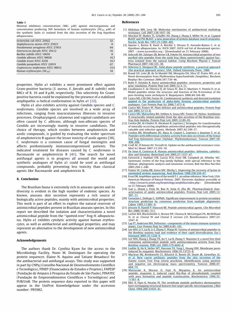
Table 1Minimal inhibitory concentrations (MIC, mM) against microorganisms and
concentration producing 50% hemolysis of human erythrocytes (HC50, mM) of
the synthetic hylin a1 isolated from the skin secretion of the frog Hypsiboas
albopunctatus.
Escherichia coli ATCC 25922 32
Staphylococcus aureus ATCC 25923 8
Pseudomonas aeruginosa ATCC 27853 64
Enterococcus faecalis ATCC 29212 16
Bacillus subtilis ATCC 19659 8
Candida albicans ATCC 90028 16.7
Candida krusei ATCC 6258 16.7
Candida parapsilosis ATCC 22019 67
Cryptococcus neoformans ATCC 90012 33.5
Human erythrocytes (HC50) 18
M.S. Castro et al. / Peptides 30 (2009) 291–296 295
properties. Hylin a1 exhibits a more prominent effect againstGram-positive bacteria (S. aureus, E. faecalis and B. subtilis) withMICs of 8, 16 and 8 mM, respectively. This selectivity for Gram-positive bacteria could be explained by the presence of a stabilizedamphipathic a-helical conformation in hylin a1 [12].
Hylin a1 also exhibits activity against Candida species and C.
neoformans. Candida species are involved in a wide array ofinfections, ranging from mucocutaneous illnesses to invasiveprocesses. Oropharyngeal, cutaneous and vaginal candidiasis areoften caused by C. albicans, although non-albicans species ofCandida are increasingly mainly in invasive candidiasis. Thechoice of therapy, which resides between amphotericin andazolic compounds, is guided by evaluating the wider spectrumof amphotericin B against the lesser toxicity of azole agents [38].C. neoformans is a common cause of fungal meningitis andaffects predominantly immunocompromised patients. Theindicated treatment for diseases caused by C. neoformans isfluconazole or amphotericin B [41]. The search for novelantifungal agents is in progress all around the world andsynthetic analogues of hylin a1 could be used as antifungalcompounds, probably presenting less toxicity than classicalagents like fluconazole and amphotericin B.
4. Conclusion
The Brazilian fauna is extremely rich in anurans species and itsdiversity is evident in the high number of endemic species. Asknown, anurans skin secretion represents a rich source ofbiologically active peptides, mainly with antimicrobial properties.This work is part of an effort to explore the natural reservoir ofantimicrobial peptides present in Brazilian anurans species. In thisreport we described the isolation and characterization a novelantimicrobial peptide from the ‘‘spotted-tree’’ frog H. albopuncta-
tus. Hylin a1 exhibits cytolytic activity against human erythro-cytes, as well as antibacterial and antifungal properties, and mayrepresent an alternative to the development of new antimicrobialagents.
Acknowledgements
The authors thank Dr. Cynthia Kyaw for the access to theMicrobiology Facility, Nuno M. Domingues for operating theprotein sequencer, Elaine N. Aquino and Tatiane Benaducci forthe antibacterial and antifungal assays. This study was supportedin part by CNPq (Conselho Nacional de Desenvolvimento Cientıficoe Tecnologico), FINEP (Financiadora de Estudos e Projetos), FAPESP(Fundacao de Amparo a Pesquisa do Estado de Sao Paulo), FINATEC(Fundacao de Empreendimentos Cientıficos e Tecnologicos) andFUB/UnB. The protein sequence data reported in this paper willappear in the UniProt Knowledgebase under the accessionnumber: P85982.
References
[1] Alekshun MN, Levy SB. Molecular mechanisms of antibacterial multidrugresistance. Cell 2007;128:1037–50.
[2] Altschul SF, Maden TL, Schaffer AA, Zhang J, Zhang Z, Miller W, et al. GappedBLAST and PSI-BLAST: a new generation of protein database search programs.Nucleic Acids Res 1997;25:3389–402.
[3] Aquino L, Bastos R, Kwet A, Reichle S, Silvano D, Azevedo-Ramos C, et al.Hypsiboas albopunctatus. In: IUCN 2007; IUCN red list of threatened species;2007. <www.iucnredlist.org> [Downloaded on 21 February 2008].
[4] Ashcroft JW, Zalinger ZB, Bevier CR, Fekete FA. Antimicrobial properties of twopurified skin peptides from the mink frog (Rana septentrionalis) against bac-teria isolated from the natural habitat. Comp Biochem Physiol C ToxicolPharmacol 2007;146:325–30.
[5] Atherton E, Sheppard RC. Solid phase peptide synthesis: a practical approach(the practical approach series). USA: Oxford University Press; 1989.
[6] Brand GD, Leite JR, de Sa Mandel SM, Mesquita DA, Silva LP, Prates MV, et al.Novel dermaseptins from Phyllomedusa hypochondrialis (Amphibia). BiochemBiophys Res Commun 2006;347:739–46.
[7] Bulet P, Stocklin R. Insect antimicrobial peptides: structures, properties andgene regulation. Protein Pept Lett 2005;12:3–11.
[8] Casallanovo F, de Oliveira FJ, de Souza FC, Ros U, Martınez Y, Penton D, et al.Model peptides mimic the structure and function of the N-terminus of thepore-forming toxin sticholysin II. Biopolymers 2006;84:169–80.
[9] Castro MS, Cilli EM, Fontes W. Combinatorial synthesis and directed evolutionapplied to the production of alpha-helix forming antimicrobial peptidesanalogues. Curr Protein Pept Sci 2006;7:473–8.
[10] Castro MS, Fontes W. Plant defense and antimicrobial peptides. Protein PeptLett 2005;12:13–8.
[11] Castro MS, Matsushita RH, Sebben A, Sousa MV, Fontes W. Hylins: bombininsH structurally related peptides from the skin secretion of the Brazilian tree-frog Hyla biobeba. Protein Pept Lett 2005;12:89–93.
[12] Conlon JM, Al-Ghaferi N, Abraham B, Leprince J. Strategies for transformationof naturally-occurring amphibian antimicrobial peptides into therapeuticallyvaluable anti-infective agents. Methods 2007;42:349–57.
[13] Conlon JM, Woodhams DC, Raza H, Coquet L, Leprince J, Jouenne T, et al.Peptides with differential cytolytic activity from skin secretions of the lemurleaf frog Hylomantis lemur (Hylidae: Phyllomedusinae). Toxicon 2007;50:498–506.
[14] Croft AC, D’Antoni AV, Terzulli SL. Update on the antibacterial resistance crisis.Med Sci Monit 2007;13:103–18.
[15] De Smet K, Contreras R. Human antimicrobial peptides: defensins, cathelici-dins and histatins. Biotechnol Lett 2005;27:1337–47.
[16] Faivovich J, Haddad CFB, Garcia PCA, Frost DR, Campbell JA, Wheller WC.Systematic review of the frog family Hylidae, with special reference to theHylinae: phylogenetic analysis and taxonomic revision. Bull Am Mus Nat Hist2005;294:1–240.
[17] Fontes W, Cunha RB, Sousa MV, Morhy L. Improving the recovery of lysine inautomated protein sequencing. Anal Biochem 1998;258:259–67.
[18] Frost DR. Amphibian species of the world 5.1, an online reference. New York, USA:American Museum of Natural History; 2007. Electronic database accessible at<http://research.amnh.org/herpetology/amphibia/index.php> [Downloadedon 21 February 2008].
[19] Gao L, Zhang J, Feng W, Bao N, Song D, Zhu BC. Pharmacological char-acterisation of spider antimicrobial peptides. Protein Pept Lett 2005;12:507–11.
[20] Geourjon C, Deleage G. SOPMA: Significant improvement in protein secondarystructure prediction by consensus prediction from multiple alignments.Cabios 1995;11:681–4.
[21] Jenssen H, Hamill P, Hancock RE. Peptide antimicrobial agents. Clin MicrobiolRev 2006;19:491–511.
[22] Larkin MA, Blackshields G, Brown NP, Chenna R, McGettigan PA, McWilliamH, et al. Clustal W and Clustal X version 2.0. Bioinformatics 2007;23:2947–8.
[23] Lay FT, Anderson MA. Defensins—components of the innate immune system inplants. Curr Protein Pept Sci 2005;6:85–101.
[24] Lee WH, Li Y, Lai R, Li S, Zhang Y, Wang W. Variety of antimicrobial peptides inthe Bombina maxima toad and evidence of their rapid diversification. Eur JImmunol 2005;35:1220–9.
[25] Lee WH, Zhang J, Zhang YX, Jin Y, Lai R, Zhang Y. Maximin 9, a novel free thiolcontaining antimicrobial peptide with antimycoplasma activity from frogBombina maxima. FEBS Lett 2005;579:4443–8.
[26] Ludtke SJ, He K, Heller WT, Harroun TA, Yang L, Huang HW. Membrane poresinduced by magainin. Biochemistry 1996;35:13723–8.
[27] Maclean MJ, Brinkworth CS, Bilusich D, Bowie JH, Doyle JR, Llewellyn LE,et al. New caerin antibiotic peptides from the skin secretion of theDainty Green Tree Frog Litoria gracilenta. Identification using positiveand negative ion electrospray mass spectrometry. Toxicon 2006;47:664–75.
[28] Matsuzaki K, Murase O, Fujii N, Miyajima K. An antimicrobialpeptide, magainin 2, induced rapid flip-flop of phospholipids coupledwith pore formation and peptide translocation. Biochemistry 1996;35:11361–8.
[29] Mor A, Hani K, Nicolas PJ. The vertebrate peptide antibiotics dermaseptinshave overlapping structural features but target specific microorganisms. J BiolChem 1994;269:31635–41.

M.S. Castro et al. / Peptides 30 (2009) 291–296296
[30] Nascimento A, Chapeaurouge A, Perales J, Sebben A, Sousa MV, Fontes W, et al.Purification, characterization and homology analysis of ocellatin 4, a cytolyticpeptide from the skin secretion of the frog Leptodactylus ocellatus. Toxicon2007;50:1095–104.
[31] Nascimento A, Zanotta LC, Kyaw CM, Schwartz EN, Schwartz CA, Sebben A,et al. Ocellatins: new antimicrobial peptides from the skin secretion of theSouth American frog Leptodactylus ocellatus (Anura: Leptodactylidae). Protein J2004;23:501–8.
[32] Nascimento ACC, Fontes W, Sebben A, Castro MS. Antimicrobial peptides fromanurans skin secretions. Protein Pept Lett 2003;10:227–38.
[33] National Committee for Clinical Laboratory Standards. Reference method forbroth dilution antifungal susceptibility testing of yeast; approved standardM27-A2, 2nd ed., Wayne, PA, USA: National Committee for Clinical LaboratoryStandards; 2002.
[34] Onuma Y, Satake M, Ukena T, Roux J, Chanteau S, Rasolofonirina N, et al.Identification of putative palytoxin as the cause of clupeotoxism. Toxicon1999;37:55–65.
[35] Oren Z, Shai Y. Mode of action of linear amphipathic alpha-helical antimicro-bial peptides. Biopolymers 1998;47:451–63.
[36] Pouny Y, Rapaport D, Mor A, Nicolas P, Shai Y. Interaction of antimicrobialdermaseptin and its fluorescently labeled analogues with phospholipid mem-branes. Biochemistry 1992;31:12416–23.
[37] Pukala TL, Bowie JH, Maselli VM, Musgrave IF, Tyler MJ. Host-defence peptidesfrom the glandular secretions of amphibians: structure and activity. Nat ProdRep 2006;23:368–93.
[38] Rex JH, Walsh TJ, Sobel JD, Filler SG, Pappas PG, Dismukes WE, et al.Practice guidelines for the treatment of candidiasis. Clin Infect Dis 2000;30:662–78.
[39] Ribeiro RS, Tabosa-do-Egito GTB, Haddad CFB. Chave de identificacao: anfıbiosanuros da vertente de Jundiaı da Serra do Japi, Estado de Sao Paulo. BiotaNeotropica 2005;5(2):1–15.
[40] Rinaldi AC. Antimicrobial peptides from amphibian skin: an expanding sce-nario. Curr Opin Chem Biol 2002;6:799–804.
[41] Saag MS, Graybill RJ, Larsen RA, Pappas PG, Perfect JR, Powderly WG, et al.Practice guidelines for the management of cryptococcal disease. Clin Infect Dis2000;30:710–8.
[42] Schiffer M, Edmundson AB. Use of helical wheels to represent the structures ofproteins and to identify segments with helical potential. Biophys J 1967;7:121–35.
[43] Shai Y. Mode of action of membrane active antimicrobial peptides. Biopoly-mers 2002;66:236–48.
[44] Shalev DE, Mor A, Kustanovich I. Structural consequences of carboxyamidationof dermaseptin S3. Biochemistry 2002;41:7312–7.
[45] Simmaco M, Mignogna G, Barra D. Antimicrobial peptides from amphibianskin: what do they tell us? Biopolymers 1998;47:435–50.
[46] Sun X, Chen S, Li S, Yan H, Fan Y, Mi H. Deletion of two C-terminal Gln residuesof 12–26-residue fragment of melittin improves its antimicrobial activity.Peptides 2005;26:369–75.
[47] Zasloff M. Antimicrobial peptides of multicellular organisms. Nature 2002;415:389–95.