Role of Ti/CD3, Thy1, and Ly6 in Cytolytic T-Cell Activation Analyzed with Ti Loss Variants
11
Role of Ti/CD3, Thy-1, and Ly-6 in Cytolytic T-cell Activation Analyzed with Ti Loss Variants" CLAIRE LANGLET," ANNICK GUIMEZANES," PIERRE KALDY," CLAUDE BOYER,h MICHEL BUFERNE,* MARTIN POENIE,' ROGER TSIEN,' OBERDAN LEO,d JEFFREY BLUESTONE, LEE LESERMAN.h AND "Centre d'lrnmicnologie INSERM-CNRS de Marseille-Lirminy 13288 Marseille Cede* 9, Frcince Department of Physiology University of Cnlifornia Berkeley, California "lmmirnology Branch National Cancer iristiticte National institirtes of Health Bethesdci, Mcirylcind 20892 ANNE-MARIE SCHMITT-VERHULST~ INTRODUCTION Cytolytic T cells (CTL) are among the best studied elements of the immune system because of the development of reagents permitting analysis of the molecu- lar basis of their action, and the sensitivity of target cell lysis assays. The use of T- cell growth factor (interleukin-2 (IL-2))has allowed the maintenance in long-term culture of CTL clones with defined antigen specificity and stable function. Mono- clonal antibodies (mAb) against human and mouse CTL helped define the antigen receptor and the associated CD3 (T3) structures6.' involved in antigen- dependent activation of the CTL. Similarly, antigen-independent activation path- ways such as CD2 (T11)8 and T449,i0 in humans, and Thy-l"-'4 and L Y - ~ ' ~ - ~ * in mice are also known. Cell-surface structures such as LFA-I are involved in cell adhesion, in a TUCD3-independent fashion.I9 The CD8 (T8, Lyt-2) and CD4 (T4) structures, generally expressed, respectively, on CTL specific for class I major histocompatibility complex (MHC) products, and on T-helper cells (Th) specific for class I1 MHC products, appear implicated in recognitionlactivation involving MHC, either alone or in association with a non-MHC antigen (for review, see references 20 and 21). We have studied a mouse CTL clone specific for the H-2Kb alloantigen, which is inhibited by Lyt-2-specific mAb in its H-2Kb-mediated activation, but not in activation involving anti-Ti mAb.5 We recently demonstrated, by gene transfec- tion experiments, that inhibition of this clone observed with mAb correlated with the requirement for expression of the Lyt-2 structure in recognition/activation This research was supported by institutional Grants from INSERM and CNRS. 33
Transcript of Role of Ti/CD3, Thy1, and Ly6 in Cytolytic T-Cell Activation Analyzed with Ti Loss Variants

Role of Ti/CD3, Thy-1, and Ly-6 in Cytolytic T-cell Activation Analyzed with
Ti Loss Variants" CLAIRE LANGLET," ANNICK GUIMEZANES,"
PIERRE KALDY," CLAUDE BOYER,h MICHEL BUFERNE,* MARTIN POENIE,' ROGER TSIEN,'
OBERDAN LEO,d JEFFREY BLUESTONE, LEE LESERMAN.h AND
"Centre d'lrnmicnologie INSERM-CNRS de Marseille-Lirminy 13288 Marseille Cede* 9, Frcince
Department of Physiology University of Cnlifornia
Berkeley, California "lmmirnology Branch
National Cancer iristiticte National institirtes of Health
Bethesdci, Mcirylcind 20892
ANNE-MARIE SCHMITT-VERHULST~
INTRODUCTION
Cytolytic T cells (CTL) are among the best studied elements of the immune system because of the development of reagents permitting analysis of the molecu- lar basis of their action, and the sensitivity of target cell lysis assays. The use of T- cell growth factor (interleukin-2 (IL-2)) has allowed the maintenance in long-term culture of CTL clones with defined antigen specificity and stable function. Mono- clonal antibodies (mAb) against human and mouse CTL helped define the antigen receptor and the associated CD3 (T3) structures6.' involved in antigen- dependent activation of the CTL. Similarly, antigen-independent activation path- ways such as CD2 (T11)8 and T449,i0 in humans, and Thy-l"-'4 and L Y - ~ ' ~ - ~ * in mice are also known. Cell-surface structures such as LFA-I are involved in cell adhesion, in a TUCD3-independent fashion.I9 The CD8 (T8, Lyt-2) and CD4 (T4) structures, generally expressed, respectively, on CTL specific for class I major histocompatibility complex (MHC) products, and on T-helper cells (Th) specific for class I1 MHC products, appear implicated in recognitionlactivation involving MHC, either alone or in association with a non-MHC antigen (for review, see references 20 and 21).
We have studied a mouse CTL clone specific for the H-2Kb alloantigen, which is inhibited by Lyt-2-specific mAb in its H-2Kb-mediated activation, but not in activation involving anti-Ti mAb.5 We recently demonstrated, by gene transfec- tion experiments, that inhibition of this clone observed with mAb correlated with the requirement for expression of the Lyt-2 structure in recognition/activation
This research was supported by institutional Grants from INSERM and CNRS. 33
34 ANNALS NEW YORK ACADEMY OF SCIENCES
involving the class I MHC product, but not in recognitioniactivation by way of anti-Ti mhb.'?
Data presented in this publication are aimed at defining the role of the TilCD3, Thy-1. and Ly-6 structures of mouse CTL clones for two measurable activation- dependent effector functions: target-cell killing and production of g-interferon (g- I F N ) . The killing function ic not dependent on protein synthesis and can be measured within minutes of CTL-target-cell interaction. as recently shown using digital imaging of changes in intracytoplasmic C a ' ~ concentration ([Ca- +] i ) in effector and target cell4 loaded with the Ca ' ' sensitive dye Fura-2.?7 g-IFN syn- thesis is dependent on transcriptional activation and is optimally measured in supernatants around 2 0 h after C'I'L-target-cell interaction or after binding of anti-Ti mAb. We previously showed that activation for killing and activation for g- IFN production were obtained in conditions leading to phosphorylation of distinct CD3 component\." Here. the role of the TilCD3 complex in activation mediated by lectins or by [wo phosphatidylinositol-~inchored differentiation antigens, Thy-1:' and L Y - ~ . ' ~ u.ii\i evaluated on normal cells and on variants of a CTL clone, which had lost cell-surface expression of the TilCD3 complex." Results obtained with a series of T - variants indicate that absence o f the Ti o( or /.3 chain prevents surface espre\sion of the TiiCD3 complex and that Ti variants could no longer be activated either for killing or g-IFN production with lectins. anti-Thy-l or anti- Ly-6 mAh. These results raise the following questions: What is the molecular basis for the dependence of activation mediated by Thy-I and Ly-6 upon surface expre5sion of the Ti/CD3 complex'! Is this dependence specific for activation by phosphatidylinositol nicnibi-ane~anchored structures? Is this dependence absolute or could the \ame ~ u c t u r e s signal in a TiiCD3-independent fashion on immature precursor\ of C7'L and/or on cells of a distinct lineage'.' Experimental results will be tliscus\ed that give some clues as to the response to the first question, whereas the second and third questions remain unans\\wed as yet. to the best of our knowledge.
MATERIAL AND METHODS
T-cell Clotic~: Origiri r i d Cu/tuw Cotidi~iotis
KI3.F.CN is an H-2Kh-specific CTL clone of B 10.BR origin,'x maintained in culture by weekly restimulation with irradiated C57BLlh spleen cells and IL-7- containing supernatant from phorbol myristic acetate (PMAk-stimulated EL4.c 16 cells (SCIh).'X 2y
Se/cc,r ioi i c ? f Ti- L'(iiY(it i / .r of Clotic KBS.C'Z0
Negative selection using methotrexate (M?'X)-containing. protein A-coupled liposomes (MTX-lip-PA) was adapted from Machy and Leserman3" as de- scribed." Variant KB5.C20.lip.A.7 was obtained from normal KBS.C20 cells. whereas variants TiC.x were selected from KBS.CZ0 cells that had previously been treated with the mutagen ethyl methyl sulfonate (EMS) at 400 pgiml. As the TiC variants can no longer respond to antigen btimulation. their maintenance involves. every other week. a 7 h activation step with ionomycin (iono), 1 p M . and PMA. 1.6 nM, at 37°C. followed by replacement of the medium with 10% SC16 as described."
LANGLET et a/.: ROLE OF TilCD3 35
Rerigetits
Reagents used were ionomycin (Calbiochem), PMA (Sigma), EMS (Sigma), and acetoxymethyl ester of Fura-2 (Molecular Probes).
Motloclonal Antihotiies
The characteristics and origin of the mAb used in this study are summarized in TABLE I .
g-IFN was measured either by its macrophage-activating factor (MAF) activ- ity as previously described,j? or by a solid phase radioimmunoassay using two mouse g-IFN-specific hamster mAb. kindly given by Dr. R. D. Schreiber (Wash- ington University, St Louis, MO) adapted from Schreiber et cil .??
TABLE 1. Specificity and Origin of Monoclonal Antibodies Used mAb
Desire-l H155.124.3 H140.61.2 H129.93.9. I 145.2c.11 143.4.2 19.178 100.5.28 20.8.4
Specificity
Ti (KB5.C20) Thy-l(epitope C ) Thy-l(epitope B ) Thy-l(epitope A ) CD3 ( E )
L y - 6 . X Lyt-2.2 H-2Kk H-2Kh
Reference
Hua et ril. 1986a" Pont et ti / . 1985" Pont et t r l . 198S'j Pont et t r l . 1985'? Leo et t r l . 1987' Leo c't ti / . 1987" Hammerling rt tr / . 1979' Lemke c't cil. 1979" Ozato and Sachs 1981'"
Measirre of CTL Activity
T-cell clones were incubated in V-bottomed microtitre plates with 104 "Cr- labeled target cells as described.28 'ICr released in the supernatants of triplicate cultures was measured after a 4 h or 16 h incubation at 37°C as indicated in the TABLES. Percent specific ''Cr released was expressed as [cpm (experimental) - cpm (medium)/cpm maximum ( I N HCI) - cpm (medium)] x 100. Target cells were either H-2h (EL4.BU) o r H-2k (RDM4) thymomas, o r the H-2' (P388DI) Fc receptor-positive tumor line.
Cytoflirorot?ietric Atid?sis
Immunofluorescence was performed as previously described,3J except that the fluorescein isothiocyanate (F1TC)-labeled reagents were either F(ab')? rabbit anti- mouse Ig, F(ab')? goat anti-rat Ig. o r goat anti-hamster Ig (all from Cappel), depending on the origin of the first antibody (TABLE I ) . Results are expressed as relative fluorescence intensities as described.3J

36 ANNALS NEW YORK ACADEMY OF SCIENCES
[Ca- ]i was measured using 5ingle-cell analysis and digital-image processing of cells loaded with the acetoxymethyl form of the Ca*+-sensitive dye, Fura-2 (Molecular Probes). as previously dew-ibed.r4 27
RESULTS AND DISCUSSION
Approaches to the Understanding of the Role of Distinct Cell-Surjace Structures in the Activation of CTL Effector Functions
Srlet.tioii o f Vtir-iriiir.s of C T L Clotir K 3 5 . C X
KB5.C20 is a CTL clone of BIO.BR origin (H-2'), specific for the H-2Kh alloantigen.?8 I t is dependent o n antigenic stimulation for efficient IL-2-depen- dent growth?* and for activation of its effector functions, target-cell killing and production of g-IFN . 3 > These antigen-dependent triggering events can be replaced by an anticlonotypic mAb. Desire-l,5,34.32 or by a pulse with a C a + + ionophore such as ionomycin and a protein kinase C activator such as PMA.3' Selection of variants was based on a negative selection method eliminating cells that bind and internalize liposomes containing the drug methotrexate. specifically targeted to a cell-surface molecule by way of a mAb? Use of the anticlonotypic mAb, Desire- 1, in such a selection on unmutagenized or EMS-treated KB5.C20 cells. led to the isolation of variant cells called KB5.C20.1ip.A.727 and Ti~-.x (this report), respec- tively. To culture these variants. antigen-5pecific stimulation was replaced by a pulse with ionomycin ( 1 pM) and PMA ( 1.6 nMI3' every other week, whereas IL- 2-containing medium was supplied every 3 or 4 days. We will only consider here variants that have selectively lost cell-surfdce expression of the Ti and associated CD3 structures (TABLE 2) . Among these. some have lost expression of mRNA for the a chain of the T-cell receptor." whereas the defects of others (for example variants Ti-.32. Ti-.33. and Ti-.37) are presently being characterized at the mo- lecular level (P. Kaldy rt ( I / . , in preparation).
TABLE 2. Selective Loss of Ti and CD3 Expression on Ti Clone KBS.C20
Variants of
Relative Fluorescence Intensity after Staining with mAb Specific for:
CTL Clone Kb Kk Ti(KB5.CXP Lyt-2.2" Ly-6.2C -h Thy-lh -l CD3c
KB5.CZO 3.6 200." 100. 116. 33. 0. 99. 0. 55. Ti-.32 3.7 160. 3.8 64. 22. 0. 84. 0. 0. Ti-.33 7.8 185. 6.6 51. 29. 0. 84. 0. 1.3 Ti-.37 5.4 139. 5 . 8 42. 45. 0. 81. 0. 0.
F(ab'): rabbit anti-mouse Ig-FLTC. F(ab'): goat anti-rat Ig-FITC.
' Godl anti-hamster Ig-FITC. if Bold numbers indicate positive expression of the corresponding marker.
LANGLET el al.: ROLE OF TilCD3 37
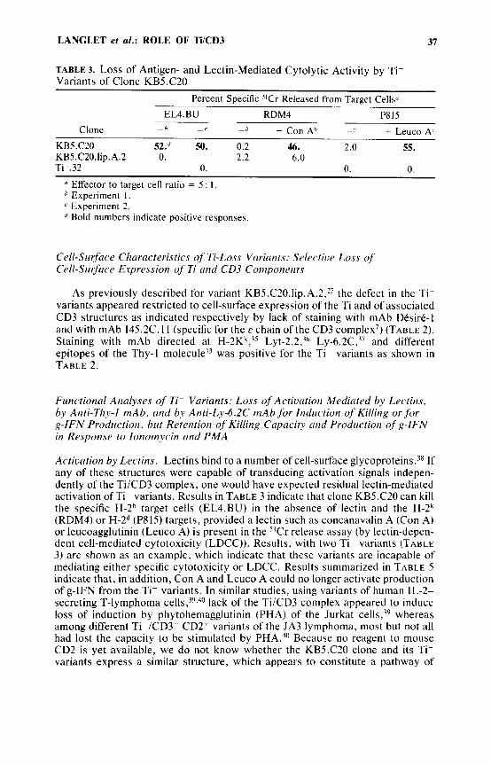
TABLE 3. Loss of Antigen- and Lectin-Mediated Cytolytic Activity by Ti- Variants of Clone KBS.C20
Percent Specific "Cr Released from Target Cellsi
EL4.BU RDM4 P815 + Leuco AL Clone - h - < _ h + Con Ah - c
KB5.C20 52." 50. 0.2 46. 2.0 55. KB5.C20.lip.A.2 0. 2.2 6.0 Ti-.32 0. n. n
Effector to target cell ratio = 5 : I . Experiment 1. Experiment 2. Bold numbers indicate positive responses.
Cell-Sirrface Characteristics of Ti-Loss Vriricints: Selective Loss of Cell-Sirrface Espression of Ti m d CD3 Coriiporierits
As previously described for variant KB5.C20.1ip.A.2,27 the defect in the Ti- variants appeared restricted to cell-surface expression of the Ti and of associated CD3 structures as indicated respectively by lack of staining with mAb Desire-I and with mAb 145.2C. 1 1 (specific for the E chain of the CD3 complex') (TABLE 2). Staining with mAb directed at H-2Kk,j5 L ~ t - 2 . 2 , ~ ~ Ly-6.2C,j7 and different epitopes of the Thy-1 moleculel3 was positive for the Ti- variants as shown in TABLE 2.
Firnctional Anrilyses of Ti- Variants: Loss of Actiurition Mediated by Lectins, by Anti-Thy-I mAb, rind by Anti-Ly-6.2C riiAb for Indirction of Killing or f o r g-IFN Prodirctiori, but Retention of Killing Capricitv rind Prodirctioti of g-IFN in Response to Iotioniycin and PMA
Activation by Lectins. Lectins bind to a number of cell-surface glycoproteins.?* If any of these structures were capable of transducing activation signals indepen- dently of the TilCD3 complex, one would have expected residual lectin-mediated activation of Ti- variants. Results in TABLE 3 indicate that clone KBS.C20 can kill the specific H-2" target cells (EL4.BU) in the absence of lectin and the H-2k (RDM4) or H-2d (PSIS) targets, provided a lectin such as concanavalin A (Con A) or leucoagglutinin (Leuco A) is present in the "Cr release assay (by lectin-depen- dent cell-mediated cytotoxicity (LDCC)). Results, with two Ti- variants (TABLE 3 ) are shown as an example, which indicate that these variants are incapable of mediating either specific cytotoxicity or LDCC. Results summarized in TABLE 5 indicate that, in addition, Con A and Leuco A could no longer activate production of g-IFN from the Ti- variants. In similar studies, using variants of human IL-2- secreting T-lymphoma lack of the Ti/CD3 complex appeared to induce loss of induction by phytohemagglutinin (PHA) of the Jurkat cells,39 whereas among different TiClCD3- CD2+ variants of the JA3 lymphoma, most but not all had lost the capacity to be stimulated by PHA.40 Because no reagent to mouse CD2 is yet available, we do not know whether the KBS.C20 clone and its Ti- variants express a similar structure, which appears to constitute a pathway of
38 ANN.ALS NEW YORK ACADEMY OF SCIENCES
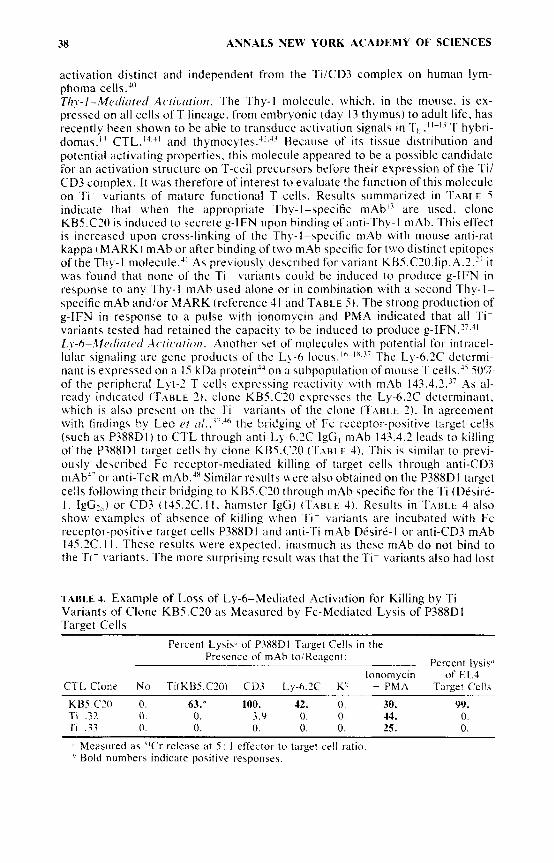
activation distinct and independent from the TilCD3 complex on human lym- phoma cells.'" T/I?.-I-M~,ditrtrcl ilctiuntiori. The Thy-l molecule. which. in the mouse, is ex- pressed on all cells of T lineage. from embryonic (day 13 thymus) to adult life. has recently been shown to be able to transduce activation signals in Th . ' ' - I 3 T hybri- domas." CTL."." and thymocytes.-".J7 Because of its tissue distrihution and potential activating properties. this niolecule appeared to be a possible candidate for an activation structure on 'T-cell precursors before their evprrssion of the Ti/ CD3 complex. I t was therefore of interest to evaluate thc function of this molecule on Ti variants of mature functional T cells. Results summarized in TABLE 5 indicate that when the appropriate Thy-I-specific mAb" are used, clone KH5.C2O i F induced to cecrete g-IFN upon binding of anti-Thy-l mAb. 'This effect is increased upon cross-linking of the Thy-]-specific mAb with mouse anti-rat kappa ( M A R K ) mAb or after binding of two mAb specific for two distinct epitopes of the Thy-1 molecule.'l As previously described for variant KB5.C20.lip.A.2," it v,'a\ found that none of the Ti- variants co~i ld be induced to produce g-IFN in response to any Thy-I mAb used alone or in combination with a second Thy-I- specific mAb andlor MARK (reference 41 and TABLE 5 ) . The strong production of g-IFN in response to a pulse with ionomycin and PMA indicated that all Ti- variants tested had retained the capacity to be induced to produce g-IFN.27.'' L!-rS-.ZlcclitrtPtI Actirtrtioti. Another set of molecules with potential for intracel- lular signaling are gene products of the Ly-6 locus. ' h - I X . 3 i The Ly-6.2C determi- nant is e x p t ~ s c e d on a 15 kDa protein'" on ;i subpopulation of mouse T cells,J' 50% of the peripheral Lyt-2 T cells exprr4sing rriictivit), w i th mAb 143.4.2." As al- read!, indicated ( ' T A H L E 2) . clone KBS.C?O evpresseh the L y - 6 . X determinant, which is also present on the Ti variants of the clone (Tt\Ri F. 2 ) . I n agreement with findings by Leo t'f o/..'-.'~ the bridging of Fc receptor-positive target cells (such as P388DI) to CTL through anti-Ly-6.2C I g G niAb 143.3.1 leads to killing of the P3XXDl target cells by clone KB5.C2O (TAHI E 4). This is similar to previ- ously described Fc receptor-mediated killing of target cells through anti-CD3 mAb'- or anti-TcR mAb.l' Similar results were also obtained on the P3XXDI target cells following their bridging to KB5.C20 through inAh specific for the Ti (Desire- I . I&?,,) or CD3 (145.2C.ll. ham5ter IgG) ( T : \ B L E 4). Results in 'I'ABLL 4 also show esamples of absence of killing Lvhen 'fit variants are incubated with Fc receptor-positive target cells P388DI and anti-Ti mAb DCsire-l or anti-CD3 mAb 145.2C. I I . These results were expected. inasmuch a s these mAb d o not bind to the Ti- variants. The more surprising result was that the T - variants also had lost
TABLE 4. Example of Loss of Ly-6-Mediated Activation for Killing by Ti- Variants of Clone KB5.C20 as Measured by Fc-Mediated Lysis of P3X8DI Target Cells
Percent Lysis" of P3XXDI Target Cells in the Pre\ence of mAb to;Reagent: Percent lysisc'
CTL Clone N o Ti(KB5.CX) CD3 Ly-6 .X K h + PMA Target Cells lonomycin of EL4
K B 5 . C N 0. 63.* 100. 42. 0. 30. 99. Ti .32 0 . 0. 3.9 0. 0. 44. 0. Ti ~ .33 0. 0. 0. 0. 0. 25. 0.
" Measured as ''Cr release at 5 : 1 effector to target cell ]ratio '' Bold numbers indicate positive response$.

LANGLET et al.: ROLE OF TilCD3 39
TABLE 5. Summary of Stimuli Leading to Activation for Killing, g-IFN Production, or Increase in [Ca'+]i by Clone KBS.C2O (Ti+) and Antigen Receptor Loss Variants (Ti-)
Target g-IFN Increase in Cell LysIs Production [Ca++]i by Clone by Clone by Clone KB5.CZO KBS.C20 KB5.C20
Stimuli Ti- Ti- Ti+ Ti- Ti+ Ti ~
- + H-2Kb cells I + Concanavalin A +
Leucoagglutinin + N D N D + anti-Ti (DCsire-I) I
anti-CD3 i - ND ND + + anti-Thy-l/C N D ' N D + + anti-Ly-6.2C +
PMA + + - ND ND Ionom ycin - ( + I - ( + I - -(+) + + PMA + ionomycin + i + + ND N D
- + + + I
- - - -
- -
- - -
-
- -
- - + -
-
ND = not done.
the capacity to kill the P388D1 target cells through bridging by the anti-Ly-6.2C mAb 143.4.2. (TABLE 4), although PMA-inducedZ4 or iono plus PMA-induced target-cell killing was as efficient for the Ti- variants as for clone KBS.C20 (TA- BLE 4). Results summarized in TABLE 5 indicate that anti-Ly-6.2C mAb also can induce production of g-IFN by clone KBS.C20, but not by the Ti- variants (Guimezanes et al. in preparation).
CONCLUSION
In agreement with recent studies using human Jurkat cells transfected with the murine Thy-1.2 gene,49 our results using Ti- variants of a CTL clone had shown that Thy-1-mediated activation is dependent on the expression of the Ti/CD3 complex on mature T cells.?7 Recent work on the stimulation of subpopulations of thymocytes is also compatible with the requirement of CD3 expression associated with a Ti(cu/P) or with a Ti(y/G) for activation of immature t h y m o c y t e ~ . ~ ~ It re- mains to be established whether, as suggested for human thyrnocyte~~' and lyrn- phoma cells,40 a CD2 structure is present and can function as an alternative to the Ti/CD3 system for immature or functional murine T cells. Alternatively, as yet unknown molecules may be involved in signaling in T-cell precursors before their expression of a Ti/CD3 complex. Results presented here indicate that, in addition to the Thy-1 molecule, Ly-6C-mediated activation is similarly dependent on the expression of the Ti/CD3 complex and, therefore, suggest that molecules of the Ly-6 family are not likely candidates for early. CD3-independent activation struc- tures.
The question of the mechanism of signaling through the Thy-1 and Ly-6 mole- cules, both of which are probably phosphatidylinositol membrane-anchored structure^,^^.^^ and on the nature of its dependence on elements associated with the Ti/CD3 complex remains to be investigated. Our results have indicated that the Ca++ influx, which can be measured after binding of activating mAb to either

40 ANNALS NEW YORK ACADEMY OF SCIENCES
the Thy-l?’ or the Ly-6c ( T A B L E 5 ) molecules of clone KBZ.C?O, is no longer observed when Ti- variants of the clone are studied (reference 37 and TABL.E 5). If Ca- ’ influx is dependent on a product generated after hydrolysis of phosphatidyl- inositol bisphosphatr. as recently suggested (for review. see reference 5 2 ) . the Thy-I and Ly-6 activation pathways could depend on an element leading to acti- vation of phospholipase C on the general pathway of protein kinasc C-dcpcndent signaling.”
ACKNOWLEDGMENT
We thank M. Pierres for antibodies and discussion.
[NOTE ADDED ih PRnoF: Northern blot analyses of Ti- variants used in this study revealed that they had all lost expression of mRNA for the a chain of the Ti (P. Kaldy. unpublished).]
REFERENCES
I. K f i N H m z . E. L.. S. C. M E P E R & S. F. SCHLOSSM+N. 1983. The human T cell recep- tor: Analysis with cytolytic 7’ cell clone\. Immunol. Kev. 74: 83-1 12.
1984. Immunoprecipitation of cell surface structures of cloned cytotoxic T lympho- cytes by clone-specific antisera. Proc. Natl. Acad. Sci. USA 81: 573-577.
L A N C K I . D. W.. 0. 1. M A . W. 1. HAL‘RAN & F. W . FITCH. 1984. Cell surface struc- tures involved in T cell activation. Imniunol. Rev. 81: 65-94.
1984. Monoclonal antibodies spccific for a inurine cytotoxic ’I lymphocyte clone. Proc. Natl. Acad. Sci. L‘S.4 81: 1799-1803.
Hu.4. C.. M. B L ~ F E R N E & i\.-M. SCHMITT-VERHLILST. 1985. Lysis of hybridoma cells bearing anti-clonotypic w f ’ x e immunoglobulin by clonotype-expressing alloreac- tive cytotoxic ‘1 cells. Eur . J . Immunol. 15: 1029-1032.
MEuFR. S. C.. K. A. FITZGERALD. R. E . HLTSSEY. J . C. HODGDON. S. F. SCHLOSSMAN & E . I,. R F I N H F R Z . 1983. Clonotypic structures involved in antigen-specific human T cell function. Kelation\hip to the 1.3 molecular complex. J . Exp. Med. 157: 705- 719.
Lco. 0.. M. Foo. D. H . SACHS. L. E. SAMFI~SON & J . A. BLUESTONE. 1987. Identifica-
2. KRAIUZ. D. hi. . D. H. S H E R M A N . M. V. SITOVSRY. M. S. PASTERNACR & H . N . EISEN.
3.
3 . SlAEKZ. U . D. . M. S. P.ASTERN.AC.K. J . R. K L F I N . J . D. BENEDETTO & M. J . BEVAN.
5 .
6.
7 . tion ofa monoclonal antibody specific for inurine T3. Proc. Natl. Acad. Sci. USA 84: 1374-1 37x.
8. MFLIFR. S. C.. R. E . HL‘SSEY. M. F.ABBI. D. Fox. 0. ACLJTO. K. A. E‘ITZGERALD, J . C. HODGDON. J . P. PROTENTIS. S. k-. ScHi.osSxiAN & E. L. KEINHERZ. 1984. An alternative path\bay of T-cell acrivation: a functional I-ole for the 50 K D TI 1 sheep erythrocyte receptor protein. Cell 36: 897-906.
9. H ~ R A . T.. S. M. F u B( J . A . H.ANSEN. 1985. Human T cell activation. 11. A new activation pathway u\ed bq a major T cell population via a disulfide-bonded dimer of a 44 K D polypeptide (9.3 antigen). J . Exp. Med. 161: 1513-1524.
MORETTA. A . . G . PANTAI.FO. M. LOPEZ-HOTET B( I.. MORETTA. 1985. Involvement of the T44 molecules in an antigen-independent pathway of T cell activation. Analysis of the correlations t o the T cell antigen-receptor cornple\. J . Exp. Med. 162: 823- 83X.
KONAKA. Y. . M. ‘4. NORCROSS. V . C. MAIN^ & R. T. S%iirri. 1981. Anti-Thy-l mediated T cell activation. Role of soluble factors and expression of interleukin-’ receptors on T cellb. Eur. J . Imniunol. 11: 435-450.
GUNTER. K . C.. T. R. MALEK & E. M. SHEVACH. 1984. T cell-activating properties of an anti-Thy-I monoclonal antibody. J . Eup. Med. 159: 716-730.
10.
I I .
12.
LANGLET et a/.: ROLE OF TilCD3 41
13.
14.
IS.
16.
17.
18.
19.
20. 21.
22.
23.
24.
25.
26.
27.
28.
29.
30.
31.
PONT, S . , A. R~GNIER-VIGOUROUX. P. NAQUET, D. BLANC, A. PIERRES, S. MAR- CHETTO & M. PIERRES. 1985. Analysis of the Thy-I pathway of T cell hybridoma activation using 17 rat monoclonal antibodies reactive with distinct Thy-] epitopes. Eur. J. Immunol. 15: 1222-1228.
1985. Production and characterization of monoclonal anti-Thy-1 antibodies that stim- ulate lymphokine production by cytolytic T cell clones. Eur. J . Immunol. 15: 495- 501.
ROCK, K. L. , E. T. H . YEH. C. F. GRAMM, S. I . HABER, H. REISER & B. BENACER- RAF. 1986. TAP, a novel T cell-activating protein involved in the stimulation of MHC-restricted T lymphocytes. J . Exp. Med. 163: 315333.
REISER, H. , E . T. H. YEH, C. F. GRAMM, B. BENACERRAF & K. L. ROCK. 1986. Gene encoding T-cell-activating protein TAP maps to the Ly-6 locus. Proc. Natl. Acad. Sci. USA 83: 2954-2958.
MALEK. T. R.. G. ORTEGA. C. CHAN, R. A. KROCZEK & E. M. SHEVACH. 1986. Role of Ly-6 in lymphocyte activation. 11. Induction of T cell activation by monoclonal anti-Ly-6 antibodies. J . Exp. Med. 164: 709-722.
DUMONT, F. 1987. Ly-6C-mediated T cell activation in normal, /ppr/lpr and g/d/g/d mice. J . Immunol. 138: 4106-4113.
SANDERS. 1986. Two antigen-independent adhesion pathways used by human cyto- toxic T cell clones. Nature (London) 323: 262-264.
MACDONALD, H . R. , c. BRON, M. ROUSSEALJX. c. HORVATH & J . c. CEROTTINI.
SHAW. S . . G. E. GINTHER LUCE, R. QUINONES, R. E. GRESS. T. A. SPRINGER & D. E.
MOLLER, G., Ed . 1982. Immunol. Rev. 68: 1-218. BURAKOFF. S. J. , organizer, 16'h Forum in Immunol. 1987. The role of the CD4(L3T4)
molecule in T cell activation. Ann. Inst. PasteuriImmunol. 138: 125-169. GABERT. J. , C. LANGLET, R. ZAMOYSKA, J . R. PARNES. A.-M. SCHMITT-VERHULST &
B. MALISSEN. 1987. Reconstitution of MHC-class 1 specificity by T-cell receptor and Lyt-2 gene transfer. Cell. 50: 545-554.
POENIE. M . , R. Y. TSIEN & A.-M. SCHMITT-VERHULST. 1987. Sequential activation and lethal hit measured by [Ca-+]i in individual cytolytic T cells and targets. EMBO J. 6: 2223-2232.
BOYER. C.. C. LANGLET. A. GUIMEZANES. M. BUFERNE. C. HUA & A:M. SCHMITT- VERHULST. 1987. Phosphorylation of T-cell antigen receptor-associated proteins: correlation with activation for killing and/or for gamma-interferon production by a cytolytic T-cell clone. Ann. Inst. Pasteur/lmmunol. 138: 65-82.
TSE, A. G. D., A. N . BARCLAY. A. WATTS & A. WILLIAMS. 1985. A glycophospholipid tail at the carboxyl terminus of the Thy-I glycoprotein of neurons and thymocytes. Science 230: 1003-1008.
REISER. H. . H . OETTGEN, E. T. H . YEH, C. TERHORST. M. G. L o w , B. BENACERRAF & K. I,. ROCK. 1986. Structural characterization of the TAP molecule: a phosphati- dylinositol-linked glycoprotein distinct from the T cell receptoriT3 complex and Thy-] . Cell 47: 365-370.
M. BUFERNE, C. H U A & L. D. LESERMAN. 1987. Pleiotropic loss of activation pathways in a T-cell receptor a-chain deletion variant of a cytolytic T-cell clone. Nature (London) 325: 628-631.
ALBERT, F.. M. BUFERNE. C. BOYER & A.-M. SCHMITT-VERHULST. 1982. Interactions between MHC-encoded products and cloned T-cells. I. Fine specificity of induction of proliferation and lysis. lmmunogenetics 16: 533-549.
ALBERT. F.. C. BOYER. M. BUFERNE & A.-M. SCHMITT-VERHULST. 1984. Interactions between MHC-encoded products and cloned T-cells. 11. Analysis of physiological requirements indicates two different pathways of stimulation by class I alloantigens. lmmunogenetics 19: 279-294.
MACHY, P. & L. D . LESERMAN. 1984. Elimination or rescue of cells in culture by specifically targeted liposomes containing methotrexate or formyl-tetrahydrofolate. EMBO J. 3: 1971-1977.
Distinction between antigen receptor and 11-2 receptor triggering events in the activa-
SCHMITT-VERHULST, A.-M., A. GUIMEZANES, C. BOYER, M. POENIE, R. Y. TSIEN,
ALBERT, F., c . H U A . A. TRUNEH, M. PlERRES & A.-M. SCHMITT-VERHULST. 1985.
42 ANNALS NEW YORK ACADEMY OF SClENCES
3 2 .
33.
34.
35.
36.
37.
38.
39.
40.
41
42.
33.
44.
45.
46.
41.
48.
49.
tion of alloreactive T-cell clones with calcium ionophore and phorbol esters. J . lmmunol. 134: 3649-3655.
HUA. C.. C. BOYER. A. GUIMEZANES. F. ALBERT & A.-M. SCHMITT-~ERHUI.ST. 1986b. Analysis of T cell activation requirements with the use of alloantigens or anti- clonotypic monoclonal antibody. J . Immunol. 136: 1927-1936.
SCHREIBER. R. D.. L. J . HICKS. A. CEI-ADA. N. A. BUCHMEIER & P. W. GRAY. 1985. Monoclonal antibodies to murine gamma-interferon which differentially modulate macrophage activation and anti-viral activity. J . Immunol. 134: 1609-1618.
HLA. C.. C . BOYER. M. BUFERNE & A.-M. SCHMITT-VERHUI.ST. 1986a. Monoclonal antibodies against a n H-2Kh-specific cytotoxic T cell clone detect several clone- specific molecules. J . Immunol. 136: 1937-1944.
LFMKL. H.. G. J . HAhIS1ERI.ING & U . H~XISIERI . INC. 1979. Fine specificity analysis with monoclonal antibodies of antigens controlled by the major histocompatibility complex and by the Qa/TL region in mice. Immunol. Rev. 47: 175-306.
HAMMERI I N G . G . J . . U. HAMMERLING & L. FLAHERT'I'. 1979. Qat-4 and Qat-5. new murine T-cell antigens governed by the Tla region and identified by monoclonal antibodieb. J . Exp. Med. 150: 108-1 16.
LEO. 0.. M. Foo. D. M. SEGAI.. E . SHEVACH & J . A. BLUESTONE. 1987. Activation of murine T lymphocytes with monoclonal antibodies: detection on Lyt-2' cells of an activation antigen not associated w.ith the T cell receptor complex. J . Immunol. 139: I 2 14-1292.
S I T K ~ V S K ) . . M. V . . M. S. PASTERNACK. J . P. LUGO. J . K I F I N & H . N. E I S E N . 1984. Iwlation and partial characteriration of concanawlin A receptors on cloned cyto- toxic T lymphocytes. Proc. Natl. Acad. Sci. USA 81: 1519-1533.
WEISS. A. & J . D. STOBO. 1984. Requirement for the coexpression of T3 and the T cell antigen receptor of a malignant human T cell line. J . Exp. Med. 160: 1284-1299.
MORFTTA. A, , A. POGGI. I). OLIVE. C. BOTTINO. C. FORTIS, G. PANTALEO & L. MORETTA. 1987. Selection and characterization of T-cell variants lacking molecules involved in T-cell activation iT3 T-cell receptor. T44. and T I I ) : analysis of the functional relationship among different pathways of activation. Proc. Natl. Acad. Sci. USA 84: 1654-1658.
GUIMEZANES. A.. M. BUFERNE. S. PONT. M. PIERRES & A.-M. SCHMITT-VERHUI.ST. 1988. Interactions between the Thy-l and T cell antigen receptor pathways in the activation of cytotoxic T cells: evidence from synergistic effects, loss variants and antLCD8 antibody mediated inhibition. Cell Immunol. In press.
PONT. S.. A. R ~ G N I E R - V I G O U R O U X . S . MARCHETTO & M. PIERRES. 1987. A Thy-1.1- specific monoclonal alloantibody activates both mouze and rat T cells. Cell. Im- munol. 107: 64-68.
H o w r . R . C. & H. R. MACDONALD. 1988. Heterogeneity of immature (1,yt-Z- 16374 ) thvmocytes. J . Immunol. 140: 1047-1055.
T A K F I . F. 1982. Biochemical characterization of H9!25. an allospecificity encoded by the Ly-6 region. Immunogenetics 16: 201-208.
KIML'RA. S., N. TADA, Y . L I U - L A M & U. HAMMERI.ING. 1984. Studies of the mouse Ly-6 alloantigen system. 11. Complexities of the Ly-6 region. Immunogenetics 20: 47-56.
LEO. 0.. D. H . SACHS. L. E. SAMEI.SON, M. Foo. R . QUINONES, R. GRESS & J . A. BI-UESTONE. 1986. Identification of monoclonal antibodies specific for the T cell receptor complex by Fc receptor-mediated CTL lysis. J . Immunol. 137: 3874-3880.
SPITS. H. . H. YSSEL, J. LEEUWENBERG & J . E. DE VRIES. 1985. Antigen-specific cytotoxic T cell and antigen-specific proliferating T cell clones can be induced to cytolytic activity by monoclonal antibodies against T3. Eur. J . Immunol. 15: 88- 91.
STAERZ. U . D. & M. J . BEVAN. 1985. Cytotoxic T lymphocyte-mediated lysis via the E'c receptor of target cells. Eur. J . Immunol. 15: 1172-1 177.
GUNTER. K. C.. R. N. G E R M A I N . R. A. KROCZEK. T. SAITO, W. M. YOKOYAMA. C. CHAN. A. WEISS & E. M. SHFVACH. 1987. Thy-I-mediated T-cell activation requires co-expression of CD3ITi complex. Nature (London) 326: 505-507.

LANGLET et al.: ROLE OF T E D 3 43
SO. OZATO, K. & D. H. SACHS. 1981. Monoclonal antibodies to mouse MHC antigens. 111. Hybridoma antibodies reacting to antigens of the H-zh haplotype reveal genetic control of isotype expression. J. Immunol. 126 317-320.
Fox, D. A,, R. E. HUSSEY, K. A. FITZGERALD, A. BENSUSSAN, J . F. DALEY. S. F. SCHLOSSMAN & E. L. REINHERZ. 1985. Activation of human thymocytes via the 50 KD TI 1 sheep erythrocyte binding protein induces the expression of interleukin 2 receptors on both T3' and T3- populations. J . Immunol. 134: 330-335.
HOUSLAY, M. D. 1987. Egg activation unscrambles a potential role for IP4. Trends Biochem. Sci. 12: 1-2.
NISHIZUKA, Y. 1984. The role of protein kinase C in cell surface signal transduction and turnour promotion. Nature (London) 308: 693-698.
POENIE. M.. J. ALDERTON. R. A. STEINHARDT & R. Y. TSIEN. 1986. Calcium rises abruptly and briefly throughout the cell at the onset of anaphase. Science 233: 886- 889.
51.
52.
53.
54.