Morphology and function of the forelimb in arboreal frogs: specializations for grasping ability?
12
J. Anat. (2008) 213, pp296–307 doi: 10.1111/j.1469-7580.2008.00929.x © 2008 The Authors Journal compilation © 2008 Anatomical Society of Great Britain and Ireland Blackwell Publishing Ltd Morphology and function of the forelimb in arboreal frogs: specializations for grasping ability? Adriana S. Manzano, 1 Virginia Abdala 2 and Anthony Herrel 3 1 CONICET-UADER, Matteri y España (3105), Diamante, Entre Ríos, Argentina 2 Instituto de Herpetología, Fundación Miguel Lillo-CONICET, Fac. de Cs. Naturales (UNT) Miguel Lillo 251 4000 Tucumán, Argentina 3 Department of Biology, Laboratory of Functional Morphology University of Antwerp, Universiteitsplein 1, B-2610 Wilrijk, Antwerp, Belgium Abstract Frogs are characterized by a unique morphology associated with their saltatory lifestyle. Although variation in the form and function of the pelvic girdle and associated appendicular system related to specialized locomotor modes such as swimming or burrowing has been documented, the forelimbs have typically been viewed as relatively unspecialized. Yet, previous authors have noted versatility in forelimb function among arboreal frogs associated with feeding. Here we study the morphology and function of the forelimb and hand during locomotion in two species of arboreal frogs (Litoria caerulea and Phyllomedusa bicolor). Our data show a complex arrangement of the distal forelimb and hand musculature with some notable differences between species. Analyses of high-speed video and video fluoroscopy recordings show that forelimbs are used in alternating fashion in a diagonal sequence footfall pattern and that the position of the hand is adjusted when walking on substrates of different diameters. Electromyographic recordings show that the flexors of the hand are active during substrate contact, suggesting the use of gripping to generate a stabilizing torque. Measurements of grasping forces in vivo and during stimulation experiments show that both species, are capable of executing a so-called power grip but also indicates marked differences between species, in the magnitude of forces generated. Stimulation experiments showed an increased control of digit flexion in the more specialized of the two species, allowing it to execute a precision grip paralleled only by that seen in primates. Key words arboreal frogs; grasping; muscle morphology. Introduction Frogs are characterized by a specialized morphology including a shortened trunk and tail, elongated ilia, and elongated hind limbs, all traits thought to be associated with their saltatory mode of life (Gans & Parsons, 1966; Lutz & Rome, 1994; Shubin & Jenkins, 1995). This morphology was already present in the earliest fossils assigned to the Anura (Shubin & Jenkins, 1995; Jenkins & Shubin, 1998). Despite this common body plan, diverse lifestyles have evolved among frogs including specialist aquatic, fossorial and arboreal species characterized by unique modes of locomotion (Duellman & Trueb, 1986; Frost et al. 2006). Pipid frogs, for example, are highly specialized aquatic frogs characterized by a sliding pelvis thought to enhance their swimming capacity (Videler & Jorna, 1985). Pseudis and Lysapsus, aquatic hylids frogs, have ilio-sacral spe- cializations related to their floating behaviour at the water surface (Manzano & Barg, 2005). Many burrowers, by contrast, show specializations of the pelvic girdle and hind-limbs thought to improve their burrowing ability (Emerson, 1976). In contrast to the hindlimbs, the forelimbs are generally considered to be conserved among frogs. Their main function is thought to be associated with providing body support during sitting or walking, and/or the absorption of impact forces during landing (Nauwelaerts & Aerts, 2006). Frog forelimbs are typically short as the hind limbs are the prin- cipal limb pair generating propulsion. Moreover, while at rest most of the body weight is also displaced towards the hind limbs in frogs. However, distinct sexual dimorphism in forelimb length has been noted and is thought to be related to the ability of males to hold on to females during amplexus (Emerson, 1991). The pectoral girdle is also rela- tively unspecialized, although two structurally different types have been noted (Havelková & RoCek, 2006). One major exception to the relative lack of specialization among frog forelimbs is found in arboreal frogs. Arboreal frogs often have relatively long forelimbs that are capable of considerable dexterity during feeding (Gray et al. 1997). For example, although the use of the hands during Correspondence Adriana S. Manzano, CONICET-UADER, Matteri y España (3105), Diamante, Entre Ríos, Argentina. E-mail: [email protected] Accepted for publication 29 April 2008 Article published online 19 June 2008
-
Upload
fhaycs-uader -
Category
Documents
-
view
3 -
download
0
Transcript of Morphology and function of the forelimb in arboreal frogs: specializations for grasping ability?

J. Anat.
(2008)
213
, pp296–307 doi: 10.1111/j.1469-7580.2008.00929.x
© 2008 The AuthorsJournal compilation © 2008 Anatomical Society of Great Britain and Ireland
Blackwell Publishing Ltd
Morphology and function of the forelimb in arboreal frogs: specializations for grasping ability?
Adriana S. Manzano,
1
Virginia Abdala
2
and Anthony Herrel
3
1
CONICET-UADER, Matteri y España (3105), Diamante, Entre Ríos, Argentina
2
Instituto de Herpetología, Fundación Miguel Lillo-CONICET, Fac. de Cs. Naturales (UNT) Miguel Lillo 251 4000 Tucumán, Argentina
3
Department of Biology, Laboratory of Functional Morphology University of Antwerp, Universiteitsplein 1, B-2610 Wilrijk, Antwerp, Belgium
Abstract
Frogs are characterized by a unique morphology associated with their saltatory lifestyle. Although variation in theform and function of the pelvic girdle and associated appendicular system related to specialized locomotor modessuch as swimming or burrowing has been documented, the forelimbs have typically been viewed as relativelyunspecialized. Yet, previous authors have noted versatility in forelimb function among arboreal frogs associatedwith feeding. Here we study the morphology and function of the forelimb and hand during locomotion in twospecies of arboreal frogs (
Litoria caerulea
and
Phyllomedusa bicolor
). Our data show a complex arrangement ofthe distal forelimb and hand musculature with some notable differences between species. Analyses of high-speedvideo and video fluoroscopy recordings show that forelimbs are used in alternating fashion in a diagonal sequencefootfall pattern and that the position of the hand is adjusted when walking on substrates of different diameters.Electromyographic recordings show that the flexors of the hand are active during substrate contact, suggestingthe use of gripping to generate a stabilizing torque. Measurements of grasping forces
in vivo
and during stimulationexperiments show that both species, are capable of executing a so-called power grip but also indicates markeddifferences between species, in the magnitude of forces generated. Stimulation experiments showed an increasedcontrol of digit flexion in the more specialized of the two species, allowing it to execute a precision grip paralleledonly by that seen in primates.
Key words
arboreal frogs; grasping; muscle morphology.
Introduction
Frogs are characterized by a specialized morphologyincluding a shortened trunk and tail, elongated ilia, andelongated hind limbs, all traits thought to be associatedwith their saltatory mode of life (Gans & Parsons, 1966;Lutz & Rome, 1994; Shubin & Jenkins, 1995). This morphologywas already present in the earliest fossils assigned to theAnura (Shubin & Jenkins, 1995; Jenkins & Shubin, 1998).Despite this common body plan, diverse lifestyles haveevolved among frogs including specialist aquatic, fossorialand arboreal species characterized by unique modes oflocomotion (Duellman & Trueb, 1986; Frost et al. 2006).Pipid frogs, for example, are highly specialized aquaticfrogs characterized by a sliding pelvis thought to enhancetheir swimming capacity (Videler & Jorna, 1985).
Pseudis
and
Lysapsus
, aquatic hylids frogs, have ilio-sacral spe-
cializations related to their floating behaviour at the watersurface (Manzano & Barg, 2005). Many burrowers, by contrast,show specializations of the pelvic girdle and hind-limbsthought to improve their burrowing ability (Emerson, 1976).
In contrast to the hindlimbs, the forelimbs are generallyconsidered to be conserved among frogs. Their main functionis thought to be associated with providing body supportduring sitting or walking, and/or the absorption of impactforces during landing (Nauwelaerts & Aerts, 2006). Frogforelimbs are typically short as the hind limbs are the prin-cipal limb pair generating propulsion. Moreover, while atrest most of the body weight is also displaced towards thehind limbs in frogs. However, distinct sexual dimorphismin forelimb length has been noted and is thought to berelated to the ability of males to hold on to females duringamplexus (Emerson, 1991). The pectoral girdle is also rela-tively unspecialized, although two structurally differenttypes have been noted (Havelková & Ro
C
ek, 2006).One major exception to the relative lack of specialization
among frog forelimbs is found in arboreal frogs. Arborealfrogs often have relatively long forelimbs that are capableof considerable dexterity during feeding (Gray et al. 1997).For example, although the use of the hands during
Correspondence
Adriana S. Manzano, CONICET-UADER, Matteri y España (3105), Diamante, Entre Ríos, Argentina. E-mail: [email protected]
Accepted for publication
29 April 2008
Article published online
19 June 2008

Frog grasping abilities, A. S. Manzano et al.
© 2008 The Authors Journal compilation © 2008 Anatomical Society of Great Britain and Ireland
297
feeding is not unusual among frogs, many arboreal frogsuse their hands to manipulate food and even bring foodto the mouth using complex rotations at the wrist.Moreover, these complex behaviours arose independentlyat least three times in arboreal frogs (Gray et al. 1997).
Consequently, the ability to execute these complex move-ments was interpreted as an exaptation of the specializationof the forelimbs for arboreal locomotion (Gray et al. 1997).
Indeed, it can be expected that for arboreal frogs tomove across narrow substrates they not only need to movetheir arms independently from one another (in contrast totypical bilaterally simultaneous movements duringlanding or hopping), but will also need to be able to closethe hand (i.e. execute a power grip
sensu
Napier 1956) togenerate a balancing torque. Increased flexion capacity ofthe manus and increased mobility at the wrist seem to beimportant features as these allow closure of the handaround the substrate (Cartmill, 1985; Isler, 2005). Generatinga balancing torque is probably crucial when moving onsubstrates equal to or narrower than the width of thebody to counteract the moment of force induced by lateraldisplacements of the centre of mass during locomotion(Cartmill, 1985; Sargis, 2001; Schmitt & Lemelin, 2004). Indeed,the evolution of grasping is often thought to be associatedwith specialized arboreal habits in ancestral or earlyprimates (Napier, 1967; Martin, 1990; Sargis, 2001; Bloch &Boyer, 2002).
Unfortunately, little is known about the morphologyand function of the forelimbs in frogs with the exceptionof studies investigating the role thereof during landing(Nauwelaerts & Aerts, 2006), the morphology of theintercalary elements (Manzano et al. 2007), and themechanism of attachment and detachment of the toe padsin arboreal frogs (Hanna & Barnes, 1991). Thus, we decidedto examine the morphology and function of the forelimbduring locomotion to understand better the origin ofthe increased mobility of the hand and wrist observed inarboreal frogs (Gray et al. 1997). We selected two species,one a more generalized arboreal frog,
Litoria caerulea
,and the other a representative of highly specializedarboreal frogs well known for their slow but precise limbmovements (
Phyllomedusa
). Phyllomedusine frogs areparticularly interesting to study as an unusual degree ofdexterity was previously described (Blaylock et al. 1976), asthese frogs use their hands and feet to distribute seroussubstances over their bodies. Specifically, we study thedetailed anatomy of the forelimb and hand muscles,quantify how the forelimbs and hands are used whilewalking on a narrow substrate, investigate the muscleactivity patterns during locomotion, quantify graspingperformance, and explore potential for muscular controlof the digits using stimulation experiments. Based on theknown dexterity of
Phyllomedusa
we predicted anatomicaldifferences that would also be reflected in grasping abilityand movement patterns during locomotion in these frogs.
Materials and methods
Animals
Three adult
Litoria caerulea
(snout–vent length, SVL = 69.7 ± 2.2 mm)and one adult
Phyllomedusa bicolor
(SVL = 105.7 mm) obtainedthrough the pet trade were used in the experiments. Animalswere kept in separate terraria with dense vegetation and weremisted daily. Animals were fed
ad libitum
and were maintained ina climate-controlled room at 25
°
C. Before the experiments, allspecimens were weighed and the dimensions of the body (SVL),head, forelimbs and hind-limbs were determined using digitalcalipers (Mitutoyo CD-30C and CD-15B; ±0.01 mm). One adultpreserved specimen of
Phyllomedusa bicolor
, three adult
P. sauvagii
and two adults
L. caerulea
were used for morphological analysis.Specimens of
P. sauvagii
are deposited in the herpetologicalcollections of Fundacion Miguel Lillo – Tucumán, Argentina(FML04899, two specimens) and CICyTTP-CONICET-Entre Ríos,Argentina (DIAM 0337, one specimen). Specimens of
L. caerulea
and
P. bicolor
are deposited in the personal collection of A. Herrel,and one specimen of
L. caerulea
in CICyTTP-CONICET-Entre Ríos,Argentina (DIAM 0313).
High-speed video recordings
Animals were filmed in lateral view walking on a narrow dowel(17 mm) using a Redlake MotionPro 500 camera set at 100 framess
–1
. At least five walking sequences were recorded and analysedfor each individual. Videos were reviewed in a Midas player(version 2.1.5; Xcitex Inc.) and contact times and durations wererecorded. Qualitative descriptions of the placement of the handonto the substrate were made based on these videos as well.Additionally, we recorded the number of times an animal lostbalance while moving across the narrow dowel.
X-ray recordings and electromyography
Before X-ray recordings were made, animals were anaesthetizedusing a buffered MS222 solution, and small metal markers wereinserted subcutaneously at the proximal and distal ends of thehumerus, at the proximal and distal ends of the radius, at the baseof the carpals, at the base of the phalanges and at the last phalanxof digit II. Markers were implanted in the muscle tissue close tothe bone using hypodermic needles and marker placement waschecked on X-rays. Animals were filmed in lateral view whilemoving on a narrow dowel (17 mm). X-rays were generated usinga Philips optimus M200 X-ray generator and recorded using aPhilips image intensifier with a Redlake MotionPro2000 cameraattached. At least five trials were recorded for each animal. Thefollowing points were digitized using Didge (version 2.2.0.; A.Cullum) for the frame where the hand was in full contact withthe substrate (mid stance) and the frame just before release of thesubstrate (toe-off) for all steps recorded in each sequence: theshoulder, the elbow, the wrist, the base of digits 3 and 4, the tipof digits 3 and 4, and the tip of the snout. Based on these points,the elbow, wrist and hand angles were calculated as well as theaverage velocity of movement (Fig. 1).
Bipolar Ni–Cr twisted hook electrodes were inserted percutaneouslyinto the following muscles in
P. bicolor
, m. flexor proprius digiti II,m. palmaris profundus, m. flexor digitorum communis longus, m.biceps brachii, m. extensor digitorum communis longus, m. abductor

Frog grasping abilities, A. S. Manzano et al.
© 2008 The AuthorsJournal compilation © 2008 Anatomical Society of Great Britain and Ireland
298
indicis longus and the m. triceps brachii. For
L. caerulea
, electrodeswere inserted in the same muscles with the exception of the m.palmaris profundus. Signals were amplified 10 000 times usingGould Universal pre-amplifiers with notch filter and HoneywellAccudata 117DC amplifiers. Signals were recorded digitally ontape using a TEAC 145T DAT recorder. To allow synchronizationbetween the X-ray video recordings and muscle activity patterns,a synchronization signal from the X-ray generator was recordedon tape. Data were transferred digitally to a PC using the TEACQuickVu software, and the onset and duration of the muscular activityrelative to substrate contact was quantified in Microsoft Excel.
Grasping force
Grasping forces were measured using a Kistler Squirrel force plat-form. A glass dowel was mounted on the force plate and animalswere allowed to grasp the dowel with both hands. Next, animalswere pulled off the dowel at constant speed at an angle of 45
°
tothe horizontal. Three trials including at least three pull-offs eachwere recorded for every animal. Trials were analysed using theKistler Bioware software and peak forces in the
x
,
y
and
z
directionas well as the resultant forces were extracted. The peak grip(resultant force, including friction generated by the adhesivepads) and grasp (vertical component only) forces were recordedfor each individual and species means were calculated.
Kinematic analyses
All variables were log
10
transformed before analyses, and normalityand homoscedasticity were tested with Shapiro–Wilks and Levene’stests, respectively (Sokal & Rolph, 1995). First, we tested for differencesin the velocity of movement between species. As movement velocitywas not significantly different between species (
F
1,0.96
= 1.21;
P
= 0.48),we did not use velocity as a covariate in our analysis. Next, nestedanalyses of variance, with individual assigned as random factor andnested within species, were used to test for differences in kinematicsbetween species and contact time. Non-significant interaction effectswere removed from the analysis.
Stimulation experiment
Bipolar Ni–Cr twisted electrodes were inserted in the followingmuscles in
P. bicolor
: the m. flexor digitorum communis longus,
the m. flexor carpi radialis, the m. epitrochleocubitalis, the m.flexor proprius digiti II, the m. lumbricalis longus digit IV, and them. palmaris profundus. In
L. caerulea
, electrodes were insertedinto the same muscles with the exception of the m. epitrochleocu-bitalis. Stimulations were performed on one
P. bicolor
and two
L. caerulea
. Animals were brought under deep anaesthesia usingketamine (225 mg kg
–1
body mass) and the muscles of the rightforelimb were exposed. Electrodes were inserted in the middle ofthe respective muscle bellies and connected to a stimulator (GrassS48). The stimulation circuit was charge balanced by a couplingcapacitor and bleed resistor (Loeb & Gans, 1986) to avoid muscledamage and undue fatigue.
Muscles were stimulated at 12 V with a pulse train of 500 ms at70 Hz, and 3-ms pulse duration. Stimulation voltage was graduallyincreased from 5 V upwards until no further increase in wristflexion could be observed. Animals were positioned on their backon a custom-made platform and the lower arm was immobilizedto allow visualization of movements at the wrist and hand. Animalswere filmed in combined ventral and lateral view using a mirrorpositioned at an angle of 45
°
to the horizontal at the level of thearm. Muscles were stimulated one by one and movements wererecorded. Next, combined stimulations were performed to understandthe consequences of co-activation of the different muscles. Finally,the hand of the animal was positioned around two custom-madesemicircular plates attached to a Kistler force transducer (type9207, ±5 N) and portable charge amplifier (type 5995). All muscleswere stimulated at once, and both the stimulus and the corre-sponding grasping forces were recorded digitally on tape using aTEAC DAT recorder (Fig. 2). Three to five trials were performed foreach individual and the maximal medially directed force perindividual was retained and a species average was calculated.Forces were multiplied by two to allow for a comparison with theforces exerted using both hands in the
in vivo
trials using the forceplate. At the end of the experiments, animals were killed via anoverdose of ketamine (400 mg kg
–1
body mass).All experiments were approved by the animal ethics committee
at the University of Antwerp.
Results
Morphology
In the following descriptions of the muscles we follow theterminology of Gaupp (1896) unless otherwise noted, andfor bones we follow Fabrezi (1992) and Fabrezi & Alberch(1996). Below, we describe those muscles specificallyrelevant to hand flexion in addition to those used duringelectromyographic and stimulation experiments.
Extensor digitorum communis longus
(e.c.l. Fig. 3A,B):This is a superficial, long, broad muscle that covers thedorsal surface of the radio-ulna. It originates on the distalcondyle of the humerus and inserts on digits III, IV and V.Distally it divides into three branches, the lateral oneinserting on the lateral base of metacarpus V by a shorttendon, the central branch inserting on the middle ofmetacarpus IV by a tendon and the medial one insertingon the medial base of digit III by a short tendon. Nodifferences related to this muscle were observed betweenthe three species.
Fig. 1 Image from a high-speed X-ray recording of Phyllomedusa bicolor walking on a narrow substrate. Indicated are the points digitized and the angles used to describe differences in forelimb movement during locomotion.

Frog grasping abilities, A. S. Manzano et al.
© 2008 The Authors Journal compilation © 2008 Anatomical Society of Great Britain and Ireland
299
Extensor indicis brevis superficialis
(e.b.s. Fig. 3A,B):This is one of the three branches of the m. extensor brevissuperficialis that, in
L. caerulea
, originates on the ulnarside of the distal epicondyle of the radio-ulna and extendsobliquely onto the dorsal face of the carpals. It insertson the dorsum of metacarpal II and continues with atendinous fascia to the metacarpal–phalangeal joint. In
P. sauvagii
it originates on the dorsum of the radiale andextends over almost the entire dorsal surface of digit II. Isa triangular and broad muscle, larger than in
L. caerulea
,which inserts on the metacarpal–phalangeal joint by atendon. It is partially covered by the long and triangular
m. abductor indicis longus that inserts on the dorsalface of the first phalanx by means of a wide and broadtendon.
Deltoideus
(delt. Figs 3A,B and 4B): In
L. caerulea
this isa broad and long muscle that covers the entire ventro-lateral surface of the humerus. It has three branches thatjoin on the proximal condyle of the humerus: a pars episternalisarising from the base of the omosternum; a pars clavicula-ris arising from the proximal extreme of the epicoracoidcartilage; and a pars scapularis (delt.p.sc. Fig. 3A,B) arisingfrom the proximal scapulo-clavicular joint. It inserts on thedistal extreme of the humerus. In
P. sauvagii
the muscle
Fig. 2 Representative traces of a stimulation experiment in Phyllomedusa bicolor. Depicted are the 12-V stimulus train (A) and resulting grasping force measured using a force transducer (B).
Fig. 3 Dorsal view of the hand showing the extensor musculature: (A) Litoria caerulea, right hand. (B) Phyllomedusa sauvagii, left hand. Abbreviations: e.c.l., m. extensor communis longus; e.b.s., m. extensor brevis superficialis; e.b.m., m. extensor brevis medius; delt.p.sc., m. deltoideus pars scapularis; t.b., m. triceps brachii; add.i.l., m. adductor indicis longus; epic., m. epicondylo-cubitalis. Scale bars = 5 mm.
Frog grasping abilities, A. S. Manzano et al.
© 2008 The AuthorsJournal compilation © 2008 Anatomical Society of Great Britain and Ireland
300
covers the deltoid crest and inserts on the ventro-lateralface of the proximal half of the humerus.
Triceps brachii
(anconeus
sensu
Gaupp, 1896) (t.b.Figs 3A,B and 4B): In
L. caerulea
and
P. sauvagii
, this is abroad and bulky muscle that covers the entire ventro-lateral and dorso-lateral surfaces of the humerus. It has threebranches: a ventral branch originating on the ventro-lateral base of the proximal condyle of the humerus andcontinuing to give rise to the elbow aponuerosis; a dorsalbranch arising from the dorso-lateral base of the proximalcondyle of the humerus and merging with the elbowaponeurosis; and a lateral branch arising by a short andbroad tendon, from the proximal and posterior border ofthe scapula. It extends on the lateral surface of the humerus,covering part of the other two branches.
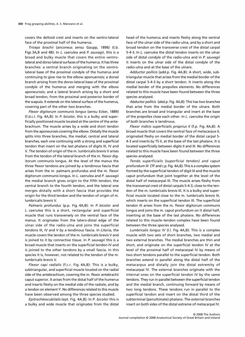
Flexor digitorum communis longus
(
sensu
Ecker, 1889)(f.d.c.l. Fig. 4A,B): In
P. bicolor
, this is a bulky and super-ficially positioned muscle located at the centre of the ante-brachium. The muscle arises by a wide and short tendonfrom the aponeurosis covering the elbow. Distally the musclesplits into three branches, the medial, central and lateralbranches, each one continuing with a strong and superficialtendon that insert on the last phalanx of digits III, IV andV. The tendon of origin of the m. lumbricalis brevis V arisesfrom the tendon of the lateral branch of the m. flexor dig-itorum communis longus. At the level of the manus thethree flexor tendons are joined by a tendinous fascia thatarises from the m. palmaris profundus and the m. flexordigitorum communis longus. In
L. caerulea
and
P. sauvagii
the medial branch gives origin to the fifth tendon, thecentral branch to the fourth tendon, and the lateral onemerges distally with a short fascia that provides theorigin for the third tendon and the tendon of origin of m.lumbricalis brevis V.
Palmaris profundus
(p.p. Fig. 4A,B): In
P. bicolor
and
L. caerulea
this is a short, rectangular and superficialmuscle that runs transversely on the ventral face of themanus. It originates from the latero-distal edge of theulnar side of the radio-ulna and joins the superficialtendons III, IV and V by a tendinous fascia. In
Litoria
, themuscle covers the tendon of the m. lumbricalis brevis V andis joined to it by connective tissue. In
P. sauvagii
this is abroad muscle that inserts on the superficial tendon IV andis joined to the other tendons by a small fascia. In thisspecies it is, however, not related to the tendon of the m.lumbricalis brevis V.
Flexor capi radialis
(f.c.r. Fig. 4A,B): This is a bulky,subtriangular, and superficial muscle located on the radialside of the antebrachium, covering the m. flexor antebrachiicaput superior. It arises from the distal half of the humerusand inserts fleshy on the medial side of the radiale, and bya tendon on element Y. No differences related to this musclehave been observed among the three species studied.
Epitrochleocubitalis
(ept. Fig. 4A,B): In
P. bicolor
this isa bulky and wide muscle that originates from the distal
head of the humerus and inserts fleshy along the ventralface of the ulnar side of the radio-ulna, and by a short andbroad tendon on the transverse crest of the distal carpal5-4-3. In
L. caerulea
the distal tendon inserts on the ulnarside of distal condyle of the radio-ulna and in
P. sauvagii
it inserts on the ulnar side of the distal condyle of theradio-ulna and at the base of the ulnare.
Adductor pollicis
(add.p. Fig. 4A,B): A short, wide, sub-triangular muscle that arises from the medial border of thedistal carpal 5-4-3 by a short tendon. It inserts along themedial border of the prepollex elements. No differencesrelated to this muscle have been found between the threespecies analysed.
Abductor pollicis
(abd.p. Fig. 4A,B): This has two branchesthat arise from the medial border of the ulnare. Bothbranches are broad and triangular and insert at the baseof the prepollex close each other. In
L. caerulea
the originof both branches is tendinous.
Flexor indicis superficialis proprius II
(f.p. Fig. 4A,B): Abroad muscle that covers the ventral face of metacarpus II,originated fleshy on medial border of the distal carpal 5-4-3 and inserts by TS II, at the base of the last phalanx. It islocated superficially between digits II and III. No differencesrelated to this muscle have been found between the threespecies analysed.
Tendo superficialis (superficial tendon) and caputprofundum III
(TF and c.p. Fig. 4A,B): This is a complex systemformed by the superficial tendon of digit III and the musclecaput profundum that joint together at the level of thedistal half of metacarpal III. The muscle arises fleshy fromthe transversal crest of distal carpals 5-4-3, close to the ten-don of the m. lumbricalis brevis III. It is a bulky and super-ficial muscle located close to the m. lumbricalis brevis III,which inserts on the superficial tendon III. The superficialtendon III arises from the m. flexor digitorum communislongus and joins the m. caput profundum on it distal half,inserting at the base of the last phalanx. No differencesrelated to this muscle–tendon complex have been foundbetween the three species analysed.
Lumbricalis longus IV
(l.l. Fig. 4A,B): This is a complexmuscle with two sets of short branches, two medial andtwo external branches. The medial branches are thin andshort, and originate on the superficial tendon IV at thelevel of the proximal half of metacarpal IV by means oftwo short tendons parallel to the superficial tendon. Bothbranches extend in parallel along the distal half of themetacarpus and distally join the distal extremity ofmetacarpal IV. The external branches originate with theinternal ones on the superficial tendon IV by the sametendons. They run in parallel between the superficial tendonand the medial branch, continuing forward by means oftwo long tendons. These tendons run in parallel to thesuperficial tendon and insert on the distal third of thesubterminal (penultimate) phalanx. The external branchesinsert on both sides of the distal extreme of metacarpal IV.

Frog grasping abilities, A. S. Manzano et al.
© 2008 The Authors Journal compilation © 2008 Anatomical Society of Great Britain and Ireland
301
In
L. caerulea
the muscle is single but continues forward viatwo tendons similar to the medial branch described above.
Movement patterns
Our analysis of the high-speed video recordings indicatesthat the overall forelimb movement pattern is very similarin the two species (Fig. 5). During the swing phase the digitsare flexed and digit 2 is adducted while the elbow is flexedand the humerus protracted. Close to substrate contactthe elbow and wrist are extended, and the fingers extendedand spread fully. During substrate contact, the fingers areflexed around the dowel and the wrist and elbow areflexed during stance. One notable difference that can beobserved between species is the degree to which they canclose the hand around the dowel. Whereas in
P. bicolor
closure is typically complete, in
L. caerulea
, the terminalphalanx of the third or fourth digits of the contralateralhand is not flexed and remains visible in lateral view(Fig. 5).
One other striking difference between the two specieswas that whereas
P. bicolor
, despite its larger size, neverlost balance or stumbled when walking across the narrowestsubstrate,
L. caerulea
does. In slightly over half of the trials(53.85%)
L. caerulea
lost balance or stumbled whenwalking across the same substrate. Our analysis of the stepparameters indicates that this may be due to the longercontact time observed in
P. bicolor
(1.19 ± 0.46 s) comparedwith
L. caerulea
(0.68 ± 0.41 s). Limb movements in generalare about twice as slow in
P. bicolor
as in
L. caerulea
movingacross the same substrate, as indicated by the swing phaseduration (0.53 ± 0.12 vs. 0.29 ± 0.21 s).
An analysis of the elbow angle showed no significantspecies (
F
1,0.93
= 0.1;
P
= 0.81), contact time (
F
1,85
= 0.81;
P
= 0.37) or interaction effects (
F
1,84
= 3.93;
P
= 0.05). Wristangle, by contrast, showed significant interaction effects(F1,84 = 11.43; P = 0.001). Differences in wrist angle duringthe different contact phases were also significant (F1,84 = 10.54;P = 0.002) with angles during mid-stance being greater(i.e. wrist more extended than during toe-off). Species
Fig. 4 Ventral view of the hand showing the flexor musculature. (A) Litoria caerulea, left hand. Scale bar = 1 mm. (B) Phyllomedusa sauvagii, left hand. Scale bar = 5 mm. Abbreviations: f.d.c.l., m. flexor digitorum communis longus; ept., m. epitrochleocubitalis; p.p., m. palmaris profundus; abd.s., abductor secundus digiti V; l.b., m. lumbricalis brevis; l.l., m. lumbricalis longus; c.p., caput profundus digiti III; f.p., m. flexor indicis superficialis proprius digiti II; f.c.r., m. flexor carpis radialis; T.F., main flexor tendons; delt., m. deltoideus; t.b., m. triceps brachii.

Frog grasping abilities, A. S. Manzano et al.
© 2008 The AuthorsJournal compilation © 2008 Anatomical Society of Great Britain and Ireland
302
were different in wrist angle only during toe-off (F1,46 = 37.54;P < 0.001), with L. caerulea having greater angles and thusa more extended wrist than P. bicolor. Wrist angle was,however, not significantly different during mid-stance(F1,39 = 0.84; P = 0.37). Hand angle 1 was not differentbetween species (F1,0.68 = 0.64; P = 0.62), or contact phase(F1,85 = 1.04; P = 0.31) and also showed no significant inter-action effects (F1,84 = 0.87; P = 0.36). Hand angle 2 showedsignificant interaction (F1,84 = 4.10; P = 0.04) and contactphase (F1,84 = 3.98; P = 0.049) effects, with angles beingsmaller (i.e. the toe being more adducted) during toe-off.Species were different only during mid-stance (F1,39 = 11.86;P = 0.001), with P. bicolor displaying greater angles thanL. caerulea, but not during toe-off (F1,46 = 0.99; P = 0.33).
Muscle activity patterns
Our electromyographic recordings show that the flexors ofthe hand are active during substrate contact in bothL. caerulea (m. flexor digitorum communis longus; Fig. 6)and P. bicolor (combined activity of m. flexor digitorumcommunis longus, m. palmaris profundus and m. flexorindicis superficialis proprius II; Fig. 7). The onset of activityof the m. flexor digitorum communis longus was 50 msafter the onset of contact on average, and remained activefor an average of 500 ms in L. caerulea. In P. bicolor, them. palmaris profundus was active on average for 400 msfollowing initial substrate contact. Interestingly, themuscle also showed distinct activity during swing, coincidingwith a flexion of the fingers (Fig. 5). Unfortunately, theelectromyographic signal of the m. flexor digitorumcommunis longus in P. bicolor was too noisy to quantify itsactivity. The m. flexor i. s. proprius II (m. flexor indicissuperficialis proprius II) was active for 200 ms on averageduring stance and during the entire swing phase, causingadduction of digit 2 (Fig. 5). These data suggest that inboth species the hand is actively flexed after beingpositioned onto the substrate. Two to 300 ms before theonset of the swing phase, the flexor muscles cease theiractivity to allow extension of the hand in preparation forthe swing phase in both species. This is corroborated bythe late onset of the m. abductor indicis longus during latestance and early swing in L. caerulea (Fig. 7).
Activities of other muscles studied were more variable.In general, the m. triceps brachii was active during stancein L. caerulea, although occasionally a distinct activityburst was present during the swing phase (Fig. 6). Similarly,the wrist extensor (m. extensor digitorum communislongus) in P. bicolor showed a pronounced activity burst ofvariable duration during stance. The m. deltoideus inP. bicolor showed a pronounced activity during the swingphase but invariably showed a second activity burst duringstance. In L. caerulea the activity of the m. deltoideus wasvariable, but again showed activity during both stance andswing phases.
Fig. 5 Selected images from high-speed video recordings (100 frames per second) of walking on a narrow substrate in Litoria caerulea (A–C) and Phyllomedusa bicolor (D–F). Note the flexion of the hand and adduction of digit 2 during the swing phase (A, D) and extension and abduction of the digits right before substrate contact (B, E) in both species. During substrate contact, however, P. bicolor is able to close its fingers more completely and actively flexes the last phalanx of each digit; L. caerulea, by contrast, cannot fully flex the last phalanges (arrow) when grasping the substrate.

Frog grasping abilities, A. S. Manzano et al.
© 2008 The Authors Journal compilation © 2008 Anatomical Society of Great Britain and Ireland
303
Grasping forces
Whereas L. caerulea was able to generate 1.32 ± 0.10 N ofgrasping force on average, the one P. bicolor for whichmeasurements were obtained was able to generate 2.41 Nof force (Fig. 8). This difference was significant (F1,2 = 47.82;P = 0.02) but should be interpreted with some cautiongiven that only a single individual of P. bicolor was meas-ured. Maximal grasping forces obtained through stimulationof the forearm and hand flexors (Fig. 8) were somewhatlower for both P. bicolor (1.99 N) and L. caerulea (0.79 ± 0.30).
Stimulation experiment
M. flexor digitorum communis longus: In L. caerulea, stimula-tion of the m. flexor digitorum communis longus causes
flexion of the wrist to about 90° relative to the horizontal.Additionally, stimulation of this muscle causes flexion ofthe digits at all the different phalangeal joints. Stimulationof the same muscle in P. bicolor causes a similar degree offlexion at the wrist but resulted in flexion of the digits atthe metacarpo-phalangeal joints only.
M. flexor carpi radialis: Stimulation of the m. flexor carpiradialis causes flexion of the wrist and a rotation of thehand towards the side of digit 2 (endorotation) in bothspecies studied.
M. epitrochleocubitalis: The action of this muscle wasinvestigated in P. bicolor only. Stimulation of the m.epitrochleocubitalis causes a rotation at the wrist towardsthe side of digit 5 (exorotation). When the lower arm is notstabilized relative to the substrate, stimulation of thismuscle causes elbow flexion to an angle of about 90°.
Fig. 6 Representative electromyographic traces of selected forelimb muscles in Litoria caerulea. Grey bars represent the ipsilateral contact phase; yellow bars represent the swing phase. Note how the flexor becomes active slightly after substrate contact, suggesting that the hand is first put down and subsequently flexed. Also note how the triceps (elbow extensor) is active during the contact phase but may also show activity during the swing phase as seen in the last step.

Frog grasping abilities, A. S. Manzano et al.
© 2008 The AuthorsJournal compilation © 2008 Anatomical Society of Great Britain and Ireland
304
Little or no flexion of the wrist is observed upon stimulationof this muscle.
M. palmaris profundus: Stimulation of this muscle inL. caerulea causes an adduction of digit 5 and a slight butmarked exorotation of the hand. In P. bicolor, however,stimulation of the m. palmaris profundus causes a displacementof the tendon of the m. flexor digitorum communis longus2–3 mm towards the side of digit 5.
M. lumbricalis longus digiti IV: Stimulation of the m.lumbricalis longus digiti IV causes complete flexion of digit4 in both species.
M. flexor indicis superficialis proprius II: Stimulation ofthe m. flexor i.s. proprius II causes flexion of digit 2 in bothspecies. In P. bicolor, a pronounced adduction of digit 2 isalso observed upon stimulation of the muscle.
Combined stimulations: A combined stimulation of them. flexor digitorum communis longus, m. palmaris profundus,m. lumbricalis of digit 4 and m. flexor i. s. proprius II ofdigit 2 results in a flexion of the wrist and closure of the
hand in both species. Interestingly, stimulation of the m.lumbricalis of digit 4 and the m. flexor i. s. proprius II ofdigit 2 in P. bicolor results in a precision grip betweendigits 2 and 4. In L. caerulea the same stimulation resultsin flexion of digits 2 and 4 but the digits do not touch. Acombined stimulation of the m. flexor digitorum communislongus and the m. palmaris profundus in P. bicolor resultedin a marked increase in the flexion at the wrist comparedwith a stimulation of the m. flexor digitorum communislongus by itself.
Discussion
Morphology of the forearm and hand
When comparing the anatomy of the forearm and hand ofthe species examined here with that observed for moregeneralized frogs (Gaupp, 1896; Burton, 1998), thereappear to be some muscular characters related to the
Fig. 7 Representative electromyographic traces of selected forelimb muscles in Phyllomedusa bicolor. Grey bars represent the ipsilateral contact phase; yellow bars represent the swing phase. Note the activity of the m. palmaris profundus, important in flexing the hand and adducting the fingers during the contact phase. The m. deltoideus and the m. flexor i. s. proprius, on the other hand, show the greatest activity during the swing phase, suggesting flexion at the elbow.

Frog grasping abilities, A. S. Manzano et al.
© 2008 The Authors Journal compilation © 2008 Anatomical Society of Great Britain and Ireland
305
ability to climb, for example the elongation of the mm.extensores breves profundi and the presence of the mm.extensores breves distalis (Burton, 1998), and the intercalaryelement forming a complex system that appears to haveevolved early in the history of frogs (Manzano et al. 2007).In some scansorial frogs, such as Eleutherodactylus, and inarboreal frogs such as most of the Hylids, Centrolenids,Rhacophorids and Hyperolids, a direct connection betweenthe m. palmaris longus and the lateral tendo superficialisimplies a reduction of the palmar aponeurosis that coversthe hand musculature. This aponeurosis, which arisesfrom the palmaris longus in most frogs (and even mostvertebrates), gives origin to the superficial tendons ofeach digit. The species that we analysed have no aponeu-rosis on the palmar surface, and consequently the mainflexor tendons arise directly from a muscle we consider tobe the m. flexor digitorum communis longus. The mainflexor tendons also show a close relationship with the m.palmaris profundus that joins these tendons by connectivetissue and in Phyllomedusa species even attaches onto
superficial tendon IV. In Litoria and Phyllomedusa speciesthe m. lumbricalis brevis V originates with the superficialtendon III on the lateral branch of the flexor digitorumcommunis longus and only in Litoria is there a connectionbetween this muscle and the m. palmaris profundus.
The hand musculature of the species of Litoria andPhyllomedusa examined here is very similar. There are,however, some peculiarities in Phyllomedusa: a generalelongation and increase in the size of the muscles, thepresence of strong and long tendons (like those of the m.extensor brevis or the m. adductor indicis longus); and thepresence of elongated and naked bony areas (i.e. notcovered by muscle; Manzano & Lavilla, 1995). The in-dependence of the main flexor tendons from each other(resulting in the ability of each digit to flex independently),and the presence of muscles with accessory branches(resulting in additional insertion sites; Manzano & Lavilla,1995) are some of the features unique to Phyllomedusaand may be related to their increased dexterity.
Indeed, our analysis of the use of the forelimbs duringlocomotion on a narrow substrate suggests that bothspecies actively adjust the position of the hands andinclude a grasping type of support. On the narrow dowel,both species use a diagonal sequence gait typical of primatesand other arboreal mammals when walking on narrowsubstrates (Jenkins, 1974; Sargis, 2001; Schmitt & Lemelin,2004). Interestingly, even though both species appear touse a similar type of power grip when holding on to a nar-row substrate, despite its larger body size and longer limbsPhyllomedusa appears much more stable and secure whenmoving across narrow substrates. Detailed observationsbased on the high-speed recordings, as well as the analysisof the X-ray data, suggest that this is because Phyllomedusais able to generate a greater abduction of digit V and con-sequently is able to achieve a more complete (i.e. coveringmore of the substrate) and more secure grip on the sub-strate. Phyllomedusa bicolor also showed a greater flexionat the wrist, allowing it to maintain its grasp on thesubstrate for a longer time than L. caerulea.
That both species actively grasp the substrate is indi-cated by the results of our electromyographic analysis. InL. caerulea, the flexor digitorum communis longus showsactivity during the stance phase, ending before the end ofstance and coinciding with contact of the contralaterallimb on the substrate. Thus, these data suggest an activeflexion of the hand during stance. Although the quality ofthe data for this muscle in P. bicolor is not great, they dosuggest a similar pattern of activity. Corroborating thispattern is the activity of the m. palmaris profundus, which,as shown by the stimulation experiment, increases themoment arm of the m. flexor digitorum communis longusand thus actively assists hand and wrist flexion. In addition,the activity of the flexor i. s. proprius II of digit 2 duringstance corroborates this idea. Consequently, both speciesactively create a grasping posture of the hand during
Fig. 8 (A) Graph illustrating in vivo grasp forces in Phyllomedusa bicolor and Litoria caerulea. Note how forces are lower in L. caerulea than in Phyllomedusa bicolor. (B) Graph illustrating the maximal grasping forces obtained by electrical stimulation of the hand flexors. Both in vivo and stimulation data indicate that P. bicolor can generate higher grasp forces than L. caerulea. Bars represent average maximal grasp forces + one standard deviation.

Frog grasping abilities, A. S. Manzano et al.
© 2008 The AuthorsJournal compilation © 2008 Anatomical Society of Great Britain and Ireland
306
stance which is maintained until contra-lateral hand con-tact. Although to our knowledge no comparative data areavailable on the activity of hand flexor muscles duringgrasping associated with locomotion on narrow substrates,Tuttle & Basmajian (1974) do describe distinct activity inthe superficial and deep m. flexor digitorum in gorillaswhile grasping objects such as food or toys, suggesting thatthese muscles may be important during grasping in general.
Our in vivo measurements of grasping force and theresults of the stimulation experiment suggest that bothspecies of frog are able to exert considerable centripetallydirected force, and can thus indeed use this power grip togenerate a counter-torque on the substrate to help stabi-lize their body. The ability of these animals to flex thehand into a power grip posture thus appears to be closelyassociated with the locomotion on these narrow substrates.Phyllomedusa, which is more specialized in its habitat use,moves more securely on narrow substrates, and is capableof generating higher grasping forces. Although these datasuggest that the evolution of a high-performance powergrip has gone hand in hand with the occupation of com-plex arboreal substrates with supports of narrow diameter,this hypothesis needs to be tested in a broad comparativeframework. Arboreality has arisen many times independ-ently in frogs and the group thus presents an ideal systemto study the potential co-evolution of grasping andlocomotion on narrow substrates.
One unexpected outcome of our stimulation experimentsis that Phyllomedusa is mechanically capable of executingwhat is called a precision grip, known only from higherprimates and so characteristic of human manipulativeskills (Napier, 1956; Landsmeer, 1962; Marzke et al. 1992).A precision grip involves the adduction of the thumbtowards the digits such that the palmar surfaces of thethumb and digit touch each other. The combined stimulationof the m. lumbricalis of digit 4 and the flexor i. s. propriusof digit 2 produced exactly such a precision grip. Interest-ingly, P. sauvagii was observed using this type of grip dur-ing locomotion on very narrow branches as well as duringwiping behavior (Blaylock et al. 1976). This suggests that aprecision grip may be used during locomotion on verynarrow substrates and/or in the manipulation of small fooditems (Gray et al. 1997) in phyllomedusine frogs in general.When moving on very narrow substrates, a typical powergrip would result in the digits of the fingers overlappingand thus potentially hindering the creation of a securegrip. Adduction of the first finger (digit 2 in this case)towards digit 3 combined with flexion of the remainingdigits may (the way humans hold a stick or pen whenpointing at an object), however, allow a secure grip onvery narrow substrates. Vertical climbing on narrow sub-strates probably also benefits from a modified grip. Tokeep the centre of mass close to the substrate, and thusallow an efficient climbing style, the hand cannot beclosed around the substrate in a typical power grip (with
flexed thumb), but rather involves adduction of a straightthumb towards the palmar side of the other digits (Isler,2005). Clearly these hypotheses need to be tested byobserving locomotion of these animals on very narrowsubstrates of different orientations. Moreover, the potentialuse of the hand to manipulate small food objects,although common in arboreal frogs (Gray et al. 1997),remains to be investigated in this species.
In summary, we suggest that arboreal frogs may be amodel system to understand the ecological context of theevolution of grasping. Despite long-standing interest in theevolution of human grasping and object manipulation skills,a true understanding of the origin of this functional capacityhas been lacking due to the lack of independent origins ofthe behaviour among mammals. Frogs, despite their distantphylogenetic affinity, may thus provide us with a window tounderstand the evolution of human grasping abilities.
Acknowledgements
We would like to thank Vicky Schaerlaeken for help with experimentsand data collection; and Bieke Vanhooydonck and Vicky Schaer-laeken for measuring animals and sending data to Argentinawhich allowed us to finish the paper in a timely fashion. This researchwas supported by a collaborative project between the FWO-Flanders and SECyT-Argentina, PIP CONICET 6347, and PI-UADER.
References
Blaylock L, Ruibal R, Platt-Aloia K (1976) Skin structure and wipingbehavior of Phyllomedusinae frogs. Copeia 2, 283–295.
Bloch JI, Boyer DM (2002) Grasping primate origins. Science 298,1606–1610.
Burton TC (1998) Are the distal extensor muscles of the fingers ofanuran an adaptation to arboreality? J Herpetol 32, 611–617.
Cartmill M (1985) Climbing. In Functional Vertebrate Morphology(eds Hildebrand M, Bramble DM, Liem KF, Wake DB), pp. 73–88.Cambridge, MA: Belknap Press.
Duellman WE, Trueb L (1986) Biology of Amphibians. New York:McGraw-Hill.
Ecker A (1889) The Anatomy of the Frog. Oxford: Clarendon Press.Emerson SB (1976) Burrowing in frogs. J Morphol 149, 437–457.Emerson SB (1991) A biomechanical perspective on the use of
forelimb length as a measure of sexual selection in frogs. J EvolBiol 4, 671–678.
Fabrezi M (1992) El carpo de los anuros. Alytes 10, 1–29.Fabrezi M, Alberch P (1996) The carpal elements of anurans.
Herpetologica 52, 188–204.Frost DR, Grant T, Faivovich J, Bain RH, et al. (2006) The amphibian
tree of life. Bull Am Mus Nat Hist 297, 1–370.Gans C, Parsons T (1966) On the origin of the jumping mechanism
in frogs. Evolution 20, 92–99.Gaupp E (1896) Anatomie des Frosches. Braunschweig: Friedrich
Vieweg und Sohn.Gray L, O’Reilly JC, Nishikawa KC (1997) Evolution of forelimb
movement patterns for prey manipulation in anurans. J ExpZool 277, 417–424.
Hanna G, Barnes WJP (1991) Adhesion and detachment of the toepads of tree frogs. J Exp Biol 155: 103–125.

Frog grasping abilities, A. S. Manzano et al.
© 2008 The Authors Journal compilation © 2008 Anatomical Society of Great Britain and Ireland
307
Havelková P, Ro!ek Z (2006) Transformation of the pectoral girdlein the evolutionary origin in frogs: insights from the primitiveanuran Discoglossus. J Anat 209, 1–11.
Isler K (2005) 3D-kinematics of vertical climbing in hominoids.Am J Phys Anthropol 126, 66–81.
Jenkins FA (1974) Tree shrew locomotion and the origins of primatearborealism. In Primate Locomotion (ed. Jenkins FA), pp. 85–115. New York: Academic Press.
Jenkins FA, Shubin HH (1998) Prosalirus bitis and the anurancaudopelvic mechanism. J Vert Paleontol 18, 495–510.
Landsmeer JMF (1962) Power grip and precision handling. AnnRheum Dis 21, 164–170.
Loeb GE, Gans C (1986) Electromyography for Experimentalists.Chicago: The University of Chicago Press.
Lutz GJ, Rome LC (1994) Built for jumping: the design of the frogmuscular system. Science 263, 370–372.
Manzano AS, Barg M (2005) The iliosacral articulation in Pseudinae(Anura: Hylidae). Herpetologica 61, 259–267.
Manzano AS, Fabrezi M, Vences M (2007) Intercalary elements,treefrogs, and the early differentiation of a complex system inthe Neobatrachia. Anat Rec 290, 1551–1567.
Manzano AS, Lavilla EO (1995) Notas sobre la miología apendicular dePhyllomedusa hypocondrialis (Anura, Hylidae). Alytes 12, 169–174.
Martin RD (1990) Primate Origins and Evolution. Princeton, NJ:Princeton University Press.
Marzke MW, Wullstein KL, Viegas SF (1992) Evolution of thepower (‘squeeze’) grip and its morphological correlates inhominids. Am J Phys Anthropol 89, 283–298.
Napier JR (1956) The prehensile movements of the human hand. JBone Joint Surg 38, 902–913.
Napier JR (1967) Evolutionary aspects of primate locomotion. AmJ Phys Anthropol 27, 333–342.
Nauwelaerts S, Aerts P (2006) Take-off and landing forces in jumpingfrogs. J Exp Biol 209, 66–77.
Sargis EJ (2001) The grasping behavior, locomotion and substrateuse of the tree shrews Tupaia minor and T. tana (Mammalia,Scandentia). J Zool Lond 253, 485–490.
Schmitt D, Lemelin P (2004) Locomotor mechanics of the slenderloris (Loris tardigradus). J Hum Evol 47, 85–94.
Shubin NH, Jenkins FA (1995) An early Jurassic jumping frog.Nature 377, 49–52.
Sokal RR, Rolph FJ (1995) Biometry, 3rd edn. New York: W. H.Freeman and Company.
Tuttle RH, Basmajian JV (1974) Electromyography of forearmmusculature in Gorilla and problems related to knuckle walking.In Primate Locomotion (ed. Jenkins FA), pp. 293–347. New York:Academic Press.
Videler JJ, Jorna JT (1985) Functions of the sliding pelvis in Xeno-pus laevis. Copeia 1985, 254–257.