hutan sabah
30
-
Upload
independent -
Category
Documents
-
view
1 -
download
0
Transcript of hutan sabah


Fig. 1. Expression of hGSTA1-1 in hGSTA1 transfected cellsHL60 (A) and K562 (B) cells were transfected with the cDNA of hGSTA1 cloned in thepTarget-T mammalian expression vector or the vector alone as described in the Methodssection. The supernatant fraction (28,000g) of homogenates of the wild-type (WT), vector (VT)and hGSTA1-transfected (hGSTA1-Tr) HL60 cells containing 30µg of protein was subjectedto SDS-PAGE in 12% gel. Expression of hGSTA1-1 in the stable-transfected clone selectedin G418 (300µg/ml) was analyzed by Western blot analysis using polyclonal primaryantibodies against human α-class GSTs raised in rabbits and peroxidase-conjugated goat anti-rabbit secondary antibodies. The blot was developed using chemiluminescence (SupersignalWest Pico, Pierce) reagents. Lanes have been appropriately marked on the immunoblots.Representative immunoblot from one of the several hGSTA1-1expressing clones selected isshown. C. Levels of 4-HNE in SFN treated VT and hGSTA1-1 expressing HL60 and K562cells: VT and hGSTA1-1 expressing cells (2×10⁷) were incubated with RPMI completemedium containing SFN (0–40µM) for 5h at 37°C. After pelleting them by centrifugation, cells
Sharma et al. Page 15
Biochemistry. Author manuscript; available in PMC 2011 April 13.
NIH
-PA Author Manuscript
NIH
-PA Author Manuscript
NIH
-PA Author Manuscript

were washed and sonicated (3× 10s, 30W) in PBS containing BHT (5mM final concentration)on ice. 4-HNE levels in the cell pellets were measured by using LPO-586 kit as per themanufacturer’s instructions. Data presented are Mean ± SD (n=3, * and ** represent asignificant difference in 4-HNE levels of VT and hGSTA1-1 expressing HL60 and K562 cellsrespectively; p< 0.05). D. Immunofluorescence analysis of 4-HNE adducts in SFN treatedVT and hGSTA1-1 expressing HL60 cells: VT and hGSTA1-1 expressing HL60 cells(1×10⁶) were treated with SFN (20µM) for 2h at 37°C in RPMI complete medium. Afterpelleting them by centrifugation at 1000rpm (5min), cells were washed and resuspended inPBS. Aliquots of cell suspensions were cytospun at 500rpm for 5 min onto the superfrost Fisherbrand slides and fixed in 4% paraformaldehyde for 20min. The cells were incubated with theprimary antibodies against 4-HNE-protein adduct (1:500) prepared in the blocking buffer (1%BSA +1% goat serum in PBS) overnight at 4°C in a humidified chamber. After three washingswith PBS (5min each), cells were incubated with TRITC conjugated secondary antibodies(1:500) for 2h at room temperature. Subsequently, this was once again followed by threewashings with PBS (10min each), mounted with Vecta Shield containing DAPI and observedunder the Nikon Eclipse E800 fluorescence microscope with a 40x objective. Different panelsof photographs with DAPI and TRITC stains have been appropriately marked in the figure.
Sharma et al. Page 16
Biochemistry. Author manuscript; available in PMC 2011 April 13.
NIH
-PA Author Manuscript
NIH
-PA Author Manuscript
NIH
-PA Author Manuscript
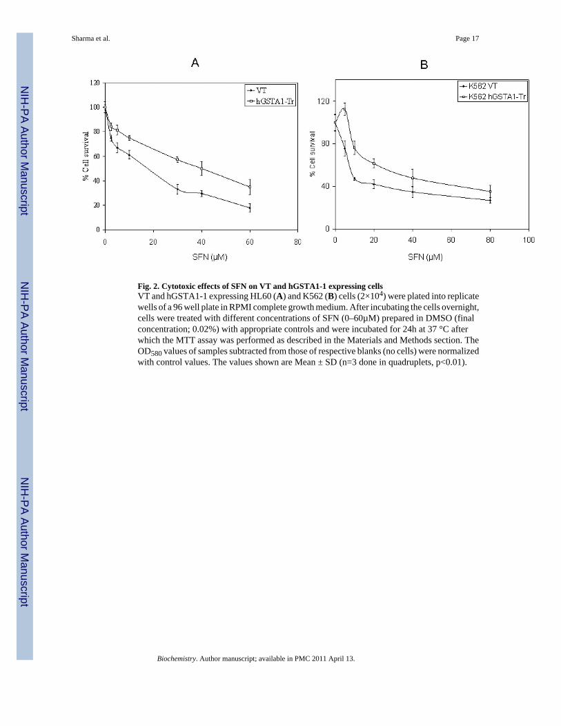
Fig. 2. Cytotoxic effects of SFN on VT and hGSTA1-1 expressing cellsVT and hGSTA1-1 expressing HL60 (A) and K562 (B) cells (2×10⁴) were plated into replicatewells of a 96 well plate in RPMI complete growth medium. After incubating the cells overnight,cells were treated with different concentrations of SFN (0–60µM) prepared in DMSO (finalconcentration; 0.02%) with appropriate controls and were incubated for 24h at 37 °C afterwhich the MTT assay was performed as described in the Materials and Methods section. TheOD580 values of samples subtracted from those of respective blanks (no cells) were normalizedwith control values. The values shown are Mean ± SD (n=3 done in quadruplets, p<0.01).
Sharma et al. Page 17
Biochemistry. Author manuscript; available in PMC 2011 April 13.
NIH
-PA Author Manuscript
NIH
-PA Author Manuscript
NIH
-PA Author Manuscript
Sharma et al. Page 18
Biochemistry. Author manuscript; available in PMC 2011 April 13.
NIH
-PA Author Manuscript
NIH
-PA Author Manuscript
NIH
-PA Author Manuscript

Sharma et al. Page 19
Biochemistry. Author manuscript; available in PMC 2011 April 13.
NIH
-PA Author Manuscript
NIH
-PA Author Manuscript
NIH
-PA Author Manuscript
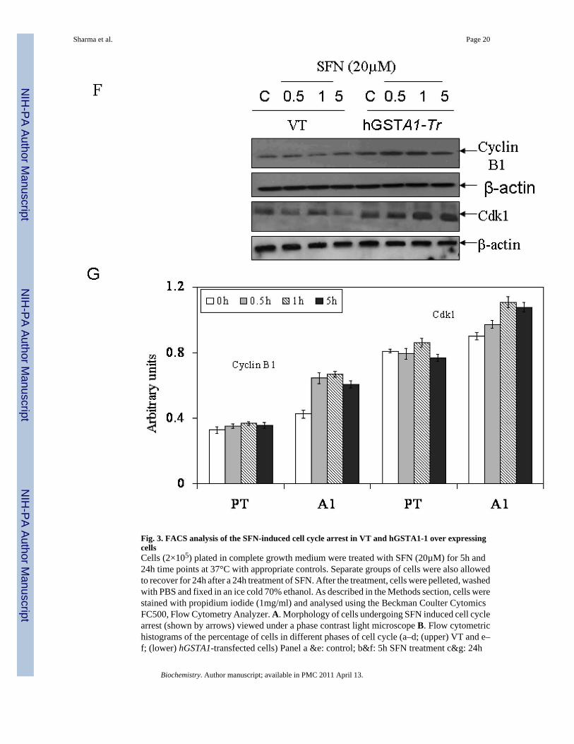
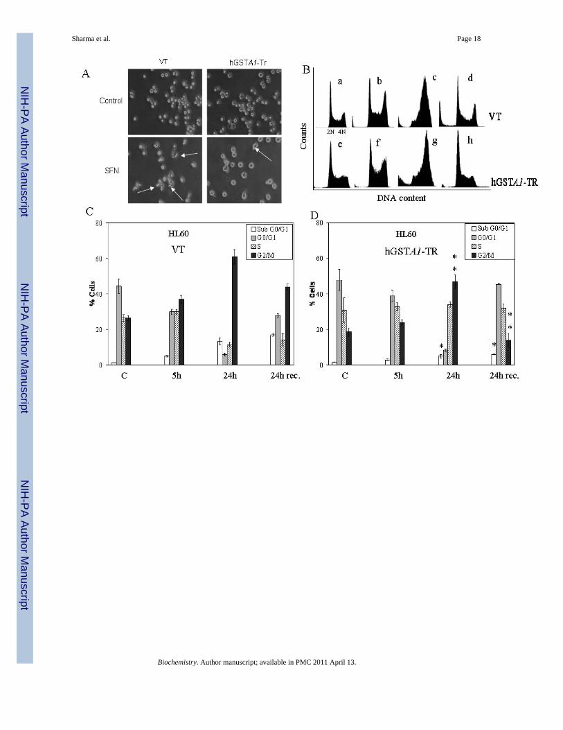
Fig. 3. FACS analysis of the SFN-induced cell cycle arrest in VT and hGSTA1-1 over expressingcellsCells (2×10⁵) plated in complete growth medium were treated with SFN (20µM) for 5h and24h time points at 37°C with appropriate controls. Separate groups of cells were also allowedto recover for 24h after a 24h treatment of SFN. After the treatment, cells were pelleted, washedwith PBS and fixed in an ice cold 70% ethanol. As described in the Methods section, cells werestained with propidium iodide (1mg/ml) and analysed using the Beckman Coulter CytomicsFC500, Flow Cytometry Analyzer. A. Morphology of cells undergoing SFN induced cell cyclearrest (shown by arrows) viewed under a phase contrast light microscope B. Flow cytometrichistograms of the percentage of cells in different phases of cell cycle (a–d; (upper) VT and e–f; (lower) hGSTA1-transfected cells) Panel a &e: control; b&f: 5h SFN treatment c&g: 24h
Sharma et al. Page 20
Biochemistry. Author manuscript; available in PMC 2011 April 13.
NIH
-PA Author Manuscript
NIH
-PA Author Manuscript
NIH
-PA Author Manuscript
SFN treatment d&h: 24h recovered cells after 24h of SFN treatment. C and D, Bar chartsshowing the percentage of cells in the respective phases (sub G0/G1, G0/G1, S and G2/M) ofthe cell cycle (Mean ± SD) from three independent experiments. E. Bar chart showing theeffect of SFN on the cell cycle of K562 (VT and hGSTA1 transfected) cells. F Western blotanalysis of cell cycle related proteins Cdk1 and cyclin B1expression respectively in controland SFN treated VT and hGSTA1-1 expressing HL60 cells. G. Densitometric analysis of bandsobtained for cyclin B1 and cdk1 on immunoblots.* and **represent significant differences inthe percentages of cells in apoptosis (sub G0/G1) and G2/M Phase respectively in VT andhGSTA1-1.
Sharma et al. Page 21
Biochemistry. Author manuscript; available in PMC 2011 April 13.
NIH
-PA Author Manuscript
NIH
-PA Author Manuscript
NIH
-PA Author Manuscript
Sharma et al. Page 22
Biochemistry. Author manuscript; available in PMC 2011 April 13.
NIH
-PA Author Manuscript
NIH
-PA Author Manuscript
NIH
-PA Author Manuscript
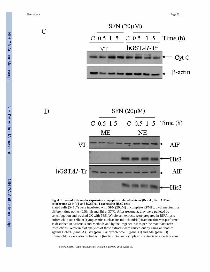
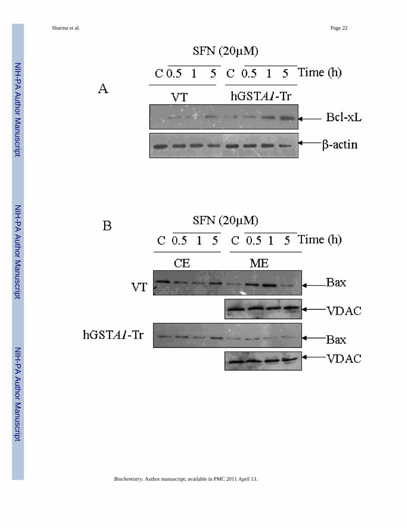
Fig. 4. Effects of SFN on the expression of apoptosis related proteins (Bcl-xL, Bax, AIF andcytochrome C) in VT and hGSTA1-1 expressing HL60 cellsPlated cells (5×10⁶) were incubated with SFN (20µM) in complete RPMI growth medium fordifferent time points (0.5h, 1h and 5h) at 37°C. After treatment, they were pelleted bycentrifugation and washed 2X with PBS. Whole cell extracts were prepared in RIPA lysisbuffer while sub cellular (cytoplasmic, nuclear and mitochondrial) fractionation was performedas described in Materials and Methods and by the Imgenex Kit as per the manufacturer’sinstructions. Western blot analyses of these extracts were carried out by using antibodiesagainst Bcl-xL (panel A), Bax (panel B); cytochrome C (panel C) and AIF (panel D).Immunoblots were also probed with β-actin (total and cytoplasmic extracts to ascertain equal
Sharma et al. Page 23
Biochemistry. Author manuscript; available in PMC 2011 April 13.
NIH
-PA Author Manuscript
NIH
-PA Author Manuscript
NIH
-PA Author Manuscript
loading of proteins. The blot was developed using chemiluminescence (Supersignal West Pico,Pierce) reagents to detect the bands on immunoblots.
Sharma et al. Page 24
Biochemistry. Author manuscript; available in PMC 2011 April 13.
NIH
-PA Author Manuscript
NIH
-PA Author Manuscript
NIH
-PA Author Manuscript
Sharma et al. Page 25
Biochemistry. Author manuscript; available in PMC 2011 April 13.
NIH
-PA Author Manuscript
NIH
-PA Author Manuscript
NIH
-PA Author Manuscript
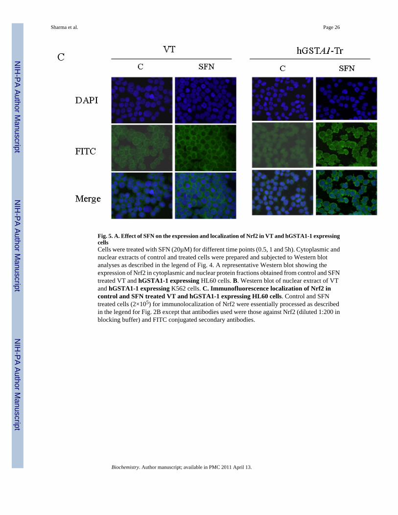
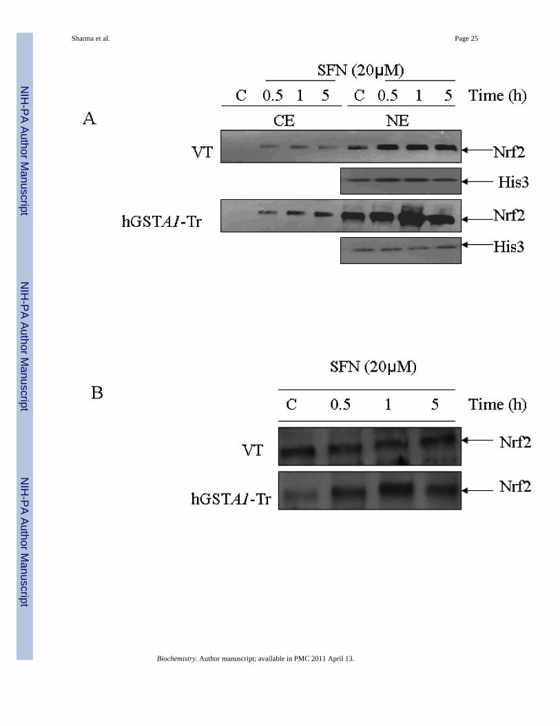
Fig. 5. A. Effect of SFN on the expression and localization of Nrf2 in VT and hGSTA1-1 expressingcellsCells were treated with SFN (20µM) for different time points (0.5, 1 and 5h). Cytoplasmic andnuclear extracts of control and treated cells were prepared and subjected to Western blotanalyses as described in the legend of Fig. 4. A representative Western blot showing theexpression of Nrf2 in cytoplasmic and nuclear protein fractions obtained from control and SFNtreated VT and hGSTA1-1 expressing HL60 cells. B. Western blot of nuclear extract of VTand hGSTA1-1 expressing K562 cells. C. Immunofluorescence localization of Nrf2 incontrol and SFN treated VT and hGSTA1-1 expressing HL60 cells. Control and SFNtreated cells (2×10⁵) for immunolocalization of Nrf2 were essentially processed as describedin the legend for Fig. 2B except that antibodies used were those against Nrf2 (diluted 1:200 inblocking buffer) and FITC conjugated secondary antibodies.
Sharma et al. Page 26
Biochemistry. Author manuscript; available in PMC 2011 April 13.
NIH
-PA Author Manuscript
NIH
-PA Author Manuscript
NIH
-PA Author Manuscript
Sharma et al. Page 27
Biochemistry. Author manuscript; available in PMC 2011 April 13.
NIH
-PA Author Manuscript
NIH
-PA Author Manuscript
NIH
-PA Author Manuscript
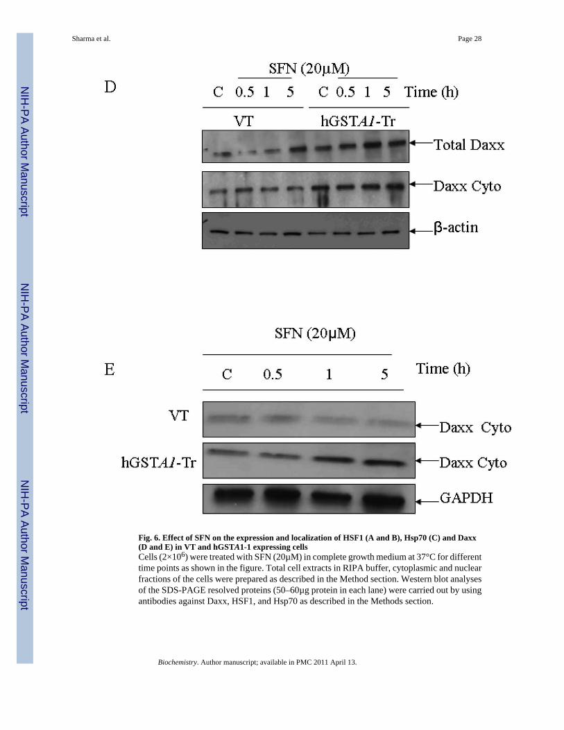
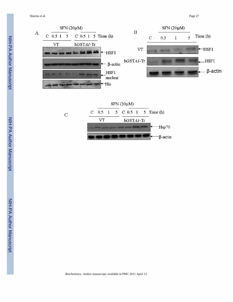
Fig. 6. Effect of SFN on the expression and localization of HSF1 (A and B), Hsp70 (C) and Daxx(D and E) in VT and hGSTA1-1 expressing cellsCells (2×10⁶) were treated with SFN (20µM) in complete growth medium at 37°C for differenttime points as shown in the figure. Total cell extracts in RIPA buffer, cytoplasmic and nuclearfractions of the cells were prepared as described in the Method section. Western blot analysesof the SDS-PAGE resolved proteins (50–60µg protein in each lane) were carried out by usingantibodies against Daxx, HSF1, and Hsp70 as described in the Methods section.
Sharma et al. Page 28
Biochemistry. Author manuscript; available in PMC 2011 April 13.
NIH
-PA Author Manuscript
NIH
-PA Author Manuscript
NIH
-PA Author Manuscript
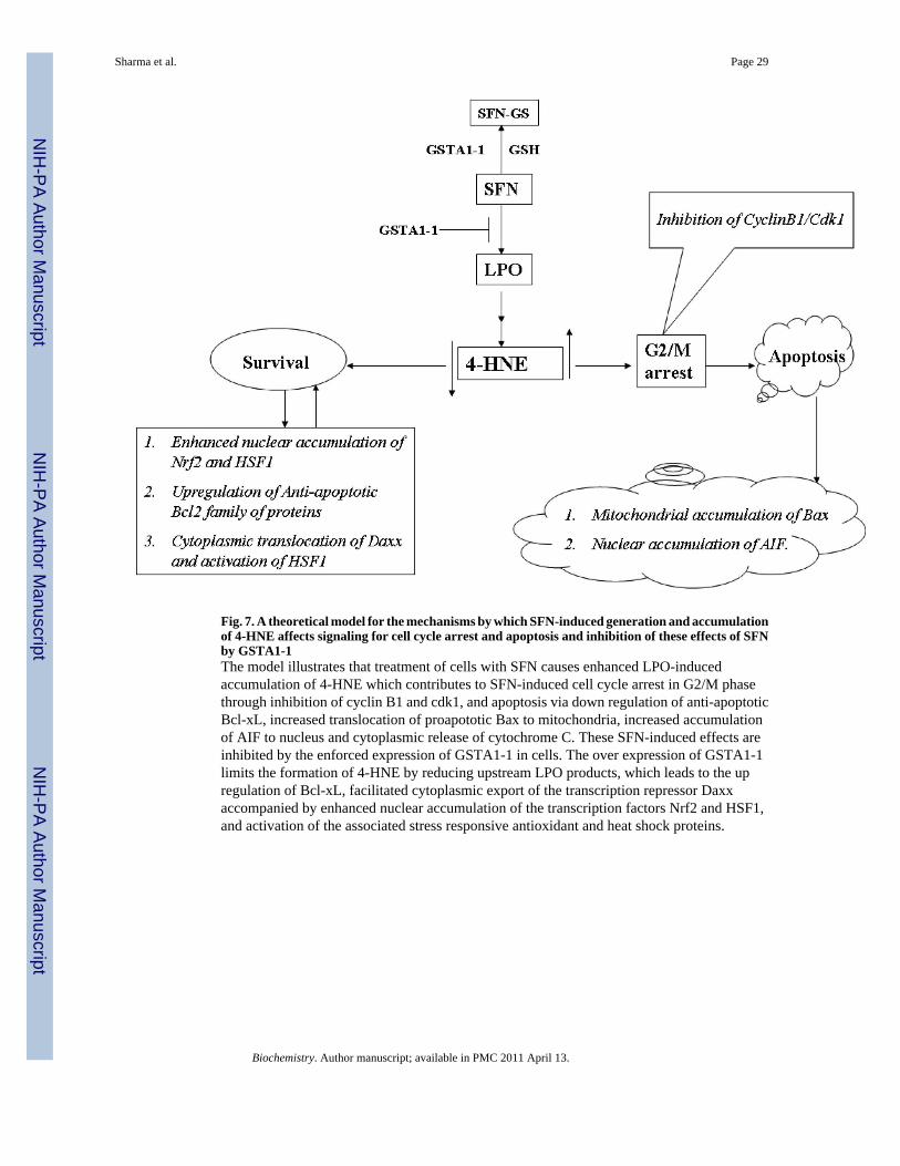
Fig. 7. A theoretical model for the mechanisms by which SFN-induced generation and accumulationof 4-HNE affects signaling for cell cycle arrest and apoptosis and inhibition of these effects of SFNby GSTA1-1The model illustrates that treatment of cells with SFN causes enhanced LPO-inducedaccumulation of 4-HNE which contributes to SFN-induced cell cycle arrest in G2/M phasethrough inhibition of cyclin B1 and cdk1, and apoptosis via down regulation of anti-apoptoticBcl-xL, increased translocation of proapototic Bax to mitochondria, increased accumulationof AIF to nucleus and cytoplasmic release of cytochrome C. These SFN-induced effects areinhibited by the enforced expression of GSTA1-1 in cells. The over expression of GSTA1-1limits the formation of 4-HNE by reducing upstream LPO products, which leads to the upregulation of Bcl-xL, facilitated cytoplasmic export of the transcription repressor Daxxaccompanied by enhanced nuclear accumulation of the transcription factors Nrf2 and HSF1,and activation of the associated stress responsive antioxidant and heat shock proteins.
Sharma et al. Page 29
Biochemistry. Author manuscript; available in PMC 2011 April 13.
NIH
-PA Author Manuscript
NIH
-PA Author Manuscript
NIH
-PA Author Manuscript
NIH
-PA Author Manuscript
NIH
-PA Author Manuscript
NIH
-PA Author Manuscript
Sharma et al. Page 30
Table 1
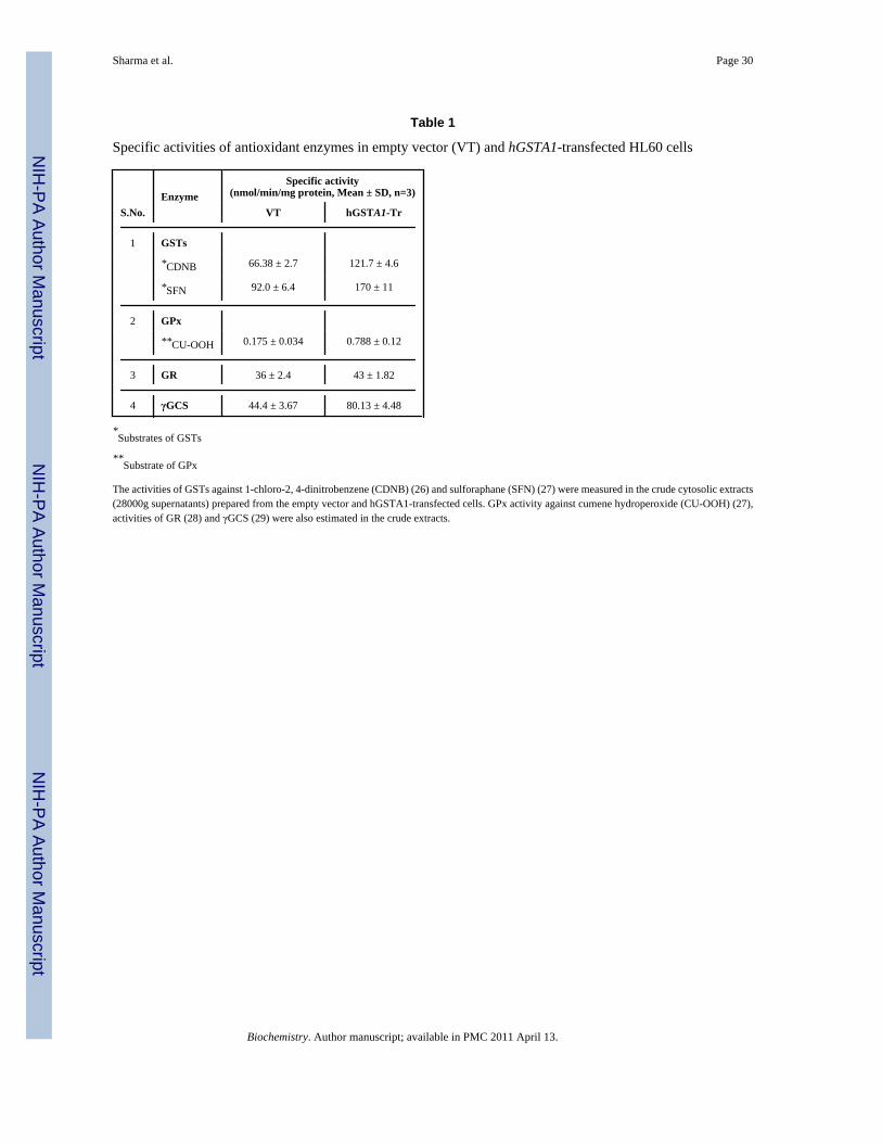
Specific activities of antioxidant enzymes in empty vector (VT) and hGSTA1-transfected HL60 cells
S.No.Enzyme
Specific activity(nmol/min/mg protein, Mean ± SD, n=3)
VT hGSTA1-Tr
1 GSTs
*CDNB 66.38 ± 2.7 121.7 ± 4.6
*SFN 92.0 ± 6.4 170 ± 11
2 GPx
**CU-OOH 0.175 ± 0.034 0.788 ± 0.12
3 GR 36 ± 2.4 43 ± 1.82
4 γGCS 44.4 ± 3.67 80.13 ± 4.48
*Substrates of GSTs
**Substrate of GPx
The activities of GSTs against 1-chloro-2, 4-dinitrobenzene (CDNB) (26) and sulforaphane (SFN) (27) were measured in the crude cytosolic extracts(28000g supernatants) prepared from the empty vector and hGSTA1-transfected cells. GPx activity against cumene hydroperoxide (CU-OOH) (27),activities of GR (28) and γGCS (29) were also estimated in the crude extracts.
Biochemistry. Author manuscript; available in PMC 2011 April 13.