Human hepatic stem cells from fetal and postnatal donors
15
The Journal of Experimental Medicine ARTICLE JEM © The Rockefeller University Press $15.00 Vol. 204, No. 8, August 6, 2007 1973-1987 www.jem.org/cgi/doi/ 1973 10.1084/jem.20061603 The role of human hepatic stem cells (hHpSCs), particularly in the maintenance and regenera- tion of the adult liver, has been a subject of debate without a clear consensus (1–11). Dur- ing embryonic development, endodermal cells in the mid-region of the embryo bulge into the cardiac mesenchyme, are affected by criti- cal signaling from endothelia forming vascula- ture, and form the liver bud (6, 7). The cells within the liver bud are recognized as hepato- blasts because of the expression of a signature marker, -fetoprotein (AFP), and are bipotent, giving rise to hepatocytes and bile-duct epithe- lial cells, which are called cholangiocytes (11). We and others have described the isolation and expansion in culture of AFP cells from fetal and adult livers of several species (8–10). Clo- nogenic expansion assays of rodent hepato- blasts under wholly defined conditions have demonstrated that hepatoblasts are capable of extensive expansion ex vivo, as well as differ- entiation to both hepatocytic and biliary lin- eages (8). The findings from investigations of liver organogenesis, as well as the ex vivo stud- ies of hepatoblasts, have led to a long-standing assumption that hHpSCs correspond to hepa- toblasts, and that hHpSCs would express AFP. However, AFP cells are rare in normal adult livers (0.01%), except in livers with severe injury or disease (11–13). In addition, the re- nowned replicative capacity of hepatocytes in vivo (14) has led to the opinion that adult livers do not have hHpScs and that all regenerative responses are from mature parenchymal cells, except in certain disease states (1). CORRESPONDENCE L.M. Reid: [email protected] Abbreviations used: AFP, -fetoprotein; CK, cytokeratin; EpCAM, epithelial cell adhesion molecule; hHpSC, human he- patic stem cell; ICAM, intercel- lular adhesion molecule; KM, Kubota’s Medium; NCAM, neural cell adhesion molecule; VEGFr, vascular endothelial growth factor receptor. E. Schmelzer, L. Zhang, and A. Bruce are co–first authors; and M.E. Furth and L. M. Reid are co–senior authors. L. Zhang’s present address is The First Affiliated Hospital of Nanjing Medical University, Nanjing, PR China, 210029. The online version of this article contains supplemental material. Human hepatic stem cells from fetal and postnatal donors Eva Schmelzer, 1 Lili Zhang, 1 Andrew Bruce, 4 Eliane Wauthier, 1 John Ludlow, 4 Hsin-lei Yao, 1,2 Nicholas Moss, 1 Alaa Melhem, 1 Randall McClelland, 1 William Turner, 1,2 Michael Kulik, 4 Sonya Sherwood, 4 Tommi Tallheden, 1 Nancy Cheng, 1 Mark E. Furth, 5 and Lola M. Reid 1,2,3 1 Departments of Cell and Molecular Physiology and 2 Biomedical Engineering, 3 Program in Molecular Biology and Biotechnology, School of Medicine, University of North Carolina, Chapel Hill, NC 27599 4 Vesta Therapeutics, Durham, NC 27713 5 Institute for Regenerative Medicine, Wake Forest Baptist Medical Center, Winston Salem, NC 27157 Human hepatic stem cells (hHpSCs), which are pluripotent precursors of hepatoblasts and thence of hepatocytic and biliary epithelia, are located in ductal plates in fetal livers and in Canals of Hering in adult livers. They can be isolated by immunoselection for epithelial cell adhesion molecule–positive (EpCAM+) cells, and they constitute 0.5–2.5% of liver pa- renchyma of all donor ages. The self-renewal capacity of hHpSCs is indicated by phenotypic stability after expansion for 150 population doublings in a serum-free, defined medium and with a doubling time of 36 h. Survival and proliferation of hHpSCs require paracrine signaling by hepatic stellate cells and/or angioblasts that coisolate with them. The hHpSCs are 9 m in diameter, express cytokeratins 8, 18, and 19, CD133/1, telomerase, CD44H, claudin 3, and albumin (weakly). They are negative for -fetoprotein (AFP), intercellular adhesion molecule (ICAM) 1, and for markers of adult liver cells (cytochrome P450s), hemopoietic cells (CD45), and mesenchymal cells (vascular endothelial growth factor receptor and desmin). If transferred to STO feeders, hHpSCs give rise to hepatoblasts, which are recognizable by cordlike colony morphology and up-regulation of AFP, P4503A7, and ICAM1. Transplantation of freshly isolated EpCAM cells or of hHpSCs expanded in culture into NOD/SCID mice results in mature liver tissue expressing human-specific proteins. The hHpSCs are candidates for liver cell therapies. on September 6, 2016 jem.rupress.org Downloaded from Published July 30, 2007 http://jem.rupress.org/content/suppl/2007/08/01/jem.20061603.DC1.html Supplemental Material can be found at:
-
Upload
independent -
Category
Documents
-
view
3 -
download
0
Transcript of Human hepatic stem cells from fetal and postnatal donors

The
Journ
al o
f Exp
erim
enta
l M
edic
ine
ARTICLE
JEM © The Rockefeller University Press $15.00
Vol. 204, No. 8, August 6, 2007 1973-1987 www.jem.org/cgi/doi/
1973
10.1084/jem.20061603
The role of human hepatic stem cells (hHpSCs), particularly in the maintenance and regenera-tion of the adult liver, has been a subject of debate without a clear consensus ( 1 – 11 ). Dur-ing embryonic development, endodermal cells in the mid-region of the embryo bulge into the cardiac mesenchyme, are aff ected by criti-cal signaling from endothelia forming vascula-ture, and form the liver bud ( 6, 7 ). The cells within the liver bud are recognized as hepato-blasts because of the expression of a signature marker, � -fetoprotein (AFP), and are bipotent, giving rise to hepatocytes and bile-duct epithe-lial cells, which are called cholangiocytes ( 11 ). We and others have described the isolation and
expansion in culture of AFP� cells from fetal and adult livers of several species ( 8 – 10 ). Clo-nogenic expansion assays of rodent hepato-blasts under wholly defi ned conditions have demonstrated that hepatoblasts are capable of extensive expansion ex vivo, as well as diff er-entiation to both hepatocytic and biliary lin-eages ( 8 ). The fi ndings from investigations of liver organogenesis, as well as the ex vivo stud-ies of hepatoblasts, have led to a long-standing assumption that hHpSCs correspond to hepa-toblasts, and that hHpSCs would express AFP. However, AFP� cells are rare in normal adult livers (�0.01%), except in livers with severe injury or disease ( 11 – 13 ). In addition, the re-nowned replicative capacity of hepatocytes in vivo ( 14 ) has led to the opinion that adult livers do not have hHpScs and that all regenerative responses are from mature parenchymal cells, except in certain disease states ( 1 ).
CORRESPONDENCE
L.M. Reid:
Abbreviations used: AFP,
� -fetoprotein; CK, cytokeratin;
EpCAM, epithelial cell adhesion
molecule; hHpSC, human he-
patic stem cell; ICAM, intercel-
lular adhesion molecule; KM,
Kubota ’ s Medium; NCAM,
neural cell adhesion molecule;
VEGFr, vascular endothelial
growth factor receptor.
E. Schmelzer, L. Zhang, and A. Bruce are co–fi rst authors;
and M.E. Furth and L. M. Reid are co–senior authors.
L. Zhang ’ s present address is The First Affi liated Hospital of
Nanjing Medical University, Nanjing, PR China, 210029.
The online version of this article contains supplemental material.
Human hepatic stem cells from fetal and postnatal donors
Eva Schmelzer, 1 Lili Zhang, 1 Andrew Bruce, 4 Eliane Wauthier, 1 John Ludlow, 4 Hsin-lei Yao, 1,2 Nicholas Moss, 1 Alaa Melhem, 1 Randall McClelland, 1 William Turner, 1,2 Michael Kulik, 4 Sonya Sherwood, 4 Tommi Tallheden, 1 Nancy Cheng, 1 Mark E. Furth, 5 and Lola M. Reid 1,2,3
1 Departments of Cell and Molecular Physiology and 2 Biomedical Engineering, 3 Program in Molecular Biology
and Biotechnology, School of Medicine, University of North Carolina, Chapel Hill, NC 27599
4 Vesta Therapeutics, Durham, NC 27713
5 Institute for Regenerative Medicine, Wake Forest Baptist Medical Center, Winston Salem, NC 27157
Human hepatic stem cells (hHpSCs), which are pluripotent precursors of hepatoblasts and
thence of hepatocytic and biliary epithelia, are located in ductal plates in fetal livers and in
Canals of Hering in adult livers. They can be isolated by immunoselection for epithelial cell
adhesion molecule – positive (EpCAM+) cells, and they constitute � 0.5 – 2.5% of liver pa-
renchyma of all donor ages. The self-renewal capacity of hHpSCs is indicated by phenotypic
stability after expansion for �150 population doublings in a serum-free, defi ned medium
and with a doubling time of � 36 h. Survival and proliferation of hHpSCs require paracrine
signaling by hepatic stellate cells and/or angioblasts that coisolate with them. The hHpSCs
are � 9 � m in diameter, express cytokeratins 8, 18, and 19, CD133/1, telomerase, CD44H,
claudin 3, and albumin (weakly). They are negative for � -fetoprotein (AFP), intercellular
adhesion molecule (ICAM) 1, and for markers of adult liver cells (cytochrome P450s),
hemopoietic cells (CD45), and mesenchymal cells (vascular endothelial growth factor
receptor and desmin). If transferred to STO feeders, hHpSCs give rise to hepatoblasts, which
are recognizable by cordlike colony morphology and up-regulation of AFP, P4503A7, and
ICAM1. Transplantation of freshly isolated EpCAM� cells or of hHpSCs expanded in culture
into NOD/SCID mice results in mature liver tissue expressing human-specifi c proteins. The
hHpSCs are candidates for liver cell therapies.
on Septem
ber 6, 2016jem
.rupress.orgD
ownloaded from
Published July 30, 2007
http://jem.rupress.org/content/suppl/2007/08/01/jem.20061603.DC1.html Supplemental Material can be found at:

1974 HUMAN HEPATIC STEM CELLS | Schmelzer et al.
hepatoblasts, the distribution of CK19 is more particulate and less intense than that in ductal plate cells. EpCAM expression in AFP� cells, both in fetal and postnatal livers, occurred at the membrane only. In pediatric and adult livers, hepatoblasts were found as individual cells or small clusters of cells teth-ered to the ends of the Canals of Hering ( Fig. 1 ). The hepa-toblasts in nondiseased, postnatal livers constitute �0.01% of cells and express AFP weakly.
Flow cytometry of EpCAM+ cells
Using fl ow cytometry, we observed EpCAM+ cells in human liver cell suspensions of all donor ages ( Fig. 2 A ). Suspensions from fetal livers from which hemopoietic cells had been purged contained, on average, 12% EpCAM� cells. How-ever, the percentage could be as high as 20% depending on the gestational age of the fetus. Most of the EpCAM� cells in fe-tal livers were of nonhepatic lineage and were predominantly hemopoietic. The vast majority (�90%) of the EpCAM� cells in fetal livers coexpress AFP, albumin, and CK19 ( Fig. 2 ). They could be subdivided into two subpopulations: (a) hepa-toblasts showing expression of ICAM1, AFP, albumin, CK19, CD133/1, P450A7, and CD44H (hyaluronan receptor), and (b) hHpSCs, constituting � 5% of EpCAM� cells from fetal
We defi ne a novel class of AFP-negative cells in fetal and adult human livers that are precursors to hepatoblasts and have properties consistent with hHpSCs. The hHpSCs are negative for AFP, but positive for epithelial cell adhesion molecule (EpCAM; CD326, C017-1A antigen, and GA733-2). This protein, encoded by the tumor-associated calcium signal transducer 1 gene, is expressed by many carcinomas and serves a regulatory function in certain normal epithelia, in-cluding all of those derived from endoderm (liver, lung, pan-creas, and intestine) ( 15, 16 ). By immunohistochemistry, Balzar et al. observed that hepatoblasts in embryonic hu-man liver are EpCAM�, whereas mature hepatocytes are EpCAM� ( 15 ). In adult livers, most, but not all, bile duct epithelia are EpCAM�. Also, expanded ductular structures, seen in cases of focal nodular hyperplasia or biliary cirrhosis, contain numerous EpCAM� cells ( 15 ).
We have previously reported that EpCAM�, AFP� cells from human livers are hHpSCs, and we have compared their pattern of gene expression with that of hepatoblasts and ma-ture liver parenchyma ( 17 ). We now show that the hHpSCs are located in ductal plates in fetal and neonatal livers and in the proximal branches of the intrahepatic biliary tree, the Ca-nals of Hering, in pediatric and adult livers of all donor ages, with the frequency of hHpSCs remaining relatively constant throughout life. We further document the immunoselection of these cells using monoclonal antibodies to EpCAM and test whether they meet the defi ning criteria for stem cells, i.e., self-renewal and pluripotency.
RESULTS
In vivo localization of EpCAM+ hHpSCs
Sections of fetal and adult livers were stained for EpCAM and for liver-specifi c markers (albumin, AFP, and CK19; Fig. 1 ). We found that ductal plates, bands of tissue encircling each of the portal triads in fetal and neonatal livers, have small cells (7 – 10 � m) with a paucity of cytoplasm, and stained intensely both cytoplasmically and at the surface for CK19 and EpCAM, and weakly for albumin, but are negative for AFP. In non-diseased, postnatal (pediatric and adult) livers, cells staining positively for EpCAM by immunohistochemistry appear ex-clusively in the Canals of Hering in the vicinity of the portal triads of acini. Theise et al. ( 18 ) reported that cells lining these ductules express cytokeratin (CK) 19, which is present in biliary epithelia, but not in hepatocytes. Our data confi rm their study and demonstrate that the CK19� cells within the Canals of Hering express EpCAM, and that subpopulations of them also express albumin ( Fig. 1 ; yellow color is caused by overlap of CK19 and albumin expression). The coexpression of CK19 and albumin is consistent with hHpSCs and corrob-orates the hypothesis of others that the Canals of Hering comprise a niche for hHpSCs ( 19 ). Shown are data from ex vivo studies of EpCAM� cells supportive of the interpreta-tion that they include hHpSCs.
Most parenchymal cells of fetal and neonatal livers con-sisted of hepatoblasts, slightly larger cells (10 – 12 � m in diam-eter) that stained positively for albumin, AFP, and CK19. In
Figure 1. Immunohistochemical studies on human fetal livers.
Confocal microscopic images on 5-�m liver sections. The antigenic pro-
fi les are given in the table (bottom left). In human fetal livers, sections
were stained for: EpCAM (green) and CK19 (red) (A); EpCAM (green) and
AFP (red) (B); CK19 (green) and albumin (red) (C); CK19 (green) and AFP
(red) (D). In adult livers, sections were stained for: EpCAM (E). (F) Canals of
Hering around portal triad with EpCAM (green) and CK19 (red). (G) A Canal
of Hering showing EpCAM+ cells (green), some of which also express
albumin (red).
on Septem
ber 6, 2016jem
.rupress.orgD
ownloaded from
Published July 30, 2007

JEM VOL. 204, August 6, 2007
ARTICLE
1975
cases of overt hepatic disease (e.g., cirrhosis; unpublished data). Neonatal livers, including some from premature births, showed rapidly decreasing levels of AFP� cells as a function of age, falling below detection level by a few months after birth. Based on considerations detailed in our study and from our previously published work ( 17 ), we identify the EpCAM� cells from pediatric and adult livers as almost exclusively hHpSCs, not hepatoblasts.
Culture selection on plastic and in serum-free Kubota ’ s
medium (KM) isolates hHpSCs, but not hepatoblasts
Suspensions of liver cells plated in KM, which is a serum-free medium optimized for ex vivo expansion of hepatic progeni-tors ( 8 ), either on tissue culture plastic or on embryonic stromal
livers, had an overlapping, but distinct, profi le; they were pos-itive for albumin (weak), CK19, CD44H, CD133/1�, neural cell adhesion molecule – positive (NCAM�), and claudin 3, but negative for AFP and P450A7.
Cell suspensions from adult human livers averaged 1.3% EpCAM+ cells, and those from neonatal and pediatric do-nors two- to threefold higher. Preparations having EpCAM� cell populations at levels �1.5% were typically obtained from livers subjected to ischemia (cold or warm) before organ pro-curement and/or during transportation. This suggests that the EpCAM� liver cells are more resistant to ischemia than mature liver cells. The postnatal EpCAM� liver cells also coexpress CK19 and albumin (unpublished data), but have no detectable AFP� cells (by fl ow cytometry), except in rare
Figure 2. Flow cytometric characterization of EpCAM� cells. (A) The percentage of EpCAM� cells in livers of varying donor ages. The numbers
for fetal livers have been previously reported ( 28 ), but are presented here for comparison to fi ndings in livers from older donors. (B) Analyses of EpCAM�
cells from fetal livers (similar fi ndings occur with EpCAM� cells from adult livers, except that few cells express AFP). (C) Quantitative RT-PCR assays on
freshly isolated EpCAM� versus EpCAM� cells from fetal versus postnatal livers. These data are compared with the fi ndings from colonies of hHpSC
grown on plastic and in serum-free KM for 30 – 60 d.
on Septem
ber 6, 2016jem
.rupress.orgD
ownloaded from
Published July 30, 2007

1976 HUMAN HEPATIC STEM CELLS | Schmelzer et al.
matrix substrata (unpublished data). By 2 – 3 wk, hHpSC col-onies typically contained many thousands of cells.
The hHpSC colonies were assessed for expression of lineage markers by immunofl uorescent staining. The ex-pression pattern closely resembled that of ductal plate cells in vivo. They were positive for CK19, NCAM, EpCAM, and CD44H ( Figs. 3, F – L ). In addition, they were positive for albumin (weak), E-cadherin, N-cadherin, CK8 and 18, CD133/1, integrin � -1 (CD29), claudin 3, and telomer-ase (unpublished data). They were negative for AFP, any form of cytochrome P450, hemopoietic markers (CD34, CD45, CD38, CD14, CD90, and glycophorin A), endothe-lial cell markers (vascular endothelial growth factor recep-tor [VEGFr], von Willebrand factor, and platelet/endothelial cell adhesion molecule or CD31), and mesenchymal markers, such as those for hepatic stellate cells (CD146, desmin, and � -smooth muscle actin). The expression of NCAM by the hHpSCs is important because previous studies have shown that this marker is present on the ductal plate in fetal livers and evident on liver cell populations proliferating under vari-ous disease states ( 20 – 23 ).
Immunoselection using EpCAM isolates hepatoblasts from
fetal and neonatal livers; immunoselection using EpCAM
or NCAM isolates hHpSCs from livers of all donor ages
To enrich for hepatic progenitors from liver cell suspen-sions, we explored several fractionation strategies, including
cell feeders, yielded parenchymal cell colonies with two dis-tinct morphologies. Type I colonies consisted of cells forming a cordlike morphology interspersed with clear channels and expressing EpCAM, albumin, CK19, ICAM, and AFP, but not NCAM (Figs. S1 – S4, available at http://www.jem.org/cgi/content/full/jem.20061603/DC1). Type 2 colonies con-sisted of densely packed, morphologically uniform cells, strongly expressing EpCAM, NCAM, CD44H, and claudin 3; weakly expressing or negative for albumin; and negative for AFP and ICAM-1 ( Fig. 3 and Fig. S3). We interpret the type 1 colonies as corresponding to hepatoblasts and the type 2 colonies as corresponding to hHpSCs ( Table I ).
In cultures on plastic, by 5 – 7 d (mean 5.2 1.6 d; maxi-mum number of days), the hepatoblast colonies disappeared. However, if cultured on STO feeders, hepatoblasts survived for up to 2 mo, continuing to show coexpression of albumin, AFP, and CK19 (Figs. S1 and S2). The hepatoblast colonies typically contained fewer than � 100 cells.
In contrast to hepatoblasts, hHpSC colonies on plastic con-tinued to expand. A time-lapse sequence of a growing hHpSC colony ( Fig. 3, A – E, and Video 1, available at http://www.jem.org/cgi/content/full/jem.20061603/DC1), in which the expansion of hHpScs seeded at very low density is shown on day 1, 3, and 8. The hHpSCs can be subcultured after me-chanical disaggregation and continue to multiply extensively. Their doubling time on plastic is � 36 h. That doubling time decreased to �24 h if they are plated on specifi c extracellular
Figure 3. hHpSCs in culture. A – D show a stem cell colony forming at 2 (A), 4 (B), 7 (C), 10 (D), and 14 d ( 14 ) in culture on plastic and in KM. Phase
contrast coupled with image of cells with staining for NCAM (E and F), CK19 (G and H), EpCAM (I and J), and CD44H (L). F and G are the phase and immuno-
histochemistry (IHC) for CK19; H and I are the phase and IHC for NCAM; J and K are the phase and IHC for EpCAM; and L is the IHC for CD44, the
hyaluronan receptor. Bar, 20 � m.
on Septem
ber 6, 2016jem
.rupress.orgD
ownloaded from
Published July 30, 2007
JEM VOL. 204, August 6, 2007
ARTICLE
1977
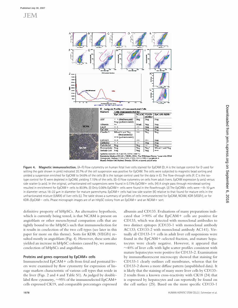
0.7% EpCAM� cells. The immunoselected population con-tained 81% EpCAM� cells, whereas the fl owthrough fraction was almost entirely depleted of EpCAM+ cells. The majority of hepatic EpCAM� cells were of 8 – 10 �m in diameter, as judged by Coulter Counter analysis, in contrast to 18 – 20 �m for diploid hepatocytes, which is the predominant population in the initial liver cell suspension. A small peak of presump-tive tetraploid cells also is evident, measuring � 25 �m in diameter. Light scatter ( “ side scatter ” ) profi les indicate that the EpCAM+ liver cells are considerably smaller and less granular than the bulk of the parenchymal cell population.
Magnetic immunoselection for NCAM� cells from fe-tal livers enriched for cells capable of forming only hHpSC colonies ( Fig. 4 ). The majority of EpCAM� cells from fe-tal liver coexpressed NCAM, whereas only � 40% of those from adult liver were also NCAM�. Therefore, sorting for NCAM� cells proved useful for isolation of hHpSCs from fetal livers, but less so from adult livers. It is unknown at this time whether NCAM and EpCAM coexpression is a
separation by buoyant density on Ficoll gradients (Table S1, available at http://www.jem.org/cgi/content/full/jem.20061603/DC1) and by immunoselection. The most satis-factory results were obtained using magnetic immunoselec-tion. Although FACS was able to yield highly purifi ed cellular subpopulations, the shear forces and the use of buff ers (PBS) that are not optimal resulted in low yields of viable cells. We used magnetic microspheres conjugated with monoclo-nal antibody to EpCAM (Miltenyi Biotec) to immunoselect EpCAM� cells from liver cell suspensions and obtained ro-bust, highly viable sorted cells that survived and expanded well when cultured ( Fig. 4 and Table S2). From postnatal livers, up to 10 billion viable cells were processed in a single pass using the CliniMACS apparatus (Miltenyi Biotec). This yielded �100 million EpCAM� cells. Purity of the enriched EpCAM� cells was typically 75 – 90%, and recovery usually exceeded 90%. Representative fractionations of a fetal liver and of a postnatal liver are depicted in Fig. 4 . A cell suspen-sion from the liver of a 2-yr-old donor was found to contain
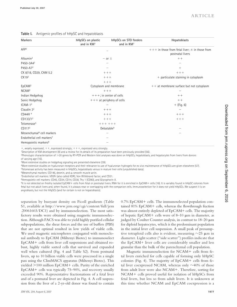
Table I . Antigenic profi les of hHpSC and hepatoblasts
Markers hHpSCs on plastic
and in KM 1
hHpSCs on STO feeders
and in KM 1
Hepatoblasts
AFP 2 _ ��� in those from fetal liver; in those from
postnatal livers
Albumin 2 � or ��
P450-3A4 2 � �
P450-A7 2 � �
CK 8/18, CD29, CAM 5.2 ��� ���
CK19 2 ��� � particulate staining in cytoplasm
EpCAM 2
���
Cytoplasm and membrane �� at membrane surface but not cytoplasm
NCAM 2 ��� �
Indian Hedgehog ���; in center of cells ��
Sonic Hedgehog ��� at periphery of cells ��
ICAM-1 2 � � ( Fig. 6 )
Claudin 3 2 ��� �
CD44H 4 ��� ���
CD133/1 2 ��� ���
Telomerase 5 ��� ���
CD117 2 Debatable 9 �
Mesenchymal 6 cell markers � �
Endothelial cell markers 7 �
Hemopoietic markers 8 �
�, weakly expressed; ��, expressed strongly; ���, expressed very strongly.
1 Description of KM development ( 9 ) and a review for its details of its preparation have been previously provided (56).
2 Phenotypic characterization of � 20 genes by RT-PCR and Western blot analyses was done on hHpSCs, hepatoblasts, and hepatocytes from livers from donors
of varying age ( 16 ).
3 More extensive studies on hedgehog signaling are presented elsewhere ( 39 ).
4 More extensive studies on hyaluronan receptors and their relevance to use of hyaluronan hydrogels for ex vivo maintenance of hHpSCs are given elsewhere (57).
5 Telomerase activity has been measured in hHpSCs, hepatoblasts versus in mature liver cells (unpublished data).
6 Mesenchymal markers: CD146, desmin, and � -smooth muscle actin.
7 Endothelial cell markers: VEGFr (also called KDR), Von Willebrand factor, and CD31.
8 Hemopoietic cell markers: CD45, CD34, CD14, CD38, Thy 1 (CD90), and Glycophorin A.
9 It is not detected on freshly isolated EpCAM� cells from fetal or postnatal livers. RNA for it is enriched in EpCAM� cells ( 16 ). It is variably found in hHpSC colonies from
fetal but not adult livers and, when found, it is always near or overlapping with the companion cells. Immunoselection for it does not yield hHpSCs. We suspect it is on
angioblasts, but not the hHpSCs (and for certain is not on hepatoblasts).
on Septem
ber 6, 2016jem
.rupress.orgD
ownloaded from
Published July 30, 2007
1978 HUMAN HEPATIC STEM CELLS | Schmelzer et al.
albumin and CD133. Evaluations of many preparations indi-cated that �90% of the EpCAM� cells are positive for CD133, which was detected with monoclonal antibodies to two distinct epitopes (CD133-1 with monoclonal antibody AC133; CD133-2 with monoclonal antibody AC141). Vir-tually all CD133-1� cells in adult liver cell suspensions were found in the EpCAM�-selected fraction, and mature hepa-tocytes were clearly negative. However, it appeared that � 40% of liver cells with light scatter profi les consistent with mature hepatocytes were positive for CD133-2. Examination by immunofl uorescent microscopy showed that staining for CD133-1 clearly outlines cell membranes, whereas that for CD133-2 shows a more diff use pattern (unpublished data). It is likely that the staining of many more liver cells by CD133-2 results from a known cross-reactivity with CK18 ( 24 ) that is expressed by hepatocytes and can reportedly be found on the cell surface ( 25 ). Based on the more specifi c CD133-1
defi nitive property of hHpSCs. An alternative hypothesis, which is currently being tested, is that NCAM is present on angioblasts or other mesenchymal companion cells that are tightly bound to the hHpSCs such that immunoselection for it results in coselection of the two cell types (see later in this paper for more on this theme). Sorts for KDR (VEGFr) re-sulted mostly in angioblasts ( Fig. 4 ). However, these sorts also yielded an increase in hHpSC colonies caused by, we assume, coselection of hHpSCs and angioblasts.
Proteins and genes expressed by EpCAM+ cells
Immunoselected EpCAM� cells from fetal and postnatal liv-ers were examined by fl ow cytometry for expression of lin-eage markers characteristic of various cell types that reside in the liver ( Figs. 2 and 4 and Table S1). As judged by double-label fl ow cytometry, � 95% of the immunoselected EpCAM+ cells expressed CK19, and comparable percentages expressed
Figure 4. Magnetic immunoselection. (A – F) Flow cytometry on human fetal liver cells stained for EpCAM (D; A is the isotype control for D used for
setting the gate shown in pink) indicated 20.7% of the cell suspension was positive for EpCAM. The cells were subjected to magnetic bead sorting and
yielded a suspension enriched for EpCAM to 54.6% of the cells (B is the isotype control used for the data in E). The fl ow-through cells (F; C is the iso-
type control for F) were depleted in EpCAM, yielding 7.15% of the cells. (G – I) Flow cytometry on cells from adult livers. EpCAM expression (y axis) versus
side scatter (x axis). In the original, unfractionated cell suspensions were found in 0.73% EpCAM� cells. (H) A single pass through microbead sorting
resulted in enrichment for EpCAM� cells to 80.9%. (I) Only 0.06% EpCAM� cells were found in the fl owthrough. (J) The EpCAM+ cells were � 9 – 10 �m
in diameter versus 18 – 22 �m in diameter for mature parenchyma. EpCAM� cells had low side scatter (K) relative to that found for mature cells in the
unfractionated mixture (UMIX) of liver cells (L). The table shows a summary of profi les of cells immuno selected for EpCAM, NCAM, KDR (VEGFr), or for
KDR-/EpCAM� cells. Phase micrograph images are of an hHpSC colony from an EpCAM� and an NCAM� sort.
on Septem
ber 6, 2016jem
.rupress.orgD
ownloaded from
Published July 30, 2007

JEM VOL. 204, August 6, 2007
ARTICLE
1979
from postnatal livers strongly enriched for transcripts encoding EpCAM, CK19, CD133, and CD117 (c-Kit); these transcripts were barely detectable in the EpCAM� cells. AFP transcripts were not detectable in EpCAM� or EpCAM� cells from postnatal livers.
Although immunoselected cells are enriched for relative expression of CD117 mRNA, we have not observed the cor-responding protein by immunostaining of freshly isolated cells from fetal or postnatal livers, or on cultured cells from postnatal livers. However, we have occasionally observed low levels of CD117 staining on cells at the periphery of hHpSC colonies from fetal livers that are located in regions where hHpSCs overlap with mesenchymal companion cells.
Cytochrome P450 3A4 (CYP3A4), which is a protein ex-pressed by mature hepatocytes, was not found at all in paren-chymal cells, either EpCAM� or EpCAM�, from fetal livers in terms of both mRNA and protein level of it. The level of mRNA for cytochrome P450 in EpCAM� cells was 20-fold lower relative to EpCAM-negative cells from postnatal livers. The small amount of CYP3A4 RNA in the EpCAM� cell fraction from postnatal livers could be accounted for by resid-ual hepatocyte contaminants. In contrast, the hepatoblasts, but not the hHpSCs, were found to express P4503A7, which is a protein found in fetal livers (unpublished data). EpCAM+ cells from postnatal livers also showed eightfold lower relative expression of albumin RNA than the fl ow-through (EpCAM-negative) population. Again, some transcripts can be attributed to incomplete removal of hepatocytes. However, the de-tection of albumin protein in EpCAM� cells by fl ow cytom-etry, together with the transcript data, demonstrates that these progenitor cells express the albumin gene, albeit at a signifi -cantly lower level than diff erentiated hepatocytes.
Assays for telomerase activity indicate signifi cant levels in freshly isolated EpCAM� cells from livers of all donor ages
antibody, we conclude that EpCAM and CD133 (prominin) are coexpressed by the vast majority of hHpSCs.
NCAM (CD56), which was previously shown to be ex-pressed by glia, muscle cells, and neurons ( 26 ), was found on the majority of hHpSCs derived from fetal and neonatal livers, but only � 40% of the EpCAM� cells from adult livers. In our prior studies, NCAM mRNA was enriched strongly in Ep-CAM� cells from both fetal and postnatal livers, but expres-sion at the protein level was variable ( 17 ). NCAM staining was most evident at the borders of the hHpSC colonies ( Fig. 3 ).
Less than 1% of the enriched EpCAM� cells stained for the hemopoietic marker CD45 (leukocyte common antigen), which is found on Kupff er cells (tissue macrophages) and lym-phocytes in the liver. The EpCAM� cells were negative for expression of other hemopoietic markers assayed (CD34, CD14, CD38, CD4, CD90, and glycophorin A), for endothelial cell markers (CD34, VEGFr or KDR, von Willebrand factor, and CD31), and for mesenchymal markers, especially those associ-ated with hepatic stellate cells (CD146, also called Mel-CAM, desmin, and � -smooth muscle actin; unpublished data). Finally, we found AFP expression at the RNA and protein levels in EpCAM� cells from fetal and neonatal livers, but not from pediatric or adult livers. As noted, small numbers of cells weakly positive for AFP, as judged by immunohistochemistry, were observed to be tethered to the ends of the Canals of Hering in sections from pediatric and adult livers ( Fig. 1 E ). In our experi-ence, these cells are too few and express AFP too weakly to per-mit recognition as a defi ned subpopulation by fl ow cytometry.
Assessment by RT-PCR of RNA expression in the EpCAM+ liver cells ( Fig. 2 ) gave results consistent with the fl ow cytometry data. Further details of these fi ndings are reported elsewhere ( 17 ). In brief, EpCAM� selection from fetal livers more than doubled the expression levels of albu-min, AFP, and CK19. Immunoselection for EpCAM� cells
Table II. Evidence for self-renewal
hHpSCs Hepatoblasts
Minimum conditions for survival KM 1 � culture plastic KM 1 � STO Feeders
Lifespan of cells Culture plastic: � 6 mo
STO feeders: indefi nite
Plastic: no survival after � 5 – 8 d
STO feeders: � 2 mo
Doubling time on plastic or feeders 2 Plastic: 1.5 – 2 d
STO feeders: 12 – 24 d
( � 24 h on certain matrix substrata 4 )
Plastic: no survival
STO Feeders: essentially no growth
2 Cell number/colony after 2 wk
( “ clonogenic ” expansion) Plastic: 1.4 10 3 � 5.2 10 2
(derived from a single hHpSC partnered with a
single companion cell; Videos 1 – 3)
Plastic: no survival
Phenotype of hHpSCs after � 150
divisions ( � 6 mo in culture)
Identical to that of cells after initial plating;
characterization summarized in Table I
Within the � 2 mo of survival time on STO feeders, cells retained
expression of albumin, AFP, and CK19
Ability to form liver tissue after
transplantation 3
Capable after 1 – 2 mo in culture on plastic and
in KM 1
Only if transplanted within � 7 d of culture on plastic
(not tested with cells on STO feeders)
1 KM � serum-free RPMI 1640 with no copper, low calcium (0.3 mM), and supplemented with zinc, selenium, insulin, transferrin, HDL, and lipids ( 12 ).
2 See Videos 1 – 3, which show colony formation at low seeding densities and over days 1-8. Clonogenic expansion occurs, but requires each hHpSC to be partnered with at
least one companion cell; the companion cells proved to be angioblasts or hepatic stellate cell precursors ( Fig. 5 ).
3 In Fig. 8 , images from liver sections from animals transplanted with hHpSCs are shown.
4 Elsewhere, we report that plating the stem cells onto specifi c forms of extracellular matrix, found in abundance in embryonic or fetal tissues enables them to go for months
through rapid divisions with doubling times of � 24 h (unpublished data).
on Septem
ber 6, 2016jem
.rupress.orgD
ownloaded from
Published July 30, 2007

1980 HUMAN HEPATIC STEM CELLS | Schmelzer et al.
with only the native feeders (the companion cells) ( Table II ). The cells maintained phenotypic stability as assessed by mor-phology and by antigenic and biochemical profi les (Tables 1 and 2). hHpSC colonies starting from 1 – 3 cells (Videos 1 – 3, available at http://www.jem.org/cgi/content/full/jem.20061603/DC1) grew to cover 4.9 0.3 mm 2 in area and contained an average of 1,400 520 cells (3 independent counts of total cells from 50 dispersed colonies). Thus, the cells in this representative experiment had gone through 10 – 11 population doublings in 2 wk, corresponding to an average doubling time of 31 – 34 h (Table S2).
Mesenchymal companion cells provide critical paracrine
signaling for hHpSCs
The tightly packed colonies of hHpSCs have a prominent ridge at the perimeter ( Figs. 3, 4, and 6 ) at which we have identifi ed mesenchymal companion cells ( Fig. 5 ). As the col-onies grow, the companion cells penetrate the colonies and are found throughout them. Time-lapse movies (Videos 1 – 3) reveal a boundary zone between the companion cells and the hHpSCS in which the companion cells fl uctuate back and forth, touching the edge of the hHpSC colony, or traversing it and moving below the colony. When removed from a culture
and in cultures of colonies of both hHpSCs and hepatoblasts. Full characterization of telomerase activity and its regulation in various fractions of human liver cells from fetal and post-natal donors is presented elsewhere (unpublished data), as are studies on the eff ects of purifi ed matrix substrata on telomer-ase activity in cultures of hHpSCs (unpublished data).
Ex vivo clonogenic expansion: evidence for self renewal
Colony formation by committed hepatic progenitors or dip-loid adult parenchyma involves a limited number of divisions (typically 5 – 7 divisions) over a relatively short period of time (2 – 3 wk) ( 8 ). In contrast, self-renewal involves clonogenic expansion that can go on for �100 population doublings with phenotypic stability, which are properties associated with stem cells. We previously found that rat hepatoblasts multiply far more extensively in KM with STO feeder cells than on tissue culture plastic ( 8 ). However, STO feeders and KM were not permissive for clonogenic expansion of hu-man hepatoblasts. Under these conditions, the hepatoblasts survived for a few months, but demonstrated limited growth. In contrast, hHpSCs from livers of all donor ages could un-dergo clonogenic expansion for �6 mo (�150 population doublings) in culture on tissue culture plastic and in KM
Figure 5. Companion cells to the hHpSC colonies comprise hepatic stellate cells and angioblasts. hHpSCs are associated with mesenchymal
companion cells with distinct antigenic profi les (Videos 1 – 3). Two types of companion cells are evident: angioblasts (positive for VEGF-R, CD133/1, and
CD117, and weakly positive for CD31 and von Willebrand ’ s factor) and hepatic stellate cells (positive for desmin and � -smooth muscle actin [aSMA]).
Videos 1 – 3 are available at http://www.jem.org/cgi/content/full/jem.20061603/DC1.
on Septem
ber 6, 2016jem
.rupress.orgD
ownloaded from
Published July 30, 2007

JEM VOL. 204, August 6, 2007
ARTICLE
1981
genic, and biochemical profi les identical to that of hepatoblasts. The cells in these erupted areas strongly expressed AFP, ICAM-1, and albumin, and were positive for cytochrome P450-A7 (not depicted), but were negative for NCAM ( Fig. 7 ). In addition, committed biliary progenitors were sometimes observed erupting from a colony of hHpSCs, as shown by staining for CK19, but not for albumin ( Fig. 7 ).
In cultures of cells from postnatal livers, in colonies stained by double-label immunofl uorescence for CK19 and albumin, we have observed distinct sectors positive for one or the other marker, but not both (Fig. S4). This was found most fre-quently in colonies of hepatoblast morphology. We interpret such sectors as deriving from unipotent cells, corresponding to committed progenitors for biliary and hepatocytic lineages, respectively. The sectoring could occur if at division a bipo-tent cell gives rise to a daughter cell that was restricted to the biliary or to the hepatocytic lineage. Occasionally, small colo-nies showed expression of only one of the lineage markers (CK19 or albumin); these colonies are assumed to have arisen from committed progenitors for the corresponding cell type.
EpCAM+ cells and colonies of hHpSCs give rise to human
liver tissue in vivo
Transplantation of freshly isolated EpCAM� cells or of hHpSC colonies, from either fetal or postnatal livers, into liv-ers of NOD/SCID mice resulted in engraftment and the for-mation of human liver tissue ( Fig. 8 ). Islands of cells staining positive for human albumin, CK19, and AFP were found within 2 d of transplantation ( Fig. 8, A, C, and E ), and they persisted within the livers for weeks ( Fig. 8 F ). The extent of engraftment and expansion of human liver cells in vivo was enhanced by treatment of the mice with carbon tetrachloride (CCL4), which is a poison for the pericentral zone of the liver acinus and is often used to create a cellular vacuum in trans-planted hosts ( Fig. 8, B, D, and G ). Human-specifi c DNA se-quences were found in the liver of animals that received the
dish, the attachment to the plastic surface is evident only at the edge of the colonies, not in the center. This suggests that attachment to the plastic is mediated either by mesenchymal cells or by cooperative interactions between the hHpSCs and the mesenchymal cells.
Phenotypic analyses of the companion cells indicates at least two distinct populations: angioblasts (VEGFr� or KDR, CD133/1�, CD117�, Von Willebrand factor, CD31 weak ); and hepatic stellate cells (desmin+, � -smooth muscle actin�, CD146�) ( Fig. 5 ). A comparison of their morphological and antigenic phenotypes is given in Fig. 5 . Cells rigorously puri-fi ed away from the companion cells (by repeated immuno-selection for EpCAM� cells) did not survive on culture plastic, but only on STO feeders (unpublished data). Immunoselec-tion of CD117� cells yielded angioblasts, but neither hepatic stellate cells nor hHpSCs (unpublished data). Immunoselec-tion for other markers found on the companion cells (VEGFr) resulted mostly in selection of the companion cells alone, though we did fi nd coselection for hHpSCs to occur at low and variable frequency ( Fig. 4 ). We still cannot rule out that the consistent enrichment of hHpSCs from fetal liver by immunoselection for NCAM could actually result from cose-lection of the stem cells via tight association with NCAM� companion cells.
Proof of pluripotency of hHpSCs
Passaging (transfer) of colonies of hHpSCs (whether derived from fetal or adult livers) from culture plastic onto feeder lay-ers of STO cells resulted, within hours, in eruption of hepa-toblasts from the periphery of hHpSC colonies ( Figs. 6 and 7 ; Table I ). After the transfer, the morphology and antigenic profi le of the cells within the hHpSC colonies proper did not change in most of the cells, although there were occasional cells with distinct gene expression within the colony ( Fig. 7 ). Instead, the colonies of hHpSCs gave rise to cordlike erup-tions from their edges, yielding cells with morphology, anti-
Figure 6. hHpSCs shifted to STO feeders erupt hepatoblasts. Passage of hHpSCs from plastic to STO feeders results in cordlike eruptions that
morphologically and antigenically are identical to hepatoblasts. (A) An hHpSC colony shortly after passaging. (B – E) A small group of passaged stem cells
appears as a tightly compacted group of cells with cords of hepatoblasts erupting at the periphery of the colonies. Shown is a colony and the steady
eruption of hepatoblasts by the end of day 1 (B), 3 (C), 5 (D), and 7 (E) after passaging.
on Septem
ber 6, 2016jem
.rupress.orgD
ownloaded from
Published July 30, 2007

1982 HUMAN HEPATIC STEM CELLS | Schmelzer et al.
As an independent test of engraftment, we assessed ex-pression of the human transferrin gene, encoding a protein characteristic of mature hepatocytes. We found by quantita-tive RT-PCR analysis with human-specifi c primers that the livers of mice sampled 1 wk after injection of the EpCAM� cells derived from postnatal human livers contained signifi -cant levels (2,100 1,140 strands/100 ng) of human trans-ferrin RNA. Such sequences were undetectable in RNA from livers of control mice (�100 strands/100 ng RNA). Al-though before transplantation �80% of cells in the test cell population expressed EpCAM and CK19, cells in recipient animals were positive for human albumin and were negative for both of the progenitor cell markers. Collectively, the data suggest that within 7 d in vivo, the engrafted hHpSCs gave rise to mature human liver cells.
DISCUSSION
Cells in the ductal plates in fetal and neonatal livers and in the Canals of Hering in pediatric and adult livers are hHpSCs.
human cell transplants, but not in other tissues or in control animals that did not receive human cells (unpublished data). Before transplantation, as expected, the cells were shown to express EpCAM and CK19 strongly and albumin weakly, but were negative for AFP at both the RNA and the protein lev-els. The liver sections from mice transplanted with hHpSCs contained cells strongly expressing human-specifi c forms of albumin, CK19, and AFP, but were negative for EpCAM. However, human cytochrome P450 3A was not detectable. Therefore, it appears that after transplantation and expansion in recipient livers, the human cells lost expression of a marker (EpCAM) found only on stem and progenitor cells, and, acquired some, but not yet all, of the functions specifi c to mature hepatocytes. We think it logical that transferrin, but not P450 3A, is expressed in transplanted cells given that in mature liver, transferrin is expressed by zone 2 parenchymal cells, whereas P450 3A by zone 3 parenchymal cells within the acinus. Thus, P450 3A is a late gene produced by cells at the end of the liver ’ s maturational lineage.
Figure 7. Shift in antigenic profi le from hHpSCs to hepatoblasts when on STO feeders. (A and B) The border between the hHpSC colony and
hepatoblast outgrowths is marked by arrowheads. (C and D) The antigenic profi le of the cords of cells erupting from the parent colony is identical to that
of hepatoblasts and includes a shift from expression of NCAM (green) to ICAM (red). Lineage restriction to committed biliary progenitors (CBPs) indicated
by staining for CK19 (green) and albumin (red). (E and F) expression of AFP (green) and albumin (red) indicates that erupting cells are hepatoblasts.
on Septem
ber 6, 2016jem
.rupress.orgD
ownloaded from
Published July 30, 2007

JEM VOL. 204, August 6, 2007
ARTICLE
1983
have long been considered hepatic stem cells ( 28 ). However, AFP − hHpSCs are actually precursors to hepatoblasts.
Recognition of the ductal plate as the liver ’ s stem cell niche provides a new insight into organogenesis. The speci-fi cation of foregut cells to a hepatic fate is associated with expansion of endoderm into the surrounding cardiac mesen-chyme, a process leading to ductal plate formation ( 27 ). Our data suggests that ductal plates are directly antecedent to the Canals of Hering, which have been identifi ed as the reservoir of stem cells in postnatal livers ( 18, 29 )
At all ages, the liver displays a remarkable capacity to regenerate after physical or toxic injury ( 1 ). Two forms of regenerative response are known. The fi rst is a hypertrophic cellular response by mature hepatocytes that undergo DNA synthesis with minimal cytokinesis, which is the predominant
They can be isolated effi ciently by selective culture condi-tions and by immunoselection for EpCAM (CD326) and/or NCAM (CD56). The hHpSCs have features typical of stem cells including Sonic and Indian Hedgehog signaling ( 27 ) and high telomerase activity (unpublished data). They are capable of self-renewal, as shown by clonogenic expansion for �150 population doublings, and are pluripotent, with the ability to give rise directly to committed biliary progenitors and hepa-toblasts, and thence to hepatocytic and biliary lineages, as well as to other endodermal cell types (our unpublished data). The hHpSCs express certain markers of both hepatocytic and biliary lineages, but lack expression of mature liver functions ( 17 ). They yield human liver tissue when transplanted intrahepati-cally in immune-defi cient mice. Hepatoblasts, expressing AFP, albumin, and CK19, and emerging in newly forming liver tissue,
Figure 8. Transplantation of EpCAM� cells (or colonies of stem cells in culture) results in engrafted liver tissue in NOD/scid mice. NOD/scid mice
were transplanted with 10 6 cells of either freshly isolated and immunoselected EpCAM� cells or colonies of hHpSCs from culture on plastic for 30 – 60 d (more
than � 40 population doublings). Similar results were obtained with both populations. After transplantation, half the mice were treated with CCL4 (representa-
tive results shown in B, D, and F; representative results from transplanted mice not treated with CCL4 are shown in A, C, E, and F. A, C, and E show sections
of murine livers stained for human-specifi c proteins 2 (A, C, and E), 8 (F), or 8 d after transplantation and 7 d after CCL4 treatment (B, D, and G). Controls
included sections of mice not transplanted (H) or sections stained with only the secondary antibody (I). All antibodies, except for the antibody to albumin, gave
little or no background; the control for albumin is shown. The arrows denote the location of human liver tissue within the section of murine liver tissue.
on Septem
ber 6, 2016jem
.rupress.orgD
ownloaded from
Published July 30, 2007

1984 HUMAN HEPATIC STEM CELLS | Schmelzer et al.
of EpCAM is believed to diminish the eff ectiveness of E-cad-herin binding through impairment of the inter action be-tween � -catenin and the actin cytoskeleton ( 16 ). This role may be physiologically relevant for hHpSCs and hepato-blasts, as E-cadherin was expressed with the same distribution as EpCAM.
The role of NCAM in the biology of hHpSCs requires further elucidation. We found that the hHpSCs from fetal and neonatal liver consistently show strong NCAM expression, and that immunoselection for NCAM enriches for hHpSCs. However, only � 40% of hHpSCs from adult livers are posi-tive for this antigen. In cultures of hHpSCs, NCAM expres-sion is observed in a characteristic scalloped pattern located most prominently at the borders of the colonies. Thus far, we have not been able to ascertain unequivocally whether NCAM is expressed by hHpSCs, by tightly associated mes-enchymal companion cells, or both. Ultrastructural studies by electron microscopy are needed to resolve this point. NCAM is a member of the Ig superfamily, with �20 alternatively spliced mRNAs encoding multiple protein isoforms ( 22, 37 ). It is the only sialated cell adhesion molecule, and it forms homotypic cell – cell attachments that are inversely propor-tional to the degree of sialation; an increase in sialation re-sults in muted cell – cell adhesion and consequent increase in migration and invasion ( 22 ). Several groups have reported that NCAM is expressed by ductal plate cells within the fetal liver and, interestingly, also by proliferative ductular cells that characterize pathologies collectively termed ductal plate mal-formations, such as primary biliary atresia ( 20, 38 – 41 ). Thus, positive staining for NCAM, in addition to albumin, CK19, and CK8/18, supports the interpretation of these cells as ones derived from the ductal plate.
We found consistent expression of CD133 (prominin-1) by hHpSCs cultured on both plastic and on STO substrata, and by �90% of EpCAM� cells immunoselected from adult livers. Although angioblasts also are CD133�, the staining in hHpSC colonies was associated with most or all cells, indicat-ing that CD133 is expressed by the hHpSCs and not only by companion cells. This pentaspan transmembrane glycoprotein was fi rst identifi ed on hematopoietic stem cells, and its expres-sion also has been observed on stem/progenitor cells of a vari-ety of lineages, including endothelia, muscle, neural, prostatic, epidermal, and others ( 42, 43 ). The role, if any, of CD133 in the self-renewal and diff erentiative capacity of hHpSCs is not yet understood. However, it may be signifi cant that an isoform of CD133 specifi cally associated with stem cells was found in cells of the basal layer of human neonatal epidermis, and co-expressed there with integrin � -1, which is also expressed by hHpSCs and hepatoblasts. Furthermore, CD133 expression was lost as the epidermal cells stopped proliferating and ac-quired a diff erentiated phenotype in culture ( 44 ).
Association of mesenchymal “ companion ” cells
with hHpSCs
The specifi c association of multiple adhesion molecules with hHpSCs and hepatoblasts suggests that they play important
mechanism of regeneration after partial hepatectomy in post-natal livers ( 30, 31 ). The other is a hyperplastic response by both progenitor cells and diploid hepatocytes after signifi cant loss of liver cells in zones 2 and 3, as previously detailed ( 2, 32, 33 ). Contributions of progenitors to regeneration after partial hepatectomy have been presumed negligible based on as-sumptions that they should be AFP� ( 1 ). Because hHpSCs are AFP-, their role in liver regeneration in adults requires re-evaluation by tracking the involvement of EpCAM� and NCAM� cells. The frequency of EpCAM� cells in suspen-sions prepared from postnatal human livers is consistent at all ages beyond a few months, in the range of 0.5 – 2.5%. We in-fer that a substantial pool of hHpSCs is maintained through-out life. The number of hHpSCs is much higher than estimates based on the frequency of AFP� cells (�0.01%). Some au-thors have argued that the mature liver contains only “ faculta-tive ” stem cells, which are activated in response to pathological states and injuries that invoke a hyperplastic response ( 3, 18, 33, 34 ). We have previously raised the alternative hypothesis that hHpSCs function routinely to replenish the liver as mature cells are lost slowly through terminal diff erentiation ( 17, 35, 36 ). The presence of a much larger pool of hHpSCs than previously suspected in normal adult livers provides a further rationale to examine this possibility more carefully.
During development, a limited number of hHpSCs are associated with developing portal tracts and steadily give rise to hepatoblasts that we hypothesize are the liver ’ s transit-amplifying cells. The hepatoblasts, in turn, are precursors to committed hepatocytic and biliary progenitors. Further evi-dence for hepatoblasts in normal adult livers is given elsewhere (unpublished data). In the fi ndings reported in this work, we show the generation of hepatoblasts and unipotent progenitors from hHpSC colonies in culture. This occurs spontaneously from discrete regions at the periphery of hHpSC colonies and may correspond to a localized signal that triggers a rapid burst of expansion from one or more cells. As noted below, cell – cell interactions are key to both maintenance and diff erentiation of hHpSCs. STO feeder cells promote diff erentiation of hHpSCs, whereas they contribute primarily to survival and expansion of rodent hepatoblasts, and may off er a tool to identify some of the diff erentiation-promoting signals for hHsPCs.
Functions of cell surface markers of hHpSCs
Characteristic cell surface antigens of hHpSCs and hepato-blasts overlap extensively, with both populations expressing EpCAM, E-cadherin, integrin � -1 (CD29), and CD133. The hHpSCs and hepatoblasts are negative for markers of hema-topoietic (CD34, CD45, CD38, CD14, CD90, and glycopho-rin A), endothelial (VEGFr, von Willebrand ’ s factor, and CD31), and mesenchymal (CD146, desmin, and � -smooth muscle actin) cell lineages.
EpCAM is present on proliferating epithelial cells in most, if not all, organs derived from endoderm (liver, lung, pan-creas, and intestine). The extracellular domain of EpCAM is thought to generate a relatively weak homotypic bond between adjacent cells ( 15 ). Conversely, the cytosolic domain
on Septem
ber 6, 2016jem
.rupress.orgD
ownloaded from
Published July 30, 2007

JEM VOL. 204, August 6, 2007
ARTICLE
1985
mesenchymal cell markers are also an example of coselec-tion ( 51 ).
The hHpSC populations from both fetal and adult livers appear essentially negative for CD45, CD34, and CD117. However, CD34 and CD117 are expressed by angioblasts, which we have found in companion cells associated with hHpSCs. Based on immunofl uorescent staining, CD117 was variably present in the border zone between the com-panion cells and some (but not all) hHpSC colonies cultured on plastic. Also, transcript analysis revealed a slight enrich-ment in CD117 mRNA in EpCAM� cell populations from fetal livers and a signifi cantly greater enrichment in Ep-CAM� cells from postnatal livers ( 17 ). Nonetheless, im-munoselection for CD117 or CD34 yielded angioblasts, not hHpSCs. In summary, our data remain inconclusive; we cannot rule out that a minor subpopulation of hHpSCs cells express CD117.
It is conceivable that phenotypic diff erences between hHpSCs obtained by diff erent isolation procedures refl ect varying stages within a common lineage and/or subtle eff ects of in vitro selection protocols. However, it is clearly impor-tant to be aware of the physical and functional interaction of hepatic (endodermal lineage) and mesenchymal lineage cells both in the developing liver and the adult organ, and the pos-sibility of ascribing properties to a single cell type that actually correspond to a mixed population. In any case, we argue that the relative frequency and anatomical location of EpCAM�, CD133�, and CD34� cells, and the growth and diff erentia-tion capacity of these cells, provide strong evidence that the hHpSCs described here are authentic stem cells in fetal and postnatal livers.
Purifi ed EpCAM� cells from fetal or postnatal livers are able to engraft the livers of immunodefi cient adult mice, with or without prior injury, yielding mature human liver tissue. The engrafted cells lose expression of stem cell markers (EpCAM, CD133, and CK19) and show enhanced expres-sion of mature human proteins and mRNAs characteristic of hepatocytes (albumin and transferrin). The use of human-specifi c antibodies and sequence probes confi rmed that these were made by donor origin cells. The extent of humaniza-tion of the murine livers was greatly enhanced by treatment of mice with CCl4, which is known to selectively kill mature parenchymal cells, and thereby, to create a cellular vacuum in the host.
The effi ciency with which EpCAM� cells can be isolated from human livers, their ability to clonogenically expand ex vivo, their pluripotency, and the evidence that they yield mature liver tissue after transplantation encourage consider-ation of their clinical utility. Potential applications include cell-based therapies of liver disease and generation of cells for bioartifi cial livers.
MATERIALS AND METHODS Human liver sourcing Fetal livers. Liver tissue was provided by an accredited agency (Advanced
Biological Resources) from fetuses between 18 – 22 wk gestational age that
regulatory functions in modulating interactions with cells that comprise local inductive environments and/or stem cell niches. A critical, enabling event, required for formation of the liver, is that angioblasts from the septum transversum in-duce the hepatic bud to form ( 7 ). Such key interactions are now amenable to study in vitro using hHpSCs. We observed that colony expansion and cell outgrowth of hHpSCs de-pends on the mesenchymal companion cells that are promi-nent at the periphery of hHpSC colonies and identifi ed by antigenic profi les as angioblasts (positive for VEGFr, CD31 or platelet/endothelial cell adhesion molecule, CD117, and CD133) or hepatic stellate cell precursors (positive for CD146 [MEL-CAM and MCAM], desmin, and � -smooth muscle actin). These fi ndings parallel our prior work defi ning hepatic stellate cell precursors as supportive of rodent hepatic pro-genitors and in which we fi nd that they express not only vitamin A, desmin, and � -smooth muscle actin but also vari-ous markers associated with endothelia, such as VCAM ( 45 ).
CD146 forms homotypic cell – cell connections that were fi rst localized on melanoma cells and, subsequently, at cell junctions within endothelial cell layers ( 46 ). CD146 has now been identifi ed on many diff erent cell types, including ke-ratinocytes and hair follicle epithelia, stromal cells in adipose tissue and bone marrow, and most cell types in the thymus ( 47, 48 ). The expression of CD146 on cells at the periphery of colonies of hHpSCs, and extending to the innermost cells of aging colonies on plastic, is consistent with the known anticohesive activity of CD146. In this capacity, CD146 func-tions as an outside-in transducer that suppresses gap junction connections, inhibits � -1 – mediated integrin binding, and disturbs E-cadherin – based adherens junctions ( 46, 49 ). Furthermore, the strong expression of ICAM-1 by more diff erentiated cells emerging from hHpSC colonies may also refl ect modula-tion of cell – cell interactions. ICAM has been shown to act in conjunction with CD146 to disturb E-cadherin – based cell junctions ( 50 ). The dramatic increase in expression of CD146, in association with diff erentiation of the hHpSCs into hepa-toblasts, is assumed to model angiogenesis in the forming liver, an interpretation that is supported by the fi ndings in a study of Sonic and Indian Hedgehog signaling and the Patched re-ceptor in the ridge formed by the angioblasts and the hepatic stellate cells ( 27 ).
The possibility of coisolation of hHpSCs with mesenchy-mal cells may account for some apparent diff erences between the hHpSCs described in this work and candidate hHpSCs reported by others ( 51 ). For example, liver-derived, stemlike cells have been reported to express markers shared with hematopoietic progenitors, including CD45 (leukocyte com-mon antigen), CD34, and CD117, and/or to be capable of giving rise to both hepatic and endothelial cells ( 52 ). Another study describes candidate hepatic stem cells found in regener-ation after massive hepatic necrosis as “ lymphoid blastlike ” cells that express CD133 and CD117, but not CD45 ( 53 ). We suspect that the multipotent stem cells found in fetal liver and reported to give rise to liver and also mesenchymal lineages (cartilage) and that coexpress EpCAM and various
on Septem
ber 6, 2016jem
.rupress.orgD
ownloaded from
Published July 30, 2007

1986 HUMAN HEPATIC STEM CELLS | Schmelzer et al.
supplemental table 1. In supplemental table 2, we present the number and
type of colonies obtained after fractionating and plating cells from postnatal
human livers of various ages. Supplemental fi gures 1 and 2 show hepatoblast
morphology, antigenic profi le, and immunostaining results when cultured
on plastic or feeder cells. Microscopy images of hepatic progenitors with
specifi c immunohistochemistry markers can be seen in supplemental fi gures
3 and 4. Clonogenic expansion of hepatic stem cells from days 1, 3, and 8
can be seen in supplemental movies 1-3. The online version of this article is
available at http://www.jem.org/cgi/content/full/jem.20061603/DC1.
We thank Thomas Asfeldt, Lucendia English, and especially Dr. Robert Susick for
their support and assistance.
Funding was provided by a sponsored research grant (Vesta Therapeutics), by
National Institutes of Health grants (RO1 DK52851, RO1 AA014243, and RO1 IP30-
DK065933), and by a U.S. Department of Energy grant (DE-FG02-02ER-63477).
L. Zhang received partial salary support from the Chinese government.
Patents have been fi led on the intellectual property, are owned by University
of North Carolina, and are licensed to Vesta Therapeutics.
The authors have no confl icts of interest.
Submitted: 31 July 2006
Accepted: 28 June 2007
REFERENCES 1 . Michalopoulos , G.K. , and M.C. DeFrances . 1997 . Liver regeneration.
Science . 276 : 60 – 66 . 2 . Thorgeirsson , S.S. V.M. Factor, and J.W. Grisham. 2004 . Stem cells. In
Methods of Tissue Engineering. W.L. Lanza, R. Langer, and J. Vacanti, editors. Elsevier, London. 497 – 512.
3 . Alison , M.R. , P. Vig , F. Russo , B.W. Bigger , E. Amofah , M. Themis , and S. Forbes . 2004 . Hepatic stem cells: from inside and outside the liver? Cell Prolif. 37 : 1 – 21 .
4 . Schmelzer , E. , L. Zhang , A. Melhem , H. Yao , W. Turner , R. McClelland , E. Wauthier , M. Furth , D. Gerber , S. Gupta , and L. Reid . 2006 . Hepatic stem cells. In Tissue Stem Cells. C.S. Potten, R.B. Clarke, J. Wilson, and A.G. Renehan, editors. Taylor and Francis Group, New York. 161 – 214.
5 . Oh , S.H. , H.M. Hatch , and B.E. Petersen . 2002 . Hepatic oval ‘ stem ’ cell in liver regeneration. Semin. Cell Dev. Biol. 13 : 405 – 409 .
6 . Lemaigre , F. , and K.S. Zaret . 2004 . Liver development update: new embryo models, cell lineage control, and morphogenesis. Curr. Opin. Genet. Dev. 14 : 582 – 590 .
7 . Matsumoto , K. , H. Yoshitomi , J. Rossant , and K. Zaret . 2001 . Liver organogenesis promoted by endothelial cells prior to vascular function. Science . 294 : 559 – 563 .
8 . Kubota , H. , and L.M. Reid . 2000 . Clonogenic hepatoblasts, common precursors for hepatocytic and biliary lineages, are lacking classical major histocompatibility complex class I antigen. Proc. Natl. Acad. Sci. USA . 97 : 12132 – 12137 .
9 . Tanimizu , N. , M. Nishikawa , H. Saito , T. Tsujimura , and A. Miyajima . 2003 . Isolation of hepatoblasts based on the expression of Dlk/Pref-1. J. Cell Sci. 116 : 1775 – 1786 .
10 . Haruna , Y. , K. Saito , S. Spaulding , M.A. Nalesnik , and M.A. Gerber . 1996 . Identifi cation of bipotential progenitor cells in human liver devel-opment. Hepatology . 23 : 476 – 481 .
11 . Saxena , R. , and N. Theise . 2004 . Canals of Hering: recent insights and current knowledge. Semin. Liver Dis. 24 : 43 – 48 .
12 . Kubota , H. , R.W. Storms , and L.M. Reid . 2002 . Variant forms of alpha-fetoprotein transcripts expressed in human hematopoietic progen-itors. Implications for their developmental potential towards endoderm. J. Biol. Chem. 277 : 27629 – 27635 .
13 . Abelev , G.I. 1971 . Alpha-fetoprotein in ontogenesis and its association with malignant tumors. Adv. Cancer Res. 14 : 295 – 358 .
14 . Overturf , K. , M. Al-Dhalimy , M. Finegold , and M. Grompe . 1999 . The repopulation potential of hepatocyte populations diff ering in size and prior mitotic expansion. Am. J. Pathol. 155 : 2135 – 2143 .
15 . Balzar , M. , M.J. Winter , C.J. de Boer , and S.V. Litvinov . 1999 . The biology of the 17-1A antigen (Ep-CAM). J. Mol. Med. 77 : 699 – 712 .
were obtained by elective terminations of pregnancy. The research protocol
was reviewed and approved by the Institutional Review Board for Human
Research Studies at the University of North Carolina.
Postnatal livers. Intact livers from cadaveric neonatal, pediatric, and adult
donors were obtained through organ donation programs via the United
Network for Organ Sharing. Those used for these studies were considered
normal, with no evidence of disease processes. Informed consent was ob-
tained from next of kin for use of the livers for research purposes, protocols
received Institutional Review Board approval, and processing was compliant
with Good Manufacturing Practice.
Liver processing
Fetal livers. All processing and cell enrichment procedures were con-
ducted in a cell wash buff er composed of a basal medium (RPMI 1640)
supplemented with 0.1% BSA (BSA Fraction V; Sigma-Aldrich), insulin
and iron-saturated transferrin (both at 5 ug/ml; Sigma-Aldrich), trace ele-
ments (300 pM selenious acid and 50 pM ZnSO4), and antibiotics (AAS;
Invitrogen). Liver tissue was subdivided into 3-ml fragments (total volume
ranged from 2 – 12 ml) for digestion in 25 ml of cell wash buff er containing
type IV collagenase and deoxyribonuclease (both at 6 mg per ml; Sigma-
Aldrich) at 32°C with frequent agitation for 15 – 20 min. This resulted in
a homogeneous suspension of cell aggregates that were passed through a
40-gauge mesh and spun at 1,200 RPM for 5 min before resuspension
in cell wash solution. Erythrocytes were eliminated by either slow-speed
centrifugation ( 54, 55 ) or by treating suspensions with anti – human red
blood cell antibodies (1:5,000 dilution; Rockland) for 15 min, followed
by LowTox Guinea Pig complement (1:3,000 dilution; Cedarlane Labs)
for 10 min, both at 37 ° C. Estimated cell viability by Trypan blue ex-
clusion was routinely �95%. See Supplemental materials and methods
for further details (available at http://www.jem.org/cgi/content/full/
jem.20061603/DC1).
Postnatal livers. The livers were perfused through the portal vein and he-
patic artery for 15 min with EGTA-containing buff er, and then with 600
mg/liter collagenase (Sigma-Aldrich) for 30 min at 34 ° C. The organ was
mechanically dissociated in either collection buff er; the cell suspension
passed through fi lters of pore size 1,000, 500, and 150 � m; the single cells
were collected, and live cells were fractionated from dead cells and debris
using density gradient centrifugation (500 g for 15 min at room temperature)
in OptiPrep-supplemented buff er in a Cobe 2991 cell washer. The resulting
hepatic cell band residing at the interface between the OptiPrep/cell solu-
tion and the RPMI 1640 without Phenol red was collected.
Magnetic immunoselection Isolation of cells expressing EpCAM from human liver cell suspensions was
performed using the monoclonal antibody HEA-125 coupled to magnetic
microbeads, and separated using a miniMACS, a MidiMACS, an AutoMACS,
or a CliniMACS magnetic column separation system from Miltenyi Biotec,
following the manufacturer ’ s recommended procedures. Similar protocols
were used for sorts for NCAM�, CD117�, VEGFr (KDR)�, CD34�, and
CD146� cells.
Colony formation Cells were plated in serum-free, hormonally defi ned medium (KM) in 6-
well tissue culture dishes seeded with monolayers of Mitomycin-treated
STO feeder cells, as described by Kubota and Reid ( 8 ). Medium was changed
every 2 – 4 d. Colonies were observed within 7 – 10 d and were followed for
up to 6 mo using an inverted microscope (1X-FLAIII; Olympus).
Online supplemental material We present details for liver processing, histology, fl ow cytometry and
immuno histochemistry in the supplemental materials and methods avail-
able online. In addition, results of the antigenic profi le can be found in
on Septem
ber 6, 2016jem
.rupress.orgD
ownloaded from
Published July 30, 2007

JEM VOL. 204, August 6, 2007
ARTICLE
1987
16 . Balzar , M. , H.A. Bakker , I.H. Briaire-de-Bruijn , G.J. Fleuren , S.O. Warnaar , and S.V. Litvinov . 1998 . Cytoplasmic tail regulates the inter-cellular adhesion function of the epithelial cell adhesion molecule. Mol. Cell. Biol. 18 : 4833 – 4843 .
17 . Schmelzer , E. , E. Wauthier , and L.M. Reid . 2006 . The phenotypes of pluripotent human hepatic progenitors. Stem Cells . 24 : 1852 – 1858 .
18 . Theise , N.D. , R. Saxena , B.C. Portmann , S.N. Thung , H. Yee , L. Chiriboga , A. Kuman , and J.M. Crawford . 1999 . The Canals of Hering and hepatic stem cells in humans. Hepatology . 30 : 1425 – 1433 .
19 . Roskams , T.A. , N.D. Theise , C. Balabaud , G. Bhagat , P.S. Bhathal , P. Bioulac-Sage , E.M. Brunt , J.M. Craqford , H.A. Crosby , V. Desmet , et al . 2004 . Nomenclature of the fi ner branches of the biliary tree: canals, ductules, and ductular reactions in human livers. Hepatology . 39 : 1739 – 1745 .
20 . Libbrecht , L. , D. Cassiman , V. Desmet , and T. Roskams . 2001 . Expression of neural cell adhesion molecule in human liver development and in con-genital and acquired liver diseases. Histochem. Cell Biol. 116 : 233 – 239 .
21 . Roskams , T. , R. De Vos , P. Van Eyken , H. Myazaki , B. Van Damme , and V. Desmet . 1998 . Hepatic OV-6 expression in human liver disease and rat experiments: evidence for hepatic progenitor cells in man. J. Hepatol. 29 : 455 – 463 .
22 . Fujimoto , I. , J.L. Bruses , and U. Rutishauser . 2001 . Regulation of cell adhesion by polysialic acid. Eff ects on cadherin, immunoglobulin cell adhesion molecule, and integrin function and independence from neu-ral cell adhesion molecule binding or signaling activity. J. Biol. Chem. 276 : 31745 – 31751 .
23 . Van Den Heuvel , M.C. , M.J. Slooff , L. Visser , M. Muller , K.P. De Jong , S. Poppema , and A.S. Gouw . 2001 . Expression of anti-OV6 an-tibody and anti-N-CAM antibody along the biliary line of normal and diseased human livers. Hepatology . 33 : 1387 – 1393 .
24 . Potgens , A.J. , U. Schmitz , P.M. Kaufmann , and H.G. Frank . 2002 . Monoclonal antibody CD133-2 (AC141) against hematopoietic stem cell antigen CD133 shows crossreactivity with cytokeratin 18. J. Histochem. Cytochem. 50 : 1131 – 1134 .
25 . Wells , M.J. , M.W. Hatton , B. Hewlett , T.J. Podor , W.P. Sheffi eld , and M.A. Blajchman . 1997 . Cytokeratin 18 is expressed on the hepatocyte plasma membrane surface and interacts with thrombin-antithrombin complexes. J. Biol. Chem. 272 : 28574 – 28581 .
26 . Goridis , C. , and J.F. Brunet . 1992 . Neural cell adhesion molecule (NCAM): structural diversity, function and regulation of expression. Semin. Cell Biol. 3 : 189 – 197 .
27 . Sicklick , J.K. , Y.X. Li , A. Melhem , E. Schmelzer , M. Zdanowicz , J. Huang , M. Caballero , J.H. Fair , J.W. Ludlow , R.E. McClelland , et al . 2005 . Hedgehog signaling maintains resident hepatic progenitors through-out life. Am. J. Physiol. Gastrointest. Liver Physiol. 290 : G859 – G870 .
28 . Fausto , N. , J.M. Lemire , and N. Shiojiri . 1993 . Cell lineages in hepatic development and the identifi cation of progenitor cells in normal and injured liver. Proc. Soc. Exp. Biol. Med. 204 : 237 – 241 .
29 . Crawford , J.M. 2002 . Development of the intrahepatic biliary tree. Semin. Liver Dis. 22 : 213 – 226 .
30 . Forbes , S. , P. Vig , R. Poulsom , H. Thomas , and M. Alison . 2002 . Hepatic stem cells. J. Pathol. 197 : 510 – 518 .
31 . Sigal , S.H. , P. Rajvanshi , G.R. Gorla , R.P. Sokhi , R. Saxena , D.R. Febhard Jr ., L.M. Reid , and S. Gupta . 1999 . Partial Hepatectomy-Induced Polyploidy Attenuates Hepatocyte Replication and Activates Cell Aging Events. Am. J. Physiol. 276 : 1260 – 1272 .
32 . Grisham , J.W. , and S.S. Thorgeirsson . 1997 . Liver stem cells. In Stem Cells. C.S. Potter, editor. Academic Press, London. 233-282.
33 . Thorgeirsson , S.S. , and J.W. Grisham . 2003 . Overview of recent ex-perimental studies on liver stem cells. Semin. Liver Dis. 23 : 303 – 312 .
34 . Alison , M.R. , M. Golding , C.E. Sarraf , R.J. Edwards , and E.N. Lalani . 1996 . Liver damage in the rat induces hepatocyte stem cells from biliary epithelial cells . Gastroenterology . 110 : 1182 – 1190 .
35 . Sigal , S.H. , S. Brill , A.S. Fiorino , and L.M. Reid . 1992 . The liver as a stem cell and lineage system. Am. J. Physiol. 263 : G139 – G148 .
36 . Sigal , S.H. , P. Rajvanshi , G.R. Gorla , R.P. Sokhi , R. Saxena , D.R. Gebhard Jr ., L.M. Reid , and S. Gupta . 1999 . Partial hepatectomy-induced polyploidy attenuates hepatocyte replication and activates cell aging events. Am. J. Physiol. 276 : G1260 – G1272 .
37 . Cunningham , B.A. , J.J. Hemperly , B.A. Murray , E.A. Prediger , R. Brackenbury , and G.M. Edelman . 1987 . Neural cell adhesion molecule:structure, immunoglobulin-like domains, cell surface modulation, and alternative RNA splicing. Science . 236 : 799 – 806 .
38 . Fabris , L. , M. Strazzabosco , H.A. Crosby , G. Ballardini , S.G. Hubscher , D.A. Kelly , J.M. Neuberger , A.J. Strain , and R. Joplin . 2000 . Characterization and isolation of ductular cells coexpressing neural cell adhesion molecule and Bcl-2 from primary cholangiopathies and ductal plate malformations. Am. J. Pathol. 156 : 1599 – 1612 .
39 . Van Den Heuvel , M. , M. Sloof , L. Visser , M. Muller , K. De Jong , S. Poppema , and A. Gouw . 2001 . Expression of anti-OV6 antibody and anti-N-CAM antibody along the biliary line of normal and diseased human livers. Hepatology . 33 : 1387 – 1393 .
40 . Neubauer , K. , T. Knittel , S. Aurisch , P. Fellmer , and G. Ramadori . 1996 . Glial fi brillary acidic protein – a cell type specifi c marker for Ito cells in vivo and in vitro. J. Hepatol. 24 : 719 – 730 .
41 . Anatskaya , O.V. , A.E. Vinogradov , and B.N. Kudryavtsev . 1994 . Hepatocyte polyploidy and metabolism/life-history traits: hypotheses testing. J. Theor. Biol. 168 : 191 – 199 .
42 . Jiang , Y. , B.N. Jahagirdar , R.L. Reinhardt , R.E. Schwartz , C.D. Keene , X.R. Ortiz-Gonzalez , M. Reyes , T. Lenvik , T. Lund , M. Blackstad , et al . 2002 . Pluripotency of mesenchymal stem cells derived from adult marrow. Nature . 418 : 41 – 49 .
43 . Bhatia , M. 2001 . AC133 expression in human stem cells. Leukemia . 15 : 1685 – 1688 .
44 . Yu , Y. , A. Flint , E.L. Dvorin , and J. Bischoff . 2002 . AC133-2, a novel isoform of human AC133 stem cell antigen. J. Biol. Chem. 277 : 20711 – 20716 .
45 . Kubota , H. , H. Yao , and L.M. Reid . 2007 . Identifi cation and charac-terization of vitamin A-storing cells in fetal liver. Stem Cells . In press .
46 . Bardin , N. , F. Anfosso , J.M. Masse , E. Cramer , F. Sabatier , A. Le Bivic , J. Sampol , and F. Dignat-George . 2001 . Identifi cation of CD146 as a component of the endothelial junction involved in the control of cell-cell cohesion. Blood . 98 : 3677 – 3684 .
47 . Gronthos , S. , D.M. Franklin , H.A. Leddy , P.G. Robey , R.W. Storms , and J.M. Gimble . 2001 . Surface protein characterization of human adi-pose tissue-derived stromal cells. J. Cell. Physiol. 189 : 54 – 63 .
48 . Seftalioglu , A. , and L. Karakoc . 2000 . Expression of CD146 adhesion molecules (MUC18 or MCAM) in the thymic microenvironment. Acta Histochem. 102 : 69 – 83 .
49 . Johnson , J.P. 1999 . Cell adhesion molecules in the development and pro-gression of malignant melanoma. Cancer Metastasis Rev. 18 : 345 – 357 .
50 . Benveniste , H. , G. Einstein , K.R. Kim , C. Hulette , and G.A. Johnson . 1999 . Detection of neuritic plaques in Alzheimer ’ s disease by magnetic resonance microscopy. Proc. Natl. Acad. Sci. USA . 96 : 14079 – 14084 .
51 . Dan , Y.Y. , K.J. Riehle , C. Lazaro , N. Teoh , J. Haque , J.S. Campbell , and N. Fausto . 2006 . Isolation of multipotent progenitor cells from hu-man fetal liver capable of diff erentiating into liver and mesenchymal lineages. Proc. Natl. Acad. Sci. USA . 103 : 9912 – 9917 .
52 . Crosby , H.A. , S.S. Nijjar , D.A. Kelly , and A.J. Strain . 2002 . Progenitor cells of the biliary epithelial cell lineage. Semin. Cell Dev. Biol. 13 : 397 – 403 .
53 . Craig , C.E. , A. Quaglia , C. Selden , M. Lowdell , H. Hodgson , and A.P. Dhillon . 2004 . The histopathology of regeneration in massive hepatic necrosis. Semin. Liver Dis. 24 : 49 – 64 .
54 . Lilja , H. , N. Arkadopoulos , P. Blanc , S. Eguchi , Y. Middleton , S. Meurling , A.A. Demetriou , and J. Rozga . 1997 . Fetal rat hepatocytes: isolation, characterization, and transplantation in the Nagase analbumin-emic rats. Transplantation . 64 : 1240 – 1248 .
55 . Lilja , H. , P. Blanc , A.A. Demetriou , and J. Rozga . 1998 . Response of cultured fetal and adult rat hepatocytes to growth factors and cyclospo-rine. Cell Transplant. 7 : 257 – 266 .
56 . Macdonald , J.M. , A.S.L. Xu , H. Kubota , E. LeCluyse , G. Hamillton , H. Liu , Y.W. Rong , N. Moss , C. Lodestro , T. Luntz , et al . 2002 . Ex vivo maintenance of cells from the liver lineage. In Methods of Tissue Engineering. W.L. Lanza, R. Langer, and J. Vacanti, editors. Academic Press, San Diego. 151-201.
57. Turner, W.S., E. Schmelzer, R.E. Mclelland, E. Wauthier, W. Chen, and L.M. Reid . 2007 . Human hepatoblast phenotype maintained by hyaluronan hydrogels. Biomed. Mater. Res. B Appl. Biomater. 82:156–158.
on Septem
ber 6, 2016jem
.rupress.orgD
ownloaded from
Published July 30, 2007