Prevalence of hepatitis B virus infection among blood donors ...
10
International Journal of Medicine and Medical Sciences Vol. 4(6), pp. 128 - 137, August 2012 Available online at http://www.academicjournals.org/IJMMS DOI: 10.5897/IJMMS12.011 ISSN 2006-9723 ©2012 Academic Journals Full Length Research paper Prevalence of hepatitis B virus infection among blood donors with antibodies to hepatitis B core antigen V. Lavanya 1 , T. Viswanathan 2 , S. Arul Sheeba Malar 1 , A. Malarvizhi 1 and K. Moorthy 1 * 1 Department of Microbiology, Vivekanandha College of Arts and Sciences for Women, Elayampalayam, Tiruchengode, India. 2 Department of Microbiology, Government Arts College (Men), Krishnagiri, India. Accepted 14 May, 2012 Transfusion associated hepatitis B virus (TAHBV) infection continues to be a major problem despite mandatory screening for hepatitis B surface antigen (HBsAg). This is because HBsAg is not detected during the window period of the infection. The present study was designed to assess the prevalence and socio-demographic distribution of hepatitis B virus (HBV) infection in healthy blood donors of Erode District, Tamil Nadu. Between November 2010 and February 2011, 200 blood donors were screened and after obtaining written consent and answering a predetermined questionnaire, the samples were screened for the presence of HBsAg, anti-HBc, and anti-HBs using standard enzyme- linked immunosorbent assay (ELISA) technique. The prevalence of HBsAg, anti-HBc total (IgG and IgM), anti-HBc IgM and anti-HBs were investigated and was found to be 3.5, 10.9, 5.7, and 3%, respectively. The prevalence of HBV markers were found to be instigated with factors like alcoholism, smoking, tattooing, ear piercing, visiting barber’s shop, and family history of jaundice, and were statistically significant. The findings of the current study recommends that all blood units should be tested for anti- HBc IgM to understand the infectivity status of the blood donors in the window period and to discard blood if positive. Key words: Hepatitis B virus, window period, enzyme-linked immunosorbent assay (ELISA), anti-HBc, anti- HBs. INTRODUCTION Blood transfusion service (BTS) is an integral and indispensable part of the healthcare system. The priority objective of BTS is to ensure safety, adequacy, accessibility, and efficiency of blood supply at all levels (Islam, 2009). Transfusion of blood and blood components, as a specialized modality of patient management saves millions of lives worldwide each year and reduces morbidity. It is well known that blood transfusion is associated with a large number of complications, some are only trivial and others are potentially life threatening, demanding for meticulous pre- transfusion testing and screening. The use of unscreened blood transfusion keeps the patient at risk of acquiring many transfusion transmitted infections (TTI) like *Corresponding author. E-mail: [email protected]. hepatitis viruses (HBV, HCV), human immuno-deficiency viruses (HIV), syphilis, malaria, etc. Transfusion departments have always been a major portal to screen, monitor, and control infections transmitted by blood transfusion. Blood transfusion departments do not only screen TTI, but also give clue about the prevalence of these infections in healthy populations (Khan et al., 2007). Hepatitis B virus (HBV) infection is a serious global health problem affecting 2 billion people world-wide and 350 million people suffer from chronic HBV infection (Dhawan et al., 2008). Countries are classified on the basis of endemicity of HBV infect ion into high (≥8%), intermediate (2 to 7%) or low (≤2%) incidence countries. The prevalence of chronic HBV infection in India ranges from 2 to 10%. India therefore comes under the intermediate to high endemicity category (Karandeep et al., 2009). HBV is highly infectious and can be transmitted
-
Upload
khangminh22 -
Category
Documents
-
view
0 -
download
0
Transcript of Prevalence of hepatitis B virus infection among blood donors ...

International Journal of Medicine and Medical Sciences Vol. 4(6), pp. 128 - 137, August 2012 Available online at http://www.academicjournals.org/IJMMS DOI: 10.5897/IJMMS12.011 ISSN 2006-9723 ©2012 Academic Journals
Full Length Research paper
Prevalence of hepatitis B virus infection among blood donors with antibodies to hepatitis B core antigen
V. Lavanya1, T. Viswanathan2, S. Arul Sheeba Malar1, A. Malarvizhi1 and K. Moorthy1*
1Department of Microbiology, Vivekanandha College of Arts and Sciences for Women, Elayampalayam, Tiruchengode,
India. 2Department of Microbiology, Government Arts College (Men), Krishnagiri, India.
Accepted 14 May, 2012
Transfusion associated hepatitis B virus (TAHBV) infection continues to be a major problem despite mandatory screening for hepatitis B surface antigen (HBsAg). This is because HBsAg is not detected during the window period of the infection. The present study was designed to assess the prevalence and socio-demographic distribution of hepatitis B virus (HBV) infection in healthy blood donors of Erode District, Tamil Nadu. Between November 2010 and February 2011, 200 blood donors were screened and after obtaining written consent and answering a predetermined questionnaire, the samples were screened for the presence of HBsAg, anti-HBc, and anti-HBs using standard enzyme-linked immunosorbent assay (ELISA) technique. The prevalence of HBsAg, anti-HBc total (IgG and IgM), anti-HBc IgM and anti-HBs were investigated and was found to be 3.5, 10.9, 5.7, and 3%, respectively. The prevalence of HBV markers were found to be instigated with factors like alcoholism, smoking, tattooing, ear piercing, visiting barber’s shop, and family history of jaundice, and were statistically significant. The findings of the current study recommends that all blood units should be tested for anti -HBc IgM to understand the infectivity status of the blood donors in the window period and to discard blood if positive.
Key words: Hepatitis B virus, window period, enzyme-linked immunosorbent assay (ELISA), anti-HBc, anti-
HBs. INTRODUCTION
Blood transfusion service (BTS) is an integral and indispensable part of the healthcare system. The priority objective of BTS is to ensure safety, adequacy, accessibility, and efficiency of blood supply at all levels (Islam, 2009). Transfusion of blood and blood components, as a specialized modality of patient management saves millions of lives worldwide each year and reduces morbidity. It is well known that blood transfusion is associated with a large number of complications, some are only trivial and others are potentially life threatening, demanding for meticulous pre-transfusion testing and screening. The use of unscreened blood transfusion keeps the patient at risk of acquiring many transfusion transmitted infections (TTI) like *Corresponding author. E-mail: [email protected].
hepatitis viruses (HBV, HCV), human immuno-deficiency viruses (HIV), syphilis, malaria, etc. Transfusion departments have always been a major portal to screen, monitor, and control infections transmitted by blood transfusion. Blood transfusion departments do not only screen TTI, but also give clue about the prevalence of these infections in healthy populations (Khan et al., 2007).
Hepatitis B virus (HBV) infection is a serious global health problem affecting 2 billion people world-wide and 350 million people suffer from chronic HBV infection (Dhawan et al., 2008). Countries are classified on the basis of endemicity of HBV infection into high (≥8%), intermediate (2 to 7%) or low (≤2%) incidence countries. The prevalence of chronic HBV infection in India ranges from 2 to 10%. India therefore comes under the intermediate to high endemicity category (Karandeep et al., 2009). HBV is highly infectious and can be transmitted

Lavanya et al. 129
Figure 1. The Erode District map.
transmitted covertly by percutaneous routes and overtly by blood transfusion. HBV infection is the leading cause of chronic hepatitis, cirrhosis, and hepatocellular carcinoma (HCC) (Surendra et al., 2008).
Transfusion associated hepatitis B viral infection (TAHBV) continues to be a major problem in India even after adoption of mandatory screening of hepatitis B surface antigen (HBsAg) by enzyme-linked immuno-sorbent assay (ELISA). The high incidence of TAHBV is reported in patients receiving multiple blood transfusions. It has been demonstrated that some HBsAg negative donors who are anti-HBc positive (antibody to hepatitis core antigen) continue to replicate hepatitis B virus (Kumar et al., 2007). They may harbour and maintain HBV-DNA sequences in their liver and blood, thus, representing potential sources of HBV transmission (Kumar et al., 2007). Thus, the absence of HBsAg in blood of apparently healthy individuals may not be sufficient to ensure lack of circulating HBV. Blood containing anti-HBc with or without detectable presence of HBsAg might be infectious; therefore, routine blood donor screening for anti-HBc has been implemented in some countries resulting in a decrease in the risk of post transfusion HBV infection. But, in India anti-HBc detection is not mandatory in donor selection procedure.
Most of the studies done to estimate anti-HBc among blood donors have used kits for total anti-HBc (both IgG and IgM) (Findik et al., 2007; Hollinger, 2008; Koten et al., 2011). But, the anti-HBc IgM is a marker of recent infection and it is considered to be a more specific marker for HBV infection during ‘window period’. In most western countries, anti-HBc positive donors are excluded from
blood donation. However, due to limited resources and the potential exclusion of too many blood donors, this anti-HBc screening is seldom practiced in countries like India. Factors such as blood donations during the window period, emergence of newer transmissible pathogens, and prevalence of asymptomatic carriers pose a serious challenge to blood safety. Hence, constant monitoring and retrospective analysis of the incidence of TTIs, notably HBV, hepatitis C virus (HCV), human immuno-deficiency virus (HIV), and syphilis among voluntary blood donors becomes essential to evaluate the prevalence of TTIs in the population. The present study was conducted to understand the seroprevalence of various HBV markers among blood donors of Erode district, Tamil Nadu. This study was also aimed to evaluate whether anti-HBc detection could be adopted in India as a routine screening assay for HBV in addition to HBsAg to improve the safety of blood transfusion. MATERIALS AND METHODS
Study area and population
This is a descriptive cross-sectional study and was conducted by obtaining the blood samples from a private blood bank of the Erode District, Tamil Nadu from November 2010 to February 2011. Erode District lies on the extreme north of Tamil Nadu. It is situated between 10 36” and 11 58” north latitude and between 76 49” and 77 58” east longitude (Figure 1). It has about 25, 81,500 population strength as per 2001 census and it approximately covers 5,714 km2 area. The district has a large industrial and agricultural base and many people from other places reside here for education, job and business.

130 Int. J. Med. Med. Sci. Blood samples All the blood donors, donating blood in the blood bank were considered as the study population. About 200 samples were collected for the study during the four months period. The participant donors were from both urban and rural areas of the district. Study procedure All the individuals coming for blood donations were examined for blood pressure, pulse, hemoglobin content, etc., and other general health check up were done. Apparently, healthy persons of age 18 to 60 years with body weight above 45 kg would qualify for donations. Inclusion criteria All the donors who satisfied the qualifying criteria for the donation were included in the study. The study included both the voluntary blood donors (altruistic donations) and the replacement blood donors (blood donated to replace blood utilized, and often includes friends or relatives of patients). Exclusion criteria Persons belonging to high risk groups such as patients from thalassemia clinics, sexually transmitted diseases clinics, professional blood donors, drug abusers, dialysis patients, sex workers, pregnant women, etc. were excluded from the study. Donor study The donors visiting the blood bank were explained about the purpose and objective of the study. Those individuals who volunteered to participate in the study were accepted alone after obtaining a written informed consent. A detailed pre-donation questionnaire was used to collect their socio-demographic characteristics including age, gender, occupation, marital status, pre-donation status, etc. They were also asked about family history of jaundice, previous surgical procedures, history of past or current use of intra venous drugs, tattooing, visiting community barber’s shop in males, history of injections received in past one year, dental services under taken, etc.
Some questions about high risk behaviours, such as premarital and extra martial sexual behaviour were also investigated. The test results were kept confidential and the blood bank official referred the infected individuals to the nearest public health care facilities.
Ethical approval The present study was approved by the ethical committee of Swamy Vivekananda College of Pharmacy, Tiruchengode, Namakkal District (IEC/July/10/11). Study method
Blood collection
Five milliliters of blood was collected by standard aseptic technique with the help of 5 ml disposable syringe, and was transferred to a sterile test tube. The blood was allowed to clot and the serum was
separated by centrifugation at 3000 rpm. The serum samples were transferred to vials and were preserved at -20°C until use. Processing of samples All the serum samples were screened for the HBsAg using ELISA (Omega diagnostics, Glaxo III) and for anti-HBc total, anti-HBc IgM, and anti-HBs (DiaSorin S.p.A. Kits, 13040 Saluggia (Vercelli)-Italy). Those samples that are non-reactive for HBsAg were chosen for further studies. The HBsAg negative samples were tested for the presence of antibodies to Hepatitis B core antigen (anti-HBc total and anti-HBc IgM). All the anti-HBc positive samples were retested with the same assay for confirmation of anti-HBc positivity. The presence of antibodies to Hepatitis B surface antigen (anti-HBs) was also investigated in all the anti-HBc positive serum samples. Statistical analysis The prevalence of HBsAg and anti-HBc was determined from the proposition of seropositive individuals in the total donor population studied and was expressed as a percentage. Descriptive statistics of socio-demographic variable and other characteristics of sampled population were computed. Percentage with 95% confidential interval (CI) was used to describe the prevalence. Odd ratio (OR) and 95% CI was calculated for each association. A p value less than 0.05 was calculated to be statistically significant. The statistical difference was also evaluated by applying the Chi-square test. All the statistical analysis was done using the Statistical Package for Social Sciences (SPSS) software package version 10 (SPSS Inc. Chicago, Illnois, USAT).
RESULTS
A total of 200 blood donors were screened during the study, and among them, a vast majority (184: 92%) were males, with a male to female ratio of 11.5:1. It was observed that there were 172 voluntary donors (males, 158 and Female, 14). Majority of the donors belong to the age group of 26 to 30 years. Among the blood groups studied, ‘O’ group was found to be the maximum (75) followed by ‘B’ group (62). There were 137 first time donors, and 63 of them were repeated donors. 75 of them were students and 62 were labourers.
Prevalence of HBsAg
Out of the 200 samples studied, 7 of them were found to be positive for HBsAg. The prevalence rate of HBsAg was (7/200) 3.5%. All the positive cases were male and no female donors were found to be positive (Table 1). Age wise prevalence was found to be more in 31 to 35 years group with 8.8% (Table 2). Replacement donors showed more seropositivity 14.3% (4/28) than the voluntary donors (Table 3). Among the blood group, wise prevalence maximum HBsAg positive cases were seen in ‘B’ blood group 4.8% (3/62). Majority HBsAg positive subjects were labourers (4.8%) and students (4.0%).
The prevalence of HBsAg based on various signs and symptoms include: loss of appetite (7.3%), abdominal
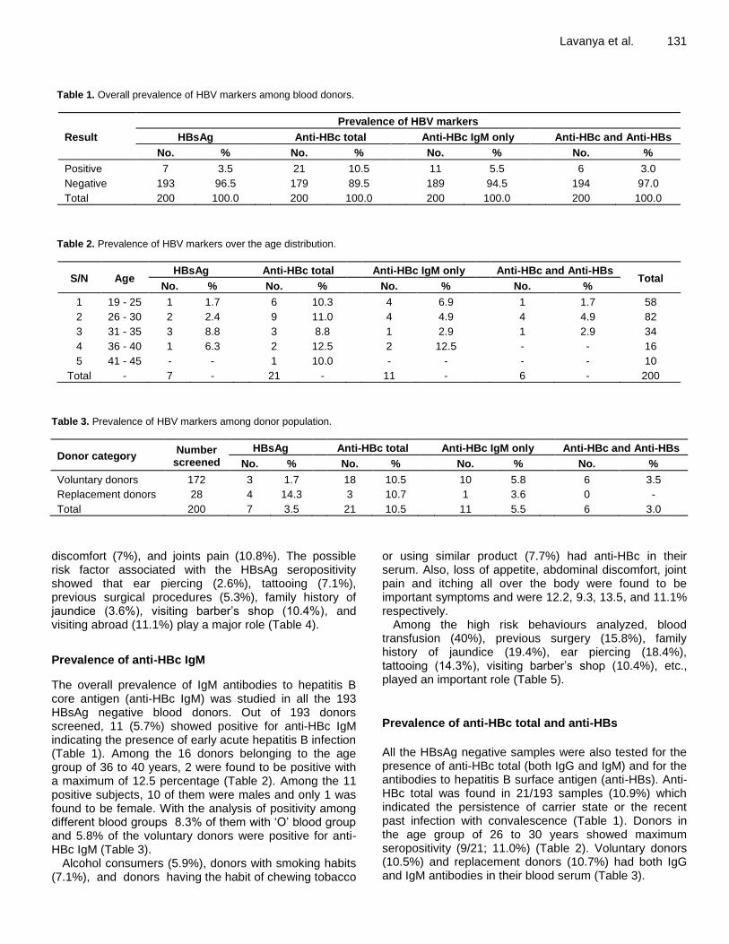
Lavanya et al. 131 Table 1. Overall prevalence of HBV markers among blood donors.
Result
Prevalence of HBV markers
HBsAg Anti-HBc total Anti-HBc IgM only Anti-HBc and Anti-HBs
No. % No. % No. % No. %
Positive 7 3.5 21 10.5 11 5.5 6 3.0
Negative 193 96.5 179 89.5 189 94.5 194 97.0
Total 200 100.0 200 100.0 200 100.0 200 100.0
Table 2. Prevalence of HBV markers over the age distribution.
S/N Age HBsAg Anti-HBc total Anti-HBc IgM only Anti-HBc and Anti-HBs
Total No. % No. % No. % No. %
1 19 - 25 1 1.7 6 10.3 4 6.9 1 1.7 58
2 26 - 30 2 2.4 9 11.0 4 4.9 4 4.9 82
3 31 - 35 3 8.8 3 8.8 1 2.9 1 2.9 34
4 36 - 40 1 6.3 2 12.5 2 12.5 - - 16
5 41 - 45 - - 1 10.0 - - - - 10
Total - 7 - 21 - 11 - 6 - 200
Table 3. Prevalence of HBV markers among donor population.
Donor category Number
screened
HBsAg Anti-HBc total Anti-HBc IgM only Anti-HBc and Anti-HBs
No. % No. % No. % No. %
Voluntary donors 172 3 1.7 18 10.5 10 5.8 6 3.5
Replacement donors 28 4 14.3 3 10.7 1 3.6 0 -
Total 200 7 3.5 21 10.5 11 5.5 6 3.0
discomfort (7%), and joints pain (10.8%). The possible risk factor associated with the HBsAg seropositivity showed that ear piercing (2.6%), tattooing (7.1%), previous surgical procedures (5.3%), family history of jaundice (3.6%), visiting barber’s shop (10.4%), and visiting abroad (11.1%) play a major role (Table 4).
Prevalence of anti-HBc IgM
The overall prevalence of IgM antibodies to hepatitis B core antigen (anti-HBc IgM) was studied in all the 193 HBsAg negative blood donors. Out of 193 donors screened, 11 (5.7%) showed positive for anti-HBc IgM indicating the presence of early acute hepatitis B infection (Table 1). Among the 16 donors belonging to the age group of 36 to 40 years, 2 were found to be positive with a maximum of 12.5 percentage (Table 2). Among the 11 positive subjects, 10 of them were males and only 1 was found to be female. With the analysis of positivity among different blood groups 8.3% of them with ‘O’ blood group and 5.8% of the voluntary donors were positive for anti-HBc IgM (Table 3).
Alcohol consumers (5.9%), donors with smoking habits (7.1%), and donors having the habit of chewing tobacco
or using similar product (7.7%) had anti-HBc in their serum. Also, loss of appetite, abdominal discomfort, joint pain and itching all over the body were found to be important symptoms and were 12.2, 9.3, 13.5, and 11.1% respectively.
Among the high risk behaviours analyzed, blood transfusion (40%), previous surgery (15.8%), family history of jaundice (19.4%), ear piercing (18.4%), tattooing (14.3%), visiting barber’s shop (10.4%), etc., played an important role (Table 5).
Prevalence of anti-HBc total and anti-HBs
All the HBsAg negative samples were also tested for the presence of anti-HBc total (both IgG and IgM) and for the antibodies to hepatitis B surface antigen (anti-HBs). Anti-HBc total was found in 21/193 samples (10.9%) which indicated the persistence of carrier state or the recent past infection with convalescence (Table 1). Donors in the age group of 26 to 30 years showed maximum seropositivity (9/21; 11.0%) (Table 2). Voluntary donors (10.5%) and replacement donors (10.7%) had both IgG and IgM antibodies in their blood serum (Table 3).

132 Int. J. Med. Med. Sci. Table 4. Socio-demographic characteristics and risk factors associated with HBsAg seropositivity in blood donors.
Variable
Prevalence of HBsAg Total Chi square
test P value
Odds ratio
95% CI
Positive Negative Lower limit
Upper limit No. % No. % No. %
Sex
Male 7 3.80 177 96.20 184 100.00 0.631 0.427 - - -
Female 0 - 16 100.00 16 100.00
Blood group
O 3 4.00 72 96.00 75 100.00
1.640 0.65 - - - A 0 - 35 100.00 35 100.00
B 3 4.80 59 95.20 62 100.00
AB 1 3.60 27 96.40 28 100.00
STD awareness
Yes 7 5.40 122 94.60 129 100.00 3.992 0.046** - - -
No 0 - 71 100.00 71 100.00
Alcoholism
Yes 6 5.00 113 95.00 119 100.00 2.069 0.15 4.25 0.50 35.97
No 1 1.20 80 98.80 81 100.00
Smoking habit
Yes 7 6.10 108 93.90 115 100.00 5.362 0.021** - - -
No 0 - 85 100.00 85 100.00
Chewing tobacco
Yes 2 2.60 76 97.40 78 100.00 0.332 0.565 0.62 0.12 3.26
No 5 4.10 117 95.90 122 100.00
Blood transfusions
Yes 0 - 10 100.00 10 100.00 0.382 0.537 0.00 - -
No 7 3.70 183 96.30 190 100.00
Previous surgery
Yes 1 5.30 18 94.70 19 100.00 0.193 0.66 1.62 0.18 14.22
No 6 3.30 175 96.70 181 100.00
Family history of jaundice
Yes 1 3.20 30 96.80 31 100.00 0.008 0.928 0.91 0.11 7.79
No 6 3.60 163 96.40 169 100.00
Ear piercing
Yes 1 2.60 37 97.40 38 100.00 0.105 0.746 0.70 0.08 6.02
No 6 3.70 156 96.30 162 100.00
Tattooing
Yes 2 7.10 26 92.90 28 100.00 1.279 0.258 2.57 0.47 13.94
No 5 2.90 167 97.10 172 100.00
Visiting barber’s shop
Yes 5 10.40 43 89.60 48 100.00 8.946 0.003*** 8.72 1.63 46.54
No 2 1.30 150 98.70 152 100.00

Lavanya et al. 133 Table 4. Contd.
Visiting abroad
Yes 2 11.10 16 88.90 18 100.00 3.393 0.065 4.43 0.79 24.66
No 5 2.70 177 97.30 182 100.00
Extra marital sexual behaviour
Yes 1 12.50 7 87.50 8 100.00 1.999 0.157 4.43 0.47 41.91
No 6 3.10 186 96.90 192 100.00
**Significant at 5% level, ***Significant at 1% level.
Six of 193 HBsAg negative subjects were found to be anti-HBs positive (3.0%) (Table 1). All the 6 anti-HBs positive donors were also found to be positive for anti-HBc. The overall prevalence of anti-HBs and anti-HBc combined seropositivity was (6/21) 28.5%. Anti-HBs prevalence was found maximum among the age group of 26 to 30 years (4.9%). Statistical analysis of prevalence
The statistical analysis of the prevalence of HBsAg and anti-HBc showed that there were no significant correlation between the prevalence rate and the socio-demographic characteristics like age, gender, blood group, and occupation. The prevalence of HBsAg was statistically significant with the type of the donors (P = 0.001), sexually transmitted diseases (STD) awareness (P = 0.04), smoking (P = 0.02), joint pain (P = 0.007), and frequent visits to the barber’s shop (P = 0.003) (Table 4).
The statistical significance in the case of anti-HBc seropositivity was observed in symptoms like loss of appetite (P = 0.03) and joint pain (P = 0.01). Also, risk factors such as blood transfusion, previous surgery, family history of jaundice, ear piercing, and tattooing were all found to be positively correlating the anti-HBc prevalence and was statistically significant (P <0.05) (Table 5). DISCUSSION
The discovery of the HBsAg was a major breakthrough in decreasing the incidence of post transfusion hepatitis. Following infection by the hepatitis B virus (HBV), the first serological marker to appear in the blood is the HBVDNA, followed by HBsAg, the DNA polymerase and the hepatitis B ‘e’ antigen (HBeAg). Thereafter, the antibodies to the hepatitis B core antigen (anti-HBc), hepatitis B ‘e’ antigen and the HBsAg can be detected. Screening of donated blood by ELISA for HBsAg is the common method for detecting hepatitis B infection. Screening of blood for the detection of this viral marker, however, does not rule out the risk of transmission of
hepatitis B totally, because during the host serological response to infection, there is a phase during which the HBsAg cannot be detected in the blood, although, hepatitis B infection is present. This phase is called the ‘window period’. It represents a carrier state of the disease. Therefore, a definite hazard of transmission of hepatitis B to recipients of such units of donated blood exists. During this ‘window period’, detection of the anti-HBc serves as a useful serological marker for hepatitis B infection. The IgM class of the anti-HBc is the first to appear, and indicates a recent infection. The IgG variety of anti-HBc appears later during the infection and points to a past HBV infection. Individuals with IgG variety of anti-HBc may not be infectious as they may have sufficiently high titres of antibodies to HBsAg (anti-HBs), which are protective in nature and the affected individuals may actually be disease free. With the fairly high incidence of HBsAg in India, there is a definite risk of inadvertently transfusing HBV infected blood. It is therefore strongly felt that a marker must be utilized for the screening of blood in the Indian population to detect the presence of hepatitis B during the window period (Hoofnagle et al., 1978; Doglas et al., 1993). The safety of blood products is one of the major problems concerned with the transfusion medicine. At present, HBsAg detection is the only diagnostic screening test for HBV infection identification in the blood transfusion centers of India. The prevalence of anti-HBc in the sera of healthy blood donors negative for HBsAg was not much considered. Since anti-HBc detection is not mandatory in India, the present study was aimed to evaluate if anti-HBc could be adopted as an additional screening assay for blood donation.
To address this issue, a total of 200 healthy blood donors were screened for a period of 4 months. In the present study, the frequency of HBsAg, anti-HBc, and anti-HBs seropositivity in the blood donors of Erode district of Tamil Nadu was investigated.
Among the 200 screened samples, 7 of them (3.5%) were found positive for HBsAg. Similar type of results was found in an Indian study which showed that there was HBsAg prevalence of 3.43% during the year 2008 (Nilima et al., 2010). In contrast, seropositivity in another study was observed to be as low as 1.55% in 1996 and

134 Int. J. Med. Med. Sci. Table 5. Socio-demographic characteristics and risk factors associated with anti -HBc seropositivity in blood donors.
Variable
Prevalence of Anti-HBc Total Chi
square test
P value
Odds ratio
95% CI
Positive Negative Lower limit
Upper limit No. % No. % No. %
Sex
Male 10 5.40 174 94.60 184 100.00 0.019 0.891 0.86 0.10 7.20
Female 1 6.30 15 93.80 16 100.00
Blood group
O 6 8.30 66 91.70 72 100.00
3.069 0.381 5.09 0.59 43.56 A 1 1.80 56 98.20 57 100.00
B 2 4.30 44 95.70 46 100.00
AB 2 8.00 23 92.00 25 100.00
STD awareness
Yes 8 6.20 121 93.80 129 100.00 0.344 0.557 1.50 0.38 5.84
No 3 4.20 68 95.80 71 100.00
Alcoholism
Yes 7 5.90 112 94.10 119 100.00 0.083 0.774 1.20 0.34 4.25
No 4 4.90 77 95.10 81 100.00
Smoking habit
Yes 5 4.30 110 95.70 115 100.00 0.691 0.406 0.60 0.18 2.03
No 6 7.10 79 92.90 85 100.00
Chewing tobacco
Yes 6 7.70 72 92.30 78 100.00 1.182 0.277 1.95 0.57 6.62
No 5 4.10 117 95.90 122 100.00
Blood transfusions
Yes 4 40.00 6 60.00 10 100.00 24.106
0.00***
17.43 3.99 76.05 No 7 3.70 183 96.30 190 100.00
Previous surgery
Yes 3 15.80 16 84.20 19 100.00 4.277
0.039**
4.05 0.98 16.81 No 8 4.40 173 95.60 181 100.00
Family history of jaundice
Yes 6 19.40 25 80.60 31 100.00 13.549
0.00***
7.87 2.23 27.73 No 5 3.00 164 97.00 169 100.00
Ear piercing
Yes 7 18.40 31 81.60 38 100.00 15.07
0.00***
8.92 2.46 32.32 No 4 2.50 158 97.50 162 100.00
Tattooing
Yes 4 14.30 24 85.70 28 100.00 4.835
0.028**
3.93 1.07 14.43 No 7 4.10 165 95.90 172 100.00
Visiting barber’s shop
Yes 5 10.40 43 89.60 48 100.00 2.937 0.087 2.83 0.82 9.72
No 6 3.90 146 96.10 152 100.00

Lavanya et al. 135 Table 5. Contd.
Visiting abroad
Yes 1 5.60 17 94.40 18 100.00 0.00 0.991 1.01 0.12 8.39
No 10 5.50 172 94.50 182 100.00
Extra marital sexual behaviour
Yes 1 12.50 7 87.50 8 100.00 0.786 0.375 2.60 0.29 23.23
No 10 5.20 182 94.80 192 100.00
**Significant at 5% level, ***Significant at 1% level.
0.99% in 2002 (Sharma et al., 2004). A community cluster survey on STD prevalence conducted in Tamil Nadu showed an HBsAg prevalence rate of about 5.7% (Kurien et al., 2005). Also, in another study conducted among the blood donors in tertiary hospital in Tabuk, North Western Saudi Arabia showed a prevalence of about 3.0% (El Beltagy et al., 2008) which was similar to our results.
Age wise prevalence percentage in the present study was found to be maximum in the age group of 31 to 35 years. These results correlated with other studies which also showed the maximum prevalence in the age groups of 26 to 36 years (Tessema et al., 2010; Baba et al., 2000; Ejele et al., 2005). HBV infection was found to be 1.7% in the voluntary donors studied. In contrast, a study conducted in voluntary blood donors in Yola, Nigeria had showed a higher prevalence of 2.4% and another study indicated that the prevalence of 2.2% was seen in the Pakistani donors (Olokoba et al., 2009; Bhatti et al., 2007). Almost similar figures were obtained in a study in Niger Delta region of Nigeria, which was only 1.1% (Ejele et al., 2005). The difference between males and females could not be evaluated because all subjects were male.
The prevalence of HBsAg among the replacement donors was found to be 2.45% in the study conducted in the blood bank, Department of Pathology, MIMS Medical College, Nellimarla, Vizianagaram (Gulia et al., 2011). But, the current study showed a higher prevalence rate of 14.3% seropositivity. The prevalence of HBsAg was significantly high in the first time donors when compared to that of the repeated donors. These results are in agreement with the previous study conducted among blood donors at Gondar University Teaching Hospital, Northwest Ethiopia between January 2003 and December 2007 (Tessema et al., 2010). The increased seroprevalence among the first time donors might be due to the fact that people who regularly donate blood were frequently subjected to screening many times.
The socio-demographic and risk factors associated with prevalence of HBsAg were also analyzed statistically. It was not found to be statistically associated with the age group (P = 0.3), gender (P = 0.4), and blood group (P = 0.65). Similar type of results was obtained in a study conducted in the Christian Medical College and Hospital,
Vellore, Tamil Nadu (Kurien et al., 2005). The analyzed risk factors showed that alcohol consumption (P = 0.15), chewing tobacco (P = 0.5), blood transfusion (P = 0.5), previous surgery (P = 0.6), family history of Jaundice (P = 0.9), tattooing (P = 0.2) and extra marital sexual behaviour (P = 0.1) did not show any statistical significance with the prevalence of HBsAg. Kurien et al. (2005) also showed similar type of insignificant correlations with various risk factors such as alcoholism, tattooing, blood transfusion, extra marital sex, etc.
The prevalence of hepatitis B infection was highly significant in those subjects who had the habit of visiting the community barber’s shop frequently (P = 0.003). But, there were no such related investigations in other studies with reference to the Tamil Nadu state.
The overall prevalence of anti-HBc in the present study was found to be 10.5% for anti-HBc total (IgG and IgM) and 5.5% for anti-HBc IgM only. The present results showed a lower prevalence when compared with a study conducted at the blood bank of Armed Forces Medical College which had a high prevalence of 15.9% of anti-HBc total (Kumar et al., 2007). In another study on the prevalence of hepatitis markers in New Delhi showed a prevalence of 11.6% of anti-HBc total amongst the blood donors (Makroo et al., 1998). In India, the incidence of anti-HBc among the blood donor community ranges from 17 to 29% (Banerjee et al., 1992). The Red Cross blood bank in the city of Behrampur in Ganjam district, Orissa conducted a study to determine the prevalence of anti-HBc positivity in blood donations. They found that out of 729 HBsAg negative donors, 220 (30.1%) were found to be positive for anti-HBc (Panigrahi et al., 2010). With respect to all these studies conducted in various parts of India, the present study had reported a relatively low prevalence of anti-HBc. But, in contrast, the prevalence of the same in Europe and North America was found to be quite low. An anti-HBc prevalence was 0.07% in the UK and 1.5% in Germany has been reported. Another study in Japan has reported a prevalence of 1.1%. This low prevalence might be due to stringent donor selection criteria, high literary rates and the more availability of voluntary donors in these countries (Dhawan et al., 2008).
The current study showed an age wise prevalence of

136 Int. J. Med. Med. Sci. 12.5 and 11% for the age groups of 36 to 40 and 26 to 30 years, respectively. Similar results were obtained in a study among the blood donors in tertiary hospital in Tabuk, Saudi Arabia (El Beltagy et al., 2008). The prevalence rate of HBV markers is high in older age groups and this could be due to the greater number of years of potential exposures and the lack of awareness of HBV infections in earlier decades. Young blood donors should be encouraged to donate blood to help ensure a long-term increase in the blood supply without jeopardizing safety.
The present study noted that the prevalence of anti-HBc total was around 10.7% in the replacement donors and about 10.5% in the voluntary donors. A relatively less prevalence was seen in a study conducted by the Department of Transfusion Medicine, PGIMER, Chandigarh. The study revealed an anti-HBc prevalence of 6.9% in voluntary donors and 10.4% in replacement donors (Dhawan et al., 2008). Another study conducted in Ganjam, West Bengal blood donors in 2004 and 2005 showed anti-HBc seropositivity as high as 18.3% in voluntary blood donors. Also, a report from New Delhi revealed that 6.92% of voluntary donors and 12.53% of the replacement donors were found to be positive for anti-HBc (Bhattacharya et al., 2007; Chaudhuri et al., 2003).
With regards to the socio demographic characters and associated risk factors of the studied subjects, it was found that there was no statistical significance of anti-HBc positivity with the age, gender, blood group, and type of donors. Even though the positive values were obtained, the risk factors such as alcoholism, smoking and tobacco chewing did not have any significance (P > 0.05). A study conducted in Saudi Arabia in 2008 also had the same type of results and they also did not have any statistical significance with HBV prevalence (El Beltagy et al., 2008).
Exposure to the high risky procedures or behaviours in the current study reported a significant association between the anti-HBc prevalence and blood transfusion, previous surgery, ear piercing, family history of Jaundice, tattooing, and visiting barber’s shop. But, these results were in contrast with the study in Saudi Arabia. They showed significance only with the family history of HBV infection. All other risk factors showed no significant association (El Beltagy et al., 2008).
In the present study, the prevalence of anti-HBs among the anti-HBc positive donors was recorded as 3.0%. This was in high controversy with the study conducted in Iran. The Iranian study showed a higher prevalence rate of 37.5% for both anti-HBc and anti-HBs (Behzad-Behbahani et al., 2005). These individuals may have recovered from previous infection, but may have persistent low levels of HBV.
Our study raises serious concerns regarding the safety blood supply even after donor screening for HBsAg. In India, transfusion associated HBV is estimated to be
approximately 50% or more in multiple transfused patients and 1.5% in post surgical recipients according to a study on post transfusion hepatitis B (Saraswat et al., 1996). Thus, the absence of HBsAg in the blood of healthy donors may not be sufficient to ensure lack of circulating HBV.
The transmission of hepatitis B following transfusion of HBsAg negative blood containing anti-HBc, was first described by Hoofnagle in 1978 (Kumar et al., 2007). The most widely used HBV marker, namely, HBsAg, is not usually detected during the window period of the infection. Therefore, transfusion of blood collected from a donor who is in the window period may lead to post transfusion hepatitis B infection in the recipients. Anti-HBc has been found to be an excellent indicator of hepatitis B infection during the window period, and hence, the detection of anti-HBc can contribute significantly in reducing the incidence of post transfusion hepatitis B infection.
Screening of the donor’s blood for anti-HBc is practiced in the western world and this has significantly brought down the possibilities of TAHBV infection in these countries. The introduction of anti-HBc screening, at least in the first time donors might prevent the HBV positive subjects from being included in the donor pool. Since the exclusion of anti-HBc positive units from the donor pool is not practical in areas with intermediate HBsAg prevalence rates such as India where the anti-HBc positivity ranges from 4.2 to 18.3% in healthy donors. Discarding anti-HBc positive units would result in unacceptably high rates of donor rejection.
However, the usefulness of screening for anti-HBc as an additional screening test to improve the safety of blood supply in India deserves still more national wide study and the introduction of polymerase chain reaction (PCR) based screenings like nucleic acid testing (NAT) should also be considered for the Indian blood donors. A national study, including a statistically significant number of blood donors from different blood donation centres across the country, should be carried out to determine the screening for anti-HBc in addition to HBsAg detection.
Conclusively, this study supports the implementation of anti-HBc screening in the donor selection criteria of India, to rule out and identify the HBV infections in blood donors’ and emphasizes on the need for establishing safe and healthy blood transfusion in the nation.
ACKNOWLEDGEMENTS
The authors are grateful to the Dr. M. Karunanithi, Chairman and Secretary, in Vivekananda Educational Institutions and the authorities of Kongu Blood bank, Erode for their support and technical assistance. REFERENCES
Baba MM, Hassan AW, Gashau W (2000). Prevalence of hepatitis B

antigenaemia and human immunodeficiency virus in blood donors in Maidugiri, Nigeria. Niger. J. Med. 9:10-12.
Banerjee K, Sarin SK, Naik SR (1992). Polymerase Chain Reaction based diagnostic assay for identification of Hepatitis B virus. Ind. J. Med. Res. 95:173-178.
Behzad-Behbahani A, Mafi Nejad A, Tabei SZ, Lankarani KB, Rashidi M, Rasouli M, Pourabbas B, Torab A, Salah AR (2005). Indication of anti-HBc antibody screening and HBV DNA detection in diagnosing latent Hepatitis B virus infection. Iran. J. Med. Sci. 30(1):28-33.
Bhattacharya P, Chandra PK, Datta S, Banerjee A, Chakraborty S, Rajendran K, Basu Sk, Bhattacharya SK, Chakravarty R (2007). Significant increase in HBV, HCV, HIV and Syphilis infections among blood donors in West Bengal, Eastern India 2004-2005: Exploratory screening reveals high frequency of occult HBV infection. World J. Gastroenterol. 13:3730-3733.
Bhatti FA, Ullah Z, Salawat N, Ayub M, Ghani E (2007). Anti-Hepatitis B core antigen testing, viral markers and Occult hepatitis B virus infection in Pakistani blood donors: implications for transfusion practice. Transfusion 47(1):74-79.
Chaudhuri V, Nanu A, Panda SK, Chand P (2003). Evaluation of serological screening of blood donors in India reveals a lack of correlation between anti-HBc titers and PCR amplified HBV DNA. Transfusion 43:1442-1448.
Dhawan HK, Marwaha N, Sharma RR, Chawla Y, Thakral B, Saluja K, Sharma SK, Thakur MK, Jain A (2008). Anti-HBc screening in Indian blood donors: Still an unresolved issue. World J. Gastroenterol. 14(34):5327-5330.
Douglas DD, Taswell HF, Rakela J, Rabe D (1993). Absence of hepatitis B DNA detected by polymerase chain reaction in blood donors who are hepatitis B surface antigen negative and antibody to hepatitis B core antigen positive from a United States population with a low prevalence of hepatitis B serologic markers. Transfusion 33:212-216.
Ejele OA, Erhabor O, Nwauche CA (2005). Trends in the prevalence of some transfusion transmissible infections among blood donors in Port Harcourt, Niger. Haema. 8:273-277.
Findik D, Arslan U, Baykan M (2007). Determination of hepatitis B virus DNA incidence, viral load, and mutations in blood donors with HBsAg and anti-HBs-negative serology and antibodies to hepatitis B core antigen. Eur. J. Med. 18:571-575.
Gulia SP, Panda S, Sitaraman E, Reddy KP (2011). Seroprevalence of Hepatitis B Virus infection among blood donors in local population. Internet. J. Pathol. 12(1).
Hollinger FB (2008). Hepatitis B virus infection and transfusion medicine: science and the occult. Transfusion: 48: 1001-1026.
Islam MB (2009). Blood transfusion services in Bangladesh Asian J. Transf. Sci. 3:108-110.
El Beltagy KE, Al Balawi IA, Maha A, Ziad AM (2008). Prevalence of hepatitis B virus markers among blood donors in a tertiary hospital in Tabuk, Northwestern Saudi Arabia. Int. J. Infect. Dis. 12:495-499. Karandeep Singh, Sudha Bhat, Shamee Shastry (2009). Trend in seroprevalence of Hepatitis B virus infection among blood donors of coastal Karnataka, India. J. Infect. Dev. Ctries. 3(5):376-379.
Lavanya et al. 137 Khan ZT, Asim S, Tariz Z, Ehsan IA, Malik RA, Ashfaq (2007).
Prevalence of Transfusion transmitted infections in healthy blood donors in Rawalpindi District, Pakistan–a five year study. Int. J. Pathol. 5:21-25.
Koten F, Oztop Y, Engin A (2011). Investigation of the HBV DNA in isolated hepatitis B core antibody positive blood donors. Sci. Res. Essays 6(13):2720-2723.
Kumar CH, Lt Col Gupta PK, Jaiprakash M (2007). The role of anti-HBc IgM in screening of blood donors. MJAFI, 63:350-352.
Kurien T, Thyagarajan SP, Jeyaseelan L, Peedicayil A, Rajendran P, Sivaram S, Hansdak SG, Renu G, Krishnamurthy P, Sudhakar K, Varghese JC (2005). Community prevalence of hepatitis B infection and modes of transmission in Tamil Nadu, India. Indian J. Med. Res. 670-675.
Makroo RN, Raina V, Kaushik V (1998). Prevalence of hepatitis markers (HBsAg, anti-HBc and anti-HCV) in Indian blood donors. Vox Sang 74:1142.
Nilima S, Sawke GK, Chawla S (2010). Seroprevalence of common Transfusion Transmitted Infections among blood donors. People’s J. Sci. Res. 3(1):5-7.
Olokoba AB, Salawu FK, Danburam A, Desalu OO, Olokaba LB, Wahab KW, Badung LH, Tidi Sk, Midala OM, Aderibigbe S, Abdulrahman MB, Babalola OM, Abdulkkarim A (2009). Viral Hepatitides in Voluntary Blood donors in Yola, Niger. Eur. J. Sci. Res. 31(3):329-334.
Panigrahi R, Biswas A, Datta S, Banerjee A, Chandra PK, Mahapatra PK, Patnaik B, Chakrabarti S, Chakravarty R (2010). Anti-hepatitis B core antigen testing with detection and characterization of occult hepatitis B virus by an in-house nucleic acid testing among blood donors in Behrampur, Ganjam, Orissa in Southeastern India: Implications for transfusion. Virol. J. 7:204-210.
Saraswat S, Banerjee K, Chaundhury N (1996). Post Transfusion hepatitis type B following multiple transfusion of HBsAg negative blood. J. Hepatol. 25:639-643.
Sharma RR, Cheema R, Vajpayee M, Rao U, Kumar S, Marwaha N, Agnihotri SK (2004). Prevalence of markers of Transfusion transmissible diseases in voluntary and replacement blood donors. The Nat. Med. J. India 171:19-21.
Surendra K, Prakash G, Bishnu RT, Manita R (2008). HBsAg Serosurveillance among Nepalese blood donors. Ann. Trop. Med. Public Health 1(1):15-18.
Tessema B, Yismaw G, Kassu A, Amsalu A, Mulu A, Emmrich F, Sack U (2010). Seroprevalence of HIV, HBV, HCV and Syphilis infections among blood donors at Gondar University Teaching Hospital, Northwest Ethiopia: declining trends over a period a five years. BMC Infect. Dis. 10:111-117.