H2S Donors and Their Use in Medicinal Chemistry - MDPI
51
biomolecules Review H 2 S Donors and Their Use in Medicinal Chemistry Elisa Magli, Elisa Perissutti , Vincenzo Santagada, Giuseppe Caliendo, Angela Corvino , Gianluca Esposito, Giovanna Esposito, Ferdinando Fiorino , Marco Migliaccio, Antonia Scognamiglio , Beatrice Severino , Rosa Sparaco and Francesco Frecentese * Citation: Magli, E.; Perissutti, E.; Santagada, V.; Caliendo, G.; Corvino, A.; Esposito, G.; Esposito, G.; Fiorino, F.; Migliaccio, M.; Scognamiglio, A.; et al. H 2 S Donors and Their Use in Medicinal Chemistry. Biomolecules 2021, 11, 1899. https://doi.org/ 10.3390/biom11121899 Academic Editor: Anna Sparatore Received: 5 November 2021 Accepted: 10 December 2021 Published: 18 December 2021 Publisher’s Note: MDPI stays neutral with regard to jurisdictional claims in published maps and institutional affil- iations. Copyright: © 2021 by the authors. Licensee MDPI, Basel, Switzerland. This article is an open access article distributed under the terms and conditions of the Creative Commons Attribution (CC BY) license (https:// creativecommons.org/licenses/by/ 4.0/). Department of Pharmacy, School of Medicine, University of Naples Federico II, Via D. Montesano 49, 80131 Napoli, Italy; [email protected] (E.M.); [email protected] (E.P.); [email protected] (V.S.); [email protected] (G.C.); [email protected] (A.C.); [email protected] (G.E.); [email protected] (G.E.); fefi[email protected] (F.F.); [email protected] (M.M.); [email protected] (A.S.); [email protected] (B.S.); [email protected] (R.S.) * Correspondence: [email protected]; Tel.: +39-081-679-829 Abstract: Hydrogen sulfide (H 2 S) is a ubiquitous gaseous signaling molecule that has an impor- tant role in many physiological and pathological processes in mammalian tissues, with the same importance as two others endogenous gasotransmitters such as NO (nitric oxide) and CO (carbon monoxide). Endogenous H 2 S is involved in a broad gamut of processes in mammalian tissues in- cluding inflammation, vascular tone, hypertension, gastric mucosal integrity, neuromodulation, and defense mechanisms against viral infections as well as SARS-CoV-2 infection. These results suggest that the modulation of H 2 S levels has a potential therapeutic value. Consequently, synthetic H 2 S- releasing agents represent not only important research tools, but also potent therapeutic agents. This review has been designed in order to summarize the currently available H 2 S donors; furthermore, herein we discuss their preparation, the H 2 S-releasing mechanisms, and their -biological applications. Keywords: hydrogen sulfide; natural H 2 S donors; synthetic H 2 S donors; H 2 S release; medicinal chemistry 1. Introduction 1.1. Hydrogen Sulfide: Chemical Properties Hydrogen sulfide (H 2 S) is a colorless, flammable, and water-soluble gas well known for its characteristic odor of rotten eggs. The acute effects of H 2 S in humans are directly proportional to its concentration and these include eye, nose, throat, and respiratory system irritation (low concentrations) and rapid loss of consciousness and death (high concentrations) [1–3]. Recently H 2 S has been considered as a member of the gasotransmitter family because it shows some common characteristics with its congeners NO (nitric oxide) and CO (carbon monoxide) [4–11]. Hydrogen sulfide has a chemical structure similar to that of water, but it is much less polar than water because the sulfur atom is less electronegative than the oxygen present in water. For this reason, the intermolecular forces for H 2 S are relatively weaker and consequently the melting and boiling points are much lower than they are for water. H 2 S can be considered as a weak acid that gives rise to two dissociations in aqueous solution (Ka 1 = 1.3 × 10 -7 M, Ka 2 =1 × 10 -19 M). From the first dissociation hydronium cation (H 3 O + ) and hydrosulfide anion (HS - ) are obtained; the second dissociation leads to H 3 O + and sulfide ions (S 2- ). In biological fluids and under physiological conditions hydrogen sulfide is mainly undissociated [12]. Hydrogen sulfide represents a reductant species because of the oxidation state of the sulfur atom in H 2 S that is –2; it can also be considered a potent nucleophile at physiological pH values. These features make H 2 S a very reactive acid that can react with many biological molecules. Hydrogen sulfide can give rise to two kinds of redox reactions: the first Biomolecules 2021, 11, 1899. https://doi.org/10.3390/biom11121899 https://www.mdpi.com/journal/biomolecules
-
Upload
khangminh22 -
Category
Documents
-
view
2 -
download
0
Transcript of H2S Donors and Their Use in Medicinal Chemistry - MDPI

biomolecules
Review
H2S Donors and Their Use in Medicinal Chemistry
Elisa Magli, Elisa Perissutti , Vincenzo Santagada, Giuseppe Caliendo, Angela Corvino , Gianluca Esposito,Giovanna Esposito, Ferdinando Fiorino , Marco Migliaccio, Antonia Scognamiglio , Beatrice Severino ,Rosa Sparaco and Francesco Frecentese *
�����������������
Citation: Magli, E.; Perissutti, E.;
Santagada, V.; Caliendo, G.; Corvino,
A.; Esposito, G.; Esposito, G.; Fiorino,
F.; Migliaccio, M.; Scognamiglio, A.;
et al. H2S Donors and Their Use in
Medicinal Chemistry. Biomolecules
2021, 11, 1899. https://doi.org/
10.3390/biom11121899
Academic Editor: Anna Sparatore
Received: 5 November 2021
Accepted: 10 December 2021
Published: 18 December 2021
Publisher’s Note: MDPI stays neutral
with regard to jurisdictional claims in
published maps and institutional affil-
iations.
Copyright: © 2021 by the authors.
Licensee MDPI, Basel, Switzerland.
This article is an open access article
distributed under the terms and
conditions of the Creative Commons
Attribution (CC BY) license (https://
creativecommons.org/licenses/by/
4.0/).
Department of Pharmacy, School of Medicine, University of Naples Federico II, Via D. Montesano 49,80131 Napoli, Italy; [email protected] (E.M.); [email protected] (E.P.); [email protected] (V.S.);[email protected] (G.C.); [email protected] (A.C.); [email protected] (G.E.);[email protected] (G.E.); [email protected] (F.F.); [email protected] (M.M.);[email protected] (A.S.); [email protected] (B.S.); [email protected] (R.S.)* Correspondence: [email protected]; Tel.: +39-081-679-829
Abstract: Hydrogen sulfide (H2S) is a ubiquitous gaseous signaling molecule that has an impor-tant role in many physiological and pathological processes in mammalian tissues, with the sameimportance as two others endogenous gasotransmitters such as NO (nitric oxide) and CO (carbonmonoxide). Endogenous H2S is involved in a broad gamut of processes in mammalian tissues in-cluding inflammation, vascular tone, hypertension, gastric mucosal integrity, neuromodulation, anddefense mechanisms against viral infections as well as SARS-CoV-2 infection. These results suggestthat the modulation of H2S levels has a potential therapeutic value. Consequently, synthetic H2S-releasing agents represent not only important research tools, but also potent therapeutic agents. Thisreview has been designed in order to summarize the currently available H2S donors; furthermore,herein we discuss their preparation, the H2S-releasing mechanisms, and their -biological applications.
Keywords: hydrogen sulfide; natural H2S donors; synthetic H2S donors; H2S release; medicinalchemistry
1. Introduction1.1. Hydrogen Sulfide: Chemical Properties
Hydrogen sulfide (H2S) is a colorless, flammable, and water-soluble gas well knownfor its characteristic odor of rotten eggs. The acute effects of H2S in humans are directlyproportional to its concentration and these include eye, nose, throat, and respiratorysystem irritation (low concentrations) and rapid loss of consciousness and death (highconcentrations) [1–3]. Recently H2S has been considered as a member of the gasotransmitterfamily because it shows some common characteristics with its congeners NO (nitric oxide)and CO (carbon monoxide) [4–11].
Hydrogen sulfide has a chemical structure similar to that of water, but it is much lesspolar than water because the sulfur atom is less electronegative than the oxygen presentin water. For this reason, the intermolecular forces for H2S are relatively weaker andconsequently the melting and boiling points are much lower than they are for water. H2Scan be considered as a weak acid that gives rise to two dissociations in aqueous solution(Ka1 = 1.3 × 10⁻⁷ M, Ka2 = 1 × 10⁻¹⁹ M). From the first dissociation hydronium cation(H3O+) and hydrosulfide anion (HS−) are obtained; the second dissociation leads to H3O+
and sulfide ions (S2−). In biological fluids and under physiological conditions hydrogensulfide is mainly undissociated [12].
Hydrogen sulfide represents a reductant species because of the oxidation state of thesulfur atom in H2S that is –2; it can also be considered a potent nucleophile at physiologicalpH values. These features make H2S a very reactive acid that can react with many biologicalmolecules. Hydrogen sulfide can give rise to two kinds of redox reactions: the first
Biomolecules 2021, 11, 1899. https://doi.org/10.3390/biom11121899 https://www.mdpi.com/journal/biomolecules
Biomolecules 2021, 11, 1899 2 of 51
oxidation should form a highly reactive HS• radical (for example in the reaction withmetal centers) [13], while the second kind of redox reaction of H2S with many biologicallyimportant reactive oxygen species (ROS) can also lead to the formation of species suchas hydrogen peroxide (H2O2), superoxide (O2), peroxynitrite (ONOO), and NO. [14–16].Hydrogen sulfide is also able to react with S-nitroso thiols to give thionitrous acid (HSNO)that probably acts as a cell-permeable nitrosylating agent that might represent the linkand explain the cell membrane–impermeable transnitrosation mechanism [17]. Someevidence reported in the literature suggests that H2S also mediates an important oxidativepost-translational modification and in particular protein S-sulfuration, generating, forexample, S-SH groups on cysteine residues. Even if the detailed mechanism(s) is not yetknown, this reaction could represent the chemical route involving H2S for the modificationof the functions of a broad range of cellular proteins and enzymes. In the literature,authors have generally agreed that H2S reacts with cysteine-modified residues, such asdisulfides (S-S–), sulfenic acids (S-OH), nitrosothiols (S-NO), etc., to form S-sulfhydratedproducts. Some authors have suggested that protein cysteines directly react with some H2Smetabolites or precursors, such as sulfane sulfurs and hydrogen polysulfides, to obtainS-SH products [18–27]. However, further studies are needed to elucidate this mechanismin detail. Finally, H2S is able to bind transition metals by coordination bonds. In particular,it is able to bind strongly to copper, and this feature has allowed the development offluorescent probes [28]. It has been shown that the activation of odorant receptors mediatedby the sulfur–copper coordination leads to an increase in human sensitivity to the smell ofvolatile sulfur compounds [29].
The toxicity of hydrogen sulfide to mammals is due to the interactions with cy-tochrome c oxidase (CcO) with the consequent inhibition of mitochondrial respiration;however, the significant capacity of H2S to induce a suspended animation-like state [30]is believed to be associated with a reversible inhibition of CcO by sulfide coordinationchemistry [31].
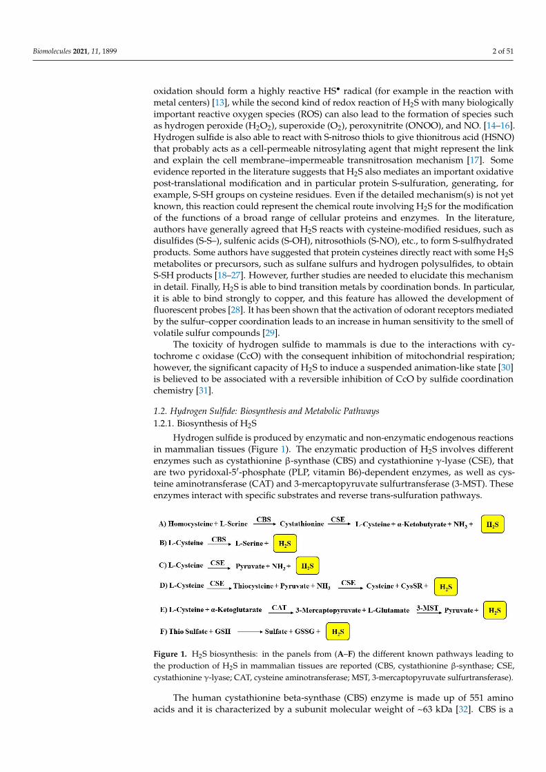
1.2. Hydrogen Sulfide: Biosynthesis and Metabolic Pathways1.2.1. Biosynthesis of H2S
Hydrogen sulfide is produced by enzymatic and non-enzymatic endogenous reactionsin mammalian tissues (Figure 1). The enzymatic production of H2S involves differentenzymes such as cystathionine β-synthase (CBS) and cystathionine γ-lyase (CSE), thatare two pyridoxal-5′-phosphate (PLP, vitamin B6)-dependent enzymes, as well as cys-teine aminotransferase (CAT) and 3-mercaptopyruvate sulfurtransferase (3-MST). Theseenzymes interact with specific substrates and reverse trans-sulfuration pathways.
Biomolecules 2021, 10, x 2 of 52
many biological molecules. Hydrogen sulfide can give rise to two kinds of redox reac-
tions: the first oxidation should form a highly reactive HS• radical (for example in the
reaction with metal centers) [13], while the second kind of redox reaction of H2S with
many biologically important reactive oxygen species (ROS) can also lead to the for-
mation of species such as hydrogen peroxide (H2O2), superoxide (O2), peroxynitrite
(ONOO), and NO. [14–16]. Hydrogen sulfide is also able to react with S-nitroso thiols to
give thionitrous acid (HSNO) that probably acts as a cell-permeable nitrosylating agent
that might represent the link and explain the cell membrane–impermeable transnitrosa-
tion mechanism [17]. Some evidence reported in the literature suggests that H2S also
mediates an important oxidative post-translational modification and in particular pro-
tein S-sulfuration, generating, for example, S-SH groups on cysteine residues. Even if the
detailed mechanism(s) is not yet known, this reaction could represent the chemical route
involving H2S for the modification of the functions of a broad range of cellular proteins
and enzymes. In the literature, authors have generally agreed that H2S reacts with cyste-
ine-modified residues, such as disulfides (S-S–), sulfenic acids (S-OH), nitrosothiols (S-
NO), etc., to form S-sulfhydrated products. Some authors have suggested that protein
cysteines directly react with some H2S metabolites or precursors, such as sulfane sulfurs
and hydrogen polysulfides, to obtain S-SH products [18–27]. However, further studies
are needed to elucidate this mechanism in detail. Finally, H2S is able to bind transition
metals by coordination bonds. In particular, it is able to bind strongly to copper, and this
feature has allowed the development of fluorescent probes [28]. It has been shown that
the activation of odorant receptors mediated by the sulfur–copper coordination leads to
an increase in human sensitivity to the smell of volatile sulfur compounds [29].
The toxicity of hydrogen sulfide to mammals is due to the interactions with cyto-
chrome c oxidase (CcO) with the consequent inhibition of mitochondrial respiration;
however, the significant capacity of H2S to induce a suspended animation-like state [30]
is believed to be associated with a reversible inhibition of CcO by sulfide coordination
chemistry [31].
1.2. Hydrogen Sulfide: Biosynthesis and Metabolic Pathways
1.2.1. Biosynthesis of H2S
Hydrogen sulfide is produced by enzymatic and non-enzymatic endogenous reac-
tions in mammalian tissues (Figure 1). The enzymatic production of H2S involves differ-
ent enzymes such as cystathionine -synthase (CBS) and cystathionine -lyase (CSE),
that are two pyridoxal-5′-phosphate (PLP, vitamin B6)-dependent enzymes, as well as
cysteine aminotransferase (CAT) and 3-mercaptopyruvate sulfurtransferase (3-MST).
These enzymes interact with specific substrates and reverse trans-sulfuration pathways.
Figure 1. H2S biosynthesis: in the panels from (A–F) the different known pathways leading to the
production of H2S in mammalian tissues are reported (CBS, cystathionine -synthase; CSE, cysta-
thionine -lyase; CAT, cysteine aminotransferase; MST, 3-mercaptopyruvate sulfurtransferase).
Figure 1. H2S biosynthesis: in the panels from (A–F) the different known pathways leading tothe production of H2S in mammalian tissues are reported (CBS, cystathionine β-synthase; CSE,cystathionine γ-lyase; CAT, cysteine aminotransferase; MST, 3-mercaptopyruvate sulfurtransferase).
The human cystathionine beta-synthase (CBS) enzyme is made up of 551 aminoacids and it is characterized by a subunit molecular weight of ~63 kDa [32]. CBS is a
Biomolecules 2021, 11, 1899 3 of 51
tetrameric protein, and each subunit binds to one heme and one pyridoxal 5′-phosphate(PLP). The catalytic center is similar to that of other members of the β- or fold II class of PLP-dependent enzymes and represents the conserved part of the protein [33]. ProtoporphyrinIX represents a structural requirement of cystathionine β-synthase, and this feature isunique in the family of PLP-dependent enzymes. Moreover, a conserved glycine richloop of fold II enzymes (G256–T257–G258–G259–T260) is responsible for multifariouselectrostatic interactions with the phosphate portion of PLP [33].
The three-dimensional structures of human and yeast cystathionine gamma-lyase(CSE) have been determined through X-ray crystallography studies [34]. CSE is charac-terized by four equal monomers with a molecular weight of ~45 kDa; in each monomerthe pyridoxal 5′-phosphate (PLP) cofactor is linked with a covalent bond. Nevertheless,some studies have reported differential PLP binding affinities within the monomers of CSEand a momentary dissociation or dismissal of PLP from the enzyme during the catalyticaction [35]. Based on studies reported in the literature, several active site residues havebeen identified as being involved in the catalysis of the α,β-elimination reaction, leadingto the production of H2S [36]. In the crystal structures of yeast and human CSE, someresidues participate in the binding of the PLP cofactor. These residues are represented byTyr60 and Arg62 from the adjacent monomer; Tyr114 that forms the π-stacking interactionwith the pyridoxal moiety of the cofactor; Asp187 that establishes hydrogen bonds with thepyridoxal nitrogen; and Ser209 and Thr211 that form hydrogen bonds with the phosphategroup. Some studies have also reported that Lys212 represents a significant catalytic residueconsidering that it forms a covalent bond with the PLP cofactor and facilitates protontransfer reactions during the α,γ-elimination reaction of L-cystathionine. Site-directedmutagenesis studies have demonstrated that the PLP cofactor also binds to Tyr60 and Arg62
from the adjacent subunit through a hydrogen bond [37].CBS and CSE catalyze reactions that represent the primary pathways for H2S produc-
tion (Figure 1). In particular, CBS is involved in the β-replacement reaction of homocysteinewith serine to give cystathionine. CSE later takes part in the α,γ-elimination of cystathion-ine that leads to cysteine, α-ketobutyrate, NH3, and hydrogen sulfide (Figure 1A) [38].
Either CBS or CSE can also produce H2S from L-cysteine via β-elimination reac-tions [39]. In particular, CBS forms L-serine and H2S from L-cysteine (Figure 1B), whileCSE generates pyruvate (using L-cysteine as the substrate), NH3 and H2S (Figure 1C) orit firstly forms thiocysteine, pyruvate, and NH3 and subsequently cysteine, S-alkenyl-mercaptocysteine (CysSR), and H2S (Figure 1D). Furthermore, CSE catalyzes the reactionof L-homoserine to form 2-oxobutanoate, NH3, and H2O.
CAT and 3-MST represent two other enzymes involved in the biosynthesis of hydrogensulfide; CAT is able to transfer the amine group from cysteine to a keto acid (for exampleα-ketoglutarate) forming 3-mercaptopyruvate. Subsequently, 3-MST leads to the formationof the persulfide, 3-MST-SSH, through the desulfuration of 3-mercaptopyruvate (Figure 1E).3-mercaptopyruvate sulfur-transferase then transfers the sulfur from 3-mercaptopyruvateto sulfite or other sulfur acceptors or forms elemental sulfur. The reaction is catalyzed byCAT and 3-MST and produces sulfane sulfur (or bound sulfur); subsequently, a reductionof the sulfur atom or a release from the thiosulfate or persulfides leads to the production ofH2S. Therefore, the presence of reductants [40] and specific enzymes such as thiosulfatesulfur transferase or thiosulfate reductase is necessary to obtain hydrogen sulfide [41].The enzymes involved in hydrogen sulfide biosynthesis are widely distributed. Both CBSand CSE represent cytosolic enzymes. CBS was initially thought to be predominantlyinvolved in the production of H2S in the brain, while CSE was thought to be predominantlyinvolved in the synthesis of H2S in the heart and blood vessels [42]. Subsequent studieshave established a broader distribution of these enzymes and in particular, it was seenthat CBS is present in vascular endothelium, CAT and 3-MST are present in the vascularendothelium and the brain, and 3-MST, but not CAT, is present in the vascular smoothmuscle [43]. CAT and 3-MST constitute both mitochondrial and cytosolic enzymes [44],though 3-MST is believed to be predominantly present in the mitochondrion [45].

Biomolecules 2021, 11, 1899 4 of 51
PGG (propargylglycine), BCA (β-cyanoalanine), AOAA (aminooxyacetic acid), trifluo-roalanine, and HA (hydroxylamine) represent the most commonly used agents that inhibitH2S biosynthesis; in particular PGG and BCA can be considered specific CSE inhibitors,while AOAA can be considered as a CBS-selective inhibitor [7,46]. In particular, PGGis a specific CSE inhibitor in comparison to CBS, but it is able to interact with differentPLP-dependent enzymes that are not necessarily involved in H2S production. In a study re-ported in the literature that monitored vascular disposition in cerebellar slices and in intactmouse brains and that foresaw the use of two-photon intravital laser scanning microscopy,Morikawa et al. analyzed the pathway mediating hypoxia-induced cerebral vasodilation.It was established that either H2S, obtained by cystathionine β-synthase (CBS), or CO,formed by heme oxygenase (HO)-2hypoxia caused cerebral vasodilation. Hypoxia reducesCO generation by HO-2, an oxygen sensor. The constitutive CO physiologically inhibitsCBS, and hypoxia increases the H2S levels that leads to the vasodilation of precapillaryarterioles. It has been observed that mice with a targeted deletion of HO-2 or CBS showedchanged vascular responses to hypoxia. Therefore, the imaging mass spectrometry of anintact adult brain cerebral cortex of HO-2–null mice showed an impaired ability to preserveATP levels during hypoxia [47].
As previously reported, H2S is also involved in non-enzymatic reactions (Figure 1F).In particular, the non-enzymatic reduction of elemental sulfur leads to a minor endogenoussource of hydrogen sulfide; this reaction occurs using reducing equivalents obtained fromthe oxidation of glucose in erythrocytes [48]. Human erythrocytes are able to obtain H2Sin the presence of elemental sulfur or inorganic polysulfides. Non-enzymatic oxidationsulfides provide thiosulfate and these can be converted to sulfite by enzymatic reactionswith thiosulfate reductase in the liver, kidney, or brain, or by thiosulfate sulfur-transferasein the liver. H2S can also be released from thiosulfate and persulfides. Garlic-derivedorganic polysulfides have been proven to release hydrogen sulfide in a thiol-dependentmanner [49]. Garlic (Allium sativum) has long been considered beneficial as an antioxidant;recent studies have demonstrated several favorable effects of garlic derived from H2Sproduction. Allicin (diallylthiosulfinate) is the best characterized naturally occurringH2S-donating garlic compound; it decomposes in water to form compounds such asdiallyldisulfide (DADS) and diallyltrisulfide (DATS).
1.2.2. H2S Catabolic Pathways
The catabolism of hydrogen sulfide includes non-enzymatic and enzymatic reactions.In the first reaction of a catabolic pathway, H2S is rapidly oxidized, mainly in the mitochon-dria and in a non-enzymatic manner, to thiosulfate; subsequently, through an enzymaticreaction, thiosulfate is converted to sulfate and/or sulfite by TST (thiosulfate cyanide sulfur-transferase) (Figure 2A). Sulfite obtained by this reaction is then rapidly oxidized to sulfate;the latter is the major end-product of H2S catabolism under physiological conditions [50].Another catabolic pathway of H2S is represented by the methylation to methanethiol anddimethyl sulfide mediated by TSMT (thiol-S-methyltransferase) (Figure 2B). This lattercatabolic pathway occurs in the cytosol of different cells of the gastrointestinal tract [51,52].The last catabolic reaction of H2S involves the methemoglobin that is able to bind toendogenous gases (for example CO and NO); moreover, it is able to bind H2S to givesulfhemoglobin (Figure 2C) [53].
Biomolecules 2021, 10, x 5 of 52
Figure 2. H2S catabolic pathways. (A) mitochondrial oxidation; (B) cytosolic methylation; (C) binding to hemoglobin.
TST: thiosulfate cyanide sulfur transferase (rhodanese). TSMT: thiol-S-thiomethyl transferase.
The catabolism of H2S in the mitochondria is related to the coupled activity of sev-
eral mitochondrial enzymes (SQR, ETHE1, SO, of which the initial one (SQR) is oxygen-
independent) and their substrates (CoC, GSH glutathione, O2). In order to form thiosul-
fate, H2S is initially processed by SQR and GSH to form glutathione persulfide (GSSH)
that serves as a substrate for both sulfite and thiosulfate formation. Alternatively, H2S
can be processed by SQR and sulfite to form thiosulfate. Most of the catabolic reactions
regarding H2S in mitochondria are enzymatic [17].
1.3. Hydrogen Sulfide: Biological Functions
The scientific literature concerning the biological roles for endogenous hydrogen
sulfide is continuously growing. Several studies have reported that H2S performs physi-
ological effects at a broad range of concentrations (10–300 M) [5–7]. It is known that
H2S participates in the regulation of numerous physiological responses, for example, an-
ti-inflammation [54], oxidative stress [55], neuromodulation [56], vasoregulation [57],
protection from reperfusion injury after myocardial infarction [58], and insulin re-
sistance [59]. Furthermore, scientific research has continued to investigate H2S in relation
to its involvement in various phases of cellular signaling, cell function, and cytoprotec-
tion.
1.3.1. Cardiovascular System
As reported in several studies, H2S has biological significance in the control of car-
diovascular homeostasis. Particularly, it has been shown that H2S presents all the posi-
tive effects of NO without forming toxic metabolites [60]. Lately, it has been observed
that the function of H2S in cardiovascular homeostasis is more important and crucial in
cases of NO-mediated control compromise such as in the endothelial dysfunction. Hy-
drogen sulfide induces relaxing effects in the vascular smooth muscle and this effect has
been observed in large vessels, for example, the rat thoracic aorta and portal vein and al-
so in peripheral resistance vessels, which have a more substantial role than large conduit
arteries in the modulation of vascular resistance and blood pressure [61]. L-cysteine rep-
resents a source of H2S; in fact, the vasorelaxing activity of L-cysteine is reduced by the
CSE inhibitor PGG. Genetic deletion of CSE in mice significantly decreases H2S levels in
the serum, heart, and aorta, and mutant mice lacking CSE presented with marked hyper-
tension and a reduced endothelium-dependent vasorelaxant effect [62]. In fact, it is
known that H2S relaxes blood vessels primarily, but not exclusively, by opening the KATP
channels of the vascular smooth muscle cells. In addition to the vasorelaxing effect, H2S,
as well as NO, is characterized by a broad range of other biological effects, which are
significant for the polyhedric control of the cardiovascular system. For example, H2S is
able to inhibit the platelet aggregation/adhesion induced by ADP, collagen, epinephrine,
arachidonic acid, thromboxane mimetic U46619, and thrombin [63].
1.3.2. Immune System and Inflammation
The production and functional role of H2S is important in the several cell types clas-
sically related to innate immunity (such as neutrophils, eosinophils, basophils macro-
phages, dendritic cells, natural killer cells, mast cells,) and adaptive immunity (T and B
Figure 2. H2S catabolic pathways. (A) mitochondrial oxidation; (B) cytosolic methylation; (C) binding to hemoglobin. TST:thiosulfate cyanide sulfur transferase (rhodanese). TSMT: thiol-S-thiomethyl transferase.

Biomolecules 2021, 11, 1899 5 of 51
The catabolism of H2S in the mitochondria is related to the coupled activity of sev-eral mitochondrial enzymes (SQR, ETHE1, SO, of which the initial one (SQR) is oxygen-independent) and their substrates (CoC, GSH glutathione, O2). In order to form thiosulfate,H2S is initially processed by SQR and GSH to form glutathione persulfide (GSSH) thatserves as a substrate for both sulfite and thiosulfate formation. Alternatively, H2S can beprocessed by SQR and sulfite to form thiosulfate. Most of the catabolic reactions regardingH2S in mitochondria are enzymatic [17].
1.3. Hydrogen Sulfide: Biological Functions
The scientific literature concerning the biological roles for endogenous hydrogensulfide is continuously growing. Several studies have reported that H2S performs phys-iological effects at a broad range of concentrations (10–300 µM) [5–7]. It is known thatH2S participates in the regulation of numerous physiological responses, for example, anti-inflammation [54], oxidative stress [55], neuromodulation [56], vasoregulation [57], protec-tion from reperfusion injury after myocardial infarction [58], and insulin resistance [59].Furthermore, scientific research has continued to investigate H2S in relation to its involve-ment in various phases of cellular signaling, cell function, and cytoprotection.
1.3.1. Cardiovascular System
As reported in several studies, H2S has biological significance in the control of cardio-vascular homeostasis. Particularly, it has been shown that H2S presents all the positiveeffects of NO without forming toxic metabolites [60]. Lately, it has been observed thatthe function of H2S in cardiovascular homeostasis is more important and crucial in casesof NO-mediated control compromise such as in the endothelial dysfunction. Hydrogensulfide induces relaxing effects in the vascular smooth muscle and this effect has beenobserved in large vessels, for example, the rat thoracic aorta and portal vein and also inperipheral resistance vessels, which have a more substantial role than large conduit arteriesin the modulation of vascular resistance and blood pressure [61]. L-cysteine represents asource of H2S; in fact, the vasorelaxing activity of L-cysteine is reduced by the CSE inhibitorPGG. Genetic deletion of CSE in mice significantly decreases H2S levels in the serum,heart, and aorta, and mutant mice lacking CSE presented with marked hypertension anda reduced endothelium-dependent vasorelaxant effect [62]. In fact, it is known that H2Srelaxes blood vessels primarily, but not exclusively, by opening the KATP channels of thevascular smooth muscle cells. In addition to the vasorelaxing effect, H2S, as well as NO,is characterized by a broad range of other biological effects, which are significant for thepolyhedric control of the cardiovascular system. For example, H2S is able to inhibit theplatelet aggregation/adhesion induced by ADP, collagen, epinephrine, arachidonic acid,thromboxane mimetic U46619, and thrombin [63].
1.3.2. Immune System and Inflammation
The production and functional role of H2S is important in the several cell types classi-cally related to innate immunity (such as neutrophils, eosinophils, basophils macrophages,dendritic cells, natural killer cells, mast cells,) and adaptive immunity (T and B lympho-cytes). H2S is also associated with the progress of various inflammatory and immunediseases. In this context, H2S has physiological and pathophysiological roles especially inthe oral cavity and in the colon, where endogenous bacteria produce H2S giving rise to a“H2S environment” to which the immune cells and the parenchymal cells are subjected; this“H2S environment” significantly regulates their viability and function. The development ofspontaneous autoimmune disease, the onset, or the worsening of the severity of variousimmune-mediated diseases (such as autoimmune rheumatoid arthritis or asthma) could beconnected to the downregulation of endogenous H2S-producing enzymes, or to the geneticdefects in H2S biosynthetic enzyme systems. Small amounts of H2S, delivered by donors,enhance the function of various immune cells, and defend them against the dysfunctionscaused by various noxious stimuli. The effects of H2S preserve the immune functions,

Biomolecules 2021, 11, 1899 6 of 51
increase the antimicrobial defenses, and exercise anti-inflammatory therapeutic effects invarious diseases.
As is already known, nonsteroidal anti-inflammatory drugs (NSAIDs) are able toinduce gastroenteropathy [64]. Studies reported in the scientific literature suggest thatNSAIDs repress endogenous H2S synthesis by decreasing the expression of CSE. Theadditional decrease in H2S synthesis may sequentially cause an expansion in leukocyteadherence leading to the development of the gastric injury that has been noted subsequentto NSAID administration [65]. Equally, the administration of exogenous H2S decreasesthe capacity of these agents to affect gastric injury. Exogenous administration of H2Srepressed NSAID-induced granulocyte infiltration, the expression of endothelial and leuko-cyte adhesion molecules, and the expression of TNFα (tumor necrosis factor α) [64]. Ithas been shown that leukocyte adhesion to the vascular endothelium produced by aspirininjury decreases after improvements in H2S bioavailability and that CSE inhibition withpropargylglycine (PGG) impaired aspirin-mediated mucosal injury and inflammation. Ithas been also observed that the leukocyte expression of LFA-1 is repressed by exogenousH2S. It is also important to highlight a molecular characteristic of the H2S-induced anti-inflammatory effects: H2S donors reduced aspirin-induced leukocyte adhesion as a resultof the activation of KATP channels and the inhibition of the CSE activity that encouragesleukocyte adhesion. Further studies have shown that the co-administration of an H2Sdonor with an NSAID leads to the inhibition of NSAID-induced leukocyte adherence anda decrease in the gravity of gastric injury [66].
1.3.3. Respiratory System
The relaxant effects of H2S have also been explored on the rings of the bronchialsmooth muscle of two rodent species. Fu et al. demonstrated that H2S produced astrong relaxation in the isolated bronchus rings from the mouse but generated only minorrelaxation in guinea pig rings. H2S content and CSE activity were also improved in anisolated rat lung which presented with an ischemia/reperfusion injury, and a preventativeperfusion with H2S reduced this injury, decreasing malondyaldehyde (MDA) productionand increasing superoxide dismutase and catalase activity [67].
The endogenous CSE/H2S pathway seems to have an important role in pulmonaryhypertension (PH). Specifically, this pathway was down-regulated in hypoxic PH (HPH),causing a reduction in endogenous H2S production in rat lung tissues as a result of oxida-tive stress. In lung tissues, the gene expression, and the activity of the CSE enzyme weresuppressed during HPH, but the exogenous amount of H2S led to an improvement in CSEactivity, an upregulation of CSE gene expression in the lung tissue, and a decrease in theremodeling of the pulmonary vascular structure during HPH [68]. Subsequently, after thetreatment with exogenous H2S, HPH was reduced because of a direct scavenging of oxi-dized glutathione (GSSG), an improved total antioxidant capacity [69], and the inhibitoryeffect of H2S on pulmonary vascular inflammation, connected to a high I-B (inhibitorof NF-B) expression and a down-regulation of NF-B p65 expression [70]. Moreover, theCSE/H2S pathway seems to also have a favorable effect in asthma and chronic obstructivepulmonary disease (COPD), two of the most important obstructive airway diseases. Inmilder COPD, enhanced levels of H2S serum may have a beneficial effect in airway protec-tion, antagonizing oxidative stress and airway inflammation and blocking the progress ofCOPD [71]. Scientific research has shown many positive effects for H2S in ischemia for sev-eral tissues. H2S has a broad range of protective functions related to ischemia/reperfusion(I/R) injury all through the body. The control of intracellular [Ca2+] through the stimulationof the KATP channel opening, anti-apoptotic pathway (ERK1/2/MAPK, PI-3 kinase/Akt,JAK-STAT) activation, mitochondrial protection through the preservation of ∆Ψm and theinhibition of the mPTP opening, and the anti-inflammatory effects through the activationof eNOS and p38 MAPK represent the mechanisms of protection [72]. H2S has been alsorevealed to be protective against pulmonary ischemia injury and the endogenous CSE/H2Spathway could be implicated in the pathogenesis of lung I/R injury [67]. In a recent study,
Biomolecules 2021, 11, 1899 7 of 51
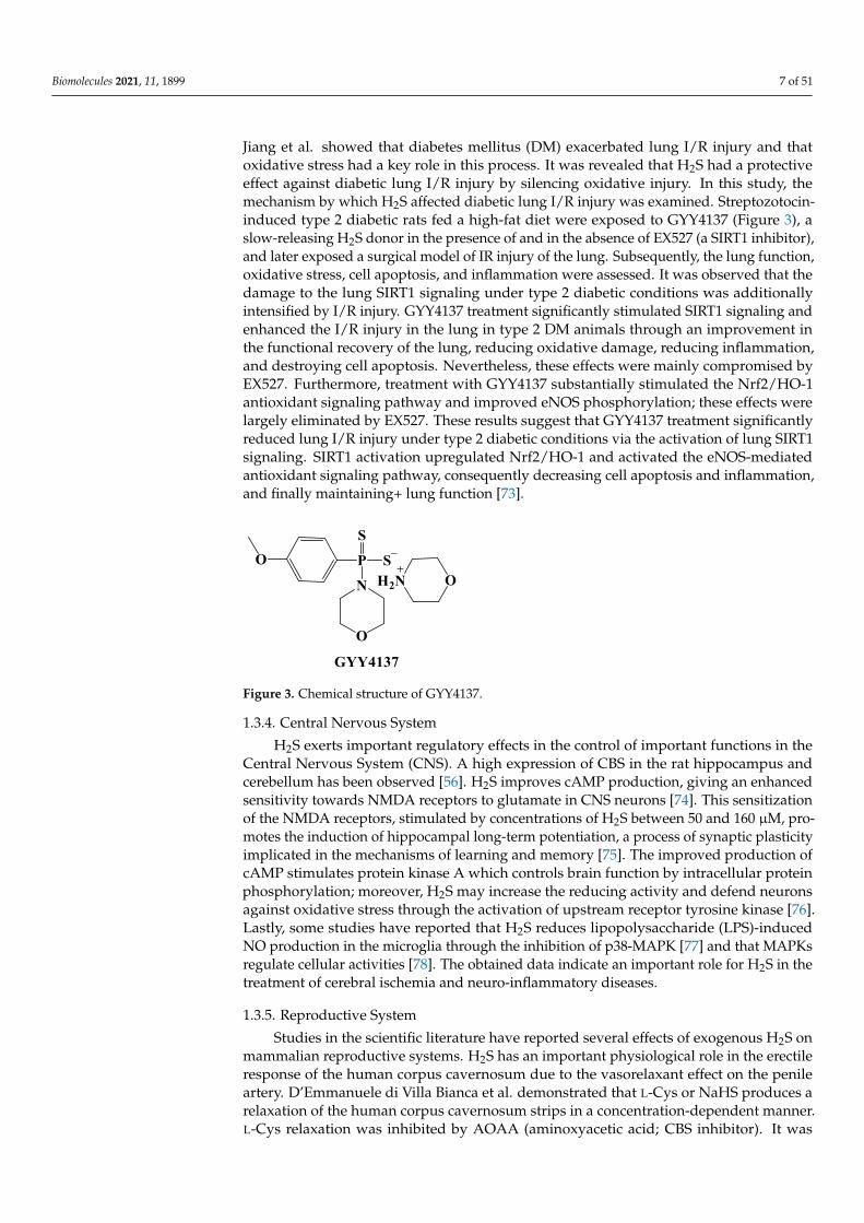
Jiang et al. showed that diabetes mellitus (DM) exacerbated lung I/R injury and thatoxidative stress had a key role in this process. It was revealed that H2S had a protectiveeffect against diabetic lung I/R injury by silencing oxidative injury. In this study, themechanism by which H2S affected diabetic lung I/R injury was examined. Streptozotocin-induced type 2 diabetic rats fed a high-fat diet were exposed to GYY4137 (Figure 3), aslow-releasing H2S donor in the presence of and in the absence of EX527 (a SIRT1 inhibitor),and later exposed a surgical model of IR injury of the lung. Subsequently, the lung function,oxidative stress, cell apoptosis, and inflammation were assessed. It was observed that thedamage to the lung SIRT1 signaling under type 2 diabetic conditions was additionallyintensified by I/R injury. GYY4137 treatment significantly stimulated SIRT1 signaling andenhanced the I/R injury in the lung in type 2 DM animals through an improvement inthe functional recovery of the lung, reducing oxidative damage, reducing inflammation,and destroying cell apoptosis. Nevertheless, these effects were mainly compromised byEX527. Furthermore, treatment with GYY4137 substantially stimulated the Nrf2/HO-1antioxidant signaling pathway and improved eNOS phosphorylation; these effects werelargely eliminated by EX527. These results suggest that GYY4137 treatment significantlyreduced lung I/R injury under type 2 diabetic conditions via the activation of lung SIRT1signaling. SIRT1 activation upregulated Nrf2/HO-1 and activated the eNOS-mediatedantioxidant signaling pathway, consequently decreasing cell apoptosis and inflammation,and finally maintaining+ lung function [73].
Biomolecules 2021, 10, x 7 of 52
structive airway diseases. In milder COPD, enhanced levels of H2S serum may have a
beneficial effect in airway protection, antagonizing oxidative stress and airway inflam-
mation and blocking the progress of COPD [71]. Scientific research has shown many
positive effects for H2S in ischemia for several tissues. H2S has a broad range of protec-
tive functions related to ischemia/reperfusion (I/R) injury all through the body. The con-
trol of intracellular [Ca2+] through the stimulation of the KATP channel opening, anti-
apoptotic pathway (ERK1/2/MAPK, PI-3 kinase/Akt, JAK-STAT) activation, mitochon-
drial protection through the preservation of ΔΨm and the inhibition of the mPTP open-
ing, and the anti-inflammatory effects through the activation of eNOS and p38 MAPK
represent the mechanisms of protection [72]. H2S has been also revealed to be protective
against pulmonary ischemia injury and the endogenous CSE/H2S pathway could be im-
plicated in the pathogenesis of lung I/R injury [67]. In a recent study, Jiang et al. showed
that diabetes mellitus (DM) exacerbated lung I/R injury and that oxidative stress had a
key role in this process. It was revealed that H2S had a protective effect against diabetic
lung I/R injury by silencing oxidative injury. In this study, the mechanism by which H2S
affected diabetic lung I/R injury was examined. Streptozotocin-induced type 2 diabetic
rats fed a high-fat diet were exposed to GYY4137 (Figure 3), a slow-releasing H2S donor
in the presence of and in the absence of EX527 (a SIRT1 inhibitor), and later exposed a
surgical model of IR injury of the lung. Subsequently, the lung function, oxidative stress,
cell apoptosis, and inflammation were assessed. It was observed that the damage to the
lung SIRT1 signaling under type 2 diabetic conditions was additionally intensified by
I/R injury. GYY4137 treatment significantly stimulated SIRT1 signaling and enhanced
the I/R injury in the lung in type 2 DM animals through an improvement in the func-
tional recovery of the lung, reducing oxidative damage, reducing inflammation, and de-
stroying cell apoptosis. Nevertheless, these effects were mainly compromised by EX527.
Furthermore, treatment with GYY4137 substantially stimulated the Nrf2/HO-1 antioxi-
dant signaling pathway and improved eNOS phosphorylation; these effects were largely
eliminated by EX527. These results suggest that GYY4137 treatment significantly re-
duced lung I/R injury under type 2 diabetic conditions via the activation of lung SIRT1
signaling. SIRT1 activation upregulated Nrf2/HO-1 and activated the eNOS-mediated
antioxidant signaling pathway, consequently decreasing cell apoptosis and inflamma-
tion, and finally maintaining+ lung function [73].
Figure 3. Chemical structure of GYY4137.
1.3.4. Central Nervous System
H2S exerts important regulatory effects in the control of important functions in the
Central Nervous System (CNS). A high expression of CBS in the rat hippocampus and
cerebellum has been observed [56]. H2S improves cAMP production, giving an enhanced
sensitivity towards NMDA receptors to glutamate in CNS neurons [74]. This
sensitization of the NMDA receptors, stimulated by concentrations of H2S between 50
and 160 M, promotes the induction of hippocampal long-term potentiation, a process of
synaptic plasticity implicated in the mechanisms of learning and memory [75]. The
improved production of cAMP stimulates protein kinase A which controls brain
function by intracellular protein phosphorylation; moreover, H2S may increase the
Figure 3. Chemical structure of GYY4137.
1.3.4. Central Nervous System
H2S exerts important regulatory effects in the control of important functions in theCentral Nervous System (CNS). A high expression of CBS in the rat hippocampus andcerebellum has been observed [56]. H2S improves cAMP production, giving an enhancedsensitivity towards NMDA receptors to glutamate in CNS neurons [74]. This sensitizationof the NMDA receptors, stimulated by concentrations of H2S between 50 and 160 µM, pro-motes the induction of hippocampal long-term potentiation, a process of synaptic plasticityimplicated in the mechanisms of learning and memory [75]. The improved production ofcAMP stimulates protein kinase A which controls brain function by intracellular proteinphosphorylation; moreover, H2S may increase the reducing activity and defend neuronsagainst oxidative stress through the activation of upstream receptor tyrosine kinase [76].Lastly, some studies have reported that H2S reduces lipopolysaccharide (LPS)-inducedNO production in the microglia through the inhibition of p38-MAPK [77] and that MAPKsregulate cellular activities [78]. The obtained data indicate an important role for H2S in thetreatment of cerebral ischemia and neuro-inflammatory diseases.
1.3.5. Reproductive System
Studies in the scientific literature have reported several effects of exogenous H2S onmammalian reproductive systems. H2S has an important physiological role in the erectileresponse of the human corpus cavernosum due to the vasorelaxant effect on the penileartery. D’Emmanuele di Villa Bianca et al. demonstrated that L-Cys or NaHS produces arelaxation of the human corpus cavernosum strips in a concentration-dependent manner.L-Cys relaxation was inhibited by AOAA (aminoxyacetic acid; CBS inhibitor). It was

Biomolecules 2021, 11, 1899 8 of 51
shown that PAG (propargylglycine; CSE inhibitor) or AOAA potentiated the electrical fieldstimulation of human penile tissue. In rats, L-Cys and NaHS were shown to promote penileerection and the response to L-Cys was impeded by PAG. The acquired data suggest that theL-Cys/H2S pathway facilitates human corpus cavernosum smooth-muscle relaxation [79].
1.3.6. Gastrointestinal System
Several papers concerning the role of H2S in the gastrointestinal (GI) tract have beendescribed in the scientific literature. Studies on H2S-induced chloride secretion provedthat H2S is able to stimulate primary afferent nerve fibers in the mucosa/submucosaof the colon in guinea pigs and humans and this leads to increased chloride secretion.Hydrogen sulfide determines the contractile function either acting directly on the smoothmuscle itself or by stimulating neurons in the enteric nervous system that influence smoothmuscle function. Even if the effect of H2S on inflammation is controversial and manystudies have demonstrated its anti-inflammatory effect at physiological concentrations,H2S can be considered as a proinflammatory mediator in abdominal sepsis, endotoxemia,and pancreatitis counter to its anti-inflammatory effects in animal models of gastritis andcolitis [80–82].
In recent years, the role of H2S and other gaseous mediators (such as CO and NO)in digestive systems has been highlighted. The cytoprotective and anti-inflammatoryproperties of H2S are correlated to mucosal defense and ulcer healing in the GI system [83].H2S is also involved in the physiology of the intestinal microbiota and in particular, it isable to inhibit pathogenic bacteria and to promote mucus production [84]. Furthermore,H2S-releasing derivatives of non-steroidal anti-inflammatory drugs (HS–NSAIDs) havebeen proven to reduce the gastric damage in comparison to the corresponding parentdrugs [85]. H2S donors have also been shown to alleviate visceral pain and HS–trimebutinehas been tested in clinical trials as an abdominal analgesic (NCT01926444) [86–88].
1.4. Hydrogen Sulfide and Diseases
Hydrogen sulfide is involved in several physiological and pathophysiological pro-cesses; this endogenous mediator is in fact associated with the homeostasis of the cardio-vascular, renal, and central nervous systems and to dermatological diseases, various typesof cancer, and viral infections as well as the SARS-CoV-2 infection that leads to COVID-19.
Hypertension, atherosclerosis, hyperhomocysteinemia, and diabetes represent car-diovascular diseases that are connected to vascular endothelial dysfunction; research hasshown that H2S can be considered as a vasoprotective gasotransmitter in diseases whereendothelial dysfunction is the central issue [89]. Moreover, morphological and functionalmodifications at the endothelial level correspond to the first events of cell senescencethat inexorably result in vascular dysfunction and the subsequent atherosclerosis thatprofoundly impact cardiovascular health. In fact, myocardial hypertrophy and fibrosis cancertainly arise and can lead to a compromise in the general cardiac output. Interestingly,hydrogen sulfide has been recently identified as a gaseous molecule able to manipulatethe intracellular pathways implicated in cell senescence and so it is considered as a pos-sible target for blocking cardiovascular diseases. In particular, the correlation betweenthe decrease in endogenous H2S levels and the beginning of several cardiovascular cellsenescence-related diseases has been demonstrated [90].
Numerous studies have clearly demonstrated that H2S has a considerable protectiveeffect in myocardial ischemia. The processes for H2S cardioprotection involve the regula-tion of ion channels; antifibrotic and antiapoptotic effects; a reduction in oxidative stressand inflammatory response; the protection of mitochondria; the regulation of microRNAexpression; and the promotion of angiogenesis. H2S donors could be used to developeffective drugs for the care of cardiovascular diseases such as myocardial ischemia [91,92].
Hydrogen sulfide has a crucial role in blood pressure control and renal physiologytoo. In the kidney, H2S is a significant regulator of vascular and cellular function, althoughthe processes that influence the (sub)cellular levels of H2S are not exactly understood.
Biomolecules 2021, 11, 1899 9 of 51
H2S regulates systemic and renal blood flow, the amount of glomerular filtration, and therenin–angiotensin axis by direct inhibition of nitric oxide synthesis. In addition, H2S affectscellular events by altering protein activity through post-translational protein modifica-tion. This process represents persulfidation and is able to modulate protein localization,protein activity, and protein–protein interactions. Furthermore, acute kidney injury (AKI)caused by mitochondrial dysfunction, which happens in the course of hypoxia or ischemia-reperfusion (IR), is mitigated by H2S. H2S increases ATP production, inhibits the injurycaused by free radicals, and controls endoplasmic reticulum stress during IR [93]. Therefore,forerunners for H2S endogenous synthesis, H2S donors, and natural plant-derivative com-pounds may serve as strategies to prevent hypertension and kidney disease [94]. Corvinoet al. recently reported a survey of currently available H2S donors and their effects on car-diovascular disease; these H2S donors have been described as the basis of the mechanismthat triggers the release of H2S and underline their possible use as favorable drugs in thetreatment of cardiovascular diseases [95].
H2S, NO, and their crosstalk, have been described as helpful in Acute Kidney Injury(AKI), a syndrome mainly produced by ischemia and reperfusion (IR) injury, which momen-tarily impedes the blood flow, improves inflammation processes, and stimulates oxidativestress. Both these endogenous gasotransmitters act by reducing inflammation, regulatingreactive oxygen species (ROS) concentrations, and modulating pro-inflammatory cytokines,as well supporting vasodilation and reducing hypertrophy, apoptosis, and autophagy [96].
Some studies reported in the scientific literature describe the correlation between H2Sand cisplatin-induced nephrotoxicity. Precisely, the decrease in H2S by cystathionine γ-lyase (CSE) downregulation, subsequent to cisplatin treatment, may contribute to cisplatin-induced renal cell injury, probably through the increase in the production of endogenousreactive oxygen species (ROS); on the other hand, H2S prevents successive renal disordersthrough the inhibition of the activation of NADPH oxidase. Interestingly, GYY-4137, aslow-releasing H2S donor, has been proven to improve the anticancer activity of cisplatinin various cancer cell lines, maybe because of its specific anticancer effect. Nevertheless,the effectiveness of H2S donors in tumor-bearing animals continues to be examined interms of renal protection, cancer inhibition, and later, cisplatin treatment. Moreover, moredata regarding the use of polysulfide, a H2S derived compound, in the therapy of cisplatin-induced nephrotoxicity, have been additionally reported [97].
During recent years, a wide range of studies have focused on the function of H2Sin the central nervous system (CNS), providing evidence that H2S has an important rolein neuroprotection activities because it constitutes a signaling molecule that controls thefunction of the CNS. Neurodegenerative diseases are linked to endoplasmic reticulumstress (ERS) and protein misfolding. H2S regulates ERS and shows a broad variety of cyto-protective and physiological functions in CNS degenerative diseases. The neuroprotectiveeffect of H2S for ERS has an important role in various CNS diseases as well as Parkinson’sdisease, depression disorders, and Alzheimer’s disease [98].
In bone tissue, H2S has a cytoprotective role and stimulates bone formation; recently,scientific studies have established its role as a therapeutic agent in bone pathologies. H2Shas been proven to stimulate several signaling pathways implicated in several phases of theprocesses of bone repair. In particular, H2S seems to be able to modulate osteogenic differ-entiation in osteoprogenitor cells through the induction of S-sulfhydration in TRP channels,increasing Ca2+ influx in the mesenchymal stromal cells (MSCs); H2S donors encouragethe expression of osteogenic signaling by the stimulation of multiple signaling pathways atthe transcriptional level. Concerning inflammation, hydrogen sulfide activates antioxidantsignaling in osteoclasts (OCs), which includes the improved nuclear translocations ofNRF-2 and leads to the inhibition of OC differentiation. Pharmacological administrationof H2S has led to promising outcomes in preclinical studies in the treatment of systemicbone diseases, for example, osteoporosis; nevertheless, local delivery of H2S offers furtheropportunities for treatment. Moreover, new H2S donors and biomaterials have recently

Biomolecules 2021, 11, 1899 10 of 51
been studied, paving the way for the advancement of H2S-releasing procedures for bonere-forming medicine [99].
Alterations in H2S production are involved in several dermatological diseases, forexample, psoriasis, melanoma, and other dermatoses. The consequences of H2S in theskin involve the stimulation of keratinocyte differentiation; the control of vasodilatation;wound healing; the regulation of apoptosis; the modulation of pruritus; and the resolu-tion of inflammation. Regarding the H2S pathway in the skin, the information availablereferences the connection between this molecule and the physiological and pathologicaleffects. Generally, most of the effects of H2S are clearly concentration or dose-dependent.Due to the important role of H2S in skin physiology, it is not unexpected that modifiedH2S production leads to the pathogenesis of several cutaneous diseases. Many H2S donorshave been used in experiments, and different results have been acquired. Nevertheless,these compounds present different intrinsic characteristics such as the capacity to enterthe cell or organelle specifically, H2S-releasing kinetics, and pH dependence, but certainly,these chemical entities include a new family of potential therapeutic tools for the treatmentof various skin diseases by targeting their differential etiological aspects. Obviously, thedevelopment of H2S donors will necessarily rely on an better understanding of the exactrole of H2S and its goals in the various phases of skin biology [100].
Several studies reported in the scientific literature have shown an effective role ofH2S in the framework of cancer biology. CSE, CBS, and 3MST, the three H2S-producingenzymes, are expressed in several types of cancer. Furthermore, the inhibition of CBSleads to anti-tumor activity especially in breast cancer, ovarian cancer, and colon cancer,while the effect of CSE or 3-MST inhibition has been largely unexplored in cancer cells.Interestingly, H2S donation has been shown to induce cancer cell apoptosis in in vitro andin vivo studies. In particular, low levels of H2S may show pro-cancer effects, while higherlevels of H2S may lead to cancer cell death. This leads us to assume that the inhibitionof H2S biosynthesis and H2S supplementation work in two different ways in relation tocancer treatment. This enigmatic role of H2S is interesting in relation to the developmentof innovative CBS inhibitors, H2S donors, and H2S-releasing hybrids that could thereforehave potential for cancer therapy [101].
Finally, it is possible to affirm that hydrogen sulfide represents a natural defenseagainst the infections from enveloped RNA viruses and is implicated in COVID-19. Ithas previously been reported that H2S inhibits the growth and pathogenic mechanisms ofseveral enveloped RNA viruses, and it has been observed that H2S plasmatic levels areelevated in COVID-19 survivors compared to fatal cases [102]. The release of H2S is trig-gered by carbon monoxide (CO) from the catabolism of heme by inducible heme oxygenase(HO-1) and heme proteins have the catalytic activity required for H2S signaling by proteinpersulfidation. Subjects with a long promoter for the HMOX1 gene, coding for HO-1, havea lower efficiency of this mechanism. SARS-CoV-2 prevents the formation of the heme ofhemoglobin and other hemeproteins, so impeding both the signaling and release of H2S.Deficiency of the H2S-induced persulfidation of the KATP channels of leucocytes triggersthe adhesion and release of the inflammatory cytokines, lung infiltration, and systemic en-dothelial damage with hyper-coagulability. These outcomes mainly justify the sex and agedistribution, clinical manifestations, and co-morbidities of COVID-19 [102]. The involve-ment of H2S in COVID-19 therapy is also supported by its anti-inflammatory and antiviralfunctions. As is already known, H2S has a pivotal role in regulating the inflammatory andpro-inflammatory cytokine cascade and certain H2S donors are effective in the treatmentof acute lung inflammation. The latest studies have elucidated the mechanism of actionfor the antiviral effects of hydrogen sulfide. Moreover, certain preliminary clinical resultshave indicated an inverse relationship between endogenous H2S levels and the severity ofCOVID-19. H2S donors, then, could represent a potential therapeutic opportunity for thetreatment of COVID-19 [103]. The alteration of H2S levels in COVID-19 represent furtherevidence of the need to regulate the homeostasis of this gasotransmitter, considering thatthe alteration of H2S levels has an important role in several diseases [89–104].

Biomolecules 2021, 11, 1899 11 of 51
Finally, H2S is especially controlled in experimental glaucoma. By scavenging reactiveoxygen species and distending retinal vessels, H2S protects RGCs from pressure andoxidative stress both in vitro and in vivo. Consequently, H2S is considered to be a newneuro-protectant in glaucoma [104].
2. H2S Donors2.1. Gaseous H2S
On the basis of the broad and active physiological effects of H2S, some researchgroups have described the protective role of H2S donors in various disease models. H2Sgas has been clearly applied in these fields. The most prominent method of deliveryis the direct inhalation of this gas. The delivery method has been often assessed, eventhough H2S is a toxic and flammable gas. It has been demonstrated that gaseous H2Sgenerates a suspended animation-like state in mice [30] and it has been proposed that theseeffects are caused by the reversible and competitive inhibition of the mitochondrial enzymecytochrome c oxidase [31]. Moreover, H2S gas demonstrated a reversible reduction in motoractivity and body temperature with a subsequent growth in blood sulfide concentration inmice [105,106]. Consequently, inhalation of H2S is used as a technique to inhibit pulmonaryand systemic inflammatory reactions in the course of physiological stress in hypothermicand normothermic mice [107,108]. The reversible H2S-induced hibernation state has beenshown to protect mice from lethal hypoxia [109]. H2S gas can also promote glucose uptakeand provide amelioration in type II diabetes [110]. H2S inhalation allowed a modulationof the delivery by precisely modulating pressure over time and it bypassed some of theissues with intravenous delivery as well as the oxidation and volatilization of suspendedH2S. On the other hand, H2S inhalation has various inherent shortcomings because of thetoxicity and flammability of the gas, including gas storage, secure administration, andtargeting. From the experimental point of view, it is difficult to consider H2S gas as an idealdrug because of the problems in acquiring accurately regulated concentrations and for thepotential toxic effect of an excess of H2S.
2.2. Inorganic Salts
Inorganic salts, as well as sodium hydrosulfide (NaHS), sodium sulfide (Na2S), andcalcium sulfide (CaS), are believed to be exogenous H2S donors. These inorganic salts,which represent standard tools, have been long employed in biological studies to investigatethe therapeutic effects of exogenous hydrogen sulfide, although several significant limitshave been proved.
Indeed, sulfide salts hydrolyze instantaneously following dissolution in water, es-tablishing an equilibrium between hydrogensulfide ions (HS−), sulfide ions (S2−), andmolecular hydrogen sulfide (H2S) which depend on some parameters such as pH, tem-perature, and pressure [111]. When this equilibrium is determined, volatilization of H2Sappears, thus reducing the general concentration of the sulfur species in the solution.Moreover, HS air oxidation−, catalyzed by metals in water, lowers the concentration ofH2S solutions [112]. These chemical limitations restrict the use of salts in clinical settingseven though the administration of sulfide salts to animals, tissues, or cells has been demon-strated to have protective effects for a large number of diseases. Some researchers havehighlighted the short- and long-term cardioprotective effects of H2S using Na2S as a H2Sdonor in a mouse model of myocardial ischemia injury (MI/R). The results have suggestedthat a 7-day treatment of Na2S resulted in a reduction in the dilatation of the left ventricle, aloss of cardiac hypertrophy, and an increase in cardiac function. Moreover, treatment withNa2S decreased the oxidative stress related to heart failure [113]. Aqueous NaHS solutiondelivered in aortic rings led to a 60% increase in relaxation, proving the vasorelaxant effectsof H2S [114]; this salt reduced ischemia–reperfusion injury [115], and protected againsthemorrhagic shock [116] and myocardial infarction (MI) [117].
Moreover, sulfide salts have protective effects for other diseases as well as inflam-mation [118]. Osteoarthritis (OA) is a form of arthritis characterized by degenerative and

Biomolecules 2021, 11, 1899 12 of 51
inflammatory processes and by higher levels of pro-inflammatory proteins (IL-6 and IL-8).A study reported in the literature demonstrated that the short-term treatment of humancells with NaHS is enough to down-regulate IL-6 and IL-8 expression, which in turn maycause anti-inflammatory effects of H2S against OA [119]. However, it has also been ob-served that the anti-inflammatory effects of NaHS translated into pro-inflammatory effectswhen the NaHS incubation time was protracted (1 h). In fact, it has been reported thatsulfide salts as H2S donors showed both anti-inflammatory and pro-inflammatory effects.
Some researchers have indicated that the leukocyte-regulated inflammation of theknee joint was significantly hindered after Na2S injection [120]. In an independent study,Chen et al. determined that the administration of NaHS into the lungs in an asthmatic ratmodel attenuated inflammation [71]. Through the use of a smoke inhalation-induced lunginjury model, Esechie et al. proved that Na2S exhibited protective actions against severelung injury [118]. These results have suggested that H2S (using NaHS or Na2S) is a potentanti-inflammatory agent.
In contrast, the pro-inflammatory effects of H2S have also been highlighted. Indeed, Liet al. proved that H2S exhibited pro-inflammatory effects in an animal model of endotoxicshock [121]. Furthermore, plasma H2S concentrations, the synthesizing ability of H2S intissue, and CSE expression were all proven to be enhanced in several animal models ofinflammation [122–124]. This conclusion has indicated the potential pro-inflammatoryeffects of H2S.
In a separate study, the administration of NaHS solution by intravenous injectionalleviated the degree of ALI by decreasing IL-6 and IL-8 levels while concurrently increasingIL-10 levels in the plasma and lung tissue [125]. This study also substantiated the hypothesisthat the down-regulation of endogenous H2S levels in the cardiovascular system is presentin ALI pathogenesis.
Parkinson’s disease (PD) manifests a progressive loss of dopaminergic neurons in thesubstantia nigra and a depletion of the neurotransmitter dopamine in the striatum [126].Hu et al. indicated that a systemic administration of H2S (using NaHS) could reducebehavioral symptoms and dopaminergic neuronal degeneration in 6-hydroxydopamine(6-OHDA)-induced PD models [127]. By employing such models, they determinedthat endogenous H2S production is lowered during the development of PD, and theresults have suggested that the administration of H2S could have therapeutic benefits forParkinson’s disease.
Comparatively, H2S has proven to be useful for Alzheimer’s disease (AD). The amountof H2S biosynthesis in the brain is lowered in AD patients and the levels of H2S in theplasma are linked to the severity of AD [44]. Giuliani et al. observed how treatment witha H2S donor induced neuroprotection and slowed down the progression of AD. Studieswith sodium hydrosulfide and Tabiano’s spa-water were conducted in three experimentalmodels of AD. Short-term and long-term treatments with sodium hydrosulfide and/orTabiano’s spa-water significantly protected against impairments in learning and memoryin rat models of AD induced by an injection of β-amyloid1–40 (Aβ) or streptozotocin in thebrain, and in an AD mouse model harboring the human transgenes APPSwe, PS1M146V,and tauP301L (3xTg-AD mice). The improvement in behavioral performance was related tothe size of the Aβ plaques in the hippocampus and the preservation of the morphologicalpicture, as found in AD rats. In addition, the reduced concentration/phosphorylationlevels of proteins were proven to be essential in AD pathophysiology. Specifically, theamyloid precursor protein, presenilin-1, Aβ1-42 and tau phosphorylated at Thr181, Ser396and Ser202, were identified in 3xTg-AD mice treated with spa-water. The excitotoxicity-triggered oxidative and nitrosative stress was found in 3xTg-AD mice, as indicated bythe decreased levels of malondialdehyde and nitrites in the cerebral cortex. The limitedactivity of c-jun N-terminal kinases, extracellular signal-regulated kinases, and p38 inthe hippocampus presented a well-established role both in the phosphorylation of thetau protein and in inflammation and apoptosis. Consistently, the decrease in the tumornecrosis factor-a level, the up-regulation of Bcl-2, and the down-regulation of BAX and the
Biomolecules 2021, 11, 1899 13 of 51
downstream executioner caspase-3 in the hippocampus of 3xTg-AD mice after treatmentwith Tabiano’s spa-water implied that it is also able to modulate inflammation and apop-tosis. These results indicate that appropriate treatments with H2S donors and Tabiano’sspa-waters and other spa-waters rich in H2S content might represent an innovative ap-proach to slow down AD progression in humans by targeting multiple pathophysiologicalmechanisms [128].
The low pharmacokinetic profile of sulfide salts limits the use of these salts as potentialtherapeutics and has led to the research into innovative organic H2S donors, includingnaturally occurring donors and synthetic compounds.
2.3. Natural H2S Donors
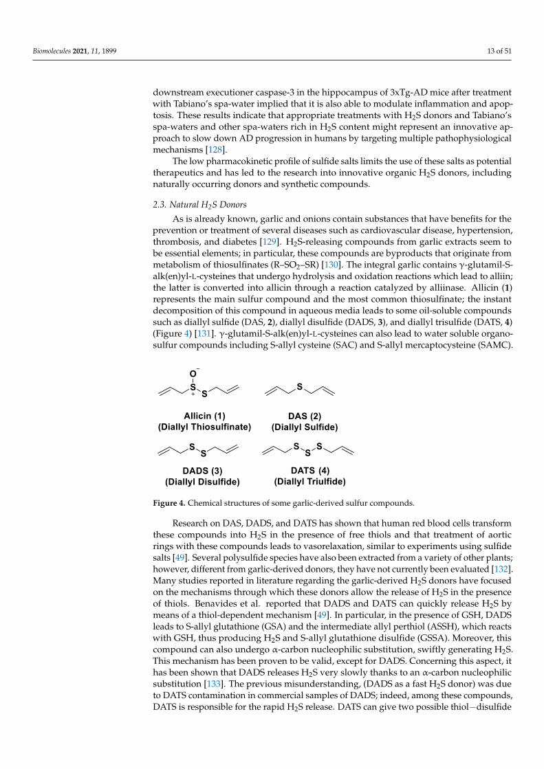
As is already known, garlic and onions contain substances that have benefits for theprevention or treatment of several diseases such as cardiovascular disease, hypertension,thrombosis, and diabetes [129]. H2S-releasing compounds from garlic extracts seem tobe essential elements; in particular, these compounds are byproducts that originate frommetabolism of thiosulfinates (R–SO2–SR) [130]. The integral garlic contains γ-glutamil-S-alk(en)yl-L-cysteines that undergo hydrolysis and oxidation reactions which lead to alliin;the latter is converted into allicin through a reaction catalyzed by alliinase. Allicin (1)represents the main sulfur compound and the most common thiosulfinate; the instantdecomposition of this compound in aqueous media leads to some oil-soluble compoundssuch as diallyl sulfide (DAS, 2), diallyl disulfide (DADS, 3), and diallyl trisulfide (DATS, 4)(Figure 4) [131]. γ-glutamil-S-alk(en)yl-L-cysteines can also lead to water soluble organo-sulfur compounds including S-allyl cysteine (SAC) and S-allyl mercaptocysteine (SAMC).
Biomolecules 2021, 10, x 14 of 52
Figure 4. Chemical structures of some garlic-derived sulfur compounds.
Research on DAS, DADS, and DATS has shown that human red blood cells trans-
form these compounds into H2S in the presence of free thiols and that treatment of aortic
rings with these compounds leads to vasorelaxation, similar to experiments using sul-
fide salts [49]. Several polysulfide species have also been extracted from a variety of oth-
er plants; however, different from garlic-derived donors, they have not currently been
evaluated [132]. Many studies reported in literature regarding the garlic-derived H2S
donors have focused on the mechanisms through which these donors allow the release
of H2S in the presence of thiols. Benavides et al. reported that DADS and DATS can
quickly release H2S by means of a thiol-dependent mechanism [49]. In particular, in the
presence of GSH, DADS leads to S-allyl glutathione (GSA) and the intermediate allyl
perthiol (ASSH), which reacts with GSH, thus producing H2S and S-allyl glutathione di-
sulfide (GSSA). Moreover, this compound can also undergo α-carbon nucleophilic sub-
stitution, swiftly generating H2S. This mechanism has been proven to be valid, except for
DADS. Concerning this aspect, it has been shown that DADS releases H2S very slowly
thanks to an α-carbon nucleophilic substitution [133]. The previous misunderstanding,
(DADS as a fast H2S donor) was due to DATS contamination in commercial samples of
DADS; indeed, among these compounds, DATS is responsible for the rapid H2S release.
DATS can give two possible thiol−disulfide exchange reaction pathways: primarily, the
nucleophilic attack of GSH on allylic sulfur of DATS that generates GSSA and ASSH,
with the latter possibly reduced by GSH and then releasing H2S; further, the nucleophilic
attack of GSH on the central sulfur atom of DATS can lead to the production of ASH and
GSSSA. The latter rapidly produces hydrogen sulfide.
Another class of natural H2S donors is composed by some isothiocyanates (Figure
4); these compounds represent natural donors that have attracted attention due to their
biological and pharmacological effects in the prevention of important human diseases,
as well as cancer, neurodegenerative processes, and cardiovascular diseases [134]. The
isothiocyanate moiety has been thoroughly investigated due to its use in synthetic mole-
cules for structure activity relationship studies. In a recent study, the hydrogen sulfide-
releasing properties of some synthetic aryl isothiocyanate derivatives were reported, in-
dicating that the isothiocyanate function can be considered as a suitable slow hydrogen
sulfide-releasing moiety, along with the pharmacological potential typical of this gas-
otransmitter. Many isothiocyanate derivatives (as a result of the myrosinase-mediated
transformation of glucosinolates) are well-known secondary metabolites of plants be-
longing to the family Brassicaceae, a large botanical family comprising of many edible
species. The phytotherapeutic and nutraceutic usefulness of Brassicaceae in the preven-
tion of important human diseases, such as cancer, neurodegenerative processes, and
cardiovascular diseases has been widely discussed in the scientific literature. Although
these effects have been associated with isothiocyanates, by and large, the exact mecha-
nism of action is still unknown.
In a recent experimental study, the hydrogen sulfide-releasing capacity of some
important natural isothiocyanates has been investigated (AITC (5), ERU (6), BITC (7),
Figure 4. Chemical structures of some garlic-derived sulfur compounds.
Research on DAS, DADS, and DATS has shown that human red blood cells transformthese compounds into H2S in the presence of free thiols and that treatment of aorticrings with these compounds leads to vasorelaxation, similar to experiments using sulfidesalts [49]. Several polysulfide species have also been extracted from a variety of other plants;however, different from garlic-derived donors, they have not currently been evaluated [132].Many studies reported in literature regarding the garlic-derived H2S donors have focusedon the mechanisms through which these donors allow the release of H2S in the presenceof thiols. Benavides et al. reported that DADS and DATS can quickly release H2S bymeans of a thiol-dependent mechanism [49]. In particular, in the presence of GSH, DADSleads to S-allyl glutathione (GSA) and the intermediate allyl perthiol (ASSH), which reactswith GSH, thus producing H2S and S-allyl glutathione disulfide (GSSA). Moreover, thiscompound can also undergo α-carbon nucleophilic substitution, swiftly generating H2S.This mechanism has been proven to be valid, except for DADS. Concerning this aspect, ithas been shown that DADS releases H2S very slowly thanks to an α-carbon nucleophilicsubstitution [133]. The previous misunderstanding, (DADS as a fast H2S donor) was dueto DATS contamination in commercial samples of DADS; indeed, among these compounds,DATS is responsible for the rapid H2S release. DATS can give two possible thiol−disulfide

Biomolecules 2021, 11, 1899 14 of 51
exchange reaction pathways: primarily, the nucleophilic attack of GSH on allylic sulfurof DATS that generates GSSA and ASSH, with the latter possibly reduced by GSH andthen releasing H2S; further, the nucleophilic attack of GSH on the central sulfur atomof DATS can lead to the production of ASH and GSSSA. The latter rapidly produceshydrogen sulfide.
Another class of natural H2S donors is composed by some isothiocyanates (Figure 4);these compounds represent natural donors that have attracted attention due to their bi-ological and pharmacological effects in the prevention of important human diseases, aswell as cancer, neurodegenerative processes, and cardiovascular diseases [134]. The isoth-iocyanate moiety has been thoroughly investigated due to its use in synthetic moleculesfor structure activity relationship studies. In a recent study, the hydrogen sulfide-releasingproperties of some synthetic aryl isothiocyanate derivatives were reported, indicatingthat the isothiocyanate function can be considered as a suitable slow hydrogen sulfide-releasing moiety, along with the pharmacological potential typical of this gasotransmitter.Many isothiocyanate derivatives (as a result of the myrosinase-mediated transformation ofglucosinolates) are well-known secondary metabolites of plants belonging to the familyBrassicaceae, a large botanical family comprising of many edible species. The phytothera-peutic and nutraceutic usefulness of Brassicaceae in the prevention of important humandiseases, such as cancer, neurodegenerative processes, and cardiovascular diseases has beenwidely discussed in the scientific literature. Although these effects have been associatedwith isothiocyanates, by and large, the exact mechanism of action is still unknown.
In a recent experimental study, the hydrogen sulfide-releasing capacity of some im-portant natural isothiocyanates has been investigated (AITC (5), ERU (6), BITC (7), HBITC(8)—Figure 5) by using in vitro amperometric detection. Some of the tested natural isothio-cyanates exhibited a significant release of hydrogen sulfide, thus implying that hydrogensulfide may be, at least in some measure, a relevant player accounting for several biologicaleffects of Brassicaceae [135].
Biomolecules 2021, 10, x 15 of 52
HBITC (8) —Figure 5) by using in vitro amperometric detection. Some of the tested nat-
ural isothiocyanates exhibited a significant release of hydrogen sulfide, thus implying
that hydrogen sulfide may be, at least in some measure, a relevant player accounting for
several biological effects of Brassicaceae [135].
Figure 5. General structure of isothiocyanates and the chemical structures of natural isothiocya-
nates tested in [135].
In addition, it has been proven that some selected aryl isothiocyanates are able to
release H2S in a biological environment and produce a vasorelaxant effect in rat aortic
rings, strongly antagonized by a specific Kv7-blocker, and in the coronary vascular bed,
causing an increase in the basal coronary flow [136]. Furthermore, the H2S-releasing
properties of the secondary metabolites of Brassicaceae, isothiocyanates, are also derived
from the quick reaction with thiols, namely cysteine. The Cys-ITC adduct formed un-
dergoes an intramolecular cyclization followed by a release of an organic amine R−NH2
and raphanusamic acid (RA), on the one hand, and H2S and 2-carbylamino-4,5-
dihydrothiazole-4-carboxylic acid, on the other hand [137].
Although naturally occurring H2S donors represent an attractive tool for in vivo
studies, the tendency of these compounds to also bring about some byproducts, which
are not related to H2S production, has prompted researchers to develop novel synthetic
H2S donors with more favorable pharmacokinetic and safety profiles.
Indeed, synthetic donors, which provide tunable H2S release rates via structural
modifications with discrete byproducts, allow for a more in-depth analysis of the physi-
ological roles of H2S and, optimistically, clinically relevant H2S-releasing prodrugs.
In this review, H2S donors are presented according to their main role in several con-
texts: cardiovascular diseases, cancer, nervous system diseases, gastrointestinal diseases,
dermatological diseases, viral infections, and other diseases.
3. H2S Donors and Diseases
3.1. H2S Donors and Cardiovascular Diseases
As already reported, the current scientific literature has proven the protective ac-
tions of H2S in cardiovascular diseases, for instance hypertension, atherosclerosis, cardi-
ac hypertrophy, heart failure, and myocardial ischemia/reperfusion (I/R) injury. The in-
volvement of H2S in the cardiovascular system is due to its role as a blood pressure and
heart rate regulator, an activator of angiogenesis, and a basal vasorelaxant agent. Fur-
thermore, it has been proven that antioxidation, the inhibition of cell apoptosis, pro-
angiogenesis, the anti-inflammatory effect, and the regulation of ion channels represent
the mechanisms of action that lead to cardioprotective activity [95].
Based on the involvement of H2S in the cardiovascular system, scientific research
has shown a strong interest in the development of H2S donors, which could represent
biological instruments and encouraging cardioprotective agents.
Figure 5. General structure of isothiocyanates and the chemical structures of natural isothiocyanatestested in [135].
In addition, it has been proven that some selected aryl isothiocyanates are able torelease H2S in a biological environment and produce a vasorelaxant effect in rat aortic rings,strongly antagonized by a specific Kv7-blocker, and in the coronary vascular bed, causingan increase in the basal coronary flow [136]. Furthermore, the H2S-releasing properties ofthe secondary metabolites of Brassicaceae, isothiocyanates, are also derived from the quickreaction with thiols, namely cysteine. The Cys-ITC adduct formed undergoes an intramolec-ular cyclization followed by a release of an organic amine R−NH2 and raphanusamic acid(RA), on the one hand, and H2S and 2-carbylamino-4,5-dihydrothiazole-4-carboxylic acid,on the other hand [137].
Although naturally occurring H2S donors represent an attractive tool for in vivostudies, the tendency of these compounds to also bring about some byproducts, which are
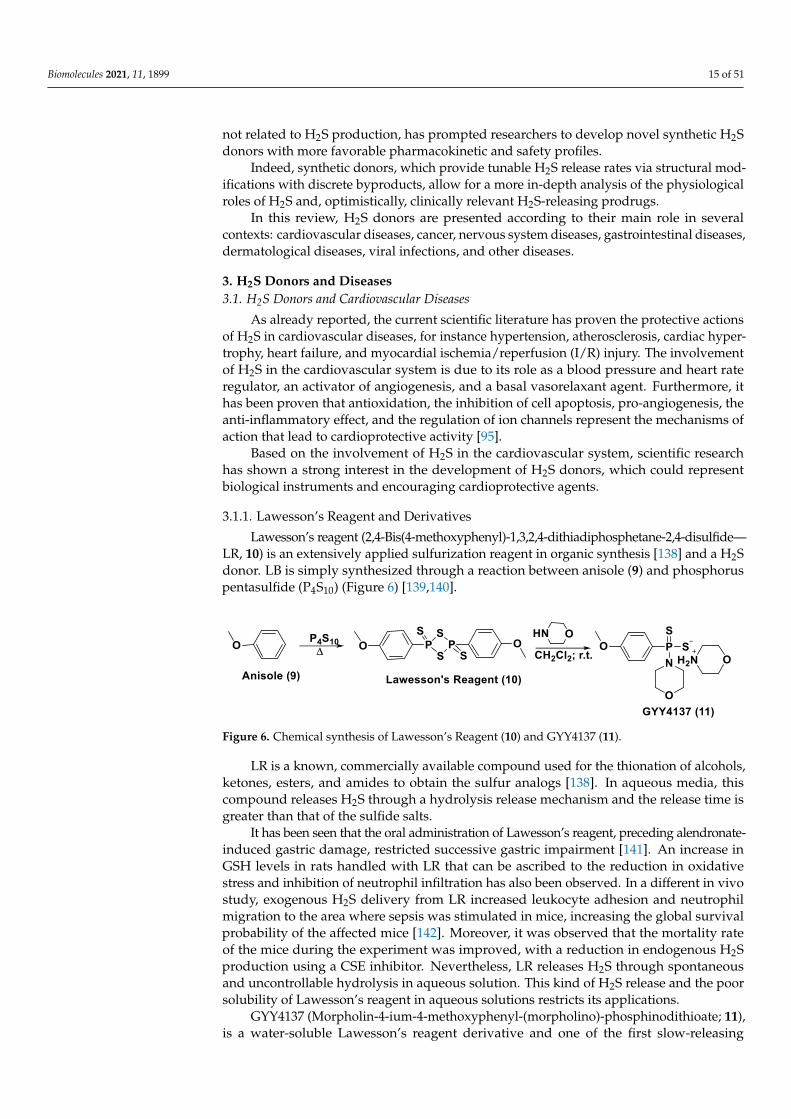
Biomolecules 2021, 11, 1899 15 of 51
not related to H2S production, has prompted researchers to develop novel synthetic H2Sdonors with more favorable pharmacokinetic and safety profiles.
Indeed, synthetic donors, which provide tunable H2S release rates via structural mod-ifications with discrete byproducts, allow for a more in-depth analysis of the physiologicalroles of H2S and, optimistically, clinically relevant H2S-releasing prodrugs.
In this review, H2S donors are presented according to their main role in severalcontexts: cardiovascular diseases, cancer, nervous system diseases, gastrointestinal diseases,dermatological diseases, viral infections, and other diseases.
3. H2S Donors and Diseases3.1. H2S Donors and Cardiovascular Diseases
As already reported, the current scientific literature has proven the protective actionsof H2S in cardiovascular diseases, for instance hypertension, atherosclerosis, cardiac hyper-trophy, heart failure, and myocardial ischemia/reperfusion (I/R) injury. The involvementof H2S in the cardiovascular system is due to its role as a blood pressure and heart rateregulator, an activator of angiogenesis, and a basal vasorelaxant agent. Furthermore, ithas been proven that antioxidation, the inhibition of cell apoptosis, pro-angiogenesis, theanti-inflammatory effect, and the regulation of ion channels represent the mechanisms ofaction that lead to cardioprotective activity [95].
Based on the involvement of H2S in the cardiovascular system, scientific researchhas shown a strong interest in the development of H2S donors, which could representbiological instruments and encouraging cardioprotective agents.
3.1.1. Lawesson’s Reagent and Derivatives
Lawesson’s reagent (2,4-Bis(4-methoxyphenyl)-1,3,2,4-dithiadiphosphetane-2,4-disulfide—LR, 10) is an extensively applied sulfurization reagent in organic synthesis [138] and a H2Sdonor. LB is simply synthesized through a reaction between anisole (9) and phosphoruspentasulfide (P4S10) (Figure 6) [139,140].
Biomolecules 2021, 10, x 16 of 52
3.1.1. Lawesson’s Reagent and Derivatives
Lawesson’s reagent (2,4-Bis(4-methoxyphenyl)-1,3,2,4-dithiadiphosphetane-2,4-disulfide—LR, 10) is an extensively applied sulfurization reagent in organic synthesis
[138] and a H2S donor. LB is simply synthesized through a reaction between anisole (9)
and phosphorus pentasulfide (P4S10) (Figure 6) [139,140].
LR is a known, commercially available compound used for the thionation of alco-
hols, ketones, esters, and amides to obtain the sulfur analogs [138]. In aqueous media,
this compound releases H2S through a hydrolysis release mechanism and the release
time is greater than that of the sulfide salts.
It has been seen that the oral administration of Lawesson’s reagent, preceding alen-
dronate-induced gastric damage, restricted successive gastric impairment [141]. An in-
crease in GSH levels in rats handled with LR that can be ascribed to the reduction in ox-
idative stress and inhibition of neutrophil infiltration has also been observed. In a differ-
ent in vivo study, exogenous H2S delivery from LR increased leukocyte adhesion and
neutrophil migration to the area where sepsis was stimulated in mice, increasing the
global survival probability of the affected mice [142]. Moreover, it was observed that the
mortality rate of the mice during the experiment was improved, with a reduction in en-
dogenous H2S production using a CSE inhibitor. Nevertheless, LR releases H2S through
spontaneous and uncontrollable hydrolysis in aqueous solution. This kind of H2S release
and the poor solubility of Lawesson’s reagent in aqueous solutions restricts its applica-
tions.
GYY4137 (Morpholin-4-ium-4-methoxyphenyl-(morpholino)-phosphinodithioate;
11), is a water-soluble Lawesson’s reagent derivative and one of the first slow-releasing
H2S donors developed [143–145]. This reagent is the compound that is most used to ex-
amine the role of H2S in the biological districts. It can be synthesized by reacting Lawes-
son’s reagent [139,140] with morpholine in methylene chloride at room temperature
(Figure 6) [143].
Figure 6. Chemical synthesis of Lawesson’s Reagent (10) and GYY4137 (11).
GYY4137 exhibits anti-inflammatory, antioxidant, and anticancer properties [146].
In addition, this reagent triggers the activation of the vascular smooth muscle KATP
channels and relaxes rat aortic rings and the renal blood vessels that lead to anti-
hypertensive activity [143]. Furthermore, GYY4137 interfered with platelet activation
and adhesion molecule-mediated aggregation reducing the formation of the microvascu-
lar thrombus and stabilizing atherosclerosis plaque [147]. It seems to protect against dia-
betic myocardial I/R injury (activating the PHLPP-1/Akt/Nrf2 pathway) [148] and
against cerebral I/R injury (via the p38 MAPK mediated anti-apoptotic signaling path-
ways) in rats [149]. It has been proven that GYY4137 slowly releases H2S upon hydroly-
sis. To explain the two-step hydrolytic decomposition pathway of the slow-release H2S
donor GYY4137, a combination of NMR spectroscopy, mass spectrometry, and chemical
synthesis has been used and the key decomposition product has been also obtained by
an independent synthetic strategy. The (dichloromethane-free) sodium salt of the phos-
phonamidodithioate GYY4137 has also been made as a pharmaceutically more appro-
priate salt. It has been shown that the decomposition product does not produce H2S and
does not have cytoprotective or anti-inflammatory effects in oxidatively stressed human
Jurkat T-cells and LPS-treated murine RAW264.7 macrophages, in contrast with
GYY4137 and its sodium salt. The decomposition product signifies a helpful control
Figure 6. Chemical synthesis of Lawesson’s Reagent (10) and GYY4137 (11).
LR is a known, commercially available compound used for the thionation of alcohols,ketones, esters, and amides to obtain the sulfur analogs [138]. In aqueous media, thiscompound releases H2S through a hydrolysis release mechanism and the release time isgreater than that of the sulfide salts.
It has been seen that the oral administration of Lawesson’s reagent, preceding alendronate-induced gastric damage, restricted successive gastric impairment [141]. An increase inGSH levels in rats handled with LR that can be ascribed to the reduction in oxidativestress and inhibition of neutrophil infiltration has also been observed. In a different in vivostudy, exogenous H2S delivery from LR increased leukocyte adhesion and neutrophilmigration to the area where sepsis was stimulated in mice, increasing the global survivalprobability of the affected mice [142]. Moreover, it was observed that the mortality rateof the mice during the experiment was improved, with a reduction in endogenous H2Sproduction using a CSE inhibitor. Nevertheless, LR releases H2S through spontaneousand uncontrollable hydrolysis in aqueous solution. This kind of H2S release and the poorsolubility of Lawesson’s reagent in aqueous solutions restricts its applications.
GYY4137 (Morpholin-4-ium-4-methoxyphenyl-(morpholino)-phosphinodithioate; 11),is a water-soluble Lawesson’s reagent derivative and one of the first slow-releasing
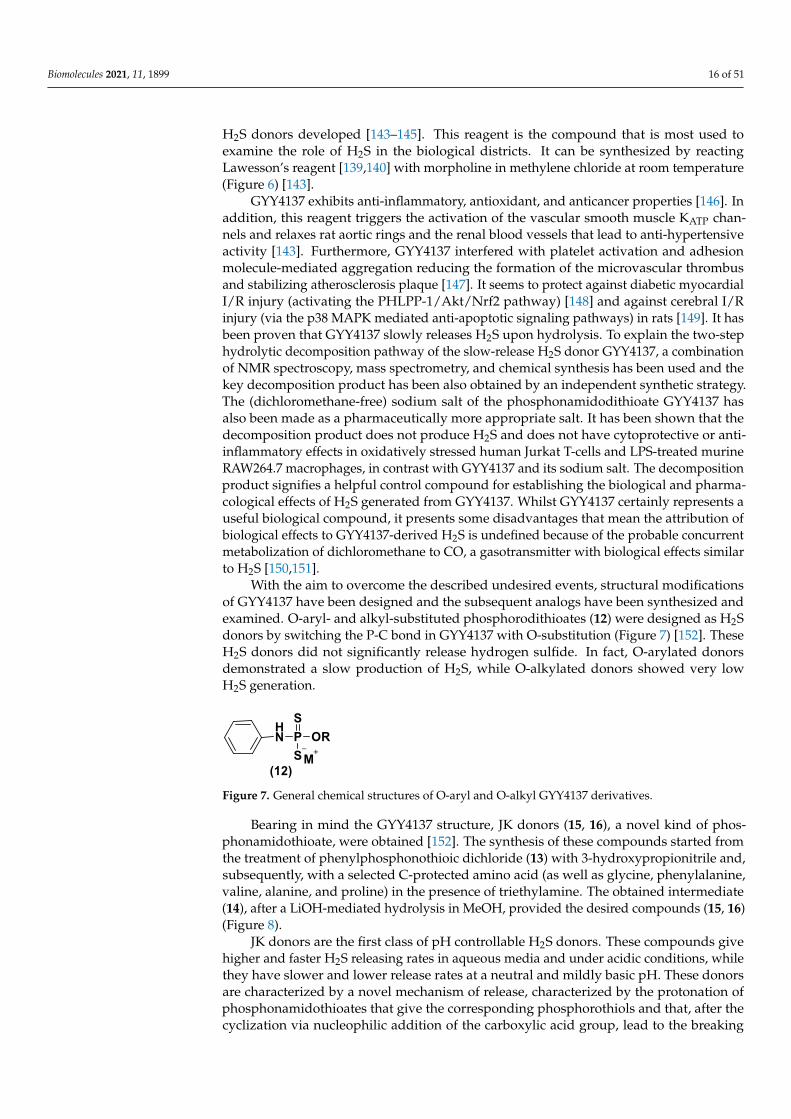
Biomolecules 2021, 11, 1899 16 of 51
H2S donors developed [143–145]. This reagent is the compound that is most used toexamine the role of H2S in the biological districts. It can be synthesized by reactingLawesson’s reagent [139,140] with morpholine in methylene chloride at room temperature(Figure 6) [143].
GYY4137 exhibits anti-inflammatory, antioxidant, and anticancer properties [146]. Inaddition, this reagent triggers the activation of the vascular smooth muscle KATP chan-nels and relaxes rat aortic rings and the renal blood vessels that lead to anti-hypertensiveactivity [143]. Furthermore, GYY4137 interfered with platelet activation and adhesionmolecule-mediated aggregation reducing the formation of the microvascular thrombusand stabilizing atherosclerosis plaque [147]. It seems to protect against diabetic myocardialI/R injury (activating the PHLPP-1/Akt/Nrf2 pathway) [148] and against cerebral I/Rinjury (via the p38 MAPK mediated anti-apoptotic signaling pathways) in rats [149]. It hasbeen proven that GYY4137 slowly releases H2S upon hydrolysis. To explain the two-stephydrolytic decomposition pathway of the slow-release H2S donor GYY4137, a combinationof NMR spectroscopy, mass spectrometry, and chemical synthesis has been used and thekey decomposition product has been also obtained by an independent synthetic strategy.The (dichloromethane-free) sodium salt of the phosphonamidodithioate GYY4137 hasalso been made as a pharmaceutically more appropriate salt. It has been shown that thedecomposition product does not produce H2S and does not have cytoprotective or anti-inflammatory effects in oxidatively stressed human Jurkat T-cells and LPS-treated murineRAW264.7 macrophages, in contrast with GYY4137 and its sodium salt. The decompositionproduct signifies a helpful control compound for establishing the biological and pharma-cological effects of H2S generated from GYY4137. Whilst GYY4137 certainly represents auseful biological compound, it presents some disadvantages that mean the attribution ofbiological effects to GYY4137-derived H2S is undefined because of the probable concurrentmetabolization of dichloromethane to CO, a gasotransmitter with biological effects similarto H2S [150,151].
With the aim to overcome the described undesired events, structural modificationsof GYY4137 have been designed and the subsequent analogs have been synthesized andexamined. O-aryl- and alkyl-substituted phosphorodithioates (12) were designed as H2Sdonors by switching the P-C bond in GYY4137 with O-substitution (Figure 7) [152]. TheseH2S donors did not significantly release hydrogen sulfide. In fact, O-arylated donorsdemonstrated a slow production of H2S, while O-alkylated donors showed very lowH2S generation.
Biomolecules 2021, 10, x 17 of 52
compound for establishing the biological and pharmacological effects of H2S generated
from GYY4137. Whilst GYY4137 certainly represents a useful biological compound, it
presents some disadvantages that mean the attribution of biological effects to GYY4137-
derived H2S is undefined because of the probable concurrent metabolization of di-
chloromethane to CO, a gasotransmitter with biological effects similar to H2S [150,151].
With the aim to overcome the described undesired events, structural modifications
of GYY4137 have been designed and the subsequent analogs have been synthesized and examined. O-aryl- and alkyl-substituted phosphorodithioates (12) were designed as H2S
donors by switching the P-C bond in GYY4137 with O-substitution (Figure 7) [152].
These H2S donors did not significantly release hydrogen sulfide. In fact, O-arylated do-
nors demonstrated a slow production of H2S, while O-alkylated donors showed very
low H2S generation.
Figure 7. General chemical structures of O-aryl and O-alkyl GYY4137 derivatives.
Bearing in mind the GYY4137 structure, JK donors (15, 16), a novel kind of phos-
phonamidothioate, were obtained [152]. The synthesis of these compounds started from
the treatment of phenylphosphonothioic dichloride (13) with 3-hydroxypropionitrile
and, subsequently, with a selected C-protected amino acid (as well as glycine, phenylal-
anine, valine, alanine, and proline) in the presence of triethylamine. The obtained inter-
mediate (14), after a LiOH-mediated hydrolysis in MeOH, provided the desired com-
pounds (15, 16) (Figure 8).
Figure 8. Synthesis of JK donors.
JK donors are the first class of pH controllable H2S donors. These compounds give
higher and faster H2S releasing rates in aqueous media and under acidic conditions,
while they have slower and lower release rates at a neutral and mildly basic pH. These
donors are characterized by a novel mechanism of release, characterized by the protona-
tion of phosphonamidothioates that give the corresponding phosphorothiols and that,
after the cyclization via nucleophilic addition of the carboxylic acid group, lead to the
breaking of the P-S bond and to H2S release. JK-1 and JK-2, the two main donors of the
series (Figure 8), had protective effects on cellular and murine models of myocardial I/R
injury and significantly reduced infarct size and the circulating troponin-I level [153].
Ammonium tetrathiomolybdate ((NH4)2MoS4, ATTM) is a copper chelator and rep-
resents another pH dependent H2S donor [154]. It is able to release H2S even under
strong acidic conditions (5% H2SO4).
ATTM is a water-soluble and slow releasing H2S donor; studying the gaseous re-
lease in different pH buffers (from 5 to 8), it has been shown that the level of H2S at pH 5
Figure 7. General chemical structures of O-aryl and O-alkyl GYY4137 derivatives.
Bearing in mind the GYY4137 structure, JK donors (15, 16), a novel kind of phos-phonamidothioate, were obtained [152]. The synthesis of these compounds started fromthe treatment of phenylphosphonothioic dichloride (13) with 3-hydroxypropionitrile and,subsequently, with a selected C-protected amino acid (as well as glycine, phenylalanine,valine, alanine, and proline) in the presence of triethylamine. The obtained intermediate(14), after a LiOH-mediated hydrolysis in MeOH, provided the desired compounds (15, 16)(Figure 8).
JK donors are the first class of pH controllable H2S donors. These compounds givehigher and faster H2S releasing rates in aqueous media and under acidic conditions, whilethey have slower and lower release rates at a neutral and mildly basic pH. These donorsare characterized by a novel mechanism of release, characterized by the protonation ofphosphonamidothioates that give the corresponding phosphorothiols and that, after thecyclization via nucleophilic addition of the carboxylic acid group, lead to the breaking

Biomolecules 2021, 11, 1899 17 of 51
of the P-S bond and to H2S release. JK-1 and JK-2, the two main donors of the series(Figure 8), had protective effects on cellular and murine models of myocardial I/R injuryand significantly reduced infarct size and the circulating troponin-I level [153].
Biomolecules 2021, 10, x 17 of 52
compound for establishing the biological and pharmacological effects of H2S generated
from GYY4137. Whilst GYY4137 certainly represents a useful biological compound, it
presents some disadvantages that mean the attribution of biological effects to GYY4137-
derived H2S is undefined because of the probable concurrent metabolization of di-
chloromethane to CO, a gasotransmitter with biological effects similar to H2S [150,151].
With the aim to overcome the described undesired events, structural modifications
of GYY4137 have been designed and the subsequent analogs have been synthesized and examined. O-aryl- and alkyl-substituted phosphorodithioates (12) were designed as H2S
donors by switching the P-C bond in GYY4137 with O-substitution (Figure 7) [152].
These H2S donors did not significantly release hydrogen sulfide. In fact, O-arylated do-
nors demonstrated a slow production of H2S, while O-alkylated donors showed very
low H2S generation.
Figure 7. General chemical structures of O-aryl and O-alkyl GYY4137 derivatives.
Bearing in mind the GYY4137 structure, JK donors (15, 16), a novel kind of phos-
phonamidothioate, were obtained [152]. The synthesis of these compounds started from
the treatment of phenylphosphonothioic dichloride (13) with 3-hydroxypropionitrile
and, subsequently, with a selected C-protected amino acid (as well as glycine, phenylal-
anine, valine, alanine, and proline) in the presence of triethylamine. The obtained inter-
mediate (14), after a LiOH-mediated hydrolysis in MeOH, provided the desired com-
pounds (15, 16) (Figure 8).
Figure 8. Synthesis of JK donors.
JK donors are the first class of pH controllable H2S donors. These compounds give
higher and faster H2S releasing rates in aqueous media and under acidic conditions,
while they have slower and lower release rates at a neutral and mildly basic pH. These
donors are characterized by a novel mechanism of release, characterized by the protona-
tion of phosphonamidothioates that give the corresponding phosphorothiols and that,
after the cyclization via nucleophilic addition of the carboxylic acid group, lead to the
breaking of the P-S bond and to H2S release. JK-1 and JK-2, the two main donors of the
series (Figure 8), had protective effects on cellular and murine models of myocardial I/R
injury and significantly reduced infarct size and the circulating troponin-I level [153].
Ammonium tetrathiomolybdate ((NH4)2MoS4, ATTM) is a copper chelator and rep-
resents another pH dependent H2S donor [154]. It is able to release H2S even under
strong acidic conditions (5% H2SO4).
ATTM is a water-soluble and slow releasing H2S donor; studying the gaseous re-
lease in different pH buffers (from 5 to 8), it has been shown that the level of H2S at pH 5
Figure 8. Synthesis of JK donors.
Ammonium tetrathiomolybdate ((NH4)2MoS4, ATTM) is a copper chelator and repre-sents another pH dependent H2S donor [154]. It is able to release H2S even under strongacidic conditions (5% H2SO4).
ATTM is a water-soluble and slow releasing H2S donor; studying the gaseous releasein different pH buffers (from 5 to 8), it has been shown that the level of H2S at pH 5 isconsiderably higher than the levels at other pH values [155]. Furthermore, it has beenobserved that H2S concentrations were stable for long time in all solutions, demonstratingthat ATTM is a stable and slow releasing H2S donor.
ATTM had protective effects on the heart and brain in rat models of myocardial andcerebral I/R injury and reduces I/R injury [156]. In fact, certain studies are currentlyevaluating the potential dual role of ATTM as a cardioprotective and anticancer agent inpatients with cancer treated with doxorubicin or Adriamycin [157–159]. Moreover, ATTMhas recently progressed in clinical trials for the treatment of breast cancer because of itscopper depletion effects [160].
3.1.2. DTTs
DTTs (1,2-Dithiole-3-thiones, 17) are another class of hydrolysis-triggered H2S donors.Even if their H2S-releasing mechanism is not totally delineated, it is commonly agreed thatthe production of H2S from DTTs occurs via hydrolysis (Figure 9).
Biomolecules 2021, 10, x 18 of 52
is considerably higher than the levels at other pH values [155]. Furthermore, it has been
observed that H2S concentrations were stable for long time in all solutions, demonstrat-
ing that ATTM is a stable and slow releasing H2S donor.
ATTM had protective effects on the heart and brain in rat models of myocardial and
cerebral I/R injury and reduces I/R injury [156]. In fact, certain studies are currently
evaluating the potential dual role of ATTM as a cardioprotective and anticancer agent in
patients with cancer treated with doxorubicin or Adriamycin [157–159]. Moreover,
ATTM has recently progressed in clinical trials for the treatment of breast cancer because
of its copper depletion effects [160].
3.1.2. DTTs
DTTs (1,2-Dithiole-3-thiones, 17) are another class of hydrolysis-triggered H2S do-
nors. Even if their H2S-releasing mechanism is not totally delineated, it is commonly
agreed that the production of H2S from DTTs occurs via hydrolysis (Figure 9).
It has been proven that DTT derivatives progressively release H2S, converting it in-
to the corresponding 1,2-dithiole-3-one (18) with heating to 120 °C and in aqueous solu-
tion [161].
Figure 9. H2S release from DTTs.
ADT (Anethole dithiolethione, 19) and ADT-OH (5-(p-hydroxyphenyl)-3H-1,2-
dithiole-3-thione, 20), a O-demethylated ADT derivative, are the most employed DTTs
as H2S donors (Figure 10).
Figure 10. Chemical structures of ADT and ADT-OH.
In terms of the mechanism of H2S release by this class of compounds, it must be
specified that to date not all the steps, metabolic and/or chemicals, have been elucidated.
In particular, important information comes from the observation that S-diclofenac re-
leases H2S in a time-dependent manner in both rat plasma and liver homogenates. This
implies a metabolic step as the initiation of the release mechanism. A similar result is al-
so obtained by the incubation of ADT-OH. To date, the nature of this activation step,
which does not involve enzymes belonging to the esterase class, remains unidentified
[162].
The synthesis of DTTs is easy and provides the reaction of anethole with elemental
sulfur; these compounds can also be easily added to other molecules to obtain drug–
DTT conjugates. Nevertheless, different synthetic methods have been proposed for their
preparation [163]. The most used synthetic strategy is based on the dehydrogenation
and sulfurization of an allylic methyl group (a) which involves treatment with elemental
sulfur or phosphorus pentasulfide (Figure 10). On the other hand, β-ketoesters (b) have
Figure 9. H2S release from DTTs.
It has been proven that DTT derivatives progressively release H2S, converting itinto the corresponding 1,2-dithiole-3-one (18) with heating to 120 ◦C and in aqueoussolution [161].
ADT (Anethole dithiolethione, 19) and ADT-OH (5-(p-hydroxyphenyl)-3H-1,2-dithiole-3-thione, 20), a O-demethylated ADT derivative, are the most employed DTTs as H2Sdonors (Figure 10).
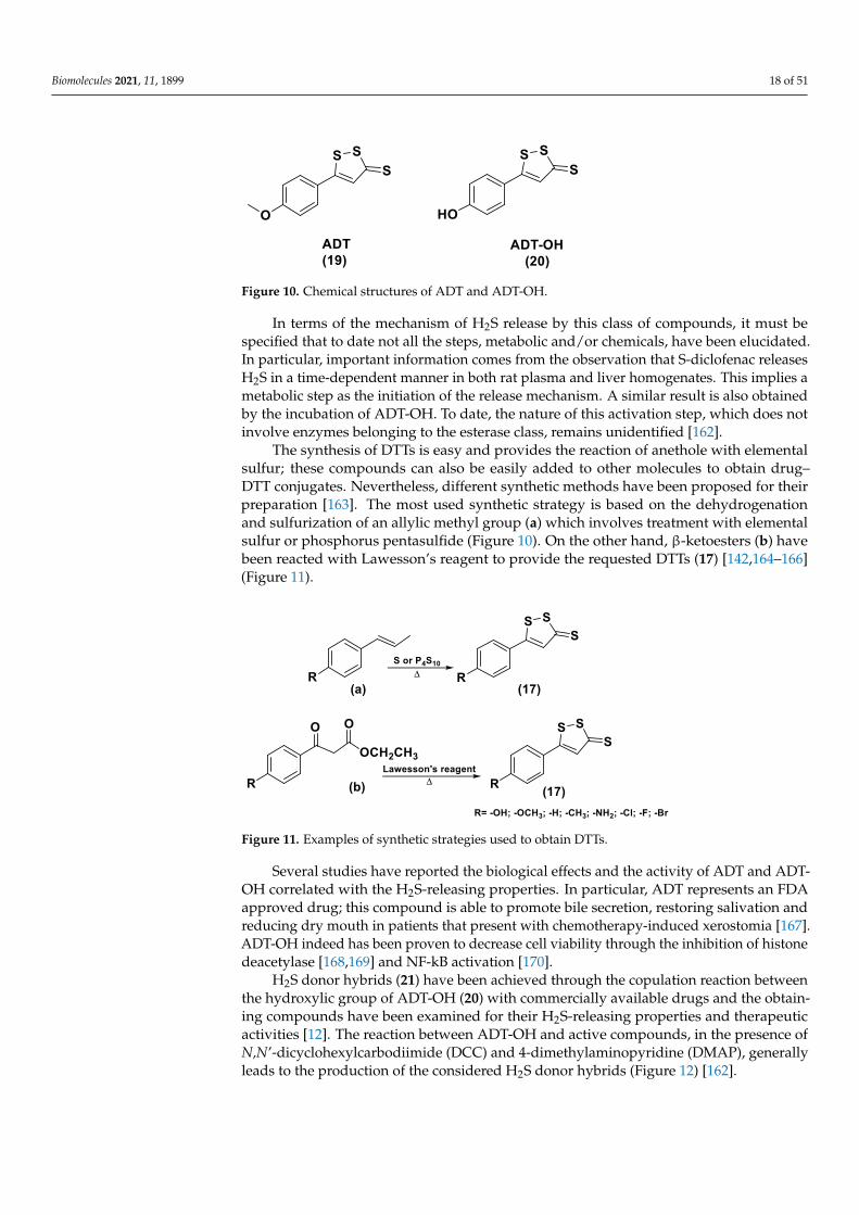
Biomolecules 2021, 11, 1899 18 of 51
Biomolecules 2021, 10, x 18 of 52
is considerably higher than the levels at other pH values [155]. Furthermore, it has been
observed that H2S concentrations were stable for long time in all solutions, demonstrat-
ing that ATTM is a stable and slow releasing H2S donor.
ATTM had protective effects on the heart and brain in rat models of myocardial and
cerebral I/R injury and reduces I/R injury [156]. In fact, certain studies are currently
evaluating the potential dual role of ATTM as a cardioprotective and anticancer agent in
patients with cancer treated with doxorubicin or Adriamycin [157–159]. Moreover,
ATTM has recently progressed in clinical trials for the treatment of breast cancer because
of its copper depletion effects [160].
3.1.2. DTTs
DTTs (1,2-Dithiole-3-thiones, 17) are another class of hydrolysis-triggered H2S do-
nors. Even if their H2S-releasing mechanism is not totally delineated, it is commonly
agreed that the production of H2S from DTTs occurs via hydrolysis (Figure 9).
It has been proven that DTT derivatives progressively release H2S, converting it in-
to the corresponding 1,2-dithiole-3-one (18) with heating to 120 °C and in aqueous solu-
tion [161].
Figure 9. H2S release from DTTs.
ADT (Anethole dithiolethione, 19) and ADT-OH (5-(p-hydroxyphenyl)-3H-1,2-
dithiole-3-thione, 20), a O-demethylated ADT derivative, are the most employed DTTs
as H2S donors (Figure 10).
Figure 10. Chemical structures of ADT and ADT-OH.
In terms of the mechanism of H2S release by this class of compounds, it must be
specified that to date not all the steps, metabolic and/or chemicals, have been elucidated.
In particular, important information comes from the observation that S-diclofenac re-
leases H2S in a time-dependent manner in both rat plasma and liver homogenates. This
implies a metabolic step as the initiation of the release mechanism. A similar result is al-
so obtained by the incubation of ADT-OH. To date, the nature of this activation step,
which does not involve enzymes belonging to the esterase class, remains unidentified
[162].
The synthesis of DTTs is easy and provides the reaction of anethole with elemental
sulfur; these compounds can also be easily added to other molecules to obtain drug–
DTT conjugates. Nevertheless, different synthetic methods have been proposed for their
preparation [163]. The most used synthetic strategy is based on the dehydrogenation
and sulfurization of an allylic methyl group (a) which involves treatment with elemental
sulfur or phosphorus pentasulfide (Figure 10). On the other hand, β-ketoesters (b) have
Figure 10. Chemical structures of ADT and ADT-OH.
In terms of the mechanism of H2S release by this class of compounds, it must bespecified that to date not all the steps, metabolic and/or chemicals, have been elucidated.In particular, important information comes from the observation that S-diclofenac releasesH2S in a time-dependent manner in both rat plasma and liver homogenates. This implies ametabolic step as the initiation of the release mechanism. A similar result is also obtainedby the incubation of ADT-OH. To date, the nature of this activation step, which does notinvolve enzymes belonging to the esterase class, remains unidentified [162].
The synthesis of DTTs is easy and provides the reaction of anethole with elementalsulfur; these compounds can also be easily added to other molecules to obtain drug–DTT conjugates. Nevertheless, different synthetic methods have been proposed for theirpreparation [163]. The most used synthetic strategy is based on the dehydrogenationand sulfurization of an allylic methyl group (a) which involves treatment with elementalsulfur or phosphorus pentasulfide (Figure 10). On the other hand, β-ketoesters (b) havebeen reacted with Lawesson’s reagent to provide the requested DTTs (17) [142,164–166](Figure 11).
Biomolecules 2021, 10, x 19 of 52
been reacted with Lawesson’s reagent to provide the requested DTTs (17) [142,164–166]
(Figure 11).
Figure 11. Examples of synthetic strategies used to obtain DTTs.
Several studies have reported the biological effects and the activity of ADT and
ADT-OH correlated with the H2S-releasing properties. In particular, ADT represents an
FDA approved drug; this compound is able to promote bile secretion, restoring saliva-
tion and reducing dry mouth in patients that present with chemotherapy-induced xero-
stomia [167]. ADT-OH indeed has been proven to decrease cell viability through the in-
hibition of histone deacetylase [168,169] and NF-kB activation [170].
H2S donor hybrids (21) have been achieved through the copulation reaction be-
tween the hydroxylic group of ADT-OH (20) with commercially available drugs and the
obtaining compounds have been examined for their H2S-releasing properties and thera-
peutic activities [12]. The reaction between ADT-OH and active compounds, in the pres-
ence of N,N’-dicyclohexylcarbodiimide (DCC) and 4-dimethylaminopyridine (DMAP),
generally leads to the production of the considered H2S donor hybrids (Figure 12) [162].
Figure 12. General synthetic strategy used to obtain HS–NSAIDs.
Non-steroidal anti-inflammatory drugs (NSAIDs), as well as ibuprofen and aspirin,
have been broadly applied for inflammation treatment [171]. Some studies have re-
vealed that the unpleasant effects induced by NSAIDs (for example, stomach bleeding
and gastrointestinal ulceration) can be substantially diminished by combining DTT mol-
ecules with the parent NSAIDs [12]. For example, a H2S–aspirin hybrid has been synthe-sized (ACS-14, S-aspirin, MZe786 (22); Figure 13) and the negative gastric effects caused
by ACS-14 and aspirin in rats have been compared [172]. It has been shown that after
oral administration of aspirin for 7 days, an extensive hemorrhagic lesion in the stomach
of rats occurred. By contrast, rats given an equivalent dose of ACS-14 did not present
with gastric damage. Then the H2S levels in plasma before and after ACS-14 were com-
pared, and the obtained results revealed that drug administration led to a three-fold in-
crease in the H2S concentration in the plasma. These results indicate that the advantages
of ACS-14 in gastric protection are probably correlated with the increase in H2S for-
mation. ACS-14 has also shown very important protective activities in pathological car-
diovascular alterations in rats induced by a GSH-synthase inhibitor (BSO, buthionine
sulfoximine) by improving systolic blood pressure and reducing heart rates. Further-
more, BSO-induced hypertensive effects were reduced by oral administration of ACS-14,
Figure 11. Examples of synthetic strategies used to obtain DTTs.
Several studies have reported the biological effects and the activity of ADT and ADT-OH correlated with the H2S-releasing properties. In particular, ADT represents an FDAapproved drug; this compound is able to promote bile secretion, restoring salivation andreducing dry mouth in patients that present with chemotherapy-induced xerostomia [167].ADT-OH indeed has been proven to decrease cell viability through the inhibition of histonedeacetylase [168,169] and NF-kB activation [170].
H2S donor hybrids (21) have been achieved through the copulation reaction betweenthe hydroxylic group of ADT-OH (20) with commercially available drugs and the obtain-ing compounds have been examined for their H2S-releasing properties and therapeuticactivities [12]. The reaction between ADT-OH and active compounds, in the presence ofN,N’-dicyclohexylcarbodiimide (DCC) and 4-dimethylaminopyridine (DMAP), generallyleads to the production of the considered H2S donor hybrids (Figure 12) [162].

Biomolecules 2021, 11, 1899 19 of 51
Biomolecules 2021, 10, x 19 of 52
been reacted with Lawesson’s reagent to provide the requested DTTs (17) [142,164–166]
(Figure 11).
Figure 11. Examples of synthetic strategies used to obtain DTTs.
Several studies have reported the biological effects and the activity of ADT and
ADT-OH correlated with the H2S-releasing properties. In particular, ADT represents an
FDA approved drug; this compound is able to promote bile secretion, restoring saliva-
tion and reducing dry mouth in patients that present with chemotherapy-induced xero-
stomia [167]. ADT-OH indeed has been proven to decrease cell viability through the in-
hibition of histone deacetylase [168,169] and NF-kB activation [170].
H2S donor hybrids (21) have been achieved through the copulation reaction be-
tween the hydroxylic group of ADT-OH (20) with commercially available drugs and the
obtaining compounds have been examined for their H2S-releasing properties and thera-
peutic activities [12]. The reaction between ADT-OH and active compounds, in the pres-
ence of N,N’-dicyclohexylcarbodiimide (DCC) and 4-dimethylaminopyridine (DMAP),
generally leads to the production of the considered H2S donor hybrids (Figure 12) [162].
Figure 12. General synthetic strategy used to obtain HS–NSAIDs.
Non-steroidal anti-inflammatory drugs (NSAIDs), as well as ibuprofen and aspirin,
have been broadly applied for inflammation treatment [171]. Some studies have re-
vealed that the unpleasant effects induced by NSAIDs (for example, stomach bleeding
and gastrointestinal ulceration) can be substantially diminished by combining DTT mol-
ecules with the parent NSAIDs [12]. For example, a H2S–aspirin hybrid has been synthe-sized (ACS-14, S-aspirin, MZe786 (22); Figure 13) and the negative gastric effects caused
by ACS-14 and aspirin in rats have been compared [172]. It has been shown that after
oral administration of aspirin for 7 days, an extensive hemorrhagic lesion in the stomach
of rats occurred. By contrast, rats given an equivalent dose of ACS-14 did not present
with gastric damage. Then the H2S levels in plasma before and after ACS-14 were com-
pared, and the obtained results revealed that drug administration led to a three-fold in-
crease in the H2S concentration in the plasma. These results indicate that the advantages
of ACS-14 in gastric protection are probably correlated with the increase in H2S for-
mation. ACS-14 has also shown very important protective activities in pathological car-
diovascular alterations in rats induced by a GSH-synthase inhibitor (BSO, buthionine
sulfoximine) by improving systolic blood pressure and reducing heart rates. Further-
more, BSO-induced hypertensive effects were reduced by oral administration of ACS-14,
Figure 12. General synthetic strategy used to obtain HS–NSAIDs.
Non-steroidal anti-inflammatory drugs (NSAIDs), as well as ibuprofen and aspirin,have been broadly applied for inflammation treatment [171]. Some studies have revealedthat the unpleasant effects induced by NSAIDs (for example, stomach bleeding and gas-trointestinal ulceration) can be substantially diminished by combining DTT moleculeswith the parent NSAIDs [12]. For example, a H2S-aspirin hybrid has been synthesized(ACS-14, S-aspirin, MZe786 (22); Figure 13) and the negative gastric effects caused byACS-14 and aspirin in rats have been compared [172]. It has been shown that after oraladministration of aspirin for 7 days, an extensive hemorrhagic lesion in the stomach of ratsoccurred. By contrast, rats given an equivalent dose of ACS-14 did not present with gastricdamage. Then the H2S levels in plasma before and after ACS-14 were compared, and theobtained results revealed that drug administration led to a three-fold increase in the H2Sconcentration in the plasma. These results indicate that the advantages of ACS-14 in gastricprotection are probably correlated with the increase in H2S formation. ACS-14 has alsoshown very important protective activities in pathological cardiovascular alterations inrats induced by a GSH-synthase inhibitor (BSO, buthionine sulfoximine) by improvingsystolic blood pressure and reducing heart rates. Furthermore, BSO-induced hypertensiveeffects were reduced by oral administration of ACS-14, unlike aspirin which did not havethis effect. Additionally, administration of ACS-14 decreased myocardial I/R injury in BSOrats [173–175].
Biomolecules 2021, 10, x 20 of 52
unlike aspirin which did not have this effect. Additionally, administration of ACS-14 de-
creased myocardial I/R injury in BSO rats [173–175].
Figure 13. Chemical structures of representative H2S–NSAIDs.
Hypertensive disorder in pregnancy is a main cause of maternal and perinatal mor-
tality. Soluble Flt-1 (sFlt-1) is a protein whose values are elevated in preeclampsia and
may continue to be high during postpartum in women with a preeclampsia diagnosis.
Heme oxygenase-1 (Hmox1/HO-1) is another protein that has protective effects versus
oxidative stimuli and is altered in the placenta of pregnant women with preeclampsia.
Ahmed et al. assumed that soluble Flt-1 impeded cardiac mitochondrial activity in HO-
1-deficient mice. It was demonstrated that the deprival of HO-1 perturbed cardiac mito-
chondrial respiration and decreased mitochondrial biogenesis. The overexpression of
sFlt-1 caused the inhibition of cardiac mitochondrial activity in Hmox1+/− mice. This re-
search revealed that MZe786 restored mitochondrial activity through the stimulation of
cardiac mitochondrial biogenesis and antioxidant protection in Hmox1+/− mice and in
Hmox1+/− mice subjected to a high sFlt-1 environment [176,177]. Moreover, Ahmed et al.
determined the therapeutic value of MZe786 in HO-1 haploid-deficient (Hmox1+/−) preg-
nant mice in a high sFlt-1 environment. It was demonstrated that the animal model used
presented critical preeclampsia symptoms and a decrease in antioxidant genes. MZe786
reduced preeclampsia-stimulating antioxidant genes and then reduced hypertension
and renal damage. MZe786 was also better for fetal outcomes in comparison with aspi-
rin alone and was shown to be a good therapeutic agent in preventing preeclampsia
[178]. Moreover, H2S-releasing diclofenac (ACS-15, S-diclofenac (23); Figure 13) has been
investigated. In some studies ACS-15 demonstrated an enhanced anti-inflammatory ef-
fect in comparison with diclofenac. ACS-15 considerably decreased the lipopolysaccha-
ride-induced infiltration of neutrophils into the liver and the lungs without increasing
leukocyte adherence, as instead diclofenac [66,171].
ATB-429 (mesalamine derivative (24); Figure 13) represents a hybrid drug from
mesalamine that has been used in animal models of Crohn’s disease and ulcerative coli-
tis. In preclinical studies concerning the visceral pain associated with inflammatory
bowel disease, this compound was more successful than mesalamine [86,179].
Beside NSAIDs, other DTT–H2S donor hybrids have been developed. As is already
known, L-DOPA represents a drug used in the clinical treatment for Parkinson’s Disease
and dopamine-responsive dystonia. Some side effects of long-term L-DOPA treatment
as well as dyskinesia seem to be correlated to the oxidative stress that leads to the loss of
Figure 13. Chemical structures of representative H2S-NSAIDs.
Hypertensive disorder in pregnancy is a main cause of maternal and perinatal mortal-ity. Soluble Flt-1 (sFlt-1) is a protein whose values are elevated in preeclampsia and maycontinue to be high during postpartum in women with a preeclampsia diagnosis. Hemeoxygenase-1 (Hmox1/HO-1) is another protein that has protective effects versus oxidativestimuli and is altered in the placenta of pregnant women with preeclampsia. Ahmed et al.assumed that soluble Flt-1 impeded cardiac mitochondrial activity in HO-1-deficient mice.It was demonstrated that the deprival of HO-1 perturbed cardiac mitochondrial respiration

Biomolecules 2021, 11, 1899 20 of 51
and decreased mitochondrial biogenesis. The overexpression of sFlt-1 caused the inhibitionof cardiac mitochondrial activity in Hmox1+/− mice. This research revealed that MZe786restored mitochondrial activity through the stimulation of cardiac mitochondrial biogenesisand antioxidant protection in Hmox1+/− mice and in Hmox1+/− mice subjected to a highsFlt-1 environment [176,177]. Moreover, Ahmed et al. determined the therapeutic valueof MZe786 in HO-1 haploid-deficient (Hmox1+/−) pregnant mice in a high sFlt-1 environ-ment. It was demonstrated that the animal model used presented critical preeclampsiasymptoms and a decrease in antioxidant genes. MZe786 reduced preeclampsia-stimulatingantioxidant genes and then reduced hypertension and renal damage. MZe786 was alsobetter for fetal outcomes in comparison with aspirin alone and was shown to be a goodtherapeutic agent in preventing preeclampsia [178].
Moreover, H2S-releasing diclofenac (ACS-15, S-diclofenac (23); Figure 13) has beeninvestigated. In some studies ACS-15 demonstrated an enhanced anti-inflammatory effectin comparison with diclofenac. ACS-15 considerably decreased the lipopolysaccharide-induced infiltration of neutrophils into the liver and the lungs without increasing leukocyteadherence, as instead diclofenac [66,171].
ATB-429 (mesalamine derivative (24); Figure 13) represents a hybrid drug frommesalamine that has been used in animal models of Crohn’s disease and ulcerative colitis.In preclinical studies concerning the visceral pain associated with inflammatory boweldisease, this compound was more successful than mesalamine [86,179].
Beside NSAIDs, other DTT-H2S donor hybrids have been developed. As is alreadyknown, L-DOPA represents a drug used in the clinical treatment for Parkinson’s Diseaseand dopamine-responsive dystonia. Some side effects of long-term L-DOPA treatmentas well as dyskinesia seem to be correlated to the oxidative stress that leads to the loss ofneurons [180,181]. Because H2S diminishes oxidative stress, L-DOPA coupled with a H2S-releasing moiety could lead to improved bioactivity. In fact, Lee et al. synthesized DOPA–DTT conjugates and studied their antioxidant effects [182]. The hybrid compounds (suchas ACS-84 (25); Figure 13) released H2S in the mitochondria. Additionally, this compoundhas been shown to inhibit MAO B, the dopamine metabolizing enzyme; therefore, it couldbe useful to reestablish the Parkinson’s Disease-depleted dopamine levels in the brain.
NO-NSAIDs represent another class of hybrid drugs well studied where NO isreleased [183] (Figure 14). NO and H2S share several comparable actions, and newNO–H2S-releasing hybrids (for example NOSH-1(26)) have been designed and studied(Figure 13) [174,184,185]. These compounds are able to induce higher NO and H2S concen-trations in vivo and have shown important activities related to the inhibition of the growthin cancer cell lines and anti-inflammatory activities in animal models.
Biomolecules 2021, 10, x 21 of 52
neurons [180,181]. Because H2S diminishes oxidative stress, L-DOPA coupled with a
H2S-releasing moiety could lead to improved bioactivity. In fact, Lee et al. synthesized
DOPA–DTT conjugates and studied their antioxidant effects [182]. The hybrid com-
pounds (such as ACS-84 (25); Figure 13) released H2S in the mitochondria. Additionally,
this compound has been shown to inhibit MAO B, the dopamine metabolizing enzyme;
therefore, it could be useful to reestablish the Parkinson’s Disease-depleted dopamine
levels in the brain.
NO–NSAIDs represent another class of hybrid drugs well studied where NO is re-
leased [183] (Figure 14). NO and H2S share several comparable actions, and new NO–
H2S-releasing hybrids (for example NOSH-1(26)) have been designed and studied (Fig-
ure 13) [174,184,185]. These compounds are able to induce higher NO and H2S concen-
trations in vivo and have shown important activities related to the inhibition of the
growth in cancer cell lines and anti-inflammatory activities in animal models.
Figure 14. Chemical structures of representative NO–H2S-releasing hybrids, PEG–ADT, and AP-
39.
Recently, DTT-based polymeric H2S donors have been reported [186]. DTT has been conjugated to poly(ethylene glycol) polymer, obtaining PEG–ADT (27; Figure 14) that
represents a H2S donor that demonstrates a lower and slower H2S release when com-
pared to DTT alone, probably as a result of the steric hindrance of PEG. PEG–ADT has
also been shown to have fewer toxic effects as compared to the parent donor compound;
this important characteristic is probably due to the co-localization of the PEG–ADT con-
jugate within the endo-lysosomes without escaping to the cytoplasm. It is important to
consider that even if DTTs have been asserted as H2S donors, it is not clear if their effects
are related to H2S; in fact, DTTs can considerably improve GSH levels in vivo by induc-
ing the enzymes involved in the preservation of the balance of GSH [187]. It is probable
that the higher GSH level contributes much more than H2S to those pharmaceutical ef-
fects.
AP-39 (28) is another mitochondria-targeted H2S donor hybrid, obtained from the
reaction of triphenylphosphonium (TPP) with ADT-OH [188]; it has been investigated
for its H2S-releasing properties and pharmacological effects (Figure 14) [189]. After being
rapidly up-taken and accumulated in a mitochondrial membrane-potential (ΔΨ) de-
pendent-manner, the AP-39-induced H2S release in the mitochondria was confirmed by
several fluorescent probes. It was also found that AP-39 significantly inhibits the oxida-
tive stress-induced toxicity by silencing the mitochondrial cell death pathways and con-
sequently contributing to the maintenance of cell homeostasis. Furthermore, in vivo
Figure 14. Chemical structures of representative NO-H2S-releasing hybrids, PEG-ADT, and AP-39.
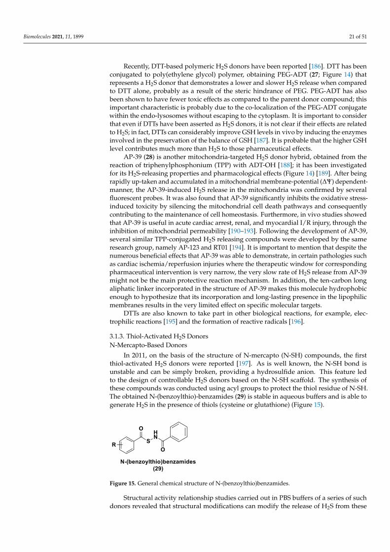
Biomolecules 2021, 11, 1899 21 of 51
Recently, DTT-based polymeric H2S donors have been reported [186]. DTT has beenconjugated to poly(ethylene glycol) polymer, obtaining PEG-ADT (27; Figure 14) thatrepresents a H2S donor that demonstrates a lower and slower H2S release when comparedto DTT alone, probably as a result of the steric hindrance of PEG. PEG-ADT has alsobeen shown to have fewer toxic effects as compared to the parent donor compound; thisimportant characteristic is probably due to the co-localization of the PEG-ADT conjugatewithin the endo-lysosomes without escaping to the cytoplasm. It is important to considerthat even if DTTs have been asserted as H2S donors, it is not clear if their effects are relatedto H2S; in fact, DTTs can considerably improve GSH levels in vivo by inducing the enzymesinvolved in the preservation of the balance of GSH [187]. It is probable that the higher GSHlevel contributes much more than H2S to those pharmaceutical effects.
AP-39 (28) is another mitochondria-targeted H2S donor hybrid, obtained from thereaction of triphenylphosphonium (TPP) with ADT-OH [188]; it has been investigatedfor its H2S-releasing properties and pharmacological effects (Figure 14) [189]. After beingrapidly up-taken and accumulated in a mitochondrial membrane-potential (∆Ψ) dependent-manner, the AP-39-induced H2S release in the mitochondria was confirmed by severalfluorescent probes. It was also found that AP-39 significantly inhibits the oxidative stress-induced toxicity by silencing the mitochondrial cell death pathways and consequentlycontributing to the maintenance of cell homeostasis. Furthermore, in vivo studies showedthat AP-39 is useful in acute cardiac arrest, renal, and myocardial I/R injury, through theinhibition of mitochondrial permeability [190–193]. Following the development of AP-39,several similar TPP-conjugated H2S releasing compounds were developed by the sameresearch group, namely AP-123 and RT01 [194]. It is important to mention that despite thenumerous beneficial effects that AP-39 was able to demonstrate, in certain pathologies suchas cardiac ischemia/reperfusion injuries where the therapeutic window for correspondingpharmaceutical intervention is very narrow, the very slow rate of H2S release from AP-39might not be the main protective reaction mechanism. In addition, the ten-carbon longaliphatic linker incorporated in the structure of AP-39 makes this molecule hydrophobicenough to hypothesize that its incorporation and long-lasting presence in the lipophilicmembranes results in the very limited effect on specific molecular targets.
DTTs are also known to take part in other biological reactions, for example, elec-trophilic reactions [195] and the formation of reactive radicals [196].
3.1.3. Thiol-Activated H2S DonorsN-Mercapto-Based Donors
In 2011, on the basis of the structure of N-mercapto (N-SH) compounds, the firstthiol-activated H2S donors were reported [197]. As is well known, the N-SH bond isunstable and can be simply broken, providing a hydrosulfide anion. This feature ledto the design of controllable H2S donors based on the N-SH scaffold. The synthesis ofthese compounds was conducted using acyl groups to protect the thiol residue of N-SH.The obtained N-(benzoylthio)-benzamides (29) is stable in aqueous buffers and is able togenerate H2S in the presence of thiols (cysteine or glutathione) (Figure 15).
Biomolecules 2021, 10, x 22 of 52
studies showed that AP-39 is useful in acute cardiac arrest, renal, and myocardial I/R in-
jury, through the inhibition of mitochondrial permeability [190–193]. Following the de-
velopment of AP-39, several similar TPP-conjugated H2S releasing compounds were de-
veloped by the same research group, namely AP-123 and RT01 [194]. It is important to
mention that despite the numerous beneficial effects that AP-39 was able to demon-
strate, in certain pathologies such as cardiac ischemia/reperfusion injuries where the
therapeutic window for corresponding pharmaceutical intervention is very narrow, the
very slow rate of H2S release from AP-39 might not be the main protective reaction
mechanism. In addition, the ten-carbon long aliphatic linker incorporated in the struc-
ture of AP-39 makes this molecule hydrophobic enough to hypothesize that its incorpo-
ration and long-lasting presence in the lipophilic membranes results in the very limited
effect on specific molecular targets.
DTTs are also known to take part in other biological reactions, for example, electro-
philic reactions [195] and the formation of reactive radicals [196].
3.1.3. Thiol-Activated H2S Donors
N-Mercapto-Based Donors
In 2011, on the basis of the structure of N-mercapto (N-SH) compounds, the first
thiol-activated H2S donors were reported [197]. As is well known, the N-SH bond is un-
stable and can be simply broken, providing a hydrosulfide anion. This feature led to the
design of controllable H2S donors based on the N-SH scaffold. The synthesis of these
compounds was conducted using acyl groups to protect the thiol residue of N-SH. The
obtained N-(benzoylthio)-benzamides (29) is stable in aqueous buffers and is able to
generate H2S in the presence of thiols (cysteine or glutathione) (Figure 15).
Figure 15. General chemical structure of N-(benzoylthio)benzamides.
Structural activity relationship studies carried out in PBS buffers of a series of such
donors revealed that structural modifications can modify the release of H2S from these
compounds. In particular, the presence of electron withdrawing groups promoted faster
H2S generation, but electron-donating groups slowed down the release of H2S. Thiol-
activated H2S donors have been also assessed in plasma and similar results in H2S re-
lease have been obtained.
The mechanism of H2S release from this N-mercapto-(N-SH) has been reported (Figure 16). Firstly, a thioester (30) exchange between the donor and cysteine occurs. The
resultant S-benzoyl cysteine (31) forms a more stable N-benzoyl cysteine (32) due to a
fast S to N acyl transfer. The N-SH intermediate (33) instead reacts with another mole-
cule of cysteine to produce benzamide and cysteine perthiol (34); the latter reacts with
cysteine to give cysteine disulfide (cystine, 35) and releasing H2S.
Figure 15. General chemical structure of N-(benzoylthio)benzamides.
Structural activity relationship studies carried out in PBS buffers of a series of suchdonors revealed that structural modifications can modify the release of H2S from these
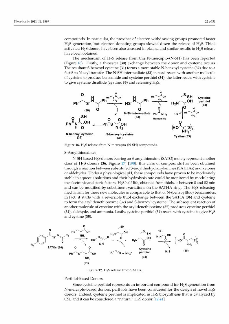
Biomolecules 2021, 11, 1899 22 of 51
compounds. In particular, the presence of electron withdrawing groups promoted fasterH2S generation, but electron-donating groups slowed down the release of H2S. Thiol-activated H2S donors have been also assessed in plasma and similar results in H2S releasehave been obtained.
The mechanism of H2S release from this N-mercapto-(N-SH) has been reported(Figure 16). Firstly, a thioester (30) exchange between the donor and cysteine occurs.The resultant S-benzoyl cysteine (31) forms a more stable N-benzoyl cysteine (32) due to afast S to N acyl transfer. The N-SH intermediate (33) instead reacts with another moleculeof cysteine to produce benzamide and cysteine perthiol (34); the latter reacts with cysteineto give cysteine disulfide (cystine, 35) and releasing H2S.
Biomolecules 2021, 10, x 23 of 52
Figure 16. H2S release from N-mercapto-(N-SH) compounds.
S-Aroylthiooximes
N-SH-based H2S donors bearing an S-aroylthiooxime (SATO) moiety represent an-other class of H2S donors (36, Figure 17) [198]; this class of compounds has been ob-
tained through a reaction between substituted S-aroylthiohydroxylamines (SATHAs)
and ketones or aldehydes. Under a physiological pH, these compounds have proven to
be moderately stable in aqueous solutions and their hydrolysis rate could be monitored
by modulating the electronic and steric factors. H2S half-life, obtained from thiols, is be-
tween 8 and 82 min and can be modified by substituent variations on the SATHA ring.
The H2S-releasing mechanism for these new molecules is comparable to that of N-
(benzoylthio) benzamides; in fact, it starts with a reversible thiol exchange between the
SATOs (36) and cysteine to form the arylidenethiooxime (37) and S-benzoyl cysteine.
The subsequent reaction of another molecule of cysteine with the arylidenethiooxime (37) produces cysteine perthiol (34), aldehyde, and ammonia. Lastly, cysteine perthiol
(34) reacts with cysteine to give H2S and cystine (35).
Figure 17. H2S release from SATOs.
Perthiol-Based Donors
Since cysteine perthiol represents an important compound for H2S generation from
N-mercapto-based donors, perthiols have been considered for the design of novel H2S
donors. Indeed, cysteine perthiol is implicated in H2S biosynthesis that is catalyzed by
CSE and it can be considered a “natural” H2S donor [12,41].
Considering this feature, a class of perthiol-based donors have been synthesized as cysteine (38) and penicillamine (39) derivatives (Figure 18) [199]. To protect the thiol res-
idue, acyl groups were used. The evaluation of the H2S-releasing profile of these donors
was carried out in the presence of thiols and in aqueous buffers and showed that cyste-
ine-based donors release very small amounts of hydrogen sulfide (probably caused by
an undesirable disulfide cleavage of the donors by thiols that leads to no H2S produc-
tion), while penicillamine-based donors have a high efficiency in H2S generation. Proba-
bly the presence of two adjacent methyl groups precluded the cleavage of the disulfide
bonds by thiols and then the diacylation and following H2S production dominated in the
reaction leading to a better H2S production.
Figure 16. H2S release from N-mercapto-(N-SH) compounds.
S-Aroylthiooximes
N-SH-based H2S donors bearing an S-aroylthiooxime (SATO) moiety represent anotherclass of H2S donors (36, Figure 17) [198]; this class of compounds has been obtainedthrough a reaction between substituted S-aroylthiohydroxylamines (SATHAs) and ketonesor aldehydes. Under a physiological pH, these compounds have proven to be moderatelystable in aqueous solutions and their hydrolysis rate could be monitored by modulatingthe electronic and steric factors. H2S half-life, obtained from thiols, is between 8 and 82 minand can be modified by substituent variations on the SATHA ring. The H2S-releasingmechanism for these new molecules is comparable to that of N-(benzoylthio) benzamides;in fact, it starts with a reversible thiol exchange between the SATOs (36) and cysteineto form the arylidenethiooxime (37) and S-benzoyl cysteine. The subsequent reaction ofanother molecule of cysteine with the arylidenethiooxime (37) produces cysteine perthiol(34), aldehyde, and ammonia. Lastly, cysteine perthiol (34) reacts with cysteine to give H2Sand cystine (35).
Biomolecules 2021, 10, x 23 of 52
Figure 16. H2S release from N-mercapto-(N-SH) compounds.
S-Aroylthiooximes
N-SH-based H2S donors bearing an S-aroylthiooxime (SATO) moiety represent an-other class of H2S donors (36, Figure 17) [198]; this class of compounds has been ob-
tained through a reaction between substituted S-aroylthiohydroxylamines (SATHAs)
and ketones or aldehydes. Under a physiological pH, these compounds have proven to
be moderately stable in aqueous solutions and their hydrolysis rate could be monitored
by modulating the electronic and steric factors. H2S half-life, obtained from thiols, is be-
tween 8 and 82 min and can be modified by substituent variations on the SATHA ring.
The H2S-releasing mechanism for these new molecules is comparable to that of N-
(benzoylthio) benzamides; in fact, it starts with a reversible thiol exchange between the
SATOs (36) and cysteine to form the arylidenethiooxime (37) and S-benzoyl cysteine.
The subsequent reaction of another molecule of cysteine with the arylidenethiooxime (37) produces cysteine perthiol (34), aldehyde, and ammonia. Lastly, cysteine perthiol
(34) reacts with cysteine to give H2S and cystine (35).
Figure 17. H2S release from SATOs.
Perthiol-Based Donors
Since cysteine perthiol represents an important compound for H2S generation from
N-mercapto-based donors, perthiols have been considered for the design of novel H2S
donors. Indeed, cysteine perthiol is implicated in H2S biosynthesis that is catalyzed by
CSE and it can be considered a “natural” H2S donor [12,41].
Considering this feature, a class of perthiol-based donors have been synthesized as cysteine (38) and penicillamine (39) derivatives (Figure 18) [199]. To protect the thiol res-
idue, acyl groups were used. The evaluation of the H2S-releasing profile of these donors
was carried out in the presence of thiols and in aqueous buffers and showed that cyste-
ine-based donors release very small amounts of hydrogen sulfide (probably caused by
an undesirable disulfide cleavage of the donors by thiols that leads to no H2S produc-
tion), while penicillamine-based donors have a high efficiency in H2S generation. Proba-
bly the presence of two adjacent methyl groups precluded the cleavage of the disulfide
bonds by thiols and then the diacylation and following H2S production dominated in the
reaction leading to a better H2S production.
Figure 17. H2S release from SATOs.
Perthiol-Based Donors
Since cysteine perthiol represents an important compound for H2S generation fromN-mercapto-based donors, perthiols have been considered for the design of novel H2Sdonors. Indeed, cysteine perthiol is implicated in H2S biosynthesis that is catalyzed byCSE and it can be considered a “natural” H2S donor [12,41].

Biomolecules 2021, 11, 1899 23 of 51
Considering this feature, a class of perthiol-based donors have been synthesizedas cysteine (38) and penicillamine (39) derivatives (Figure 18) [199]. To protect the thiolresidue, acyl groups were used. The evaluation of the H2S-releasing profile of these donorswas carried out in the presence of thiols and in aqueous buffers and showed that cysteine-based donors release very small amounts of hydrogen sulfide (probably caused by anundesirable disulfide cleavage of the donors by thiols that leads to no H2S production),while penicillamine-based donors have a high efficiency in H2S generation. Probably thepresence of two adjacent methyl groups precluded the cleavage of the disulfide bonds bythiols and then the diacylation and following H2S production dominated in the reactionleading to a better H2S production.
Biomolecules 2021, 10, x 24 of 52
Studies concerning the mechanism of H2S release from these donors showed simi-
larities to that of N-mercapto-based donors.
H2S release from penicillamine-derived perthiol donors in H9c2 cells and in in vivo
studies has been also evaluated. The cardioprotective effects of a number of compounds
have been also observed in a myocardial ischemia/reperfusion (MI/R) injury murine
model. The results revealed that these donors had therapeutic benefits reducing the in-
farct size in MI/R mice.
Figure 18. General chemical structures of perthiol-based H2S donors.
Dithioperoxy-Anhydrides
Dithioperoxy-anhydrides (40a, 40b) represent another kind of thiol-activated H2S
donor [200]. Considering the disulfide linkage, these donors have some chemical simi-
larities with perthiol-based donors. Acylpersulfides are very important intermediates for
H2S release and two probable pathways have been considered: (a) a direct reaction be-
tween acylpersulfides and thiols to form H2S and RSSAc; (b) the production of a new
perthiol leading to H2S and disulfide generation (Figure 19).
Figure 19. Chemical structures and the H2S release mechanism of dithioperoxy-anhydrides.
H2S release has been proven in both buffers and HeLa cell lysates. Moreover, the
CH3C(O)SSC(O)CH3 compound is effective in the concentration-dependent vaso-
relaxation of a pre-contracted rat aortic ring through its H2S release.
Thioamide- and Aryl Isothiocyanate-Based Donors
Arylthioamide derivatives (41) represent a class of thiol-activated H2S donors, even
if these compounds are weak and slow-releasing donors (Figure 20) [146].
Figure 18. General chemical structures of perthiol-based H2S donors.
Studies concerning the mechanism of H2S release from these donors showed similari-ties to that of N-mercapto-based donors.
H2S release from penicillamine-derived perthiol donors in H9c2 cells and in in vivostudies has been also evaluated. The cardioprotective effects of a number of compoundshave been also observed in a myocardial ischemia/reperfusion (MI/R) injury murinemodel. The results revealed that these donors had therapeutic benefits reducing the infarctsize in MI/R mice.
Dithioperoxy-Anhydrides
Dithioperoxy-anhydrides (40a, 40b) represent another kind of thiol-activated H2Sdonor [200]. Considering the disulfide linkage, these donors have some chemical similari-ties with perthiol-based donors. Acylpersulfides are very important intermediates for H2Srelease and two probable pathways have been considered: (a) a direct reaction betweenacylpersulfides and thiols to form H2S and RSSAc; (b) the production of a new perthiolleading to H2S and disulfide generation (Figure 19).
Biomolecules 2021, 10, x 24 of 52
Studies concerning the mechanism of H2S release from these donors showed simi-
larities to that of N-mercapto-based donors.
H2S release from penicillamine-derived perthiol donors in H9c2 cells and in in vivo
studies has been also evaluated. The cardioprotective effects of a number of compounds
have been also observed in a myocardial ischemia/reperfusion (MI/R) injury murine
model. The results revealed that these donors had therapeutic benefits reducing the in-
farct size in MI/R mice.
Figure 18. General chemical structures of perthiol-based H2S donors.
Dithioperoxy-Anhydrides
Dithioperoxy-anhydrides (40a, 40b) represent another kind of thiol-activated H2S
donor [200]. Considering the disulfide linkage, these donors have some chemical simi-
larities with perthiol-based donors. Acylpersulfides are very important intermediates for
H2S release and two probable pathways have been considered: (a) a direct reaction be-
tween acylpersulfides and thiols to form H2S and RSSAc; (b) the production of a new
perthiol leading to H2S and disulfide generation (Figure 19).
Figure 19. Chemical structures and the H2S release mechanism of dithioperoxy-anhydrides.
H2S release has been proven in both buffers and HeLa cell lysates. Moreover, the
CH3C(O)SSC(O)CH3 compound is effective in the concentration-dependent vaso-
relaxation of a pre-contracted rat aortic ring through its H2S release.
Thioamide- and Aryl Isothiocyanate-Based Donors
Arylthioamide derivatives (41) represent a class of thiol-activated H2S donors, even
if these compounds are weak and slow-releasing donors (Figure 20) [146].
Figure 19. Chemical structures and the H2S release mechanism of dithioperoxy-anhydrides.
H2S release has been proven in both buffers and HeLa cell lysates. Moreover, theCH3C(O)SSC(O)CH3 compound is effective in the concentration-dependent vaso-relaxationof a pre-contracted rat aortic ring through its H2S release.

Biomolecules 2021, 11, 1899 24 of 51
Thioamide- and Aryl Isothiocyanate-Based Donors
Arylthioamide derivatives (41) represent a class of thiol-activated H2S donors, even ifthese compounds are weak and slow-releasing donors (Figure 20) [146].
Biomolecules 2021, 10, x 25 of 52
Figure 20. Chemical structures of thioamide- and aryl isothiocyanate-based donors.
To evaluate the vasoconstriction effects, arylthioamides have been tested; it was ob-served that the pretreatment of aortic rings with compound 41a (p-
hydroxybenzothioamide), considerably inhibited the vasoconstriction generated by nor-
adrenaline. Oral administration of p-hydroxybenzothioamide in rats led to a decrease in
blood pressure, supporting the protective actions of this donors in cardiovascular sys-
tems. Endogenous thiols were sufficient to promote H2S release from the donors because
neither exogenous cysteine nor glutathione were added.
Aryl isothiocyanate derivatives represent another class of slow-releasing H2S do-
nors (Figure 20) [136]. Thiol activation for these derivatives proved to be required.
PhNCS (42) and PhNCS-COOH (43) exhibited vaso-relaxing effects on conductance and
coronary arteries but the biological mechanism is unclear. It has been proposed that aryl
isothiocyanates are able to react with biomolecules (for example cysteine) to produce
thioamide compounds and then H2S.
gem-Dithiol-Based H2S Donors
gem-Dithiols represent unstable compounds in aqueous solutions and H2S is the de-
composition product [201,202]. They belong to thiol-activated H2S donors (44, Figure 21)
[145,203]. These compounds have been modified by acylation and a library of donors
have been obtained. H2S release from these donors has been determined in the presence
of Cys or GSH. Donor-induced H2S release has been similarly examined in HeLa cells (in
the presence of endogenous thiols).
Figure 21. H2S release from acylated gem-dithiols.
3.1.4. Enzyme-Triggered Donors
In 2016 Zheng et al. developed a series of esterase-sensitive prodrugs, based on a
lactonization reaction, otherwise known as a ‘‘trimethyl lock” (TML) [204,205]. HP-101
(45) was the first obtained derivative (Figure 22) and it was synthesized under micro-
wave conditions starting from compound 45a by a reaction with Lawesson’s reagent
[206], to give an intermediate 45b that was subsequently treated with sodium hydroxide.
Figure 20. Chemical structures of thioamide- and aryl isothiocyanate-based donors.
To evaluate the vasoconstriction effects, arylthioamides have been tested; it was ob-served that the pretreatment of aortic rings with compound 41a (p-hydroxybenzothioamide),considerably inhibited the vasoconstriction generated by noradrenaline. Oral administra-tion of p-hydroxybenzothioamide in rats led to a decrease in blood pressure, supportingthe protective actions of this donors in cardiovascular systems. Endogenous thiols weresufficient to promote H2S release from the donors because neither exogenous cysteine norglutathione were added.
Aryl isothiocyanate derivatives represent another class of slow-releasing H2S donors(Figure 20) [136]. Thiol activation for these derivatives proved to be required. PhNCS(42) and PhNCS-COOH (43) exhibited vaso-relaxing effects on conductance and coronaryarteries but the biological mechanism is unclear. It has been proposed that aryl isothio-cyanates are able to react with biomolecules (for example cysteine) to produce thioamidecompounds and then H2S.
gem-Dithiol-Based H2S Donors
gem-Dithiols represent unstable compounds in aqueous solutions and H2S is the decom-position product [201,202]. They belong to thiol-activated H2S donors (44, Figure 21) [145,203].These compounds have been modified by acylation and a library of donors have beenobtained. H2S release from these donors has been determined in the presence of Cysor GSH. Donor-induced H2S release has been similarly examined in HeLa cells (in thepresence of endogenous thiols).
Biomolecules 2021, 10, x 25 of 52
Figure 20. Chemical structures of thioamide- and aryl isothiocyanate-based donors.
To evaluate the vasoconstriction effects, arylthioamides have been tested; it was ob-served that the pretreatment of aortic rings with compound 41a (p-
hydroxybenzothioamide), considerably inhibited the vasoconstriction generated by nor-
adrenaline. Oral administration of p-hydroxybenzothioamide in rats led to a decrease in
blood pressure, supporting the protective actions of this donors in cardiovascular sys-
tems. Endogenous thiols were sufficient to promote H2S release from the donors because
neither exogenous cysteine nor glutathione were added.
Aryl isothiocyanate derivatives represent another class of slow-releasing H2S do-
nors (Figure 20) [136]. Thiol activation for these derivatives proved to be required.
PhNCS (42) and PhNCS-COOH (43) exhibited vaso-relaxing effects on conductance and
coronary arteries but the biological mechanism is unclear. It has been proposed that aryl
isothiocyanates are able to react with biomolecules (for example cysteine) to produce
thioamide compounds and then H2S.
gem-Dithiol-Based H2S Donors
gem-Dithiols represent unstable compounds in aqueous solutions and H2S is the de-
composition product [201,202]. They belong to thiol-activated H2S donors (44, Figure 21)
[145,203]. These compounds have been modified by acylation and a library of donors
have been obtained. H2S release from these donors has been determined in the presence
of Cys or GSH. Donor-induced H2S release has been similarly examined in HeLa cells (in
the presence of endogenous thiols).
Figure 21. H2S release from acylated gem-dithiols.
3.1.4. Enzyme-Triggered Donors
In 2016 Zheng et al. developed a series of esterase-sensitive prodrugs, based on a
lactonization reaction, otherwise known as a ‘‘trimethyl lock” (TML) [204,205]. HP-101
(45) was the first obtained derivative (Figure 22) and it was synthesized under micro-
wave conditions starting from compound 45a by a reaction with Lawesson’s reagent
[206], to give an intermediate 45b that was subsequently treated with sodium hydroxide.
Figure 21. H2S release from acylated gem-dithiols.

Biomolecules 2021, 11, 1899 25 of 51
3.1.4. Enzyme-Triggered Donors
In 2016 Zheng et al. developed a series of esterase-sensitive prodrugs, based on alactonization reaction, otherwise known as a “trimethyl lock” (TML) [204,205]. HP-101(45) was the first obtained derivative (Figure 22) and it was synthesized under microwaveconditions starting from compound 45a by a reaction with Lawesson’s reagent [206], to givean intermediate 45b that was subsequently treated with sodium hydroxide. The desiredcompounds led to the release of H2S due to the cleavage of a phenolic ester by esteraseand the consequent lactonization caused by the steric repulsion of three methyl groups(Figure 22). Remarkably, HP-102 (46, Figure 22) a TML derivative, was shown to reducemyocardial I/R injury and presented a cardioprotective action [207]. HP-102 is at presentunder investigation to better identify its chemical, pharmacological, and safety profilesprior to its further progression in clinical trials.
Biomolecules 2021, 10, x 26 of 52
The desired compounds led to the release of H2S due to the cleavage of a phenolic ester
by esterase and the consequent lactonization caused by the steric repulsion of three me-thyl groups (Figure 22). Remarkably, HP-102 (46, Figure 22) a TML derivative, was
shown to reduce myocardial I/R injury and presented a cardioprotective action [207].
HP-102 is at present under investigation to better identify its chemical, pharmacological,
and safety profiles prior to its further progression in clinical trials.
Figure 22. TML chemical structures and the synthetic route for HP-101 preparation.
Carbonyl sulfide (COS)-releasing compounds are a recent class of enzyme-triggered
H2S donors; COS represent a substrate of CA (carbonic anhydrase) enzyme that catalyz-
es the reaction that leads to H2S production [208,209]. N-thiocarboxyanhydrides (NTAs)
(Figure 23) constitute dual COS/H2S donors [210,211], that advantageously release COS
and innoxious peptide byproducts at the same time [212]. NTA1 (47, Figure 22), a sarco-
sine NTA derivative was obtained starting from sarcosine (47a, N-methyl glycine) as re-
ported in Figure 23 [213].
Figure 23. General structure of N-thiocarboxyanhydrides (NTAs) and the synthetic procedure for
the synthesis of NTA1.
COS/H2S release from NTA1 is based on a cycle opening upon a nucleophilic attack
promoted by endogenous amines: the dipeptide formed has been detected by LC-MS
and GC-MS. The subsequent release of H2S from COS is due to the rapid enzymatic ac-
tion of CA (Figure 24) [212]. When endothelial cells have been treated with NTA1 im-
proved proliferation, an important step in the angiogenic process occurs [212].
Figure 22. TML chemical structures and the synthetic route for HP-101 preparation.
Carbonyl sulfide (COS)-releasing compounds are a recent class of enzyme-triggeredH2S donors; COS represent a substrate of CA (carbonic anhydrase) enzyme that catalyzesthe reaction that leads to H2S production [208,209]. N-thiocarboxyanhydrides (NTAs)(Figure 23) constitute dual COS/H2S donors [210,211], that advantageously release COSand innoxious peptide byproducts at the same time [212]. NTA1 (47, Figure 22), a sarcosineNTA derivative was obtained starting from sarcosine (47a, N-methyl glycine) as reportedin Figure 23 [213].
Biomolecules 2021, 10, x 26 of 52
The desired compounds led to the release of H2S due to the cleavage of a phenolic ester
by esterase and the consequent lactonization caused by the steric repulsion of three me-thyl groups (Figure 22). Remarkably, HP-102 (46, Figure 22) a TML derivative, was
shown to reduce myocardial I/R injury and presented a cardioprotective action [207].
HP-102 is at present under investigation to better identify its chemical, pharmacological,
and safety profiles prior to its further progression in clinical trials.
Figure 22. TML chemical structures and the synthetic route for HP-101 preparation.
Carbonyl sulfide (COS)-releasing compounds are a recent class of enzyme-triggered
H2S donors; COS represent a substrate of CA (carbonic anhydrase) enzyme that catalyz-
es the reaction that leads to H2S production [208,209]. N-thiocarboxyanhydrides (NTAs)
(Figure 23) constitute dual COS/H2S donors [210,211], that advantageously release COS
and innoxious peptide byproducts at the same time [212]. NTA1 (47, Figure 22), a sarco-
sine NTA derivative was obtained starting from sarcosine (47a, N-methyl glycine) as re-
ported in Figure 23 [213].
Figure 23. General structure of N-thiocarboxyanhydrides (NTAs) and the synthetic procedure for
the synthesis of NTA1.
COS/H2S release from NTA1 is based on a cycle opening upon a nucleophilic attack
promoted by endogenous amines: the dipeptide formed has been detected by LC-MS
and GC-MS. The subsequent release of H2S from COS is due to the rapid enzymatic ac-
tion of CA (Figure 24) [212]. When endothelial cells have been treated with NTA1 im-
proved proliferation, an important step in the angiogenic process occurs [212].
Figure 23. General structure of N-thiocarboxyanhydrides (NTAs) and the synthetic procedure for thesynthesis of NTA1.

Biomolecules 2021, 11, 1899 26 of 51
COS/H2S release from NTA1 is based on a cycle opening upon a nucleophilic attackpromoted by endogenous amines: the dipeptide formed has been detected by LC-MS andGC-MS. The subsequent release of H2S from COS is due to the rapid enzymatic actionof CA (Figure 24) [212]. When endothelial cells have been treated with NTA1 improvedproliferation, an important step in the angiogenic process occurs [212].
Biomolecules 2021, 10, x 27 of 52
Figure 24. COS/H2S release from NTA1.
ROS-triggered H2S donors (peroxyTCM-1, 2, 3; 48, 49, 50) are another class of
COS/H2S donors; the compounds were obtained [214] by applying a ROS-cleavable aryl
boronate (X) as a protecting group (Figure 25) [215,216].
Figure 25. ROS-activated H2S donor preparation.
The proposed mechanism for the release of COS/H2S is summarized in Figure 26:
these compounds carry out H2S in the presence of an oxidant, for example H2O2: the
boronate ester is transformed into a phenol by ROS, giving COS, which is then rapidly
moved into H2S by CA [214].
Figure 26. H2O2-triggered COS/H2S release.
Boronate esters are known as ROS scavengers and this effect was coupled with the
H2S cytoprotective action leading to a novel tool for the treatment of human pathologies
determined by elevated oxidative stress, for instance cardiovascular diseases [193]. Ad-
ditional in vivo studies are ongoing for the assessment of their efficacy.
3.1.5. Other H2S Donors in Cardiovascular Diseases
Thioglycine (51) and thiovaline (52), two thioamino acids, are bicarbonate-triggered
H2S donors [217]; these compounds have been synthesized starting from commercially
accessible Boc-protected amino acids (51a, 52a) that have been treated with 1,1′-
carbonyldiimidazole in DCM and then with gaseous H2S. The subsequent deprotection of intermediates 51b and 52b with TFA in dichloromethane provided the required com-
pounds (Figure 27).
Figure 24. COS/H2S release from NTA1.
ROS-triggered H2S donors (peroxyTCM-1, 2, 3; 48, 49, 50) are another class of COS/H2Sdonors; the compounds were obtained [214] by applying a ROS-cleavable aryl boronate (X)as a protecting group (Figure 25) [215,216].
Biomolecules 2021, 10, x 27 of 52
Figure 24. COS/H2S release from NTA1.
ROS-triggered H2S donors (peroxyTCM-1, 2, 3; 48, 49, 50) are another class of
COS/H2S donors; the compounds were obtained [214] by applying a ROS-cleavable aryl
boronate (X) as a protecting group (Figure 25) [215,216].
Figure 25. ROS-activated H2S donor preparation.
The proposed mechanism for the release of COS/H2S is summarized in Figure 26:
these compounds carry out H2S in the presence of an oxidant, for example H2O2: the
boronate ester is transformed into a phenol by ROS, giving COS, which is then rapidly
moved into H2S by CA [214].
Figure 26. H2O2-triggered COS/H2S release.
Boronate esters are known as ROS scavengers and this effect was coupled with the
H2S cytoprotective action leading to a novel tool for the treatment of human pathologies
determined by elevated oxidative stress, for instance cardiovascular diseases [193]. Ad-
ditional in vivo studies are ongoing for the assessment of their efficacy.
3.1.5. Other H2S Donors in Cardiovascular Diseases
Thioglycine (51) and thiovaline (52), two thioamino acids, are bicarbonate-triggered
H2S donors [217]; these compounds have been synthesized starting from commercially
accessible Boc-protected amino acids (51a, 52a) that have been treated with 1,1′-
carbonyldiimidazole in DCM and then with gaseous H2S. The subsequent deprotection of intermediates 51b and 52b with TFA in dichloromethane provided the required com-
pounds (Figure 27).
Figure 25. ROS-activated H2S donor preparation.
The proposed mechanism for the release of COS/H2S is summarized in Figure 26:these compounds carry out H2S in the presence of an oxidant, for example H2O2: theboronate ester is transformed into a phenol by ROS, giving COS, which is then rapidlymoved into H2S by CA [214].
Biomolecules 2021, 10, x 27 of 52
Figure 24. COS/H2S release from NTA1.
ROS-triggered H2S donors (peroxyTCM-1, 2, 3; 48, 49, 50) are another class of
COS/H2S donors; the compounds were obtained [214] by applying a ROS-cleavable aryl
boronate (X) as a protecting group (Figure 25) [215,216].
Figure 25. ROS-activated H2S donor preparation.
The proposed mechanism for the release of COS/H2S is summarized in Figure 26:
these compounds carry out H2S in the presence of an oxidant, for example H2O2: the
boronate ester is transformed into a phenol by ROS, giving COS, which is then rapidly
moved into H2S by CA [214].
Figure 26. H2O2-triggered COS/H2S release.
Boronate esters are known as ROS scavengers and this effect was coupled with the
H2S cytoprotective action leading to a novel tool for the treatment of human pathologies
determined by elevated oxidative stress, for instance cardiovascular diseases [193]. Ad-
ditional in vivo studies are ongoing for the assessment of their efficacy.
3.1.5. Other H2S Donors in Cardiovascular Diseases
Thioglycine (51) and thiovaline (52), two thioamino acids, are bicarbonate-triggered
H2S donors [217]; these compounds have been synthesized starting from commercially
accessible Boc-protected amino acids (51a, 52a) that have been treated with 1,1′-
carbonyldiimidazole in DCM and then with gaseous H2S. The subsequent deprotection of intermediates 51b and 52b with TFA in dichloromethane provided the required com-
pounds (Figure 27).
Figure 26. H2O2-triggered COS/H2S release.
Boronate esters are known as ROS scavengers and this effect was coupled with the H2Scytoprotective action leading to a novel tool for the treatment of human pathologies deter-mined by elevated oxidative stress, for instance cardiovascular diseases [193]. Additionalin vivo studies are ongoing for the assessment of their efficacy.

Biomolecules 2021, 11, 1899 27 of 51
3.1.5. Other H2S Donors in Cardiovascular Diseases
Thioglycine (51) and thiovaline (52), two thioamino acids, are bicarbonate-triggered H2Sdonors [217]; these compounds have been synthesized starting from commercially accessibleBoc-protected amino acids (51a, 52a) that have been treated with 1,1′-carbonyldiimidazole inDCM and then with gaseous H2S. The subsequent deprotection of intermediates 51b and52b with TFA in dichloromethane provided the required compounds (Figure 27).
Biomolecules 2021, 10, x 28 of 52
Figure 27. Synthesis of thioglycine and thiovaline.
Following a cyclization reaction, these compounds are converted into the corre-
sponding amino acid N-carboxyanhydrides in the presence of a bicarbonate and pro-
duce H2S (Figure 28). Thioamino acids are considered as in vivo H2S donors due to the
releasing mechanism and the high-level of bicarbonate in the blood.
Figure 28. H2S release from thioamino acids.
Thioglycine and thiovaline H2S release have been assessed through amperometric
studies. The compounds also improve the formation of cGMP in a concentration-
dependent manner and produce a substantial relaxation of pre-contracted mouse aortic
rings. Conversely, thioglycine and thiovaline are characterized by a high reactivity; in
fact, they can provide amidation [218–222] and oxidation reactions [190], giving prod-
ucts which could produce undesirable side-effects. ZYZ803 (2-(N-Boc-amino)-3-prop-2-ynylsulfanylpropionic acid, 53) is a novel H2S-
NO releasing molecule, recently developed through a copulation reaction between SPRC
and furoxan in the presence of Boc anhydride (Figure 29) [223,224].
Figure 29. Synthesis of ZYZ803.
SPRC (53a) releases H2S through CSE enzyme activation, whereas furoxan (53b)
represents a thiol-triggered NO-releasing moiety [225]. Consequently, ZYZ803 (53) is
able to stimulate either the expression of CSE through NO synthase (e-NOS) endothelial
activity, releasing H2S and NO, correspondingly (Figure 30), promoting angiogenesis by
the SIRT1/VEGF/cGMP pathway and through crosstalk between STAT3 and CaMKII
[226].
Figure 27. Synthesis of thioglycine and thiovaline.
Following a cyclization reaction, these compounds are converted into the correspond-ing amino acid N-carboxyanhydrides in the presence of a bicarbonate and produce H2S(Figure 28). Thioamino acids are considered as in vivo H2S donors due to the releasingmechanism and the high-level of bicarbonate in the blood.
Biomolecules 2021, 10, x 28 of 52
Figure 27. Synthesis of thioglycine and thiovaline.
Following a cyclization reaction, these compounds are converted into the corre-
sponding amino acid N-carboxyanhydrides in the presence of a bicarbonate and pro-
duce H2S (Figure 28). Thioamino acids are considered as in vivo H2S donors due to the
releasing mechanism and the high-level of bicarbonate in the blood.
Figure 28. H2S release from thioamino acids.
Thioglycine and thiovaline H2S release have been assessed through amperometric
studies. The compounds also improve the formation of cGMP in a concentration-
dependent manner and produce a substantial relaxation of pre-contracted mouse aortic
rings. Conversely, thioglycine and thiovaline are characterized by a high reactivity; in
fact, they can provide amidation [218–222] and oxidation reactions [190], giving prod-
ucts which could produce undesirable side-effects. ZYZ803 (2-(N-Boc-amino)-3-prop-2-ynylsulfanylpropionic acid, 53) is a novel H2S-
NO releasing molecule, recently developed through a copulation reaction between SPRC
and furoxan in the presence of Boc anhydride (Figure 29) [223,224].
Figure 29. Synthesis of ZYZ803.
SPRC (53a) releases H2S through CSE enzyme activation, whereas furoxan (53b)
represents a thiol-triggered NO-releasing moiety [225]. Consequently, ZYZ803 (53) is
able to stimulate either the expression of CSE through NO synthase (e-NOS) endothelial
activity, releasing H2S and NO, correspondingly (Figure 30), promoting angiogenesis by
the SIRT1/VEGF/cGMP pathway and through crosstalk between STAT3 and CaMKII
[226].
Figure 28. H2S release from thioamino acids.
Thioglycine and thiovaline H2S release have been assessed through amperometricstudies. The compounds also improve the formation of cGMP in a concentration-dependentmanner and produce a substantial relaxation of pre-contracted mouse aortic rings. Con-versely, thioglycine and thiovaline are characterized by a high reactivity; in fact, they canprovide amidation [218–222] and oxidation reactions [190], giving products which couldproduce undesirable side-effects.
ZYZ803 (2-(N-Boc-amino)-3-prop-2-ynylsulfanylpropionic acid, 53) is a novel H2S-NOreleasing molecule, recently developed through a copulation reaction between SPRC andfuroxan in the presence of Boc anhydride (Figure 29) [223,224].
Biomolecules 2021, 10, x 28 of 52
Figure 27. Synthesis of thioglycine and thiovaline.
Following a cyclization reaction, these compounds are converted into the corre-
sponding amino acid N-carboxyanhydrides in the presence of a bicarbonate and pro-
duce H2S (Figure 28). Thioamino acids are considered as in vivo H2S donors due to the
releasing mechanism and the high-level of bicarbonate in the blood.
Figure 28. H2S release from thioamino acids.
Thioglycine and thiovaline H2S release have been assessed through amperometric
studies. The compounds also improve the formation of cGMP in a concentration-
dependent manner and produce a substantial relaxation of pre-contracted mouse aortic
rings. Conversely, thioglycine and thiovaline are characterized by a high reactivity; in
fact, they can provide amidation [218–222] and oxidation reactions [190], giving prod-
ucts which could produce undesirable side-effects. ZYZ803 (2-(N-Boc-amino)-3-prop-2-ynylsulfanylpropionic acid, 53) is a novel H2S-
NO releasing molecule, recently developed through a copulation reaction between SPRC
and furoxan in the presence of Boc anhydride (Figure 29) [223,224].
Figure 29. Synthesis of ZYZ803.
SPRC (53a) releases H2S through CSE enzyme activation, whereas furoxan (53b)
represents a thiol-triggered NO-releasing moiety [225]. Consequently, ZYZ803 (53) is
able to stimulate either the expression of CSE through NO synthase (e-NOS) endothelial
activity, releasing H2S and NO, correspondingly (Figure 30), promoting angiogenesis by
the SIRT1/VEGF/cGMP pathway and through crosstalk between STAT3 and CaMKII
[226].
Figure 29. Synthesis of ZYZ803.
SPRC (53a) releases H2S through CSE enzyme activation, whereas furoxan (53b)represents a thiol-triggered NO-releasing moiety [225]. Consequently, ZYZ803 (53) isable to stimulate either the expression of CSE through NO synthase (e-NOS) endothelialactivity, releasing H2S and NO, correspondingly (Figure 30), promoting angiogenesis by theSIRT1/VEGF/cGMP pathway and through crosstalk between STAT3 and CaMKII [226].

Biomolecules 2021, 11, 1899 28 of 51Biomolecules 2021, 10, x 29 of 52
Figure 30. H2S/NO releasing pathways from ZYZ803.
In vitro and in vivo studies have shown that ZYZ803 significantly promotes endo-
thelial cell angiogenesis [227]. In an animal model of ischemia, ZYZ803 stimulated angi-
ogenesis in a more efficient way in comparison to the monotherapy administration of
SPRC or furoxan, proving the synergistic actions of H2S and NO released by ZYZ803
[227]. This interesting lead compound regulated vascular tone in rat aortic rings [223],
reduced cardiac dysfunction, and increased myocardial injury following heart failure
[228], giving a potent cardioprotective effect.
Finally, Zofenopril (54), an ACE inhibitor, is among the H2S donors with cardiovas-
cular system activity. This compound in the liver undergoes a hydrolysis reaction with
the formation of Zofenoprilat (55), and its active metabolite contains a thiol group (Fig-
ure 31).
Figure 31. Chemical structures of Zofenopril and Zofenoprilat.
Zofenopril is an already well studied drug, and it was approved for medical use in
2000. This compound has been proven to release H2S, and this gaseous transmitter in
turn mediates the beneficial anti-apoptotic, pro-angiogenic, and anti-inflammatory ef-
fects of zofenopril [229–234]. The drug improved vascular function in a murine model of
hypertension, due to its ACE inhibitor action and H2S-releasing effects, and the admin-
istration of this compound increased the plasmatic levels of H2S in mice, protecting them
from myocardial I/R injury and increasing the endocardial blood flow in myocardial is-
chemia [235].
3.2. H2S Donors and Cancer
H2S has several roles in the regulation of cellular activities, for instance, migration,
proliferation, apoptosis, and autophagy. A low production of H2S in several cancer types
has been found. The principal experimental technique applied to enhance H2S levels in
cancer cells is represented by the use of H2S donors; nevertheless, their use is connecting
to the concentration/dose, cell type, time, and administration method.
Natural and synthesized donors have been used in this context and different effects
have been observed, such as suppressant effects but also promoter effects. Following
treatment with H2S donors, numerous signaling pathways have been altered by cells,
Figure 30. H2S/NO releasing pathways from ZYZ803.
In vitro and in vivo studies have shown that ZYZ803 significantly promotes endothe-lial cell angiogenesis [227]. In an animal model of ischemia, ZYZ803 stimulated angiogene-sis in a more efficient way in comparison to the monotherapy administration of SPRC orfuroxan, proving the synergistic actions of H2S and NO released by ZYZ803 [227]. Thisinteresting lead compound regulated vascular tone in rat aortic rings [223], reduced cardiacdysfunction, and increased myocardial injury following heart failure [228], giving a potentcardioprotective effect.
Finally, Zofenopril (54), an ACE inhibitor, is among the H2S donors with cardiovascu-lar system activity. This compound in the liver undergoes a hydrolysis reaction with theformation of Zofenoprilat (55), and its active metabolite contains a thiol group (Figure 31).
Biomolecules 2021, 10, x 29 of 52
Figure 30. H2S/NO releasing pathways from ZYZ803.
In vitro and in vivo studies have shown that ZYZ803 significantly promotes endo-
thelial cell angiogenesis [227]. In an animal model of ischemia, ZYZ803 stimulated angi-
ogenesis in a more efficient way in comparison to the monotherapy administration of
SPRC or furoxan, proving the synergistic actions of H2S and NO released by ZYZ803
[227]. This interesting lead compound regulated vascular tone in rat aortic rings [223],
reduced cardiac dysfunction, and increased myocardial injury following heart failure
[228], giving a potent cardioprotective effect.
Finally, Zofenopril (54), an ACE inhibitor, is among the H2S donors with cardiovas-
cular system activity. This compound in the liver undergoes a hydrolysis reaction with
the formation of Zofenoprilat (55), and its active metabolite contains a thiol group (Fig-
ure 31).
Figure 31. Chemical structures of Zofenopril and Zofenoprilat.
Zofenopril is an already well studied drug, and it was approved for medical use in
2000. This compound has been proven to release H2S, and this gaseous transmitter in
turn mediates the beneficial anti-apoptotic, pro-angiogenic, and anti-inflammatory ef-
fects of zofenopril [229–234]. The drug improved vascular function in a murine model of
hypertension, due to its ACE inhibitor action and H2S-releasing effects, and the admin-
istration of this compound increased the plasmatic levels of H2S in mice, protecting them
from myocardial I/R injury and increasing the endocardial blood flow in myocardial is-
chemia [235].
3.2. H2S Donors and Cancer
H2S has several roles in the regulation of cellular activities, for instance, migration,
proliferation, apoptosis, and autophagy. A low production of H2S in several cancer types
has been found. The principal experimental technique applied to enhance H2S levels in
cancer cells is represented by the use of H2S donors; nevertheless, their use is connecting
to the concentration/dose, cell type, time, and administration method.
Natural and synthesized donors have been used in this context and different effects
have been observed, such as suppressant effects but also promoter effects. Following
treatment with H2S donors, numerous signaling pathways have been altered by cells,
Figure 31. Chemical structures of Zofenopril and Zofenoprilat.
Zofenopril is an already well studied drug, and it was approved for medical usein 2000. This compound has been proven to release H2S, and this gaseous transmitterin turn mediates the beneficial anti-apoptotic, pro-angiogenic, and anti-inflammatory ef-fects of zofenopril [229–234]. The drug improved vascular function in a murine modelof hypertension, due to its ACE inhibitor action and H2S-releasing effects, and the ad-ministration of this compound increased the plasmatic levels of H2S in mice, protectingthem from myocardial I/R injury and increasing the endocardial blood flow in myocardialischemia [235].
3.2. H2S Donors and Cancer
H2S has several roles in the regulation of cellular activities, for instance, migration,proliferation, apoptosis, and autophagy. A low production of H2S in several cancer typeshas been found. The principal experimental technique applied to enhance H2S levels incancer cells is represented by the use of H2S donors; nevertheless, their use is connectingto the concentration/dose, cell type, time, and administration method.
Natural and synthesized donors have been used in this context and different effectshave been observed, such as suppressant effects but also promoter effects. Followingtreatment with H2S donors, numerous signaling pathways have been altered by cells,prompting their use in cancer treatment. The good efficiency of H2S donors has silenceddifferent cancer types due the regulation of cellular activities and signaling pathways. Theevident opposite biological effects in different experiments, for example in the case of

Biomolecules 2021, 11, 1899 29 of 51
NaHS, needs to be further analyzed. The different effects of this salt seem to be correlatedto the short-time and the high concentration of H2S released from NaHS, giving firstly amomentary increase in the cellular H2S levels, followed by a subsequent decline; this kindof release is very different to the normal slow-releasing cellular mode, and therefore NaHShas been replaced in clinical applications.
Additionally, the amount of information about H2S donors (such as on metabolismand clearance) is limited and their applicability has generated many questions.
For this purpose, it is important to assess the range of concentrations for H2S donorsand to define all the potential toxicity and clearance mechanisms. Furthermore, potentialinteractions between different molecules, as in the case of molecular hybrids, requiresadditional analysis for a better realization of the side effects.
It has been observed that the combination of H2S donors and cancer drugs (for examplecisplatin and docetaxel) increases drug sensitivity and decreases resistance. The synergisticeffect of H2S donors with metformin/simvastatin has promoted anti-cancer activities dueto the elevation of intracellular acidity and the reduction in pH regulators, paving the wayfor their useful administration in the treatment of advanced stages of cancer; at present, allthe researchers agree that more preclinical and clinical trials are needed to study this aspect.
In summary, H2S donors, used both in monotherapy or in combination with otherdrugs, have interesting applications in cancer treatment; several studies are now in progressand the hope is to shed additional light on cancer therapy [236].
Triple-negative breast cancer (TNBC) is an aggressive subtype cancer form and repre-sents about 15–20% of breast cancers. Unfortunately, it is characterized by metastasis andrecurrence and the development of novel treatments of TNBC is very important. Recently,extraordinary progress in the treatment of TNBC by H2S donors has been made.
In summary, on the one hand, H2S donors efficiently modulate the proliferation andapoptosis of TNBC cells through the inhibition of the phosphorylation or expression ofproteins connected to the NF-κB, PI3K/Akt/mTOR, and Ras/Raf/MEK/ERK signalingpathways. Otherwise, H2S donors inhibit the expression of MMP-2/9 and EMT to resistTNBC metastasis through the inhibition of the abnormal activation of the β-catenin pathway.
H2S is believed to be involved in other pathways that account for TNBC preventionand this deserves further investigation. Some organic H2S donors (DATS, DADS, GYY4137,ADT-OH, thiobenzamide) are used for endogenous H2S production to exert antitumoreffects. ATB-346 (naproxen derivative, 56, Figure 32) and GIC-1001 (57, Figure 32) repre-sent some H2S donor-based therapeutics entered in Phase II clinical trials. HS-ASA (58,Figure 32) showed antitumor activity on TNBC MDA-MB-231 cells in vitro and in vivostudies. HA-ADT (59, Figure 32) was able to induce MDA-MB-231 cell apoptosis by im-proving the ratio of Bad/Bcl-xl and Bax/Bcl-2 and the expression of cleaved caspase-3/9and cleaved PARP [237].
Hybrids capable of releasing H2S or NO, or both gaseous transmitters have beendeveloped. The hybrids can be beneficial in reducing the doses of the chemotherapeuticagent in order to diminish the toxic effects of these drugs and the gaseous transmitter gainsadditional chemotherapeutic effects.
Various H2S and NO-releasing compounds have been developed for their use in cancerchemotherapy and SNOs (S-nitrosothiols) donors are to date considered the most efficientcompounds. GSNO (S-nitrosoglutathione, 60) and S-nitroso-N-acetylpenicillamine (SNAP, 61)(Figure 33) are two SNOs that have been investigated for their anti-carcinogenic properties.
The reaction between SNO donors and Cysteine leads to the formation of CysNOat the external face of the plasma membrane. This compound has a membrane that isimpermeable but passes into the cell using the L-type amino acid transporter on the plasmamembrane. The reaction between SNO donors and H2S yields HSNO which diffusesspontaneously across cell membranes. The mixed effects of CysNO and HSNO enhancethe cellular nitrosation most likely via transnitrosation processes or by enhancing thenitrosating capabilities of both compounds. Prolonged exposure of tumor cells to thesecompounds causes stress conditions and consequently leads to apoptosis [238].

Biomolecules 2021, 11, 1899 30 of 51Biomolecules 2021, 10, x 31 of 52
Figure 32. Chemical structures of H2S donors evaluated for TNBC treatment.
Hybrids capable of releasing H2S or NO, or both gaseous transmitters have been
developed. The hybrids can be beneficial in reducing the doses of the chemotherapeutic
agent in order to diminish the toxic effects of these drugs and the gaseous transmitter
gains additional chemotherapeutic effects.
Various H2S and NO-releasing compounds have been developed for their use in
cancer chemotherapy and SNOs (S-nitrosothiols) donors are to date considered the most efficient compounds. GSNO (S-nitrosoglutathione, 60) and S-nitroso-N-
acetylpenicillamine (SNAP, 61) (Figure 33) are two SNOs that have been investigated for
their anti-carcinogenic properties.
The reaction between SNO donors and Cysteine leads to the formation of CysNO at
the external face of the plasma membrane. This compound has a membrane that is im-
permeable but passes into the cell using the L-type amino acid transporter on the plasma
membrane. The reaction between SNO donors and H2S yields HSNO which diffuses
spontaneously across cell membranes. The mixed effects of CysNO and HSNO enhance
the cellular nitrosation most likely via transnitrosation processes or by enhancing the ni-
trosating capabilities of both compounds. Prolonged exposure of tumor cells to these
compounds causes stress conditions and consequently leads to apoptosis [238].
Figure 32. Chemical structures of H2S donors evaluated for TNBC treatment.Biomolecules 2021, 10, x 32 of 52
Figure 33. Chemical structures of GSNO and SNAP.
Cancer cells are distinguished by an improved uptake of sugars, for example glu-
cose. Fortunato et al. synthesized novel glycoconjugated-H2S donors in order to selec-
tively enhance the concentration of H2S in cancer cells. Dithiolethione or isothiocyanate
moieties have been considered for their H2S-releasing properties. C1-glycoconjugates
have been obtained by applying a synthetic stereoselective procedure based on trichlo-
roacetimidate glycosyl donors, while C6-glycoconjugates have been achieved through a
Mitsunobu reaction (Figure 33). Subsequently the obtained compounds have been eval-
uated for their anticancer effects in the AsPC-1 cell line. Compounds 62 and 63 (Figure
34) have been demonstrated to release H2S inside the AsPC-1 cells and to modify the ba-
sal cell cycle [239].
Figure 34. Glycoconjugated H2S donors.
Brassicaceae are a well-known plant family and have attracted attention for the
presence of the glucosinolate myrosinase system that leads to isothiocyanates upon
plant biotic and abiotic lesions.
Erucin (ERU; Figure 4), an isothiocyanate present in arugula, originates through
glucosinolate glucoerucin hydrolysis due to the myrosinase enzyme.
Natural isothiocyanates modify cancer cells stimulating anti-proliferative effects at
high concentrations. Gaseous hydrogen sulfide or non-natural H2S-releasing compounds
showed comparable anti-proliferative effects.
Isothiocyanates release H2S in biological conditions: ERU belongs to this category
and was evaluated for its anticancer effects on pancreatic adenocarcinoma cells (AsPC-
1). The obtained results showed the chemo preventive and anticancer features of this
compound. In addition, ERU presented significant anti-proliferative effects by the inhi-
bition of AsPC-1 cell viability and cell migration, modifying the AsPC-1 cell cycle, and
exhibiting a pro-apoptotic effect.
Moreover, AsPC-1 cells present a genetic mutation in KRAS that leads to KRAS hy-
per-activation and then to MAP-kinase hyper-phosphorylation. ERU repressed ERK1/2
phosphorylation. This inhibition is very important in pancreatic cancer proliferation,
growth, and survival [240].
3.3. H2S Donors and Nervous System Diseases
H2S donors have been investigated for their use in the treatment of nervous system
pathologies. The pioneer compound for this use was ACS 84 (Figure 12), a DOPA-DTT
that has been shown to have an important role in Parkinson’s Disease; successively, sev-
eral other compounds have been reported for their in vitro effects and for their potential
use in Alzheimer’s disease (AD).
Figure 33. Chemical structures of GSNO and SNAP.
Cancer cells are distinguished by an improved uptake of sugars, for example glucose.Fortunato et al. synthesized novel glycoconjugated-H2S donors in order to selectivelyenhance the concentration of H2S in cancer cells. Dithiolethione or isothiocyanate moietieshave been considered for their H2S-releasing properties. C1-glycoconjugates have beenobtained by applying a synthetic stereoselective procedure based on trichloroacetimidateglycosyl donors, while C6-glycoconjugates have been achieved through a Mitsunobureaction (Figure 33). Subsequently the obtained compounds have been evaluated for theiranticancer effects in the AsPC-1 cell line. Compounds 62 and 63 (Figure 34) have beendemonstrated to release H2S inside the AsPC-1 cells and to modify the basal cell cycle [239].

Biomolecules 2021, 11, 1899 31 of 51
Biomolecules 2021, 10, x 32 of 52
Figure 33. Chemical structures of GSNO and SNAP.
Cancer cells are distinguished by an improved uptake of sugars, for example glu-
cose. Fortunato et al. synthesized novel glycoconjugated-H2S donors in order to selec-
tively enhance the concentration of H2S in cancer cells. Dithiolethione or isothiocyanate
moieties have been considered for their H2S-releasing properties. C1-glycoconjugates
have been obtained by applying a synthetic stereoselective procedure based on trichlo-
roacetimidate glycosyl donors, while C6-glycoconjugates have been achieved through a
Mitsunobu reaction (Figure 33). Subsequently the obtained compounds have been eval-
uated for their anticancer effects in the AsPC-1 cell line. Compounds 62 and 63 (Figure
34) have been demonstrated to release H2S inside the AsPC-1 cells and to modify the ba-
sal cell cycle [239].
Figure 34. Glycoconjugated H2S donors.
Brassicaceae are a well-known plant family and have attracted attention for the
presence of the glucosinolate myrosinase system that leads to isothiocyanates upon
plant biotic and abiotic lesions.
Erucin (ERU; Figure 4), an isothiocyanate present in arugula, originates through
glucosinolate glucoerucin hydrolysis due to the myrosinase enzyme.
Natural isothiocyanates modify cancer cells stimulating anti-proliferative effects at
high concentrations. Gaseous hydrogen sulfide or non-natural H2S-releasing compounds
showed comparable anti-proliferative effects.
Isothiocyanates release H2S in biological conditions: ERU belongs to this category
and was evaluated for its anticancer effects on pancreatic adenocarcinoma cells (AsPC-
1). The obtained results showed the chemo preventive and anticancer features of this
compound. In addition, ERU presented significant anti-proliferative effects by the inhi-
bition of AsPC-1 cell viability and cell migration, modifying the AsPC-1 cell cycle, and
exhibiting a pro-apoptotic effect.
Moreover, AsPC-1 cells present a genetic mutation in KRAS that leads to KRAS hy-
per-activation and then to MAP-kinase hyper-phosphorylation. ERU repressed ERK1/2
phosphorylation. This inhibition is very important in pancreatic cancer proliferation,
growth, and survival [240].
3.3. H2S Donors and Nervous System Diseases
H2S donors have been investigated for their use in the treatment of nervous system
pathologies. The pioneer compound for this use was ACS 84 (Figure 12), a DOPA-DTT
that has been shown to have an important role in Parkinson’s Disease; successively, sev-
eral other compounds have been reported for their in vitro effects and for their potential
use in Alzheimer’s disease (AD).
Figure 34. Glycoconjugated H2S donors.
Brassicaceae are a well-known plant family and have attracted attention for thepresence of the glucosinolate myrosinase system that leads to isothiocyanates upon plantbiotic and abiotic lesions.
Erucin (ERU; Figure 4), an isothiocyanate present in arugula, originates throughglucosinolate glucoerucin hydrolysis due to the myrosinase enzyme.
Natural isothiocyanates modify cancer cells stimulating anti-proliferative effects athigh concentrations. Gaseous hydrogen sulfide or non-natural H2S-releasing compoundsshowed comparable anti-proliferative effects.
Isothiocyanates release H2S in biological conditions: ERU belongs to this category andwas evaluated for its anticancer effects on pancreatic adenocarcinoma cells (AsPC-1). Theobtained results showed the chemo preventive and anticancer features of this compound.In addition, ERU presented significant anti-proliferative effects by the inhibition of AsPC-1cell viability and cell migration, modifying the AsPC-1 cell cycle, and exhibiting a pro-apoptotic effect.
Moreover, AsPC-1 cells present a genetic mutation in KRAS that leads to KRAShyper-activation and then to MAP-kinase hyper-phosphorylation. ERU repressed ERK1/2phosphorylation. This inhibition is very important in pancreatic cancer proliferation,growth, and survival [240].
3.3. H2S Donors and Nervous System Diseases
H2S donors have been investigated for their use in the treatment of nervous systempathologies. The pioneer compound for this use was ACS 84 (Figure 12), a DOPA-DTTthat has been shown to have an important role in Parkinson’s Disease; successively, severalother compounds have been reported for their in vitro effects and for their potential use inAlzheimer’s disease (AD).
An important relationship between H2S and aging has been observed and a significantdecline in H2S levels has been seen in patients affected by Alzheimer’s disease (AD). Onthis basis, the use of H2S-donors could represent an exciting and intriguing strategy to bepursued in the treatment of neurodegenerative diseases.
Sestito et al. designed a small series of multitarget molecules combining the rivastigmine-scaffold, a well-established drug already approved for AD, with sulforaphane (SFN) anderucin (ERN), two natural products derived from the enzymatic hydrolysis of glucosi-nolates contained in broccoli and rocket, respectively, endowed with antioxidant andneuroprotective effects.
The newly obtained hybrids (64–69, Figure 35) elicited protective effects in an LPS-induced microglia inflammation model. A decrease in NO production was observed inLPS-stimulated cells pre-treated with the compounds and they also showed neuroprotectiveand antioxidant activities in human neuronal cells. Compound 64 proved to be the mostpromising agent, combining antioxidant and anti-inflammatory properties. This promisingpharmacological profile has been imputed to the slow H2S releasing profile which combinedproduced a very favorable logP value [241].

Biomolecules 2021, 11, 1899 32 of 51
Biomolecules 2021, 10, x 33 of 52
An important relationship between H2S and aging has been observed and a signifi-
cant decline in H2S levels has been seen in patients affected by Alzheimer’s disease (AD).
On this basis, the use of H2S-donors could represent an exciting and intriguing strategy
to be pursued in the treatment of neurodegenerative diseases.
Sestito et al. designed a small series of multitarget molecules combining the ri-
vastigmine-scaffold, a well-established drug already approved for AD, with sul-
foraphane (SFN) and erucin (ERN), two natural products derived from the enzymatic
hydrolysis of glucosinolates contained in broccoli and rocket, respectively, endowed
with antioxidant and neuroprotective effects.
The newly obtained hybrids (64–69, Figure 35) elicited protective effects in an LPS-
induced microglia inflammation model. A decrease in NO production was observed in
LPS-stimulated cells pre-treated with the compounds and they also showed neuropro-
tective and antioxidant activities in human neuronal cells. Compound 64 proved to be
the most promising agent, combining antioxidant and anti-inflammatory properties.
This promising pharmacological profile has been imputed to the slow H2S releasing pro-
file which combined produced a very favorable logP value [241].
Figure 35. Chemical structures of ERN- and SFN-rivastigmine hybrids.
In another study, Sestito et al. substituted the free amine group of memantine (70)
with an isothiocyanate group to achieve H2S release. The new chemical entity, named
“Memit” (70b, Figure 36), has been tested in vitro to establish the pharmacological pro-
file of the “native drug”. Indeed, it has been observed that memit releases H2S through a
cysteine-mediated mechanism, producing memantine (Figure 36). The new compound
exerted protective effects in neuronal inflammation and induced a drastic decrease in
ROS production. Memit has also been proven to decrease the aggregation of Aβ(1–42)
and had a cytoprotective effect versus Aβ oligomers which induced damage in human
neurons and rat microglia cells. Furthermore, the new molecule promoted autophagy
that is altered in neurodegenerative diseases.
Figure 36. Preparation of Memit and its mechanism of H2S release.
Memit can be considered a prodrug of memantine, although in vivo studies are im-
portant to study the substantial effects of memantine and H2S [242].
Figure 35. Chemical structures of ERN- and SFN-rivastigmine hybrids.
In another study, Sestito et al. substituted the free amine group of memantine (70)with an isothiocyanate group to achieve H2S release. The new chemical entity, named“Memit” (70b, Figure 36), has been tested in vitro to establish the pharmacological profileof the “native drug”. Indeed, it has been observed that memit releases H2S through acysteine-mediated mechanism, producing memantine (Figure 36). The new compoundexerted protective effects in neuronal inflammation and induced a drastic decrease in ROSproduction. Memit has also been proven to decrease the aggregation of Aβ(1–42) and hada cytoprotective effect versus Aβ oligomers which induced damage in human neurons andrat microglia cells. Furthermore, the new molecule promoted autophagy that is altered inneurodegenerative diseases.
Biomolecules 2021, 10, x 33 of 52
An important relationship between H2S and aging has been observed and a signifi-
cant decline in H2S levels has been seen in patients affected by Alzheimer’s disease (AD).
On this basis, the use of H2S-donors could represent an exciting and intriguing strategy
to be pursued in the treatment of neurodegenerative diseases.
Sestito et al. designed a small series of multitarget molecules combining the ri-
vastigmine-scaffold, a well-established drug already approved for AD, with sul-
foraphane (SFN) and erucin (ERN), two natural products derived from the enzymatic
hydrolysis of glucosinolates contained in broccoli and rocket, respectively, endowed
with antioxidant and neuroprotective effects.
The newly obtained hybrids (64–69, Figure 35) elicited protective effects in an LPS-
induced microglia inflammation model. A decrease in NO production was observed in
LPS-stimulated cells pre-treated with the compounds and they also showed neuropro-
tective and antioxidant activities in human neuronal cells. Compound 64 proved to be
the most promising agent, combining antioxidant and anti-inflammatory properties.
This promising pharmacological profile has been imputed to the slow H2S releasing pro-
file which combined produced a very favorable logP value [241].
Figure 35. Chemical structures of ERN- and SFN-rivastigmine hybrids.
In another study, Sestito et al. substituted the free amine group of memantine (70)
with an isothiocyanate group to achieve H2S release. The new chemical entity, named
“Memit” (70b, Figure 36), has been tested in vitro to establish the pharmacological pro-
file of the “native drug”. Indeed, it has been observed that memit releases H2S through a
cysteine-mediated mechanism, producing memantine (Figure 36). The new compound
exerted protective effects in neuronal inflammation and induced a drastic decrease in
ROS production. Memit has also been proven to decrease the aggregation of Aβ(1–42)
and had a cytoprotective effect versus Aβ oligomers which induced damage in human
neurons and rat microglia cells. Furthermore, the new molecule promoted autophagy
that is altered in neurodegenerative diseases.
Figure 36. Preparation of Memit and its mechanism of H2S release.
Memit can be considered a prodrug of memantine, although in vivo studies are im-
portant to study the substantial effects of memantine and H2S [242].
Figure 36. Preparation of Memit and its mechanism of H2S release.
Memit can be considered a prodrug of memantine, although in vivo studies areimportant to study the substantial effects of memantine and H2S [242].
3.4. H2S Donors and Gastrointestinal Diseases
S-aspirin (ACS-14), S-diclofenac (ACS-15), ATB-429 (Figure 13) and ATB-346 (Figure 32)have been already discussed above. In the same way, ATB-343 (indomethacin derivative,71) and ATB-340 (72) (Figure 37) decreases the gravity of NSAID-induced damage in therat stomach. ACS-15 (CTGPharma) and ATB-337 (Antibe Therapeutics) (Figure 13), adiclofenac derivative, exhibits increased anti-inflammatory effects in comparison to theparent drug. Indeed, whereas diclofenac damages the stomach in dose-dependent manner,ACS-15 shows substantially decreased GI toxicity and does not affect hematocrit. Thiscompound inhibits COX-1 and COX-2 activity as well as diclofenac. Moreover, differentlyfrom diclofenac, it does not cause leukocyte adherence, and is more effective in decreasingpaw edema [12].

Biomolecules 2021, 11, 1899 33 of 51
Biomolecules 2021, 10, x 34 of 52
3.4. H2S Donors and Gastrointestinal Diseases
S-aspirin (ACS-14), S-diclofenac (ACS-15), ATB-429 (Figure 13) and ATB-346 (Fig-
ure 32) have been already discussed above. In the same way, ATB-343 (indomethacin
derivative, 71) and ATB-340 (72) (Figure 37) decreases the gravity of NSAID-induced
damage in the rat stomach. ACS-15 (CTGPharma) and ATB-337 (Antibe Therapeutics)
(Figure 13), a diclofenac derivative, exhibits increased anti-inflammatory effects in com-
parison to the parent drug. Indeed, whereas diclofenac damages the stomach in dose-
dependent manner, ACS-15 shows substantially decreased GI toxicity and does not af-
fect hematocrit. This compound inhibits COX-1 and COX-2 activity as well as diclofenac.
Moreover, differently from diclofenac, it does not cause leukocyte adherence, and is
more effective in decreasing paw edema [12].
Figure 37. Chemical structures of ATB-343 and ATB-340.
Looking in more detail at the different drug applications, ATB-429 is a mesalamine
derivative and is therefore aimed at the improvement of the treatment of inflammatory
bowel disease (IBD). IBD can be separated into two subtypes: Crohn’s disease (CD) and
ulcerative colitis (UC). ATB-429 has been widely characterized in CD and UC animal
models and is significantly superior to mesalamine. It was more efficient in treating the
visceral pain associated to IBD [12]. ATB-346 represents a naproxen H2S donor-
derivative designed for osteoarthritis pain relief. Naproxen is generally associated with a
considerable risk of severe gastrointestinal bleeding and cardiovascular effects. ATB-346
showed superiority and a remarkable reduction in the gastrointestinal and cardiovascu-
lar toxicities in comparison to its parent drug in preclinical studies [12]. 4-carbamothioyl phenyl 2-acetoxybenzoate (ATB-340 (72), Figure 37) is an H2S re-
leasing aspirin. The H2S pathway is involved in the susceptibility of the gastric mucosa
to High Fructose Diet (HFD) injury. The administration of ATB-340 in elderly patients
showed an increased efficiency in the decrease in age-associated gastric mucosal de-
fense. Decreased activation of H2S signaling in the HFD model is associated with the in-
crease in mitochondrial impairment and oxidative stress that stimulates the expression
of thiosulfate-dithiol sulfurtransferase. The gastro-protection observed upon ATB-340
treatment is H2S-dependent and is related to the attenuation of oxidative stress [243].
3.5. H2S Donors and Dermatological Diseases
In the skin, H2S is generated by enzymic pathways and has a physiological role in
various functions, as well as inflammation, vasodilatation, cell proliferation, and apop-
tosis. Modifications of H2S production are involved in several dermatological diseases,
for example melanoma, psoriasis, and other dermatoses.
The data available to date in relation to H2S signaling in the skin support the in-
volvement of hydrogen sulfide in patho-physiological processes. Generally, H2S effects
are highly concentration or dose-dependent, implying the interaction with targets that
result in stimulatory or inhibitory effects that interfere with skin processes.
Due to the important role of H2S in skin physiology, altered H2S production con-
tributes to the pathogenesis of various cutaneous diseases.
Figure 37. Chemical structures of ATB-343 and ATB-340.
Looking in more detail at the different drug applications, ATB-429 is a mesalaminederivative and is therefore aimed at the improvement of the treatment of inflammatorybowel disease (IBD). IBD can be separated into two subtypes: Crohn’s disease (CD) andulcerative colitis (UC). ATB-429 has been widely characterized in CD and UC animal modelsand is significantly superior to mesalamine. It was more efficient in treating the visceral painassociated to IBD [12]. ATB-346 represents a naproxen H2S donor-derivative designed forosteoarthritis pain relief. Naproxen is generally associated with a considerable risk of severegastrointestinal bleeding and cardiovascular effects. ATB-346 showed superiority and aremarkable reduction in the gastrointestinal and cardiovascular toxicities in comparison toits parent drug in preclinical studies [12].
4-carbamothioyl phenyl 2-acetoxybenzoate (ATB-340 (72), Figure 37) is an H2S releas-ing aspirin. The H2S pathway is involved in the susceptibility of the gastric mucosa to HighFructose Diet (HFD) injury. The administration of ATB-340 in elderly patients showed anincreased efficiency in the decrease in age-associated gastric mucosal defense. Decreased ac-tivation of H2S signaling in the HFD model is associated with the increase in mitochondrialimpairment and oxidative stress that stimulates the expression of thiosulfate-dithiol sulfur-transferase. The gastro-protection observed upon ATB-340 treatment is H2S-dependentand is related to the attenuation of oxidative stress [243].
3.5. H2S Donors and Dermatological Diseases
In the skin, H2S is generated by enzymic pathways and has a physiological role invarious functions, as well as inflammation, vasodilatation, cell proliferation, and apoptosis.Modifications of H2S production are involved in several dermatological diseases, forexample melanoma, psoriasis, and other dermatoses.
The data available to date in relation to H2S signaling in the skin support the involve-ment of hydrogen sulfide in patho-physiological processes. Generally, H2S effects arehighly concentration or dose-dependent, implying the interaction with targets that resultin stimulatory or inhibitory effects that interfere with skin processes.
Due to the important role of H2S in skin physiology, altered H2S production con-tributes to the pathogenesis of various cutaneous diseases.
Several H2S-releasing compounds, for example Na2S, NaHS, DATS (Figure 4), GYY-4137 (Figure 6), JK1 (Figure 8), ATB-346 (Figure 32), and naproxen-HBTA (naproxen-4-hydroxybenzodithioate, 73) (Figure 38), have been examined in experimental studies.
Biomolecules 2021, 10, x 35 of 52
Several H2S-releasing compounds, for example Na2S, NaHS, DATS (Figure 4), GYY-
4137 (Figure 6), JK1 (Figure 8), ATB-346 (Figure 32), and naproxen-HBTA (naproxen-4-hydroxybenzodithioate, 73) (Figure 38), have been examined in experimental studies.
Figure 38. Chemical structure of naproxen-HBTA.
These compounds are potential therapeutic molecules useful for the treatment of
several skin diseases due to their different etiological aspects. The development of novel
and safer H2S-based drugs will fundamentally depend on increasing knowledge of the
exact role of H2S in the various aspects of skin biology [100].
In 2019, naproxen-HBTA, a H2S-releasing derivative of naproxen, was analyzed in a
metastatic melanoma model in in vitro and in vivo studies. The obtained results demon-
strated that naproxen-HBTA stimulated caspase 3-mediated apoptosis and reduced mo-
tility and invasiveness. Moreover, daily oral treatment with naproxen-HBTA significant-
ly suppressed melanoma growth and progression in mice. This dual approach could
represent a “combination therapy” for melanoma based on COX-2 and H2S pathways as
new therapeutic targets [244].
3.6. H2S Donors and Viral Infections
As stated above, H2S exerts several positive effects in pathologies characterized by
lung inflammation, such as COVID-19. H2S promotes the elimination of potentially dan-
gerous viruses by breaking mucin disulfide bonds, making the mucus less viscous and
facilitating the expulsion of pathogens through the respiratory ciliary apparatus.
Through S-sulfuration, H2S impedes the activation of the NF-κB pathway, and the
inhibition of the IKκβ enzyme, inhibiting the translocation of NF-κB into the nucleus,
and then the generation of the cytokine storm. Moreover, H2S stimulates the activation
of Nrf2, improving the expression of enzymes and antioxidant molecules. Furthermore,
H2S protected the tissues from oxidative stress due to direct antioxidant activity. Finally,
H2S promoted bronchodilation by activating the KATP channels expressed on the cell
membrane of the bronchial smooth muscle cells and leading to electrolyte absorption by
increasing muco-ciliary clearance and blocking the Na+/K+ ATPase pump.
Antiviral effects of organosulfur molecules have been investigated [103] and recent
studies have elucidated that these molecules are H2S donors, leading to the belief that
antiviral effects are linked to the release of H2S.
DATS (Figure 3) has been tested in cytomegalovirus (CMV)-induced hepatitis. In
this study, the polysulfide DATS decreased the viral load of CMV in the infected organs
of mice and humans by inhibiting gene transcription and promoting the amplification of
CMV-induced T-regulatory helper cells, giving an immunosuppressive effect versus the
CMV noted in murine models [103].
Similarly, the antiviral properties of the root extract of the plant Isatis indigotica L.
have been investigated. This extract contains sinigrin (a precursor of the allyl isothiocy-
anate, (74) Figure 39), a compound that has been proven to be the most effective com-
pound in 3CLpro inhibition (3-chymotrypsin-like protease; Mpro—main protease) of
SARS-CoV. Notably, this protease causes the proteolytic processing of polypeptides
leading to functional proteins required for viral replication [103].
Figure 38. Chemical structure of naproxen-HBTA.

Biomolecules 2021, 11, 1899 34 of 51
These compounds are potential therapeutic molecules useful for the treatment ofseveral skin diseases due to their different etiological aspects. The development of noveland safer H2S-based drugs will fundamentally depend on increasing knowledge of theexact role of H2S in the various aspects of skin biology [100].
In 2019, naproxen-HBTA, a H2S-releasing derivative of naproxen, was analyzed in ametastatic melanoma model in in vitro and in vivo studies. The obtained results demon-strated that naproxen-HBTA stimulated caspase 3-mediated apoptosis and reduced motilityand invasiveness. Moreover, daily oral treatment with naproxen-HBTA significantly sup-pressed melanoma growth and progression in mice. This dual approach could represent a“combination therapy” for melanoma based on COX-2 and H2S pathways as new thera-peutic targets [244].
3.6. H2S Donors and Viral Infections
As stated above, H2S exerts several positive effects in pathologies characterizedby lung inflammation, such as COVID-19. H2S promotes the elimination of potentiallydangerous viruses by breaking mucin disulfide bonds, making the mucus less viscous andfacilitating the expulsion of pathogens through the respiratory ciliary apparatus.
Through S-sulfuration, H2S impedes the activation of the NF-κB pathway, and theinhibition of the IKκβ enzyme, inhibiting the translocation of NF-κB into the nucleus,and then the generation of the cytokine storm. Moreover, H2S stimulates the activationof Nrf2, improving the expression of enzymes and antioxidant molecules. Furthermore,H2S protected the tissues from oxidative stress due to direct antioxidant activity. Finally,H2S promoted bronchodilation by activating the KATP channels expressed on the cellmembrane of the bronchial smooth muscle cells and leading to electrolyte absorption byincreasing muco-ciliary clearance and blocking the Na+/K+ ATPase pump.
Antiviral effects of organosulfur molecules have been investigated [103] and recentstudies have elucidated that these molecules are H2S donors, leading to the belief thatantiviral effects are linked to the release of H2S.
DATS (Figure 3) has been tested in cytomegalovirus (CMV)-induced hepatitis. Inthis study, the polysulfide DATS decreased the viral load of CMV in the infected organsof mice and humans by inhibiting gene transcription and promoting the amplification ofCMV-induced T-regulatory helper cells, giving an immunosuppressive effect versus theCMV noted in murine models [103].
Similarly, the antiviral properties of the root extract of the plant Isatis indigotica L. havebeen investigated. This extract contains sinigrin (a precursor of the allyl isothiocyanate,(74) Figure 39), a compound that has been proven to be the most effective compound in3CLpro inhibition (3-chymotrypsin-like protease; Mpro—main protease) of SARS-CoV. No-tably, this protease causes the proteolytic processing of polypeptides leading to functionalproteins required for viral replication [103].
Biomolecules 2021, 10, x 36 of 52
Figure 39. Chemical structures of sinigrin, sulforaphane, and XM-01.
Data obtained exploring the effects of sulforaphane (75, present in various plants of
Brassicaceae family; Figure 39) suggest that the antiviral effect of Brassicaceae extracts
can be largely ascribed to their isothiocyanate compounds. Sulforaphane was proven to
have antiviral activity through the influenza A/WSN/33 (H1N1) virus by decreasing vi-
rus replication in vitro. These results have been confirmed by a randomized, double-
blind, placebo-controlled clinical study. Sulforaphane improved virus-induced
granzyme B (a protease released by NK or T cells that stimulates cell apoptosis) through
the NK cells. The increase in granzyme B levels was adversely related to influenza RNA
levels in nasal lavage fluid cells, proving a sulforaphane antiviral role [103].
GYY4137 (Figure 3) has been used by the Casola group for studying the antiviral
role of H2S in RSV (respiratory syncytial virus). RSV infection is characterized by low
levels of CSE mRNA and protein expression in airway epithelial cells, consequently
leading to a reduction in the levels of H2S. Scientists have reported increased viral repli-
cation and airway inflammation in RSV-infected CSE knockout mice in comparison to
wild-type mice. Bearing in mind these findings, GYY4137 has been used in order to set
the role of H2S in paramyxovirus infections and a considerable reduction in the produc-
tion and viral replication of pro-inflammatory mediators was found. The antiviral effect
of this compound was shown to be related to the inhibition of syncytia formation, viral
assembly, and release. Moreover, GYY4137 revealed anti-inflammatory effects by the
regulation of NF-κB and IRF-3 activation. This effect was confirmed in vivo during RSV
infections through the intranasal administration of GYY4137 to infected mice: the com-
pound inhibited viral replication when administered within 24 h of infection, decreased
pro-inflammatory mediators, and reduced airway dysfunction. These effects have also
been observed in vitro even on different RNA virus infections, for example influenza A
and B and Ebola virus.
In in vitro and in vivo models, gem-dithiol-based H2S donors reduced viral replica-
tion and deceased proinflammatory mediators, confirming once again that H2S was re-
sponsible for the observed antiviral effects [103].
More recently, XM-01 (76; Figure 39) a disulfide compound, has been investigated
for its antiviral effect on different viral strains (RSV, influenza virus (A/WSN/33 strain),
rotavirus). The effects of XM-01 (a cysteine-based perthiol derivative) correlated with its
H2S release, were reported in a study based on the identification of cardioprotective H2S
donors. Moreover, XM-01 demonstrated antiviral effects on enveloped viruses but did
not show activity through non-enveloped viruses.
Figure 39. Chemical structures of sinigrin, sulforaphane, and XM-01.

Biomolecules 2021, 11, 1899 35 of 51
Data obtained exploring the effects of sulforaphane (75, present in various plants ofBrassicaceae family; Figure 39) suggest that the antiviral effect of Brassicaceae extractscan be largely ascribed to their isothiocyanate compounds. Sulforaphane was proven tohave antiviral activity through the influenza A/WSN/33 (H1N1) virus by decreasing virusreplication in vitro. These results have been confirmed by a randomized, double-blind,placebo-controlled clinical study. Sulforaphane improved virus-induced granzyme B (aprotease released by NK or T cells that stimulates cell apoptosis) through the NK cells.The increase in granzyme B levels was adversely related to influenza RNA levels in nasallavage fluid cells, proving a sulforaphane antiviral role [103].
GYY4137 (Figure 3) has been used by the Casola group for studying the antiviral roleof H2S in RSV (respiratory syncytial virus). RSV infection is characterized by low levelsof CSE mRNA and protein expression in airway epithelial cells, consequently leadingto a reduction in the levels of H2S. Scientists have reported increased viral replicationand airway inflammation in RSV-infected CSE knockout mice in comparison to wild-typemice. Bearing in mind these findings, GYY4137 has been used in order to set the roleof H2S in paramyxovirus infections and a considerable reduction in the production andviral replication of pro-inflammatory mediators was found. The antiviral effect of thiscompound was shown to be related to the inhibition of syncytia formation, viral assembly,and release. Moreover, GYY4137 revealed anti-inflammatory effects by the regulationof NF-κB and IRF-3 activation. This effect was confirmed in vivo during RSV infectionsthrough the intranasal administration of GYY4137 to infected mice: the compound inhibitedviral replication when administered within 24 h of infection, decreased pro-inflammatorymediators, and reduced airway dysfunction. These effects have also been observed in vitroeven on different RNA virus infections, for example influenza A and B and Ebola virus.
In in vitro and in vivo models, gem-dithiol-based H2S donors reduced viral replicationand deceased proinflammatory mediators, confirming once again that H2S was responsiblefor the observed antiviral effects [103].
More recently, XM-01 (76; Figure 39) a disulfide compound, has been investigatedfor its antiviral effect on different viral strains (RSV, influenza virus (A/WSN/33 strain),rotavirus). The effects of XM-01 (a cysteine-based perthiol derivative) correlated with itsH2S release, were reported in a study based on the identification of cardioprotective H2Sdonors. Moreover, XM-01 demonstrated antiviral effects on enveloped viruses but did notshow activity through non-enveloped viruses.
Thus, as the antiviral action may be due to alterations of the viral membrane, GYY4137and its analogues could be useful against enveloped viruses inhibiting the entry phase ofthe host cell.
In a very recent paper, H2S antiviral activity versus SARS-CoV-2 has been reporteddescribing its interference with ACE and TMPRSS2 [103].
3.7. H2S Donors and Other Diseases
H2S is implicated in other diseases, such as diabetes, pain, and bone tissue alterations.Type 2 diabetes mellitus (DM) represents an important chronic disease correlated to
severe vascular and microvascular complications (including neuropathy, cerebrovascularand peripheral arterial diseases, and retinopathy), that often leads to the progressionof atherosclerosis.
Dietary therapy represents the first step in the treatment of diabetic patients. Medicinalplants are growing in importance among the current therapeutic options due to theireffective and economical therapeutic support and reduced side effects.
Organosulfur compounds present anti-inflammatory, antioxidant, hypoglycemic, andimmunomodulatory effects; for these effects they have usually been considered as cardio-protective agents in type 2 DM and therefore, organosulfur compounds derived from garlic(Figure 4), signify an appreciated support in the type 2 DM diet.
Several studies, ranging from preclinical to clinical studies, have demonstrated therelevant effects of hydrogen sulfide in human DM; however, further evidence is necessary

Biomolecules 2021, 11, 1899 36 of 51
to establish the validity of the administration of organosulfur compounds in the treatmentof DM. Globally, stability is one of the most relevant problems with natural compounds;in fact, allicin and its derivatives (Figure 4), quickly degrade. Consequently, to improvestability and bioavailability, several researchers are developing new strategies for theadministration of these natural compounds (nano-emulsions, capsules containing garlicoil). The preparation of combined formulations containing other nutraceuticals, withsynergistic effects, is a very focused challenge for many companies. The variability of thechemical composition of vegetables, dependent on environmental conditions and countriesof production, represents another relevant problem. Therefore, vegetable trials have tocontain information on the chemical composition and standardized preparations. Thestudy of garlic organosulfur compounds represents a stimulating field where inflammation,redox biology, tissue repair, detoxification, and regeneration are connected for favorableeffects on human health [245].
Trimebutine maleate, a spasmolytic FDA and EMA-approved drug, has been recentlyevaluated for the treatment of patients undergoing sedation-free colonoscopy. A new trime-butine H2S-releasing salt has been recently disclosed and has been proven to release in vivothe gasotransmitter that allows a reduction in nociception. The new compound (GIC-1001(57), Figure 32) has been obtained combining trimebutine and 3-thiocarbamoylbenzoate(3TCB). It has been tested in a mouse model of colorectal distension: the oral administra-tion of GIC-1001 led to a significant reduction in the nociceptive responses to all noxiousstimuli. This effect was significantly superior in comparison to the parent compound atequimolar doses. The preliminary results of GIC-1001 suggest that this compound mayrepresent a good compound to reduce the visceral pain and discomfort caused by lumenaldistension [246].
BPs (Bisphosphonates, 77, Figure 40) represent the principal treatment for bone lossdue to various pathological conditions. Considering their high bone affinity, BPs have beenutilized to develop conjugates with anti-catabolic and pro-anabolic drugs. H2S stimulatesosteogenesis and reduces osteoclast differentiation; following this rationale, an innovativecompound, DM-22 (78, Figure 40), has been prepared combining alendronate (AL) andan arylisothiocyanate. DM-22 and AL have been tested in vitro to evaluate the effects onh-OCs (human osteoclasts) and h-MSCs (mesenchymal stromal cells) during osteogenicdifferentiation. Amperometric analysis showed that DM-22 slowly produces H2S in athiol-dependent manner. DM-22 significantly reduced h-OCs differentiation and function,preserving a residual viability of h-OCs. In h-MSCs, DM-22 did not produce cytotoxicityas showed by a LDH assay, considerably stimulating bone mineralization, and enhancingmRNA expression of Collagen I.
Biomolecules 2021, 10, x 38 of 52
duces H2S in a thiol-dependent manner. DM-22 significantly reduced h-OCs differentia-
tion and function, preserving a residual viability of h-OCs. In h-MSCs, DM-22 did not
produce cytotoxicity as showed by a LDH assay, considerably stimulating bone mineral-
ization, and enhancing mRNA expression of Collagen I.
Figure 40. General chemical structure of BPs and chemical structure of DM-22.
DM-22 is therefore a new bisphosphonate without cytotoxicity and represents a
candidate for a different kind of osteoanabolic drug [247].
3.8. Other H2S Donors
In this section further examples of H2S donors recently reported in the literature are
discussed.
Research in the field of H2S is at present compromised by the lack of pharmacologi-
cal tools, for example, selective enzymatic inhibitors exclusively target only one of the
H2S pathway components. In 2016, Corvino et al. applied a new approach based on a
hybrid method to design a novel selective CSE inhibitor. A library of compounds was
developed; oxothiazolidine derivatives (79, Figure 41) demonstrated maximal inhibitory
effects in tissues and were shown to be superior to PAG (propargylglycine). Moreover,
metabolomics assays on the purified enzymes demonstrated their full selectivity as CSE
inhibitors. In comparison to PAG, compound 79 behaved in a different way: both com-
pounds inhibited CSE through the production of pyruvate, ammonia, and H2S, but un-
expectedly, in the presence of PAG, CSE used the substrate L-Cys and produced cystine
and lanthionine. In contrast, 79 completely inhibited the enzyme, without an effect on
the concentration of the substrate. This resulted in the chance of utilizing lower doses of
79 in comparison to PAG, thus decreasing all the adverse effects. Compound 79, but not
PAG, is not a suicidal inhibitor and this represents an additional advantage. In order to
further progress into clinical studies, a careful pharmacological investigation will be re-
quired in order to evaluate whether the new compound is associated with unwanted
side effects. This highly selective CSE inhibitor could be useful to better understand the
role of CSE vs. CBS in the pathophysiology of the diseases in which a role for the H2S
pathway has been suggested. The development of selective CSE inhibitors, particularly
characterized by good water solubility, will further shed new light on the crosstalk be-
tween H2S and other relevant pathways of transmitters (e.g., NO, CO) [248].
More recently, Severino et al. reported the preparation of novel H2S donors bearing
an 1,2,4-thiadiazolidine-3,5-diones scaffold as new donors for a controlled release of H2S.
Amperometric approaches have been used to evaluate the H2S-releasing properties and
the vasorelaxant ability on aorta rings of the new compounds. Moreover, a study on the
release mechanism has been conducted on THIA 3 (80, Figure 41), the most efficient H2S
donor; the modification of structural substituents determined a reduction in the release
of H2S. The influence of THIAs on the vasoconstrictor effects of PE (L-phenylephrine)
was tested on isolated mouse aortic rings, proving that the presence of THIA 3 com-
pletely reverted the PE effect in evoking vasoconstriction. By using HPLC, MS, and
NMR techniques, the molecular mechanism responsible for H2S release has been stud-
ied. 1,2,4-thiadiazolidine-3,5-dione has been confirmed as a H2S-releasing moiety and its
H2S releasing properties have been modulated by substitutions on the aromatic rings
[249].
Figure 40. General chemical structure of BPs and chemical structure of DM-22.
DM-22 is therefore a new bisphosphonate without cytotoxicity and represents acandidate for a different kind of osteoanabolic drug [247].
3.8. Other H2S Donors
In this section further examples of H2S donors recently reported in the literatureare discussed.
Research in the field of H2S is at present compromised by the lack of pharmacologicaltools, for example, selective enzymatic inhibitors exclusively target only one of the H2S

Biomolecules 2021, 11, 1899 37 of 51
pathway components. In 2016, Corvino et al. applied a new approach based on a hybridmethod to design a novel selective CSE inhibitor. A library of compounds was developed;oxothiazolidine derivatives (79, Figure 41) demonstrated maximal inhibitory effects intissues and were shown to be superior to PAG (propargylglycine). Moreover, metabolomicsassays on the purified enzymes demonstrated their full selectivity as CSE inhibitors. Incomparison to PAG, compound 79 behaved in a different way: both compounds inhibitedCSE through the production of pyruvate, ammonia, and H2S, but unexpectedly, in thepresence of PAG, CSE used the substrate L-Cys and produced cystine and lanthionine.In contrast, 79 completely inhibited the enzyme, without an effect on the concentrationof the substrate. This resulted in the chance of utilizing lower doses of 79 in comparisonto PAG, thus decreasing all the adverse effects. Compound 79, but not PAG, is not asuicidal inhibitor and this represents an additional advantage. In order to further progressinto clinical studies, a careful pharmacological investigation will be required in order toevaluate whether the new compound is associated with unwanted side effects. This highlyselective CSE inhibitor could be useful to better understand the role of CSE vs. CBS in thepathophysiology of the diseases in which a role for the H2S pathway has been suggested.The development of selective CSE inhibitors, particularly characterized by good watersolubility, will further shed new light on the crosstalk between H2S and other relevantpathways of transmitters (e.g., NO, CO) [248].
Biomolecules 2021, 10, x 39 of 52
Figure 41. Chemical structures of compound 79 and 80 in [248,249].
In 2016 Marino et al. reported 4-carboxy-phenyl isothiocyanate as a new H2S donor.
As reported above, H2S regulates inflammation and allergic reactions, in which
mast cells represent major effector cells. IgE receptor (FcεRI) cross linking gives an en-
hancement to intracellular calcium ([Ca2+]i), a crucial step in mast cell degranulation. The
role of H2S in a [Ca2+]i-dependent mast cell activation has been evaluated and the effects
of H2S, principally released from PhNCS-COOH (Figure 21), on the degranulation of na-
tive murine mast cells has been analyzed, as well as its effect on RBL-2H3 mast cell lines.
The release of β-hexosaminidase and renin, two specific mast cell degranulation mark-
ers, has been described, together with changes in [Ca2+]i and the phosphorylation of pro-
teins downstream of FcεRI activation. H2S released by PhNCS-COOH decreased
degranulation and renin release in all mast cell types. Moreover, PhNCS-COOH also in-
hibited the increase in [Ca2+]i caused by Ca2+ ionophore, FcεRI activation, and thapsigar-
gin. Additionally, PhNCS-COOH diminished the phosphorylation of Syk, cPLA-2, and
PLCγ1 in antigen-stimulated RBL-2H3 cells. Taken together, the results demonstrated
that H2S reduces [Ca2+]i availability through the reduction in the phosphorylation of pro-
teins downstream of FcεRI cross-linking on the mast cells, and then mast cell degranula-
tion and renin release. The data indicate that PhNCS-COOH is a therapeutic instrument
in mast cell-mediated allergic disorders [250].
In 2017, a new set of iminothioethers (ITEs) with H2S-releasing properties and char-acterized by vasorelaxing effects on rat aortic rings was reported. Compounds 81 and 82
(Figure 42), were chosen as representative of slow and fast rate H2S donors, correspond-
ingly, a full recovery of the basal coronary flow, giving back the AngII-induced effects in
isolated rat hearts was determined. Studies on HASMCs (human aortic smooth muscle
cells) showed membrane hyperpolarizing effects, linked to the intracellular generation
of H2S. These findings highlighted compounds 81 and 82 as new pharmacological H2S
donors useful for cardiovascular drug discovery [251].
Figure 42. Chemical structures of compounds 81 and 82 in [251] and compounds 83, 84, and 85 in
[252].
In 2019, two series of thiophosphamide derivatives were studied in vitro and in vi-vo. Compounds 83 and 85 (Figure 42) were shown to release H2S under measured condi-
tion; the H2S-releasing rates in tissues and organs were different in PBS, but the H2S-
Figure 41. Chemical structures of compound 79 and 80 in [248,249].
More recently, Severino et al. reported the preparation of novel H2S donors bearingan 1,2,4-thiadiazolidine-3,5-diones scaffold as new donors for a controlled release of H2S.Amperometric approaches have been used to evaluate the H2S-releasing properties and thevasorelaxant ability on aorta rings of the new compounds. Moreover, a study on the releasemechanism has been conducted on THIA 3 (80, Figure 41), the most efficient H2S donor;the modification of structural substituents determined a reduction in the release of H2S.The influence of THIAs on the vasoconstrictor effects of PE (L-phenylephrine) was testedon isolated mouse aortic rings, proving that the presence of THIA 3 completely revertedthe PE effect in evoking vasoconstriction. By using HPLC, MS, and NMR techniques, themolecular mechanism responsible for H2S release has been studied. 1,2,4-thiadiazolidine-3,5-dione has been confirmed as a H2S-releasing moiety and its H2S releasing propertieshave been modulated by substitutions on the aromatic rings [249].
In 2016 Marino et al. reported 4-carboxy-phenyl isothiocyanate as a new H2S donor.As reported above, H2S regulates inflammation and allergic reactions, in which mast
cells represent major effector cells. IgE receptor (FcεRI) cross linking gives an enhancementto intracellular calcium ([Ca2+]i), a crucial step in mast cell degranulation. The role ofH2S in a [Ca2+]i-dependent mast cell activation has been evaluated and the effects ofH2S, principally released from PhNCS-COOH (Figure 21), on the degranulation of nativemurine mast cells has been analyzed, as well as its effect on RBL-2H3 mast cell lines. Therelease of β-hexosaminidase and renin, two specific mast cell degranulation markers, hasbeen described, together with changes in [Ca2+]i and the phosphorylation of proteinsdownstream of FcεRI activation. H2S released by PhNCS-COOH decreased degranulationand renin release in all mast cell types. Moreover, PhNCS-COOH also inhibited the increasein [Ca2+]i caused by Ca2+ ionophore, FcεRI activation, and thapsigargin. Additionally,PhNCS-COOH diminished the phosphorylation of Syk, cPLA-2, and PLCγ1 in antigen-stimulated RBL-2H3 cells. Taken together, the results demonstrated that H2S reduces

Biomolecules 2021, 11, 1899 38 of 51
[Ca2+]i availability through the reduction in the phosphorylation of proteins downstreamof FcεRI cross-linking on the mast cells, and then mast cell degranulation and renin release.The data indicate that PhNCS-COOH is a therapeutic instrument in mast cell-mediatedallergic disorders [250].
In 2017, a new set of iminothioethers (ITEs) with H2S-releasing properties and char-acterized by vasorelaxing effects on rat aortic rings was reported. Compounds 81 and 82(Figure 42), were chosen as representative of slow and fast rate H2S donors, correspond-ingly, a full recovery of the basal coronary flow, giving back the AngII-induced effects inisolated rat hearts was determined. Studies on HASMCs (human aortic smooth musclecells) showed membrane hyperpolarizing effects, linked to the intracellular generationof H2S. These findings highlighted compounds 81 and 82 as new pharmacological H2Sdonors useful for cardiovascular drug discovery [251].
Biomolecules 2021, 10, x 39 of 52
Figure 41. Chemical structures of compound 79 and 80 in [248,249].
In 2016 Marino et al. reported 4-carboxy-phenyl isothiocyanate as a new H2S donor.
As reported above, H2S regulates inflammation and allergic reactions, in which
mast cells represent major effector cells. IgE receptor (FcεRI) cross linking gives an en-
hancement to intracellular calcium ([Ca2+]i), a crucial step in mast cell degranulation. The
role of H2S in a [Ca2+]i-dependent mast cell activation has been evaluated and the effects
of H2S, principally released from PhNCS-COOH (Figure 21), on the degranulation of na-
tive murine mast cells has been analyzed, as well as its effect on RBL-2H3 mast cell lines.
The release of β-hexosaminidase and renin, two specific mast cell degranulation mark-
ers, has been described, together with changes in [Ca2+]i and the phosphorylation of pro-
teins downstream of FcεRI activation. H2S released by PhNCS-COOH decreased
degranulation and renin release in all mast cell types. Moreover, PhNCS-COOH also in-
hibited the increase in [Ca2+]i caused by Ca2+ ionophore, FcεRI activation, and thapsigar-
gin. Additionally, PhNCS-COOH diminished the phosphorylation of Syk, cPLA-2, and
PLCγ1 in antigen-stimulated RBL-2H3 cells. Taken together, the results demonstrated
that H2S reduces [Ca2+]i availability through the reduction in the phosphorylation of pro-
teins downstream of FcεRI cross-linking on the mast cells, and then mast cell degranula-
tion and renin release. The data indicate that PhNCS-COOH is a therapeutic instrument
in mast cell-mediated allergic disorders [250].
In 2017, a new set of iminothioethers (ITEs) with H2S-releasing properties and char-acterized by vasorelaxing effects on rat aortic rings was reported. Compounds 81 and 82
(Figure 42), were chosen as representative of slow and fast rate H2S donors, correspond-
ingly, a full recovery of the basal coronary flow, giving back the AngII-induced effects in
isolated rat hearts was determined. Studies on HASMCs (human aortic smooth muscle
cells) showed membrane hyperpolarizing effects, linked to the intracellular generation
of H2S. These findings highlighted compounds 81 and 82 as new pharmacological H2S
donors useful for cardiovascular drug discovery [251].
Figure 42. Chemical structures of compounds 81 and 82 in [251] and compounds 83, 84, and 85 in
[252].
In 2019, two series of thiophosphamide derivatives were studied in vitro and in vi-vo. Compounds 83 and 85 (Figure 42) were shown to release H2S under measured condi-
tion; the H2S-releasing rates in tissues and organs were different in PBS, but the H2S-
Figure 42. Chemical structures of compounds 81 and 82 in [251] and compounds 83, 84, and 85in [252].
In 2019, two series of thiophosphamide derivatives were studied in vitro and in vivo.Compounds 83 and 85 (Figure 42) were shown to release H2S under measured condition;the H2S-releasing rates in tissues and organs were different in PBS, but the H2S-releasingmechanism was the same. All the thiophosphamide derivatives exhibited low toxicitywhen tested in five cancer cell lines and in W138 cell lines and these significantly decreasedthe level of TNF-α and improved the level of IL-10. Moreover, the compounds preservedH9c2 cells from injury and myocardial injury by an antioxidation effect. Compounds 84and 85 (Figure 42) proved to be the most promising derivatives. In conclusion, these thio-phosphamide derivatives showed low toxicity, great activity, and a good pharmacokineticprofile and could be candidates for additional investigation [252].
Recently, thioamides have attracted attention especially when included with H2S–NSAID hybrids and showed low toxicity, but regrettably the mechanism of H2S releasefrom thioamides continues to be unclear.
The synthesis and the evaluation of SulfenylTCMs, (86; Figure 43), a H2S-releasingclass of thioamide-derived sulfenyl thiocarbamates, has been described to overcome thisissue. SulfenylTCMs release COS (carbonyl sulfide), which is immediately converted intoH2S by CA (carbonic anhydrase), due to the activation by cellular thiols. These compoundsshowed excellent H2S release and worked by mechanistically well-defined pathways. Thesulfenyl thiocarbamate group is easily bonded to common NSAIDs, for example naproxen,to generate new hybrids, such as YZ-597 (87, Figure 43), able to release H2S in cellularenvironments. This new class of H2S donors offers an important starting point for thedevelopment of new donors [253].
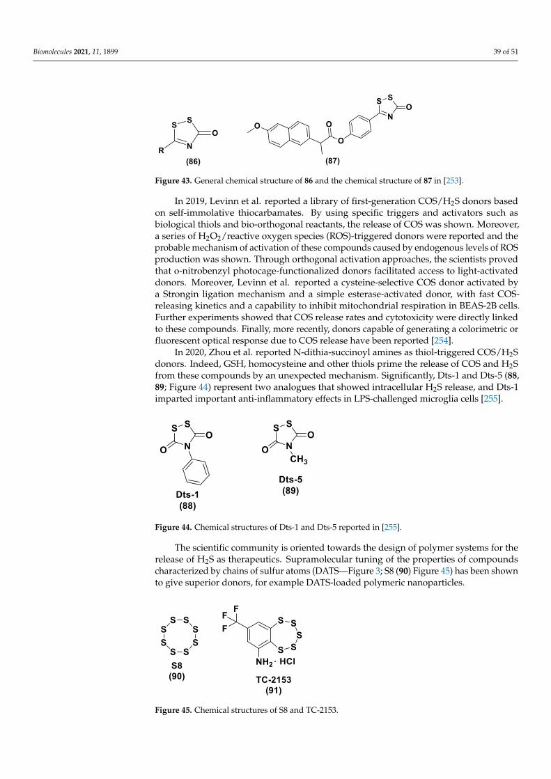
Biomolecules 2021, 11, 1899 39 of 51
Biomolecules 2021, 10, x 40 of 52
releasing mechanism was the same. All the thiophosphamide derivatives exhibited low
toxicity when tested in five cancer cell lines and in W138 cell lines and these significantly
decreased the level of TNF-α and improved the level of IL-10. Moreover, the compounds
preserved H9c2 cells from injury and myocardial injury by an antioxidation effect.
Compounds 84 and 85 (Figure 42) proved to be the most promising derivatives. In con-
clusion, these thiophosphamide derivatives showed low toxicity, great activity, and a
good pharmacokinetic profile and could be candidates for additional investigation [252].
Recently, thioamides have attracted attention especially when included with H2S–
NSAID hybrids and showed low toxicity, but regrettably the mechanism of H2S release
from thioamides continues to be unclear. The synthesis and the evaluation of SulfenylTCMs, (86; Figure 43), a H2S-releasing
class of thioamide-derived sulfenyl thiocarbamates, has been described to overcome this
issue. SulfenylTCMs release COS (carbonyl sulfide), which is immediately converted in-
to H2S by CA (carbonic anhydrase), due to the activation by cellular thiols. These com-
pounds showed excellent H2S release and worked by mechanistically well-defined
pathways. The sulfenyl thiocarbamate group is easily bonded to common NSAIDs, for
example naproxen, to generate new hybrids, such as YZ-597 (87, Figure 43), able to re-
lease H2S in cellular environments. This new class of H2S donors offers an important
starting point for the development of new donors [253].
Figure 43. General chemical structure of 86 and the chemical structure of 87 in [253].
In 2019, Levinn et al. reported a library of first-generation COS/H2S donors based
on self-immolative thiocarbamates. By using specific triggers and activators such as bio-
logical thiols and bio-orthogonal reactants, the release of COS was shown. Moreover, a
series of H2O2/reactive oxygen species (ROS)-triggered donors were reported and the
probable mechanism of activation of these compounds caused by endogenous levels of
ROS production was shown. Through orthogonal activation approaches, the scientists
proved that o-nitrobenzyl photocage-functionalized donors facilitated access to light-
activated donors. Moreover, Levinn et al. reported a cysteine-selective COS donor acti-
vated by a Strongin ligation mechanism and a simple esterase-activated donor, with fast
COS-releasing kinetics and a capability to inhibit mitochondrial respiration in BEAS-2B
cells. Further experiments showed that COS release rates and cytotoxicity were directly
linked to these compounds. Finally, more recently, donors capable of generating a color-
imetric or fluorescent optical response due to COS release have been reported [254].
In 2020, Zhou et al. reported N-dithia-succinoyl amines as thiol-triggered COS/H2S
donors. Indeed, GSH, homocysteine and other thiols prime the release of COS and H2S
from these compounds by an unexpected mechanism. Significantly, Dts-1 and Dts-5 (88,
89; Figure 44) represent two analogues that showed intracellular H2S release, and Dts-1
imparted important anti-inflammatory effects in LPS-challenged microglia cells [255].
Figure 43. General chemical structure of 86 and the chemical structure of 87 in [253].
In 2019, Levinn et al. reported a library of first-generation COS/H2S donors basedon self-immolative thiocarbamates. By using specific triggers and activators such asbiological thiols and bio-orthogonal reactants, the release of COS was shown. Moreover,a series of H2O2/reactive oxygen species (ROS)-triggered donors were reported and theprobable mechanism of activation of these compounds caused by endogenous levels of ROSproduction was shown. Through orthogonal activation approaches, the scientists provedthat o-nitrobenzyl photocage-functionalized donors facilitated access to light-activateddonors. Moreover, Levinn et al. reported a cysteine-selective COS donor activated bya Strongin ligation mechanism and a simple esterase-activated donor, with fast COS-releasing kinetics and a capability to inhibit mitochondrial respiration in BEAS-2B cells.Further experiments showed that COS release rates and cytotoxicity were directly linkedto these compounds. Finally, more recently, donors capable of generating a colorimetric orfluorescent optical response due to COS release have been reported [254].
In 2020, Zhou et al. reported N-dithia-succinoyl amines as thiol-triggered COS/H2Sdonors. Indeed, GSH, homocysteine and other thiols prime the release of COS and H2Sfrom these compounds by an unexpected mechanism. Significantly, Dts-1 and Dts-5 (88,89; Figure 44) represent two analogues that showed intracellular H2S release, and Dts-1imparted important anti-inflammatory effects in LPS-challenged microglia cells [255].
Biomolecules 2021, 10, x 41 of 52
Figure 44. Chemical structures of Dts-1 and Dts-5 reported in [255].
The scientific community is oriented towards the design of polymer systems for the
release of H2S as therapeutics. Supramolecular tuning of the properties of compounds
characterized by chains of sulfur atoms (DATS—Figure 3; S8 (90) Figure 45) has been
shown to give superior donors, for example DATS-loaded polymeric nanoparticles.
SG1002 is a water-insoluble, alpha-sulfur rich microcrystalline material including
traces of ionic substances (sodium sulfate, sodium thiosulfate, and sodium polythio-
nates). Promising results in animal models of several diseases (for example heart failure,
ischemic damage, atherosclerosis, Duchenne muscular dystrophy) have been found by using this compound. Compound 91 (TC-2153, 8-(Trifluoromethyl)-1,2,3,4,5-
benzopentathiepin-6-amine hydrochloride, Figure 45) gave good results in age-related
memory decline, Alzheimer’s disease, fragile X syndrome, schizophrenia, and cocaine
addiction. Finally, DATS has been tested in colon, gastric, brain, and breast cancer.
SG1002 could represent the preferred H2S donor for the evaluation of the gasotransmit-
ter’s effects in animal models, due to its distinctive capability to slowly produce H2S
without by-products and in a dose-independent and enzyme-independent sustained
mode [256,257].
Figure 45. Chemical structures of S8 and TC-2153.
In a very recent paper, Giordano et al. reported hybrids between H2S-donors and
betamethasone 17-valerate or triamcinolone acetonide. Considering the broad use of
glucocorticoids for the treatment of inflammation in asthmatic patients and the positive
effect of H2S on this disease, novel betamethasone 17-valerate and triamcinolone ace-
tonide hybrids linked to H2S-donor moieties have been designed and synthesized. The
obtained compounds were assessed for the potential H2S-releasing profile in a cell-free
environment and in BSMCs (bronchial smooth muscle cells). Molecular modelling stud-
ies carried out with two hybrids 92 and 93 (Figure 46) have been reported and the results
showed that their different H2S-releasing properties may be due to the steric accessibility
of the isothiocyanate carbon atom. Moreover, the most interesting hybrids 92 and 93
have been examined for their inhibitory effect on mast cell degranulation and for their
capability to produce cell membrane hyperpolarization in BSMCs. Important inhibitory
effects on mast cell degranulation have been observed, due to a reduction in the release
of β-hexosaminidase which was more effective compared to the corresponding native
drugs. Compounds 92 and 93 established an important membrane hyperpolarization of
Figure 44. Chemical structures of Dts-1 and Dts-5 reported in [255].
The scientific community is oriented towards the design of polymer systems for therelease of H2S as therapeutics. Supramolecular tuning of the properties of compoundscharacterized by chains of sulfur atoms (DATS—Figure 3; S8 (90) Figure 45) has been shownto give superior donors, for example DATS-loaded polymeric nanoparticles.
Biomolecules 2021, 10, x 41 of 52
Figure 44. Chemical structures of Dts-1 and Dts-5 reported in [255].
The scientific community is oriented towards the design of polymer systems for the
release of H2S as therapeutics. Supramolecular tuning of the properties of compounds
characterized by chains of sulfur atoms (DATS—Figure 3; S8 (90) Figure 45) has been
shown to give superior donors, for example DATS-loaded polymeric nanoparticles.
SG1002 is a water-insoluble, alpha-sulfur rich microcrystalline material including
traces of ionic substances (sodium sulfate, sodium thiosulfate, and sodium polythio-
nates). Promising results in animal models of several diseases (for example heart failure,
ischemic damage, atherosclerosis, Duchenne muscular dystrophy) have been found by using this compound. Compound 91 (TC-2153, 8-(Trifluoromethyl)-1,2,3,4,5-
benzopentathiepin-6-amine hydrochloride, Figure 45) gave good results in age-related
memory decline, Alzheimer’s disease, fragile X syndrome, schizophrenia, and cocaine
addiction. Finally, DATS has been tested in colon, gastric, brain, and breast cancer.
SG1002 could represent the preferred H2S donor for the evaluation of the gasotransmit-
ter’s effects in animal models, due to its distinctive capability to slowly produce H2S
without by-products and in a dose-independent and enzyme-independent sustained
mode [256,257].
Figure 45. Chemical structures of S8 and TC-2153.
In a very recent paper, Giordano et al. reported hybrids between H2S-donors and
betamethasone 17-valerate or triamcinolone acetonide. Considering the broad use of
glucocorticoids for the treatment of inflammation in asthmatic patients and the positive
effect of H2S on this disease, novel betamethasone 17-valerate and triamcinolone ace-
tonide hybrids linked to H2S-donor moieties have been designed and synthesized. The
obtained compounds were assessed for the potential H2S-releasing profile in a cell-free
environment and in BSMCs (bronchial smooth muscle cells). Molecular modelling stud-
ies carried out with two hybrids 92 and 93 (Figure 46) have been reported and the results
showed that their different H2S-releasing properties may be due to the steric accessibility
of the isothiocyanate carbon atom. Moreover, the most interesting hybrids 92 and 93
have been examined for their inhibitory effect on mast cell degranulation and for their
capability to produce cell membrane hyperpolarization in BSMCs. Important inhibitory
effects on mast cell degranulation have been observed, due to a reduction in the release
of β-hexosaminidase which was more effective compared to the corresponding native
drugs. Compounds 92 and 93 established an important membrane hyperpolarization of
Figure 45. Chemical structures of S8 and TC-2153.

Biomolecules 2021, 11, 1899 40 of 51
SG1002 is a water-insoluble, alpha-sulfur rich microcrystalline material includingtraces of ionic substances (sodium sulfate, sodium thiosulfate, and sodium polythionates).Promising results in animal models of several diseases (for example heart failure, is-chemic damage, atherosclerosis, Duchenne muscular dystrophy) have been found by usingthis compound. Compound 91 (TC-2153, 8-(Trifluoromethyl)-1,2,3,4,5-benzopentathiepin-6-amine hydrochloride, Figure 45) gave good results in age-related memory decline,Alzheimer’s disease, fragile X syndrome, schizophrenia, and cocaine addiction. Finally,DATS has been tested in colon, gastric, brain, and breast cancer. SG1002 could representthe preferred H2S donor for the evaluation of the gasotransmitter’s effects in animal mod-els, due to its distinctive capability to slowly produce H2S without by-products and in adose-independent and enzyme-independent sustained mode [256,257].
In a very recent paper, Giordano et al. reported hybrids between H2S-donors andbetamethasone 17-valerate or triamcinolone acetonide. Considering the broad use ofglucocorticoids for the treatment of inflammation in asthmatic patients and the positiveeffect of H2S on this disease, novel betamethasone 17-valerate and triamcinolone acetonidehybrids linked to H2S-donor moieties have been designed and synthesized. The obtainedcompounds were assessed for the potential H2S-releasing profile in a cell-free environmentand in BSMCs (bronchial smooth muscle cells). Molecular modelling studies carriedout with two hybrids 92 and 93 (Figure 46) have been reported and the results showedthat their different H2S-releasing properties may be due to the steric accessibility of theisothiocyanate carbon atom. Moreover, the most interesting hybrids 92 and 93 have beenexamined for their inhibitory effect on mast cell degranulation and for their capabilityto produce cell membrane hyperpolarization in BSMCs. Important inhibitory effectson mast cell degranulation have been observed, due to a reduction in the release of β-hexosaminidase which was more effective compared to the corresponding native drugs.Compounds 92 and 93 established an important membrane hyperpolarization of BSMCsand have been shown to be more effective than the reference compound NS1619 [258].
Biomolecules 2021, 10, x 42 of 52
BSMCs and have been shown to be more effective than the reference compound NS1619
[258].
In a similar paper, Corvino et al. reported a library of hybrids obtained by the con-
densation of corticosteroids with different H2S donor moieties. Through an amperomet-
ric study, all the hybrids released H2S and their chemical stability was explored. The prednisone–TBZ hybrid (94, Figure 46) was chosen for additional evaluations. The re-
sults of in vitro and in vivo studies evidently showed the anti-inflammatory properties of the released H2S. Due to the protective effect on airway remodeling, compound 94 can
be considered an interesting molecule for reducing allergic asthma symptoms and exac-
erbations [259].
Figure 46. Chemical structures of compounds 92 and 93 in [258] and compound 94 in [259].
4. Conclusions
Considering the therapeutic potential of H2S, H2S donor chemistry has remarkably
progressed. The progress of chemical synthesis has acted as the driving force for H2S re-
search, and has particularly focused on the preparation of H2S donors useful for biologi-
cal studies on the physiological and pathophysiological effects of this molecule.
Some H2S donors are or have been under pharmacological evaluation in clinical tri-
als. Remarkable examples include GIC-1001 (GIcare Pharma Inc.), a trimebutine maleate
salt and a H2S donor used as an alternative to sedatives in colonoscopies; ATB-346, a
naproxen-based H2S donor (Antibe Therapeutics), that shows anti-inflammatory effects
and that, in 2016, was included in a phase II study concerning the pain associated with
osteoarthritis; SG-1002 (SulfaGENIX), that has been assessed as part of an investigation
aimed at increasing circulating H2S and NO levels subsequently heart failure; ammoni-
um tetrathiomolybdate (ATTM, (NH4)2MoS4), which has been subjected to clinical trials
for the treatment of breast cancer owing to its copper depletion effects; and finally, hu-
man clinical studies have supported the chemo preventive effects of sulforaphane on
carcinogenesis [260] and antiviral action through the influenza A/WSN/33 (H1N1) virus.
Crucial doubts persist in this field and these issues will necessitate combined efforts
from chemists, biologists, and pharmacologists. One question is related to the determi-
nation of the H2S therapeutic window in different targets; another is the elucidation of
Figure 46. Chemical structures of compounds 92 and 93 in [258] and compound 94 in [259].

Biomolecules 2021, 11, 1899 41 of 51
In a similar paper, Corvino et al. reported a library of hybrids obtained by thecondensation of corticosteroids with different H2S donor moieties. Through an amper-ometric study, all the hybrids released H2S and their chemical stability was explored.The prednisone–TBZ hybrid (94, Figure 46) was chosen for additional evaluations. Theresults of in vitro and in vivo studies evidently showed the anti-inflammatory propertiesof the released H2S. Due to the protective effect on airway remodeling, compound 94can be considered an interesting molecule for reducing allergic asthma symptoms andexacerbations [259].
4. Conclusions
Considering the therapeutic potential of H2S, H2S donor chemistry has remarkablyprogressed. The progress of chemical synthesis has acted as the driving force for H2Sresearch, and has particularly focused on the preparation of H2S donors useful for biologicalstudies on the physiological and pathophysiological effects of this molecule.
Some H2S donors are or have been under pharmacological evaluation in clinical tri-als. Remarkable examples include GIC-1001 (GIcare Pharma Inc.), a trimebutine maleatesalt and a H2S donor used as an alternative to sedatives in colonoscopies; ATB-346, anaproxen-based H2S donor (Antibe Therapeutics), that shows anti-inflammatory effectsand that, in 2016, was included in a phase II study concerning the pain associated withosteoarthritis; SG-1002 (SulfaGENIX), that has been assessed as part of an investigationaimed at increasing circulating H2S and NO levels subsequently heart failure; ammoniumtetrathiomolybdate (ATTM, (NH4)2MoS4), which has been subjected to clinical trials forthe treatment of breast cancer owing to its copper depletion effects; and finally, humanclinical studies have supported the chemo preventive effects of sulforaphane on carcino-genesis [260] and antiviral action through the influenza A/WSN/33 (H1N1) virus.
Crucial doubts persist in this field and these issues will necessitate combined effortsfrom chemists, biologists, and pharmacologists. One question is related to the determi-nation of the H2S therapeutic window in different targets; another is the elucidation ofthe pathways responsible for donor effects. Clarification of the biological activity and thespecies involved will be necessary to increase the biological knowledge of donor triggersand by-products, species interchange, signaling mechanisms, and redox chemistry of H2Sdonors. Moreover, the release of reactive by-products, including unidentified sulfur species,from H2S donors represent a topic that needs further attention.
All these aspects must necessarily be considered in order to broaden this field. Inces-sant innovation in synthetic donors and an improved knowledge of H2S physiology arepaving the way for H2S therapeutics to be used in clinics.
Author Contributions: Conceptualization, E.M., G.C., E.P. and V.S.; resources, B.S., R.S., G.E. (Gian-luca Esposito), G.E. (Giovanna Esposito), M.M. and A.C. and A.S.; writing—original draft preparation,E.M., F.F. (Francesco Frecentese); writing—review and editing, F.F. (Francesco Frecentese) and F.F.(Ferdinando Fiorino); supervision, F.F. (Francesco Frecentese) and E.M. All authors have read andagreed to the published version of the manuscript.
Funding: This research received no external funding.
Institutional Review Board Statement: Not applicable.
Informed Consent Statement: Not applicable.
Data Availability Statement: No new data were created or analyzed in this paper. Data sharing isnot applicable to this article.
Conflicts of Interest: The authors declare no conflict of interest.
References1. Evans, C.L. The toxicity of hydrogen sulphide and other sulphides. J. Exp. Physiol. 1967, 52, 231–248. [CrossRef]2. Li, Q.; Lancaster, J.R., Jr. Chemical foundations of hydrogen sulfide biology. Nitric Oxide 2013, 35, 21–34. [CrossRef] [PubMed]

Biomolecules 2021, 11, 1899 42 of 51
3. Lindenmann, J.; Matzi, V.; Neuboeck, N.; Ratzenhofer-Komenda, B.; Maier, A.; Smolle-Juettner, F.M. Severe hydrogen sulphidepoisoning treated with 4-dimethylaminophenol and hyperbaric oxygen. Diving Hyperb. Med. 2010, 40, 213–217. [PubMed]
4. Vandiver, M.S.; Snyder, S.H. Hydrogen sulfide: A gasotransmitter of clinical relevance. J. Mol. Med. 2012, 90, 255–263. [CrossRef]5. Wang, R. Physiological implications of hydrogen sulfide: A whiff exploration that blossomed. Physiol. Rev. 2012, 92, 791–896.
[CrossRef]6. Li, L.; Moore, P.K. Hydrogen sulfide and cell signaling. Annu. Rev. Pharmacol. Toxicol. 2011, 51, 169–187. [CrossRef]7. Szabo, C. Hydrogen sulphide and its therapeutic potential. Nat. Rev. Drug Discov. 2007, 6, 917–935. [CrossRef]8. Kolluru, G.K.; Shen, X.; Bir, S.C.; Kevil, C.G. Hydrogen sulfide chemical biology: Pathophysiological roles and detection. Nitric
Oxide 2013, 35, 5–20. [CrossRef] [PubMed]9. Fukuto, J.M.; Carrington, S.J.; Tantillo, D.J.; Harrison, J.G.; Ignarro, L.J.; Freeman, B.A.; Chen, A.; Wink, D.A. Small molecule
signaling agents: The integrated chemistry and biochemistry of nitrogen oxides, oxides of carbon, dioxygen, hydrogen sulfide,and their derived species. Chem. Res. Toxicol. 2012, 25, 769–793. [CrossRef]
10. Olson, K.R.; Donald, J.A.; Dombkowski, R.A.; Perry, S.F. Evolutionary and comparative aspects of nitric oxide, carbon monoxideand hydrogen sulfide. Respir. Physiol. Neurobiol. 2012, 184, 117–129. [CrossRef] [PubMed]
11. Olson, K.R. A practical look at the chemistry and biology of hydrogen sulfide. Antioxid. Redox Signal. 2012, 17, 32–44.[CrossRef] [PubMed]
12. Caliendo, G.; Cirino, G.; Santagada, V.; Wallace, J.L. Synthesis and biological effects of hydrogen sulfide (H2S): Development ofH2S-releasing drugs as pharmaceuticals. J. Med. Chem. 2010, 53, 6275–6286. [CrossRef]
13. Vitvitsky, V.; Miljkovic, J.L.; Bostelaar, T.; Adhikari, B.; Yadav, P.K.; Steiger, A.K.; Torregrossa, R.; Pluth, M.D.; Whiteman, M.;Banerjee, R.; et al. Cytochrome c Reduction by H2S Potentiates Sulfide Signaling. ACS Chem. Biol. 2018, 13, 2300–2307. [CrossRef]
14. Filipovic, M.R.; Miljkovic, J.L.; Nauser, T.; Royzen, M.; Klos, K.; Shubina, T.; Koppenol, W.H.; Lippard, S.J.; Ivanovic-Burmazovic, I.Chemical characterization of the smallest S-nitrosothiol, HSNO; cellular cross-talk of H2S and S-nitrosothiols. J. Am. Chem. Soc.2012, 134, 12016–12027. [CrossRef]
15. Carballal, S.; Trujillo, M.; Cuevasanta, E.; Bartesaghi, S.; Moller, M.N.; Folkes, L.K.; Garcia-Bereguian, M.A.; Gutierrez-Merino, C.;Wardmann, P.; Denicola, A.; et al. Reactivity of hydrogen sulfide with peroxynitrite and other oxidants of biological interest. FreeRadic. Biol. Med. 2011, 50, 196–205. [CrossRef]
16. Eberhardt, M.; Dux, M.; Namer, B.; Miljkovic, J.; Cordasic, N.; Will, C.; Kichko, T.I.; de la Roche, J.; Fischer, M.; Suárez, S.A.; et al.H2S and NO cooperatively regulate vascular tone by activating a neuroendocrine HNO–TRPA1–CGRP signalling pathway. Nat.Commun. 2014, 5, 4381. [CrossRef] [PubMed]
17. Filipovic, M.R.; Zivanovic, J.; Alvarez, B.; Banerjee, R. Chemical Biology of H2S Signaling through Persulfidation. Chem. Rev.2018, 118, 1253–1337. [CrossRef] [PubMed]
18. Paulsen, C.E.; Carroll, K.S. Cysteine-mediated redox signaling: Chemistry, biology, and tools for discovery. Chem. Rev. 2013,113, 4633–4679. [CrossRef]
19. Pan, J.; Carroll, K.S. Persulfide reactivity in the detection of protein S-sulfhydration. ACS Chem. Biol. 2013, 8, 1110–1116.[CrossRef]
20. Zhang, D.; Macinkovic, I.; Devarie-Baez, N.O.; Pan, J.; Park, C.M.; Carroll, K.S.; Filipovic, M.R.; Xian, M. Detection of proteinS-sulfhydration by a tag-switch technique. Angew. Chem. Int. Ed. 2014, 53, 575–581. [CrossRef]
21. Mustafa, A.K.; Gadalla, M.M.; Sen, N.; Kim, S.; Mu, W.; Gazi, S.K.; Barrow, R.K.; Yang, G.; Wang, R.; Snyder, S.H. H2S signalsthrough protein S-sulfhydration. Sci. Signal. 2009, 2, ra72. [CrossRef]
22. Gadalla, M.M.; Snyder, S.H. Hydrogen sulfide as a gasotransmitter. J. Neurochem. 2010, 113, 14–26. [CrossRef]23. Sen, N.; Paul, B.D.; Gadalla, M.M.; Mustafa, A.K.; Sen, T.; Xu, R.; Kim, S.; Snyder, S.H. Hydrogen sulfide-linked sulfhydration of
NF-κb mediates its antiapoptotic actions. Mol. Cell 2012, 45, 13–24. [CrossRef] [PubMed]24. Krishnan, N.; Fu, C.; Pappin, D.J.; Tonks, N.K. H2S-Induced sulfhydration of the phosphatase PTP1B and its role in the
endoplasmic reticulum stress response. Sci. Signal. 2011, 4, ra86. [CrossRef] [PubMed]25. Vandiver, M.S.; Paul, B.D.; Xu, R.; Karuppagounder, S.; Rao, F.; Snowman, A.M.; Ko, H.S.; Lee, Y.I.; Dawson, V.L.; Dawson,
T.M.; et al. Sulfhydration mediates neuroprotective actions of parkin. Nat. Commun. 2013, 4, 1626. [CrossRef]26. Paul, B.D.; Snyder, S.H. H2S signaling through protein sulfhydration and beyond. Nat. Rev. Mol. Cell Biol. 2012, 13, 499–507.
[CrossRef] [PubMed]27. Yang, G.; Zhao, K.; Ju, Y.; Mani, S.; Cao, Q.; Puukila, S.; Khaper, N.; Wu, L.; Wang, R. Hydrogen sulfide protects against cellular
senescence via S-sulfhydration of Keap1 and activation of Nrf2. Antioxid. Redox Signal. 2013, 18, 1906–1919. [CrossRef]28. Sasakura, K.; Hanaoka, K.; Shibuya, N.; Mikami, Y.; Kimura, Y.; Komatsu, T.; Ueno, T.; Terai, T.; Kimura, H.; Nagano,
T. Development of a highly selective fluorescence probe for hydrogen sulfide. J. Am. Chem. Soc. 2011, 133, 18003–18005.[CrossRef] [PubMed]
29. Duan, X.; Block, E.; Li, Z.; Connelly, T.; Zhang, J.; Huang, Z.; Su, X.; Pan, Y.; Wu, L.; Chi, Q.; et al. Crucial role of copper indetection of metal-coordinating odorants. Proc. Natl. Acad. Sci. USA 2012, 109, 3492–3497. [CrossRef]
30. Blackstone, E.; Morrison, M.; Roth, M.B. H2S induces a suspended animation-like state in mice. Science 2005, 308, 518. [CrossRef]31. Collman, J.P.; Ghosh, S.; Dey, A.; Decre’au, R.A. Using a functional enzyme model to understand the chemistry behind hydrogen
sulfide induced hibernation. Proc. Natl. Acad. Sci. USA 2009, 106, 22090–22095. [CrossRef]
Biomolecules 2021, 11, 1899 43 of 51
32. Kraus, J.P.; Williamson, C.L.; Firgaira, F.A.; Yang-Feng, T.L.; Münke, M.; Francke, U.; Bukovska, G.; Rosenberg, L.E. Cloningand sequencing nanogram amounts of immunopurified mRNAs: cDNA cloning and chromosomal mapping of cystathionineβ-synthase and the β subunit of propionyl CoA carboxylase. Proc. Natl. Acad. Sci. USA 1986, 83, 2047–2051. [CrossRef]
33. Alexander, F.W.; Sandmeier, E.; Mehta, P.K.; Christen, P. Evolutionary relationships among pyridoxal-5′-phosphate-dependentenzymes. Regio-specific alpha, beta and gamma families. Eur. J. Biochem. 1994, 219, 953–960. [CrossRef]
34. Sun, Q.; Collins, R.; Huang, S.; Holmberg-Schiavone, L.; Anand, G.S.; Tan, C.H.; van-den-Berg, S.; Deng, L.W.; Moore, P.K.;Karlberg, T.; et al. Structural basis for the inhibition mechanism of human cystathionine-gammalyase: An enzyme responsible forthe production of H2S. J. Biol. Chem. 2009, 284, 3076–3085. [CrossRef]
35. Messerschmidt, A.; Worbs, M.; Steegborn, C.; Wahl, M.C.; Huber, R.; Laber, B.; Clausen, T. Determinants of enzymatic specificityin the Cys-Met metabolism PLPdependent enzyme family: Crystal structure of cystathionine γ-lyase from yeast and intrafamiliarstructure comparison. Biol. Chem. 2003, 384, 373–386. [CrossRef]
36. Huang, S.; Chua, J.H.; Yew, W.S.; Sivaraman, J.; Moore, P.K.; Tan, C.H.; Deng, L.W. Site-directed mutagenesis on humanCystathionine-γ-lyase reveals insights into the modulation of H2S production. J. Mol. Biol. 2010, 396, 708–718. [CrossRef]
37. DeLano, W.L. The PyMOL Molecular Graphics System; DeLano Scientific LLC: Palo Alto, CA, USA, 2002.38. Stipanuk, M.H. Sulfur amino acid metabolism: Pathways for production and removal of homocysteine and cysteine. Annu. Rev.
Nutr. 2004, 24, 539–577. [CrossRef]39. Singh, S.; Padovani, D.; Leslie, R.A.; Chiku, T.; Banerjee, R. Relative contributions of cystathionine beta-synthase and gamma-
cystathionase to H2S biogenesis via alternative trans-sulfuration reactions. J. Biol. Chem. 2009, 284, 22457–22466. [CrossRef]40. Kabil, O.; Banerjee, R. The redox biochemistry of hydrogen sulfide. J. Biol. Chem. 2010, 285, 21903–21907. [CrossRef]41. Stipanuk, M.H.; Beck, P.W. Characterization of the enzymic capacity for cysteine desulphhydration in liver and kidney of the rat.
Biochem. J. 1982, 206, 267–277. [CrossRef]42. Kimura, H. Hydrogen sulfide: Its production, release and functions. Amino Acids 2011, 41, 113–121. [CrossRef]43. Olson, K.R.; Whitfield, N.L.; Bearden, S.E.; St Leger, J.; Nilson, E.; Gao, Y.; Madden, J.A. Hypoxic pulmonary vasodilation:
A paradigm shift with a hydrogen sulfide mechanism. Am. J. Physiol. Regul. Integr. Comp. Physiol. 2010, 298, 51–60. [CrossRef]44. Kamoun, P. Endogenous production of hydrogen sulfide in mammals. Amino Acids 2004, 26, 243–254. [CrossRef]45. Mikami, Y.; Shibuya, N.; Kimura, Y.; Nagahara, N.; Ogasawara, Y.; Kimura, H. Thioredoxin and dihydrolipoic acid are required
for 3-mercaptopyruvate sulfurtransferase to produce hydrogen sulfide. Biochem. J. 2011, 439, 479–485. [CrossRef]46. Whiteman, M.; Le Trionnaire, S.; Chopra, M.; Fox, B.; Whatmore, J. Emerging role of hydrogen sulfide in health and disease:
Critical appraisal of biomarkers and pharmacological tools. Clin. Sci. 2011, 121, 459–488. [CrossRef]47. Morikawa, T.; Kajimura, M.; Nakamura, T.; Hishiki, T.; Nakanishi, T.; Yukutake, Y.; Nagahata, Y.; Ishikawa, M.; Hattori, K.;
Takenouchi, T.; et al. Hypoxic regulation of the cerebral microcirculation is mediated by a carbon monoxide-sensitive hydrogensulfide pathway. Proc. Natl. Acad. Sci. USA 2012, 109, 1293–1298. [CrossRef]
48. Searcy, D.G.; Lee, S.H. Sulfur reduction by human erythrocytes. J. Exp. Zool. 1998, 282, 310–322. [CrossRef]49. Benavides, G.A.; Squadrito, G.L.; Mills, R.W.; Patel, H.D.; Isbell, T.S.; Patel, R.P.; DarleyUsmar, V.M.; Doeller, J.; Kraus, D.W.
Hydrogen sulfide mediates the vasoactivity of garlic. Proc. Natl. Acad. Sci. USA 2007, 104, 17977–17982. [CrossRef] [PubMed]50. Belardinelli, M.C.; Chabli, A.; Chadefaux-Vekemans, B.; Kamoun, P. Urinary sulfur compounds in Down syndrome. Clin. Chem.
2001, 47, 1500–1501. [CrossRef]51. Levitt, M.D.; Furne, J.; Springfield, J.; Suarez, F.; DeMaster, E. Detoxification of hydrogen sulfide and methanethiol in the cecal
mucosa. J. Clin. Investig. 1999, 104, 1107–1114. [CrossRef]52. Picton, R.; Eggo, M.C.; Merrill, G.A.; Langman, M.J.; Singh, S. Mucosal protection against sulfide: Importance of the enzyme
rhodanese. Gut 2002, 50, 201–205. [CrossRef]53. Kurzban, G.P.; Chu, L.; Ebersole, J.L.; Holt, S.C. Sulfhemoglobin formation in human erythrocytes by cystalysin, an L-cysteine
desulfhydrase from Treponema denticola. Oral Microbiol. Immunol. 1999, 14, 153–164. [CrossRef]54. Zanardo, R.C.; Brancaleone, V.; Distrutti, E.; Fiorucci, S.; Cirino, G.; Wallace, J.L. Hydrogen sulfide is an endogenous modulator of
leukocyte-mediated inflammation. FASEB J. 2006, 20, 2118–2120. [CrossRef]55. Yonezawa, D.; Sekiguchi, F.; Miyamoto, M.; Taniguchi, E.; Honjo, M.; Masuko, T.; Nishikawa, H.; Kawabata, A. A protective role
of hydrogen sulfide against oxidative stress in rat gastric mucosal epithelium. Toxicology 2007, 241, 11–18. [CrossRef]56. Abe, K.; Kimura, H. The possible role of hydrogen sulfide as an endogenous neuromodulator. J. Neurosci. 1996, 16, 1066–1071.
[CrossRef]57. Laggner, H.; Hermann, M.; Esterbauer, H.; Muellner, M.K.; Exner, M.; Gmeiner, B.M.; Kapiotis, S. The novel gaseous vasorelaxant
hydrogen sulphide inhibits angiotensin-converting enzyme activity of endothelial cells. J. Hypertens. 2007, 25, 2100–2104.[CrossRef] [PubMed]
58. Sivarajah, A.; Collino, M.; Yasin, M.; Benetti, E.; Gallicchio, M.; Mazzon, E.; Cuzzocrea, S.; Fantozzi, R.; Thiemermann, C. Anti-apoptotic and anti-inflammatory effects of hydrogen sulfide in a rat model of regional myocardial I/R. Shock 2009, 31, 267–274.[CrossRef]
59. Zhang, H.; Huang, Y.; Chen, S.; Tang, C.; Wang, G.; Du, J.; Jin, J. Hydrogen sulfide regulates insulin secretion and insulin resistancein diabetes mellitus, a new promising target for diabetes mellitus treatment? A review. J. Adv. Res. 2021, 27, 19–30. [CrossRef]
60. Lefer, D.J. A new gaseous signaling molecule emerges: Cardioprotective role of hydrogen sulfide. Proc. Natl. Acad. Sci. USA 2007,46, 17907–17908. [CrossRef]

Biomolecules 2021, 11, 1899 44 of 51
61. Cheng, Y.; Ndisang, J.F.; Tang, G.; Cao, K.; Wang, R. Hydrogen sulfide-induced relaxation of resistance mesenteric artery beds ofrats. Am. J. Physiol. Heart Circ. Physiol. 2004, 287, H2316–H2323. [CrossRef]
62. Yang, G.; Wu, L.; Jiang, B.; Yang, W.; Qi, J.; Cao, K.; Meng, Q.; Mustafa, A.K.; Mu, W.; Zhang, S.; et al. H2S as a physiologicvasorelaxant: Hypertension in mice with deletion of cystathionin gamma-lyase. Science 2008, 322, 587–590. [CrossRef]
63. Zagli, G.; Patacchini, R.; Trevisani, M.; Abbate, R.; Cinotti, S.; Gensini, G.F.; Masotti, G.; Geppetti, P. Hydrogen sulfide inhibitshuman platelet aggregation. Eur. J. Pharmacol. 2007, 559, 65–68. [CrossRef]
64. Fiorucci, S.; Antonelli, E.; Distrutti, E.; Rizzo, G.; Mencarelli, A.; Orlandi, S.; Zanardo, R.; Renga, B.; Di Sante, M.; Morelli,A.; et al. Inhibition of hydrogen sulfide generation contributes to gastric injury caused by anti-inflammatory nonsteroidal drugs.Gastroenterology 2005, 129, 1210–1224. [CrossRef]
65. Perini, R.; Fiorucci, S.; Wallace, J.L. Mechanisms of nonsteroidal anti-inflammatory drug-induced gastrointestinal injury andrepair: A window of opportunity for cyclooxygenase-inhibiting nitric oxide donors. Can. J. Gastroenterol. 2004, 18, 229–236.[CrossRef]
66. Wallace, J.L.; Caliendo, G.; Santagada, V.; Cirino, G.; Fiorucci, S. Gastrointestinal safety and anti-inflammatory effects of ahydrogen sulfide-releasing diclofenac derivative in the rat. Gastroenterology 2007, 132, 261–271. [CrossRef]
67. Fu, Z.; Liu, X.; Geng, B.; Fang, L.; Tang, C. Hydrogen sulfide protects rat lung from ischemia-reperfusion injury. Life Sci. 2008,82, 1196–1202. [CrossRef]
68. Zhang, C.; Du, J.; Bu, D.; Yan, H.; Tang, X.; Tang, C. The regulatory effect of hydrogen sulphide on hypoxic pulmonaryhypertension in rats. Biochem. Biophys. Res. Commun. 2003, 302, 810–816.
69. Wei, H.L.; Zhang, C.Y.; Jin, H.F.; Tang, C.S.; Du, J.B. Hydrogen sulphide regulates lung tissue oxidized glutathione and totalantioxidant capacity in hypoxic pulmonary hypertensive rats. Acta Pharmacol. Sin. 2008, 29, 670–676. [CrossRef]
70. Jin, H.F.; Liang, C.; Liang, J.M.; Tang, C.S.; Du, J.B. Effects of hydrogen sulphide on vascular inflammation in pulmonaryhypertension induced by high pulmonary blood flow: Experiment with rats. Zhonghua Yi Xue Za Zhi 2008, 88, 2235–2239.
71. Chen, Y.H.; Wu, R.; Geng, B.; Qi, Y.F.; Wang, P.P.; Yao, W.Z.; Tang, C.S. Endogenous hydrogen sulfide reduces airway inflammationand remodeling in a rat model of asthma. Cytokine 2009, 45, 117–123. [CrossRef]
72. Nicholson, C.K.; Calvert, J.W. Hydrogen Sulfide and Ischemia—Reperfusion Injury. Pharmacol. Res. 2010, 62, 289–297. [CrossRef]73. Jiang, T.; Yang, W.; Zhang, H.; Song, Z.; Liu, T.; Lv, X. Hydrogen Sulfide Ameliorates Lung Ischemia-Reperfusion Injury through
SIRT1 Signaling Pathway in Type 2 Diabetic Rats. Front. Physiol. 2020, 11, 596. [CrossRef]74. Kimura, H. Hydrogen sulfide induces cyclic AMP and modulates the NMDA receptor. Biochem. Biophys. Res. Commun. 2000,
267, 129–133. [CrossRef]75. Kimura, H.; Nagai, Y.; Umemura, K.; Kimura, Y. Physiological roles of hydrogen sulfide: Synaptic modulation, neuroprotection,
and smooth muscle relaxation. Antioxid. Redox Signal. 2005, 7, 795–803. [CrossRef]76. Tan, B.H.; Wong, P.T.-H.; Bian, J.S. Hydrogen sulphide: A novel signalling molecule in the central nervous system. Neurochem. Int.
2010, 56, 3–10. [CrossRef]77. Hu, L.F.; Wong, P.T.; Moore, P.K.; Bian, J.S. Hydrogen sulphide attenuates lipopolysaccharide-induced inflammation by inhibition
of p38 mitogen-activated protein kinase in microglia. J. Neurochem. 2007, 100, 1121–1128. [CrossRef]78. Roux, P.P.; Blenis, J. ERK and p38 MAPK-activated protein kinases: A family of protein kinases with diverse biological functions.
Microbiol. Mol. Biol. Rev. 2004, 68, 320–344. [CrossRef]79. d’Emmanuele di Villa Bianca, R.; Sorrentino, R.; Maffia, P.; Mirone, V.; Imbimbo, C.; Fusco, F.; De Palma, R.; Ignarro, L.J.; Cirino,
G. Hydrogen sulfide as a mediator of human corpus cavernosum smooth-muscle relaxation. Proc. Natl. Acad. Sci. USA 2009,106, 4513–4518. [CrossRef]
80. Fiorucci, S.; Santucci, L.; Distrutti, E. NSAIDs, coxibs, CINOD and H2S-releasing NSAIDs: What lies beyond the horizon. Dig.Liver Dis. 2007, 39, 1043–1051. [CrossRef]
81. Wallace, J.L.; Vong, L. NSAID-induced gastrointestinal damage and the design of GI-sparing NSAIDs. Curr. Opin. Investig. Drugs2008, 9, 1151–1156. [CrossRef]
82. Wallace, J.L. Building a better aspirin: Gaseous solutions to a century-old problem. Br. J. Pharmacol. 2007, 152, 421–428. [CrossRef]83. Wallace, J.L.; Ianaro, A.; de Nucci, G. Gaseous Mediators in Gastrointestinal Mucosal Defense and Injury. Dig. Dis. Sci. 2017, 62,
2223–2230. [CrossRef]84. Glowacka, U.; Brzozowski, T.; Magierowski, M. Synergisms, discrepancies and interactions between hydrogen sulfide and carbon
monoxide in the gastrointestinal and digestive system physiology, pathophysiology and pharmacology. Biomolecules 2020, 10, 445.[CrossRef]
85. Sulaieva, O.; Wallace, J.L. Gaseous mediator-based anti-inflammatory drugs. Curr. Opin. Pharm. 2015, 25, 1–6. [CrossRef]86. Distrutti, E.; Sediari, L.; Mencarelli, A.; Renga, B.; Orlandi, S.; Russo, G.; Caliendo, G.; Santagada, V.; Cirino, G.; Wallace, J.L.; et al.
5-Amino-2-hydroxybenzoic acid 4-(5-thioxo-5H-[1,2]dithiol-3yl)-phenyl ester (ATB-429), a hydrogen sulfide-releasing derivativeof mesalamine, exerts antinociceptive effects in a model of postinflammatory hypersensitivity. J. Pharm. Exp. 2006, 319, 447–458.[CrossRef]
87. Ekundi-Valentim, E.; Santos, K.T.; Camargo, E.A.; Denadai-Souza, A.; Teixeira, S.A.; Zanoni, C.I.; Grant, A.D.; Wallace, J.; Muscara,M.N.; Costa, S.K. Differing effects of exogenous and endogenous hydrogen sulphide in carrageenan-induced knee joint synovitisin the rat. Br. J. Pharmacol. 2010, 159, 1463–1474. [CrossRef]
Biomolecules 2021, 11, 1899 45 of 51
88. Distrutti, E.; Sediari, L.; Mencarelli, A.; Renga, B.; Orlandi, S.; Antonelli, E.; Roviezzo, F.; Morelli, A.; Cirino, G.; Wallace,J.L.; et al. Evidence that hydrogen sulfide exerts antinociceptive effects in the gastrointestinal tract by activating KATP channels.J. Pharmacol. Exp. Ther. 2006, 316, 325–335. [CrossRef]
89. Citi, V.; Martelli, A.; Gorica, E.; Brogi, S.; Testai, L.; Calderone, V. Role of hydrogen sulfide in endothelial dysfunction: Pathophysi-ology and therapeutic approaches. J. Adv. Res. 2021, 27, 99–113. [CrossRef]
90. Testai, L.; Citi, V.; Martelli, A.; Brogi, S.; Calderone, V. Role of hydrogen sulfide in cardiovascular ageing. Pharmacol. Res. 2020,160, 105–125. [CrossRef]
91. Chen, Y.; Zhang, F.; Yin, J.; Wu, S.; Zhou, X. Protective mechanisms of hydrogen sulfide in myocardial ischemia. J. Cell. Physiol.2020, 235, 9059–9070. [CrossRef]
92. Kang, S.C.; Sohn, E.H.; Lee, S.R. Hydrogen Sulfide as a Potential Alternative for the Treatment of Myocardial Fibrosis. Oxid. Med.Cell. Longev. 2020, 23, 4105382. [CrossRef]
93. Roorda, M.; Miljkovic, J.L.; van Goor, H.; Henning, R.H.; Bouma, H.R. Spatiotemporal regulation of hydrogen sulfide signaling inthe kidney. Redox Biol. 2021, 43, 101961. [CrossRef] [PubMed]
94. Hsu, C.N.; Tain, Y.L. Hydrogen Sulfide in Hypertension and Kidney Disease of Developmental Origins. Int. J. Mol. Sci. 2018,19, 1438. [CrossRef] [PubMed]
95. Corvino, A.; Frecentese, F.; Magli, E.; Perissutti, E.; Santagada, V.; Scognamiglio, A.; Caliendo, G.; Fiorino, F.; Severino, B. Trendsin H2S-Donors Chemistry and Their Effects in Cardiovascular Diseases. Antioxidants 2021, 10, 429. [CrossRef] [PubMed]
96. Pieretti, J.C.; Cruz Junho, C.V.; Sorelli Carneiro-Ramos, M.; Barozzi Seabra, A. H2S- and NO-releasing gasotransmitter platform:A crosstalk signaling pathway in the treatment of acute kidney injury. Pharmacol. Res. 2020, 161, 105121. [CrossRef]
97. Cao, X.; Zhang, W.; Moore, P.K.; Bian, J. Protective Smell of Hydrogen Sulfide and Polysulfide in Cisplatin-Induced Nephrotoxicity.Int. J. Mol. Sci. 2019, 20, 313. [CrossRef]
98. Zhong, H.; Yu, H.; Chen, J.; Sun, J.; Guo, L.; Huang, P.; Zhong, Y. Hydrogen Sulfide and Endoplasmic Reticulum Stress: A PotentialTherapeutic Target for Central Nervous System Degeneration Diseases. Front. Pharmacol. 2020, 11, 702. [CrossRef]
99. Gambari, L.; Grigolo, B.; Grassi, F. Hydrogen Sulfide in Bone Tissue Regeneration and Repair: State of the Art and NewPerspectives. Int. J. Mol. Sci. 2019, 20, 5231. [CrossRef]
100. Coavoy-Sánchez, S.A.; Costa, S.K.P.; Muscará, M.N. Hydrogen sulfide and dermatological diseases. Br. J. Pharmacol. 2020,177, 857–865. [CrossRef]
101. Cao, X.; Ding, L.; Xie, Z.Z.; Yang, Y.; Whiteman, M.; Moore, P.K.; Bian, J.S. A Review of Hydrogen Sulfide Synthesis, Metabolism,and Measurement: Is Modulation of Hydrogen Sulfide a Novel Therapeutic for Cancer? Antioxid. Redox Signal. 2019, 31, 1–38.[CrossRef] [PubMed]
102. Dattilo, M. The role of host defences in Covid 19 and treatments thereof. Mol. Med. 2020, 26, 90. [CrossRef] [PubMed]103. Citi, V.; Martelli, A.; Brancaleone, V.; Brogi, S.; Gojon, G.; Montanaro, R.; Morales, G.; Testai, L.; Calderone, V. Anti-inflammatory
and antiviral roles of hydrogen sulfide: Rationale for considering H2S donors in COVID-19 therapy. Br. J. Pharmacol. 2020,177, 4931–4941. [CrossRef]
104. Liu, H.; Anders, F.; Thanos, S.; Mann, C.; Liu, A.; Grus, F.H.; Pfeiffer, N.; Prokosch-Willing, V. Hydrogen Sulfide Protects RetinalGanglion Cells Against Glaucomatous Injury In Vitro and In Vivo. Investig. Ophthalmol. Vis. Sci. 2017, 58, 5129–5141. [CrossRef]
105. Struve, M.F.; Brisbois, J.N.; James, R.A.; Marshall, M.W.; Dorman, D.C. Neurotoxicological effects associated with short-termexposure of Sprague-Dawley rats to hydrogen sulfide. Neurotoxicology 2001, 22, 375–385. [CrossRef]
106. Volpato, G.P.; Searles, R.; Yu, B.; Scherrer-Crosbie, M.; Bloch, K.D.; Ichinose, F.; Zapol, W.M. Inhaled Hydrogen Sulfide: A RapidlyReversible Inhibitor of Cardiac and Metabolic Function in the Mouse. Anesthesiology 2008, 108, 659–668. [CrossRef] [PubMed]
107. Faller, S.; Ryter, S.W.; Choi, A.M.K.; Loop, T.; Schmidt, R.; Hoetzel, A. Inhaled hydrogen sulfide protects against ventilator-inducedlung injury. Anesthesiology 2010, 113, 104–115. [CrossRef]
108. Wagner, F.; Wagner, K.; Weber, S.; Stahl, B.; Knöferl, M.W.; Huber-Lang, M.; Seitz, D.H.; Asfar, P.; Calzia, E.; Senftleben, U.; et al.Inflammatory Effects of Hypothermia and Inhaled H2S During Resuscitated, Hyperdynamic Murine Septic Shock. Shock 2011,35, 396–402. [CrossRef] [PubMed]
109. Blackstone, E.; Roth, M.B. Suspended animation-like state protects mice from lethal hypoxia. Shock 2007, 27, 370–372. [CrossRef]110. Xue, R.; Hao, D.; Sun, J.; Li, W.; Zhao, M.; Li, X.; Chen, Y.; Zhu, J.; Ding, Y.; Liu, J.; et al. Hydrogen Sulfide Treatment Promotes
Glucose Uptake by Increasing Insulin Receptor Sensitivity and Ameliorates Kidney Lesions in Type 2 Diabetes. Antioxid. RedoxSignal. 2013, 19, 5. [CrossRef]
111. Chen, K.Y.; Morris, J.C. Kinetics of oxidation of aqueous sulfide by oxygen. Environ. Sci. Technol. 1972, 6, 537. [CrossRef]112. Hughes, M.N.; Centelles, M.N.; Moore, K.P. Making and working with hydrogen sulfide. Free Radic. Biol. Med. 2009, 47, 1346–1353.
[CrossRef]113. Calvert, J.W.; Elston, M.; Nicholson, C.K.; Gundewar, S.; Jha, S.; Elrod, J.W.; Ramachandran, A.; Lefer, D.J. Genetic and Pharmaco-
logic Hydrogen Sulfide Therapy Attenuates Ischemia-Induced Heart Failure in Mice. Circulation 2010, 122, 11. [CrossRef]114. Zhao, W.; Zhang, J.; Lu, Y.; Wang, R. The vasorelaxant effect of H2S as a novel endogenous gaseous KATP channel opener. EMBO J.
2001, 20, 6008–6016. [CrossRef]115. Mard, S.A.; Neisi, N.; Solgi, G.; Hassanpour, M.; Darbor, M.; MaMaleki, M. Gastroprotective effect of NaHS against mucosal
lesions induced by ischemia-reperfusion injury in rat. Dig. Dis. Sci. 2012, 57, 1496–1503. [CrossRef]

Biomolecules 2021, 11, 1899 46 of 51
116. Gao, C.; Xu, D.Q.; Gao, C.J.; Ding, Q.; Yao, L.N.; Li, Z.C.; Chai, W. An exogenous hydrogen sulphide donor, NaHS, inhibits thenuclear factor κB inhibitor kinase/nuclear factor κB inhibitor/nuclear factor-κB signaling pathway and exerts cardioprotectiveeffects in a rat hemorrhagic shock model. Biol. Pharm. Bull. 2012, 35, 1029–1034. [CrossRef] [PubMed]
117. Li, N.; Wang, M.J.; Jin, S.; Bai, Y.D.; Hou, C.L.; Ma, F.F.; Li, X.H.; Zhu, Y.C. The H2S donor NaHS changes the expression pattern ofH2S-Producing enzymes after myocardial infarction. Oxid. Med. Cell. Longev. 2016, 2016, 6492469. [CrossRef]
118. Esechie, A.; Kiss, L.; Olah, G.; Horvath, E.M.; Hawkins, H.; Szabo, C.; Traber, D.L. Protective effect of hydrogen sulfide in amurine model of combined burn and smoke inhalation-induced acute lung injury. Clin. Sci. 2008, 115, 91. [CrossRef]
119. Kloesch, B.; Liszt, M.; Steiner, G.; Broll, J. Inhibitors of p38 and ERK1/2 MAPkinase and hydrogen sulphide block constitutiveand IL-1β-induced IL-6 and IL-8 expression in the human chondrocyte cell line C-28/I2. Rheumatol. Int. 2012, 32, 729.[CrossRef] [PubMed]
120. Andruski, B.; McCafferty, D.M.; Ignacy, T.; Millen, B.; McDougall, J.J. Leukocyte trafficking and pain behavioral responses to ahydrogen sulfide donor in acute monoarthritis. Am. J. Physiol. Regul. Integr. Comp. Physiol. 2008, 295, 814–820. [CrossRef] [PubMed]
121. Li, L.; Bhatia, M.; Zhu, Y.Z.; Zhu, Y.C.; Ramnath, R.D.; Wang, Z.J.; Anuar, F.B.M.; Whiteman, M.; Salto-Tellez, M.; Moore, P.K.Hydrogen sulfide is a novel mediator of lipopolysaccharideinduced inflammation in the mouse. FASEB J. 2005, 19, 1196–1198.[CrossRef]
122. Bhatia, M.; Wong, F.L.; Fu, D.; Lau, H.Y.; Moochhala, S.M.; Moore, P.K. Role of hydrogen sulfide in acute pancreatitis andassociated lung injury. FASEB J. 2005, 19, 623–625. [CrossRef]
123. Mok, Y.Y.; Moore, P.K. Hydrogen sulfide is pro-inflammatory in haemorrhagic shock. Inflamm. Res. 2008, 57, 512–528. [CrossRef]124. Mok, Y.Y.; Atan, M.S.; Yoke Ping, C.; Zhong Jing, W.; Bhatia, M.; Moochhala, S.; Moore, P.K. Role of hydrogen sulfide in
haemorrhagic shock in the rat: Protective effect of inhibitors of hydrogen sulfide biosynthesis. Br. J. Pharmacol. 2004, 143, 881–889.[CrossRef]
125. Li, T.; Zhao, B.; Wang, C.; Wang, H.; Liu, Z.; Li, W.; Jin, H.; Tang, C.; Du, J. Regulatory Effects of Hydrogen Sulfide on IL-6, IL-8and IL-10 Levels in the Plasma and Pulmonary Tissue of Rats with Acute Lung Injury. Exp. Biol. Med. 2008, 233, 1081–1087.[CrossRef] [PubMed]
126. Moore, D.J.; West, A.B.; Dawson, V.L.; Dawson, T.M. Molecular pathophysiology of Parkinson’s disease. Annu. Rev. Neurosci.2005, 28, 57–87. [CrossRef] [PubMed]
127. Hu, L.F.; Lu, M.; Tiong, C.X.; Dawe, G.S.; Hu, G.; Bian, J.S. Neuroprotective effects of hydrogen sulfide on Parkinson’s disease ratmodels. Aging Cell 2010, 9, 135–146. [CrossRef]
128. Giuliani, D.; Ottani, A.; Zaffe, D.; Galantucci, M.; Strinati, F.; Lodi, R.; Guarini, S. Hydrogen sulfide slows down progression ofexperimental Alzheimer’s disease by targeting multiple pathophysiological mechanisms. Neurobiol. Learn. Mem. 2013, 104, 82–91.[CrossRef] [PubMed]
129. Bayan, L.; Koulivand, P.H.; Gorji, A. Garlic: A review of potential therapeutic effects. Avicenna J. Phytomed. 2014, 4, 1–14.[PubMed]
130. Lawson, L.D.; Wood, S.G.; Hughes, B.G. HPLC analysis of allicin and other thiosulfinates in garlic clove homogenates. PlantaMed. 1991, 57, 263–270. [CrossRef]
131. Brodnitz, M.H.; Pascale, J.V.; Van Derslice, L. Flavor components of garlic extract. J. Agric. Food Chem. 1971, 19, 273–275.[CrossRef]
132. Pluth, M.D.; Bailey, T.S.; Hammers, M.D.; Hartle, M.D.; Henthorn, H.A.; Steiger, A.K. Natural Products Containing HydrogenSulfide Releasing Moieties. Synlett 2015, 26, 2633–2643. [CrossRef]
133. Liang, D.; Wu, H.; Wong, M.W.; Huang, D. Diallyl Trisulfide Is a Fast H2S Donor, but Diallyl Disulfide Is a Slow One: TheReaction Pathways and Intermediates of Glutathione with Polysulfides. Org. Lett. 2015, 17, 4196–4199. [CrossRef]
134. Dinkova-Kostova, A.T.; Kostov, R.V. Glucosinolates and isothiocyanates in health and disease. Trends Mol. Med. 2012, 18, 337–347.[CrossRef] [PubMed]
135. Citi, V.; Martelli, A.; Testai, L.; Marino, A.; Breschi, M.C.; Calderone, V. Hydrogen Sulfide Releasing Capacity of NaturalIsothiocyanates: Is It a Reliable Explanation for the Multiple Biological Effects of Brassicaceae? Planta Med. 2014, 80, 610–613.[CrossRef]
136. Martelli, A.; Testai, L.; Citi, V.; Marino, A.; Bellagambi, F.G.; Ghimenti, S.; Breschi, M.C.; Calderone, V. Pharmacologicalcharacterization of the vascular effects of aryl isothiocyanates: Is hydrogen sulfide the real player? Vascul. Pharmacol. 2014,60, 32–41. [CrossRef]
137. Lin, Y.; Yang, X.; Lu, Y.; Liang, D.; Huang, D. Isothiocyanates as H2S Donors Triggered by Cysteine: Reaction Mechanism andStructure and Activity Relationship. Org. Lett. 2019, 21, 5977–5980. [CrossRef]
138. Ozturk, T.; Ertas, E.; Mert, O. Use of Lawesson’s Reagent in Organic Syntheses. Chem. Rev. 2007, 107, 5210–5278. [CrossRef] [PubMed]139. Lecher, H.Z.; Greenwood, R.A.; Whitehouse, K.C.; Chao, T.H. The Phosphonation of Aromatic Compounds with Phosphorus
Pentasulfide. J. Am. Chem. Soc. 1956, 78, 5018. [CrossRef]140. Thomsen, I.; Clausen, K.; Scheibye, S.; Lawesson, S.O. Thiation with 2,4-bis(4-methoxyphenyl)-1,3,2,4-dithiadiphosphetane
2,4-disulfide: N-methylthiopyrrolidone. Org. Synth. 1984, 62, 158.141. Nicolau, L.A.D.; Silva, R.O.; Damasceno, S.R.B.; Carvalho, N.S.; Costa, N.R.D.; Aragão, K.S.; Barbosa, A.L.R.; Soares, P.M.G.;
Souza, M.H.L.P.; Medeiros, J.V.R. The hydrogen sulfide donor, Lawesson’s reagent, prevents alendronate-induced gastric damagein rats. Braz. J. Med. Biol. Res. 2013, 46, 708–714. [CrossRef] [PubMed]
Biomolecules 2021, 11, 1899 47 of 51
142. Spiller, F.; Orrico, M.I.L.; Nascimento, D.C.; Czaikoski, P.G.; Souto, F.O.; Alves-Filho, J.C.; Freitas, A.; Carlos, D.; Montenegro,M.F.; Neto, A.F.; et al. Hydrogen Sulfide Improves Neutrophil Migration and Survival in Sepsis via K+ATP channel activation.Am. J. Respir. Crit. Care Med. 2010, 182, 360–368. [CrossRef]
143. Li, L.; Whiteman, M.; Guan, Y.Y.; Neo, K.L.; Cheng, Y.; Lee, S.W.; Zhao, Y.; Baskar, R.; Tan, C.H.; Moore, P.K. Characterizationof a novel, water-soluble hydrogen sulfide-releasing molecule (GYY4137): New insights into the biology of hydrogen sulfide.Circulation 2008, 117, 2351–2360. [CrossRef] [PubMed]
144. Rose, P.; Dymock, B.W.; Moore, P.K. GYY4137, a novel water-soluble, H2S-releasing molecule. Methods Enzymol. 2015, 554, 143–167.145. Zhao, Y.; Biggs, T.D.; Xian, M. Hydrogen sulfide (H2S) releasing agents: Chemistry and biological applications. Chem. Commun.
2014, 50, 11788–11805. [CrossRef] [PubMed]146. Martelli, A.; Testai, L.; Citi, V.; Marino, A.; Pugliesi, I.; Barresi, E.; Nesi, G.; Rapposelli, S.; Taliani, S.; Da Settimo, F.; et al.
Arylthioamides as H2S donors: L-cysteine-activated releasing properties and vascular effects in vitro and in vivo. ACS Med.Chem. Lett. 2013, 4, 904–908. [CrossRef] [PubMed]
147. Karwi, Q.G.; Whiteman, M.; Wood, M.E.; Torregrossa, R.; Baxter, G.F. Pharmacological postconditioning against myocardialinfarction with a slow-releasing hydrogen sulfide donor, GYY4137. Pharmacol. Res. 2016, 111, 442–451. [CrossRef]
148. Qiu, Y.; Wu, Y.; Meng, M.; Luo, M.; Zhao, H.; Sun, H.; Gao, S. GYY4137 protects against myocardial ischemia/reperfusion injuryvia activation of the PHLPP-1/Akt/Nrf2 signaling pathway in diabetic mice. J. Surg. Res. 2018, 225, 29–39. [CrossRef]
149. Han, X.; Mao, Z.C.; Wang, S.; Xin, Y.; Li, P.; Maharjan, S.; Zhang, B. GYY4137 protects against MCAO via p38 MAPK mediatedanti-apoptotic signaling pathways in rats. Brain Res. Bull. 2020, 158, 59–65. [CrossRef] [PubMed]
150. Alexander, B.E.; Coles, S.J.; Fox, B.C.; Khan, T.F.; Maliszewski, J.; Perry, A.; Pitak, M.B.; Whiteman, M.; Wood, M.E. Investigatingthe generation of hydrogen sulfide from the phosphonamidodithioate slow-release donor GYY4137. Med. Chem. Commun. 2015,6, 1649. [CrossRef]
151. Moore, P.K.; Whiteman, M. Chemistry, Biochemistry and Pharmacology of Hydrogen Sulfide; Springer: Berlin/Heidelberg, Germany,2015; pp. 344–354.
152. Park, C.; Zhao, Y.; Zhu, Z.; Pacheco, A.; Peng, B.; Devarie-Baez, N.O.; Bagdon, P.; Zhang, H.; Xian, M. Synthesis and evaluation ofphosphorodithioate-based hydrogen sulfide donors. Mol. Biosyst. 2013, 9, 2430–2434. [CrossRef]
153. Kang, J.; Li, Z.; Organ, C.L.; Park, C.M.; Yang, C.T.; Pacheco, A.; Wang, D.; Lefer, D.J.; Xian, M. pH-controlled hydrogen sulfiderelease for myocardial ischemia-reperfusion injury. J. Am. Chem. Soc. 2016, 138, 6336–6339. [CrossRef] [PubMed]
154. Wang, H.W.; Skeldon, P.; Thompson, G.E.; Wood, G.C. Synthesis and characterization of molybdenum disulphide formed fromammonium tetrathiomolybdate. J. Mater. Sci. 1997, 32, 497. [CrossRef]
155. Xu, S.; Yang, C.T.; Meng, F.H.; Pacheco, A.; Chen, L.; Xian, M. Ammonium tetrathiomolybdate as a water-soluble and slow-releasehydrogen sulfide donor. Bioorg. Med. Chem. Lett. 2016, 26, 1585–1588. [CrossRef]
156. Dyson, A.; Dal-Pizzol, F.; Sabbatini, G.; Lach, A.B.; Galfo, F.; Dos Santos Cardoso, J.; Pescador Mendonça, B.; Hargreaves, I.; BollenPinto, B.; Bromage, D.I.; et al. Ammonium tetrathiomolybdate following ischemia/reperfusion injury: Chemistry, pharmacology,and impact of a new class of sulfide donor in preclinical injury models. PLoS Med. 2017, 14, 1002310. [CrossRef]
157. Zhang, H.; Zhang, A.; Guo, C.; Shi, C.; Zhang, Y.; Liu, Q.; Sparatore, A.; Wang, C. S-diclofenac protects against doxorubicin-induced cardiomyopathy in mice via ameliorating cardiac gap junction remodeling. PLoS ONE 2011, 6, e26441. [CrossRef]
158. Wu, J.; Guo, W.; Lin, S.Z.; Wang, Z.J.; Kan, J.T.; Chen, S.Y.; Zhu, Y.Z. Gp130-mediated STAT3 activation by S-propargyl-cysteine,an endogenous hydrogen sulfide initiator, prevents doxorubicin-induced cardiotoxicity. Cell. Death Dis. 2016, 7, e2339. [CrossRef]
159. Su, Y.W.; Liang, C.; Jin, H.F.; Tang, X.Y.; Han, W.; Chai, L.J.; Zhang, C.Y.; Geng, B.; Tang, C.S.; Du, J.B. Hydrogen sulfide regulatescardiac function and structure in adriamycin-induced cardiomyopathy. Circ. J. 2009, 73, 741–749. [CrossRef] [PubMed]
160. Chan, N.; Willis, A.; Kornhauser, N.; Ward, M.M.; Lee, S.B.; Nackos, E.; Seo, B.R.; Chuang, E.; Cigler, T.; Moore, A.; et al. Influencingthe tumor microenvironment: A phase II study of copper depletion using tetrathiomolybdate in patients with breast cancer at highrisk for recurrence and in preclinical models of lung metastases. Clin. Cancer Res. 2017, 23, 666–676. [CrossRef] [PubMed]
161. Zanatta, S.D.; Jarrott, B.; Williams, S.J. Synthesis and preliminary pharmacological evaluation of aryl dithiolethiones withcyclooxygenase-2-selective inhibitory activity and hydrogen sulfide-releasing properties. Aust. J. Chem. 2010, 63, 946–957.[CrossRef]
162. Li, L.; Rossoni, G.; Sparatore, A.; Lee, L.C.; Del Soldato, P.; Moore, P.K. Anti-inflammatory and gastrointestinal effects of a noveldiclofenac derivative. Free Radic. Biol. Med. 2007, 42, 706. [CrossRef]
163. Rakitin, O.A. Synthesis and Reactivity of 3H-1,2-dithiole-3-thiones. Molecules 2021, 26, 3595. [CrossRef] [PubMed]164. Landis, P.S. The chemistry of 1,2-dithiole-3-thiones. Chem. Rev. 1965, 65, 237. [CrossRef]165. Curphey, T.J.; Libby, A.A. Dianions of 3-Oxo dithioic Acids: Preparation and Conversion to 3H-1,2-Dithiole-3-thiones. Tetrahedron
Lett. 2000, 41, 6977. [CrossRef]166. Pedersen, B.S.; Lawesson, S.O. Syntheses of 3H-1,2-dithiole-3-thiones and 4H-1,3,2-oxazaphosphorine derivatives from the dimer
of p-methoxyphenyl-thionophosphine sulfide and der. Tetrahedron 1979, 35, 2433–2437. [CrossRef]167. Hamada, T.; Nakane, T.; Kimura, T.; Arisawa, K.; Yoneda, K.; Yamamoto, T.; Osaki, T. Treatment of xerostomia with the bile
secretion-stimulating drug anethole trithione: A clinical trial. Am. J. Med. Sci. 1999, 318, 146–151. [CrossRef]168. Perrino, E.; Cappelletti, G.; Tazzari, V.; Giavini, E.; Soldato, P.D.; Sparatore, A. New sulfurated derivatives of valproic acid with
enhanced histone deacetylase inhibitory activity. Bioorg. Med. Chem. Lett. 2008, 18, 1893–1897. [CrossRef] [PubMed]
Biomolecules 2021, 11, 1899 48 of 51
169. Tazzari, V.; Cappelletti, G.; Casagrande, M.; Perrino, E.; Renzi, L.; Del Soldato, P.; Sparatore, A. New aryldithiolethione derivativesas potent histone deacetylase inhibitors. Bioorg. Med. Chem. 2010, 18, 4187–4194. [CrossRef]
170. Sen, C.K.; Traber, K.E.; Packer, L. Inhibition of NF-jB activation in human T-cell lines by anetholdithiolthione. Biochem. Biophys.Res. Commun. 1996, 218, 148–153. [CrossRef]
171. Qandil, A.M. Prodrugs of Nonsteroidal Anti-Inflammatory Drugs (NSAIDs), More Than Meets the Eye: A Critical Review. Int. J.Mol. Sci. 2012, 13, 17244. [CrossRef]
172. Sparatore, A.; Perrino, E.; Tazzari, V.; Giustarini, D.; Rossi, R.; Rossoni, G.; Erdman, K.; Schröder, H.; Del Soldato, P. Pharmacolog-ical profile of a novel H2S-releasing aspirin. Free Radic. Biol. Med. 2009, 46, 586–592. [CrossRef]
173. Rossoni, G.; Manfredi, B.; Tazzari, V.; Sparatore, A.; Trivulzio, S.; Del Soldato, P.; Berti, F. Activity of a new hydrogen sulfide-releasing aspirin (ACS14) on pathological cardiovascular alterations induced by glutathione depletion in rats. Eur. J. Pharmacol.2010, 648, 139–145. [CrossRef]
174. Kodela, R.; Chattopadhyay, M.; Kashfi, K. NOSH-Aspirin: A novel nitric oxide–hydrogen sulfide-releasing hybrid: A new class ofanti-inflammatory pharmaceuticals. ACS Med. Chem. Lett. 2012, 3, 257–262. [CrossRef]
175. Rossoni, G.; Berti, M.; Colonna, V.D.; Bernareggi, M.; Del Soldato, P.; Berti, F. Myocardial protection by the nitroderivative ofaspirin, NCX 4016: In vitro and in vivo experiments in the rabbit. Italian Heart J. 2000, 1, 146–155.
176. Ahmed, A.; Rezai, H.; Broadway-Stringer, S. Evidence-Based Revised View of the Pathophysiology of Preeclampsia. Adv. Exp.Med. Biol. Adv. Intern. Med. 2017, 2, 355–374. [CrossRef]
177. Sanchez-Aranguren, L.C.; Rezai, H.; Ahmad, S.; Alzahrani, F.A.; Sparatore, A.; Wang, K.; Ahmed, A. MZe786 Rescues CardiacMitochondrial Activity in High sFlt-1 and Low HO-1 Environment. Antioxidants 2020, 9, 598. [CrossRef] [PubMed]
178. Rezai, H.; Ahmad, S.; Alzahrani, F.A.; Sanchez-Aranguren, L.; Dias, I.H.K.; Agrawal, S.; Sparatore, A.; Wang, K.; Ahmed, A.MZe786, a hydrogen sulfide-releasing aspirin prevents preeclampsia in heme oxygenase-1 haplodeficient pregnancy under highsoluble flt-1 environment. Redox Biol. 2021, 38, 101768. [CrossRef] [PubMed]
179. Fiorucci, S.; Orlandi, S.; Mencarelli, A.; Caliendo, G.; Santagada, V.; Distrutti, E.; Santucci, L.; Cirino, G.; Wallace, J.L. Enhancedactivity of a hydrogen sulphide releasing derivative of mesalamine (ATB-429) in a mouse model of colitis. Br. J. Pharmacol. 2007,150, 996–1002. [CrossRef] [PubMed]
180. Fahn, S. An open trial of high-dosage antioxidants in early Parkinson’s disease. Am. J. Clin. Nutr. 1991, 53, 380–382. [CrossRef]181. Rascol, O.; Brooks, D.J.; Korczyn, A.D.; De Deyn, P.P.; Clarke, C.E.; Lang, A.E. A five-year study of the incidence of dyskinesia
in patients with early Parkinson’s disease who were treated with ropinirole or levodopa. N. Engl. J. Med. 2000, 342, 1484–1491.[CrossRef]
182. Lee, M.; Tazzari, V.; Giustarini, D.; Rossi, R.; Sparatore, A.; Del Soldato, P.; McGeer, E.; McGeer, P.L. Effects of hydrogensulfide-releasing L-DOPA derivatives on glial activation. J. Biol. Chem. 2010, 285, 17318–17328. [CrossRef]
183. Kashfi, K. Anti-inflammatory agents as cancer therapeutics. Adv. Pharmacol. 2009, 57, 31–89. [PubMed]184. Chattopadhyay, M.; Kodela, R.; Olson, K.R.; Kashfi, K. NOSH-aspirin (NBS-1120), a novel nitric oxide- and hydrogen sulfide-
releasing hybrid is a potent inhibitor of colon cancer cell growth in vitro and in a xenograft mouse model. Biochem. Biophys. Res.Commun. 2012, 419, 523–528. [CrossRef]
185. Kashfi, K. Anti-cancer activity of new designer hydrogen sulfide-donating hybrids. Antioxid. Redox Signal. 2014, 20, 831–846.[CrossRef]
186. Hasegawa, U.; van del Vlies, A.J. Design and synthesis of polymeric hydrogen sulfide donors. Bioconjug. Chem. 2014, 25, 1290–1300.[CrossRef] [PubMed]
187. Zhang, Y.; Munday, R. Dithiolethiones for cancer chemoprevention: Where do we stand? Mol. Cancer Ther. 2008, 7, 3470–3479.[CrossRef]
188. Wood, M.E.; Whiteman, M.; Perry, A. Hydrogen Sulfide Releasing Compounds and Their Use. U.S. Patent No. WO2013045951,4 April 2013.
189. Szczesny, B.; Módis, K.; Yanagi, K.; Coletta, C.; Le Trionnaire, S.; Perry, A.; Wood, M.E.; Whiteman, M.; Szabo, C. AP39, a novelmitochondria-targeted hydrogen sulfide donor, stimulates cellular bioenergetics, exerts cytoprotective effects and protects againstthe loss of mitochondrial DNA integrity in oxidatively stressed endothelial cells in vitro. Nitric Oxide 2014, 41, 120–130. [CrossRef]
190. Ikeda, K.; Marutani, E.; Hirai, S.; Wood, M.E.; Whiteman, M.; Ichinose, F. Mitochondria-targeted hydrogen sulfide donor AP39improves neurological outcomes after cardiac arrest in mice. Nitric Oxide 2015, 49, 90–96. [CrossRef] [PubMed]
191. Ahmad, A.; Olah, G.; Szczesny, B.; Wood, M.E.; Whiteman, M.; Szabo, C. AP39, a mitochondrially targeted hydrogen sulfidedonor, exerts protective effects in renal epithelial cells subjected to oxidative stress in vitro and in acute renal injury in vivo. Shock2016, 45, 88–97. [CrossRef] [PubMed]
192. Chatzianastasiou, A.; Bibli, S.I.; Andreadou, I.; Efentakis, P.; Kaludercic, N.; Wood, M.E.; Whiteman, M.; Di Lisa, F.; Daiber, A.;Manolopoulos, V.G.; et al. Cardioprotection by H2S donors: Nitric oxide-dependent and independent mechanisms. J. Pharmacol.Exp. Ther. 2016, 358, 431–440. [CrossRef]
193. Karwi, Q.G.; Bornbaum, J.; Boengler, K.; Torregrossa, R.; Whiteman, M.; Wood, M.E.; Schulz, R.; Baxter, G.F. AP39, a mitochondria-targeting hydrogen sulfide (H2S) donor, protects against myocardial reperfusion injury independently of salvage kinase signalling.Br. J. Pharmacol. 2017, 174, 287–301. [CrossRef]
Biomolecules 2021, 11, 1899 49 of 51
194. Gero, D.; Torregrossa, R.; Perry, A.; Waters, A.; Le-Trionnaire, S.; Whatmore, J.L.; Wood, M.; Whiteman, M. The novel mitochondria-targeted hydrogen sulfide (H2S) donors AP123 and AP39 protect against hyperglycemic injury in microvascular endothelial cellsin vitro. Pharmacol. Res. 2016, 113, 186–198. [CrossRef] [PubMed]
195. Talalay, P.; De Long, M.J.; Prochaska, H.J. Identification of a common chemical signal regulating the induction of enzymes thatprotect against chemical carcinogenesis. Proc. Natl. Acad. Sci. USA 1988, 85, 8261–8265. [CrossRef] [PubMed]
196. Kim, W.; Gates, K.S. Evidence for thiol-dependent production of oxygen radicals by 4-methyl-5-pyrazinyl-3H-1, 2-dithiole-3-thione: Possible relevance to the anticarcinogenic properties of 1, 2-dithiole-3-thiones. Chem. Res. Toxicol. 1997, 10, 296–301.[CrossRef]
197. Zhao, Y.; Wang, H.; Xian, M. Cysteine-activated hydrogen sulfide (H2S) donors. J. Am. Chem. Soc. 2011, 133, 15–17. [CrossRef]198. Foster, J.C.; Powell, C.R.; Radzinski, S.C.; Matson, J.B. S-Aroylthiooximes: A facile route to hydrogen sulfide releasing compounds
with structure-dependent release kinetics. Org. Lett. 2014, 16, 1558–1561. [CrossRef]199. Zhao, Y.; Bhushan, S.; Yang, C.; Otsuka, H.; Stein, J.D.; Pacheco, A.; Peng, B.; Devarie-Baez, N.O. Aguilar myocardial ischemia-
reperfusion injury. ACS Chem. Biol. 2013, 8, 1283–1290. [CrossRef] [PubMed]200. Roger, T.; Raynaud, F.; Bouillaud, F.; Ransy, C.; Simonet, S.; Crespo, C.; Bourguignon, M.P.; Villeneuve, N.; Vilaine, J.P.; Artaud,
I.; et al. New biologically active hydrogen sulfide donors. ChemBioChem 2013, 14, 2268–2271. [CrossRef] [PubMed]201. Cairns, T.L.; Evans, G.L.; Larchar, A.W.; Mckusick, B.C. gem-Dithiols. J. Am. Chem. Soc. 1952, 74, 3982–3989. [CrossRef]202. Berchtold, G.A.; Edwards, B.E.; Campaigne, E.; Carmack, M. The preparation of a crystalline gem-dithiol under mild conditions.
J. Am. Chem. Soc. 1959, 81, 3148. [CrossRef]203. Zhao, Y.; Kang, J.; Park, C.M.; Bagdon, P.E.; Peng, B.; Xian, M. Thiol-activated gem-dithiols: A new class of controllable hydrogen
sulfide (H2S) donors. Org. Lett. 2014, 16, 4536–4539. [CrossRef] [PubMed]204. Zheng, Y.; Yueqin, Z.; Ji, K.; Pan, Z.; Chittavong, V.; Wang, B. Esterase-Sensitive Prodrugs with Tunable Release Rates and Direct
Generation of Hydrogen Sulfide. Angew. Chem. Int. Ed. 2016, 55, 4514–4518. [CrossRef]205. Borchardt, R.T.; Cohen, L.A. Stereopopulation Control. III. Facilitation of Intramolecular Conjugate Addition of the Carboxyl
Group. J. Am. Chem. Soc. 1972, 94, 9175–9182. [CrossRef]206. Rao, Y.; Li, X.; Nagorny, P.; Hayashida, J.; Danishefsky, S.J. A Simple Method for the Conversion of Carboxylic Acids into
Thioacids with Lawesson’s Reagent. Tetrahedron Lett. 2009, 50, 6684–6686. [CrossRef] [PubMed]207. Li, Z.; Organ, C.L.; Zheng, Y.; Wang, B.; Lefer, D.J. Abstract 17903: Novel Esterase Activated Hydrogen Sulfide Donors Attenuate
Myocardial Ischemia/ Reperfusion Injury. Circulation 2016, 134, A17903.208. Chengelis, C.P.; Neal, R.A. Studies of Carbonyl Sulfide Toxicity: Metabolism by Carbonic Anhydrase. Toxicol. Appl. Pharmacol.
1980, 55, 198–202. [CrossRef]209. Chengelis, C.P.; Neal, R.A. Hepatic Carbonyl Sulfide Metabolism. Biochem. Biophys. Res. Commun. 1979, 90, 993–999. [CrossRef]210. Steiger, A.K.; Marcatti, M.; Szabo, C.; Szczesny, B.; Pluth, M.D. Inhibition of Mitochondrial Bioenergetics by Esterase-Triggered
COS/H2S Donors. ACS Chem. Biol. 2017, 12, 2117–2123. [CrossRef]211. Steiger, A.K.; Pardue, S.; Kevil, C.G.; Pluth, M.D. Self-Immolative Thiocarbamates Provide Access to Triggered H2S Donors and
Analyte Replacement Fluorescent Probes. J. Am. Chem. Soc. 2016, 138, 7256–7259. [CrossRef]212. Powell, C.R.; Foster, J.C.; Okyere, B.; Theus, M.H.; Matson, J.B. Therapeutic Delivery of H2S via COS: Small Molecule and
Polymeric Donors with Benign Byproducts. J. Am. Chem. Soc. 2016, 138, 13477–13480. [CrossRef] [PubMed]213. Tao, X.; Deng, Y.; Shen, Z.; Ling, J. Controlled Polymerization of N-Substituted Glycine N-Thiocarboxyanhydrides Initiated by
Rare Earth Borohydrides toward Hydrophilic and Hydrophobic Polypeptoids. Macromolecules 2014, 47, 6173–6180. [CrossRef]214. Zhao, Y.; Pluth, M.D. Hydrogen Sulfide Donors Activated by Reactive Oxygen Species. Angew. Chem. Int. Ed. 2016, 55, 14638–14642.
[CrossRef]215. Dickinson, B.C.; Huynh, C.; Chang, C.J. A Palette of Fluorescent Probes with Varying Emission Colors for Imaging Hydrogen
Peroxide Signaling in Living Cells. J. Am. Chem. Soc. 2010, 132, 5906–5915. [CrossRef]216. Miller, E.W.; Albers, A.E.; Pralle, A.; Isacoff, E.Y.; Chang, C.J. Boronate-Based Fluorescent Probes for Imaging Cellular Hydrogen
Peroxide. J. Am. Chem. Soc. 2005, 127, 16652–16659. [CrossRef]217. Zhou, Z.; Rekowski, M.V.W.; Coletta, C.; Szabo, C.; Bucci, M.; Cirino, G.; Topouzis, S.; Papapetropoulos, A.; Giannis, A.
Thioglycine and L-Thiovaline: Biologically Active H2S-donors. Bioorganic Med. Chem. 2012, 20, 2675–2678. [CrossRef]218. Pan, J.; Devarie-Baez, N.O.; Xian, M. Facile Amide Formation viaS-Nitrosothioacids. Org. Lett. 2011, 13, 1092–1094. [CrossRef]219. Rao, Y.; Li, X.; Danishefsky, S.J. Thio FCMA Intermediates as Strong Acyl Donors: A General Solution to the Formation of
Complex Amide Bonds. J. Am. Chem. Soc. 2009, 131, 12924–12926. [CrossRef] [PubMed]220. Wang, P.; Danishefshy, S.J. A Promising General Solution to the Problem of Ligating Peptides and Glycopeptides. J. Am. Chem.
Soc. 2010, 132, 17045–17051. [CrossRef] [PubMed]221. Liu, R.; Orgel, L.E. Oxidative Acylation Using Thioacids. Nat. Cell Biol. 1997, 389, 52–54. [CrossRef]222. Dawson, E.P.; Muir, T.W.; Clark-Lewis, I.; Kent, S.B. Synthesis of Proteins by Native Chemical Ligation. Science 1994, 266, 776–779.
[CrossRef]223. Crich, D.; Sharma, I. Epimerization-Free Block Synthesis of Peptides from Thioacids and Amines with the Sanger and Mukaiyama
Reagents. Angew. Chem. Int. Ed. 2009, 48, 2355–2358. [CrossRef] [PubMed]224. Wu, D.; Hu, Q.; Ma, F.; Zhu, Y.Z. Vasorelaxant Effect of a New Hydrogen Sulfide-Nitric Oxide Conjugated Donor in Isolated Rat
Aortic Rings through cGMP Pathway. Oxidative Med. Cell. Longev. 2016, 2016, 7075682 . [CrossRef] [PubMed]

Biomolecules 2021, 11, 1899 50 of 51
225. Liu, C.; Guo, W.; Shi, X.; Kaium, M.; Gu, X.; Zhu, Y.Z. Leonurine-Cysteine Analog Conjugates as a New Class of MultifunctionalAnti-myocardial Ischemia Agent. Eur. J. Med. Chem. 2011, 46, 3996–4009. [CrossRef] [PubMed]
226. Ignarro, L.J.; Napoli, C.; Loscalzo, J. Nitric Oxide Donors and Cardiovascular Agents Modulating the Bioactivity of Nitric Oxide:An Overview. Circ. Res. 2002, 90, 21–28. [CrossRef]
227. Xiong, Y.; Chang, L.-L.; Tran, B.; Dai, T.; Zhong, R.; Mao, Y.C.; Zhu, Y.Z. ZYZ-803, a Novel Hydrogen SulfiDe-nitric OxideConjugated Donor, Promotes Angiogenesis via Cross-Talk between STAT3 and CaMKII. Acta Pharmacol. Sin. 2020, 41, 218–228.[CrossRef] [PubMed]
228. Hu, Q.; Wu, D.; Ma, F.; Yang, S.; Tan, B.; Xin, H.; Gu, X.; Chen, X.; Chen, S.; Mao, Y.; et al. Novel Angiogenic Activity andMolecular Mechanisms of ZYZ-803, a Slow-Releasing Hydrogen Sulfide-Nitric Oxide Hybrid Molecule. Antioxid. Redox Signal.2016, 25, 498–514. [CrossRef]
229. Wu, D.; Hu, Q.; Xiong, Y.; Zhu, D.; Mao, Y.; Zhu, Y.Z. Novel H2S-NO Hybrid Molecule (ZYZ-803) Promoted Synergistic EffectsAgainst Heart Failure. Redox Biol. 2018, 15, 243–252. [CrossRef]
230. Bucci, M.; Vellecco, V.; Cantalupo, A.; Brancaleone, V.; Zhou, Z.; Evangelista, S.; Calderone, V.; Papapetropoulos, A.; Cirino, G.Hydrogen Sulfide Accounts for the Peripheral Vascular Effects of Zofenopril Independently of ACE Inhibition. Cardiovasc. Res.2014, 102, 138–147. [CrossRef]
231. Terzuoli, E.; Monti, M.; Vellecco, V.; Bucci, M.; Cirino, G.; Ziche, M.; Morbidelli, L. Characterization of Zofenoprilat as an Inducerof Functional Angiogenesis through Increased H2S Availability. Br. J. Pharmacol. 2015, 172, 2961–2973. [CrossRef]
232. Monti, M.; Terzuoli, E.; Ziche, M.; Morbidelli, L. The Sulphydryl Containing ACE Inhibitor Zofenoprilat Protects CoronaryEndothelium from Doxorubicin-Induced Apoptosis. Pharmacol. Res. 2013, 76, 171–181. [CrossRef]
233. Monti, M.; Terzuoli, E.; Ziche, M.; Morbidelli, L. H2S Dependent and Independent Anti-inflammatory Activity of Zofenoprilat inCells of the Vascular Wall. Pharmacol. Res. 2016, 113, 426–437. [CrossRef]
234. Furuta, S.; Kiyosawa, K.; Higuchi, M.; Kasahara, H.; Saito, H.; Shioya, H.; Oguchi, H. Pharmacokinetics of Temocapril, an ACEInhibitor with Preferential Biliary Excretion, in Patients with Impaired Liver Function. Eur. J. Clin. Pharmacol. 1993, 44, 383–385.[CrossRef]
235. Donnarumma, E.; Ali, M.J.; Rushing, A.M.; Scarborough, A.L.; Bradley, J.M.; Organ, C.L.; Islam, K.N.; Polhemus, D.J.; Evangelista,S.; Cirino, G.; et al. Zofenopril Protects Against Myocardial Ischemia–Reperfusion Injury by Increasing Nitric Oxide andHydrogen Sulfide Bioavailability. J. Am. Hear. Assoc. 2016, 5, 1–17. [CrossRef]
236. Ngowi, E.E.; Afzal, A.; Sarfra, M.; Khattak, S.; Zaman, S.U.; Khan, N.H.; Li, T.; Jiang, Q.Y.; Zhang, X.; Duan, S.F.; et al. Role ofhydrogen sulfide donors in cancer development and progression. Int. J. Biol. Sci. 2021, 17, 73–88. [CrossRef]
237. Li, H.; Xu, F.; Gao, G.; Gao, X.; Wu, B.; Zheng, C.; Wang, P.; Li, Z.; Hua, H.; Li, D. Hydrogen sulfide and its donors: Novelantitumor and antimetastatic therapies for triple-negative breast cancer. Redox Biol. 2020, 34, 101564. [CrossRef]
238. Cardoso Amorim Reis, A.K.; Stern, A.; Pequeno Monteiro, H. S-nitrosothiols and H2S donors: Potential chemo-therapeutic agentsin Cancer. Redox Biol. 2019, 27, 101190. [CrossRef]
239. Fortunato, S.; Lenzi, C.; Granchi, C.; Citi, V.; Martelli, A.; Calderone, V.; Di Pietro, S.; Signore, G.; Di Bussolo, V.; Minutolo, F. FirstExamples of H2S-Releasing Glycoconjugates: Stereoselective Synthesis and Anticancer Activities. Bioconjugate Chem. 2019, 30, 614–620.[CrossRef] [PubMed]
240. Citi, V.; Piragine, E.; Pagnotta, E.; Ugolini, L.; Di Cesare Mannelli, L.; Testai, L.; Ghelardini, C.; Lazzeri, L.; Calderone, V.; Martelli,A. Anticancer properties of erucin, an H2S-releasing isothiocyanate, on human pancreatic adenocarcinoma cells (AsPC-1).Phytother. Res. 2019, 33, 845–855.
241. Sestito, S.; Pruccoli, l.; Runfola, M.; Citi, V.; Martelli, A.; Saccomanni, G.; Calderone, V.; Tarozzi, T.; Rapposelli, S. Design and synthesisof H2S-donor hybrids: A new treatment for Alzheimer’s disease? Eur. J. Med. Chem. 2019, 184, 111745. [CrossRef] [PubMed]
242. Sestito, S.; Daniele, S.; Pietrobono, D.; Citi, V.; Bellusci, L.; Chiellini, G.; Calderone, V.; Martini, C.; Rapposelli, S. Memantineprodrug as a new agent for Alzheimer’s Disease. Sci. Rep. 2019, 9, 4612. [CrossRef] [PubMed]
243. Pavlovskiy, Y.; Yashchenko, A.; Zayachkivska, O. H2S Donors Reverse Age-Related Gastric Malfunction Impaired Due toFructose-Induced Injury via CBS, CSE, and TST expression. Front. Pharmacol. 2020, 11, 1134. [CrossRef] [PubMed]
244. Ercolano, G.; De Cicco, P.; Frecentese, F.; Saccone, I.; Corvino, A.; Giordano, F.; Magli, E.; Fiorino, F.; Severino, B.; Calderone,V.; et al. Anti-metastatic Properties of Naproxen-HBTA in a Murine Model of Cutaneous Melanoma. Front. Pharmacol. 2019, 10, 66.[CrossRef] [PubMed]
245. Melino, S.; Leo, S.; Papajani, V.T. Natural Hydrogen Sulfide Donors from Allium sp. As a Nutraceutical Approach in Type 2Diabetes Prevention and Therapy. Nutrients 2019, 11, 1581. [CrossRef]
246. Cenac, N.; Castro, M.; Desormeaux, C.; Colin, P.; Sie, M.; Ranger, M.; Vergnolle, N. A novel orally administered trimebutinecompound (GIC-1001) is anti-nociceptive and features peripheral opioid agonistic activity and Hydrogen Sulphide-releasingcapacity in mice. Eur. J. Pain 2016, 20, 723–730. [CrossRef]
247. Rapposelli, S.; Gambari, L.; Digiacomo, M.; Citi, V.; Lisignoli, G.; Manferdini, C.; Calderone, V.; Grassi, F. A Novel H2S-releasingAmino- Bisphosphonate which combines bone anti-catabolic and anabolic functions. Sci. Rep. 2017, 7, 11940. [CrossRef] [PubMed]
248. Corvino, A.; Severino, B.; Fiorino, F.; Frecentese, F.; Magli, E.; Perissutti, E.; Santagada, V.; Bucci, M.; Cirino, G.; Kelly, G.; et al.Fragment-based de novo design of a cystathionine γ-lyase selective inhibitor blocking hydrogen sulfide production. Sci. Rep.2016, 6, 34398. [CrossRef]

Biomolecules 2021, 11, 1899 51 of 51
249. Severino, B.; Corvino, A.; Fiorino, F.; Luciano, P.; Frecentese, F.; Magli, E.; Saccone, I.; Di Vaio, P.; Citi, V.; Calderone, V.; et al.1,2,4-Thiadiazolidin-3,5-diones as novel hydrogen sulfide donors. Eur. J. Med. Chem. 2018, 143, 1677–1686. [CrossRef] [PubMed]
250. Marino, A.; Martelli, A.; Citi, V.; Fu, M.; Wang, R.; Calderone, V.; Levi, R. The novel H2S donor 4-carboxy-pheny isothiocyanateinhibits mast cell degranulation and renin release by decreasing intracellular calcium. Br. J. Pharmacol. 2016, 173, 3222–3234.[CrossRef]
251. Barresi, E.; Nesi, G.; Citi, V.; Piragine, E.; Piano, I.; Taliani, S.; Da Settimo, F.; Rapposelli, S.; Testai, L.; Breschi, M.C.; et al.Iminothioethers as Hydrogen Sulfide Donors: From the Gasotransmitter Release to the Vascular Effects. J. Med. Chem. 2017,60, 7512–7523. [CrossRef]
252. Zhang, J.; Zhang, Q.; Wang, Y.; Li, J.; Bai, Z.; Zhao, Q.; He, D.; Wang, Z.; Zhang, J.; Chen, Y. Toxicity, bioactivity, release of H2Sin vivo and pharmaco-kinetics of H2S-donors with thiophosphamide structure. Eur. J. Med. Chem. 2019, 176, 456–475. [CrossRef]
253. Zhao, Y.; Steiger, A.K.; Pluth, M.D. Cyclic Sulfenyl Thiocarbamates Release Carbonyl Sulfide and Hydrogen Sulfide Independentlyin Thiol-Promoted Pathways. J. Am. Chem. Soc. 2019, 141, 13610–13618. [CrossRef] [PubMed]
254. Levinn, C.M.; Cerda, M.M.; Pluth, M.D. Development and Application of Carbonyl Sulfide-Based Donors for H2S Delivery. Acc.Chem. Res. 2019, 52, 2723–2731. [CrossRef]
255. Zhou, S.; Mou, Y.; Liu, M.; Du, Q.; Ali, B.; Ramprasad, J.; Qiao, C.; Hu, L.F.; Ji, X. Insights into the Mechanism of Thiol-TriggeredCOS/H2S Release from N-Dithiasuccinoyl Amine. J. Org. Chem. 2020, 85, 8352–8359. [CrossRef]
256. Connal, L.A. The benefits of macromolecular hydrogen sulfide prodrugs. J. Mater. Chem. B 2018, 6, 7122. [CrossRef] [PubMed]257. Gojon, G.; Morales, G.A. SG1002 and Catenated Divalent Organic Sulfur Compounds as Promising Hydrogen Sulfide Prodrugs.
Antioxid. Redox Signal. 2020, 33. [CrossRef] [PubMed]258. Giordano, F.; Corvino, A.; Scognamiglio, A.; Citi, V.; Gorica, E.; Fattorusso, C.; Persico, M.; Caliendo, G.; Fiorino, F.; Magli, E.; et al.
Hybrids Between H2S-donors And Betamethasone 17-Valerate or Triamcinolone Acetonide Inhibit Mast Cell Degranulation andPromote Hyperpolarization of Bronchial Smooth Muscle Cells. Eur. J. Med. Chem. 2021, 221, 113517. [CrossRef]
259. Corvino, A.; Citi, V.; Fiorino, F.; Frecentese, F.; Magli, E.; Perissutti, E.; Santagada, V.; Calderone, V.; Martelli, A.; Gorica, E.; et al.H2S donating corticosteroids: Design, synthesis and biological evaluation in a murine model of asthma. J. Adv. Res. 2021, in press.[CrossRef]
260. Jiang, X.; Liu, Y.; Ma, L.; Ji, R.; Qu, Y.; Xin, Y.; Lv, G. Chemopreventive activity of sulforaphane. Drug Des. Dev. Ther. 2018,12, 2905–2913. [CrossRef] [PubMed]