HCV NS3 Serine Protease-neutralizing Single-chain Antibodies Isolated by a Novel Genetic Screen
13
HCV NS3 Serine Protease-neutralizing Single-chain Antibodies Isolated by a Novel Genetic Screen Meital Gal-Tanamy 1,2 , Romy Zemel 2 , Yevgeny Berdichevsky 1 Larissa Bachmatov 2 , Ran Tur-Kaspa 2,3 * and Itai Benhar 1 * 1 Department of Molecular Microbiology and Biotechnology, The George S. Wise Faculty of Life Sciences Green Building, Room 202 Tel-Aviv University, Ramat Aviv, Israel 2 Molecular Hepatology Research Laboratory Felsenstein Medical Research Center, Sackler School of Medicine, Tel-Aviv University Petah Tikva, Israel 3 Department of Medicine D and Liver Institute, Rabin Medical Center, Beilinson Campus Petah Tikva, Israel Hepatitis C virus (HCV) infection is a major world-wide health problem causing chronic hepatitis, liver cirrhosis and primary liver cancer. The high frequency of treatment failure points to the need for more specific, less toxic and more active antiviral therapies for HCV. The HCV NS3 is currently regarded as a prime target for anti-viral drugs, thus specific inhibitors of its activity are of utmost importance. Here, we report the development of a novel bacterial genetic screen for inhibitors of NS3 catalysis and its application for the isolation of single-chain antibody-inhibitors. Our screen is based on the concerted co-expression of a reporter gene, of recombinant NS3 protease and of fusion-stabilized single-chain antibodies (scFvs) in Escherichia coli. The reporter system had been constructed by inserting a short peptide corresponding to the NS5A/B cleavage site of NS3 into a permissive site of the enzyme b-galactosidase. The resulting engineered lacZ gene, coding for an NS3-cleavable b-galactosidase, is carried on a low copy plasmid that also carried the NS3 protease-coding sequence. The resultant b-galacto- sidase enzyme is active, conferring a Lac C phenotype (blue colonies on indicator 5-bromo-4-chloro-3-indolyl b-D-galactoside (X-gal) plates), while induction of NS3 expression results in loss of b-galactosidase activity (transparent colonies on X-gal plates). The identification of inhibitors, as shown here by isolating NS3-inhibiting single-chain antibodies, expressed from a compatible high copy number plasmid, is based on the appearance of blue colonies (NS3 inhibited) on the background of colorless colonies (NS3 active). Our source of inhibitory scFvs was an scFv library that we prepared from spleens of NS3-immunized mice and subjected to limited affinity selection. Once isolated, the inhibitors were validated as genuine and specific NS3 binders by an enzyme-linked immunosorbent assay and as bone fide NS3 serine protease inhibitors by an in vitro catalysis assay. We further show that upon expression as cytoplasmic intracellular antibodies (intrabodies) in NS3-expressing mammalian cells, three of the scFvs inhibit NS3-mediated cell proliferation. Although applied here for the isolation of antibody-based inhibitors, our genetic screen should be applicable for the identification of candidate inhibitors from other sources. q 2005 Elsevier Ltd. All rights reserved. Keywords: genetic screen; HCV; intrabodies; NS3 protease; scFv * Corresponding authors 0022-2836/$ - see front matter q 2005 Elsevier Ltd. All rights reserved. Abbreviations used: CDR, complementarity-determining region; DIP, delayed infectivity panning; EGFP, enhanced green fluorescent protein; ELISA, enzyme-linked immunosorbent assay; HCV, hepatitis C virus; HRP, horseradish peroxidase; MBP, maltose-binding protein; ONPG, o-nitrophenyl b-D-galactoside; scFv, single chain antibody fragment; siRNA, small-interfering RNAs; V H , heavy chain variable region; V L , light chain variable region; X-gal, 5-bromo-4- chloro-3-indolyl b-D-galactoside. E-mail addresses of the corresponding authors: [email protected]; [email protected] doi:10.1016/j.jmb.2005.02.020 J. Mol. Biol. (2005) 347, 991–1003
Transcript of HCV NS3 Serine Protease-neutralizing Single-chain Antibodies Isolated by a Novel Genetic Screen

doi:10.1016/j.jmb.2005.02.020 J. Mol. Biol. (2005) 347, 991–1003
HCV NS3 Serine Protease-neutralizing Single-chainAntibodies Isolated by a Novel Genetic Screen
Meital Gal-Tanamy1,2, Romy Zemel2, Yevgeny Berdichevsky1
Larissa Bachmatov2, Ran Tur-Kaspa2,3* and Itai Benhar1*
1Department of MolecularMicrobiology andBiotechnology, The George S.Wise Faculty of Life SciencesGreen Building, Room 202Tel-Aviv University, RamatAviv, Israel
2Molecular HepatologyResearch LaboratoryFelsenstein Medical ResearchCenter, Sackler School ofMedicine, Tel-Aviv UniversityPetah Tikva, Israel
3Department of Medicine D andLiver Institute, Rabin MedicalCenter, Beilinson CampusPetah Tikva, Israel
0022-2836/$ - see front matter q 2005 E
Abbreviations used: CDR, complgreen fluorescent protein; ELISA, enperoxidase; MBP, maltose-binding psiRNA, small-interfering RNAs; VH
chloro-3-indolyl b-D-galactoside.E-mail addresses of the correspon
[email protected]; rturkaspa@cl
Hepatitis C virus (HCV) infection is a major world-wide health problemcausing chronic hepatitis, liver cirrhosis and primary liver cancer. The highfrequency of treatment failure points to the need for more specific, less toxicand more active antiviral therapies for HCV. The HCV NS3 is currentlyregarded as a prime target for anti-viral drugs, thus specific inhibitors of itsactivity are of utmost importance.
Here, we report the development of a novel bacterial genetic screen forinhibitors of NS3 catalysis and its application for the isolation ofsingle-chain antibody-inhibitors. Our screen is based on the concertedco-expression of a reporter gene, of recombinant NS3 protease and offusion-stabilized single-chain antibodies (scFvs) in Escherichia coli. Thereporter system had been constructed by inserting a short peptidecorresponding to the NS5A/B cleavage site of NS3 into a permissive siteof the enzyme b-galactosidase. The resulting engineered lacZ gene, codingfor an NS3-cleavable b-galactosidase, is carried on a low copy plasmid thatalso carried the NS3 protease-coding sequence. The resultant b-galacto-sidase enzyme is active, conferring a LacC phenotype (blue colonies onindicator 5-bromo-4-chloro-3-indolyl b-D-galactoside (X-gal) plates), whileinduction of NS3 expression results in loss of b-galactosidase activity(transparent colonies on X-gal plates). The identification of inhibitors, asshown here by isolating NS3-inhibiting single-chain antibodies, expressedfrom a compatible high copy number plasmid, is based on the appearanceof blue colonies (NS3 inhibited) on the background of colorless colonies(NS3 active). Our source of inhibitory scFvs was an scFv library that weprepared from spleens of NS3-immunized mice and subjected to limitedaffinity selection. Once isolated, the inhibitors were validated as genuineand specific NS3 binders by an enzyme-linked immunosorbent assay andas bone fide NS3 serine protease inhibitors by an in vitro catalysis assay. Wefurther show that upon expression as cytoplasmic intracellular antibodies(intrabodies) in NS3-expressing mammalian cells, three of the scFvs inhibitNS3-mediated cell proliferation. Although applied here for the isolation ofantibody-based inhibitors, our genetic screen should be applicable for theidentification of candidate inhibitors from other sources.
q 2005 Elsevier Ltd. All rights reserved.
Keywords: genetic screen; HCV; intrabodies; NS3 protease; scFv
*Corresponding authorslsevier Ltd. All rights reserved.
ementarity-determining region; DIP, delayed infectivity panning; EGFP, enhancedzyme-linked immunosorbent assay; HCV, hepatitis C virus; HRP, horseradishrotein; ONPG, o-nitrophenyl b-D-galactoside; scFv, single chain antibody fragment;, heavy chain variable region; VL, light chain variable region; X-gal, 5-bromo-4-
ding authors:alit.org
992 Genetic Screen for Protease Inhibitors
Introduction
Hepatitis C virus (HCV) is an RNA virus thatcauses acute or chronic hepatitis, cirrhosis, andhepatocellular carcinoma (HCC).1,2 Despite recentadvances in the therapy of HCV, even the mostrecent combination of pegylated a-interferon andribavirin fails to eliminate infection in nearly 50% ofthose infected.3,4 This high frequency of treatmentfailure points to the need for more specific, less toxicandmore active antiviral therapies for HCV.3,5 HCVis a single-stranded, RNA virus whose 9.6 kbgenome is organized to contain a single, largetranslational open-reading frame that encodes alarge polyprotein precursor of 3010–3033 aminoacid residues.1,6 Most precursor proteolytic proces-sing events are directed by the virally encoded NS3serine protease that requires the adjacent NS4Acofactor for efficient cleavage activity. NS3 directsthe proteolytic cleavages at the NS3/4A, NS4A/4B,NS4B/5A, and NS5A/5B junctions and is thusessential for replication of the virus.7,8 In addition,expression of NS3 has been found to interfere withsignal transduction pathways, to promote cellproliferation and to cause cell transformation.9–12
Due to its essential role in viral replication and itseffects on the physiology of the infected cell, NS3presents a prime target for antiviral therapy.
The generation of therapies based on theinhibition of site-specific proteolysis has beenillustrated clearly in the development of effectiveinhibitors of HIV-1.13 The quest for effective NS3inhibitors was facilitated by studies of the structureand function of the HCV NS3 serine proteasedomain. X-ray crystallography14–16 and NMRanalyses17,18 of the NS3 protease domain haveprovided a refined picture of the NS3 structurethat has underscored the challenge for inhibitordesign and isolation. Given the featureless andshallow appearance of the substrate-bindinggroove, the binding of small molecule inhibitors isquite inefficient. However, since the active site ofthe NS3 protease is different from that of cellularserine proteases, it was suggested that specific non-toxic inhibitors of NS3 could be developed.19
To date, several types of rationally designed NS3serine protease inhibitors have been discovered.These include substrate-based peptide inhibitorstargeting the active site, strong electrophiles locatedat the position of the scissile amide bond, phenan-threnequinones, thiazolidines, benzoylamides andsmall-interfering RNAs (siRNA).20–23 The vastinvestment in searching for NS3 inhibitors hasthus far yielded only two small-molecule inhibitorsshowing promising results in clinical trials.24,25
Because of the aforementioned difficulties of denovo design of efficient non-toxic small-moleculeNS3 inhibitors, several groups have turned to high-throughput screening of chemical and naturalproduct libraries to search for novel lead molecules.However, in most cases the tested compounds alsoinhibited human serine proteases such as chymo-trypsin and elastase, making them inappropriate
candidates for clinical use.19,26–29 Some groups haveturned to screening libraries of human pancreaticsecretory trypsin inhibitor and minibodies,30 ofcamelized antibody VH domains31 and anti-bodies32,33 for NS3 protease inhibitors. Thesestudies used the isolated molecules as leads towarddeveloping small-molecule inhibitors.34,35 We haverecently reported the isolation of a panel of anti-NS3single-chain antibodies (scFvs) from an antibodyphage-display library. Unfortunately, these scFvswere non-inhibitory and upon their expression asintracellular antibodies (intrabodies) in NS3-expressing cells they only partially inhibited celltransformation by diverting NS3 from the cellcytoplasm to the nucleus.36
All the NS3-inhibiting antibodies isolated thus farhave been isolated by virtue of NS3 binding andsubsequent testing for inhibition of catalysis. Weopted for an approach that would facilitate thedirect isolation of NS3-inhibitory scFvs by virtue oftheir inhibition of catalysis. We report the designand evaluation of a novel bacterial genetic screenfor enzyme inhibitors and its application for theisolation of NS3 inhibiting scFvs from a murineimmune antibody library. These inhibitors werevalidated as genuine and specific NS3 binders andas bone fide NS3 serine protease inhibitors by anin vitro catalysis assay. Recently, we reported thatthe expression of NS3 in the cytoplasm of ratfibroblasts enhances cell proliferation as evidentfrom increased incorporation of [3H]thymidine.36
We applied this system as a model for preliminaryevaluation of the ability of our scFvs to perform asNS3 inhibitors when expressed as cytoplasmicintrabodies. We show that the three scFvs we testedeffectively neutralize NS3-mediated enhancementof cell proliferation. These results suggest that ourscFvs may have a potential for inhibiting HCVreplication.
Results and Discussion
A bacterial genetic screen for the isolation ofNS3 protease inhibitors
We established a novel bacterial genetic screenbased on phenotypic changes that result from theconcerted expression of enzyme, substrate andpotential inhibitor, to identify NS3-neutralizingantibodies directly from a large pool of clones.Vector pMGT14 (Figure 1) was designed to allowthe expression of the enzyme MBP-scNS3 and thesubstrate, an engineered derivative of b-galacto-sidase, which is cleaved by NS3 at the NS5A/B site.Co-expression of an inhibitor in pMGT14 carryingE. coli cells results in the phenotypic changes thatmake it possible to identify and characterize suchNS3 inhibitors.
The genetic screen is based on the lacZ as areporter gene that yields blue colonies whenexpressed in E. coli cells that are plated on5-bromo-4-chloro-3-indolyl b-D-galactoside (X-gal)

Figure 1. Steps in evaluation of the bacterial genetic screen for NS3 inhibitors and its application for the isolation ofinhibitory scFvs. (a) Test bacteria that express active NS3 (pMGT14) form white colonies on X-gal-supplemented plates,while test bacteria that express inactive NS3 (pMGT15) form blue colonies on such plates. (b) Induction of active MBP-scNS3 (grey bars), but not of inactive MBP-scNS3 (black bars) expression with arabinose results in a dose-dependentreduction in b-galactosidase activity as measured by an ONPG hydrolysis assay. Error bars represent the standarddeviation of the data. (c) Evaluation of the dependence of digestion of the engineered b-galactosidase on MBP-scNS3induction time by an immunoblot. Total protein extracts prepared from uninduced test bacteria (lane 1) or that wereinduced for 10 minutes, 20 minutes, 30 minutes, 60 minutes and 120 minutes were loaded in lanes 2, 3, 4, 5 and 6,respectively. Extracts prepared from uninduced test bacteria that express the inactive S-1165-A NS3 mutant or inducedfor 60 minutes and 120 minutes were loaded in lanes 7, 8 and 9, respectively. Purified b-galactosidase was loaded in laneM. The upper arrow marks the position of the engineered b-galactosidase, while the lower arrow marks the position ofMBP-scNS3. (d) Application of the genetic screen for the isolation of NS3-inhibitory scFvs. Affinity-selected scFv inpMALc-NNwere introduced into test bacteria and plated on X-gal-supplemented plates. At the bottom is a comparisonof the color developed by genetic screen bacteria that express four of the inhibitory scFvs on the left, and four controlscFvs on the right.29
Genetic Screen for Protease Inhibitors 993
indicator plates. The engineered lacZ gene in oursystem was designed for constitutive expression ata low level. Thus, we chose the trpR promoter-controlled trpR-lacZ fusion construct.37 We trans-ferred the reporter gene onto a low copy numberp15A replicon plasmid that carries a kanamycin-resistance gene (plasmid pEB13) to make itcompatible with subsequently introduced inhibitor-coding plasmids that are based on colE1 replicon,high copy number, ampicillin-resistance carryingplasmids.
Initially, we inserted two tandem sfiI sitesseparated by a TAA stop codon between codons279 and 280 of the lacZ gene. This resulted in theisolation of plasmid pEB13-Sfi that does not encodea functional b-galactosidase because its open read-ing frame is interrupted by the inserted stop codon.Plasmid pEB13-Sfi may serve as a general purposesubstrate plasmid by the insertion of any desirablesequence that specifies a protease cleavage site as astaggered DNA duplex. Such insertion restores thelacZ open reading frame allowing the identificationof the cleavage site engineered lacZ carryingplasmids as blue colonies upon plating the trans-formed E. coli cells on X-gal containing plates. Inour case, a sequence specifying the NS5A/B site
was inserted, yielding a NS3-cleavable b-galactosi-dase enzyme.The insertion site for enzyme cleavage sequences
in lacZ was chosen on the basis of studies ofengineered b-galactosidase as a biosensor forantibody binding (our unpublished results).38,39
These studies identified surface-exposed loops ofb-galactosidase that could be used for insertion ofsequences that specify antibody epitopes. Thebinding of the corresponding antibodies to theengineered b-galactosidase sensors resulted inmeasurable changes in b-galactosidase enzymaticactivity. In these aforementioned studies, the inser-tion of the antibody epitopes into b-galactosidaseresulted in an enzyme with a reduced (but still highenough to be useful) specific activity. To test theactivity of our NS5A/B site-containing b-galacto-sidase encoded by vector pEB13-lacZNS5A/B, weapplied an o-nitrophenyl b-D-galactoside (ONPG)hydrolysis assay essentially as described.40 Indeed,as with the epitope-containing b-galactosidasederivatives, our enzyme had 12% b-galactosidaseenzymatic activity in comparison to the wild-typelacZ carried on plasmid pEB13 (Table 1). Still, itformed a blue phenotype on X-gal indicator plates(not shown).
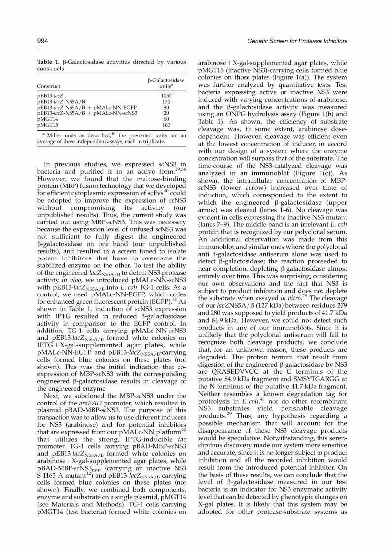
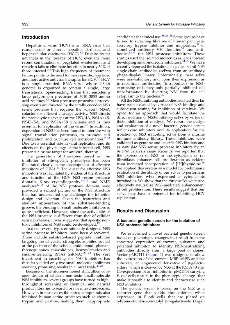
Table 1. b-Galactosidase activities directed by variousconstructs
Constructb-Galactosidase
unitsa
pEB13-lacZ 1057pEB13-lacZ-NS5A/B 130pEB13-lacZ-NS5A/B C pMALc-NN-EGFP 80pEB13-lacZ-NS5A/B C pMALc-NN-scNS3 20pMGT14 60pMGT15 160
a Miller units as described;40 the presented units are anaverage of three independent assays, each in triplicate.
994 Genetic Screen for Protease Inhibitors
In previous studies, we expressed scNS3 inbacteria and purified it in an active form.29,36
However, we found that the maltose-bindingprotein (MBP) fusion technology that we developedfor efficient cytoplasmic expression of scFvs40 couldbe adopted to improve the expression of scNS3without compromising its activity (ourunpublished results). Thus, the current study wascarried out using MBP-scNS3. This was necessarybecause the expression level of unfused scNS3 wasnot sufficient to fully digest the engineeredb-galactosidase on one hand (our unpublishedresults), and resulted in a screen tuned to isolatepotent inhibitors that have to overcome thestabilized enzyme on the other. To test the abilityof the engineered lacZNS5A/B to detect NS3 proteaseactivity in vivo, we introduced pMALc-NN-scNS3with pEB13-lacZNS5A/B into E. coli TG-1 cells. As acontrol, we used pMALc-NN-EGFP, which codesfor enhanced green fluorescent protein (EGFP).40 Asshown in Table 1, induction of scNS3 expressionwith IPTG resulted in reduced b-galactosidaseactivity in comparison to the EGFP control. Inaddition, TG-1 cells carrying pMALc-NN-scNS3and pEB13-lacZNS5A/B formed white colonies onIPTGCX-gal-supplemented agar plates, whilepMALc-NN-EGFP and pEB13-lacZNS5A/B-carryingcells formed blue colonies on those plates (notshown). This was the initial indication that co-expression of MBP-scNS3 with the correspondingengineered b-galactosidase results in cleavage ofthe engineered enzyme.
Next, we subcloned the MBP-scNS3 under thecontrol of the araBAD promoter, which resulted inplasmid pBAD-MBP-scNS3. The purpose of thistransaction was to allow us to use different inducersfor NS3 (arabinose) and for potential inhibitorsthat are expressed from our pMALc-NN platform40
that utilizes the strong, IPTG-inducible tacpromoter. TG-1 cells carrying pBAD-MBP-scNS3and pEB13-lacZNS5A/B formed white colonies onarabinoseCX-gal-supplemented agar plates, whilepBAD-MBP-scNS3mut (carrying an inactive NS3S-1165-A mutant12) and pEB13-lacZNS5A/B-carryingcells formed blue colonies on those plates (notshown). Finally, we combined both components,enzyme and substrate on a single plasmid, pMGT14(see Materials and Methods). TG-1 cells carryingpMGT14 (test bacteria) formed white colonies on
arabinoseCX-gal-supplemented agar plates, whilepMGT15 (inactive NS3)-carrying cells formed bluecolonies on those plates (Figure 1(a)). The systemwas further analyzed by quantitative tests. Testbacteria expressing active or inactive NS3 wereinduced with varying concentrations of arabinose,and the b-galactosidase activity was measuredusing an ONPG hydrolysis assay (Figure 1(b) andTable 1). As shown, the efficiency of substratecleavage was, to some extent, arabinose dose-dependent. However, cleavage was efficient evenat the lowest concentration of inducer, in accordwith our design of a system where the enzymeconcentration will surpass that of the substrate. Thetime-course of the NS3-catalyzed cleavage wasanalyzed in an immunoblot (Figure 1(c)). Asshown, the intracellular concentration of MBP-scNS3 (lower arrow) increased over time ofinduction, which corresponded to the extent towhich the engineered b-galactosidase (upperarrow) was cleaved (lanes 1–6). No cleavage wasevident in cells expressing the inactive NS3 mutant(lanes 7–9). The middle band is an irrelevant E. coliprotein that is recognized by our polyclonal serum.An additional observation was made from thisimmunoblot and similar ones where the polyclonalanti b-galactosidase antiserum alone was used todetect b-galactosidase; the reaction proceeded tonear completion, depleting b-galactosidase almostentirely over time. This was surprising, consideringour own observations and the fact that NS3 issubject to product inhibition and does not depletethe substrate when assayed in vitro.29 The cleavageof our lacZNS5A/B (127 kDa) between residues 279and 280 was supposed to yield products of 41.7 kDaand 84.9 kDa. However, we could not detect suchproducts in any of our immunoblots. Since it isunlikely that the polyclonal antiserum will fail torecognize both cleavage products, we concludethat, for an unknown reason, these products aredegraded. The protein termini that result fromdigestion of the engineered b-galactosidase by NS3are QRASEDVVCC at the C terminus of theputative 84.9 kDa fragment and SMSYTGARGG atthe N terminus of the putative 41.7 kDa fragment.Neither resembles a known degradation tag forproteolysis in E. coli,41 nor do other recombinantNS3 substrates yield perishable cleavageproducts.29 Thus, any hypothesis regarding apossible mechanism that will account for thedisappearance of these NS3 cleavage productswould be speculative. Notwithstanding, this seren-dipitous discovery made our system more sensitiveand accurate, since it is no longer subject to productinhibition and all the recorded inhibition wouldresult from the introduced potential inhibitor. Onthe basis of these results, we can conclude that thelevel of b-galactosidase measured in our testbacteria is an indicator for NS3 enzymatic activitylevel that can be detected by phenotypic changes onX-gal plates. It is likely that this system may beadopted for other protease-substrate systems as

Genetic Screen for Protease Inhibitors 995
long as the protease can be expressed in an activeform in E. coli.
Genetic screens for proteases,42 for proteaseinhibitors in general,43,44 and for NS3 inhibitors inparticular,45 have been described but came short ofproviding the desired protease inhibitors. To thebest of our knowledge, ours is the first geneticscreen to actually yield potentially useful inhibitors(see below). We are currently evaluating the utilityof our test bacteria to identify other proteininhibitors co-expressed as a genetic screen orsmall-molecule inhibitors added to the culturemedium.
Application of the genetic screen for theidentification of NS3-inhibiting scFvs
For the isolation of NS3 inhibitory antibodies, weconstructed a diverse immune antibody phagedisplay library. The source of antibody genes wasa pool of spleens from three mice that wereimmunized with MBP-scNS3 and had serum titersO600,000 after the third boost. Total RNA wasrecovered and an scFv phage display library wasconstructed as described in Materials and Methods.The total library size is O107 individual clones.A single cycle of delayed infectivity panning (DIP)46
and of biopanning47 affinity selections of the phageantibodies were applied (separately) to partiallyenrich for NS3 binders. This enrichment cycle wasnecessary in order to reduce the library size forscreening a reasonable number of clones onindicator plates. The scFv clones obtained asaffinity-selection output (about 106 clones) werepooled and subcloned into pMALc-NN asdescribed in Materials and Methods. We chose totest the scFvs for potential inhibition of NS3 in theform ofMBP-scFvs, since we found that in that formthe scFvs are stabilized as soluble active binders inthe reducing environment of the cytoplasm.40 ThepMALc-NN-scFv pool was introduced into testbacteria resulting in a bacterial genetic screencarrying two compatible plasmids with threedifferent regulatory mechanisms for expression:IPTG-inducible, high-level scFv expression; arabi-nose-inducible, medium-level NS3 expression; andconstitutive low-level b-galactosidase expression.The system is thus tuned such that sufficient NS3 ismade to fully digest the engineered b-galactosidase,unless inhibited by an excess of inhibitor that isproduced at a still higher concentration.
The transformants (about 2!106 colonies) werescreened on X-gal indicator agar plates(Figure 1(d)). A total of 100 blue colonies werepicked and analyzed by BstNI fingerprinting asdescribed.47 Many clones were duplicates, whichresulted in 25 clones being identified as unique. Theresiding pMALc-NN-scFv plasmids were recoveredfrom them and were re-introduced into test bacteriato validate the results of the initial screen. All 25clones regained their blue phenotype, confirmingthat the phenotypic change was indeed a directresult of scFvs expression. A sample of four of the
clones (10, 11, 35, and 171) is shown in Figure 1(e) incomparison to four control scFvs, three of which(37Y, 44Y, and 53Y) that were isolated from a humansynthetic antibody library,36 bind NS3 withoutinhibiting it and one (Anti Tac) totally irrelevant.48
Characterization of specificity and bindingaffinity of selected scFvs
The scFvs that were isolated as blue colonies inthe bacterial genetic screen were expressed andpurified as soluble proteins, and evaluated forscNS3 binding in an enzyme-linked immuno-sorbent assay (ELISA) with a panel of irrelevantproteins serving as specificity controls. As a control,we used the non-inhibitory, NS3 binding scFv 53Y.36
As shown in Figure 2(a), seven of the isolated scFvsbound scNS3 specifically. Although MBP-scNS3was used for immunization and scFv identification,the binding studies that were carried out withunfused scNS3 confirm that MBP did not play a rolein their antigen binding. Binding affinities wereestimated from half-maximal binding signals ofELISAs. As shown in Figure 2(b), affinities of theselected scFvs ranged between 80 nM and 300 nM,which are moderate binding affinities. The remain-ing 18 scFvs were weaker or less specific binders orweaker inhibitors in the quantitative assay (seebelow) and were not studied further. Thus, in itspresent form, our genetic screen is very sensitive inidentifying potential inhibitors that would escapedetection by in vitro approaches. This is probablydue to the MBP-fusion technology we chose forscFv expression, where the very high intracellularconcentration of MBP-scFvs may compensate for itsweak affinity.40
To evaluate whether the scFvs NS3 epitopesoverlap, we performed a competition ELISA asdescribed in Materials and Methods. As shown inFigure 2(c), scFvs 1, 10, 11, 35, 53Y and 171 couldcompete with scFvs 171 and 35 for specific bindingto scNS3, while scFv 162 apparently did not.Because our tracers were not radioactive, but ratherthe same scFvs with different epitope tags, we hadto use relatively high concentrations, which pre-vented a full competition of binding. This is evidentfrom the inferior inhibition obtained when thehigher-affinity scFv 171 is used as a tracer. As aresult, these data cannot be used to calculatebinding affinities. Nonetheless, they do suggestthat the binding epitopes of most of our scFvsoverlap at least partially, and are probably locatedaround the NS3 catalytic site. Interestingly, thecontrol scFv 53Y,36 which is non-inhibitory, alsocompeted the binding of both scFvs 35 and 171 toscNS3 (Figure 2(c)). This highlights the advantage ofusing a screen for inhibitory antibodies that is basedon actual inhibition rather than merely on antigenbinding. Due to their large size (in comparison tosmall-molecule inhibitors), scFvs may inhibit NS3catalysis by blocking the access of the substrate tothe active site without engaging it directly.The scFvs sequence analysis (Table 2) revealed a

Figure 2. Evaluation of the selected scFvs binding to NS3. (a) Binding specificity to scNS3 compared to five irrelevant proteins determined by ELISA. The scFvs tested at30 mg/ml, 15 mg/ml, and 7.5 mg/ml are shown as black, grey and white bars, respectively. (b) Binding curves of the selected scFvs to scNS3 determined by ELISA. Error barsrepresent the standard deviation of the data. (c) Competition ELISA showing the competition of scFvs 171 and 35 as tracers binding to scNS3 by the selected scFvs.

Table 2. Deduced amino acid sequences of the CDR regions of the NS3-inhibitory scFvs
ScFv VH CDR1 VH CDR2 VH CDR3 Germlinea
1 DYYIK VINPGSGGTNYNEKFKG RAGTGYYFDY J558.310 NYWIE GILPGSGSTNYNEKFKG DGPPGFDY J558.1711 DYYMH RIYPGNGQTDYNGKFKG GLFAY J558.1835 DYYMH WIDPENGDTEYTQKFKG ITTDYYFDY V130148 DTFVH RIDPANGNTKYDPKFQG GLDYYAMDY V130162 DYYMH WIDPANGNTKYDPKFQG WDWYFDV V130171 DYYMH WIDPENGDTEYAPKFQG WGGSSDYYAMDY V130
ScFv VL CDR1 VL CDR2 VL CDR3 Germlinea
1 RSSQSIVHSNGNTYLE KVSNRFS FQGSHVPLT cr110 KSSQSLLDSDGKTYLN LVSKLDS FQGSHVPFT bd211 RSSQSLANSYGNTYLS GISNRFS LQGTHQPLT cs135 RSSQSLVHSNGNTYLE KVSNRFS SQSTHVPLT cr1148 RSSQSLVHSNGNTYLH KVSNRFS SQSTHVPWT bb1162 RSSQSIVHSNGNTYLE KVSNRFS FQGSHVPWT cr1171 RSSQSLVHSNGNTYLH KVSNRFS FQGSHVPWT cr1
a Determined by alignment of the VH and VL coding sequences to mouse V-gene germline genes using Ig-Blast: (http://www.ncbi.nlm.nih.gov/igblast/).
Genetic Screen for Protease Inhibitors 997
high degree of sequence identity in the light chainvariable region (VL) complementarity-determiningregion (CDR) sequences in contrast to a higherdiversity observed in the heavy chain variableregion (VH) CDR sequences, particularly at CDR3.In contrast, a more limited repertoire of germlinegenes appears in the selected VH domains than inthe VL domains. Still, each VH utilizes different Dand J segments in the assembly of CDR3, while allthe selected VLs utilize a single J segment, J5. Thissuggests that the VL CDR3 was strongly selected byour genetic screen and is probably of criticalimportance. The high degree of similarity in theVL domains may suggest that the bindingspecificity of scFvs to NS3 may be determinedlargely by the VLs, while the VHs may play anaccessory role.
Validation of the selected scFvs as NS3inhibitors in vitro
The potential of the selected scFvs to inhibit NS3catalysis was evaluated quantitatively by measur-ing the b-galactosidase level that serves as anindicator for NS3 enzymatic activity level in geneticscreen bacteria. An ONPG hydrolysis assay wascarried out on cultures of the seven selected clonesdescribed above. As shown in Figure 3(a), culturesof the selected scFvs had high levels of b-galacto-sidase for all the scFvs tested, as compared to thecontrol non-inhibitory scFv 53Y. The levels ofb-galactosidase were in correlation to the level ofscFvs expression, but could be detected evenwithout IPTG induction, due to the leakiness ofthe Ptac promoter under our assay conditions.These results indicate that NS3 catalysis is inhibitedby the selected scFvs in a dose-dependent manner.
To further confirm the inhibitory effect of scFvson NS3 catalysis we applied an in vitro fluorometricassay for the measurement of NS3 protease catalysisand inhibition as described in Materials andMethods. As shown in Figure 3(b), all the tested
scFvs inhibited NS3 catalysis in a dose-dependentmanner as compared to the control non-inhibitoryscFv 53Y. Different levels of inhibition could bedetected between the different scFvs, and NS3catalysis ranged between 12% and 54%. On thebasis of Figure 3(b) and additional unpublisheddata, the IC50 values of the isolated scFvs are about2.4 mM for scFvs 1, 11 and 148, about 1.2 mM forscFvs 10 and 162, and about 0.6 mM for scFvs 35 and171. These results validated the suggestion that theselected scFvs that were identified initially as bluecolonies in the bacterial genetic screen and latervalidated as specific NS3 binders are genuine NS3catalytic activity inhibitors. As shown in Figure 3(c),the binding affinity of the scFvs largely correlated(with scFv 1 as an exception) with their potency ofin vitro inhibition. However, the b-galactosidaselevels as measured by ONPG hydrolysis assayfollow a different trend, probably due to differentlevels of accumulation and different stabilities of thescFvs in the cytoplasmic milieu.
Inhibition of NS3-mediated cell proliferation bythe anti-NS3 intrabodies
Our goal in isolating scFvs that inhibit NS3 is fortheir application as intracellular antibodies in vivo.Previously, we have shown that the expression ofcatalytically active NS3 (without the NS4A cofactor), but not of the S-1165-A inactive mutantresulted in enhanced proliferation of rat fibroblasts(RF cells). Furthermore, this proliferation could beinhibited by small-molecule protease inhibitors.12
More recently, we demonstrated that scNS3 (withthe NS4A co-factor) expression in RF cells alsoenhances cell proliferation as evident frommeasuring the incorporation of [3H]thymidine.36
To evaluate the potential of our scFvs to inhibit NS3expressed at different levels, we stably transfectedRF cells with scNS3. Stable clones were selected andthe expression level of scNS3 was evaluated using areal-time PCR analysis.49 As shown in Figure 4(a),

Figure 3. Evaluation of the selected scFvs inhibition ofNS3 catalysis. (a) b-Galactosidase activity determined byan ONPG hydrolysis by uninduced or IPTG-inducedgenetic screen bacteria carrying selected scFvs. (b) In vitroinhibition of NS3 catalysis at two concentrations of testedscFvs, 2.4 mM (striped bars) and 1.2 mM (filled bars). Errorbars represent the standard deviation of the data. (c) Atable ranking the scFvs according to their potency in NS3catalysis inhibition in vitro. Affinity: apparent kD (in nM)estimated from the binding curves in Figure 2(b); b-gal,b-galactosidase taken from Figure 3(a); % inhibition,values were obtained by subtracting the percentagecatalysis value shown in Figure 3(b) from 100%.
Figure 4. Evaluation of the selected scFvs inhibition ofNS3-mediated cell proliferation. RF cells were stablytransfected with vector pYB50 and stable clones wereselected. (a) Real-time PCR analysis of scNS3 expression;the results are shown as relative quantity normalized tothe control housekeeping gene. (b) Cell proliferationanalyzed using a [3H]thymidine uptake assay. (c) Thestable clones 1 (very low NS3), 4 (low NS3) and 16 (highNS3) were transiently transfected with the tested scFvs(inset) and cell proliferation was analyzed using a[3H]thymidine uptake assay. Error bars represent thestandard deviation of the data.
998 Genetic Screen for Protease Inhibitors
scNS3 was expressed at various levels in differentclones. Clone 1 was expressed 1000-fold less thanclone 4, but still a real-time PCR signal could bedetected above that resulting from RNA recoveredfrom NS3-less cells.
Next, the NS3-mediated phenotype was studiedby evaluating the relative rate of cell proliferationby a [3H]thymidine uptake assay. As shown inFigure 4(b), the increase in the cell proliferation rateof the stable scNS3-expressing RF cells correlatedwith the expression level of scNS3 shown inFigure 4(a).
To evaluate the effect of NS3-neutralizing intra-body expression on the NS3-mediated phenotype,stable RF clones number 1 (very low NS3expression), number 4 (low NS3 expression), andnumber 16 (high level of NS3 expression) weretransiently transfected with the tested intrabodies.As shown in Figure 4(c), intrabodies 35, 171 and 10inhibited NS3-mediated cell proliferation as com-
pared to the control, non-inhibitory scFv 53Y. Themost efficient intrabody in this test, 35, inhibited theincorporation of [3H]thymidine by 74–83% withseemingly more efficient inhibition observed at thelow level of NS3 expression. Considering thatthe expression level of NS3 in viral infected cellsis probably in the low range we observe here, ourresults confirm the potential of our scFvs to functionas inhibitory intrabodies for NS3-mediated pheno-type in mammalian cells. Indeed, preliminarystudies suggest that our intrabodies inhibit sub-genomic HCV RNA replicons (our unpublishedresults).
Intrabodies with inhibitory properties present a

Genetic Screen for Protease Inhibitors 999
new class of neutralizing molecules with greatpotential for gene therapy. scFvs have been shownto possess favorable properties as therapeuticagents. Indeed, anti-HCV intrabody-basedapproaches were suggested by studies of theisolation of an scFvs that binds and inhibits the NS3helicase using phage display technology.50,51 As foranti-NS3 protease intrabodies, we have recentlyreported the isolation of non-neutralizing anti-NS3scFvs by antibody phage-display that, as intra-bodies, partially inhibited NS3-mediated cell trans-formation by diverting NS3 from the cell cytoplasmto the nucleus.36 Inhibition of NS3 catalysis bysmall-molecule inhibitors has been a formidablechallenge, due to the featureless appearance of theNS3 catalytic groove that results in weak binding.Antibodies may offer a better solution by forming a“clamp” that involves interactions with NS3 thatare located away from the active site on one hand,and blocking or interfering with substrate bindingon the other. In our in vitro assay, our scFvs inhibitNS3 at micromolar concentrations, thus they shouldprobably be improved to make more potentinhibitors. The fact that their affinities lie in thelow micromolar range as well suggest that theycould be manipulated easily by approaches such asin vitro affinity maturation by phage display52 or byfurther exploiting our genetic screen towards suchan end. Still, considering that we had a transfectionefficiency of about 50%, the in vivo inhibition ofNS3-mediated cell proliferation we show here isquite efficient. This is probably because we areusing fusion-stabilized scFvs that accumulate athigh levels in the cell cytoplasm of bacteria40 and ofmammalian cells (S. Shaki-Loewenstein et al.,unpublished results).
There are numerous technical challengesassociated with translation of the use of HCVNS3-inhibitory scFvs to clinical therapeutics. Still,it is our belief that our scFvs or improvedderivatives thereof, via gene delivery or proteindelivery approaches53 should be explored, as theymay represent the effective interventions that maylead to the eradication of HCV infection. Con-comitantly, our current efforts are focused on usingcomputer-docking studies combined with muta-genesis to elucidate the binding and inhibitionmechanism of our scFvs and to generate improvedones, and on studying NS3 inhibition by our scFvsusing other models of HCVreplication such as RNAreplicons.
†www.neb.com/nebecomm/tech_reference/restriction_enzymes/sequences/pacyc177.seq
Materials and Methods
General recombinant DNA techniques and reagentswere as described.29
Construction of the bacterial genetic screen
The p15A replicon-based low copy number reporterplasmid pMGT14, carries both the substrate and theenzyme components of the bacterial genetic screen, whilepotential inhibitors are carried on a second compatible
plasmid. Plasmid pMGT14 (Figure 1) was constructed inthree steps as follows: in the first step, an NS3-cleavableb-galactosidase-encoding engineered lacZ gene carryingplasmid was prepared. Plasmid pIB1337 carries the 5 0 77codons of the E. coli trpR gene fused to the eighth codon ofthe E. coli lacZ gene, under the control of the trpRpromoter on a pBR322 plasmid backbone (colE1 replicon).pIB13 DNA was digested with the restriction enzymesClaI and DraI, and a 4089 bp DNA fragment wasrecovered. A p15A replicon and kanamycin-resistancecassette carrying 3054 bp DNA fragment was isolated byDraI and BstBI digestion of pACYC177 plasmid DNA†.The two DNA fragments were ligated and used totransform E. coli TG-1 cells, which resulted in plasmidpEB13.Two tandem sfiI restriction sites separated by a TAA
stop codon were inserted between lacZ codons 279 and280 by overlap-extension PCR-based mutagenesis asfollows:29 pEB13 DNA was used as template in twoPCR reactions using primers LacZ-Sfi-279-280-BACKwith LacZ-520 FOR and LacZ-70-BACK with LacZ-279-FOR (Table 3). All the PCR reactions were carried outusing 30 cycles of 95 8C for 30 s, 55 8C for one minute and72 8C for oneminute with a final extension at 72 8C for fiveminutes. The resulting PCR products of 667 bp and763 bp, respectively, were mixed and assembled by PCRamplification using primers LacZ-70-BACK and LacZ-520-FOR. The resulting PCR product was digested withBsu36I and BssHII and cloned into pEB13 DNA digestedwith the same enzymes. Ligated DNA was used totransform DH5a E. coli cells (Invitrogen, USA) that wereplated on LB agar plates supplemented with 0.04% (w/v)X-gal. White (colorless) colonies that resulted frominterruption of the lacZ open reading frame by theinserted stop codon were analyzed by DNA sequencing.The resulting plasmid was pEB13-Sfi. This plasmid servesas a universal plasmid for insertion of foreign sequencesinto a permissive site of lacZ.38
The NS5A/B cleavage site of NS329 was engineeredbetween codons 279 and 280 of the lacZ gene on plasmidpEB13-Sfi as follows: a DNA duplex formed by annealingprimers NS5A/B-SE and NS5A/B-AS (Table 3) wasinserted into pEB13-Sfi plasmid DNA that had beendigested with SfiI. Ligated DNA was used to transformTG-1 E. coli cells (Amersham Biosciences, USA) that wereplated on X-gal-supplemented LB agar plates. Bluecolonies that resulted from restoration of the lacZ openreading frame by the inserted fragment were analyzed byDNA sequencing. The resulting plasmid, pEB13-lacZNS5A/B encodes an engineered lacZ gene encodingfor NS3-cleavable b-galactosidase.In the second step, a construct for expression of fusion-
stabilized NS3 was prepared. Single-chain NS3 protease(scNS3)29 was stabilized by its expression as an MBPfusion,40 and cloned under the control of the araBADpromoter as follows: Plasmid pYB4329 was used astemplate in a PCR reaction with primers NS3-Nco-BACK and NS3-Not-FOR (Table 3). The resulting PCRproduct was digested with NcoI and NotI and cloned intopMALc-NN DNA40 digested with the same enzymes.The resulting plasmid was pMALc-NN-scNS3, whereexpression of MBP-scNS3 is still controlled by the tacpromoter.To convert MBP-scNS3 to an arabinose-inducible gene,
the MBP-scNS3-carrying fragment was recovered byPCR using pMALc-NN-scNS3 as template with primers

Table 3. Oligonucleotide primers
A. Primers for construction of pEB13-SfiLacZ-Sfi-279-280-BACK: 50 GAAATTATCGATGAGGGCCAACGTGGCCGGTAATT TTTGGCCTCTGGGGCCCGTGGTGGTTATGCC 30
LacZ-520-FOR: 5 0 CAGCCGGGAAGGGCTGGTCTT 3 0
LacZ-70-BACK: 50 CCGGAAAGCTGGCTGGAGTGC 3 0
LacZ-279-FOR: 5 0 GGCCCTCATCGATAATTTCAC 3 0
B. Primers for NS5A/B site duplexNS5A/B-SE: 5 0 AGCTAGCGAGGACGTCGTCTGCTGCTCGATGTCCTACACTG 3 0
NS5A/B-AS: 5 0 TGTAGGACATCGAGCAGCAGACGACGTCCTCGCTAGCTCGT 3 0
C. Primers for SphI-NotI sites duplexpEB13-dup-SE: 50 AATTGCATGCCTGCAGGCGGCCGCG 3 0
pEB13-dup-AS: 5 0 AATTCGCGGCCGCCTGCAGGCATGC 3 0
D. Primers for MBP-scNS3 cloningNS3-Nco-BACK: 5 0 TCAGTACCATGGCGCCTATCGGCTCAGTAGTA 3 0
NS3-Not-FOR: 5 0 GGGAAAGCGGCCGCTTACCGCATAGTGGTTTCCATAGA 3 0
MalE-BspHI-BACK: 5 0 CCCTAATCATGAAAACTGAAGAAGGTAAA 3 0
E. Primers for real-time PCR detection of NS3Txt-156F: 5 0 CCTTTACCTGGTCACGAAACATG 3 0
txt-257R: 5 0 CCCTTCAAGTAGGAGATGGGTCTA 3 0
1000 Genetic Screen for Protease Inhibitors
MalE-BspHI-BACK and NS3-Not-FOR. The resultingPCR product was digested with BspHI and NotI andcloned into pBAD-TOPO DNA (Invitrogen, USA)digested with NcoI and NotI. The resulting plasmid wasnamed pBAD-MBP-scNS3. A similar plasmid, pBAD-MBP-scNS3mut was constructed for expression of aninactive mutant S-1165-A NS3 to serve as a negativecontrol.12
In the third step, we constructed a bi-cistronic plasmidcarrying both lacZ with the inserted NS3 cleavage site(lacZNS5A/B) and MBP-scNS3 as follows: restriction sitesfor SphI and NotI were inserted into vectorpEB13lacZNS5A/B as a DNA duplex formed by annealingprimers pEB13-dup-SE and pEB13-dup-AS (Table 3). Theduplex was inserted into pEB13-lacZNS5A/B plasmid DNAthat had been linearized at the unique EcoRI site. Theresulting plasmid was digested with SphI and NotI toobtain a vector fragment. An insert fragment correspond-ing to MBP-scNS3 with its controlling araBAD promoterand the regulatory araC gene was obtained by digestingplasmid pBAD-MBP-scNS3 with SphI and NotI. Thetwo fragments were ligated to generate plasmid pMGT14.A similar plasmid, pMGT15 expressing an inactivemutant S-1165-A NS312 was constructed using the SphI-NotI fragment of plasmid pBAD-MBP-scNS3mut to serveas a negative control of catalysis. In both plasmids theexpression of the engineered lacZ gene is weaklyconstitutive in trpRC E. coli strains, while MBP-scNS3 isinducible by arabinose.
Construction and affinity selection of the antibodylibrary
MBP-scNS3 protein was expressed and purified essen-tially as described for MBP-scFvs.40 Balb/c mice wereimmunized with 50 mg of purified MBP-scNS3 in com-plete Freunds adjuvant and boosted three times atintervals of two weeks using 50 mg of purified MBP-scNS3 in incomplete Freunds adjuvant. The mice werebled three days after each boost. The serum anti-NS3antibody titer was determined by an ELISA.Total RNAwas extracted from 107 cells recovered from
the immune mice spleens according to the instructions ofthe Tri Reagent total RNA extraction kit (Talron, Israel).A total of 250 mg of RNA was obtained. cDNA wasproduced from mRNA by reverse transcription reactionsusing 1 mg of total RNA as template. Heavy and light
chain variable domains were amplified in PCR using aprimer set designed for the construction of mouse scFvlibraries.47 The antibody variable cassettes were combi-natorially assembled into complete scFv constructs andcloned into pCC phagemid vector54 essentially asdescribed.47 The cloned vectors were transferred intocompetent E. coli TG-1 cells by electroporation and librarydiversity was tested as described.47
The library was affinity-selected using the DIP methodwith NS3-displaying E. coli cells as described.46 Thelibrary was also affinity-selected by mixing 1011 libraryphages with 25 mg of MBP-scNS3 to form phage-antigencomplexes. This affinity selection was carried out essen-tially as described;47 however, since MBP-scNS3 is notbiotinylated, the streptavidin coated magnetic beads werereplaced with amylose resin (New England Biolabs, USA)and magnetic capture was replaced by centrifugation.
Applying the bacterial genetic screen to isolate NS3-inhibitory scFvs
Affinity-selected phages were pooled and used assource for scFv DNA by digestion of phagemid DNAwith NcoI and NotI. The pooled insert DNA was clonedinto pMALc-NN that had been digested with the sameenzymes essentially as described.40 In pMALc-NN,expression of MBP-scFvs is under the control of anIPTG-inducible tac promoter and the plasmid carries anampicillin-resistance cassette and the colE1 origin ofreplication, and is thus compatible with pMGT. Theplasmid pool was introduced into E. coli TG-1 cellsalready containing the pMGT plasmid. The transformantswere plated to yield individual colonies on 2!YT agarplates supplemented with 0.004% (w/v) X-gal, 100 mg/mlof ampicillin, 50 mg/ml of kanamycin, 0.2% (w/v)arabinose and 0.05 mM IPTG. The plates were screenedand blue colonies were picked. pMALc-NN-scFv DNAwere recovered from them and re-introduced into pMGT-expressing cells to validate the results of the initial screen.
Binding assays
Selected MBP-scFvs were expressed and purified fromthe soluble fraction of IPTG-induced plasmid-carryingE. coli BL21 cells using amylose resin chromatographyessentially as described.40 ScNS3 was overexpressed in

Genetic Screen for Protease Inhibitors 1001
pYB-43 carrying E. coli BL-21(DE3) cells by a modificationof our published method.29 The bacteria were inducedwith 1 mM IPTG at 37 8C for three hours, which resultedin accumulation of the scNS3 as inclusion bodies. Theinclusion bodies were solubilized in 6 M guanidiniumhydrochloride and purified by Talon (Clontech, USA)IMAC chromatography under denaturing conditions. Thepurified protein (0.5 mg/ml) was stored atK80 8C in 6 Mguanidinium hydrochloride containing 150 mM imida-zole. ELISA plates were coated with 4 mg/ml of antigen in50 mM NaHCO3 buffer (pH 9.6). ELISAs were processedas described,47 using a mouse monoclonal anti-MBPantibody (Sigma, Israel) followed by horseradish peroxi-dase (HRP)-conjugated goat anti-mouse antibodies(Jackson, USA).For competition ELISAs, we subcloned the scFvs that
were used as tracers into a derivative of pMALc-NNwhere the C-terminal FLAG epitope40 was replaced witha myc tag. Competing scFvs were added to scNS3-coatedplates at various concentrations and incubated at roomtemperature for one hour before adding the tracer scFvs.Binding of the tracers was monitored using an HRP-conjugated anti-myc antibody (Sigma, Israel) followed byHRP-conjugated goat anti-mouse antibodies (Jackson,USA). ELISAs were developed using the HRP chromo-genic substrate TMB (Dako, USA) and read at 450 nm.
NS3 catalysis inhibition assays
Blue colonies of E. coli TG-1 cells carrying pMGT14 andpMALc-NN-scFv that were isolated by the genetic screenwere grown in LB medium supplemented with100 mg/ml of ampicillin, 50 mg/ml of kanamycin andinduced with 0.2% (w/v) arabinose and 0. 5 mM IPTG atA600 nmZ0.6 for four hours at 30 8C. To measure theb-galactosidase activity, we carried out an ONPGhydrolysis assay essentially as described.40 An in vitrofluorometric assay for the measurement of NS3 proteasecatalysis inhibition by the purified scFvs was carried outessentially as described,29 with the following modifi-cations: the EFGP-NS5A/B-CBD substrate wasimmobilized onto cellulose prior to its exposure toenzyme and inhibitor. The reactions were carried out in96-well plates in a volume of 100 ml containing 5 mMimmobilized substrate, 100 nMMBP-scNS3 and 2.4 mMor1.2 mM tested MBP-scFv.
Immunoblot
Immunoblotting was carried out to follow the time-course on NS3 induction and its effect on cleavage oflacZNS5A/B. TG-1 cells carrying plasmids pMGT14 orpMGT15 were grown in LB medium supplemented with100 mg/ml of ampicillin, and 50 mg/ml of kanamycin toan A600 nm Z0.6. The cells were than induced with 0.2%(w/v) arabinose for 10 minutes, 20 minutes, 30 minutes,60 minutes and 120 minutes at 30 8C. Then 20 mg of totalcell extracts was separated by SDS/PAGE and transferredonto nitrocellulose membranes as described.29 Themembranes were blocked and reacted with mousepolyclonal anti-b-galactosidase antiserum (from ourlaboratory collection) and a mouse anti-MBP antibodyand detected by HRP-conjugated goat anti-mouse anti-body. The membrane was developed by using ECLreagents as described.29
Expression of scNS3 in mammalian cells
All the cell culture reagents and antibiotics were from
Beit-Haemek, Israel. To express scNS3 in mammaliancells, we used vector pYB50.36 RF cells were stablytransfected by Lipofectamine plus reagent (Invitrogen,USA) with vector pYB50 and stable transfectants wereselected in a medium containing hygromycine (200 mg/ml). Stable clones were picked up and maintainedroutinely in DMEM containing 10% (v/v) fetal calfserum (FCS) and supplemented with hygromycine (at200 mg/ml). To detect the level of expression of scNS3 inthe different cell clones we performed a real-time (RT)PCR analysis (SYBR Green I dye chemistry, AppliedBiosystemns, USA) as follows. RNA extraction andRT-PCRwas carried out essentially as described.49 Briefly,1 mg of total RNAwas converted to cDNA using randomprimers in a total volume of 20 ml. Then 1 ml of the RTreaction was used as template for the RT-PCR reactionusing primers txt-156F and txt-257R (Table 3) for the NS3gene that were designed using the Primer Expresssoftware (Applied Biosystem, USA). The reaction wascarried out and analyzed as recommended by thesupplier (Applied Biosystem, USA). The measurementof NS3 mRNA expression was performed utilizing anABI prism 7000 Sequence Detection System (AppliedBiosystem, USA). The RT-PCR amplification was relatedto a standard curve obtained using standard house-keeping genes.
Cloning of intrabodies to mammalian plasmid
For evaluation as cytoplasmic intrabodies, weexpressed our scFv as fusion-stabilized, MBP-scFv.40
The construction of plasmid pCMV/H6myc/Cyto-MBPused for cytoplasmic expression of fusion stabilized scFvswill be described elsewhere (S. Shaki-Loewenstein et al.,unpublished results). Briefly, the MBP-coding malE genewas amplified by PCR using plasmid pMALc-NN40 astemplate and inserted 5 0 to the scFv insertion sites ofplasmid pCMV/myc/cyto (Invitrogen USA, a vector forcytoplasmic expression of intrabodies). The plasmid wasfurther modified with a sequence encoding HisCmyctags 3 0 to the scFv insertion sites. DNA fragments codingfor anti-NS3 and control scFvs were recovered fromvector pMALc-NN-scFv plasmid DNA by digestion withNcoI and NotI, and subcloned into this mammalianexpression vector.
[3H]thymidine uptake assay
Cell proliferation was evaluated by measuring[3H]thymidine uptake essentially as described.55 StablescNS3-expressing RF cells were plated in triplicate intowells of flat bottom 96-well plates in DMEM containing10% FCS. Twenty four hours later the cells weretransiently transfected with pCMV/H6myc/Cyto-MBPplasmids using Lipofectamine plus reagent. At 48 hourspost transfection, cells were pulsed with[methyl-3H]thymidine, 1 mCi/well, and the cells werefurther incubated for 18 hours. The cells were washedand collected onto glass microfiber filters using a cellharvester (Dynatech, USA). Incorporated [3H]thymidinethat was retained on the filter was determined using aPackard Matrix-9600 direct b counter.
Acknowledgements
This work was supported by research grants from

1002 Genetic Screen for Protease Inhibitors
the United States Israel Binational ScienceFoundation (BSF) and from the Israel CancerResearch Fund (ICRF). M.G.-T. was supported bya travel fellowship from the Joan and JaimeConstantiner Institute for Molecular Genetics.
References
1. Purcell, R. (1997). The hepatitis C virus: overview.Hepatology, 26, 11S–14S.
2. Poynard, T., Yuen, M. F., Ratziu, V. & Lai, C. L. (2003).Viral hepatitis C. Lancet, 362, 2095–2100.
3. Pawlotsky, J. M. (2000). Hepatitis C virus resistance toantiviral therapy. Hepatology, 32, 889–896.
4. Manns, M. P., McHutchison, J. G., Gordon, S. C.,Rustgi, V. K., Shiffman, M., Reindollar, R. et al. (2001).Peginterferon alfa-2b plus ribavirin compared withinterferon alfa-2b plus ribavirin for initial treatment ofchronic hepatitis C: a randomised trial. Lancet, 358,958–965.
5. Molla, A. & Kohlbrenner, W. (2003). Evolving thera-peutic paradigms for HIV and HCV. Curr. Opin.Biotechnol. 14, 634–640.
6. Brechot, C. (1996). Hepatitis C virus: molecularbiology and genetic variability. Dig. Dis. Sci. 41,6S–11S.
7. Failla, C., Tomei, L. & De Francesco, R. (1994). BothNS3 and NS4A are required for proteolytic processingof hepatitis C virus nonstructural proteins. J. Virol. 68,3753–3760.
8. Suzuki, R., Suzuki, T., Ishii, K., Matsuura, Y. &Miyamura, T. (1999). Processing and functions ofHepatitis C virus proteins. Intervirology, 42, 145–152.
9. Borowski, P., Kuhl, R., Laufs, R., Schulze zur Wiesch,J. & Heiland, M. (1999). Identification and character-ization of a histone binding site of the non-structuralprotein 3 of hepatitis C virus. J. Clin. Virol. 13, 61–69.
10. Borowski, P., Heiland, M., Feucht, H. & Laufs, R.(1999). Characterisation of non-structural protein 3 ofhepatitis C virus as modulator of protein phosphoryl-ation mediated by PKA and PKC: evidences for actionon the level of substrate and enzyme. Arch. Virol. 144,687–701.
11. Ishido, S. & Hotta, H. (1998). Complex formation ofthe nonstructural protein 3 of hepatitis C virus withthe p53 tumor suppressor. FEBS Letters, 438, 258–262.
12. Zemel, R., Gerechet, S., Greif, H., Bachmatove, L.,Birk, Y., Golan-Goldhirsh, A. et al. (2001). Celltransformation induced by hepatitis C virus NS3serine protease. J. Viral. Hepatol. 8, 96–102.
13. Kaufmann, G. R. & Cooper, D. A. (2000). Antiretro-viral therapy of HIV-1 infection: established treatmentstrategies and new therapeutic options. Curr. Opin.Microbiol. 3, 508–514.
14. Kim, J. L., Morgenstern, K. A., Lin, C., Fox, T., Dwyer,M. D., Landro, J. A. et al. (1996). Crystal structure ofthe hepatitis C virus NS3 protease domain complexedwith a synthetic NS4A cofactor peptide. Cell, 87,343–355.
15. Love, R. A., Parge, H. E., Wickersham, J. A.,Hostomsky, Z., Habuka, N., Moomaw, E. W. et al.(1996). The crystal structure of hepatitis C virus NS3proteinase reveals a trypsin-like fold and a structuralzinc binding site. Cell, 87, 331–342.
16. Yan, Y., Li, Y., Munshi, S., Sardana, V., Cole, J. L.,Sardana, M. et al. (1998). Complex of NS3 protease
and NS4A peptide of BK strain hepatitis C virus: a 2.2A resolution structure in a hexagonal crystal form.Protein Sci. 7, 837–847.
17. Cicero, D. O., Barbato, G., Koch, U., Ingallinella, P.,Bianchi, E., Nardi, M. C. et al. (1999). Structuralcharacterization of the interactions of optimizedproduct inhibitors with the N-terminal proteinasedomain of the hepatitis C virus (HCV) NS3 protein byNMR andmodelling studies. J. Mol. Biol. 289, 385–396.
18. Barbato, G., Cicero, D. O., Nardi, M. C., Steinkuhler,C., Cortese, R., De Francesco, R. & Bazzo, R. (1999).The solution structure of the N-terminal proteinasedomain of the hepatitis C virus (HCV) NS3 proteinprovides new insights into its activation and catalyticmechanism. J. Mol. Biol. 289, 371–384.
19. Kwong, A. D., Kim, J. L., Rao, G., Lipovsek, D. &Raybuck, S. A. (1998). Hepatitis C virus NS3/4Aprotease. Antiviral Res. 40, 1–18.
20. Randall, G. & Rice, C. M. (2004). Interfering withhepatitis C virus RNA replication. Virus Res. 102,19–25.
21. Kakiuchi, N., Komoda, Y., Komoda, K., Takeshita, N.,Okada, S., Tani, T. & Shimotohno, K. (1998). Non-peptide inhibitors of HCV serine proteinase. FEBSLetters, 421, 217–220.
22. Steinkuhler, C., Koch, U., Narjes, F. & Matassa, V. G.(2001). Hepatitis C virus protease inhibitors: currentprogress and future challenges. Curr. Med. Chem. 8,919–932.
23. De Francesco, R., Tomei, L., Altamura, S., Summa, V.& Migliaccio, G. (2003). Approaching a new era forhepatitis C virus therapy: inhibitors of the NS3-4Aserine protease and the NS5B RNA-dependent RNApolymerase. Antiviral Res. 58, 1–16.
24. Lin, C., Lin, K., Luong, Y. P., Rao, B. G., Wei, Y. Y.,Brennan, D. L. et al. (2004). In vitro resistance studiesof hepatitis C virus serine protease inhibitors, VX-950and BILN 2061: structural analysis indicatesdifferent resistance mechanisms. J. Biol. Chem. 279,17508–17514.
25. Lamarre, D., Anderson, P. C., Bailey, M., Beaulieu, P.,Bolger, G., Bonneau, P. et al. (2003). An NS3 proteaseinhibitor with antiviral effects in humans infectedwith hepatitis C virus. Nature, 426, 186–189.
26. Johansson, A., Poliakov, A., Akerblom, E., Lindeberg,G., Winiwarter, S., Samuelsson, B. et al. (2002).Tetrapeptides as potent protease inhibitors ofhepatitis C virus full-length NS3 (protease-helicase/NTPase). Bioorg. Med. Chem. 10, 3915–3922.
27. Sperandio, D., Gangloff, A. R., Litvak, J., Goldsmith,R., Hataye, J. M., Wang, V. R. et al. (2002). Highlypotent non-peptidic inhibitors of the HCVNS3/NS4A serine protease. Bioorg. Med. Chem. Letters,12, 3129–3133.
28. Wyss, D. F., Arasappan, A., Senior, M. M., Wang, Y. S.,Beyer, B. M., Njoroge, F. G. & McCoy, M. A. (2004).Non-peptidic small-molecule inhibitors of the single-chain Hepatitis C virus NS3 protease/NS4A cofactorcomplex discovered by structure-based NMRscreening. J. Med. Chem. 47, 2486–2498.
29. Berdichevsky, Y., Zemel, R., Bachmatov, L.,Abramovich, A., Koren, R., Sathiyamoorthy, P. et al.(2003). A novel high throughput screening assay forHCV NS3 serine protease inhibitors. J. Virol. Methods,107, 245–255.
30. Dimasi, N., Martin, F., Volpari, C., Brunetti, M.,Biasiol, G., Altamura, S. et al. (1997). Characterizationof engineered hepatitis C virus NS3 protease

Genetic Screen for Protease Inhibitors 1003
inhibitors affinity selected from human pancreaticsecretory trypsin inhibitor and minibody repertoires.J. Virol. 71, 7461–7469.
31. Martin, F., Volpari, C., Steinkuhler, C., Dimasi, N.,Brunetti, M., Biasiol, G. et al. (1997). Affinity selectionof a camelized V(H) domain antibody inhibitor ofhepatitis C virus NS3 protease. Protein Eng. 10,607–614.
32. Ueno, T., Misawa, S., Ohba, Y., Matsumoto, M.,Mizunuma, M., Kasai, N. et al. (2000). Isolation andcharacterization of monoclonal antibodies that inhibithepatitis C virus NS3 protease. J. Virol. 74, 6300–6308.
33. Kasai, N., Tsumoto, K., Niwa, S., Misawa, S., Ueno, T.,Hayashi, H. & Kumagai, I. (2001). Inhibition of thehepatitis C virus NS3 protease activity by Fv fragmentof antibody 8D4. Biochem. Biophys. Res. Commun. 281,416–424.
34. Tsumoto, K., Misawa, S., Ohba, Y., Ueno, T., Hayashi,H., Kasai, N. et al. (2002). Inhibition of hepatitis Cvirus NS3 protease by peptides derived from com-plementarity-determining regions (CDRs) of themonoclonal antibody 8D4: tolerance of a CDR peptideto conformational changes of a target. FEBS Letters,525, 77–82.
35. Martin, F., Steinkuhler, C., Brunetti, M., Pessi, A.,Cortese, R., De Francesco, R. & Sollazzo, M. (1999).A loop-mimetic inhibitor of the HCV-NS3 proteasederived from a minibody. Protein Eng. 12, 1005–1011.
36. Zemel, R., Berdichevsky, Y., Bachmatov, L., Benhar, I.& Tur-Kaspa, R. (2004). Inhibition of hepatitis C virusNS3-mediated cell transformation by recombinantintracellular antibodies. J. Hepatol. 40, 1000–1007.
37. Benhar, I. & Engelberg-Kulka, H. (1993). Frameshift-ing in the expression of the E. coli trpR gene occurs bythe bypassing of a segment of its coding sequence.Cell, 72, 121–130.
38. Feliu, J. X. & Villaverde, A. (1998). Engineering ofsolvent-exposed loops in Escherichia coli beta-galacto-sidase. FEBS Letters, 434, 23–27.
39. Feliu, J. X., Ferrer-Miralles, N., Blanco, E., Cazorla, D.,Sobrino, F. & Villaverde, A. (2002). Enhancedresponse to antibody binding in engineered beta-galactosidase enzymatic sensors. Biochim. Biophys.Acta, 1596, 212–224.
40. Bach, H., Mazor, Y., Shaky, S., Shoham-Lev, A.,Berdichevsky, Y., Gutnick, D. L. & Benhar, I. (2001).Escherichia colimaltose-binding protein as a molecularchaperone for recombinant intracellular cytoplasmicsingle-chain antibodies. J. Mol. Biol. 312, 79–93.
41. Herman, C., Thevenet, D., Bouloc, P., Walker, G. C. &D’Ari, R. (1998). Degradation of carboxy-terminal-tagged cytoplasmic proteins by the Escherichia coliprotease HflB (FtsH). Genes Dev. 12, 1348–1355.
42. Sices, H. J. & Kristie, T. M. (1998). A genetic screen forthe isolation and characterization of site-specificproteases. Proc. Natl Acad. Sci. USA, 95, 2828–2833.
43. Martinez, M. A., Cabana, M., Parera, M., Gutierrez,
A., Este, J. A. & Clotet, B. (2000). A bacteriophagelambda-based genetic screen for characterization ofthe activity and phenotype of the human immuno-deficiency virus type 1 protease. Antimicrob. AgentsChemother. 44, 1132–1139.
44. Dautin, N., Karimova, G., Ullmann, A. & Ladant, D.(2000). Sensitive genetic screen for protease activitybased on a cyclic AMP signaling cascade in Escherichiacoli. J. Bacteriol. 182, 7060–7066.
45. Martinez, M. A. & Clotet, B. (2003). Genetic screen formonitoring hepatitis C virus NS3 serine proteaseactivity. Antimicrob. Agents Chemother. 47, 1760–1765.
46. Benhar, I., Azriel, R., Nahary, L., Shaky, S.,Berdichevsky, Y., Tamarkin, A. & Wels, W. (2000).Highly efficient selection of phage antibodiesmediated by display of antigen as Lpp-OmpA 0
fusions on live bacteria. J. Mol. Biol. 301, 893–904.47. Benhar, I. & Reiter, Y. (2002). Phage display of single-
chain antibodies (scFvs). In Current Protocols inImmunology (Colligan, J., ed.), vol. 23, p. 10.19B.1,John Wiley & Sons, Inc., USA.
48. Chaudhary, V. K., Queen, C., Junghans, R. P.,Waldmann, T. A., FitzGerald, D. J. & Pastan, I.(1989). A recombinant immunotoxin consisting oftwo antibody variable domains fused to Pseudomonasexotoxin. Nature, 339, 394–397.
49. Komurian-Pradel, F., Paranhos-Baccala, G., Sodoyer,M., Chevallier, P., Mandrand, B., Lotteau, V. & Andre,P. (2001). Quantitation of HCV RNA using real-timePCR and fluorimetry. J. Virol. Methods, 95, 111–119.
50. Tessmann, K., Erhardt, A., Haussinger, D. & Heintges,T. (2002). Cloning and molecular characterization ofhuman high affinity antibody fragments againstHepatitis C virus NS3 helicase. J. Virol. Methods, 103,75–88.
51. Sullivan, D. E., Mondelli, M. U., Curiel, D. T.,Krasnykh, V., Mikheeva, G., Gaglio, P. et al. (2002).Construction and characterization of an intracellularsingle-chain human antibody to hepatitis C virus non-structural 3 protein. J. Hepatol. 37, 660–668.
52. Benhar, I. (2001). Biotechnological applications ofphage and cell display. Biotechnol. Advan. 19, 1–33.
53. Cohen-Saidon, C., Nechushtan, H., Kahlon, S., Livni,N., Nissim, A. & Razin, E. (2003). A novel strategyusing single-chain antibody to show the importanceof Bcl-2 in mast cell survival. Blood, 102, 2506–2512.
54. Berdichevsky, Y., Ben-Zeev, E., Lamed, R. & Benhar, I.(1999). Phage display of a cellulose binding domainfrom Clostridium thermocellum and its application as atool for antibody engineering. J. Immunol. Methods,228, 151–162.
55. Gieni, R. S., Li, Y. & HayGlass, K. T. (1995).Comparison of [3H]thymidine incorporation withMTT- and MTS-based bioassays for human andmurine IL-2 and IL-4 analysis. Tetrazolium assaysprovide markedly enhanced sensitivity. J. Immunol.Methods. 187, 85–93.
Edited by M. Yaniv
(Received 28 December 2004; received in revised form 2 February 2005; accepted 7 February 2005)