Utilidades gatovolador net issuu down php url http 3A 2F 2Fi
Upload
independentCategory
view
0download
0
RESEARCH Open Access
Down-regulation of IRES containing 5’UTR ofHCV genotype 3a using siRNAsSaba Khaliq*, Shah Jahan, Asim Pervaiz, Usman Ali Ashfaq and Sajida Hassan
Abstract
Background: Hepatitis C virus (HCV) is a major causative agent of liver associated diseases leading to thedevelopment of hepatocellular carcinoma (HCC) all over the world and genotype-3a responsible for most of thecases in Pakistan. Due to the limited efficiency of current chemotherapy of interferon-a (IFN-a) and ribavirinagainst HCV infection alternative options are desperately needed out of which the recently discovered RNAirepresent a powerful silencing approach for molecular therapeutics through a sequence-specific RNA degradationprocess to silence virus infection or replication. HCV translation is mediated by a highly conserved internalribosome entry site (IRES) within the 5’UTR region making it a relevant target for new drug development.
Materials and methods: The present study was proposed to assess and explore the possibility of HCV silencingusing siRNA targeting 5’UTR. For this analysis full length HCV 5’UTR of HCV-3a (pCR3.1/5’UTR) was tagged with GFPprotein for in vitro analysis in Huh-7 cells. siRNA targeting 5’UTR were designed, and tested against constructedvector in Huh-7 cell line both at RNA and Protein levels. Furthermore, the effect of these siRNAs was confirmed inHCV-3a serum infected Huh-7 cell line.
Results: The expression of 5’UTR-GFP was dramatically reduced both at mRNA and protein levels as comparedwith Mock transfected and control siRNAs treated cells using siRNAs against IRES of HCV-3a genotype. Thepotential of siRNAs specificity to inhibit HCV-3a replication in serum-infected Huh-7 cells was also investigated;upon treatment with siRNAs a significant decrease in HCV viral copy number and protein expression was observed.
Conclusions: Overall, the present work of siRNAs against HCV 5’UTR inhibits HCV-3a expression and representseffective future therapeutic opportunities against HCV-3a genotype.
BackgroundA large number of people die each year from liver fail-ure and cancer caused by HCV infection as more than3% world population is chronically infected with thisviral pathogen especially in developing countries includ-ing Pakistan where 6% of population is infected [1,2]. In40-60% of HCV infected individuals persistent infectionis mainly associated with liver cirrhosis and steatosisleading to HCC [3,4]. The standard treatment for HCV,a combination therapy of pegylated interferon a (PEG-IFN-a) and guanosine analog ribavirin, has limited effi-ciency, significant expense, poor tolerability and assurelong term eradication of the virus in less than half pro-portion of treated patients largely dependent on the sig-nificant variation among different HCV genotypes [5].
In Pakistan, about 75% of patients have no therapeuticbenefit to current therapy and approximately 20% ofpatients have to discontinue therapy due to adverse sideeffects [6,7]. In Pakistan the major HCV genotype is 3afollowed by 3b and 1a with a strong correlation betweenchronic HCV infection and HCC with genotype 3a [8,9].Due to the limitations of current therapy the develop-ment of better tolerated therapeutic option for HCV is amajor objective of the present era. Currently research isfocused on exploiting new viral drug targets such assequence-specific and endogenous mechanism of genesilencing like RNA interference (RNAi) for human ther-apy and gene function studies.RNAi, a recently described phenomenon in which
post-transcriptional regulation of protein expression, isdone by small double stranded RNA (dsRNA), called assiRNA, inducing sequence-specific degradation of homo-logous target mRNA recognized by antisense strand of
* Correspondence: [email protected] and Functional Genomics Lab, Centre of Excellence in MolecularBiology, University of the Punjab. Lahore, Pakistan
Khaliq et al. Virology Journal 2011, 8:221http://www.virologyj.com/content/8/1/221
© 2011 Khaliq et al; licensee BioMed Central Ltd. This is an Open Access article distributed under the terms of the Creative CommonsAttribution License (http://creativecommons.org/licenses/by/2.0), which permits unrestricted use, distribution, and reproduction inany medium, provided the original work is properly cited.

siRNA [10-16]. siRNAs can be used as potential thera-peutic agent against HCV, because HCV replication takesplace in cytoplasm of liver cells, primary target, withoutintegration into host genome. Moreover, its genomefunctions both as mRNA and a replication template. Sothe destruction of HCV RNA could eliminate not onlyprotein synthesis but also viral replication. siRNA direc-ted against the viral genes including 5’untranslated region(5’UTR) of HCV 1a, 1b and 3a genotype (recently by ourgroup) effectively blocked the replication of viral repli-cons in Huh-7 derived cell lines [17-30]. The develop-ment of siRNA targeted to 5’UTR of local genotype 3awhich are crucial for initiation of viral translation pro-vides better options for developing a rational antiviralstrategy against this local HCV genotype.HCV is a positive single-stranded RNA (ssRNA) envel-
oped virus approximately 9.6 kb in length with an openreading frame (ORF) encoding a large viral polyproteinof about 3010 amino acids [31,32]. Viral translation ismediated through an internal ribosome entry site (IRES)found within the 5’UTR. The sequence of 5’UTR ~341 bpin length is highly conserved even between different HCVisolates. 5’UTR does not encode for functional protein andcontains IRES that initiate translation of the viral polypro-tein in a cap-independent manner. The IRES has a keyrole in translational events as it binds independently to the40S ribosomal subunit and directs the ribosome to theinitiation codon of the HCV mRNA in order to facilitatetranslation in a cap-independent manner [33,34]. It con-tains four highly structured stem-lopped domains (domainI-IV) that facilitate the translation of HCV RNA [35,36].Domain I is not required for IRES activity but essential forHCV replication, IRES in Domain II-IV mediates the capindependent translation of viral genes [35,37,38]. DomainIII contains subdomains which are essential for the bind-ing of 40S ribosomal subunit [39].Viral escape and off-target effects due to RNA silencing
is a major problem in development of effective RNAibased antiviral therapy but that can be overcome by find-ing highly effective target sites. Huh-7 cells are highlypermissive for HCV replication and are widely used forthe study of HCV-associated diseases and antiviral strate-gies like RNAi. The present study was undertaken tostudy the effect of HCV genotype 3a 5’UTR specific siR-NAs using a tagged mammalian expression vectors ofHCV 5’UTR-GFP and potential of these siRNAs inreduction of viral titer in serum-infected Huh-7 cells.
Materials and methodsSource of samplesThe local HCV-3a patient’s serum samples used in thisstudy were obtained from the CAMB (Center forApplied Molecular Biology) diagnostic laboratory,Lahore, Pakistan after quantification and genotype
assessment. Serum samples were stored at -80°C priorto RNA extraction for cloning and viral inoculationexperiments. Patient’s written consent and approval forthis study was obtained from institutional ethicscommittee.
Plasmid construction and siRNA designingFor the construction of expression plasmid, viral RNAwas isolated from 100 μl serum aliquots using GentraRNA isolation kit (Gentra System Pennsylvania, USA)according to the manufacturer’s instructions. 100-200ng extracted viral RNA was used for RT-PCR using theSuperScript III one step RT-PCR system (Invitrogen Lifetechnologies, USA). HCV complementary DNA (cDNA)of full length 5’UTR was amplified using specific pri-mers; 5’UTR-F 5’GCAAGCTTACCTGCCTCTTAC-GAGGC’3 and 5’UTR-R 5’AAGATATCGTTGCACGGTCTACG’3. After amplification, PCR product wastagged with GFP gene, as 5’UTR contain IRES and doesnot encode any protein, isolated from pUbC-GFP vector.The ligated gene product was digested along withpCR3.1 vector (kindly provided by Dr. Zafar Nawaz,University of Miami, USA) employing primers withEcoRV and XbaI restriction sites.To express RNAi mechanism against 5’UTR region of
HCV-3a genome, siRNA oligonucleotides were designedusing the Ambion’s siRNA design tool http://www.ambion.com/techlib/misc/siRNA_finder.html. Thedesigned siRNAs (HCV-3a 5’UTR and control scrambledsiRNA) were synthesized using Silencer siRNA construc-tion kit according to the manufacturer’s instruction(Ambion, USA). Negative control siRNA (scrambledsiRNA) with the same nucleotide composition as theexperimental siRNA but lacks significant sequencehomology to the HCV and human genome was design(Table 1).
Cell culture and transfectionHuh-7 cell line was kindly provided by Dr. Zafar Nawaz(University of Miami, USA) and routinely maintained inDulbecco’s modified eagle medium (DMEM) supplemented
Table 1 Sequence of siRNA oligonucleotides directedagainst 5’UTR of HCV 3a genotype
No. siRNA name Sequences 5’-3’
1 Usi170- antisense AATCGCTGGGGTGACCGGGTCCCTGTCTC
2 Usi170-sense AAGACCCGGTCACCCCAGCGACCTGTCTC
3 Usi212- antisense AATACCCAGAAATTTGGGCGTCCTGTCTC
4 Usi212- sense AAACGCCCAAATTTCTGGGTACCTGTCTC
5 Usi272- antisense AAAGGCCTTGTGGTACTGCCTCCTGTCTC
6 Usi272- sense AAAGGCAGTACCACAAGGCCTCCTGTCTC
7 Sc-antisense AACCTGCATACGCGACTCGACCCTGTCTC
8 Sc-sense AAGTCGAGTCGCGTATGCAGGCCTGTCTC
Khaliq et al. Virology Journal 2011, 8:221http://www.virologyj.com/content/8/1/221
Page 2 of 9
with 100 μg/ml penicillin:streptomycin and 10% fetalbovine serum (Sigma Aldrich, USA) at 37°C with 5%CO2. To examine the effects of HCV-3a 5’UTR siRNAs,Huh-7 cells were transfected with specific or scrambledsiRNAs along with HCV-3a 5’UTR-GFP vector. Briefly,cells were seeded in 24-well (1 × 10⁵/well) or 6-well (5 ×10⁵/well) plates and cultured in complete medium untilthey became 60-80% confluent. Cells in 24-well plateswere transiently transfected with 10, 20, 40 nM/well ofspecific siRNAs or scrambled siRNA along with 0.4 μg ofHCV-3a constructed vectors in serum free media usingLipofectamine™ 2000 (Invitrogen Life technologies, CA)according to the manufacturer’s protocol. After 6 hrsincubation at 37°C in 5% CO2, complete medium wasadded to the cells. Protein analysis was carried out forabove mentioned experiments in 6-well plates with100 μM/well of each siRNA. Cells were harvested at 24and 48 hrs post-transfection for gene expression analysis.
Isolation of total RNA and gene expressionTotal RNA from transfected and non-transfected cellswas isolated using TRIzol reagent (Invitrogen life tech-nologies, CA) 24 hrs and 48 hrs post-transfection. Toanalyze the effect of siRNA, cDNA was synthesized with1 μg of total isolated RNA, using Superscript III cDNAsynthesis kit (Invitrogen life technologies, CA) andsemi-quantitative RT-PCR was done using primers of5’UTR and GAPDH as control. Quantitative Real TimePCR was carried out using Real Time ABI 7500 system(Applied Biosystems Inc, USA) with SYBR Green mix(Fermentas International Inc, Canada) using gene speci-fic primers 5’UTR-F 5’TCACTCCCCTGTGAGGA-ACT’3, 5’UTR-R 5’TCCCGGGGCACTCGCAAGCA’3.GAPDH gene was used for normalization as controlusing GAPDH-F 5’ACCACAGTCCATGCCATCAC’3and GAPDH-R 5’TCCACCACCCTGTTGCTGTA’3.The relative gene expression analysis was carried out bythe SDS 3.1 software (Applied Biosystems Inc, USA).Each individual experiment was performed in triplicate.
Western BlottingTo determine the protein expression levels of GFP geneunder the control of 5’UTR, the transfected and non-transfected (with and without siRNAs) cells were lysedwith ProteoJET mammalian cell lysis reagent (Fermentas,Canada). Equal amounts of total proteins were subjectedto electrophoresis on 12% SDS-PAGE and electrophoreti-cally transferred to a nitrocellulose membrane followingthe manufacturer’s protocol (Bio-Rad, CA). After block-ing non-specific binding sites with 5% skimmed milk,blots were incubated with primary monoclonal antibodiesspecific to GFP, Core, E1, E2 and GAPDH (Santa CruzBiotechnology Inc, USA) and secondary Horseradish per-oxidase-conjugated anti-goat anti-mouse antibody
(Sigma Aldrich, USA). The protein expressions wereevaluated using chemiluminescence’s detection kit(Sigma Aldrich, USA).
Viral inoculation and co-transfection with siRNAHuh-7 cell line was used to establish the in vitro replica-tion of HCV. A similar protocol was used for viralinoculation as described previously [28,40]. Briefly, highviral titer > 1 × 10⁸ IU/ml from HCV-3a patient’s serawas used as principle inoculum in these experiments.Huh-7 cells were maintained in 6-well culture plates tosemi-confluence, washed twice with serum-free medium,and then inoculated with 5 × 10⁷IU/well viral load ofHCV-3a sera and 500 μl serum free media. Cells weremaintained overnight at 37°C in 5% CO2. Next day,adherent cells were washed three times with 1× PBS,complete medium was added and incubation was con-tinued for 48 hrs. Cells harvested and were assessed forviral RNA quantitatively by Real Time PCR. To analyzethe effect of siRNA on HCV infection, serum infectedHuh-7 cells were again seeded after three days of infec-tion in 24-well plates and grown to 80% confluence with2 ml medium. The cells were transfected with or with-out 40 μM/well of siRNAs using Lipofectamine™ 2000(Invitrogen Life technologies, CA) according to themanufacturer’s protocol.
Viral LoadCells were harvested for intracellular viral RNA determi-nation using Gentra RNA isolation kit (Gentra SystemPennsylvania, USA) according to the manufacturer’sinstructions. For viral quantification (assay based on thedetection of 5’UTR) of viral copies) Sacace HCV quanti-tative analysis kit (Sacace Biotechnologies Caserta, Italy)was used. Briefly, 10 μl of extracted viral RNA wasmixed with an internal control derived from 5’UTR pro-vided by Sacace HCV Real TM Quant kit and subjectedto viral quantification using Real Time PCR SmartCyclerII system (Cepheid Sunnyvale, USA). For viral proteinexpression analysis at day 3 of viral inoculation, cellswere washed 3 times before harvesting and Westernblotting was performed.
Statistical AnalysisAll statistical analysis was done using SPSS software(version 16.0, SPSS Inc). Data are presented as mean ±SD. Numerical data were analyzed using student’s t-testand ANOVA. P value < 0.05 was considered statisticallysignificant.
ResultsScreening of siRNAs against HCV 5’UTR at RNA levelDue to highly conserved region of HCV is indispensiblefor both RNA translation and replication 5’UTR has
Khaliq et al. Virology Journal 2011, 8:221http://www.virologyj.com/content/8/1/221
Page 3 of 9

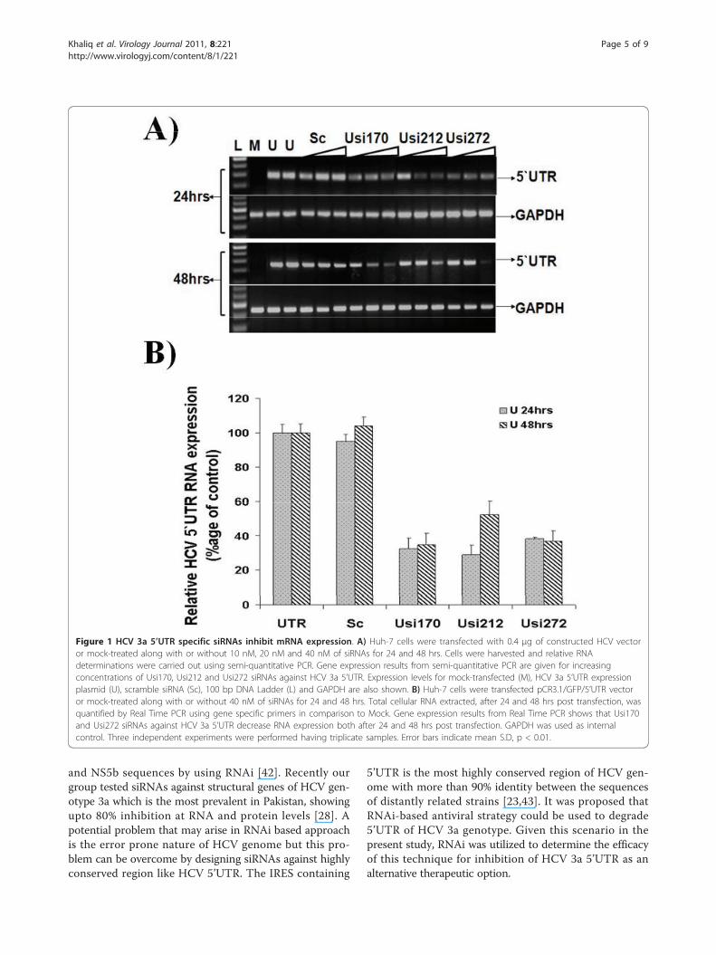
been a focus of antiviral research, therefore in the pre-sent study we designed and tested siRNAs targeting5’UTR of HCV-3a to evaluate their antiviral activityagainst important domains for translation initiation. Totest the HCV 3a directed siRNAs for their ability to sup-press the well conserved HCV IRES (5’UTR) mediatedtranslation, three siRNAs were designed to trigger RNAiand co-transfected with constructed vector into Huh-7cells in different concentrations. Huh-7 cells were trans-fected with pCR3.1/GFP/5’UTR, that expresses GFPunder the control of HCV 5’UTR, with and without5’UTR specific siRNAs. All siRNAs inhibited the expres-sion of GFP gene in a dose dependent manner (10, 20and 40 nM) compared with control siRNA. The inhibi-tory effect of Usi170 siRNA against 5’UTR is stronger at24 hrs and 48 hrs post transfection even at low concen-tration, while Usi212 and Usi272 shows more effectafter 24 and 48 hrs transfection at 40 nM comparedwith scramble siRNA which showed no significantchange in expression of transfected vector to controlcells. In all the siRNA screening experiments, treatmentwith synthetic and control siRNA did not affect thelevels of cellular gene GAPDH expression (Figure 1A).The results of relative quantitative analysis revealed thatthe transcript levels of the HCV 5’UTR were decreasedto 67% in treated cells with Usi170, 71% with Usi212and 62% with Usi272 at 24 hrs post-transfection, while65% with Usi170, 48% with Usi212 and 36.7% withUsi272 at 48 hrs post-transfection. A maximum inhibi-tion of 60-70% was observed with Usi170. GAPDH tran-script levels showed no change in non-transfected ortransfected cells (Figure 1B).At 24 and 48 hrs post-transfection, GFP expression in
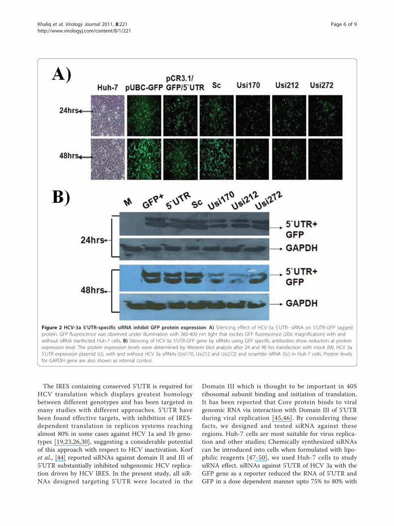
Huh-7 cells was also photographed under a fluorescentmicroscope; the results suggested that GFP fluorescencewas decreased in siRNA transfected cells as compared topUbC-GFP as a control vector, +ve control withoutsiRNA and scrambled siRNAs transfected cells. To deter-mine whether changes in GFP fluorescence accuratelyreflect underlying changes in GFP mRNA presence as aresult of RNAi triggered siRNAs, the protein extractsderived from the siRNA transfected cells were tested forthe expression of GAPDH and found to be similar in allHuh-7 cells, non-transfected and transfected with HCVplasmid. Interestingly, western blotting showed that GFPprotein expression was efficiently inhibited by 65% incells co-transfected with pCR3.1/GFP/5’UTR, and siR-NAs targeting 5’UTR, but not in the cells co-transfectedwith positive vector and scramble siRNA after 24 and48 hrs transfection. Moreover, Usi212 was found to bemore effective than Usi170 and Usi272 (Figure 2). Thisdata suggest that siRNA not only has a negative effect onHCV-RNA but also it could decrease protein productionunder the control of 5’UTR.
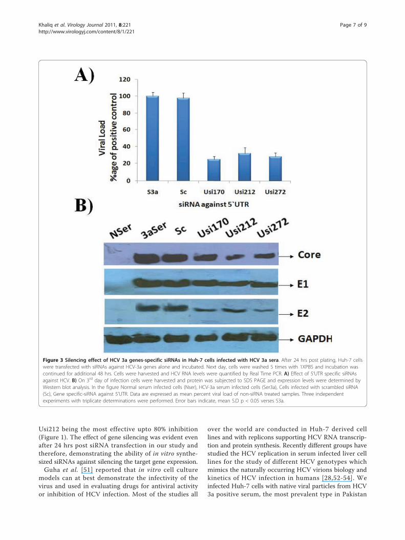
Effect of siRNAs against HCV virus in serum infected Huh-7 cellsThe present study was undertaken to design and testsiRNA as an alternative therapy against HCV, the resultsagainst constructed vector indicate that siRNA targetingHCV 5’UTR has the ability to inhibit the replication ofHCV RNA and protein in Huh-7 cells. Taken advantageof the newly developed cell culture system of HCVinfection in liver cells [27,28,41], the effect of siRNAsagainst whole virus was evaluated by treating cells withsiRNA. Huh-7 serum infected cells were treated with5’UTR siRNAs and subsequently incubated for 3 days.Total cellular RNA was extracted from Huh-7 cells andReal Time PCR was performed with 5’UTR specific pri-mers to analyze the down regulation of HCV RNA bygene specific siRNAs. Maximal inhibition of HCV tran-script levels was detected on Day 3 post transfection,the results show an approximate 70-80% decrease inHCV RNA levels in HCV serum infected Huh-7 cellsthat were incubated with different gene specific-siRNAs.This result was in accordance with Zekri et al., [27]result who also showed that best inhibitory effect of siR-NAs against 5’UTR on 3rd day of post transfection. Nosignificant inhibition was detected in cells transfectedwith the negative control siRNA. siRNAs targeting IREScontaining 5’UTR showed upto 80% inhibition of viralload. Taken together a significant level of HCV genotype3a viral RNA suppression by siRNA against 5’UTR wasobserved and these data suggest a negative impact ofgene-specific siRNAs on HCV replication (Figure 3A).Moreover, siRNAs effect on HCV proteins (Core, E1,and E2) in serum infected Huh-7 cells was detectedusing gene specific antibodies. Densitometric analysisrevealed no significant inhibition at protein levels incells transfected with the negative control siRNA(scrambled siRNA). Two siRNAs targeting IRES con-taining 5’UTR (Usi170 and Usi272) showed upto 60-70%inhibition of viral proteins expression especially E2 andupto 40% inhibition of Core protein expression (Figure 3B).Together, the data suggest a negative impact of chemicallysynthesized siRNA against 5’UTR on the down regulationof HCV genes activity as a target for preventing HCVinduced HCC development. These results are in accor-dance with our previous data in which Core and envelopegenes were targeted by siRNAs which had a inhibitoryeffect on total viral titre [28].
DiscussionAs a gene silencing mechanism, RNAi represents an excit-ing technology with potential applications for treatment ofviral diseases such as HCV. Several laboratories havedemonstrated that RNAi efficiently inhibits viral replica-tion and expression of HCV genotype 1a and 1b usingreplicon cell lines by targeted 5’UTR, Core, E2, NS3, NS4b
Khaliq et al. Virology Journal 2011, 8:221http://www.virologyj.com/content/8/1/221
Page 4 of 9
and NS5b sequences by using RNAi [42]. Recently ourgroup tested siRNAs against structural genes of HCV gen-otype 3a which is the most prevalent in Pakistan, showingupto 80% inhibition at RNA and protein levels [28]. Apotential problem that may arise in RNAi based approachis the error prone nature of HCV genome but this pro-blem can be overcome by designing siRNAs against highlyconserved region like HCV 5’UTR. The IRES containing
5’UTR is the most highly conserved region of HCV gen-ome with more than 90% identity between the sequencesof distantly related strains [23,43]. It was proposed thatRNAi-based antiviral strategy could be used to degrade5’UTR of HCV 3a genotype. Given this scenario in thepresent study, RNAi was utilized to determine the efficacyof this technique for inhibition of HCV 3a 5’UTR as analternative therapeutic option.
Figure 1 HCV 3a 5’UTR specific siRNAs inhibit mRNA expression. A) Huh-7 cells were transfected with 0.4 μg of constructed HCV vectoror mock-treated along with or without 10 nM, 20 nM and 40 nM of siRNAs for 24 and 48 hrs. Cells were harvested and relative RNAdeterminations were carried out using semi-quantitative PCR. Gene expression results from semi-quantitative PCR are given for increasingconcentrations of Usi170, Usi212 and Usi272 siRNAs against HCV 3a 5’UTR. Expression levels for mock-transfected (M), HCV 3a 5’UTR expressionplasmid (U), scramble siRNA (Sc), 100 bp DNA Ladder (L) and GAPDH are also shown. B) Huh-7 cells were transfected pCR3.1/GFP/5’UTR vectoror mock-treated along with or without 40 nM of siRNAs for 24 and 48 hrs. Total cellular RNA extracted, after 24 and 48 hrs post transfection, wasquantified by Real Time PCR using gene specific primers in comparison to Mock. Gene expression results from Real Time PCR shows that Usi170and Usi272 siRNAs against HCV 3a 5’UTR decrease RNA expression both after 24 and 48 hrs post transfection. GAPDH was used as internalcontrol. Three independent experiments were performed having triplicate samples. Error bars indicate mean S.D, p < 0.01.
Khaliq et al. Virology Journal 2011, 8:221http://www.virologyj.com/content/8/1/221
Page 5 of 9
The IRES containing conserved 5’UTR is required forHCV translation which displays greatest homologybetween different genotypes and has been targeted inmany studies with different approaches. 5’UTR havebeen found effective targets, with inhibition of IRES-dependent translation in replicon systems reachingalmost 80% in some cases against HCV 1a and 1b geno-types [19,23,26,30], suggesting a considerable potentialof this approach with respect to HCV inactivation. Korfet al., [44] reported siRNAs against domain II and III of5’UTR substantially inhibited subgenomic HCV replica-tion driven by HCV IRES. In the present study, all siR-NAs designed targeting 5’UTR were located in the
Domain III which is thought to be important in 40Sribosomal subunit binding and initiation of translation.It has been reported that Core protein binds to viralgenomic RNA via interaction with Domain III of 5’UTRduring viral replication [45,46]. By considering thesefacts, we designed and tested siRNA against theseregions. Huh-7 cells are most suitable for virus replica-tion and other studies; Chemically synthesized siRNAscan be introduced into cells when formulated with lipo-philic reagents [47-50], we used Huh-7 cells to studysiRNA effect. siRNAs against 5’UTR of HCV 3a with theGFP gene as a reporter reduced the RNA of 5’UTR andGFP in a dose dependent manner upto 75% to 80% with
Figure 2 HCV-3a 5’UTR-specific siRNA inhibit GFP protein expression. A) Silencing effect of HCV-3a 5’UTR- siRNA on 5’UTR-GFP taggedprotein. GFP fluorescence was observed under illumination with 360-400 nm light that excites GFP fluorescence (20× magnification) with andwithout siRNA tranfected Huh-7 cells. B) Silencing of HCV-3a 5’UTR-GFP gene by siRNAs using GFP specific antibodies show reduction at proteinexpression level. The protein expression levels were determined by Western blot analysis after 24 and 48 hrs transfection with mock (M), HCV 3a5’UTR expression plasmid (U), with and without HCV 3a siRNAs (Usi170, Usi212 and Usi272) and scramble siRNA (Sc) in Huh-7 cells. Protein levelsfor GAPDH gene are also shown as internal control.
Khaliq et al. Virology Journal 2011, 8:221http://www.virologyj.com/content/8/1/221
Page 6 of 9
Usi212 being the most effective upto 80% inhibition(Figure 1). The effect of gene silencing was evident evenafter 24 hrs post siRNA transfection in our study andtherefore, demonstrating the ability of in vitro synthe-sized siRNAs against silencing the target gene expression.Guha et al. [51] reported that in vitro cell culture
models can at best demonstrate the infectivity of thevirus and used in evaluating drugs for antiviral activityor inhibition of HCV infection. Most of the studies all
over the world are conducted in Huh-7 derived celllines and with replicons supporting HCV RNA transcrip-tion and protein synthesis. Recently different groups havestudied the HCV replication in serum infected liver celllines for the study of different HCV genotypes whichmimics the naturally occurring HCV virions biology andkinetics of HCV infection in humans [28,52-54]. Weinfected Huh-7 cells with native viral particles from HCV3a positive serum, the most prevalent type in Pakistan
Figure 3 Silencing effect of HCV 3a genes-specific siRNAs in Huh-7 cells infected with HCV 3a sera. After 24 hrs post plating, Huh-7 cellswere transfected with siRNAs against HCV-3a genes alone and incubated. Next day, cells were washed 5 times with 1XPBS and incubation wascontinued for additional 48 hrs. Cells were harvested and HCV RNA levels were quantified by Real Time PCR. A) Effect of 5’UTR specific siRNAsagainst HCV. B) On 3rd day of infection cells were harvested and protein was subjected to SDS PAGE and expression levels were determined byWestern blot analysis. In the figure Normal serum infected cells (Nser), HCV-3a serum infected cells (Ser3a), Cells infected with scrambled siRNA(Sc), Gene specific-siRNA against 5’UTR. Data are expressed as mean percent viral load of non-siRNA treated samples. Three independentexperiments with triplicate determinations were performed. Error bars indicate, mean S.D p < 0.05 verses S3a.
Khaliq et al. Virology Journal 2011, 8:221http://www.virologyj.com/content/8/1/221
Page 7 of 9
using the same protocol as established [27,28]. The siR-NAs against 5’UTR in the present study were furtherscreened against HCV in serum infected Huh-7 cells. Anexciting finding of this study is decline of HCV viral titerto a maximum of 80% with gene specific siRNAs followedby reduction in HCV protein expression. HCV replicationin the Huh-7 cells was observed through detection of5’UTR of viral copies by Real Time PCR in cells from 3rd
day post infection. Bian et al. [55] reported that 14 aminoacids from the C-terminus of Core gene are required forproper function of E1 and at least 12 amino acids from C-terminus of E1 genes are required for E2 function, influen-cing the proper glycosylation of E1 and E2 gene. The siR-NAs in the present study which showed reduction in viraltiter were also tested for effect on HCV proteins expres-sion as they are translated in a form of single polypeptideand found to be equally effective in HCV protein expres-sion, while the effect was much greater against E2 proteinexpression by all siRNAs (Figure 3). This effect may be asa result of 5’UTR mediated translation inhibition and alsodue to the inability of interaction between 5’UTR domainIII and Core during virion assembly. Our data is in agree-ment with Zekri et al. [27], who demonstrated that siR-NAs against 5’UTR of HCV genotype-4 inhibited HCVreplication in serum infected Huh-7 cells and Khaliq et al.[28] who tested siRNAs against HCV 3a in serum infectedcells and observed the down regulation of HCV RNA.Treatment of siRNAs revealed significant inhibitory effecton HCV copy number indicating the potential of siRNAsas an efficient therapeutic strategy.Our results demonstrate that siRNA targeting HCV
5’UTR can elicit viral RNA from infected cell and poten-tially offer an efficient therapeutic option for HCV infec-tion. These results are in agreement with the previousstudies which suggested that siRNA is the most efficientnucleic acid based antiviral approach that can be utilizedto degrade HCV genome in the infected cells. Moreover,based on these results it is suggested that RNAi-mediatedsilencing of the local HCV-3a 5’UTR may be one of theimportant therapeutic opportunities against HCV-3a gen-otype. Recently Pan et al. [56] reported that RNAi (against5’UTR) can be applied in combination with IFNa withouteffecting signal transduction to hepatocytes or interferingusing RNAi which significantly enhanced their individualantiviral effects. Therefore, it can also be speculated fromour pilot study that therapeutic induction of RNAi againstHCV-3a 5’UTR either alone or in combination with IFNtreatment might represent an alternative approach forfuture treatment of chronic infection.
List of abbreviations5’UTR: 5’ untranslated region; IRES: internal ribosome entry site; HCC:Hepatocellular carcinoma; HCV: Hepatitis C virus; PEG-INF-α: pegylatedinterferon alpha; RNAi: RNA interference; siRNAs: small interfering RNAs
AcknowledgementsFinancial support by Higher Education Commission and all facilities providedby CEMB/CAMB are highly acknowledged.
Authors’ contributionsSK, SJ and AP perform lab work and prepared the manuscript. UAA helpedSK in lab work and literature review. SK and SJ write and critically reviewedthe manuscript. SH provides all facilitates to complete this work. All authorsread and approved final manuscript.
Authors’ informationSaba Khaliq (PhD Molecular Biology), Shah Jahan (PhD Molecular Biology),Asim Pervaiz (M.Phil Molecular Biology), Usman Ali Ashfaq (PhD MolecularBiology), and Sajida Hassan (PhD Molecular Biology) at CEMB, University ofthe Punjab, Lahore.
Competing interestsThe authors declare that they have no competing interests.
Received: 12 March 2011 Accepted: 13 May 2011Published: 13 May 2011
References1. Giannini C, Brechot C: Hepatitis C virus biology. Cell Death Differ 2003,
10(Suppl 1):S27-S38.2. Parker SP, Khan HI, Cubitt WD: Detection of antibodies to hepatitis C virus
in dried blood spot samples from mothers and their offspring in Lahore,Pakistan. J Clin Microbiol 1999, 37:2061-2063.
3. Alter MJ: Epidemiology of hepatitis C. Hepatology 1997, 26:62S-65S.4. Hoofnagle JH: Course and outcome of hepatitis C. Hepatology 2002, 36:
S21-S29.5. McHutchison JG, Fried MW: Current therapy for hepatitis C: pegylated
interferon and ribavirin. Clin Liver Dis 2003, 7:149-161.6. Khakoo S, Glue P, Grellier L, Wells B, Bell A, Dash C, Murray-Lyon I,
Lypnyj D, Flannery B, Walters K, Dusheiko GM: Ribavirin and interferonalfa-2b in chronic hepatitis C: assessment of possible pharmacokineticand pharmacodynamic interactions. Br J Clin Pharmacol 1998,46:563-570.
7. Russo MW, Fried MW: Side effects of therapy for chronic hepatitis C.Gastroenterology 2003, 124:1711-1719.
8. Idrees M, Riazuddin S: Frequency distribution of hepatitis C virusgenotypes in different geographical regions of Pakistan and theirpossible routes of transmission. BMC Infect Dis 2008, 8:69.
9. Idrees M, Rafique S, Rehman I, Akbar H, Yousaf MZ, Butt S, Awan Z,Manzoor S, Akram M, Aftab M, Khubaib B, Riazuddin S: Hepatitis C virusgenotype 3a infection and hepatocellular carcinoma: Pakistanexperience. World J Gastroenterol 2009, 15:5080-5085.
10. Cullen BR: RNA interference: antiviral defense and genetic tool. NatImmunol 2002, 3:597-599.
11. Elbashir SM, Harborth J, Lendeckel W, Yalcin A, Weber K, Tuschl T: Duplexesof 21-nucleotide RNAs mediate RNA interference in cultured mammaliancells. Nature 2001, 411:494-498.
12. Elbashir SM, Lendeckel W, Tuschl T: RNA interference is mediated by 21-and 22-nucleotide RNAs. Genes Dev 2001, 15:188-200.
13. Fire A, Xu S, Montgomery MK, Kostas SA, Driver SE, Mello CC: Potent andspecific genetic interference by double-stranded RNA in Caenorhabditiselegans. Nature 1998, 391:806-811.
14. Hannon GJ: RNA interference. Nature 2002, 418:244-251.15. Paddison PJ, Hannon GJ: RNA interference: the new somatic cell
genetics? Cancer Cell 2002, 2:17-23.16. Sharp PA: RNA interference–2001. Genes Dev 2001, 15:485-490.17. Chevalier C, Saulnier A, Benureau Y, Flechet D, Delgrange D, Colbere-
Garapin F, Wychowski C, Martin A: Inhibition of hepatitis C virus infectionin cell culture by small interfering RNAs. Mol Ther 2007, 15:1452-1462.
18. Hamazaki H, Ujino S, Miyano-Kurosaki N, Shimotohno K, Takaku H:Inhibition of hepatitis C virus RNA replication by short hairpin RNAsynthesized by T7 RNA polymerase in hepatitis C virus subgenomicreplicons. Biochem Biophys Res Commun 2006, 343:988-994.
19. Kanda T, Steele R, Ray R, Ray RB: Small interfering RNA targeted tohepatitis C virus 5’ nontranslated region exerts potent antiviral effect. JVirol 2007, 81:669-676.
Khaliq et al. Virology Journal 2011, 8:221http://www.virologyj.com/content/8/1/221
Page 8 of 9

20. Kapadia SB, Brideau-Andersen A, Chisari FV: Interference of hepatitis Cvirus RNA replication by short interfering RNAs. Proc Natl Acad Sci USA2003, 100:2014-2018.
21. Kim M, Shin D, Kim SI, Park M: Inhibition of hepatitis C virus geneexpression by small interfering RNAs using a tri-cistronic full-length viralreplicon and a transient mouse model. Virus Res 2006, 122:1-10.
22. Korf M, Meyer A, Jarczak D, Beger C, Manns MP, Kruger M: Inhibition ofHCV subgenomic replicons by siRNAs derived from plasmids withopposing U6 and H1 promoters. J Viral Hepat 2007, 14:122-132.
23. Kronke J, Kittler R, Buchholz F, Windisch MP, Pietschmann T,Bartenschlager R, Frese M: Alternative approaches for efficient inhibitionof hepatitis C virus RNA replication by small interfering RNAs. J Virol2004, 78:3436-3446.
24. Prabhu R, Vittal P, Yin Q, Flemington E, Garry R, Robichaux WH, Dash S:Small interfering RNA effectively inhibits protein expression andnegative strand RNA synthesis from a full-length hepatitis C virus clone.J Med Virol 2005, 76:511-519.
25. Watanabe T, Sudoh M, Miyagishi M, Akashi H, Arai M, Inoue K, Taira K,Yoshiba M, Kohara M: Intracellular-diced dsRNA has enhanced efficacy forsilencing HCV RNA and overcomes variation in the viral genotype. GeneTher 2006, 13:883-892.
26. Yokota T, Sakamoto N, Enomoto N, Tanabe Y, Miyagishi M, Maekawa S, Yi L,Kurosaki M, Taira K, Watanabe M, Mizusawa H: Inhibition of intracellularhepatitis C virus replication by synthetic and vector-derived smallinterfering RNAs. EMBO Rep 2003, 4:602-608.
27. Zekri AR, Bahnassy AA, El-Din HM, Salama HM: Consensus siRNA forinhibition of HCV genotype-4 replication. Virol J 2009, 6:13.
28. Khaliq S, Jahan S, Ijaz B, Ahmad W, Asad S, Pervaiz A, Samreen B, Khan M,Hassan S: Inhibition of core gene of HCV 3a genotype using syntheticand vector derived siRNAs. Virol J 2010, 7:318.
29. Khaliq S, Jahan S, Ijaz B, Ahmad W, Asad S, Hassan S: Inhibition of hepatitisC virus genotype 3a by siRNAs targeting envelope genes. Arch Virol 2011,156:433-442.
30. Seo MY, Abrignani S, Houghton M, Han JH: Small interfering RNA-mediated inhibition of hepatitis C virus replication in the humanhepatoma cell line Huh-7. J Virol 2003, 77:810-812.
31. Choo QL, Kuo G, Weiner AJ, Overby LR, Bradley DW, Houghton M: Isolationof a cDNA clone derived from a blood-borne non-A, non-B viralhepatitis genome. Science 1989, 244:359-362.
32. Reed KE, Rice CM: Overview of hepatitis C virus genome structure,polyprotein processing, and protein properties. Curr Top MicrobiolImmunol 2000, 242:55-84.
33. Penin F, Dubuisson J, Rey FA, Moradpour D, Pawlotsky JM: Structuralbiology of hepatitis C virus. Hepatology 2004, 39:5-19.
34. Spahn CM, Kieft JS, Grassucci RA, Penczek PA, Zhou K, Doudna JA, Frank J:Hepatitis C virus IRES RNA-induced changes in the conformation of the40s ribosomal subunit. Science 2001, 291:1959-1962.
35. Honda M, Beard MR, Ping LH, Lemon SM: A phylogenetically conservedstem-loop structure at the 5’ border of the internal ribosome entry siteof hepatitis C virus is required for cap-independent viral translation. JVirol 1999, 73:1165-1174.
36. Wang C, Sarnow P, Siddiqui A: Translation of human hepatitis C virusRNA in cultured cells is mediated by an internal ribosome-bindingmechanism. J Virol 1993, 67:3338-3344.
37. Friebe P, Lohmann V, Krieger N, Bartenschlager R: Sequences in the 5’nontranslated region of hepatitis C virus required for RNA replication. JVirol 2001, 75:12047-12057.
38. Tsukiyama-Kohara K, Iizuka N, Kohara M, Nomoto A: Internal ribosomeentry site within hepatitis C virus RNA. J Virol 1992, 66:1476-1483.
39. Otto GA, Puglisi JD: The pathway of HCV IRES-mediated translationinitiation. Cell 2004, 119:369-380.
40. Khaliq S, Jahan S, Ijaz B, Ahmad W, Asad S, Hassan S: Inhibition of hepatitisC virus genotype 3a by siRNAs targeting envelope genes. Arch Virol 2011,156:433-442.
41. el-Awady MK, Tabll AA, el-Abd YS, Bahgat MM, Shoeb HA, Youssef SS, Baderel-Din NG, Redwan e, el-Demellawy M, Omran MH, el-Garf WT, Goueli SA:HepG2 cells support viral replication and gene expression of hepatitis Cvirus genotype 4 in vitro. World J Gastroenterol 2006, 12:4836-4842.
42. Khaliq S, Khaliq SA, Zahur M, Ijaz B, Jahan S, Ansar M, Riazuddin S, Hassan S:RNAi as a new therapeutic strategy against HCV. Biotechnol Adv 2010,28:27-34.
43. Bukh J, Purcell RH, Miller RH: Sequence analysis of the 5’ noncodingregion of hepatitis C virus. Proc Natl Acad Sci USA 1992, 89:4942-4946.
44. Korf M, Jarczak D, Beger C, Manns MP, Kruger M: Inhibition of hepatitis Cvirus translation and subgenomic replication by siRNAs directed againsthighly conserved HCV sequence and cellular HCV cofactors. J Hepatol2005, 43:225-234.
45. Tanaka Y, Shimoike T, Ishii K, Suzuki R, Suzuki T, Ushijima H, Matsuura Y,Miyamura T: Selective binding of hepatitis C virus core protein tosynthetic oligonucleotides corresponding to the 5’ untranslated regionof the viral genome. Virology 2000, 270:229-236.
46. Yu KL, Jang SI, You JC: Identification of in vivo interaction betweenHepatitis C Virus core protein and 5’ and 3’ UTR RNA. Virus Res 2009,145:285-292.
47. Lindenbach BD, Evans MJ, Syder AJ, Wolk B, Tellinghuisen TL, Liu CC,Maruyama T, Hynes RO, Burton DR, McKeating JA, Rice CM: Completereplication of hepatitis C virus in cell culture. Science 2005, 309:623-626.
48. Moreira JN, Santos A, Moura V, Pedroso de Lima MC, Simoes S: Non-virallipid-based nanoparticles for targeted cancer systemic gene silencing. JNanosci Nanotechnol 2008, 8:2187-2204.
49. Wakita T, Pietschmann T, Kato T, Date T, Miyamoto M, Zhao Z, Murthy K,Habermann A, Krausslich HG, Mizokami M, Bartenschlager R, Liang TJ:Production of infectious hepatitis C virus in tissue culture from a clonedviral genome. Nat Med 2005, 11:791-796.
50. Zhong J, Gastaminza P, Cheng G, Kapadia S, Kato T, Burton DR, Wieland SF,Uprichard SL, Wakita T, Chisari FV: Robust hepatitis C virus infection invitro. Proc Natl Acad Sci USA 2005, 102:9294-9299.
51. Guha C, Lee SW, Chowdhury NR, Chowdhury JR: Cell culture models andanimal models of viral hepatitis. Part II: hepatitis C. Lab Anim (NY) 2005,34:39-47.
52. Buck M: Direct infection and replication of naturally occurring hepatitis Cvirus genotypes 1, 2, 3 and 4 in normal human hepatocyte cultures.PLoS One 2008, 3:e2660.
53. Lazaro CA, Chang M, Tang W, Campbell J, Sullivan DG, Gretch DR, Corey L,Coombs RW, Fausto N: Hepatitis C virus replication in transfected andserum-infected cultured human fetal hepatocytes. Am J Pathol 2007,170:478-489.
54. Molina S, Castet V, Pichard-Garcia L, Wychowski C, Meurs E, Pascussi JM,Sureau C, Fabre JM, Sacunha A, Larrey D, Dubuisson J, Coste J, McKeating J,Maurel P, Fournier-Wirth C: Serum-derived hepatitis C virus infection ofprimary human hepatocytes is tetraspanin CD81 dependent. J Virol 2008,82:569-574.
55. Bian T, Zhou Y, Bi S, Tan W, Wang Y: HCV envelope protein function isdependent on the peptides preceding the glycoproteins. BiochemBiophys Res Commun 2009, 378:118-122.
56. Pan Q, Henry SD, Metselaar HJ, Scholte B, Kwekkeboom J, Tilanus HW,Janssen HL, van der Laan LJ: Combined antiviral activity of interferon-alpha and RNA interference directed against hepatitis C withoutaffecting vector delivery and gene silencing. J Mol Med 2009, 87:713-722.
doi:10.1186/1743-422X-8-221Cite this article as: Khaliq et al.: Down-regulation of IRES containing5’UTR of HCV genotype 3a using siRNAs. Virology Journal 2011 8:221.
Submit your next manuscript to BioMed Centraland take full advantage of:
• Convenient online submission
• Thorough peer review
• No space constraints or color figure charges
• Immediate publication on acceptance
• Inclusion in PubMed, CAS, Scopus and Google Scholar
• Research which is freely available for redistribution
Submit your manuscript at www.biomedcentral.com/submit
Khaliq et al. Virology Journal 2011, 8:221http://www.virologyj.com/content/8/1/221
Page 9 of 9
Copyright © 2022 FDOKUMEN