Membrane Remodeling Induced by the Dynamin-Related Protein Drp1 Stimulates Bax Oligomerization
Upload
independentCategory
view
3download
0
Molecular Biology of the CellVol. 21, 1225–1236, April 1, 2010
Growth Factor erv1-like Modulates Drp1 to PreserveMitochondrial Dynamics and Function in MouseEmbryonic Stem CellsLance R. Todd,* Matthew N. Damin,* Rohini Gomathinayagam,* Sarah R. Horn,†Anthony R. Means,† and Uma Sankar*‡
*James Graham Brown Cancer Center and Owensboro Cancer Research Program, ‡Department ofPharmacology and Toxicology, University of Louisville, Owensboro, KY 42303; and †Department ofPharmacology and Cancer Biology, Duke University Medical Center, Durham, NC 27707
Submitted November 6, 2009; Revised January 26, 2010; Accepted February 1, 2010Monitoring Editor: Donald D. Newmeyer
The relationship of mitochondrial dynamics and function to pluripotency are rather poorly understood aspects of stemcell biology. Here we show that growth factor erv1-like (Gfer) is involved in preserving mouse embryonic stem cell (ESC)mitochondrial morphology and function. Knockdown (KD) of Gfer in ESCs leads to decreased pluripotency markerexpression, embryoid body (EB) formation, cell survival, and loss of mitochondrial function. Mitochondria in Gfer-KDESCs undergo excessive fragmentation and mitophagy, whereas those in ESCs overexpressing Gfer appear elongated.Levels of the mitochondrial fission GTPase dynamin-related protein 1 (Drp1) are highly elevated in Gfer-KD ESCs anddecreased in Gfer-overexpressing cells. Treatment with a specific inhibitor of Drp1 rescues mitochondrial function andapoptosis, whereas expression of Drp1-dominant negative resulted in the restoration of pluripotency marker expressionin Gfer-KD ESCs. Altogether, our data reveal a novel prosurvival role for Gfer in maintaining mitochondrial fission–fusion dynamics in pluripotent ESCs.
INTRODUCTION
Pluripotent embryonic stem cells (ESCs) are remarkable intheir ability to undergo unlimited proliferation and self-renewal. Consequently, the capacity to proliferate indefi-nitely in culture in an undifferentiated state confer ESCswith considerable therapeutic potential, especially in thetreatment of spinal cord injury and of degenerative diseasessuch as type 1 diabetes and Parkinson’s and Alzheimer’sdiseases. To preserve their pluripotency, ESCs in prolongedculture conditions must be protected from genomic, epige-netic, oxidative, or mitochondrial damage (Zeng and Rao,2007). There are significant gaps in our knowledge regardinggenes that execute the fundamental task of maintaining ESCstability and consequentially preserving their pluripotencyin culture. A comprehensive understanding of the mecha-nisms by which ESCs maintain their pluripotency is there-fore crucial to realize their full potential in therapeutic re-generative medicine.
Growth factor erv1-like (Gfer), or augmenter of liver re-generation, is the mammalian homologue of the evolution-arily conserved yeast erv1 protein, an FAD-dependent sulf-hydryl oxidase predominantly located in the intermembrane
space (IMS) of mitochondria (Lisowsky et al., 2001). In ad-dition to mitochondrial IMS, Gfer is also present in thecytoplasm and nucleus, and a nonmitochondrial function isassociated with its role in spermatogenesis (Klissenbauer etal., 2002). Although its precise function is unknown, Gferplays roles in cytosolic Fe-S cluster assembly and mitochon-drial biogenesis (Polimeno et al., 2000; Lange et al., 2001;Gatzidou et al., 2006). In yeast, deletion of erv1 is lethal withthe mutants displaying aberrant mitochondrial morphologyand defective biogenesis of cytoplasmic Fe-S clusters (Becheret al., 1999; Lange et al., 2001; Lisowsky et al., 2001). More-over, Gfer is an important component of a disulfide redoxrelay system that mediates the import of proteins to the IMSof the mitochondria (Mesecke et al., 2005).
Gfer was identified as one of 216 (Ramalho-Santos et al.,2002) or 283 (Ivanova et al., 2002) common genes that areenriched in ESCs, neuronal and hematopoietic stem cells(SCs). Gfer maps within the t-haplotype region (Silver, 1993)of the SC gene-enriched mouse chromosome 17 (a regionsyntenic with the proximal end of the short arm of humanchromosome 16; Polimeno et al., 1999), suggesting an evolu-tionary clustering of genes important in SC biology. Gfer hasroles in the regeneration of mammalian liver, pancreas andDrosophila leg imaginal discs, and its expression correlateswith early spermatogenesis (Klissenbauer et al., 2002; Klebeset al., 2005; Gatzidou et al., 2006).
As no role has been established for Gfer in any stem cell,we used gene depletion and overexpression approaches toevaluate its role in the most primitive of SCs, the pluripotentESC. Here we show that down-regulation of Gfer in mouseESCs results in significantly reduced pluripotency markergene expression, EB formation, and cell survival. Depletionof Gfer also resulted in loss of ESC mitochondrial membrane
This article was published online ahead of print in MBoC in Press(http://www.molbiolcell.org/cgi/doi/10.1091/mbc.E09–11–0937)on February 10, 2010.
Address correspondence to: Uma Sankar ([email protected]).
Abbreviations used: SC, stem cell; Drp1, Dynamin-related protein 1;EB, embryoid body; ESC, embryonic stem cell; Gfer, growth factorerv1-like; IMS, intermembrane space; KD, knockdown; MEF, mouseembryonic fibroblast; ��m, mitochondrial membrane potential.
© 2010 by The American Society for Cell Biology 1225
potential (��m), fragmentation of mitochondria, and auto-phagy of the damaged mitochondria (mitophagy). Conversely,mitochondria in ESCs overexpressing Gfer appeared signifi-cantly elongated, with well-defined cristae. Interestingly,levels of the mitochondrial fission GTPase Drp1 were highlyelevated in Gfer-KD cells and decreased in Gfer-overex-pressing cells, indicating that the enhanced Drp1 levels inGfer-KD ESCs may be responsible for increased mitochon-drial fragmentation, loss of ��m, and apoptosis. Consistentwith this idea, treatment with mdivi-1, a specific small mol-ecule inhibitor of Drp1 (Cassidy-Stone et al., 2008) or ectopicexpression of a dominant negative K38A mutant of Drp1(Drp1DN) that inhibits GTP binding (Smirnova et al., 2001)rescued mitochondrial dysfunction, apoptosis, and pluripo-tency in Gfer-KD ESCs. A selective role for Gfer in SCs wascorroborated by our observation that depletion of Gfer fromdifferentiated cells such as primary mouse embryonic fibro-blasts (MEFs) did not affect mitochondrial morphology,function, or cell survival. Thus, during homeostasis, Gfermodulates the levels of Drp1 to preserve mouse ESC mito-chondrial morphology and function and maintain pluripo-tency marker expression in these primitive cells.
MATERIALS AND METHODS
Cell Culture and ReagentsMitomycin C and rotenone were purchased from Sigma (St. Louis, MO).Primary C57BL/6 (B6) MEFs were from ATCC (Manassas, VA) and NIH 3T3cells were a kind gift from Dr. Geoffrey Clark (James Graham Brown CancerCenter). Primary MEFs were cultured on gelatin (0.1% gelatin in water;Millipore, Billerica, MA)–coated dishes, in knockout DMEM (Invitrogen,Carlsbad, CA) containing 15% ES-screened fetal bovine serum (FBS; HyClone,Logan, UT), 0.1 mM 2-mercaptoethanol (�ME), 0.1 mM nonessential aminoacids (NEAA; all from Millipore), 2 mM l-glutamine (HyClone), and 50 U/mlpenicillin/streptomycin (P/S; Invitrogen). For the production of ESC feedercells, MEFs were treated with 10 �g/ml mitomycin C, in media for 3 h at 37°C.NIH 3T3 cells were grown in DMEM with 10% FCS (Invitrogen), 2 mMl-glutamine, and 50 U/ml P/S. For culture of ESCs, B6 ESCs were plated onfeeder cells on gelatin-coated plates, in the presence of 15% ES-FBS, 0.1 mM�ME, 2 mM l-glutamine, 0.1 mM NEAA, 50 U/ml P/S, and 1000 U/mlleukemia inhibitory factor (LIF)-ESGROW (Millipore; ESC media). For pas-saging, ESCs were harvested by adding 0.25% trypsin-EDTA or TrypLEExpress (Invitrogen). Residual feeder cells were panned by plating the entirecell suspension on regular tissue culture–coated dishes (not gelatin-coated)for 20 min at 37°C. The floating undifferentiated ESCs were harvested andreplated on fresh gelatin-coated dishes, in the presence of feeder cells and ESCmedia, at required densities (0.8 � 106/ml for normal passage). For rotenonetreatment, rotenone was dissolved in dimethyl sulfoxide (DMSO; Sigma) tomake 1 mM stock. ESCs were treated with DMSO or rotenone at 10 �M finalconcentration for 24 h. The cells were then harvested after repeated washingwith 1� phosphate-buffered saline (PBS). For culture of EBs, approximately250,000 ESCs were seeded per well of fibroblast-free Ultra-Low-Attachmentsix-well dishes (Corning) containing ESC medium without LIF.
Virus Infection of ESCsFG12-Lenti-green fluorescent protein (GFP)–short hairpin RNA (shRNA) vi-ruses were produced as described (Qin et al., 2003) using 293T cells. GfershRNA sequences are as follows: Gfer1: 5� GGAACAGCTTCCTTAGCGTT 3�and Gfer2: 5� CCAGGTGCCTCGTACCCTTCA 3�. Nanog shRNA was previ-ously described (Wang et al., 2007). ESCs were infected with lentiviruses at amultiplicity of infection (MOI) of 5:1 with 4 �g/ml Polybrene (Sigma). GFP�
cells were sorted out 72 h after infection, using a FACSAria (BD Biosciences,San Jose, CA). Full-length mouse Gfer cDNA was purchased from OpenBiosystems (Huntsville, AL) and cloned into the murine stem cell virus(MSCV)-internal ribosome entry site (IRES)-GFP system, and viruses wereproduced as described (Kitsos et al., 2005). Freshly passaged ESCs wereinfected with MSCV-IRES-GFP (control) or MSCV-IRES-GFP-Gfer (MSCV-Gfer) viruses at a MOI of 5:1 with 4 �g/ml Polybrene (Sigma). GFP� cellswere sorted out 72 h after infection as mentioned before. For ectopic expres-sion of Drp1DN, the Flag-tagged DRP1 K38A pcDNA3.1 expression plasmidwas used (a generous gift from Dr. Sally Kornbluth, Department of Pharma-cology and Cancer Biology, Duke University Medical Center, Durham, NC).Briefly, GFP� lentivirus infected ESCs were sorted out, transfected with Drp1DN
expression plasmid using the mouse ES Cell Nucleofector Kit (Lonza, Walkers-ville, MD) using the A23 program and immediately were plated on feeder
cells that had been preplated on gelatin-coated eight-well Lab-Tek ChamberSlides (Nalgene Nunc, Rochester, NY).
ESC Proliferation AssayTen thousand ES cells were plated per well of a gelatin-coated 24-well dish inthe presence of feeder cells and ESC media. The cell numbers and viability(using trypan blue) were monitored for several passages using a Vi-CELL(Beckman Coulter, Fullerton, CA). For the GFP-Competition Assay, approx-imately 7500 Lenti-GFP-shRNA virus–infected GFP� ESCs were mixed with2500 GFP-WT ESCs cells (3:1 ratio) and plated in the presence of feeder cellsand ESC media, in gelatin-coated wells. Self-renewal was measured for fivepassages by analyzing for GFP expression.
Immunoassays
Alkaline Phosphatase. ES cells were seeded at 5000 cells/well on gelatin-coated 16-well Lab-Tek Chamber Slides (Nalgene Nunc). Alkaline phospha-tase activity was measured using a kit (Chemicon International, Temecula,CA). For immunocytochemistry of ES cells, the following primary antibodieswere used: Nanog (rabbit polyclonal; Abcam, Cambridge, MA), Oct4 (mousemonoclonal; BD Transduction, Franklin Lakes, NJ), SSEA3 (mouse monoclo-nal; Millipore), and Gfer (11293–1-AP, rabbit polyclonal; PTG Labs, Chicago,IL). Cy-3 secondary antibodies (Jackson ImmunoResearch Laboratories, WestGrove, PA) were used, and cells were mounted using VectaShield MountingMedia with DAPI (Vector Laboratories, Burlingame, CA). Images were cap-tured using the Axio Observer Z1 microscope with ApoTome assembly usinga 63� oil-immersion objective (Carl Zeiss, Thornwood, NY). The signal in-tensities were calculated using the AxioVision 2.0 software. Primary antibod-ies used for immunoblotting were �-actin (clone AC-15, Sigma), Bax (p-19;Santa Cruz Biotechnology, Santa Cruz, CA), caspase-3 (AB1899, Millipore),Gfer (PTG Labs), Beclin-1 (clone 20, BD Transduction); rabbit polyclonalAtg12 (Cell Signaling Technology, Beverly, MA), Lc3B (Cell Signaling), Drp1(clone 8/DLP-1, BD Transduction, or H-300 rabbit polyclonal, Santa Cruz),OPA-1 (rabbit polyclonal, Abcam), Mfn-1 (chicken polyclonal, Novus Biologi-cals, Littleton, CO), and Mfn-2 (M6319, Sigma).
Digital Imaging of Mitochondria. The mammalian expression vectorpDsRed2-Mito (Clontech Laboratories, Mountain View, CA) was introducedinto ESCs using the mouse ES cell nucleofector kit, and the cells wereimmediately plated on feeder cells that were plated on gelatin-coated eight-well Lab-Tek chamber slides. Digital optical sectioning was performed 24 hafter nucleofection using Axio Observer Z1 inverted fluorescence microscopewith ApoTome assembly using a 100� oil-immersion objective (Plan-Neofluar; Carl Zeiss).
Mdivi-1 Treatment. Mdivi-1 (Enzo Life Sciences, Farmingdale, NY) was dis-solved in DMSO at a concentration of 25 mg/ml. Reconstituted mdivi-1 wasadded directly to the cell culture medium at a final concentration of 25 �M,and cells were cultured for 24 h.
Flow Cytometry Assays
Apoptosis Assay. Staining for annexin V/7-AAD was performed using theannexin V-PE apoptosis detection kit I (BD Biosciences). Samples were ana-lyzed using BD FACSAria (BD Biosciences). Analysis of tetramethylrhodam-ine ethyl ester (TMRE) fluorescence was conducted as follows: cells wereloaded with 50 nM TMRE (Molecular Probes, Invitrogen, Eugene, OR) ingrowth media, for 20 min under normal growth conditions. Cells were thenharvested and analyzed for PE fluorescence. The cytochrome c staining assaywas performed as described previously (Waterhouse and Trapani, 2003).Briefly, harvested cells were permeabilized with digitonin (Sigma; 50 �g/mldigitonin in PBS with 100 mM KCl) for 5 min on ice, followed by fixation in4% paraformaldehyde. Cells were incubated overnight at 4°C with 1:200anti-cytochrome c mAb (BD PharMingen) in blocking buffer (3% BSA, 0.05%Saponin (Sigma) in PBS) followed by incubation with a PE-labeled secondaryantibody. The PE-Active caspase-3 Apoptosis kit (BD PharMingen) was usedto measure active caspase-3 levels.
Electron Microscopy. Cell pellets containing 300,000 cells were washed brieflywith a 0.1 M sodium cacodylate buffer and then fixed in 3% glutaraldehyde in0.1 M sodium cacodylate overnight at 4°C. Cells were then postfixed in 1%osmium tetroxide in 0.1 M sodium cacodylate for 1 h. Cells were then brieflyrinsed with 0.1 M sodium cacodylate and then dehydrated using a series ofgraded alcohols. Cell pellets were then embedded in LX-112 epoxy resin(Ladd Research Industries, Burlington, VT), and �8-�m sections were cut onan LKB microtome (LKB Instruments, Gaithersburg, MD). Sections were thenstained using uradyl acetate and lead citrate before viewing on a CM12electron microscope equipped with a digital camera (Phillips Electronic In-struments, Mahwah, NJ).
L. R. Todd et al.
Molecular Biology of the Cell1226
Real-Time RT-PCR Analysis. Total RNA was prepared using RNAqueouskits (Ambion, Austin, TX). cDNA was prepared using High Capacity cDNAReverse Transcription Kit (Applied Biosystems, Foster City, CA). Quantitative(q) real-time RT-PCR was performed on the IQ5 I-Cycler (Bio-Rad Laborato-ries, Hercules CA) using IQ SYBR Green supermix (Bio-Rad). Nanog and Oct4primers were previously listed (Pasini et al., 2007). Other primer sequenceslisted in Supplementary Table 1.
Statistical AnalysisStudent’s t test was used to assess statistical significance, and p � 0.05 wasdeemed significant.
RESULTS
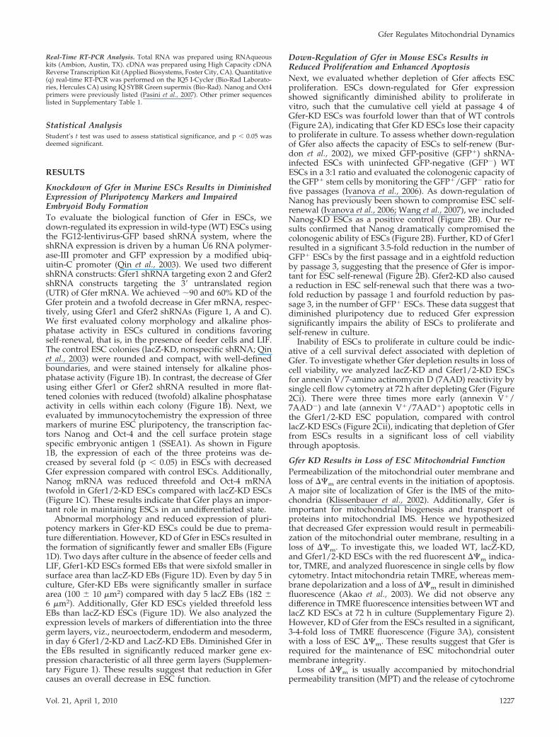
Knockdown of Gfer in Murine ESCs Results in DiminishedExpression of Pluripotency Markers and ImpairedEmbryoid Body FormationTo evaluate the biological function of Gfer in ESCs, wedown-regulated its expression in wild-type (WT) ESCs usingthe FG12-lentivirus-GFP based shRNA system, where theshRNA expression is driven by a human U6 RNA polymer-ase-III promoter and GFP expression by a modified ubiq-uitin-C promoter (Qin et al., 2003). We used two differentshRNA constructs: Gfer1 shRNA targeting exon 2 and Gfer2shRNA constructs targeting the 3� untranslated region(UTR) of Gfer mRNA. We achieved �90 and 60% KD of theGfer protein and a twofold decrease in Gfer mRNA, respec-tively, using Gfer1 and Gfer2 shRNAs (Figure 1, A and C).We first evaluated colony morphology and alkaline phos-phatase activity in ESCs cultured in conditions favoringself-renewal, that is, in the presence of feeder cells and LIF.The control ESC colonies (lacZ-KD, nonspecific shRNA; Qinet al., 2003) were rounded and compact, with well-definedboundaries, and were stained intensely for alkaline phos-phatase activity (Figure 1B). In contrast, the decrease of Gferusing either Gfer1 or Gfer2 shRNA resulted in more flat-tened colonies with reduced (twofold) alkaline phosphataseactivity in cells within each colony (Figure 1B). Next, weevaluated by immunocytochemistry the expression of threemarkers of murine ESC pluripotency, the transcription fac-tors Nanog and Oct-4 and the cell surface protein stagespecific embryonic antigen 1 (SSEA1). As shown in Figure1B, the expression of each of the three proteins was de-creased by several fold (p � 0.05) in ESCs with decreasedGfer expression compared with control ESCs. Additionally,Nanog mRNA was reduced threefold and Oct-4 mRNAtwofold in Gfer1/2-KD ESCs compared with lacZ-KD ESCs(Figure 1C). These results indicate that Gfer plays an impor-tant role in maintaining ESCs in an undifferentiated state.
Abnormal morphology and reduced expression of pluri-potency markers in Gfer-KD ESCs could be due to prema-ture differentiation. However, KD of Gfer in ESCs resulted inthe formation of significantly fewer and smaller EBs (Figure1D). Two days after culture in the absence of feeder cells andLIF, Gfer1-KD ESCs formed EBs that were sixfold smaller insurface area than lacZ-KD EBs (Figure 1D). Even by day 5 inculture, Gfer-KD EBs were significantly smaller in surfacearea (100 � 10 �m2) compared with day 5 lacZ EBs (182 �6 �m2). Additionally, Gfer KD ESCs yielded threefold lessEBs than lacZ-KD ESCs (Figure 1D). We also analyzed theexpression levels of markers of differentiation into the threegerm layers, viz., neuroectoderm, endoderm and mesoderm,in day 6 Gfer1/2-KD and LacZ-KD EBs. Diminished Gfer inthe EBs resulted in significantly reduced marker gene ex-pression characteristic of all three germ layers (Supplemen-tary Figure 1). These results suggest that reduction in Gfercauses an overall decrease in ESC function.
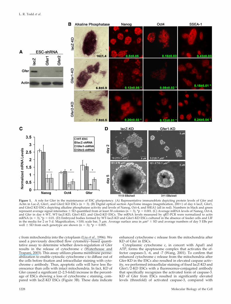
Down-Regulation of Gfer in Mouse ESCs Results inReduced Proliferation and Enhanced ApoptosisNext, we evaluated whether depletion of Gfer affects ESCproliferation. ESCs down-regulated for Gfer expressionshowed significantly diminished ability to proliferate invitro, such that the cumulative cell yield at passage 4 ofGfer-KD ESCs was fourfold lower than that of WT controls(Figure 2A), indicating that Gfer KD ESCs lose their capacityto proliferate in culture. To assess whether down-regulationof Gfer also affects the capacity of ESCs to self-renew (Bur-don et al., 2002), we mixed GFP-positive (GFP�) shRNA-infected ESCs with uninfected GFP-negative (GFP) WTESCs in a 3:1 ratio and evaluated the colonogenic capacity ofthe GFP� stem cells by monitoring the GFP�/GFP ratio forfive passages (Ivanova et al., 2006). As down-regulation ofNanog has previously been shown to compromise ESC self-renewal (Ivanova et al., 2006; Wang et al., 2007), we includedNanog-KD ESCs as a positive control (Figure 2B). Our re-sults confirmed that Nanog dramatically compromised thecolonogenic ability of ESCs (Figure 2B). Further, KD of Gfer1resulted in a significant 3.5-fold reduction in the number ofGFP� ESCs by the first passage and in a eightfold reductionby passage 3, suggesting that the presence of Gfer is impor-tant for ESC self-renewal (Figure 2B). Gfer2-KD also causeda reduction in ESC self-renewal such that there was a two-fold reduction by passage 1 and fourfold reduction by pas-sage 3, in the number of GFP� ESCs. These data suggest thatdiminished pluripotency due to reduced Gfer expressionsignificantly impairs the ability of ESCs to proliferate andself-renew in culture.
Inability of ESCs to proliferate in culture could be indic-ative of a cell survival defect associated with depletion ofGfer. To investigate whether Gfer depletion results in loss ofcell viability, we analyzed lacZ-KD and Gfer1/2-KD ESCsfor annexin V/7-amino actinomycin D (7AAD) reactivity bysingle cell flow cytometry at 72 h after depleting Gfer (Figure2Ci). There were three times more early (annexin V�/7AAD) and late (annexin V�/7AAD�) apoptotic cells inthe Gfer1/2-KD ESC population, compared with controllacZ-KD ESCs (Figure 2Cii), indicating that depletion of Gferfrom ESCs results in a significant loss of cell viabilitythrough apoptosis.
Gfer KD Results in Loss of ESC Mitochondrial FunctionPermeabilization of the mitochondrial outer membrane andloss of ��m are central events in the initiation of apoptosis.A major site of localization of Gfer is the IMS of the mito-chondria (Klissenbauer et al., 2002). Additionally, Gfer isimportant for mitochondrial biogenesis and transport ofproteins into mitochondrial IMS. Hence we hypothesizedthat decreased Gfer expression would result in permeabili-zation of the mitochondrial outer membrane, resulting in aloss of ��m. To investigate this, we loaded WT, lacZ-KD,and Gfer1/2-KD ESCs with the red fluorescent ��m indica-tor, TMRE, and analyzed fluorescence in single cells by flowcytometry. Intact mitochondria retain TMRE, whereas mem-brane depolarization and a loss of ��m result in diminishedfluorescence (Akao et al., 2003). We did not observe anydifference in TMRE fluorescence intensities between WT andlacZ KD ESCs at 72 h in culture (Supplementary Figure 2).However, KD of Gfer from the ESCs resulted in a significant,3-4-fold loss of TMRE fluorescence (Figure 3A), consistentwith a loss of ESC ��m. These results suggest that Gfer isrequired for the maintenance of ESC mitochondrial outermembrane integrity.
Loss of ��m is usually accompanied by mitochondrialpermeability transition (MPT) and the release of cytochrome
Gfer Regulates Mitochondrial Dynamics
Vol. 21, April 1, 2010 1227
c from mitochondria into the cytoplasm (Liu et al., 1996). Weused a previously described flow cytometry–based quanti-tative assay to determine whether down-regulation of Gferresults in the release of cytochrome c (Waterhouse andTrapani, 2003). This assay utilizes plasma membrane perme-abilization to enable cytosolic cytochrome c to diffuse out ofthe cells before fixation and intracellular staining with cyto-chrome c antibody. Thus, apoptotic cells will have less flu-orescence than cells with intact mitochondria. In fact, KD ofGfer caused a significant (2–2.5-fold) increase in the percent-age of ESCs showing a loss of cytochrome c staining, com-pared with lacZ-KD ESCs (Figure 3B). These data indicate
enhanced cytochrome c release from the mitochondria afterKD of Gfer in ESCs.
Cytoplasmic cytochrome c, in concert with Apaf1 andATP, forms the apoptosome complex that activates the ef-fector caspases-3, -6, and -7 (Wang, 2001). To confirm thatenhanced cytochrome c release from the mitochondria afterGfer-KD in the ESCs also resulted in elevated caspase activ-ity, we performed intracellular staining of fixed lacZ-KD andGfer1/2-KD ESCs with a fluorescence-conjugated antibodythat specifically recognizes the activated form of caspase-3.KD of Gfer from ESCs resulted in significantly elevatedlevels (threefold) of activated caspase-3, compared with
Figure 1. A role for Gfer in the maintenance of ESC pluripotency. (A) Representative immunoblots depicting protein levels of Gfer andActin in Lac-Z, Gfer1, and Gfer2 KD ESCs (n 3). (B) Digital optical section ApoTome images (magnification, 200�) of day 6 lacZ, Gfer1,and Gfer2 KD ESCs depicting alkaline phosphatase activity and levels of Nanog, Oct-4, and SSEA1 (all in red). Numbers in black and greenrepresent average signal intensities � SD quantified from at least 30 colonies (n 3), *p 0.001. (C) Average mRNA levels of Nanog, Oct-4,and Gfer in day 6 WT, WT-lacZ-KD, Gfer1-KD, and Gfer2-KD ESCs. The mRNA levels measured by qRT-PCR were normalized to actinmRNA (n 3), *p 0.01. (D) Embryoid bodies formed by WT-lacZ-KD and Gfer1-KD ESCs cultured in the absence of feeder cells and LIFin the media for 2 or 5 d. Magnification, �100; scale bar, 5 �m. Average surface area in �m2 � SD and average numbers of day 5 EBs perwell � SD from each genotype are shown (n 3); *p 0.005.
L. R. Todd et al.
Molecular Biology of the Cell1228
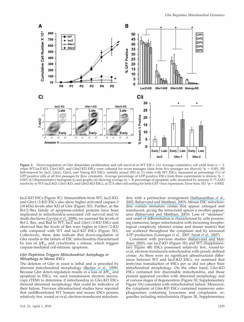
lacZ-KD ESCs (Figure 3C). Immunoblots from WT, lacZ-KD,and Gfer1/2-KD ESCs also show higher activated caspase-3(18 kDa) levels after KD of Gfer (Figure 3D). Further, as theBcl-2/Bax family of apoptosis-related proteins have beenimplicated in mitochondria-associated cell survival and/ordeath decisions (Levine et al., 2008); we assessed the levels ofBcl-2, Bax, and Bad in WT, lacZ and Gfer1/2-KD ESCs andobserved that the levels of Bax were higher in Gfer1/2-KDcells compared with WT and lacZ-KD ESCs (Figure 3D).Collectively, these data indicate that down-regulation ofGfer results in the failure of ESC mitochondria characterizedby loss of ��m and cytochrome c release, which triggerscaspase-mediated cell-intrinsic apoptosis.
Gfer Depletion Triggers Mitochondrial Autophagy orMitophagy in Mouse ESCsThe deletion of Gfer in yeast is lethal and is preceded byaberrant mitochondrial morphology (Becher et al., 1999).Because Gfer down-regulation results in a loss of ��m andapoptosis in ESCs, we used transmission electron micros-copy (TEM) to determine if mitochondria in Gfer-KD ESCsshowed abnormal morphology that could be indicative oftheir failure. Previous ultrastructural studies have reportedthat undifferentiated WT human and mouse ESCs possessrelatively few, round or oval, electron-translucent mitochon-
dria with a perinuclear arrangement (Sathananthan et al.,2002; Baharvand and Matthaei, 2003). Mouse ESC mitochon-dria contain immature cristae that appear enlarged andtranslucent, giving the intracristal spaces a swollen appear-ance (Baharvand and Matthaei, 2003). Loss of “stemness”and onset of differentiation is characterized by cells possess-ing numerous, larger mitochondria with increasing morpho-logical complexity (distinct cristae and denser matrix) thatare scattered throughout the cytoplasm and by increasedATP production (Lonergan et al., 2007; Nesti et al., 2007).
Consistent with previous studies (Baharvand and Mat-thaei, 2003), our lacZ-KD (Figure 3E) and WT (Supplemen-tary Figure 4B) ESCs possessed relatively few, round-to-oval, electron-translucent mitochondria with poorly definedcristae. As there were no significant ultrastructural differ-ences between WT and lacZ-KD ESCs, we surmised thatlentivirus transduction of ESCs per se did not affect theirmitochondrial morphology. On the other hand, Gfer-KDESCs contained few discernable mitochondria, and thosepresent appeared swollen with abnormal morphology andat various stages of degeneration (Figure 3E; SupplementaryFigure 3A) consistent with mitochondrial failure. Moreover,the cytoplasm of Gfer-KD ESCs contained numerous auto-phagosomes containing lysosomes and cytoplasmic or-ganelles including mitochondria (Figure 3E, Supplementary
Figure 2. Down-regulation of Gfer diminishes proliferation and cell survival in WT ESCs. (A) Average cumulative cell yield from n 3,when WT-lacZ-KD, Gfer1-KD, and Gfer2-KD ESCs were cultured for seven passages (data from five passages are shown); *p 0.001. (B)Self-renewal by lacZ, Gfer1, Gfer2, and Nanog KD ESCs, initially mixed (P0) at 3:1 ratio with WT ESCs, measured as percentage (%) ofGFP-positive cells at all five passages by flow cytometry. Average percentage of GFP-positive ESCs from three experiments is shown; *p 0.003. (C) Representative histogram (i) and graphs (ii) showing average (n 4) percentage of apoptotic cells, measured by annexin V/7-AADreactivity in WT-lacZ-KD, Gfer1-KD, and Gfer2-KD ESCs, at 72 h after cell sorting for lenti-GFP virus expression. Error bars, SD; *p 0.0002.
Gfer Regulates Mitochondrial Dynamics
Vol. 21, April 1, 2010 1229
Figure 3A), suggesting that the degenerated mitochondria inthese cells are being eliminated by autophagy/mitophagy.Further, levels of autophagy-associated proteins light chain(LC) 3b, Beclin-1, and Atg5/12 complexes were higher inGfer1-KD and Gfer2-KD ESCs compared with either WT orlacZ-KD ESCs (Supplementary Figure 3B). Taken together,our data reveal that the down-regulation of Gfer in the ESCsresults in mitochondrial dysfunction; the ensuing MPT trig-gers the elimination of damaged mitochondria by mitoph-agy (Elmore et al., 2001), and massive degradation of such anessential organelle initiates apoptosis.
Our observations suggest that one mechanism by whichGfer maintains ESC stemness is by preserving the integrityof their mitochondria. We surmised that any assault to the
very few mitochondria that the ESCs possess would alsotrigger mitophagy in these primitive cells. Complex I, amultisubunit redox machine, is the first enzyme in mito-chondrial electron transport chain (ETC) that plays a centralrole in mitochondrial energy metabolism and can be phar-macologically inhibited by rotenone. Inhibition of ETC com-plex I with 10 �M rotenone for 24 h resulted in enhancedearly (fivefold) and late (twofold) apoptotic populations inWT ESCs, indicating a marked reduction in cell viability(Supplementary Figure 4A). Further, treatment of ESCs withrotenone triggered mitochondrial autophagy or mitophagy,characterized by large autophagosomes containing severalmitochondria (Supplementary Figure 4B). We also observedclassic apoptotic features such as cell fragmentation within
Figure 3. KD of Gfer initiates mitochondrial dysfunction triggering mitophagy in ESCs. (A) Representative histograms depicting TMREfluorescence (PE channel) in lacZ and Gfer1/2 KD ESCs. Numbers in red (lacZ-KD) and green (Gfer1/2-KD) are average (n 3) percentage(%) of cells with TMRE fluorescence intensities within the gated region (dotted box) � SD; *p 0.0001. Mitochondrial membranedepolarization was assessed by loss of TMRE retention/fluorescence. (B) Histographic representation of the assay measuring the release ofcytochrome c in the indicated ESC genotypes. Red (lacZ-KD) and green (Gfer1/2-KD) numbers are average (n 3) percentage of cells withinthe gated region (black line) � SD; *p 0.001, measuring loss of PE-conjugated anti-cytochrome c antibody staining. (C) Percentage of cellsshowing positive staining (gated with a black line) for a PE-conjugated active caspase-3 antibody. Results represent average (n 3)percentage of cells showing positive staining in the PE channel � SD, *p 0.01, for lacZ (red) and Gfer1/2-KD (green). (D) Representativeimmunoblot (n 3) analyses of active caspase-3 and Bax in indicated ESC genotype. Gfer and actin levels are also shown. (E) Digital TEMimages depicting ultrastructural details in lacZ (left) and Gfer-1 KD (right) ESCs. Scale bars, 5 �m at �2650 and 1 �m at �7100 and �25,000magnifications. Black arrowheads, representative autophagosomes; white arrowheads, degenerating mitochondria.
L. R. Todd et al.
Molecular Biology of the Cell1230
the rotenone-treated ESCs (Supplementary Figure 4B). Thus,inhibition of ETC complex I or Gfer down-regulation se-verely damages ESC mitochondria triggering extensive mi-tophagy and apoptosis.
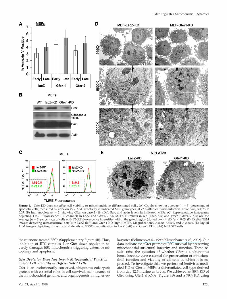
Gfer Depletion Does Not Impair Mitochondrial Functionand/or Cell Viability in Differentiated CellsGfer is an evolutionarily conserved, ubiquitous eukaryoticprotein with essential roles in cell survival, maintenance ofthe mitochondrial genome, and organogenesis in higher eu-
karyotes (Polimeno et al., 1999; Klissenbauer et al., 2002). Ourdata indicate that Gfer promotes ESC survival by preservingmitochondrial structural integrity and function. These re-sults raise the question of whether Gfer is a ubiquitoushouse-keeping gene essential for preservation of mitochon-drial function and viability of all cells in which it is ex-pressed. To investigate this, we performed lentivirus-medi-ated KD of Gfer in MEFs, a differentiated cell type derivedfrom day 12.5 murine embryos. We achieved an 80% KD ofGfer using Gfer1 shRNA (Figure 4B) and a 70% KD using
Figure 4. Gfer KD does not affect cell viability or mitochondria in differentiated cells. (A) Graphs showing average (n 3) percentage ofapoptotic cells, measured by annexin V/7-AAD reactivity in indicated MEF genotypes, at 72 h after lentivirus infection. Error bars, SD; *p �0.05. (B) Immunoblots (n 2) showing Gfer, caspase 3 (18 kDa), Bax, and actin levels in indicated MEFs. (C) Representative histogramsdepicting TMRE fluorescence (PE channel) in LacZ and Gfer1/2 KD MEFs. Numbers in red (LacZ-KD) and green (Gfer1/2-KD) are theaverage (n 3) percentage of cells with TMRE fluorescence intensities within the gated region (dotted box) � SD; *p � 0.05. (D) Digital TEMimages depicting ultrastructural details in LacZ (left) and Gfer-1 KD (right) MEFs. Magnifications, �2650, �5600, and �25,000. (E) DigitalTEM images depicting ultrastructural details at �5600 magnification in LacZ (left) and Gfer-1 KD (right) NIH 3T3 cells.
Gfer Regulates Mitochondrial Dynamics
Vol. 21, April 1, 2010 1231
Gfer2 shRNA (not shown). We then evaluated whether KDof Gfer resulted in a loss of cell viability in MEFs by moni-toring annexin V/7-AAD reactivity. Depletion of Gfer didnot initiate any significant increases in the number ofeither early or late apoptotic cells (Figure 4A). Addition-ally, as shown in Figure 4B, KD of Gfer did not alter levelsof activated form of caspase-3 or the proapoptotic Bax.Furthermore, reduction in Gfer did not alter the ��m inprimary MEFs (Figure 4C). These results indicate thatdown-regulation of Gfer does not initiate apoptosis oraffect mitochondrial function in a differentiated cell typesuch as the MEF.
We also performed TEM studies to determine whetherGfer-KD affected mitochondrial morphology in primaryMEFs. Compared with ESCs, MEFs are larger cells thatcontain numerous mitochondria with well-distinct cristae,rough endoplasmic reticulum (ER), Golgi apparatus, and afew autophagic vesicles (Figure 4D). Although the mito-chondrial cristae in Gfer-KD MEFs appeared slightly swol-len compared with those in LacZ-KD MEFs, there was noalteration in the overall mitochondrial morphology or re-duction in the number of mitochondria in these cells (Figure4D). Additionally, KD of Gfer in NIH 3T3 cells, an immor-talized MEF cell line, did not alter mitochondrial morphol-ogy (Figure 4E) or cell viability (not shown). Collectively,these data suggest that Gfer may largely be dispensable formitochondrial function and/or survival of differentiatedcells.
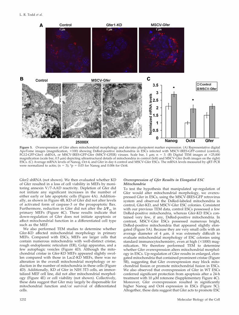
Overexpression of Gfer Results in Elongated ESCMitochondriaTo test the hypothesis that manipulated up-regulation ofGfer would alter mitochondrial morphology, we overex-pressed Gfer in ESCs, using the MSCV-IRES-GFP retrovirussystem and observed the DsRed-labeled mitochondria incontrol, Gfer-KD, and MSCV-Gfer ESC colonies. Consistentwith our previous TEM data, control ESCs possessed a fewDsRed-positive mitochondria, whereas Gfer-KD ESCs con-tained very few, if any, DsRed-positive mitochondria. Incontrast, MSCV-Gfer ESCs possessed numerous bright,DsRed-positive mitochondria that appeared slightly elon-gated (Figure 5A). Because they are very small cells with anaverage diameter of 4 �m, it was extremely difficult toevaluate mitochondrial morphology of ESC colonies usingstandard immunocytochemistry, even at high (�1000) mag-nification. We therefore performed TEM to determinewhether Gfer overexpression alters mitochondrial morphol-ogy in ESCs. Up-regulation of Gfer results in enlarged, elon-gated mitochondria that contained prominent cristae (Figure5B), suggesting that Gfer overexpression may block mito-chondrial fission or promote mitochondrial fusion in ESCs.We also observed that overexpression of Gfer in WT ESCsconferred significant protection from apoptosis after a 24-htreatment with 10 �M rotenone (Supplementary Figure 4C).Moreover, Gfer overexpression resulted in significantlyhigher Nanog and Oct4 expression in ESCs (Figure 5C).Altogether, these data suggest that Gfer acts to promote ESC
Figure 5. Overexpression of Gfer alters mitochondrial morphology and elevates pluripotent marker expression. (A) Representative digitalApoTome images (magnification, �100) showing DsRed-positive mitochondria in ESCs infected with MSCV-IRES-GFP-control (control),FG12-GFP-Gfer1 shRNA, or MSCV-IRES-GFP-Gfer (MSCV-GFER) viruses. Scale bar, 1 �m; n 3. (B) Digital TEM images at �25,000magnification (scale bar, 0.5 �m) depicting ultrastructural details of mitochondria in control (left) and MSCV-Gfer (both images on the right)ESCs. (C) Average mRNA levels of Nanog, Oct-4, and Gfer in day 6 control and MSCV-Gfer ESCs. The mRNA levels measured by qRT-PCRwere normalized to actin; (n 3); *p 0.03 for Nanog and 0.006 for Oct4.
L. R. Todd et al.
Molecular Biology of the Cell1232
pluripotency by preserving the mitochondrial structural in-tegrity and function.
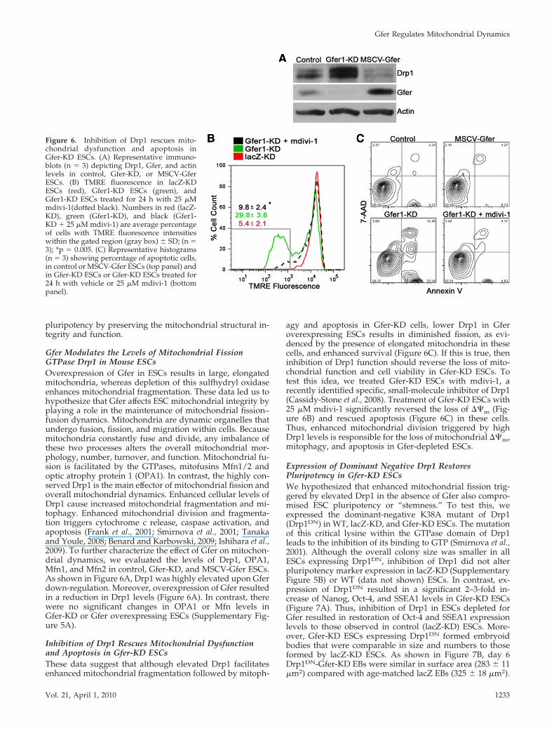
Gfer Modulates the Levels of Mitochondrial FissionGTPase Drp1 in Mouse ESCsOverexpression of Gfer in ESCs results in large, elongatedmitochondria, whereas depletion of this sulfhydryl oxidaseenhances mitochondrial fragmentation. These data led us tohypothesize that Gfer affects ESC mitochondrial integrity byplaying a role in the maintenance of mitochondrial fission–fusion dynamics. Mitochondria are dynamic organelles thatundergo fusion, fission, and migration within cells. Becausemitochondria constantly fuse and divide, any imbalance ofthese two processes alters the overall mitochondrial mor-phology, number, turnover, and function. Mitochondrial fu-sion is facilitated by the GTPases, mitofusins Mfn1/2 andoptic atrophy protein 1 (OPA1). In contrast, the highly con-served Drp1 is the main effector of mitochondrial fission andoverall mitochondrial dynamics. Enhanced cellular levels ofDrp1 cause increased mitochondrial fragmentation and mi-tophagy. Enhanced mitochondrial division and fragmenta-tion triggers cytochrome c release, caspase activation, andapoptosis (Frank et al., 2001; Smirnova et al., 2001; Tanakaand Youle, 2008; Benard and Karbowski, 2009; Ishihara et al.,2009). To further characterize the effect of Gfer on mitochon-drial dynamics, we evaluated the levels of Drp1, OPA1,Mfn1, and Mfn2 in control, Gfer-KD, and MSCV-Gfer ESCs.As shown in Figure 6A, Drp1 was highly elevated upon Gferdown-regulation. Moreover, overexpression of Gfer resultedin a reduction in Drp1 levels (Figure 6A). In contrast, therewere no significant changes in OPA1 or Mfn levels inGfer-KD or Gfer overexpressing ESCs (Supplementary Fig-ure 5A).
Inhibition of Drp1 Rescues Mitochondrial Dysfunctionand Apoptosis in Gfer-KD ESCsThese data suggest that although elevated Drp1 facilitatesenhanced mitochondrial fragmentation followed by mitoph-
agy and apoptosis in Gfer-KD cells, lower Drp1 in Gferoverexpressing ESCs results in diminished fission, as evi-denced by the presence of elongated mitochondria in thesecells, and enhanced survival (Figure 6C). If this is true, theninhibition of Drp1 function should reverse the loss of mito-chondrial function and cell viability in Gfer-KD ESCs. Totest this idea, we treated Gfer-KD ESCs with mdivi-1, arecently identified specific, small-molecule inhibitor of Drp1(Cassidy-Stone et al., 2008). Treatment of Gfer-KD ESCs with25 �M mdivi-1 significantly reversed the loss of ��m (Fig-ure 6B) and rescued apoptosis (Figure 6C) in these cells.Thus, enhanced mitochondrial division triggered by highDrp1 levels is responsible for the loss of mitochondrial ��m,mitophagy, and apoptosis in Gfer-depleted ESCs.
Expression of Dominant Negative Drp1 RestoresPluripotency in Gfer-KD ESCsWe hypothesized that enhanced mitochondrial fission trig-gered by elevated Drp1 in the absence of Gfer also compro-mised ESC pluripotency or “stemness.” To test this, weexpressed the dominant-negative K38A mutant of Drp1(Drp1DN) in WT, lacZ-KD, and Gfer-KD ESCs. The mutationof this critical lysine within the GTPase domain of Drp1leads to the inhibition of its binding to GTP (Smirnova et al.,2001). Although the overall colony size was smaller in allESCs expressing Drp1DN, inhibition of Drp1 did not alterpluripotency marker expression in lacZ-KD (SupplementaryFigure 5B) or WT (data not shown) ESCs. In contrast, ex-pression of Drp1DN resulted in a significant 2–3-fold in-crease of Nanog, Oct-4, and SSEA1 levels in Gfer-KD ESCs(Figure 7A). Thus, inhibition of Drp1 in ESCs depleted forGfer resulted in restoration of Oct-4 and SSEA1 expressionlevels to those observed in control (lacZ-KD) ESCs. More-over, Gfer-KD ESCs expressing Drp1DN formed embryoidbodies that were comparable in size and numbers to thoseformed by lacZ-KD ESCs. As shown in Figure 7B, day 6Drp1DN-Gfer-KD EBs were similar in surface area (283 � 11�m2) compared with age-matched lacZ EBs (325 � 18 �m2).
Figure 6. Inhibition of Drp1 rescues mito-chondrial dysfunction and apoptosis inGfer-KD ESCs. (A) Representative immuno-blots (n 3) depicting Drp1, Gfer, and actinlevels in control, Gfer-KD, or MSCV-GferESCs. (B) TMRE fluorescence in lacZ-KDESCs (red), Gfer1-KD ESCs (green), andGfer1-KD ESCs treated for 24 h with 25 �Mmdivi-1(dotted black). Numbers in red (lacZ-KD), green (Gfer1-KD), and black (Gfer1-KD � 25 �M mdivi-1) are average percentageof cells with TMRE fluorescence intensitieswithin the gated region (gray box) � SD; (n 3); *p 0.005. (C) Representative histograms(n 3) showing percentage of apoptotic cells,in control or MSCV-Gfer ESCs (top panel) andin Gfer-KD ESCs or Gfer-KD ESCs treated for24 h with vehicle or 25 �M mdivi-1 (bottompanel).
Gfer Regulates Mitochondrial Dynamics
Vol. 21, April 1, 2010 1233
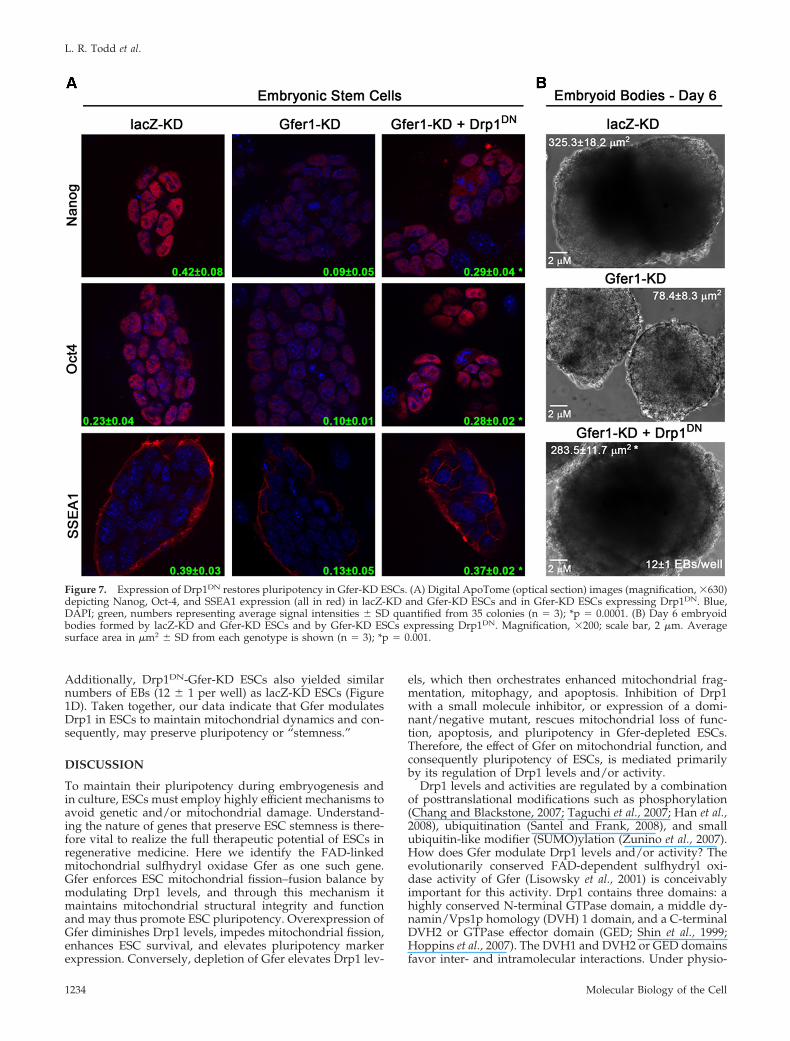
Additionally, Drp1DN-Gfer-KD ESCs also yielded similarnumbers of EBs (12 � 1 per well) as lacZ-KD ESCs (Figure1D). Taken together, our data indicate that Gfer modulatesDrp1 in ESCs to maintain mitochondrial dynamics and con-sequently, may preserve pluripotency or “stemness.”
DISCUSSION
To maintain their pluripotency during embryogenesis andin culture, ESCs must employ highly efficient mechanisms toavoid genetic and/or mitochondrial damage. Understand-ing the nature of genes that preserve ESC stemness is there-fore vital to realize the full therapeutic potential of ESCs inregenerative medicine. Here we identify the FAD-linkedmitochondrial sulfhydryl oxidase Gfer as one such gene.Gfer enforces ESC mitochondrial fission–fusion balance bymodulating Drp1 levels, and through this mechanism itmaintains mitochondrial structural integrity and functionand may thus promote ESC pluripotency. Overexpression ofGfer diminishes Drp1 levels, impedes mitochondrial fission,enhances ESC survival, and elevates pluripotency markerexpression. Conversely, depletion of Gfer elevates Drp1 lev-
els, which then orchestrates enhanced mitochondrial frag-mentation, mitophagy, and apoptosis. Inhibition of Drp1with a small molecule inhibitor, or expression of a domi-nant/negative mutant, rescues mitochondrial loss of func-tion, apoptosis, and pluripotency in Gfer-depleted ESCs.Therefore, the effect of Gfer on mitochondrial function, andconsequently pluripotency of ESCs, is mediated primarilyby its regulation of Drp1 levels and/or activity.
Drp1 levels and activities are regulated by a combinationof posttranslational modifications such as phosphorylation(Chang and Blackstone, 2007; Taguchi et al., 2007; Han et al.,2008), ubiquitination (Santel and Frank, 2008), and smallubiquitin-like modifier (SUMO)ylation (Zunino et al., 2007).How does Gfer modulate Drp1 levels and/or activity? Theevolutionarily conserved FAD-dependent sulfhydryl oxi-dase activity of Gfer (Lisowsky et al., 2001) is conceivablyimportant for this activity. Drp1 contains three domains: ahighly conserved N-terminal GTPase domain, a middle dy-namin/Vps1p homology (DVH) 1 domain, and a C-terminalDVH2 or GTPase effector domain (GED; Shin et al., 1999;Hoppins et al., 2007). The DVH1 and DVH2 or GED domainsfavor inter- and intramolecular interactions. Under physio-
Figure 7. Expression of Drp1DN restores pluripotency in Gfer-KD ESCs. (A) Digital ApoTome (optical section) images (magnification, �630)depicting Nanog, Oct-4, and SSEA1 expression (all in red) in lacZ-KD and Gfer-KD ESCs and in Gfer-KD ESCs expressing Drp1DN. Blue,DAPI; green, numbers representing average signal intensities � SD quantified from 35 colonies (n 3); *p 0.0001. (B) Day 6 embryoidbodies formed by lacZ-KD and Gfer-KD ESCs and by Gfer-KD ESCs expressing Drp1DN. Magnification, �200; scale bar, 2 �m. Averagesurface area in �m2 � SD from each genotype is shown (n 3); *p 0.001.
L. R. Todd et al.
Molecular Biology of the Cell1234
logical conditions, Drp1 exists as a dimer or tetramer and itsself-assembly into higher order structures facilitates GTPhydrolysis and mitochondrial fission (Smirnova et al., 2001;Hoppins et al., 2007). Efficacy of the chemical inhibitor ofDrp1, mdivi-1, is highly dependent on an unblocked sulfhy-dryl moiety that forms disulfide bonds on cysteine residueswithin regions outside of Drp1 GTPase domain (Cassidy-Stone et al., 2008). Thus, it is conceivable that the sulfhydryloxidase Gfer, predominantly localized in the IMS but alsopresent in the cytoplasm, catalyzes the formation of disul-fide bonds on the same cysteine residues of Drp1 to inacti-vate the GTPase and/or facilitate structural changes thatfavor Drp1 turnover. In fact, Drp1 contains four evolution-arily conserved cysteine residues outside the GTPase do-main that could serve as potential Gfer oxidization sites. Thefact that Drp1 migrates as a double band during reducingSDS-PAGE (Figure 6A; Zunino et al., 2009), suggests partialoxidization of one or more of its cysteine pairs.
On the other hand, the regulation of Drp1 by Gfer couldbe indirect, with Gfer affecting the activity of a factor thatregulates Drp1 activity. For example, overexpression of theSUMO protease SENP5 rescues SUMOylation-induced mi-tochondrial fragmentation by causing the down-regulationof DRP1; and deletion of SENP5 favors increased mitochon-drial fragmentation (Zunino et al., 2007). SENP5 possessessix highly conserved cysteine residues, at least one of which,Cys713, is important for its catalytic activity. Hence it ispossible that Gfer oxidizes disulfide bonds on one or more ofthe cysteine pairs on SENP5, thereby enhancing the protein’sactivity and stability, akin to its function in the import ofproteins to the mitochondrial IMS. In yeast, Gfer catalyzesthe formation of disulfide bridges between Mia40 and IMSproteins such as Cox17 and Tim13 to form a disulfide relaysystem that enables the import and retention of proteins inthe mitochondrial IMS, a process critical for normal mito-chondrial function (Mesecke et al., 2005). Perhaps Gfer alsocatalyzes inhibitory disulfide bridges between Drp1 andother protein(s) involved in the formation of higher-orderstructures that mediate mitochondrial fission.
Pluripotent ESCs contain a small number of mitochondriawith poorly defined (immature) cristae and consequently,possess low ATP levels (Baharvand and Matthaei, 2003;Lonergan et al., 2007; Nesti et al., 2007). Paradoxically, ESCsalso possess an unlimited proliferative potential, comparedwith differentiated cells such as the MEFs, and hence maydepend on a relatively few proteins such as Gfer to maintaintheir mitochondrial morphology, dynamics, function, andtherefore stemness. Even with 80% KD of Gfer (Figure 4B),we did not observe any associated loss of cell viabilityand/or mitochondrial morphology or function in MEFs.Although we interpreted these results to corroborate a se-lective role for Gfer in ESCs, it is also possible that theresidual (20%) Gfer in these MEFs is able to support themaintenance of their mitochondria and therefore preservecell viability. It is also possible that diminished pluripotencyin Gfer-KD ESCS is consequential to a more general loss ofits mitochondrial housekeeping function, resulting in anoverall loss of cell viability. Exactly how mitochondrial sta-bility and function contributes to stemness or pluripotencyin ESCs is a critical but poorly understood aspect of stem cellbiology.
Mitochondrial malfunction and mitophagy are associatedwith degenerative disorders such as Parkinson’s and Hun-tington’s. Elevated Drp1, enhanced mitochondrial fragmen-tation and pathological mitophagy is reported in familialand sporadic forms of Parkinson’s disease (Dagda et al.,2009). Mutations in the human Gfer gene are associated with
an infantile autosomal recessive form of myopathy, charac-terized at the cellular level by abnormal mitochondrial mor-phology, loss of mitochondrial function, and enhanced mu-tations of mtDNA (Di Fonzo et al., 2009). Moreover, reversalof cell death with a concomitant increase in Gfer expressionwas observed in a murine model of Huntington’s treatedwith inhibitors of histone deacetylation (Gardian et al., 2005).These studies underscore a cytoprotective role for Gfer inthe preservation of mitochondrial dynamics, structural in-tegrity, and function during homeostasis.
In conclusion, the data presented herein support an essen-tial prosurvival role for Gfer in the maintenance of murineESC pluripotency by preserving the structural and func-tional integrity of their mitochondria, through modulationof the key mitochondrial fission factor Drp1. Our findingshave therapeutic implications as they support a central rolefor a novel Gfer-Drp1 molecular link in preserving the stem-ness of ESCs, a primitive cell type with immense curativepotential.
ACKNOWLEDGMENTS
The authors thank Drs. Sally Kornbluth, Jeffrey Rathmell, and Tso-Pang Yao(Department of Pharmacology and Cancer Biology, Duke University MedicalCenter) and Dr. Keith R. Davis (Owensboro Cancer Research Program[OCRP] for critical reading of the manuscript and helpful discussions. We alsothank Cheryl Bock (Duke University Transgenic Core Facility) for the gener-ous gift of B6 ESCs; Dr. Ferrell C. Campbell and Cathie G. Caple (Universityof Louisville Electron Microscopy Core Facility) for help with electron mi-croscopy; and Sarah W. Grant and Cassandra Lockard for technical assistance.This work was supported by funds from the James Graham Brown CancerCenter and OCRP to U.S. and National Institutes of Health Grant R01DK074701 to A.R.M.
REFERENCES
Akao, M., O’Rourke, B., Teshima, Y., Seharaseyon, J., and Marban, E. (2003).Mechanistically distinct steps in the mitochondrial death pathway triggeredby oxidative stress in cardiac myocytes. Circ. Res. 92, 186–194.
Baharvand, H., and Matthaei, K. I. (2003). The ultrastructure of mouse em-bryonic stem cells. Reprod. Biomed. Online 7, 330–335.
Becher, D., Kricke, J., Stein, G., and Lisowsky, T. (1999). A mutant for the yeastscERV1 gene displays a new defect in mitochondrial morphology and distri-bution. Yeast 15, 1171–1181.
Benard, G., and Karbowski, M. (2009). Mitochondrial fusion and division:regulation and role in cell viability. Semin. Cell Dev. Biol. 20, 365–374.
Burdon, T., Smith, A., and Savatier, P. (2002). Signalling, cell cycle andpluripotency in embryonic stem cells. Trends Cell Biol. 12, 432–438.
Cassidy-Stone, A., Chipuk, J. E., Ingerman, E., Song, C., Yoo, C., Kuwana, T.,Kurth, M. J., Shaw, J. T., Hinshaw, J. E., Green, D. R., and Nunnari, J. (2008).Chemical inhibition of the mitochondrial division dynamin reveals its role inBax/Bak-dependent mitochondrial outer membrane permeabilization. Dev.Cell 14, 193–204.
Chang, C. R., and Blackstone, C. (2007). Cyclic AMP-dependent protein kinasephosphorylation of Drp1 regulates its GTPase activity and mitochondrialmorphology. J. Biol. Chem. 282, 21583–21587.
Dagda, R. K., Cherra, S. J., 3rd, Kulich, S. M., Tandon, A., Park, D., and Chu,C. T. (2009). Loss of PINK1 function promotes mitophagy through effects onoxidative stress and mitochondrial fission. J. Biol. Chem. 284, 13843–13855.
Di Fonzo, A., et al. (2009). The mitochondrial disulfide relay system proteinGFER is mutated in autosomal-recessive myopathy with cataract and com-bined respiratory-chain deficiency. Am. J. Hum. Genet. 84, 594–604.
Elmore, S. P., Qian, T., Grissom, S. F., and Lemasters, J. J. (2001). The mito-chondrial permeability transition initiates autophagy in rat hepatocytes.FASEB J. 15, 2286–2287.
Frank, S., Gaume, B., Bergmann-Leitner, E. S., Leitner, W. W., Robert, E. G.,Catez, F., Smith, C. L., and Youle, R. J. (2001). The role of dynamin-relatedprotein 1, a mediator of mitochondrial fission, in apoptosis. Dev. Cell 1,515–525.
Gfer Regulates Mitochondrial Dynamics
Vol. 21, April 1, 2010 1235
Gardian, G., et al. (2005). Neuroprotective effects of phenylbutyrate in theN171–82Q transgenic mouse model of Huntington’s disease. J. Biol. Chem.280, 556–563.
Gatzidou, E., Kouraklis, G., and Theocharis, S. (2006). Insights on augmenterof liver regeneration cloning and function. World J. Gastroenterol. 12, 4951–4958.
Han, X.-J., Lu, Y.-F., Li, S.-A., Kaitsuka, T., Sato, Y., Tomizawa, K., Nairn,A. C., Takei, K., Matsui, H., and Matsushita, M. (2008). CaM kinase I�-induced phosphorylation of Drp1 regulates mitochondrial morphology.J. Cell Biol. 182, 573–585.
Hoppins, S., Lackner, L., and Nunnari, J. (2007). The machines that divide andfuse mitochondria. Annu. Rev. Biochem. 76, 751–780.
Ishihara, N., et al. (2009). Mitochondrial fission factor Drp1 is essential forembryonic development and synapse formation in mice. Nat. Cell Biol. 11,958–966.
Ivanova, N., Dobrin, R., Lu, R., Kotenko, I., Levorse, J., DeCoste, C., Schafer,X., Lun, Y., and Lemischka, I. R. (2006). Dissecting self-renewal in stem cellswith RNA interference. Nature 442, 533–538.
Ivanova, N. B., Dimos, J. T., Schaniel, C., Hackney, J. A., Moore, K. A., andLemischka, I. R. (2002). A stem cell molecular signature. Science 298, 601–604.
Kitsos, C. M., Sankar, U., Illario, M., Colomer-Font, J. M., Duncan, A. W.,Ribar, T. J., Reya, T., and Means, A. R. (2005). Calmodulin-dependent proteinkinase IV regulates hematopoietic stem cell maintenance. J. Biol. Chem. 280,33101–33108.
Klebes, A., Sustar, A., Kechris, K., Li, H., Schubiger, G., and Kornberg, T. B.(2005). Regulation of cellular plasticity in Drosophila imaginal disc cells by thePolycomb group, trithorax group and lama genes. Development 132, 3753–3765.
Klissenbauer, M., Winters, S., Heinlein, U. A., and Lisowsky, T. (2002). Ac-cumulation of the mitochondrial form of the sulphydryl oxidase Erv1p/Alrpduring the early stages of spermatogenesis. J. Exp. Biol. 205, 1979–1986.
Lange, H., Lisowsky, T., Gerber, J., Muhlenhoff, U., Kispal, G., and Lill, R.(2001). An essential function of the mitochondrial sulfhydryl oxidase Erv1p/ALR in the maturation of cytosolic Fe/S proteins. EMBO Rep. 2, 715–720.
Levine, B., Sinha, S., and Kroemer, G. (2008). Bcl-2 family members: dualregulators of apoptosis and autophagy. Autophagy 4, 600–606.
Lisowsky, T., Lee, J. E., Polimeno, L., Francavilla, A., and Hofhaus, G. (2001).Mammalian augmenter of liver regeneration protein is a sulfhydryl oxidase.Dig. Liver Dis. 33, 173–180.
Liu, X., Kim, C. N., Yang, J., Jemmerson, R., and Wang, X. (1996). Induction ofapoptotic program in cell-free extracts: requirement for dATP and cyto-chrome c. Cell 86, 147–157.
Lonergan, T., Bavister, B., and Brenner, C. (2007). Mitochondria in stem cells.Mitochondrion 7, 289–296.
Mesecke, N., Terziyska, N., Kozany, C., Baumann, F., Neupert, W., Hell, K.,and Herrmann, J. M. (2005). A disulfide relay system in the intermembranespace of mitochondria that mediates protein import. Cell 121, 1059–1069.
Nesti, C., Pasquali, L., Vaglini, F., Siciliano, G., and Murri, L. (2007). The roleof mitochondria in stem cell biology. Biosci. Rep. 27, 165–171.
Pasini, D., Bracken, A. P., Hansen, J. B., Capillo, M., and Helin, K. (2007). Thepolycomb group protein Suz12 is required for embryonic stem cell differen-tiation. Mol. Cell. Biol. 27, 3769–3779.
Polimeno, L., Lisowsky, T., and Francavilla, A. (1999). From yeast to man–from mitochondria to liver regeneration: a new essential gene family. Ital. J.Gastroenterol. Hepatol. 31, 494–500.
Polimeno, L., Margiotta, M., Marangi, L., Lisowsky, T., Azzarone, A., Ierardi,E., Frassanito, M. A., Francavilla, R., and Francavilla, A. (2000). Molecularmechanisms of augmenter of liver regeneration as immunoregulator: its effecton interferon-gamma expression in rat liver. Dig. Liver Dis. 32, 217–225.
Qin, X. F., An, D. S., Chen, I. S., and Baltimore, D. (2003). Inhibiting HIV-1infection in human T cells by lentiviral-mediated delivery of small interferingRNA against CCR5. Proc. Natl. Acad. Sci. USA 100, 183–188.
Ramalho-Santos, M., Yoon, S., Matsuzaki, Y., Mulligan, R. C., and Melton,D. A. (2002). “Stemness”: transcriptional profiling of embryonic and adultstem cells. Science 298, 597–600.
Santel, A., and Frank, S. (2008). Shaping mitochondria: The complex post-translational regulation of the mitochondrial fission protein DRP1. IUBMBLife 60, 448–455.
Sathananthan, H., Pera, M., and Trounson, A. (2002). The fine structure ofhuman embryonic stem cells. Reprod. Biomed. Online 4, 56–61.
Shin, H. W., Takatsu, H., Mukai, H., Munekata, E., Murakami, K., andNakayama, K. (1999). Intermolecular and interdomain interactions of a dy-namin-related GTP-binding protein, Dnm1p/Vps1p-like protein. J. Biol.Chem. 274, 2780–2785.
Silver, L. M. (1993). The peculiar journey of a selfish chromosome: mouse thaplotypes and meiotic drive. Trends Genet. 9, 250–254.
Smirnova, E., Griparic, L., Shurland, D. L., and van der Bliek, A. M. (2001).Dynamin-related protein Drp1 is required for mitochondrial division in mam-malian cells. Mol. Biol. Cell 12, 2245–2256.
Taguchi, N., Ishihara, N., Jofuku, A., Oka, T., and Mihara, K. (2007). Mitoticphosphorylation of dynamin-related GTPase Drp1 participates in mitochon-drial fission. J. Biol. Chem. 282, 11521–11529.
Tanaka, A., and Youle, R. J. (2008). A chemical inhibitor of DRP1 uncouplesmitochondrial fission and apoptosis. Mol. Cell 29, 409–410.
Wang, J., Theunissen, T. W., and Orkin, S. H. (2007). Site-directed, virus-free,and inducible RNAi in embryonic stem cells. Proc. Natl. Acad. Sci. USA 104,20850–20855.
Wang, X. (2001). The expanding role of mitochondria in apoptosis. Genes Dev.15, 2922–2933.
Waterhouse, N. J., and Trapani, J. A. (2003). A new quantitative assay forcytochrome c release in apoptotic cells. Cell Death Differ. 10, 853–855.
Zeng, X., and Rao, M. S. (2007). Human embryonic stem cells: long termstability, absence of senescence and a potential cell source for neural replace-ment. Neuroscience 145, 1348–1358.
Zunino, R., Braschi, E., Xu, L., and McBride, H. M. (2009). Translocation ofSenP5 from the nucleoli to the mitochondria modulates DRP1-dependentfission during mitosis. J. Biol. Chem. 284, 17783–17795.
Zunino, R., Schauss, A., Rippstein, P., Andrade-Navarro, M., and McBride,H. M. (2007). The SUMO protease SENP5 is required to maintain mitochon-drial morphology and function. J. Cell Sci. 120, 1178–1188.
L. R. Todd et al.
Molecular Biology of the Cell1236
Copyright © 2022 FDOKUMEN