GLUT1-independent infection of the glioblastoma/astroglioma U87 cells by the human T cell leukemia...
12
GLUT-1-independent infection of the glioblastoma/astroglioma U87 cells by the human T cell leukemia virus type 1 Qingwen Jin c , Lokesh Agrawal a,b,1 , Zainab VanHorn-Ali a,b , Ghalib Alkhatib a,b, ⁎ a Department of Microbiology and Immunology and Indiana University School of Medicine, 635 Barnhill Drive, Rm #420, Indianapolis, IN 46202, USA b Walther Cancer Institute, Indianapolis, IN 46208, USA c Department of Neurology, Nanjing Medical University, Nanjing, Jiangsu Province, 210029, China Received 2 March 2006; returned to author for revision 28 March 2006; accepted 3 May 2006 Available online 16 June 2006 Abstract The human glucose transporter protein 1 (GLUT-1) functions as a receptor for human Tcell leukemia virus (HTLV). GLUT-1 is a twelve- transmembrane cell surface receptor with six extracellular (ECL) and seven intracellular domains. To analyze HTLV-1 cytotropism, we utilized polyclonal antibodies to a synthetic peptide corresponding to the large extracellular domain of GLUT-1. The antibodies caused significant blocking of envelope (Env)-mediated fusion and pseudotyped virus infection of HeLa cells but had no significant effect on infection of U87 cells. This differential effect correlated with the detection of high-level surface expression of GLUT-1 on HeLa cells and very weak staining of U87 cells. To investigate this in terms of viral cytotropism, we cloned GLUT-1 cDNA from U87 cells and isolated two different versions of cDNA clones: the wild-type sequence (encoding 492 residues) and a mutant cDNA with a 5-base pair deletion (GLUT-1Δ5) between nucleotides 1329 and 1333. The deletion, also detected in genomic DNA, resulted in a frame-shift and premature termination producing a truncated protein of 463 residues. Transfection of the wild-type GLUT-1 but not GLUT-1Δ5 cDNA into CHO cells resulted in efficient surface expression of the human GLUT-1. Co-expression of GLUT-1 with GLUT-1Δ5 produces a trans-inhibition by GLUT-1Δ5 of GLUT-1-mediated HTLV-1 envelope (Env)- mediated fusion. Co-immunoprecipitation experiments demonstrated physical interaction of the wild-type and mutant proteins. Northern blot and RT-PCR analyses demonstrated lower GLUT-1 RNA expression in U87 cells. We propose two mechanisms to account for the impaired cell surface expression of GLUT-1 on U87 cells: low GLUT-1 RNA expression and the formation of GLUT-1/GLUT-1Δ5 heterodimers that are retained intracellularly. Significant RNAi-mediated reduction of endogenous GLUT-1 expression impaired HTLV-1 Env-mediated fusion with HeLa cells but not with U87 cells. We propose a GLUT-1-independent mechanism of HTLV-1 infection of U87 cells. The results may have important implications for HTLV-1 neurotropism and pathogenesis. © 2006 Elsevier Inc. All rights reserved. Keywords: Virus entry; HTLV-1; Neurotropism; GLUT-1 mutations Introduction Human T-cell leukemia virus type 1 (HTLV-1), the first discovered disease-causing human retrovirus isolate, is the etiologic agent of adult T-cell leukemia/lymphoma (ATLL) and a progressive demyelinating disease known as tropical spastic paraparesis/HTLV-1-associated myelopathy (reviewed in Feuer and Green, 2005; Gallo, 2005; Yoshida, 2005). HTLV-1 is endemic in Southern Japan, West Africa, Central and South America, and the Caribbean basin (reviewed in Feuer and Green, 2005). Although HTLV-1 was the first human retrovirus to be isolated and characterized, its study has been hampered by poor viral infectivity as manifested by low cell-free and cell- Virology 353 (2006) 99 – 110 www.elsevier.com/locate/yviro Abbreviations: Env, envelope glycoprotein; Env63, wild-type HTLV-1 envelope glycoprotein; Unc63, a cleavage mutant of HTLV-1 Env63 defective in syncytium formation; wt, wild type. ⁎ Corresponding author. Department of Microbiology and Immunology and Indiana University School of Medicine, 635 Barnhill Drive, Rm #420, Indianapolis, IN 46202, USA. E-mail address: [email protected] (G. Alkhatib). 1 Current address: 251, Jefferson Alumni Hall, Department of Pathology, Anatomy and Cell Biology, Thomas Jefferson University, 1020, Locust Street, Philadelphia, PA 19107, USA. 0042-6822/$ - see front matter © 2006 Elsevier Inc. All rights reserved. doi:10.1016/j.virol.2006.05.003
-
Upload
independent -
Category
Documents
-
view
0 -
download
0
Transcript of GLUT1-independent infection of the glioblastoma/astroglioma U87 cells by the human T cell leukemia...
06) 99–110www.elsevier.com/locate/yviro
Virology 353 (20
GLUT-1-independent infection of the glioblastoma/astrogliomaU87 cells by the human T cell leukemia virus type 1
Qingwen Jin c, Lokesh Agrawal a,b,1, Zainab VanHorn-Ali a,b, Ghalib Alkhatib a,b,⁎
a Department of Microbiology and Immunology and Indiana University School of Medicine, 635 Barnhill Drive, Rm #420, Indianapolis, IN 46202, USAb Walther Cancer Institute, Indianapolis, IN 46208, USA
c Department of Neurology, Nanjing Medical University, Nanjing, Jiangsu Province, 210029, China
Received 2 March 2006; returned to author for revision 28 March 2006; accepted 3 May 2006Available online 16 June 2006
Abstract
The human glucose transporter protein 1 (GLUT-1) functions as a receptor for human T cell leukemia virus (HTLV). GLUT-1 is a twelve-transmembrane cell surface receptor with six extracellular (ECL) and seven intracellular domains. To analyze HTLV-1 cytotropism, we utilizedpolyclonal antibodies to a synthetic peptide corresponding to the large extracellular domain of GLUT-1. The antibodies caused significantblocking of envelope (Env)-mediated fusion and pseudotyped virus infection of HeLa cells but had no significant effect on infection of U87 cells.This differential effect correlated with the detection of high-level surface expression of GLUT-1 on HeLa cells and very weak staining of U87cells. To investigate this in terms of viral cytotropism, we cloned GLUT-1 cDNA from U87 cells and isolated two different versions of cDNAclones: the wild-type sequence (encoding 492 residues) and a mutant cDNA with a 5-base pair deletion (GLUT-1Δ5) between nucleotides 1329and 1333. The deletion, also detected in genomic DNA, resulted in a frame-shift and premature termination producing a truncated protein of 463residues. Transfection of the wild-type GLUT-1 but not GLUT-1Δ5 cDNA into CHO cells resulted in efficient surface expression of the humanGLUT-1. Co-expression of GLUT-1 with GLUT-1Δ5 produces a trans-inhibition by GLUT-1Δ5 of GLUT-1-mediated HTLV-1 envelope (Env)-mediated fusion. Co-immunoprecipitation experiments demonstrated physical interaction of the wild-type and mutant proteins. Northern blot andRT-PCR analyses demonstrated lower GLUT-1 RNA expression in U87 cells. We propose two mechanisms to account for the impaired cellsurface expression of GLUT-1 on U87 cells: low GLUT-1 RNA expression and the formation of GLUT-1/GLUT-1Δ5 heterodimers that areretained intracellularly. Significant RNAi-mediated reduction of endogenous GLUT-1 expression impaired HTLV-1 Env-mediated fusion withHeLa cells but not with U87 cells. We propose a GLUT-1-independent mechanism of HTLV-1 infection of U87 cells. The results may haveimportant implications for HTLV-1 neurotropism and pathogenesis.© 2006 Elsevier Inc. All rights reserved.
Keywords: Virus entry; HTLV-1; Neurotropism; GLUT-1 mutations
Abbreviations: Env, envelope glycoprotein; Env63, wild-type HTLV-1envelope glycoprotein; Unc63, a cleavage mutant of HTLV-1 Env63 defective insyncytium formation; wt, wild type.⁎ Corresponding author. Department of Microbiology and Immunology and
Indiana University School of Medicine, 635 Barnhill Drive, Rm #420,Indianapolis, IN 46202, USA.
E-mail address: [email protected] (G. Alkhatib).1 Current address: 251, Jefferson Alumni Hall, Department of Pathology,
Anatomy and Cell Biology, Thomas Jefferson University, 1020, Locust Street,Philadelphia, PA 19107, USA.
0042-6822/$ - see front matter © 2006 Elsevier Inc. All rights reserved.doi:10.1016/j.virol.2006.05.003
Introduction
Human T-cell leukemia virus type 1 (HTLV-1), the firstdiscovered disease-causing human retrovirus isolate, is theetiologic agent of adult T-cell leukemia/lymphoma (ATLL) anda progressive demyelinating disease known as tropical spasticparaparesis/HTLV-1-associated myelopathy (reviewed in Feuerand Green, 2005; Gallo, 2005; Yoshida, 2005). HTLV-1 isendemic in Southern Japan, West Africa, Central and SouthAmerica, and the Caribbean basin (reviewed in Feuer andGreen, 2005). Although HTLV-1 was the first human retrovirusto be isolated and characterized, its study has been hampered bypoor viral infectivity as manifested by low cell-free and cell-
100 Q. Jin et al. / Virology 353 (2006) 99–110
associated virus titers. Manel et al. were the first to identify theglucose transporter type 1 (GLUT-1) as a receptor for HTLV-1and HTLV-2 (Manel et al., 2003). Coskun and Sutton confirmedthat GLUT-1 expression in the relatively resistant MDBK cellsresulted in enhancement of syncytia formation and HTLV-1infection (Coskun and Sutton, 2005).
GLUT-1 protein belongs to the family of glucosetransporter proteins that have twelve membrane-spanningdomains, seven intracellular domains and six extracellulardomains (reviewed in Mueckler, 1994). The first extracellulardomain (ECL1) is the largest, with a predicted 33 aminoacids, and has a single N-linked glycosylation site. We haverecently demonstrated that antibodies to ECL1 inhibit HTLV-1Env-mediated fusion and viral infection of CD4+ Tlymphocytes and suggested that the observed in vivo cytotrop-ism may be due to the high GLUT-1 density on CD4+ Tlymphocytes (Jin et al., 2006). It remains unclear, however,whether GLUT-1 is functioning as a receptor, a coreceptor, or asan attachment cofactor. A recent report that added furthercontroversy demonstrated that heparan sulfate proteoglycan(HSPG) is as a receptor for HTLV-1 on primary CD4+ Tlymphocytes (Jones et al., 2005). HSPG has been previouslyshown to function as a non-specific attachment factor for manyviruses (reviewed in Manel et al., 2005). Manel et al. were thefirst to suggest that, in addition to GLUT-1, other cofactors/coreceptors might be involved in HTLV-1 infection (reviewedin Manel et al., 2005). However, the lack of a cell system thatdoes not express GLUT-1 has made it difficult to provideexperimental evidence for the potential expression of alternativereceptors/coreceptors.
The literature reported a number of observations that favorthe cofactor/coreceptor model. First, it is still unclear whetherthe in vivo tropism of HTLV-1 can be explained solely byGLUT-1 expression since GLUT-1 is widely expressed.Furthermore, the wide host range of HTLV-1 in vitro cannotonly be explained by GLUT-1 expression since a number astudies observed different binding and infection efficiencies ofnon-human cell lines (reviewed in Manel et al., 2005).Additionally, there has been no evidence that HTLV-1 usesGLUT-1 or other receptors to infect the different mammaliancell lines. It has been assumed that infection of mammalian celllines is mediated by endogenous GLUT-1 based on Env bindinganalysis (Jassal et al., 2001). In the present study, we describethat U87 cells, which are highly susceptible to HTLV-1infection, express very low levels of GLUT-1 protein on thecell surface and that antibodies to GLUT-1 do not inhibit HTLV-1 Env-mediated fusion and pseudotyped virus infection of U87cells. Northern and RT-PCR analyses indicated low abundanceof GLUT-1 RNA in U87 cells compared to HeLa or 293 cells.We identified a 5 bp deletion in the gene encoding the GLUT-1protein that resulted in a frame-shift and a truncated GLUT-1protein. We found that U87 cells are heterozygous for thedeleted GLUT-1 allele. We propose a GLUT-1-independentmechanism for HTLV-1 infection of U87 cells. Since U87 cellsare an astrocytoma/glioblastoma cell line derived from a humanbrain tumor, our results may have important implications forHTLV-1 neurotropism.
Results
U87 cells are highly permissive to HTLV-1
To determine the susceptibility of U87 cells to HTLV-1, weexamined these cells as targets in the Env-mediated fusion assayandmeasured their ability to support pseudotyped virus infection.We used HeLa cells as positive control since they are susceptibleto Env fusion and HTLV-1 infection. To control for non-specificEnv interactions, we used a mutant envelope protein Unc63 thathad its cleavage site at residues 309–312 (RSRR) mutated toGLGL (Jin et al., 2006). We consistently observed higher Env-mediated fusion (Fig. 1A) and infection signals (Fig. 1B) withU87 cells. We also observed some degree of permissiveness withthe non-human cell lines CHO and MDBK; however, the fusionand infectivity signals were significantly lower than observedwith U87 or HeLa cells (Figs. 1A and B). Consistent with recentdata in the literature (Coskun and Sutton, 2005),MDBK seems tobe the least susceptible to HTLV-1 Env fusion and viral infection.The competence of all cell lines to undergo cell fusion and viralinfection was confirmed by VSV Env fusion (Fig. 1C) or VSVinfection (Fig. 1D). The results demonstrate that U87 cells arehighly susceptible to HTLV-1 Env fusion and pseudotyped virusinfection.
GLUT-1 surface expression on U87 cells does not correlatewith HTLV-1 Env fusion
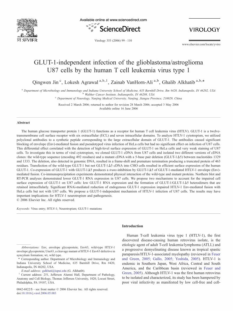
To determine whether the high susceptibility of U87 cells toHTLV-1 Env fusion is due to high expression levels of theGLUT-1 receptor, we examined the effect of GLUT-IgYantibodies on Env fusion. Pre-incubating these antibodieswith the target cells prior to mixing with Env-expressing cellsdemonstrated efficient blocking of Env fusion with HeLa cellsbut no effect on Env fusion observed with U87 cells (Fig. 2A).The inhibitory effect was specific to HTLV-1 Env-mediatedfusion since VSV fusion was not significantly altered followingincubation with GLUT-IgY (Fig. 2A). Higher concentrations ofthe antibody had no significant effect on Env fusion with U87cells (data not shown). FACS analysis was performed tocompare surface levels of U87 GLUT-1 to that expressed inHeLa cells. The antibodies used were chicken anti-humanGLUT-IgY raised against the first extracellular loop (ECL1) ofGLUT-1 (Jin et al., 2006). HeLa cells consistently expressedhigh levels of cell surface GLUT-1, whereas U87 showed veryweak staining of this antigen over the pre-immune control (Fig.2B). To determine whether this weak staining is not due toantibody recognition, we transfected the U87 with a plasmidDNA encoding wild-type GLUT-1 and analyzed expression byFACS. The results show significant increase of cell surfaceGLUT-1 on transfected cells (Fig. 2C). Western blot analysis ofGLUT-1-transfected cells demonstrated expression of therecombinant GLUT-1 in U87 cells (Fig. 2C, right). Theobserved broad protein band in U87 cells co-migrated withthat in HeLa cells indicating their source from the transfectedDNA (Fig. 2C). In contrast, western blot analysis ofendogenous GLUT-1 protein revealed the expression of a
Fig. 1. Efficient HTLV-1 Env fusion and pseudotyped virus infection of U87 cells. (A) HTLV-1 Env-mediated fusion. 293 cells were co-infected with vaccinia virusencoding either Env63, Unc63, or VSV and pT7-lacZ reporter. The target cell lines indicated at the x axis were infected with vaccinia virus expressing T7 RNApolymerase. Following overnight incubation at 31 °C, Env-expressing cells were mixed with the target cells and incubated for 2.5 h. The Env-mediated fusion wasassessed by measuring the amount of β-galactosidase produced in detergent-treated cell lysates. (B) Pseudotyped virus particles carrying Unc63, Env63, or VSV-Gwere used to infect the indicated cell lines (0.3 × 10⁶ cells/infection) in duplicates and incubated for 48 h at 37 °C. Cell lysates were prepared, and luciferase activitywas measured. The data are expressed as relative light units (RLU). C and D show Env fusion with VSV (C) and VSV infection (D) to confirm the competence of thecells in Env fusion and infection assays.
101Q. Jin et al. / Virology 353 (2006) 99–110
diffused and a faster migrating GLUT-1 protein band in U87cells compared to HeLa cells (Fig. 2D). The protein bandrepresenting GLUT-1 above the 50 kDa marker was signifi-cantly lower in U87 cells compared to HeLa cells (Fig. 2D).
To compare GLUT-1 RNA expression in U87 cells with otherhuman cell lines, we performed northern blot analysis on totalRNA and used GLUT-1 cDNA as a probe. The results showsignificant lower levels of GLUT-1 RNA in U87 cells comparedto HeLa and 293 cells (Fig. 2E). The lower levels of GLUT-1RNA in U87 cells were confirmed by RT-PCR analysis usingpurified total RNA as a template (Fig. 2F). To control for gelloading, equivalent levels of β-actin RNA were detected in allthree human cell lines (Figs. 2E and F). Together, these resultsshow defects in GLUT-1 expression at the RNA as well as at thepost-translational levels in U87 cells. We propose a GLUT-1-independent pathway for HTLV-1 infection of U87 cells.
HTLV-1 infection of U87 cells is sensitive to anti-gp46antibodies but not to GLUT-IgY antibodies
To examine the effect of GLUT-IgY on HTLV-1 infection,we compared the effect of pre-incubating these antibodies with
HeLa or U87 cells prior to infection. The results show a dose-dependent inhibition of HTLV-1 infection of HeLa cells (Fig.3A) but no significant effect observed with U87 cells (Fig. 3B).To show that infectivity signal obtained with U87 is mediatedby HTLV-1, we examined the effect of human monoclonalantibodies (HMAbs) PRH-11A (obtained from Dr. StevenFoung, Stanford University) that have been previously shown toinhibit HTLV-1 infection (Hadlock et al., 1997). The resultsshow efficient blocking of HTLV-1 infection of HeLa (Fig. 3C)as well as U87 cells (Fig. 3D). These results suggest a GLUT-1-independent pathway for the efficient infection of U87 cells.
Cloning of GLUT-1 DNA from HeLa and U87 cells
Since transfection of GLUT-1 cDNA into U87 cells restoressurface expression of the protein (Fig. 2), it was logical toanalyze the DNA sequence of the gene for potential mutations.We cloned and isolated the cDNA encoding the GLUT-1 cDNAfrom U87 and HeLa cells. Sequence comparison of the clonedGLUT-1 cDNAwith the human erythrocyte/HepG2 transportergene (GenBank NM006516) indicated a T to C transition atnucleotide 454 in exon 4, which resulted in the substitution of
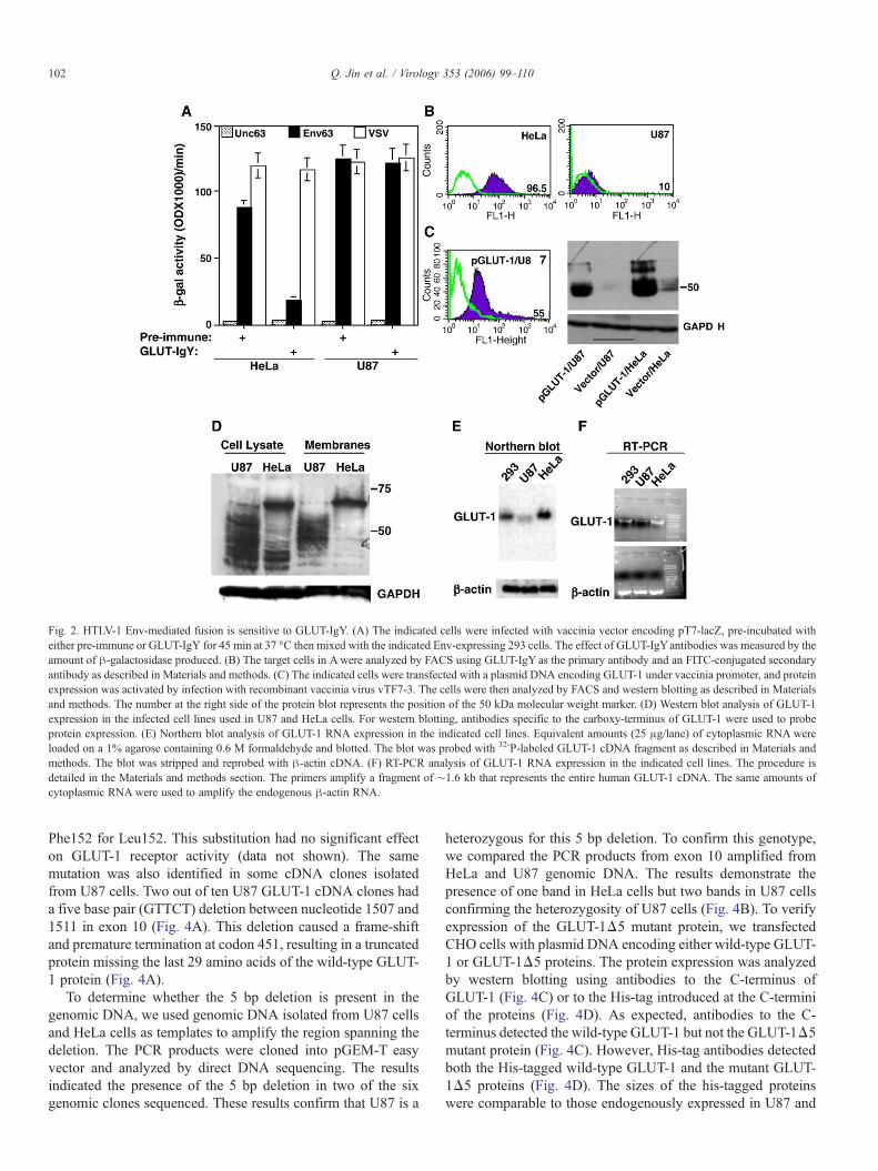
Fig. 2. HTLV-1 Env-mediated fusion is sensitive to GLUT-IgY. (A) The indicated cells were infected with vaccinia vector encoding pT7-lacZ, pre-incubated witheither pre-immune or GLUT-IgY for 45 min at 37 °C then mixed with the indicated Env-expressing 293 cells. The effect of GLUT-IgYantibodies was measured by theamount of β-galactosidase produced. (B) The target cells in Awere analyzed by FACS using GLUT-IgY as the primary antibody and an FITC-conjugated secondaryantibody as described in Materials and methods. (C) The indicated cells were transfected with a plasmid DNA encoding GLUT-1 under vaccinia promoter, and proteinexpression was activated by infection with recombinant vaccinia virus vTF7-3. The cells were then analyzed by FACS and western blotting as described in Materialsand methods. The number at the right side of the protein blot represents the position of the 50 kDa molecular weight marker. (D) Western blot analysis of GLUT-1expression in the infected cell lines used in U87 and HeLa cells. For western blotting, antibodies specific to the carboxy-terminus of GLUT-1 were used to probeprotein expression. (E) Northern blot analysis of GLUT-1 RNA expression in the indicated cell lines. Equivalent amounts (25 μg/lane) of cytoplasmic RNA wereloaded on a 1% agarose containing 0.6 M formaldehyde and blotted. The blot was probed with 32-P-labeled GLUT-1 cDNA fragment as described in Materials andmethods. The blot was stripped and reprobed with β-actin cDNA. (F) RT-PCR analysis of GLUT-1 RNA expression in the indicated cell lines. The procedure isdetailed in the Materials and methods section. The primers amplify a fragment of ∼1.6 kb that represents the entire human GLUT-1 cDNA. The same amounts ofcytoplasmic RNA were used to amplify the endogenous β-actin RNA.
102 Q. Jin et al. / Virology 353 (2006) 99–110
Phe152 for Leu152. This substitution had no significant effecton GLUT-1 receptor activity (data not shown). The samemutation was also identified in some cDNA clones isolatedfrom U87 cells. Two out of ten U87 GLUT-1 cDNA clones hada five base pair (GTTCT) deletion between nucleotide 1507 and1511 in exon 10 (Fig. 4A). This deletion caused a frame-shiftand premature termination at codon 451, resulting in a truncatedprotein missing the last 29 amino acids of the wild-type GLUT-1 protein (Fig. 4A).
To determine whether the 5 bp deletion is present in thegenomic DNA, we used genomic DNA isolated from U87 cellsand HeLa cells as templates to amplify the region spanning thedeletion. The PCR products were cloned into pGEM-T easyvector and analyzed by direct DNA sequencing. The resultsindicated the presence of the 5 bp deletion in two of the sixgenomic clones sequenced. These results confirm that U87 is a
heterozygous for this 5 bp deletion. To confirm this genotype,we compared the PCR products from exon 10 amplified fromHeLa and U87 genomic DNA. The results demonstrate thepresence of one band in HeLa cells but two bands in U87 cellsconfirming the heterozygosity of U87 cells (Fig. 4B). To verifyexpression of the GLUT-1Δ5 mutant protein, we transfectedCHO cells with plasmid DNA encoding either wild-type GLUT-1 or GLUT-1Δ5 proteins. The protein expression was analyzedby western blotting using antibodies to the C-terminus ofGLUT-1 (Fig. 4C) or to the His-tag introduced at the C-terminiof the proteins (Fig. 4D). As expected, antibodies to the C-terminus detected the wild-type GLUT-1 but not the GLUT-1Δ5mutant protein (Fig. 4C). However, His-tag antibodies detectedboth the His-tagged wild-type GLUT-1 and the mutant GLUT-1Δ5 proteins (Fig. 4D). The sizes of the his-tagged proteinswere comparable to those endogenously expressed in U87 and
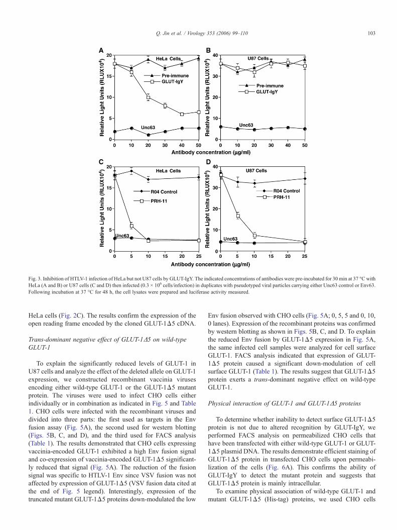
Fig. 3. Inhibition of HTLV-1 infection of HeLa but not U87 cells by GLUT-IgY. The indicated concentrations of antibodies were pre-incubated for 30 min at 37 °C withHeLa (A and B) or U87 cells (C and D) then infected (0.3 × 10⁶ cells/infection) in duplicates with pseudotyped viral particles carrying either Unc63 control or Env63.Following incubation at 37 °C for 48 h, the cell lysates were prepared and luciferase activity measured.
103Q. Jin et al. / Virology 353 (2006) 99–110
HeLa cells (Fig. 2C). The results confirm the expression of theopen reading frame encoded by the cloned GLUT-1Δ5 cDNA.
Trans-dominant negative effect of GLUT-1Δ5 on wild-typeGLUT-1
To explain the significantly reduced levels of GLUT-1 inU87 cells and analyze the effect of the deleted allele on GLUT-1expression, we constructed recombinant vaccinia virusesencoding either wild-type GLUT-1 or the GLUT-1Δ5 mutantprotein. The viruses were used to infect CHO cells eitherindividually or in combination as indicated in Fig. 5 and Table1. CHO cells were infected with the recombinant viruses anddivided into three parts: the first used as targets in the Envfusion assay (Fig. 5A), the second used for western blotting(Figs. 5B, C, and D), and the third used for FACS analysis(Table 1). The results demonstrated that CHO cells expressingvaccinia-encoded GLUT-1 exhibited a high Env fusion signaland co-expression of vaccinia-encoded GLUT-1Δ5 significant-ly reduced that signal (Fig. 5A). The reduction of the fusionsignal was specific to HTLV-1 Env since VSV fusion was notaffected by expression of GLUT-1Δ5 (VSV fusion data cited atthe end of Fig. 5 legend). Interestingly, expression of thetruncated mutant GLUT-1Δ5 proteins down-modulated the low
Env fusion observed with CHO cells (Fig. 5A; 0, 5, 5 and 0, 10,0 lanes). Expression of the recombinant proteins was confirmedby western blotting as shown in Figs. 5B, C, and D. To explainthe reduced Env fusion by GLUT-1Δ5 expression in Fig. 5A,the same infected cell samples were analyzed for cell surfaceGLUT-1. FACS analysis indicated that expression of GLUT-1Δ5 protein caused a significant down-modulation of cellsurface GLUT-1 (Table 1). The results suggest that GLUT-1Δ5protein exerts a trans-dominant negative effect on wild-typeGLUT-1.
Physical interaction of GLUT-1 and GLUT-1Δ5 proteins
To determine whether inability to detect surface GLUT-1Δ5protein is not due to altered recognition by GLUT-IgY, weperformed FACS analysis on permeabilized CHO cells thathave been transfected with either wild-type GLUT-1 or GLUT-1Δ5 plasmid DNA. The results demonstrate efficient staining ofGLUT-1Δ5 protein in transfected CHO cells upon permeabi-lization of the cells (Fig. 6A). This confirms the ability ofGLUT-IgY to detect the mutant protein and suggests thatGLUT-1Δ5 protein is mainly intracellular.
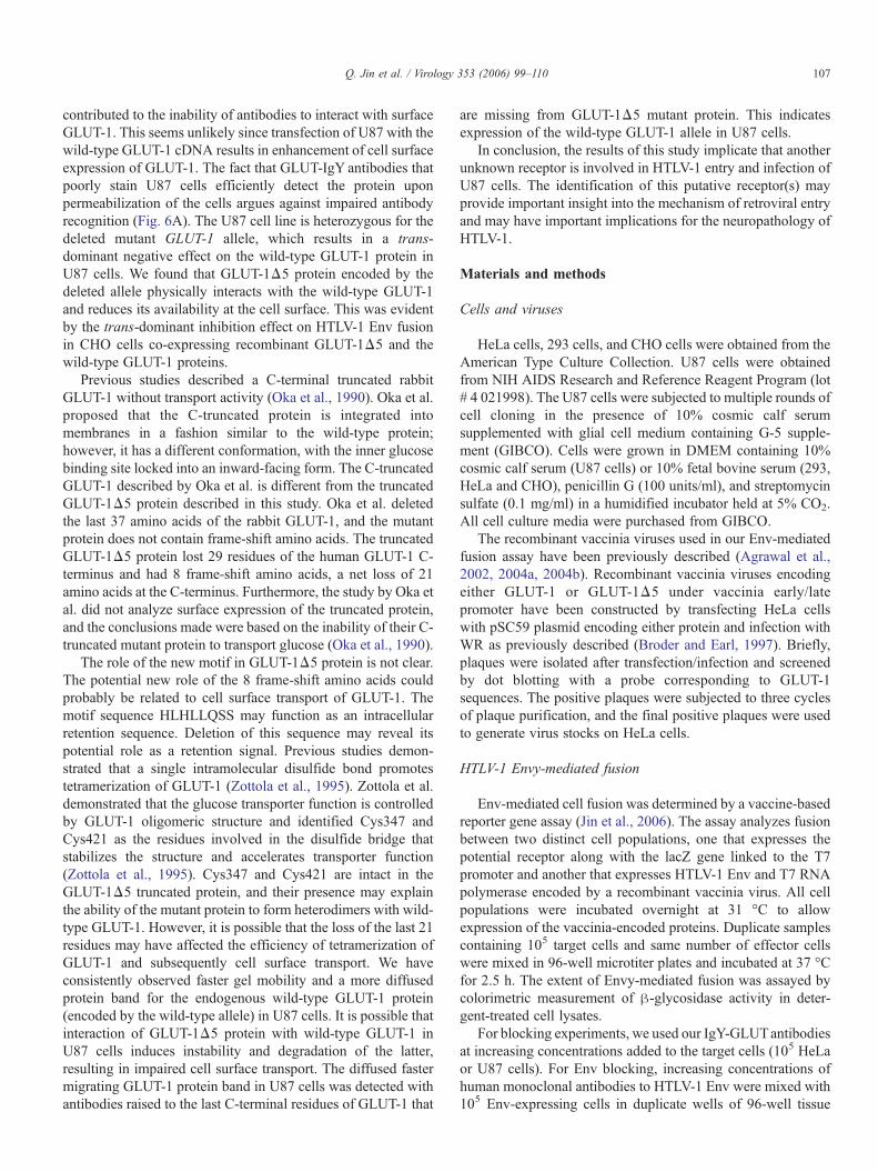
To examine physical association of wild-type GLUT-1 andmutant GLUT-1Δ5 (His-tag) proteins, we used CHO cells
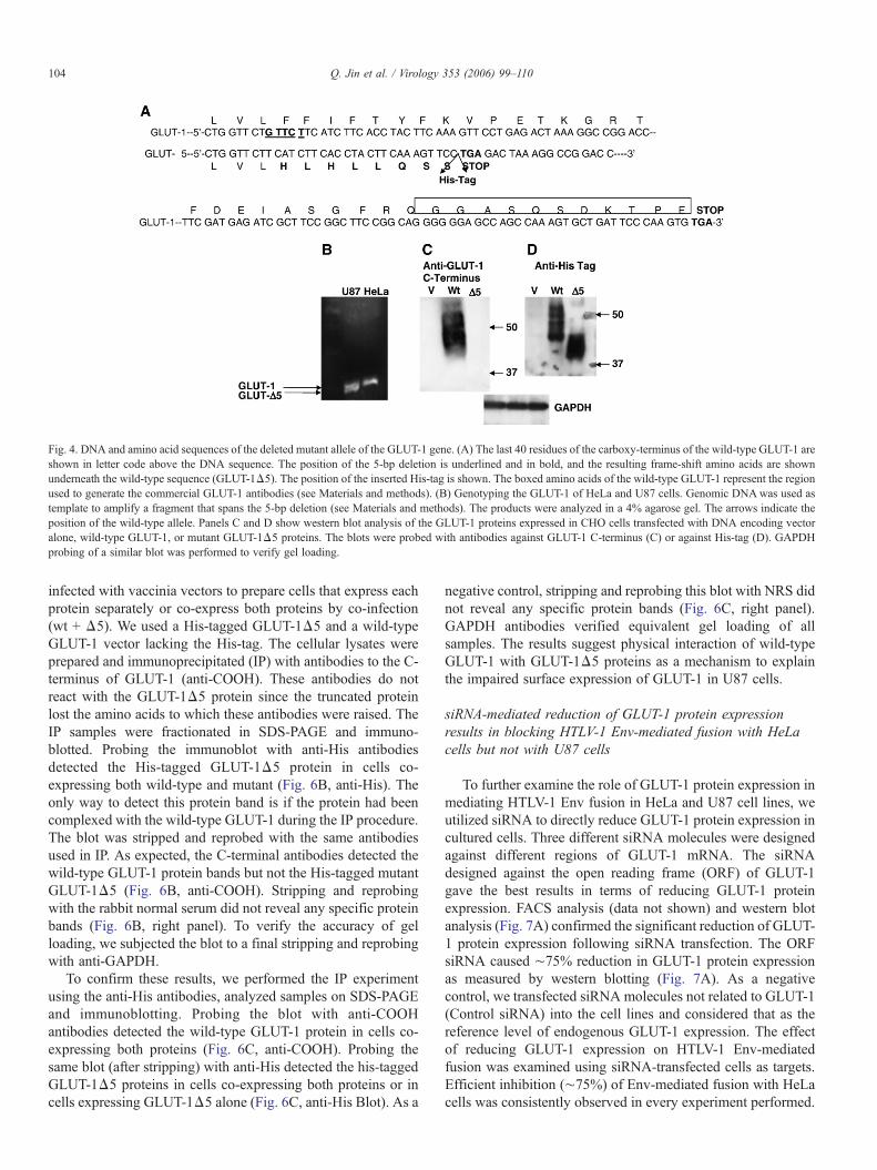
Fig. 4. DNA and amino acid sequences of the deleted mutant allele of the GLUT-1 gene. (A) The last 40 residues of the carboxy-terminus of the wild-type GLUT-1 areshown in letter code above the DNA sequence. The position of the 5-bp deletion is underlined and in bold, and the resulting frame-shift amino acids are shownunderneath the wild-type sequence (GLUT-1Δ5). The position of the inserted His-tag is shown. The boxed amino acids of the wild-type GLUT-1 represent the regionused to generate the commercial GLUT-1 antibodies (see Materials and methods). (B) Genotyping the GLUT-1 of HeLa and U87 cells. Genomic DNAwas used astemplate to amplify a fragment that spans the 5-bp deletion (see Materials and methods). The products were analyzed in a 4% agarose gel. The arrows indicate theposition of the wild-type allele. Panels C and D show western blot analysis of the GLUT-1 proteins expressed in CHO cells transfected with DNA encoding vectoralone, wild-type GLUT-1, or mutant GLUT-1Δ5 proteins. The blots were probed with antibodies against GLUT-1 C-terminus (C) or against His-tag (D). GAPDHprobing of a similar blot was performed to verify gel loading.
104 Q. Jin et al. / Virology 353 (2006) 99–110
infected with vaccinia vectors to prepare cells that express eachprotein separately or co-express both proteins by co-infection(wt + Δ5). We used a His-tagged GLUT-1Δ5 and a wild-typeGLUT-1 vector lacking the His-tag. The cellular lysates wereprepared and immunoprecipitated (IP) with antibodies to the C-terminus of GLUT-1 (anti-COOH). These antibodies do notreact with the GLUT-1Δ5 protein since the truncated proteinlost the amino acids to which these antibodies were raised. TheIP samples were fractionated in SDS-PAGE and immuno-blotted. Probing the immunoblot with anti-His antibodiesdetected the His-tagged GLUT-1Δ5 protein in cells co-expressing both wild-type and mutant (Fig. 6B, anti-His). Theonly way to detect this protein band is if the protein had beencomplexed with the wild-type GLUT-1 during the IP procedure.The blot was stripped and reprobed with the same antibodiesused in IP. As expected, the C-terminal antibodies detected thewild-type GLUT-1 protein bands but not the His-tagged mutantGLUT-1Δ5 (Fig. 6B, anti-COOH). Stripping and reprobingwith the rabbit normal serum did not reveal any specific proteinbands (Fig. 6B, right panel). To verify the accuracy of gelloading, we subjected the blot to a final stripping and reprobingwith anti-GAPDH.
To confirm these results, we performed the IP experimentusing the anti-His antibodies, analyzed samples on SDS-PAGEand immunoblotting. Probing the blot with anti-COOHantibodies detected the wild-type GLUT-1 protein in cells co-expressing both proteins (Fig. 6C, anti-COOH). Probing thesame blot (after stripping) with anti-His detected the his-taggedGLUT-1Δ5 proteins in cells co-expressing both proteins or incells expressing GLUT-1Δ5 alone (Fig. 6C, anti-His Blot). As a
negative control, stripping and reprobing this blot with NRS didnot reveal any specific protein bands (Fig. 6C, right panel).GAPDH antibodies verified equivalent gel loading of allsamples. The results suggest physical interaction of wild-typeGLUT-1 with GLUT-1Δ5 proteins as a mechanism to explainthe impaired surface expression of GLUT-1 in U87 cells.
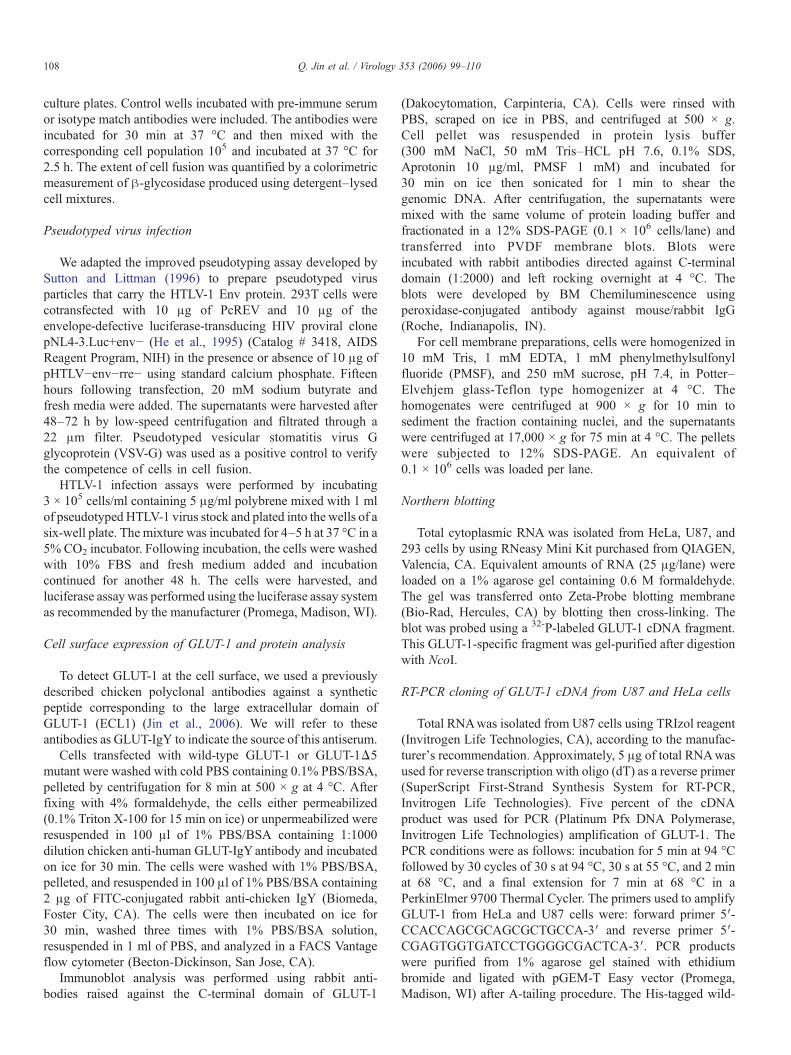
siRNA-mediated reduction of GLUT-1 protein expressionresults in blocking HTLV-1 Env-mediated fusion with HeLacells but not with U87 cells
To further examine the role of GLUT-1 protein expression inmediating HTLV-1 Env fusion in HeLa and U87 cell lines, weutilized siRNA to directly reduce GLUT-1 protein expression incultured cells. Three different siRNA molecules were designedagainst different regions of GLUT-1 mRNA. The siRNAdesigned against the open reading frame (ORF) of GLUT-1gave the best results in terms of reducing GLUT-1 proteinexpression. FACS analysis (data not shown) and western blotanalysis (Fig. 7A) confirmed the significant reduction of GLUT-1 protein expression following siRNA transfection. The ORFsiRNA caused ∼75% reduction in GLUT-1 protein expressionas measured by western blotting (Fig. 7A). As a negativecontrol, we transfected siRNA molecules not related to GLUT-1(Control siRNA) into the cell lines and considered that as thereference level of endogenous GLUT-1 expression. The effectof reducing GLUT-1 expression on HTLV-1 Env-mediatedfusion was examined using siRNA-transfected cells as targets.Efficient inhibition (∼75%) of Env-mediated fusion with HeLacells was consistently observed in every experiment performed.
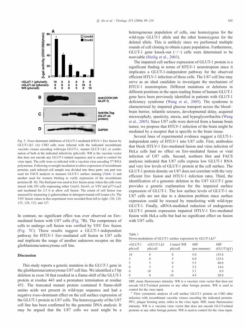
Fig. 5. Trans-dominant inhibition of GLUT-1-mediated HTLV-1 Env fusion byGLUT-1Δ5. (A) CHO cells were infected with the indicated recombinantvaccinia viruses encoding wild-type GLUT-1, mutant GLUT-1Δ5, or combi-nation of both at the indicated infectivity (pfu/cell). WR is the vaccinia vectorthat does not encode any GLUT-1-related sequence and is used to control forvirus input. The cells were co-infected with a vaccinia virus encoding T7 RNApolymerase. Following overnight incubation to allow expression of recombinantproteins, each infected cell sample was divided into three parts: one part wasused for FACS analysis to measure GLUT-1 surface staining (Table 1) andanother used for western blotting to verify expression of the recombinantproteins (B–D). The third part was used in Env fusion assay where the cells weremixed with 293 cells expressing either Unc63, Env63, or VSV and pT7-LacZand incubated for 2.5 h to allow cell fusion. The extent of cell fusion wasassessed by measuring β-galactosidase in detergent-treated cell lysates (A). TheVSV fusion values in this experiment were recorded from left to right: 130, 129,125, 128, 123, and 127.
Table 1Down-modulation of GLUT-1 surface expression by GLUT-1Δ5 a
vGLUT-1pfu/cell
vGLUT-1Δ5pfu/cell
Control WRpfu/cell
MIF(pre-immune)
MIF(GLUT-IgY)
10 0 0 5.0 155.85 0 5 6.0 125.65 5 0 5.5 60.80 5 5 5.2 14.60 10 0 5.1 8.90 0 10 4.9 20.8
MIF, mean fluorescence intensity. WR is a vaccinia virus vector that does notencode GLUT-related proteins or any other foreign protein. WR is used tocontrol for the virus input.a Flow cytometric analysis of cell surface GLUT-1 protein on CHO after
infection with recombinant vaccinia viruses encoding the indicated proteins.PFU, plaque forming units, refers to the virus input. MIF, mean fluorescenceintensity. WR is a vaccinia virus vector that does not encode GLUT-relatedproteins or any other foreign protein. WR is used to control for the virus input.
105Q. Jin et al. / Virology 353 (2006) 99–110
In contrast, no significant effect was ever observed on Env-mediated fusion with U87 cells (Fig. 7B). The competence ofcells to undergo cell fusion was verified by VSV Env fusion(Fig. 7C). These results suggest a GLUT-1-independentpathway for HTLV-1 Env-mediated cell fusion in U87 cellsand implicate the usage of another unknown receptor on thisglioblastoma/astrocytoma cell line.
Discussion
This study reports a genetic mutation in the GLUT-1 gene inthe glioblastoma/astrocytoma U87 cell line. We identified a 5 bpdeletion in exon 10 that resulted in a frame-shift of the GLUT-1protein at residue 443 and a premature termination at residue451. The truncated mutant protein contained 8 frame-shiftamino acids not present in wild-type sequence and had anegative trans-dominant effect on the cell surface expression ofthe GLUT-1 protein in U87 cells. The heterozygosity of the U87cell line has been confirmed by the genomic DNA analysis. Itmay be argued that the U87 cells we used might be a
heterogeneous population of cells, one homozygous for thewild-type GLUT-1 allele and the other homozygous for thedeleted allele. This is unlikely since we performed multiplerounds of cell cloning to obtain a pure population. Furthermore,GLUT-1 gene knock-out (−/−) cells were determined to benonviable (Heilig et al., 2003).
The impaired cell surface expression of GLUT-1 protein is asignificant finding in terms of HTLV-1 neurotropism since itimplicates a GLUT-1-independent pathway for the observedefficient HTLV-1 infection of these cells. The U87 cell line mayserve as an ideal candidate to investigate the mechanism ofHTLV-1 neurotropism. Different mutations or deletions indifferent positions in the open reading frame of human GLUT-1gene have been previously identified in patients with GLUT-1deficiency syndrome (Wang et al., 2005). The syndrome ischaracterized by impaired glucose transport across the blood–brain barrier, infantile seizures, developmental delay, acquiredmicrocephaly, spasticity, ataxia, and hypoglycorrhachia (Wanget al., 2005). Since U87 cells were derived from a human braintumor, we propose that HTLV-1 infection of the brain might bemediated by a receptor that is specific to the brain tissue.
Several lines of experimental evidence suggest a GLUT-1-independent entry of HTLV-1 into U87 cells. First, antibodiesthat block HTLV-1 Env-mediated fusion and virus infection ofHeLa cells had no effect on Env-mediated fusion or viralinfection of U87 cells. Second, northern blot and FACSanalyses indicated that U87 cells express low GLUT-1 RNAand very low levels of GLUT-1 protein at the cell surface. TheGLUT-1 protein density on U87 does not correlate with the veryefficient Env fusion and HTLV-1 infection rates. Third, theidentification of a genomic deletion in the U87 GLUT-1 geneprovides a genetic explanation for the impaired surfaceexpression of GLUT-1. The low surface levels of GLUT-1 onU87 cells are not due to a detection problem since surfaceexpression could be rescued by transfecting with wild-typeGLUT-1. Finally, siRNA-mediated reduction of endogenousGLUT-1 protein expression impaired HTLV-1 Env-mediatedfusion with HeLa cells but had no significant effect on fusionwith U87 cells.
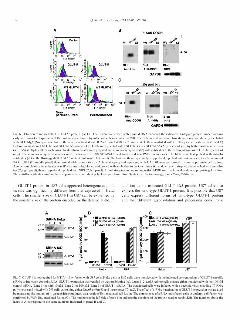
Fig. 6. Detection of intracellular GLUT-1Δ5 protein. (A) CHO cells were transfected with plasmid DNA encoding the indicated His-tagged proteins under vacciniaearly/late promoter. Expression of the protein was activated by infection with vaccinia virus WR. The cells were divided into two aliquots, one was directly incubatedwith GLUT-IgY (Non-permeabilized); the other was treated with 0.3% Triton X-100 for 20 min at 4 °C then incubated with GLUT-IgY (Permeabilized). (B and C)Heterodimerization of GLUT-1 and GLUT-1Δ5 proteins. CHO cells were infected with vGLUT-1 (wt), vGLUT-1Δ5 (Δ5), or co-infected by both recombinant viruses(wt +Δ5) at 10 pfu/cell for each virus. Total cellular lysates were prepared and immunoprecipitated (IP) with antibodies to the carboxy-terminus of GLUT-1 (detect wtonly). The immunoprecipitated samples were fractionated in 10% SDS-PAGE and transferred into PVDF membranes. The blots were first probed with anti-Hisantibodies (detect the His-tagged GLUT-1Δ5 mutant protein) (B, left panel). The blot was then sequentially stripped and reprobed with antibodies to the C-terminus ofWt GLUT-1 (B, middle panel) then normal rabbit serum (NRS). A final stripping and reprobing with GAPDH were performed to show appropriate gel loading.Another sample of cellular lysates was IP with Anti-His, blotted and probed with antibodies to the C-terminus (C, middle panel), stripped and reprobed with anti-His-tag (C, right panel), then stripped and reprobed with NRS (C, left panel). A final stripping and reprobing with GAPDHwere performed to show appropriate gel loading.The anti-His antibodies used in these experiments were rabbit polyclonal purchased from Santa Cruz Biotechnology, Santa Cruz, California.
106 Q. Jin et al. / Virology 353 (2006) 99–110
GLUT-1 protein in U87 cells appeared heterogeneous, andits size was significantly different from that expressed in HeLacells. The smaller size of GLUT-1 in U87 can be explained bythe smaller size of the protein encoded by the deleted allele. In
Fig. 7. GLUT-1 is not required for HTLV-1 Env fusion with U87 cells. HeLa cells orsiRNA or irrelevant control siRNA. GLUT-1 expression was verified by western blottcontrol siRNA (Lane 1) or with 10 nM (Lane 2) or 100 nM (Lane 3) of GLUT-1 siRpolymerase and mixed with 293 cells expressing either Unc63 or Env63 and the reporby measuring the amount of β-galactosidase produced as a result of Env-mediated ceconfirmed by VSV Env-mediated fusion (C). The numbers at the left side of each blolanes in A correspond to the same numbers indicated in panel B and C.
addition to the truncated GLUT-1Δ5 protein, U87 cells alsoexpress the wild-type GLUT-1 protein. It is possible that U87cells express different forms of wild-type GLUT-1 proteinand that different glycosylation and processing could have
U87 cells were transfected with the indicated concentrations of GLUT-1-specificing (A). Lanes 1, 2, and 3 refer to cells that are either transfected with the 100 nMNA. The transfected cells were infected with a vaccinia virus encoding T7 RNAter T7-lacZ. The effect of siRNA inactivation of GLUT-1 expression was assayedll fusion. The competence of siRNA-transfected cells to undergo cell fusion wast indicate the positions of the protein marker bands (Kd). The numbers above the
107Q. Jin et al. / Virology 353 (2006) 99–110
contributed to the inability of antibodies to interact with surfaceGLUT-1. This seems unlikely since transfection of U87 with thewild-type GLUT-1 cDNA results in enhancement of cell surfaceexpression of GLUT-1. The fact that GLUT-IgY antibodies thatpoorly stain U87 cells efficiently detect the protein uponpermeabilization of the cells argues against impaired antibodyrecognition (Fig. 6A). The U87 cell line is heterozygous for thedeleted mutant GLUT-1 allele, which results in a trans-dominant negative effect on the wild-type GLUT-1 protein inU87 cells. We found that GLUT-1Δ5 protein encoded by thedeleted allele physically interacts with the wild-type GLUT-1and reduces its availability at the cell surface. This was evidentby the trans-dominant inhibition effect on HTLV-1 Env fusionin CHO cells co-expressing recombinant GLUT-1Δ5 and thewild-type GLUT-1 proteins.
Previous studies described a C-terminal truncated rabbitGLUT-1 without transport activity (Oka et al., 1990). Oka et al.proposed that the C-truncated protein is integrated intomembranes in a fashion similar to the wild-type protein;however, it has a different conformation, with the inner glucosebinding site locked into an inward-facing form. The C-truncatedGLUT-1 described by Oka et al. is different from the truncatedGLUT-1Δ5 protein described in this study. Oka et al. deletedthe last 37 amino acids of the rabbit GLUT-1, and the mutantprotein does not contain frame-shift amino acids. The truncatedGLUT-1Δ5 protein lost 29 residues of the human GLUT-1 C-terminus and had 8 frame-shift amino acids, a net loss of 21amino acids at the C-terminus. Furthermore, the study by Oka etal. did not analyze surface expression of the truncated protein,and the conclusions made were based on the inability of their C-truncated mutant protein to transport glucose (Oka et al., 1990).
The role of the new motif in GLUT-1Δ5 protein is not clear.The potential new role of the 8 frame-shift amino acids couldprobably be related to cell surface transport of GLUT-1. Themotif sequence HLHLLQSS may function as an intracellularretention sequence. Deletion of this sequence may reveal itspotential role as a retention signal. Previous studies demon-strated that a single intramolecular disulfide bond promotestetramerization of GLUT-1 (Zottola et al., 1995). Zottola et al.demonstrated that the glucose transporter function is controlledby GLUT-1 oligomeric structure and identified Cys347 andCys421 as the residues involved in the disulfide bridge thatstabilizes the structure and accelerates transporter function(Zottola et al., 1995). Cys347 and Cys421 are intact in theGLUT-1Δ5 truncated protein, and their presence may explainthe ability of the mutant protein to form heterodimers with wild-type GLUT-1. However, it is possible that the loss of the last 21residues may have affected the efficiency of tetramerization ofGLUT-1 and subsequently cell surface transport. We haveconsistently observed faster gel mobility and a more diffusedprotein band for the endogenous wild-type GLUT-1 protein(encoded by the wild-type allele) in U87 cells. It is possible thatinteraction of GLUT-1Δ5 protein with wild-type GLUT-1 inU87 cells induces instability and degradation of the latter,resulting in impaired cell surface transport. The diffused fastermigrating GLUT-1 protein band in U87 cells was detected withantibodies raised to the last C-terminal residues of GLUT-1 that
are missing from GLUT-1Δ5 mutant protein. This indicatesexpression of the wild-type GLUT-1 allele in U87 cells.
In conclusion, the results of this study implicate that anotherunknown receptor is involved in HTLV-1 entry and infection ofU87 cells. The identification of this putative receptor(s) mayprovide important insight into the mechanism of retroviral entryand may have important implications for the neuropathology ofHTLV-1.
Materials and methods
Cells and viruses
HeLa cells, 293 cells, and CHO cells were obtained from theAmerican Type Culture Collection. U87 cells were obtainedfrom NIH AIDS Research and Reference Reagent Program (lot# 4 021998). The U87 cells were subjected to multiple rounds ofcell cloning in the presence of 10% cosmic calf serumsupplemented with glial cell medium containing G-5 supple-ment (GIBCO). Cells were grown in DMEM containing 10%cosmic calf serum (U87 cells) or 10% fetal bovine serum (293,HeLa and CHO), penicillin G (100 units/ml), and streptomycinsulfate (0.1 mg/ml) in a humidified incubator held at 5% CO2.All cell culture media were purchased from GIBCO.
The recombinant vaccinia viruses used in our Env-mediatedfusion assay have been previously described (Agrawal et al.,2002, 2004a, 2004b). Recombinant vaccinia viruses encodingeither GLUT-1 or GLUT-1Δ5 under vaccinia early/latepromoter have been constructed by transfecting HeLa cellswith pSC59 plasmid encoding either protein and infection withWR as previously described (Broder and Earl, 1997). Briefly,plaques were isolated after transfection/infection and screenedby dot blotting with a probe corresponding to GLUT-1sequences. The positive plaques were subjected to three cyclesof plaque purification, and the final positive plaques were usedto generate virus stocks on HeLa cells.
HTLV-1 Envy-mediated fusion
Env-mediated cell fusion was determined by a vaccine-basedreporter gene assay (Jin et al., 2006). The assay analyzes fusionbetween two distinct cell populations, one that expresses thepotential receptor along with the lacZ gene linked to the T7promoter and another that expresses HTLV-1 Env and T7 RNApolymerase encoded by a recombinant vaccinia virus. All cellpopulations were incubated overnight at 31 °C to allowexpression of the vaccinia-encoded proteins. Duplicate samplescontaining 105 target cells and same number of effector cellswere mixed in 96-well microtiter plates and incubated at 37 °Cfor 2.5 h. The extent of Envy-mediated fusion was assayed bycolorimetric measurement of β-glycosidase activity in deter-gent-treated cell lysates.
For blocking experiments, we used our IgY-GLUTantibodiesat increasing concentrations added to the target cells (105 HeLaor U87 cells). For Env blocking, increasing concentrations ofhuman monoclonal antibodies to HTLV-1 Env were mixed with105 Env-expressing cells in duplicate wells of 96-well tissue
108 Q. Jin et al. / Virology 353 (2006) 99–110
culture plates. Control wells incubated with pre-immune serumor isotype match antibodies were included. The antibodies wereincubated for 30 min at 37 °C and then mixed with thecorresponding cell population 105 and incubated at 37 °C for2.5 h. The extent of cell fusion was quantified by a colorimetricmeasurement of β-glycosidase produced using detergent–lysedcell mixtures.
Pseudotyped virus infection
We adapted the improved pseudotyping assay developed bySutton and Littman (1996) to prepare pseudotyped virusparticles that carry the HTLV-1 Env protein. 293T cells werecotransfected with 10 μg of PcREV and 10 μg of theenvelope-defective luciferase-transducing HIV proviral clonepNL4-3.Luc+env− (He et al., 1995) (Catalog # 3418, AIDSReagent Program, NIH) in the presence or absence of 10 μg ofpHTLV−env−rre− using standard calcium phosphate. Fifteenhours following transfection, 20 mM sodium butyrate andfresh media were added. The supernatants were harvested after48–72 h by low-speed centrifugation and filtrated through a22 μm filter. Pseudotyped vesicular stomatitis virus Gglycoprotein (VSV-G) was used as a positive control to verifythe competence of cells in cell fusion.
HTLV-1 infection assays were performed by incubating3 × 105 cells/ml containing 5 μg/ml polybrene mixed with 1 mlof pseudotyped HTLV-1 virus stock and plated into the wells of asix-well plate. The mixture was incubated for 4–5 h at 37 °C in a5% CO2 incubator. Following incubation, the cells were washedwith 10% FBS and fresh medium added and incubationcontinued for another 48 h. The cells were harvested, andluciferase assay was performed using the luciferase assay systemas recommended by the manufacturer (Promega, Madison, WI).
Cell surface expression of GLUT-1 and protein analysis
To detect GLUT-1 at the cell surface, we used a previouslydescribed chicken polyclonal antibodies against a syntheticpeptide corresponding to the large extracellular domain ofGLUT-1 (ECL1) (Jin et al., 2006). We will refer to theseantibodies as GLUT-IgY to indicate the source of this antiserum.
Cells transfected with wild-type GLUT-1 or GLUT-1Δ5mutant were washed with cold PBS containing 0.1% PBS/BSA,pelleted by centrifugation for 8 min at 500 × g at 4 °C. Afterfixing with 4% formaldehyde, the cells either permeabilized(0.1% Triton X-100 for 15 min on ice) or unpermeabilized wereresuspended in 100 μl of 1% PBS/BSA containing 1:1000dilution chicken anti-human GLUT-IgYantibody and incubatedon ice for 30 min. The cells were washed with 1% PBS/BSA,pelleted, and resuspended in 100 μl of 1% PBS/BSA containing2 μg of FITC-conjugated rabbit anti-chicken IgY (Biomeda,Foster City, CA). The cells were then incubated on ice for30 min, washed three times with 1% PBS/BSA solution,resuspended in 1 ml of PBS, and analyzed in a FACS Vantageflow cytometer (Becton-Dickinson, San Jose, CA).
Immunoblot analysis was performed using rabbit anti-bodies raised against the C-terminal domain of GLUT-1
(Dakocytomation, Carpinteria, CA). Cells were rinsed withPBS, scraped on ice in PBS, and centrifuged at 500 × g.Cell pellet was resuspended in protein lysis buffer(300 mM NaCl, 50 mM Tris–HCL pH 7.6, 0.1% SDS,Aprotonin 10 μg/ml, PMSF 1 mM) and incubated for30 min on ice then sonicated for 1 min to shear thegenomic DNA. After centrifugation, the supernatants weremixed with the same volume of protein loading buffer andfractionated in a 12% SDS-PAGE (0.1 × 10⁶ cells/lane) andtransferred into PVDF membrane blots. Blots wereincubated with rabbit antibodies directed against C-terminaldomain (1:2000) and left rocking overnight at 4 °C. Theblots were developed by BM Chemiluminescence usingperoxidase-conjugated antibody against mouse/rabbit IgG(Roche, Indianapolis, IN).
For cell membrane preparations, cells were homogenized in10 mM Tris, 1 mM EDTA, 1 mM phenylmethylsulfonylfluoride (PMSF), and 250 mM sucrose, pH 7.4, in Potter–Elvehjem glass-Teflon type homogenizer at 4 °C. Thehomogenates were centrifuged at 900 × g for 10 min tosediment the fraction containing nuclei, and the supernatantswere centrifuged at 17,000 × g for 75 min at 4 °C. The pelletswere subjected to 12% SDS-PAGE. An equivalent of0.1 × 106 cells was loaded per lane.
Northern blotting
Total cytoplasmic RNA was isolated from HeLa, U87, and293 cells by using RNeasy Mini Kit purchased from QIAGEN,Valencia, CA. Equivalent amounts of RNA (25 μg/lane) wereloaded on a 1% agarose gel containing 0.6 M formaldehyde.The gel was transferred onto Zeta-Probe blotting membrane(Bio-Rad, Hercules, CA) by blotting then cross-linking. Theblot was probed using a 32-P-labeled GLUT-1 cDNA fragment.This GLUT-1-specific fragment was gel-purified after digestionwith NcoI.
RT-PCR cloning of GLUT-1 cDNA from U87 and HeLa cells
Total RNAwas isolated from U87 cells using TRIzol reagent(Invitrogen Life Technologies, CA), according to the manufac-turer's recommendation. Approximately, 5 μg of total RNAwasused for reverse transcription with oligo (dT) as a reverse primer(SuperScript First-Strand Synthesis System for RT-PCR,Invitrogen Life Technologies). Five percent of the cDNAproduct was used for PCR (Platinum Pfx DNA Polymerase,Invitrogen Life Technologies) amplification of GLUT-1. ThePCR conditions were as follows: incubation for 5 min at 94 °Cfollowed by 30 cycles of 30 s at 94 °C, 30 s at 55 °C, and 2 minat 68 °C, and a final extension for 7 min at 68 °C in aPerkinElmer 9700 Thermal Cycler. The primers used to amplifyGLUT-1 from HeLa and U87 cells were: forward primer 5′-CCACCAGCGCAGCGCTGCCA-3′ and reverse primer 5′-CGAGTGGTGATCCTGGGGCGACTCA-3′. PCR productswere purified from 1% agarose gel stained with ethidiumbromide and ligated with pGEM-T Easy vector (Promega,Madison, WI) after A-tailing procedure. The His-tagged wild-
109Q. Jin et al. / Virology 353 (2006) 99–110
type GLUT-1 molecules were constructed by using the sameforward primer and the reverse primer: 5′-GGCTCGAGT-CAATGGTGATGGTGATGATGCACTTGGGAAT-CAGCCCC-3′. To construct a His-tagged GLUT-1 clonedfrom U87 cells, we used the same forward primer asdescribed above and the following reverse His-taggedprimer: 5′-GGCTCGAGTCAATGGTGATGGTGATGATGG-GAACTTTGAAGTAGGTG-3′. The purified fragmentscorresponding to the encoded GLUT-1 were cloned intopSC59 vector (described in Alkhatib et al., 1996) under thecontrol of vaccinia early/late promoter. The DNA sequenceof cloned DNA was verified by direct sequencing. Theplasmid DNA containing GLUT-1 from either HeLa or U87cells was transfected into CHO cells by DOTAP LiposomalTransfection Reagent (Roche, Indianapolis, IN). Expressionof GLUT-1 was activated by infection with vaccinia virus.
Cloning of genomic DNA fragments of GLUT-1 gene
Genomic DNAwas isolated from U87 and HeLa cells usingDNeasy Tissue Kit (QIAGEN, CA), according to the manufac-turer's recommendation. The concentration and purity ofgenomic DNA samples were measured, and 1 μg of genomicDNA was used for PCR. For GLUT-1 cloned from HeLa andU87 cells, the forward primer (nucleotides 1292–1312) used is:5′-CTACGTTCTTCATCATCTTCACT-3′ and the reverse-primer (nucleotides 1578–1598) used: 5′-GGAGCCCC-AGGCCCGGCTCGG-3′. PCR products were ligated withpGEM-T Easy vector, and positive clones were verified bydirect DNA sequencing.
siRNA experiments
The sequence of the synthetic siRNA complementary to theGLUT-1 open reading frame is 5′-GGGCCAAGAGUGUG-CUAAAGAAGC-3′ and complementary to the 3′UTR is 5′-GGCUGGACCUAUGUCCUAAGGACACAC-3′ (IntegratedDNA Technologies, Coralville, IA). These siRNA moleculeswere transfected together into HeLa or U87 cells using anoligofectamine reagent purchased from Invitrogen (Invitrogen,San Diego, CA). HeLa or U87 cells were plated at1.5 × 105 cells/well in a 6-well plate using serum-free mediumwithout antibiotics. The siRNA (10 or 100 nM) was gentlymixed with the oligofectamine reagent and incubated for 20 minthen added to the corresponding well. After 4-h incubation at37 °C, 0.5 ml of 3× serum was added. Following incubation for72 h in a 37 °C incubator, the transfected cells were washed andinfected with pT7-lacZ reporter. Separate populations of 293cells were infected with vaccinia viruses encoding either Unc63or Env63 and T7 RNA polymerase. At 15 h post-infection, theEnv-expressing cells were mixed (1:1) with the siRNA-transfected cells and incubated for 2.5 h to allow cell fusion.The effect of siRNA on Env-mediated fusion was assayed bymeasuring β-galactosidase produced. The control siRNA usedas a negative control was a scrambled sequence 5′-CUUCCU-CUCUUUCUCUCCCUUGUGA-3′ purchased from IntegratedDNA Technologies, Coralville, IA.
Acknowledgments
We thank Margaret Bauer for comments and the vector corefacility of Indiana University School of Medicine for assistancein constructing the Unc63 mutant. We thank Dr. Steven Foungfor the human monoclonal antibodies. We also thank Dr. M.Dokhelar, France for providing the HTLV-1 envelope DNA.The following reagent was obtained through the AIDS Researchand Reference Reagent Program, Division of AIDS, NIAID,NIH: pNL4-3.Luc.R−E− from Dr. Nathaniel Landau. This workwas supported by a grant from the National Cancer Institute1R21CA98095-01 awarded to GA and a fellowship from theChinese Scholarship Council, Beijing, China awarded to QJ.
References
Agrawal, L., Vanhorn-Ali, Z., Alkhatib, G., 2002. Multiple determinants areinvolved in HIV coreceptor use as demonstrated by CCR4/CCL22interaction in peripheral blood mononuclear cells (PBMCs). J. LeukocyteBiol. 72 (5), 1063–1074.
Agrawal, L., Lu, X., Qingwen, J., VanHorn-Ali, Z., Nicolescue, V., McDermott,D., Murphy, P.M., Alkhatib, G., 2004a. Role for CCR5Δ32 protein inresistance to R5, R5X4, and x4 human immunodeficiency virus type 1 inprimary CD4+ cells. J. Virol. 78 (5), 2277–2287.
Agrawal, L., VanHorn-Ali, Z., Berger, E.A., Alkhatib, G., 2004b. Specificinhibition of HIV-1 coreceptor activity by synthetic peptides correspondingto the predicted extracellular loops of CCR5. Blood 103 (4), 1211–1217.
Alkhatib, G., Broder, C.C., Berger, E.A., 1996. Cell type-specific fusioncofactors determine human immunodeficiency virus type 1 tropism for T-cell lines versus primary macrophages. J. Virol. 70, 5487–5494.
Broder, C.C., Earl, P.L., 1997. Design and construction of recombinant vacciniaviruses. Methods Mol. Biol. 62, 173–197.
Coskun, A.K., Sutton, R.E., 2005. Expression of glucose transporter 1 conferssusceptibility to human T-cell leukemia virus envelope-mediated fusion.J. Virol. 79 (7), 4150–4158.
Feuer, G., Green, P.L., 2005. Comparative biology of human T-cell lymphotropicvirus type 1 (HTLV-1) and HTLV-2. Oncogene 24 (39), 5996–6004.
Gallo, R.C., 2005. History of the discoveries of the first human retroviruses:HTLV-1 and HTLV-2. Oncogene 24 (39), 5926–5930.
Hadlock, K.G., Rowe, J., Perkins, S., Bradshaw, P., Song, G.Y., Cheng, C.,Yang, J., Gascon, R., Halmos, J., Rehman, S.M., McGrath, M.S., Foung,S.K., 1997. Neutralizing human monoclonal antibodies to conformationalepitopes of human T-cell lymphotropic virus type 1 and 2 gp46. J. Virol.71 (8), 5828–5840.
He, J., Choe, S., Walker, R., Di Marzio, P., Morgan, D.O., Landau, N.R., 1995.Human immunodeficiency virus type 1 viral protein R (Vpr) arrests cells inthe G2 phase of the cell cycle by inhibiting p34cdc2 activity. J. Virol. 69(11), 6705–6711.
Heilig, C., Brosius, F., Siu, B., Concepcion, L., Mortensen, R., Heilig, K., Zhu,M., Weldon, R., Wu, G., Conner, D., 2003. Implications of glucosetransporter protein type 1 (GLUT1)-haplodeficiency in embryonic stem cellsfor their survival in response to hypoxic stress. Am. J. Pathol. 163 (5),1873–1885.
Jassal, S.R., Pohler, R.G., Brighty, D.W., 2001. Human T-cell leukemia virustype 1 receptor expression among syncytium-resistant cell lines revealedby a novel surface glycoprotein-immunoadhesion. J. Virol. 75 (17),8317–8328.
Jin, Q., Agrawal, L., Vanhorn-Ali, Z., Alkhatib, G., 2006. Infection of CD4(+) Tlymphocytes by the human T cell leukemia virus type 1 is mediated by theglucose transporter GLUT-1: evidence using antibodies specific to thereceptor's large extracellular domain. Virology.
Jones, K.S., Petrow-Sadowski, C., Bertolette, D.C., Huang, Y., Ruscetti, F.W.,2005. Heparan sulfate proteoglycans mediate attachment and entry of humanT-cell leukemia virus type 1 virions into CD4+ T cells. J. Virol. 79 (20),12692–12702.

110 Q. Jin et al. / Virology 353 (2006) 99–110
Manel, N., Kim, F.J., Kinet, S., Taylor, N., Sitbon, M., Battini, J.L., 2003. Theubiquitous glucose transporter GLUT-1 is a receptor for HTLV. Cell 115 (4),449–459.
Manel, N., Battini, J.L., Taylor, N., Sitbon, M., 2005. HTLV-1 tropism andenvelope receptor. Oncogene 24 (39), 6016–6025.
Mueckler, M., 1994. Facilitative glucose transporters. Eur. J. Biochem. 219 (3),713–725.
Oka, Y., Asano, T., Shibasaki, Y., Lin, J.L., Tsukuda, K., Katagiri, H., Akanuma,Y., Takaku, F., 1990. C-terminal truncated glucose transporter is locked intoan inward-facing form without transport activity. Nature 345 (6275),550–553.
Sutton, R.E., Littman, D.R., 1996. Broad host range of human T-cell leukemia
virus type 1 demonstrated with an improved pseudotyping system. J. Virol.70 (10), 7322–7326.
Wang, D., Pascual, J.M., Yang, H., Engelstad, K., Jhung, S., Sun, R.P., De Vivo,D.C., 2005. Glut-1 deficiency syndrome: clinical, genetic, and therapeuticaspects. Ann. Neurol. 57 (1), 111–118.
Yoshida, M., 2005. Discovery of HTLV-1, the first human retrovirus, its uniqueregulatory mechanisms, and insights into pathogenesis. Oncogene 24 (39),5931–5937.
Zottola, R.J., Cloherty, E.K., Coderre, P.E., Hansen, A., Hebert, D.N.,Carruthers, A., 1995. Glucose transporter function is controlled bytransporter oligomeric structure. A single, intramolecular disulfidepromotes GLUT1 tetramerization. Biochemistry 34 (30), 9734–9747.