Corneal Langerhans Cell Dynamics After Herpes Simplex Virus Reactivation
Upload
independentCategory
view
0download
0
Human Glioblastoma Multiforme: p53 Reactivation by aNovel MDM2 InhibitorBarbara Costa1*, Sara Bendinelli1, Pamela Gabelloni1, Eleonora Da Pozzo1, Simona Daniele1,
Fabrizio Scatena2, Renato Vanacore2, Pietro Campiglia3, Alessia Bertamino4, Isabel Gomez-Monterrey4,
Daniela Sorriento5, Carmine Del Giudice5, Guido Iaccarino6,7, Ettore Novellino4, Claudia Martini1*
1Department of Pharmacy, University of Pisa, Pisa, Italy, 2U.O. Immunohaematology 2, Cisanello Hospital, Pisa, Italy, 3Department of Pharmaceutical Science, Division of
BioMedicine, University of Salerno, Salerno, Italy, 4Department of Pharmaceutical and Toxicological Chemistry, University of Naples Federico II, Naples, Italy, 5Department
of Advanced Biomedical Sciences, University of Naples Federico II, Naples, Italy, 6Department of Medicine and Surgery, University of Salerno, Salerno, Italy, 7 IRCCS
Multimedica, Milano, Italy
Abstract
Cancer development and chemo-resistance are often due to impaired functioning of the p53 tumor suppressor throughgenetic mutation or sequestration by other proteins. In glioblastoma multiforme (GBM), p53 availability is frequentlyreduced because it binds to the Murine Double Minute-2 (MDM2) oncoprotein, which accumulates at high concentrations intumor cells. The use of MDM2 inhibitors that interfere with the binding of p53 and MDM2 has become a valid approach toinhibit cell growth in a number of cancers; however little is known about the efficacy of these inhibitors in GBM. We reportthat a new small-molecule inhibitor of MDM2 with a spirooxoindolepyrrolidine core structure, named ISA27, effectivelyreactivated p53 function and inhibited human GBM cell growth in vitro by inducing cell cycle arrest and apoptosis. Inimmunoincompetent BALB/c nude mice bearing a human GBM xenograft, the administration of ISA27 in vivo activated p53,inhibited cell proliferation and induced apoptosis in tumor tissue. Significantly, ISA27 was non-toxic in an in vitro normalhuman cell model and an in vivo mouse model. ISA27 administration in combination with temozolomide (TMZ) produced asynergistic inhibitory effect on GBM cell viability in vitro, suggesting the possibility of lowering the dose of TMZ used in thetreatment of GBM. In conclusion, our data show that ISA27 releases the powerful antitumor capacities of p53 in GBM cells.The use of this MDM2 inhibitor could become a novel therapy for the treatment of GBM patients.
Citation: Costa B, Bendinelli S, Gabelloni P, Da Pozzo E, Daniele S, et al. (2013) Human Glioblastoma Multiforme: p53 Reactivation by a Novel MDM2Inhibitor. PLoS ONE 8(8): e72281. doi:10.1371/journal.pone.0072281
Editor: Guillermo Velasco, Complutense University, Spain
Received September 11, 2012; Accepted July 15, 2013; Published August 19, 2013
Copyright: � 2013 Costa et al. This is an open-access article distributed under the terms of the Creative Commons Attribution License, which permitsunrestricted use, distribution, and reproduction in any medium, provided the original author and source are credited.
Funding: Funding for this study was provided by the Italian Ministry of University and Scientific Research (PRIN2009-prot. 20098SJX4F). The funders had no rolein study design, data collection and analysis, decision to publish, or preparation of the manuscript.
Competing Interests: The authors have declared that no competing interests exist.
* E-mail: [email protected] (CM); [email protected] (BC)
Introduction
Glioblastoma multiforme (GBM) is the most common and
aggressive brain tumor in humans, and despite technical advances
in neurosurgery and clinical neuro-oncology, the prognosis for
GBM patients remains very poor [1–8]. Most patients die within
one year of diagnosis and are generally insensitive to current
therapeutic genotoxic interventions [1–8]. In the majority of GBM
cases, resistance to such genotoxic modalities has been attributed
to the attenuation of p53 function by alterations within the p53
signalling axis, including the overexpression of Murine Double
Minute-2 (MDM2) [9–13]. The MDM2 oncoprotein, a major
physiological negative regulator of p53, can bind to the p53
transactivation domain and interfere with the transcriptional
regulatory mechanisms of p53 [9–13]. MDM2 is also an E3
ubiquitin ligase that promotes p53 proteasomal degradation. For
this reason, inhibition of the interaction between MDM2 and p53
to reactivate endogenous p53 activity offers the opportunity for
therapeutic intervention, particularly in GBMs. In GBMs, the p53
gene is relatively infrequently mutated; however, wild-type p53
remains dysfunctional due to overexpressed MDM2 [9,10].
Intensive work on different classes of MDM2 inhibitors has
proven their therapeutic utility as activators of p53 in multiple
tumor models [14–17]. Indeed, it has been demonstrated that a
number of small-molecule MDM2 inhibitors can disrupt the
MDM2-p53 interaction, release p53 from negative control and
activate the p53 pathway, leading to cell cycle arrest and apoptosis
in a number of solid cancers and haematological malignancies
[14–17]. Moreover, many laboratories have shown that MDM2
inhibitors can synergise with conventional chemotherapeutic
agents, resulting in enhanced efficacy [18–22]. Interestingly,
MDM2 inhibitors have been reported to induce cancer cell
apoptosis even without the concomitant application of genotoxic
stimuli [17,18,20]. Little is known about the effects of MDM2
inhibitors on the in vitro growth of GBM cells. Recently, Nutlin-3,
the first potent MDM2 small-molecule inhibitor identified [23],
and new D-peptide derivatives [14,24] were reported to be
effective at inhibiting GBM cell growth in vitro [14,24,25],
suggesting the validity of this experimental approach for the
treatment of GBM.
In the present study, we investigated the responsiveness of
human GBM cell lines to a novel small-molecule MDM2 inhibitor
with a spirooxoindolepyrrolidine core structure, named ISA27,
PLOS ONE | www.plosone.org 1 August 2013 | Volume 8 | Issue 8 | e72281

which has been recently shown by nuclear magnetic resonance
(NMR) analysis to efficiently dissociate the reconstituted human
MDM2-p53 complex [26]. Consistently, ISA27 activated the p53
pathway in GBM cells and elicited the dose- and time-dependent
inhibition of cell growth. ISA27 induced apoptosis and evoked
cellular senescence, indicating that ISA27 promotes a pleiotropic
anticancer effect in the GBM cells. The administration of ISA27
in vivo efficiently inhibited tumor growth in nude mice bearing a
human GBM xenograft. Significantly, ISA27 was non-toxic both
in vitro in a normal human cell model and in vivo in a mouse model.
Materials and Methods
1. MaterialsISA27 was synthesised as previously reported [26]. Nutlin-3,
carbonylcyanide-m-chlorophenylhydrazone (CCCP), Nonidet P-
40 (NP-40) and cycloheximide (CHX) were obtained from Sigma–
Aldrich, Milano, Italy. Propidium iodide (PI) and the fluorescent
dye, 5,50,6,60-tetrachloro-1,10,3,30-tetraethylbenzimidazolcarbo-
cyanine iodide (JC-1) were obtained from Molecular Probes,
Invitrogen, Milano, Italy. The 3-(4,5-dimethylthiazol-2-yl)-5-(3-
carboxymethoxyphenyl)-2-(4-sulfophenyl)-2H-tetrazolium (MTS)
assay kit was from Promega Italia, Milano, Italy. The RNeasyHMini Kit was from Qiagen, Milano, Italy and the ProtoScriptHcDNA Synthesis Kit was obtained from Biolabs, Euroclone,
Milano, Italy. The mitochondrial fractionation Active MotifH Kit
was purchased from Active Motif, Rixensart, Belgium and the
Platinum Human Cytochrome C ELISA was obtained from
Bender MedSystems GmbH, Vienna, Austria. Antibodies against
p53 (FL-393) and MDM2 (C-18) were from Santa Cruz
Biotechnology.
2. GBM Cell Line Culture and Preparation of Cells fromPeripheral Blood
The U87MG, T98G and U343MG cell lines were obtained
from the National Institute for Cancer Research of Genoa (Italy),
American Type Culture Collection (USA) and Cell Lines Service
GmbH (Germany), respectively. Each cell line was monitored for
DNA profiling. The U87MG and T98G cells were cultured in
RPMI medium and Minimum essential medium Eagle, respec-
tively, supplemented with 10% FBS, 2 mM L-glutamine, 100 U/
mL penicillin, 100 mg/mL streptomycin and 1% non-essential
amino acids at 37uC in 5% CO2. The U343MG cells were
cultured in Minimum essential medium Eagle with 2 mM L-
glutamine and Earle’s BSS adjusted to contain 1.5 g/L sodium
bicarbonate and supplemented with 10% FBS, 100 U/mL
penicillin, 100 mg/mL streptomycin, 1% non-essential amino
acids and 1.0 mM sodium pyruvate at 37uC in 5% CO2.
Mononuclear cell preparation was performed according to the
method of Boyum [27]. The final cell pellet was suspended in
complete RPMI 1690 media supplemented with 15% FBS, 2 mM
L-glutamine, 100 U/mL penicillin and 100 mg/mL streptomycin.
To evaluate cell populations, random cell samples (n = 7) were
employed for flow cytometric analysis.
3. Cell TreatmentsThe human GBM cells were seeded at 5,000 cells/cm2. After
24 h, the culture medium was replaced with fresh medium
containing MDM2 inhibitor solubilised in DMSO for the
indicated incubation times. DMSO was added to control cells
(,1% v/v). For short-term treatment (up to 24 h), GBM cells were
incubated with increasing concentrations or a fixed concentration
of MDM2 inhibitor corresponding to the concentration that
inhibited 50% (IC50 value) of GBM cell survival/growth; for long-
term treatment (up to 5 days), U87MG cells and lymphomono-
cytes were incubated with 2.5 mM ISA27 or 10 mM Nutlin-3.
4. Analysis of p53 Protein StabilisationFollowing GBM cell treatment with MDM2 inhibitor, stabili-
sation of the p53 protein was evaluated as previously described
[28–30]. Briefly, GBM cells were treated with DMSO (control) or
MDM2 inhibitor for 4, 6, 8 and 12 h and then lysed for 60 min at
4uC by adding RIPA buffer (9.1 mM NaH2PO4, 1.7 mM
Na2HPO4, 150 mM NaCl, pH 7.4, 0.5% sodium deoxycholate,
1% Nonidet P-40, 0.1% SDS and a protease inhibitor cocktail).
Equal amounts of cell extracts (40 mg) from MDM2 inhibitor-
treated and untreated cells were diluted in Laemmli solution,
resolved by SDS-PAGE (8.5%), transferred to PVDF membranes
and probed overnight at 4uC with a primary anti-p53 (FL-393,
1:500) antibody. The primary antibody was detected using anti-
rabbit IgG light chains conjugated to peroxidase (diluted
1:10,000). The peroxidase was detected using a chemiluminescent
substrate (ECL, Perkin Elmer). Western blot analysis was also
performed using lysates from MDM2 inhibitor-treated and
untreated GBM cells in the absence and presence of the protein
synthesis inhibitor CHX (50 mM).
The relative quantification of p53 mRNA was performed by
real-time reverse transcription polymerase chain reaction (real-
time RT-PCR) as previously described [31] in MDM2 inhibitor-
treated and untreated GBM cells. In brief, total RNA was isolated
using the RNeasyH Mini Kit. The purity of the RNA samples was
determined by measuring the absorbance at 260:280 nm. cDNA
Table 1. Real-time RT-PCR: primer nucleotide sequences, position in mRNA sequence and amplicon length.
Name and other designationsAccess. numb.mRNA length Primer nucleotide sequences
Primer position inmRNA sequence
Tm(uC)
Ampliconlenght
Mdm2, p53 E3 ubiquitin protein ligasehomolog (mouse) (MDM2)
NM_002392.47472 pb
FOR: 59-TCTAGGAGATTTGTTTGGCGT-39;REV: 59-TCACAGATGTACCTGAGTCC-3
Exon 4 (548–568)Exon 6 (653–672)
6060
125 pb
Cyclin-dependent kinase inhibitor 1A (p21,Cip1) (CDKN1A)
NM_0003892175 pb
FOR: 59-TGCCGAAGTCAGTTCCTTG -39;REV: 59-CATGGGTTCTGACGGACATC-39
Exon 3 (50–68)Exon 4 (144–163)
6062
134 pb
Bcl2 binding component 3 (BBC3), JFY1, PUMA NM_0011272401839 pb
FOR: 59-GAGGAGGAACAGTGGGC-39;REV: 59- CTAATTGGGCTCCATCTCGG-39
Exon 4 (652–668)Exon 5 (830–849)
5662
198 pb
Tumor protein p53, antigen NYCO-13, cellulartumor antigen p53, p53 tumor suppressor
NM_0011261122588 pb
FOR: 59CTTTGAGGTGCGTGTTTGTG-39;REV: 59 GTGGTTTCTTCTTTGGCTGG-39
Exon 8 (1006–1025)Exon 9 (1147–1166)
6060
161 pb
Actin,beta (ACTB) NM_001101.31852 pb
FOR: 59-GCACTCTTCCAGCCTTCCTTCC-39REV-59-GAGCCGCCGATCCACACG-39
Exon 4 (862–884)Exon 6 (1097–1115)
7062
254 pb
doi:10.1371/journal.pone.0072281.t001
Anticancer Effect of a New MDM2 Inhibitor
PLOS ONE | www.plosone.org 2 August 2013 | Volume 8 | Issue 8 | e72281

synthesis was performed with 500 ng of RNA using the
QuantitectH reverse transcriptase kit. The primers used for the
RT-PCR were designed to span intron/exon boundaries to ensure
that products did not include genomic DNA (see Table 1). The
RT-PCR reactions consisted of 12.5 mL of BrilliantH II SYBRHGreen premix, 2.5 mL of both the forward and reverse primers (at
a 10 mM concentration), 3 mL of cDNA and 4.5 mL of H2O. All
reactions were performed for 40 cycles using the following
temperature profiles: 98uC for 30 seconds (initial denaturation);
55uC for 30 seconds (annealing) and 72uC for 3 seconds
(extension). b-actin was used as the housekeeping gene. PCR
specificity was determined using both a melting curve analysis and
gel electrophoresis, and the data were analysed by the standard
curve method. p53 mRNA levels for each sample were normalised
against b-actin mRNA levels, and relative expression was
calculated using the Ct value.
The amount of p53-MDM2 complex was determined using co-
immunoprecipitation experiments; U87MG cells were treated with
DMSO (control) or 2.5 mM ISA27 or 10 mM Nutlin-3 for 8 h.
One milligram of cell lysates, obtained as described above, was
precleared with protein A-Sepharose (1 h at 4uC) to precipitate
and eliminate IgG. Samples were then centrifuged for 10 min at
4uC (14,0006g). The supernatants were incubated with an anti-
MDM2 antibody (5 mg/sample) overnight at 4uC under constant
rotation and then immunoprecipitated with protein A-Sepharose
(2 h at 4uC). After washing, the immunocomplexes were
resuspended in Laemmli solution and boiled for 5 min, resolved
by SDS-PAGE (8.5%), transferred to PVDF membranes and
probed overnight at 4uC with primary antibodies to p53 (FL-393,
1:500) or MDM2 (C-18, 1:500) as described above.
5. Relative mRNA Quantification of p53 Target GenesThe relative mRNA quantification of p53 target genes was
performed by real-time RT-PCR as described above. Specific
primers for amplification of p21, MDM2 and PUMA mRNA were
designed to include intron/exon boundaries and are reported in
Table 1.
Figure 1. ISA27 inhibits the growth/survival of GBM cell lines expressing wild-type p53. GBM cells with wild-type p53 (U87MG, U343MG)were exposed to a range of ISA27 or Nutlin-3 concentrations for 24 h. After incubation, GBM cell viability was determined by MTS assay. A and B)Effect of ISA27 on the growth/survival of GBM cell lines expressing wild-type p53: curves for U87MG and U343MG cell samples were generated using asigmoidal dose-response curve model (GraphPad Prism 4 software) from which the IC50 values were derived. ISA27-treated cells (.), Nutlin-3-treatedcells (&); C) Effect of ISA27 on the growth/survival of the GBM T98G cell line expressing mutant p53: T98G cells were incubated with increasingconcentrations of ISA27 or Nutlin-3 (ranging from 1 mM to 25 mM), and cell viability was measured after 24 h by MTS assay. ISA27-treated cells (.),Nutlin-3-treated cells (&). The results are expressed as the percentages of cell viability with respect to the vehicle-treated sample for each cell line;the viability of the vehicle-treated sample was arbitrarily set at 100%. Points, means of three independent experiments performed in duplicate; bars,SEM.doi:10.1371/journal.pone.0072281.g001
Anticancer Effect of a New MDM2 Inhibitor
PLOS ONE | www.plosone.org 3 August 2013 | Volume 8 | Issue 8 | e72281

6. Cell Survival/growth AnalysisThe effects of MDM2 inhibitor treatment on U87MG cell
survival/growth were determined by MTS conversion and Trypan
blue exclusion assays [32,33]. The MTS assay was used to
establish the MDM2 inhibitor concentration that inhibited 50%
(IC50 value) of GBM cell survival/growth after 24 h of treatment
and was performed according to the manufacturer’s instructions.
Sigmoidal dose-response curves were generated using GraphPad
Prism 4 software (GraphPad Software Inc., San Diego, CA), from
which the IC50 values were derived. Trypan blue dye exclusion
was used to examine the long-term effects of MDM2 inhibitor
treatment on U87MG cell and lymphomonocyte viability at the
indicated times.
7. Cell Cycle AnalysisCell cycle analysis was performed by flow cytometry (FACSCa-
libur flow cytometer, Becton Dickinson, USA) [32,34]. Briefly,
after treatment, detached and trypsinized adherent cells were
collected by centrifugation at 2006g for 5 min. Pellets were gently
suspended in 500 ml of a hypotonic fluorochrome solution [PI
50 mg/ml in 0.1% (w/v) sodium citrate plus 0.01% (v/v) NP-40
and 10 mg/ml DNase-free RNase in bi-distilled water]. After
incubation in the dark at 37uC in 5% CO2 for 30 min, cell samples
were stored on ice and analysed for DNA content on a logarithmic
scale by flow cytometry. Ten thousand events per sample were
acquired. The percentage of PI-stained untreated and MDM2
inhibitor-treated cells was measured in each cell cycle phase using
the ModFit LT software program.
8. Cellular Senescence AssaysThe senescence marker, Senescence-associated beta-galactosi-
dase (SA-b-Gal), was detected as previously described [35], and
the cells were then washed in PBS (16) and photographed at 1006magnification. Images from random light microscopic fields (5
fields per well) were captured, and the cells were counted using
ImageJ software (ImageJ Software, version 1.41o; USA). The
average cell size was assessed using the Scepter 2.0 handheld
automated cell counter (Millipore, Billerica, MA, USA).
Figure 2. ISA27 increases p53 protein levels in GBM cell lines. GBM cells (U87MG, U343MG) were treated with ISA27 or Nutlin-3 for theindicated incubation times, and protein levels of p53 were analysed in whole-cell lysates by Western blot. One representative Western blot ispresented (upper panel) for each cell line. The blots show that the antibody to p53 (FL-393; Santa Cruz Biotechnology) recognised a single specificband at approximately 55 kDa, corresponding to the molecular weight of the p53 protein; b-actin is used as the loading control. Densitometricanalyses of Western blots (lower panel) demonstrated that ISA27 induced a significant enhancement of p53 protein levels in U87MG or U343MG cellswith a maximal effect at the 8 h incubation time.doi:10.1371/journal.pone.0072281.g002
Anticancer Effect of a New MDM2 Inhibitor
PLOS ONE | www.plosone.org 4 August 2013 | Volume 8 | Issue 8 | e72281

9. Analysis of Apoptotic ParametersMitochondrial membrane potential (Dym). Changes in
Dym were assessed using the fluorescent dye, JC-1, as previously
described [32,34], and fluorescence was analysed by fluocytome-
try. As a positive control, a cell pellet was incubated with the
uncoupling agent, CCCP (50 mM).
Cytosolic cytochrome C release from
mitochondria. The cytosolic fraction from untreated and
MDM2 inhibitor-treated cells was obtained using the Active
MotifH mitochondrial fractionation kit according to the manufac-
turer’s instructions. The cytosolic protein content was determined
as previously described [36], and the cytochrome C content was
determined using the Platinum Human Cytochrome C ELISA.
The minimum detectable dose of cytochrome C was 0.05 ng/mL.
DNA fragmentation analysis. DNA fragmentation was
estimated by evaluating PI staining of DNA using FACS. The
percentage of apoptotic cells was estimated as previously described
[32,34]. The percentage of cells in the sub-G0 fraction was
determined using the ModFit LT software program.
10. siRNA Mediated Inhibition of p21 Gene ExpressionThe p21siRNA and control siRNA were obtained from Santa
Cruz Biotecnology (Heidelberg, Germany). The p21siRNA was
transfected with siRNA transfection reagent to a final concentra-
tion of 50 nM, following the manufacturer’s protocol. In parallel
Figure 3. ISA27 induces the stabilisation of p53 protein levels by decreasing the interaction of p53 with MDM2 in GBM cells. A and B)Effect of ISA27 on p53 gene expression: U87MG and U343MG cells were treated with ISA27 or Nutlin-3 for 6, 12 and 24 h. After incubation, the relativequantification of p53 mRNA was performed by real-time RT-PCR. In U87MG cells, p53 mRNA levels did not change within 12 h of cell treatment; inU343 MG cells, p53 mRNA levels did not change during any of the incubation times. 3C) ISA27 sustains p53 protein expression in the presence of CHX:U87MG cells were incubated with 50 mM CHX alone or CHX in combination with ISA27 or Nutlin-3 for 0.5, 1, 2, 4 and 6 h. After incubation, cell lysateswere used for Western blot analysis. One representative Western blot is presented for each cell treatment. The inhibition of protein synthesis bytreatment with CHX alone blocked p53 protein expression after a 4-h treatment. ISA27 extended the stability of p53 in the presence of CHX. 3D) ISA27reduces MDM2-p53 complex formation: U87MG cells were incubated with ISA27 or Nutlin-3 for 8 h followed by immunoprecipitation using an anti-MDM2 antibody. The MDM2-p53 complex and the relative input of the proteins were detected by immunoblot; a significant reduction in MDM2-p53complex formation was shown in U87MG cells following 8 h of treatment with ISA27.doi:10.1371/journal.pone.0072281.g003
Anticancer Effect of a New MDM2 Inhibitor
PLOS ONE | www.plosone.org 5 August 2013 | Volume 8 | Issue 8 | e72281

to each silencing experiment, an ineffective sequence of RNA has
been used as negative control. Transfected cells were used 36 h
after siRNA transfection. The silence efficacy was verified by both
real time PCR and Western blotting analysis. For Western blotting
analysis, U87MG cells were lysed and proteins, resolved by SDS-
PAGE electrophoresis, were probed with an anti-p21 antibody
(H164, sc-756, Santa Cruz Biotecnology). After p21 genetic
inhibition, we incubated U87MG cells with ISA27 for 24 h and
assessed cell viability, cell cycle profile, Dym and phosphatidyl-
serine externalization (PE). The measurement of PE was
performed by the MuseTM Cell Analyzer using the fluorescent
dyes Annexin V and 7-AAD. Annexin-V is used as a probe to
detect cells that have expressed phosphatidylserine on the cell
surface. 7-AAD is excluded from viable cells, since it does not pass
through intact cell membranes. We simultaneously measured the
expression of phosphatidylserine on the cell surface and the
cellular plasma membrane permeabilization. Briefly, both floating
and adherent treated cells were collected, centrifuged at 3006g for
5 minutes and suspended in cell culture medium. Then, a 100 ml
aliquot of cell suspension (about 50000 cell/ml) was added to
100 ml of fluorescent reagent and incubated for 10 minutes at
room temperature. After the incubation period, the percentages of
living, apoptotic and dead cells were acquired and analysed by
Muse Cell Analyzer in accordance to the Millipore guidelines.
11. In vivo Study DesignExperiments were performed in accordance with NIH guide-
lines for animal use using 6-week-old BALB/c immunoincompe-
tent nude mice (Charles River). All animals had access to food and
water ad libitum. For tumor formation, a suspension containing
26106 U87MG cells in 200 ml of culture medium were injected
subcutaneously in the dorsal side of nude mice as previously
described [37]. Animals were anaesthetised using 2% isofluorane.
We used mice that developed tumors approximately 6 mm in size
in 2 weeks. Mice were divided into 3 groups (5 mice/group) and
given intra-tumoral or intra-peritoneal injections of the specific
treatment (ISA27) twice a week for 2 weeks. In particular, 1 group
received intra-tumoral injections of ISA27 at a dosage of 3 mg/kg,
and another group received intra-peritoneal injections of 5 mg/kg
of the drug; the control group received intra-tumoral or intra-
peritoneal injections of saline solution. Tumor growth was
measured by caliper twice a week, and volumes were calculated
using the formula, V = A6B2/2, where A and B are the major and
minor axes, respectively, of the tumor. At the end of the treatment
period, mice were sacrificed by cervical dislocation, and the
tumors were extracted and photographed.
Tumor tissues were examined for cell proliferation and
apoptosis by immunohistochemistry and Western blotting analysis.
Immunohistochemistry and Paraffin embedded sections were
processed for the triple layered immunocytochemical peroxidase
anti-peroxidase (PAP) method. Proliferating Cell Nuclear Antigen
(PCNA, HPA030522; SigmaAldrich) and cleaved caspase 3
(ab52293; Abcam) antiserum were used as cell proliferation and
apoptosis marker, respectively. The peroxidase was revealed in
presence of 0.03% hydrogen peroxide and of an electron donor,
2.5% diaminobenzidine, which becomes visible as a brown
precipitate. For negative controls, the primary antiserum was
omitted. Sections were then viewed with an Eclipse E1000
Fluorescence Microscope (Nikon) and acquired using Sigma Scan
Pro software (Jandel). The expression of PCNA and cleaved
caspase 3 was measured from digital images using a dedicated
software (NIH ImageJ64). For Western blotting analysis, tissues
were homogenized in lysis buffer (50 mM Tris-HCl, 150 mM
NaCl, 5 M NaCl, 0.25% SDS, 0.25% Sodium Deoxycholate,
1 mM EDTA, pH 7.5), containing 1% of the Proteinase inhibitor
Figure 4. ISA27 restores p53 function in GBM cells. 4A) ISA27induces p53 target gene expression: Real-time PCR analyses showed astatistically significant increase in MDM2 and p21 mRNA levels followingshort-term ISA27 treatment in U87MG cells. 4B) ISA27 induces cell cyclearrest: Cell cycle analysis (24 h) revealed a statistically significantincrease in the G1 fraction and a nearly complete depletion of the S-phase population in short-term-treated U87MG cells.doi:10.1371/journal.pone.0072281.g004
Figure 5. Long-term ISA27 treatment of U87MG cells. U87MGcells were treated with 2.5 mM ISA27 or 10 mM Nutlin-3 for 24, 48, 72, 96and 120 h. The figure shows the viable cells that were counted byTrypan blue exclusion at the indicated times. The percentage of ISA27-(.) or Nutlin-3- (&) treated viable cells was calculated with respect tountreated viable cells at the indicated incubation times. Points, meansof three independent experiments performed in duplicate; bars, SEM.doi:10.1371/journal.pone.0072281.g005
Anticancer Effect of a New MDM2 Inhibitor
PLOS ONE | www.plosone.org 6 August 2013 | Volume 8 | Issue 8 | e72281

Cocktail (Sigma P8340), and centrifuged at 10.0006g for 10 min.
Equal amounts (100 mg) of the supernatants were diluted in
Laemmli solution, resolved by SDS-PAGE (12.5%), transferred to
PVDF membranes and probed overnight at 4uC with primary
antibodies speciphic for caspase 3 (sc-7148, Santa Cruz Biotech-
nology, 1:100) or phospho-H3 (sc-12927, Santa Cruz Biotechnol-
ogy, 1:200).
Tumor tissues were examined to determine p53 activation by
Western blot analysis and real-time RT-PCR. For Western
blotting analysis, tissues were processed as above described and
probed with antibodies against p53 or p21. For real-time RT-PCR
analysis, about 20 mg of each tissue’s sample was used for total
RNA isolation and retro-transcription as reported for cell samples.
Primers for p21, MDM2 and PUMA are reported in Table 1. For
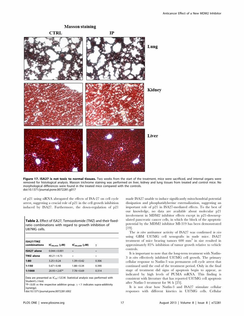
the toxicity assay, Masson trichrome staining was performed on
paraffin sections from the liver, kidney and lung of mice treated
with intra-peritoneal injections of ISA27 and control mice as
previously described [38].
12. Isobolar AnalysisA graphical assessment of synergy with regard to growth
inhibition was performed using isobolographic analysis [39,40]. In
an isobologram, the equi-effective pairs of doses of two drugs
(ISA27 and Temozolomide) are represented using rectangular
coordinates. In the present study, the dose of ISA27 required to
give a 50% effect was plotted on the abscissa, and the iso-effective
dose of Temozolomide was plotted on the ordinate. The
Figure 6. Lack of ISA27 toxicity in an in vitro human cell model (lymphomonocytes). 6A) Scatter cytogram analysis of cell populations fromperipheral blood: Scatter cytogram analysis showing cell separation by size and granularity (G2 = lymphocytes; G3=monocytes). One representativeexperiment is presented. 6B) Time-response of human lymphomonocyte viability following ISA27 treatment: lymphomonocytes were treated with2.5 mM ISA27 or 10 mM Nutlin-3 for 12, 24 and 48 h. The figure shows the viable cells and dead cells counted by Trypan blue exclusion at theindicated times. Data represent the means of three independent experiments performed in duplicate; bars, SEM. No significant differences wereobserved between MDM2 inhibitor-treated and untreated-control viable or dead cells at each incubation time.doi:10.1371/journal.pone.0072281.g006
Anticancer Effect of a New MDM2 Inhibitor
PLOS ONE | www.plosone.org 7 August 2013 | Volume 8 | Issue 8 | e72281

theoretical additive effect of the two drugs was represented by the
straight line connecting the two points. If the experimentally
determined data points and their confidence interval fall on this
line, the drug effects are additive (no interaction). If the points lie
below this line, there is superadditivity (synergy), and if the points
lie above this line, there is subadditivity (antagonism).
To determine whether the interaction between the two drugs
was synergistic, additive or antagonistic, the theoretical additive
IC50,add was estimated from the dose-response curves of each drug
administered individually, considering that the observed effect
with the combination was the sum of the individual effects of each
component. The theoretical IC50,add value was then compared
with the experimental IC50,mix to determine whether there was a
statistically significant difference [41]. The interaction index,
denoted by c, is an assessment of the degree of synergism or
antagonism. The index is defined by the isobolar relationship as
follows [42]: c= a/A+b/B where A and B are the doses of drug A
(alone) and B (alone) that give the specified effect, and (a,b) are the
combination doses that produce the same effect. The quantities in
the equation are obtained from the dose-response curves of drugs
A, B, and their combinations. If c= 1, the interaction is additive; if
c ,1, the interaction is super-additive (synergy); and if c .1, the
interaction is sub-additive (antagonism).
13. Statistical AnalysesThe nonlinear multipurpose curve-fitting program, GraphPad
Prism, was used for data analysis and graphic presentations. All
data are presented as the means 6 SEM. Statistical analysis was
performed by one-way analysis of variance (ANOVA) with
Bonferroni’s corrected t-test for post-hoc pair-wise comparisons.
The densitometric analysis of immunoreactive bands was
performed using the ImageJ program. For in vivo experimental
data, a two-way ANOVA was performed to compare the different
parameters among the different groups. P,0.05 was considered
statistically significant.
Results
1. Short-term ISA27 Treatment Inhibits GBM Cell Growth/survival
To examine the effects of ISA27 [26] (see Fig. 1 for chemical
structure) on GBM cell growth/survival, the concentration at
Figure 7. Long-term ISA27 treatment induces cell cycle arrest and a persistent increase in p21 mRNA levels in U87MG cells. 7A) Flowcytometric cell cycle profiling: the figure shows the percentage of untreated and MDM2 inhibitor-treated U87MG cells in G1-, S- and G2/M-phase. 7B)p21 mRNA evaluation: ISA27 treatment resulted in a statistically significant increase in p21 mRNA levels at 24, 48 and 72 h.doi:10.1371/journal.pone.0072281.g007
Anticancer Effect of a New MDM2 Inhibitor
PLOS ONE | www.plosone.org 8 August 2013 | Volume 8 | Issue 8 | e72281

Figure 8. ISA27 induces U87MG cell senescence. 8A) Representative images of SA-b-Gal-expressing cells: The panel shows the SA-b-Gal-expressing MDM2 inhibitor-treated and untreated cells at 72 h. 8B) Percentage of SA-b-Gal-expressing cells: The number of SA-b-Gal-expressing cellswas determined with respect to the total cells in each sample (untreated cells, ISA27- and Nutlin-3-treated cells). The percentage of SA-b-Gal-expressing MDM2 inhibitor-treated cells was then calculated with respect to the untreated cells, for which an arbitrary value of 100% was assigned.doi:10.1371/journal.pone.0072281.g008
Figure 9. U87MG cells did not recover normal growth upon removal of ISA27. U87MG cells were cultured in the presence of 2.5 mM ISA27or 10 mM Nutlin-3. After 4 days of MDM2 inhibitor treatment, cell culture medium was replaced with fresh culture medium without the MDM2inhibitor. The figure shows the number of viable cells that were counted by Trypan blue exclusion after 4 days of MDM2 inhibitor treatment and 1, 2and 3 days after MDM2 inhibitor removal. Data represent the means of three independent experiments performed in duplicate; bars, SEM.doi:10.1371/journal.pone.0072281.g009
Anticancer Effect of a New MDM2 Inhibitor
PLOS ONE | www.plosone.org 9 August 2013 | Volume 8 | Issue 8 | e72281
which this molecule inhibited 50% of cell growth/survival (IC50
value) after 24 h of treatment was assessed. We used U87MG and
U343MG cell lines that overexpress MDM2 and maintain wild-
type p53 [43]. ISA27 showed a dose-dependent inhibitory effect
on the growth/survival of U87MG and U343MG cells with IC50
values of 2.560.4 and 5.760.9 mM, respectively (the same cell
treatment with Nutlin-3 used as a control gave a value of 10.062.1
and 22.064.6 mM, respectively) (Fig. 1A and B). We also used the
GBM cell line T98G, which expresses mutant p53 [43], as a
negative control for the effect of ISA27 on GBM cell viability.
Treatment of T98G cells with ISA27 did not significantly reduce
cell growth/survival, showing approximately 80% of viable cells at
the maximum tested concentration (25 mM) (Fig. 1C). The
treatment of T98G cells with Nutlin-3 gave a similar result
(Fig. 1C).
2. Short-term ISA27 Treatment Stabilises p53 Levels andRestores p53 Function in GBM Cells Expressing Wild-typep53
We first examined the effect of ISA27 on the levels of p53 in the
GBM cells, U87MG and U343MG. Incubation of the GBM cells
with ISA27 for 4, 6, 8, and 12 h led to a time-dependent increase
in p53 protein levels (Fig. 2). The accumulation of p53 became
significant after 6 hours of treatment, reaching its highest level
after 8 h (Fig. 2). Nutlin-3 induced a significant accumulation of
p53 protein after 8 h and 12 h of treatment (Fig. 2).
MDM2 inhibition is hypothesised to stabilise p53 protein levels
by reducing MDM2-mediated p53 degradation. However, p53
stability could also be the result of enhanced transcription of the
p53 gene and subsequent p53 protein translation. To confirm that
the accumulation of p53 was due to the decreased interaction of
p53 with MDM2 rather than elevated p53 expression, we treated
GBM cells with ISA27 and monitored the levels of p53 mRNA,
p53 protein in the presence of the protein synthesis inhibitor,
cycloheximide (CHX), and the MDM2-p53 complex. The
transcription of p53 gene did not change in the GBM cells within
12 h of treatment (Fig. 3 A and B), and p53 protein levels persisted
even in the presence of CHX (Fig. 3C). Specifically, the inhibition
of protein synthesis upon treatment with CHX alone led to a
marked decrease in p53 protein expression over time. In ISA27-
treated cells, blocking protein synthesis led to an overall increase in
p53 protein expression (Fig. 3C). To test the ability of ISA27 to
disrupt the intracellular MDM2-p53 interaction, a co-immuno-
precipitation assay was done. Only minimal amounts of p53 could
be detected in MDM2 immunoprecipitates after 8 h of treatment
in GBM cells (Fig. 3D). All together these results indicate that
ISA27 is able to induce p53 up-regulation by blocking MDM2-
p53 interaction.
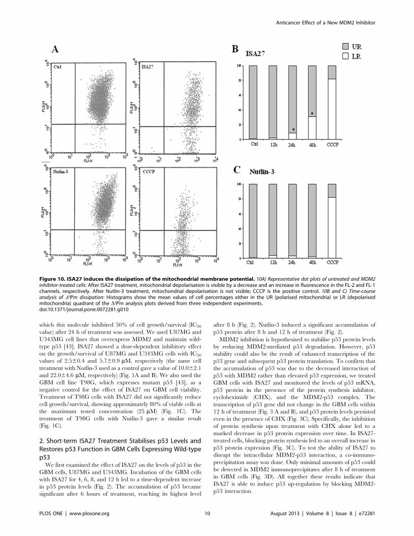
Figure 10. ISA27 induces the dissipation of the mitochondrial membrane potential. 10A) Representative dot plots of untreated and MDM2inhibitor-treated cells: After ISA27 treatment, mitochondrial depolarisation is visible by a decrease and an increase in fluorescence in the FL-2 and FL-1channels, respectively. After Nutlin-3 treatment, mitochondrial depolarisation is not visible; CCCP is the positive control. 10B and C) Time-courseanalysis of DYm dissipation: Histograms show the mean values of cell percentages either in the UR (polarised mitochondria) or LR (depolarisedmitochondria) quadrant of the DYm analysis plots derived from three independent experiments.doi:10.1371/journal.pone.0072281.g010
Anticancer Effect of a New MDM2 Inhibitor
PLOS ONE | www.plosone.org 10 August 2013 | Volume 8 | Issue 8 | e72281

Short-term ISA27 treatment of U87MG cells led to a
statistically significant increase in the mRNA levels of p53 target
genes; specifically, a 4.3- and 4.5-fold induction of MDM2 and
p21 mRNA, respectively, was observed (Fig. 4A). These results
suggested that stabilisation of p53 in ISA27-treated GBM cells led
to an increase in MDM2 and p21 mRNA levels in a manner that
was consistent with the activation of the p53 pathway. Nutlin-3
treatment resulted in a 2.3- and 4.3-fold induction of MDM2 and
p21 mRNA, respectively, in U87MG cells (Fig. 4A).
One of the main cellular consequences of p53 activation in
proliferating cells is cell cycle arrest at the G1/G2-phase, and the
cyclin-dependent kinase inhibitor p21 plays a major role in this
arrest. In accord with these data, cell cycle analysis by
cytofluorimetric DNA content analysis revealed an increase in
the G1 fraction and a nearly complete depletion of S-phase cells
after short-term treatment with ISA27 or Nutlin-3 (Fig. 4B).
3. Long-term ISA27 Treatment Leads to the CompleteInhibition of Cell Survival/growth in U87MG GBM Cells,but not in Normal Human Lymphomonocytes
A substantial decrease in U87MG cell survival/growth was
observed after one day of treatment, and the decrease continued at
each subsequent time point until almost all viable cells were
depleted after 5 days (Fig. 5).
We also examined the effect of ISA27 on the viability of normal
human lymphomonocytes. Flow cytometry was performed to
evaluate cell populations isolated from blood samples of healthy
individuals. As shown in the scatter cytogram in Fig. 6A, the two
cell populations were clearly visible (G2 = 74.0% lymphocytes;
G3 = 14.2% monocytes). As shown in Fig. 6B, ISA27-treated
lymphomonocytes were still viable at 48 h. Nutlin-3 treatment
showed a similar lack of cytotoxicity in normal peripheral blood
cells (Fig. 6B).
Figure 11. ISA27 induces an increase in PUMA mRNA levels, mitochondrial cytochrome c release, and DNA fragmentation. 11A)Relative quantification of PUMA mRNA: ISA27 induced a statistically significant increase in PUMA mRNA levels at 24 and 48 h. Nutlin-3 treatmentresulted in a statistically significant increase at 72 h. 11B) Evaluation of cytosolic cytochrome c content: ISA27-treated cells showed a 25% increase incytochrome c levels in the cytosolic fraction. Nutlin-3-treated cells did not give statistically significant results. 11C) Evaluation of DNA content:Frequency histograms from a representative experiment are shown. ISA27-treated cells showed a significant increase in the percentage of nuclei withhypodiploid DNA content at 72 h compared with control cells. In contrast, Nutlin-3 did not induce significant nuclear DNA fragmentation.doi:10.1371/journal.pone.0072281.g011
Anticancer Effect of a New MDM2 Inhibitor
PLOS ONE | www.plosone.org 11 August 2013 | Volume 8 | Issue 8 | e72281

4. Long-term ISA27 Treatment Leads to Permanent CellCycle Arrest and Apoptosis in U87MG GBM Cells
First, we analysed cell cycle profiling and p21 mRNA levels after
long-term ISA27 treatment. ISA27 treatment of U87MG cells for
24 h effectively arrested cell-cycle progression, depleted the S-
phase population (p,0.05) and increased the G0/G1-phase
population (p,0.05). Treatment for 72 h depleted the S-phase
population and increased the G2-phase population (p,0.01)
(Fig. 7A). A persistent induction of p21 mRNA was observed,
suggesting that the major transcription target of activated p53 was
involved in the cell cycle arrest (Fig. 7B). Nutlin-3 arrested cell-
cycle progression in U87MG cells 24 and 72 h after treatment,
depleting the S-phase compartment and increasing the G0/G1-
phase compartment (Fig. 7A).
To elucidate whether ISA27 induced permanent cell cycle
arrest (cellular senescence), we evaluated whether ISA27 promoted
a morphological change in U87MG cells (i.e., large and flat cells)
that was compatible with senescence and whether the widely used
senescence marker, SA-b-Gal, could be detected in ISA27-treated
cells. The ISA27-treated cells acquired an enlarged and flat
morphology as revealed by the mean cell diameter
(12.3661.56 mm and 13.5961.74 mm for control and ISA27-
treated cells, respectively). The onset of cellular senescence was
rapid; indeed, a high number of U87MG cells were positive for
SA-b-Gal after 1 day of treatment. Fig. 8A shows representative
images of SA-b-Gal detection in ISA27-treated and control cells.
As shown in Fig. 8B, ISA27 significantly enhanced the number of
SA-b-Gal-expressing cells after 24 h. Nutlin-3 caused an increase
in the number of SA-b-Gal-expressing cells at both 24 and 72 h.
Next, to assess whether U87MG cells could resume growing
after ISA27 removal, culture medium was replaced with fresh
medium not containing ISA27 after 4 days of cell treatment, and
the viable cells were assessed after 1, 2 and 3 days. After ISA27
removal, the number of viable cells further decreased at these time
points with respect to control samples, suggesting the inability to
recover normal cell growth (Fig. 9).
To investigate whether ISA27 induced apoptosis, the following
parameters were evaluated: dissipation of mitochondrial potential
Figure 12. siRNA against p21 abrogates ISA27-mediated cell growth inhibition. A) Evaluation of p21 levels after transient transfection ofU87MG cells with p21 siRNA: Western blotting and real-time RT-PCR analyses demonstrated decreased p21 levels in U87MG cells after 24 h transienttransfection. B) Evaluation of ISA27 effect on cell viability. p21 siRNA samples were exposed to a range of ISA27 concentrations for 24 h. Afterincubation, U87MG cell viability was determined by MTS assay. The IC50 value was 3.660.5 mM for random siRNA ISA27 sample. Transfection ofU87MG cells with p21 siRNA rendered ISA27 much less effective to inhibit cell viability. C) Evaluation of ISA27 effect on cell cycle in p21 siRNA sample:random siRNA and p21siRNA samples were exposed to a fixed dose of ISA27 (5 mM) or DMSO for 24 h. The ISA27-treated random siRNA sampleshowed the increase of G1 phase compared to random siRNA sample. The comparison of cell cycle phases between p21 siRNA and ISA27-treated p21siRNA sample did not show statistically significant differences.doi:10.1371/journal.pone.0072281.g012
Anticancer Effect of a New MDM2 Inhibitor
PLOS ONE | www.plosone.org 12 August 2013 | Volume 8 | Issue 8 | e72281

(DYm), cytosolic cytochrome c content, PUMA mRNA levels and
DNA fragmentation.
A decrease in DYm was indicated by a reduction in orange JC-1
aggregate fluorescence (recorded by the FL2 channel of the flow
cytometer) accompanied by a concomitant increase in green JC-1
monomer fluorescence (recorded by the FL1 channel). Represen-
tative examples of the flow cytometric analysis are shown in
Fig. 10A. The majority of untreated cells (99%) showed high
fluorescence emission in both channels and were found in the
upper right (UR) quadrant of the plot. The remaining untreated
cells (1%) showed low fluorescence emission in FL2 and were
therefore found in the lower right (LR) quadrant. Upon ISA27
treatment, an increase was observed in the percentage of the cells
plotting in the LR quadrant, consistent with DYm dissipation. In
particular, significant changes in DYm were observed after
treatment for 24 and 48 h (8.5% and 31%, respectively;
P,0.05) as shown in Fig. 10B.
Real time RT-PCR analysis showed that ISA27 treatment led to
a statistically significant increase in PUMA mRNA at 24 and 48 h
compared with the control (Fig. 11A).
Figure 11B shows that ISA27-treated cells exhibited a 25%
increase in cytochrome c in the cytosolic fraction compared with
the control.
DNA-specific PI staining showed that treatment with ISA27
caused a significant increase in the percentage of cells with
hypodiploid DNA content, a clear sign of apoptosis, as shown in
the representative frequency histograms (Fig. 11C). In particular,
the percentage of apoptotic cells (sub-G0 cells) observed after
exposure of U87MG cells to ISA27 for 72 h increased to
17.761.27% (P,0.001), whereas that of the control was
0.4360.05%. Nutlin-3 treatment did not give statistically signif-
icant results after the indicated incubation times for these
apoptotic parameters (Fig. 11B and C) with the exception of
increased PUMA mRNA levels after treatment for 72 h (Fig. 11A).
To explore the role of p21 in the ISA27-induced cell effects, we
analysed cell viability, cell cycle profile and apoptosis markers in
ISA27-treated U87MG cells in which p21 was genetically
inhibited. First, to verify p21 down-regulation by siRNA, the
levels of p21 mRNA and protein were evaluated in U87MG cells
transfected with p21 siRNA after 24 and 48 h. The Fig. 12 shows
that p21 protein (Fig. 12A) and mRNA (Fig. 12B) were effectively
down-regulated post 24 h p21 siRNA transfection. For subsequent
Figure 13. siRNA against p21 made ISA27 unable to induce DYm dissipation and PE. Random siRNA and p21siRNA samples were exposedto a fixed dose of ISA27 (5 mM) or DMSO for 24 h. A) Evaluation of ISA27 effect on the DYm in p21 siRNA samples. The ISA27-treated random siRNAsample showed dissipation of DYm compared to random siRNA sample. The comparison of DYm between p21 siRNA and ISA27-treated p21 siRNAsample did not show statistically significant differences. Upper panel shows representative dot plots of untreated and ISA27-treated random orp21siRNA samples. B) Evaluation of ISA27 effect on PE in p21 siRNA samples. The ISA27-treated random siRNA sample showed a statistical significantincrease of early and late apoptosis compared to random siRNA sample. The comparison of early and late apoptosis between p21 siRNA and ISA27-treated p21 siRNA sample did not show statistically significant differences.doi:10.1371/journal.pone.0072281.g013
Anticancer Effect of a New MDM2 Inhibitor
PLOS ONE | www.plosone.org 13 August 2013 | Volume 8 | Issue 8 | e72281
experiments, we down-regulated p21 and incubated U87MG cells
with ISA27 for 24 h. In p21 siRNA sample, ISA27 failed to reduce
U87MG cell viability (Fig. 12 B) and arrest cell cycle (Fig. 12 C).
Furthermore, DYm flow cytometry and Annexin V FITC analysis
showed that p21 down-regulation made ISA27 unable to
effectively induce mitochondrial potential dissipation (Fig. 13A)
and phosphatidylserine externalization (PE) (Fig. 13B) in U87MG
cells.
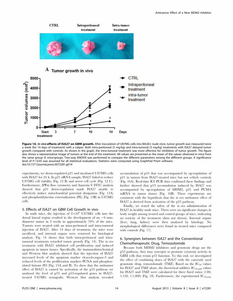
5. Effects of ISA27 on GBM Cell Growth in vivoIn nude mice, the injection of 26106 U87MG cells into the
dorsal lateral region resulted in the development of an ,6 mm-
diameter tumor in 2 weeks in approximately 70% of the mice.
Tumors were treated with an intra-peritoneal and intra-tumoral
injection of ISA27. After 14 days of treatment, the mice were
sacrificed, and internal organs were removed for histological
analysis. Fig. 14 shows that both intra-peritoneal and intra-
tumoral treatments retarded tumor growth (Fig. 14). The in vivo
treatment with ISA27 inhibited cell proliferation and induced
apoptosis in tumor tissues. Specifically, the immunohistochemical
and Western blot analysis showed that the injection of ISA27
increased levels of the apoptosis marker cleaved-caspase-3 and
reduced levels of the proliferation markers PCNA and phosphor-
ylated histone H3 (Fig. 15A and B). To show that the antitumor
effect of ISA27 is caused by activation of the p53 pathway we
analysed the level of p53 and p53-regulated genes in ISA27-
treated U87MG xenografts. Western blot analysis revealed
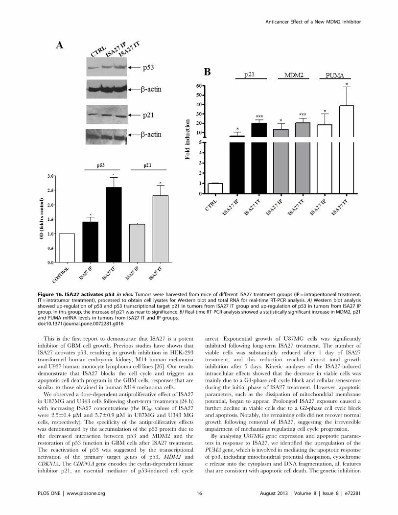
accumulation of p53 that was accompanied by up-regulation of
p21 in tumors from ISA27-treated mice but not vehicle controls
(Fig. 16A). Real-time RT-PCR data confirmed these findings and
further showed that p53 accumulation induced by ISA27 was
accompanied by up-regulation of MDM2, p21 and PUMA
mRNA in tumor tissues (Fig. 16B). These experiments are
consistent with the hypothesis that the in vivo antitumor effect of
ISA27 is derived from activation of the p53 pathway.
Finally, we tested the safety of the in vivo administration of
ISA27 in healthy nude mice. There were no significant changes in
body weight among treated and control groups of mice, indicating
no toxicity of the treatment (data not shown). Internal organs
(liver, lung, kidney) were then analysed by histology. No
morphological differences were found in treated mice compared
with controls (Fig. 17).
6. Synergism between ISA27 and the ConventionalChemotherapeutic Drug, Temozolomide
Because both MDM2 inhibitors and genotoxic drugs use the
p53 pathway, they may synergise to promote cytotoxic activity in
GBM cells that retain p53 function. To this end, we investigated
the effect of combining doses of ISA27 with the currently used
genotoxic drug, temozolomide (TMZ). Based on the IC50 values
for ISA27 and TMZ alone, the theoretical additive IC50,add values
for ISA27 and TMZ were calculated for three fixed ratios (1:80,
1:150, 1:1,000) (Fig. 18). Furthermore, the experimental IC50,mix
Figure 14. In vivo effects of ISA27 on GBM growth. After inoculation of U87MG cells into BALB/c nude mice, tumor growth was measured twicea week (for 14 days of treatment) with a caliper. Both intra-peritoneal (5 mg/kg) and intra-tumoral (3 mg/kg) treatments with ISA27 delayed tumorgrowth compared with controls. As shown in the graph, the intra-tumoral treatment was more effective for inhibition of tumor growth. The figurealso shows a representative image of tumors at the end of the treatment. All values are presented as the mean of the values observed in mice fromthe same group (5 mice/group). Two-way ANOVA was performed to compare the different parameters among the different groups. A significancelevel of P,0.05 was assumed for all statistical evaluations. Statistics were computed using GraphPad Prism software.doi:10.1371/journal.pone.0072281.g014
Anticancer Effect of a New MDM2 Inhibitor
PLOS ONE | www.plosone.org 14 August 2013 | Volume 8 | Issue 8 | e72281

values were determined for the same fixed-ratio combinations in
the viability assay (Fig. 13). The IC50,add and the IC50,mix values
are shown in Table 2. Statistical analysis of the data from
isobolographic analysis revealed synergistic interactions between
ISA27 and TMZ for the three examined fixed-ratio combinations
(Table 2; Fig. 18). Furthermore, the interaction index values of the
combinations demonstrated the following rank of potencies:
1:80.1:1,000.1:150 (ISA27:TMZ).
Discussion
The direct and specific activation of the p53 pathway without
inducing collateral DNA damage offers a tantalising solution to the
shortcomings of current therapeutic regimens and appears to be a
reasonable approach for GBM therapy in view of the infrequent
occurrence of p53 gene mutations [9,10]. The cumulative
evidence of aberrantly increased activity of the primary p53
inhibitor MDM2 in GBM [11,12] prompted us to examine the
effects of targeted inhibition of the MDM2-p53 interaction by the
spiro-oxindole analogue ISA27, a recently described small-
molecule inhibitor of MDM2 [26]. Little is known about the
effects of MDM2 inhibitors on the in vitro growth of GBM cells.
Recently, Nutlin-3, the first potent small-molecule inhibitor of
MDM2 [23], was reported to be effective at inhibiting GBM cell
growth in vitro [25], suggesting the validity of this experimental
approach for the treatment of GBM.
In this study, we investigated whether ISA27 affected the
growth of GBM cells and explored the intracellular events
following ISA27 treatment. The U87MG and U343MG cell lines
overexpress MDM2 and maintain wild-type p53 [43–45] and were
chosen as a cell culture model of human GBM. In these cell lines,
the primary mechanism of p53 inactivation is the high nuclear
MDM2 levels caused by a lack of PTEN, a tumor suppressor
protein that normally counteracts MDM2 translocation into the
nucleus [46]. The lack of PTEN makes these cell lines a suitable
representative model of GBM, as the loss of the PTEN gene locus
has been found in up to 80% of GBM cases [9,10,13].
Figure 15. ISA27 inhibits cell proliferation and induces apoptosis in tumor tissues. 14 days from starting treatment, tumors wereharvested from mice of different ISA27 treatment groups (IP = intraperitoneal treatment; IT = intratumor treatment) and processed for histological andWestern blotting analysis. A) Histological analysis: apoptosis and cell proliferation were evaluated by analysis of cleaved caspase 3 and PCNA levels byimmunohistochemistry in paraffin embedded sections of tumors. Upper panel shows representative images of immunohistochemistry analysis.ISA27-treated tumors show increased cleaved caspase 3 levels and reduced cell proliferation. Cleaved caspase 3 and PCNA levels in tumors werequantified from digital images. Results are shown in graph as percent of cleaved caspase 3 and PCNA expression respect to controls (*P,0.05 vscontrol). B) Western blotting analysis: apoptosis and cell proliferation were evaluated by analysis of cleaved caspase 3 and phosphorylated histone H3(pH3) levels by Western blotting in whole lysates of tumors using specific antibodies. Densitometric analysis shows that ISA27 induced a significantincrease in cleaved-caspase-3 levels and a significant reduction in pH3 levels (*P,0.05 vs control).doi:10.1371/journal.pone.0072281.g015
Anticancer Effect of a New MDM2 Inhibitor
PLOS ONE | www.plosone.org 15 August 2013 | Volume 8 | Issue 8 | e72281
This is the first report to demonstrate that ISA27 is a potent
inhibitor of GBM cell growth. Previous studies have shown that
ISA27 activates p53, resulting in growth inhibition in HEK-293
transformed human embryonic kidney, M14 human melanoma
and U937 human monocyte lymphoma cell lines [26]. Our results
demonstrate that ISA27 blocks the cell cycle and triggers an
apoptotic cell death program in the GBM cells, responses that are
similar to those obtained in human M14 melanoma cells.
We observed a dose-dependent antiproliferative effect of ISA27
in U87MG and U343 cells following short-term treatments (24 h)
with increasing ISA27 concentrations (the IC50 values of ISA27
were 2.560.4 mM and 5.760.9 mM in U87MG and U343 MG
cells, respectively). The specificity of the antiproliferative effects
was demonstrated by the accumulation of the p53 protein due to
the decreased interaction between p53 and MDM2 and the
restoration of p53 function in GBM cells after ISA27 treatment.
The reactivation of p53 was suggested by the transcriptional
activation of the primary target genes of p53, MDM2 and
CDKN1A. The CDKN1A gene encodes the cyclin-dependent kinase
inhibitor p21, an essential mediator of p53-induced cell cycle
arrest. Exponential growth of U87MG cells was significantly
inhibited following long-term ISA27 treatment. The number of
viable cells was substantially reduced after 1 day of ISA27
treatment, and this reduction reached almost total growth
inhibition after 5 days. Kinetic analyses of the ISA27-induced
intracellular effects showed that the decrease in viable cells was
mainly due to a G1-phase cell cycle block and cellular senescence
during the initial phase of ISA27 treatment. However, apoptotic
parameters, such as the dissipation of mitochondrial membrane
potential, began to appear. Prolonged ISA27 exposure caused a
further decline in viable cells due to a G2-phase cell cycle block
and apoptosis. Notably, the remaining cells did not recover normal
growth following removal of ISA27, suggesting the irreversible
impairment of mechanisms regulating cell cycle progression.
By analysing U87MG gene expression and apoptotic parame-
ters in response to ISA27, we identified the upregulation of the
PUMA gene, which is involved in mediating the apoptotic response
of p53, including mitochondrial potential dissipation, cytochrome
c release into the cytoplasm and DNA fragmentation, all features
that are consistent with apoptotic cell death. The genetic inhibition
Figure 16. ISA27 activates p53 in vivo. Tumors were harvested from mice of different ISA27 treatment groups (IP = intraperitoneal treatment;IT = intratumor treatment), processed to obtain cell lysates for Western blot and total RNA for real-time RT-PCR analysis. A) Western blot analysisshowed up-regulation of p53 and p53 transcriptional target p21 in tumors from ISA27 IT group and up-regulation of p53 in tumors from ISA27 IPgroup. In this group, the increase of p21 was near to significance. B) Real-time RT-PCR analysis showed a statistically significant increase in MDM2, p21and PUMA mRNA levels in tumors from ISA27 IT and IP groups.doi:10.1371/journal.pone.0072281.g016
Anticancer Effect of a New MDM2 Inhibitor
PLOS ONE | www.plosone.org 16 August 2013 | Volume 8 | Issue 8 | e72281
of p21 using siRNA abrogated the effects of ISA-27 on cell cycle
arrest, suggesting a crucial role of p21 in the cell growth inhibition
induced by ISA27. Furthermore, the down-regulation of p21
made ISA27 unable to induce significantly mitochondrial potential
dissipation and phosphatidylserine externalization, suggesting an
important role of p21 in ISA27-mediated effects. To the best of
our knowledge, no data are available about molecular p21
involvement in MDM2 inhibitor effects except in p21-downreg-
ulated pancreatic cancer cells, in which the block of the apoptotic
potential by the MDM2 inhibitor MI-319 has been demonstrated
[19].
The in vitro antitumor activity of ISA27 was confirmed in vivo
using GBM U87MG cell xenografts in nude mice. ISA27
treatment of mice bearing tumors 600 mm3 in size resulted in
approximately 85% inhibition of tumor growth relative to vehicle
controls.
It is important to note that the long-term treatment with Nutlin-
3 in vitro effectively inhibited U87MG cell growth. The primary
cellular response to Nutlin-3 was permanent cell cycle arrest that
continued until the end of the treatment period. Only in the final
stage of treatment did signs of apoptosis begin to appear, as
indicated by high levels of PUMA mRNA. This finding is
consistent with literature that has reported U87MG cell apoptosis
after Nutlin-3 treatment for 96 h [25].
It is not clear how Nutlin-3 and ISA27 stimulate cellular
responses with different kinetics in U87MG cells. Cellular
Figure 17. ISA27 is not toxic to normal tissues. Two weeks from the start of the treatment, mice were sacrificed, and internal organs wereremoved for histological analysis. Masson trichrome staining was performed on liver, kidney and lung tissues from treated and control mice. Nomorphological differences were found in the treated mice compared with the controls.doi:10.1371/journal.pone.0072281.g017
Table 2. Effect of ISA27, Temozolomide (TMZ) and their fixed-ratio combinations with regard to growth inhibition ofU87MG cells.
ISA27/TMZcombinations IC50,mix (mM) IC50,add (mM) c
ISA27 alone 0.04460.001 – –
TMZ alone 40.2164.73 – –
1:80 3.2560.24 1.3960.42 0.306
1:150 5.6760.48 1.8860.34 0.390
1:1000 20.9362.87* 7.7860.69 0.314
Data are presented as IC506S.E.M. Statistical analysis was performed withStudent’s t-test.*P,0.05 vs the respective additive group. c ,1 indicates supra-additivity(synergy).doi:10.1371/journal.pone.0072281.t002
Anticancer Effect of a New MDM2 Inhibitor
PLOS ONE | www.plosone.org 17 August 2013 | Volume 8 | Issue 8 | e72281

responses with different kinetics have been recently shown for
ISA27 and another small molecule, 10d, in the M14 human
melanoma cell line [26]. Treatment with ISA27 for 24 h induced
both cell cycle arrest and apoptosis, whereas 10d caused cell cycle
arrest only. In U87MG cells, the ability of ISA27 to promote a cell
cycle block in combination with apoptosis could be attributed to
the more rapid accumulation of p53 protein levels during
treatment with respect to Nutlin-3 treatment. The rapid ISA27-
induced increase in p53 protein could find a more favourable
cellular environment to efficiently activate p53 function as
indicated by the induction of MDM2 and proapoptotic PUMA
gene transcription. This rapid ISA27-induced antiproliferative
response may be beneficial in the treatment of human GBM,
considering that this cancer is characterised by rapid cell growth.
Additionally, a lower dose of ISA27 was efficacious when
compared with Nutlin-3. The implication of this result can be
illustrated from the recent Phase I study that showed the clinical
efficacy of the MDM2 inhibitor, JNJ-26854165, in patients with
advanced solid tumors, but at elevated doses, some toxic effects
were reported [47]. For example, lymphopoenia was observed in
the majority of the patients, and more than 20% experienced
grade 3 or 4 severity [47]. In this context, the ability of ISA27 to
maintain the viability of human lymphomonocytes is of particular
interest. A selective toxic effect of MDM2 inhibitors on cancer cells
has been shown by other authors using a number of normal cell
models. It has been demonstrated that Nutlin-3 is not toxic to
peripheral blood mononuclear cells, bone marrow-derived hae-
matopoietic progenitors and bone marrow stromal epithelial cells
[48–50]. The administration of ISA27 in vivo stimulated p53
activation in the xenograft model of human GBM, resulting in
inhibition of cell proliferation and induction of apoptosis. ISA27
showed antitumor activity without causing visible signs of toxicity
in the animals as assessed by necroscopy and body weight
assessment. These results are in agreement with previous in vivo
studies performed with Nutlin-3 and other MDM2 inhibitors
[51,52].
The precise mechanism of cell death resistance in normal cells
remains unclear. The resistance may be a consequence of the low
basal expression levels of the MDM2 oncoprotein in normal cells.
Thus, following cell treatment with the MDM2 inhibitor, the
amount of p53 protein dissociated from MDM2 and accumulated
would not be sufficient to trigger cell death. In contrast, tumor cells
overexpress MDM2, which sequesters high amounts of p53.
Consequently, after blocking the interaction between these two
proteins, the high accumulation of p53 renders the cells highly
susceptible to p53 reactivation and more sensitive to apoptosis
[52].
From a therapeutic perspective, it is interesting that ISA27 in
combination with the conventional chemotherapy drug TMZ
inhibited U87MG cell growth. This combination worked in a
synergistic manner as confirmed by isobolographic analysis (c ,1
in all cases). This result suggests the possibility of lowering the dose
of TMZ used in the treatment of GBM.
In conclusion, our data show that ISA27 disrupts the MDM2-
p53 interaction and releases the powerful antitumor capacities of
p53 in GBM cells. The use of this MDM2 inhibitor could offer a
novel therapy for the treatment of GBM patients by inhibiting
tumor growth.
Author Contributions
Conceived and designed the experiments: BC SB CM EN. Performed the
experiments: SB PG SD DS EDP CDG AB RV PC IGM. Analyzed the
data: BC SB SD GI EDP. Contributed reagents/materials/analysis tools:
FS PC IGM GI AB. Wrote the paper: BC. Revised the article critically for
important intellectual content: CM EN.
References
1. Liu J, Ma Q, Zhang M, Wang X, Zhang D, et al. (2012) Alterations of TP53 are
associated with a poor outcome for patients with hepatocellular carcinoma:
Evidence from a systematic review and meta-analysis. Eur J Cancer 48: 2328–
2338.
2. Furnari FB, Fenton T, Bachoo RM, Mukasa A, Stommel JM, et al. (2007)
Malignant astrocytic glioma: genetics, biology, and paths to treatment. Genes
Dev 21: 2683–2710.
3. Ohgaki H, Dessen P, Jourde B, Horstmann S, Nishikawa T, et al. (2004) Genetic
pathways to glioblastoma: a population-based study. Cancer Res 64: 6892–6899.
4. Conrad CA, Milosavljevic VP, Yung WK (1995) Advances in chemotherapy for
brain tumors. Neurol Clin 13: 795–812.
5. Levin VA, Leibel SA, Gutin PH (2001) Neoplasms of the central nervous system.
In: De Vita Jr VT, Hellman S, Rosenberg SA, editors. Cancer principles of
oncology. Philadelphia: Lippincott-Raven. 2100–2161.
6. Desjardins A, Rich IN, Quinn JA, Vredenburgh J, Gururangan S, et al. (2005)
Chemotherapy and novel therapeutic approaches in malignant glioma. Front
Biosci 10: 2645–2668.
7. Hess KR, Broglio KR, Bondy ML (2004) Adult glioma incidence trends in the
United States, 1977–2000. Cancer 101: 2293–2299.
8. Nagasawa DT, Chow F, Yew A, Kim W, Cremer N, et al. (2012) Temozolomide
and other potential agents for the treatment of glioblastoma multiforme.
Neurosurg Clin N Am. 23: 307–322.
9. Ohgaki H, Kleihues P (2009) Genetic alterations and signaling pathways in the
evolution of gliomas. Cancer Sci 100: 2235–2241.
10. Cerami E, Demir E, Schultz N, Taylor BS, Sander C (2010) Automated network
analysis identifies core pathways in glioblastoma. PLoS One 5: e8918.
11. He J, Reifenberger G, Liu L, Collins VP, James CD (1994) Analysis of glioma
cell lines for amplification and overexpression of MDM2. Genes Chrom Cancer
11: 91–96.
Figure 18. Synergistic effect of ISA27 and temozolomide on thesurvival/growth of GBM cells. Isobologram 2-D showing theinteractions between TMZ and ISA27 in MTS viability tests performedin U87MG cells treated for 72 h with TMZ and/or ISA27. The IC50 valuesfor TMZ and ISA27 are shown on the X- and Y-axes, respectively. Theisobole of additivity is shown as a solid line drawn between theaforementioned IC50 values of TMZ and ISA27 and connects the X- andY-axes. The open points (#) on the additivity line depict the theoreticalIC50,add values for total dose expressed as the proportion of TMZ andISA27 that produced a 50% effect. The solid points (N) depict theexperimental IC50,mix values for total dose expressed as the proportionof TMZ and ISA27 that produced a 50% effect. The experimental IC50,mix
values of the mixture of ISA27 and TMZ for the fixed-ratio combinationsof 1:80, 1:150 and 1:1,000 were found to be significantly below thetheoretical isoboles of additivity, indicating super-additive (synergy)interactions.doi:10.1371/journal.pone.0072281.g018
Anticancer Effect of a New MDM2 Inhibitor
PLOS ONE | www.plosone.org 18 August 2013 | Volume 8 | Issue 8 | e72281

12. Freedman DA, Wu L, Levine AJ (1999) Functions of the MDM2 oncoprotein.
Cell Mol Life Sci 55: 96–107.
13. Halatsch ME, Schmidt U, Unterberg A, Vougioukas VI (2006) Uniform MDM2
overexpression in a panel of glioblastoma multiforme cell lines with divergent
EGFR and p53 expression status. Anticancer Res 26: 4191–4194.
14. Li C, Shen J, Wei X, Xie C, Lu W (2012) Targeted delivery of a novel
palmitylated D-peptide for antiglioblastoma molecular therapy. J Drug Target20: 264–271.
15. Wang B, Fang L, Zhao H, Xiang T, Wang D (2012) MDM2 inhibitor Nutlin-3a
suppresses proliferation and promotes apoptosis in osteosarcoma cells. ActaBiochim Biophys Sin (Shanghai). 44: 685–691.
16. Henze J, Muhlenberg T, Simon S, Grabellus F, Rubin B, et al. (2012) p53modulation as a therapeutic strategy in gastrointestinal stromal tumors. PLoS
One 7: e37776.
17. Shangary S, Wang S (2009) Small-molecule inhibitors of the MDM2-p53protein-protein interaction to reactivate p53 function: a novel approach for
cancer therapy. Annu Rev Pharmacol Toxicol 49: 223–241.
18. Vu BT, Vassilev L (2011) Small-molecule inhibitors of the p53-MDM2
interaction. Curr Top Microbiol Immunol 348: 151–172.
19. Azmi AS, Aboukameel A, Banerjee S, Wang Z, Mohammad M, et al. (2010)MDM2 inhibitor MI-319 in combination with cisplatin is an effective treatment
for pancreatic cancer independent of p53 function. Eur J Cancer 46: 1122–1131.
20. Sonnemann J, Palani CD, Wittig S, Becker S, Eichhorn F, et al. (2011)
Anticancer effects of the p53 activator nutlin-3 in Ewing’s sarcoma cells.Eur J Cancer 47: 1432–1441.
21. Ghassemifar S, Mendrysa SM. (2012) MDM2 antagonism by nutlin-3 induces
death in human medulloblastoma cells. Neurosci Lett. 513: 106–110.
22. Chappell WH, Lehmann BD, Terrian DM, Abrams SL, Steelman LS, et al.
(2012) p53 expression controls prostate cancer sensitivity to chemotherapy andthe MDM2 inhibitor Nutlin-3. Cell Cycle 11: 4579–4588.
23. Vassilev LT, Vu BT, Graves B, Carvajal D, Podlaski F, et al. (2004) In vivo
activation of the p53 pathway by small-molecule antagonists of MDM2. Science.303: 844–888.
24. Liu M, Li C, Pazgier M, Li C, Mao Y, et al. (2010) D-peptide inhibitors of thep53-MDM2 interaction for targeted molecular therapy of malignant neoplasms.
Proc Natl Acad Sci U S A. 107: 14321–14326.
25. Villalonga-Planells R, Coll-Mulet L, Martınez-Soler F, Castano E, Acebes JJ, etal. (2011) Activation of p53 by nutlin-3a induces apoptosis and cellular
senescence in human glioblastoma multiforme. PLoS One 6: e18588.
26. Gomez-Monterrey I, Bertamino A, Porta A, Carotenuto A, Musella S, et al.
(2010) Identification of the Spiro(oxindole-3,39-thiazolidine)-Based Derivatives
as Potential p53 Activity Modulators. J Med Chem 53: 8319–8329.
27. Boyum A (1968) Isolation of mononuclear cells and granulocytes from human
blood. Isolation of monuclear cells by one centrifugation and of granulocytes bycombining centrifugation and sedimentation at 1 g. Scand J Clin Lab Invest
Suppl 97: 77–89.
28. Lee SK, Kim YC, Song SB, Kim YS (2010) Stabilization and translocation of
p53 to mitochondria is linked to Bax translocation to mitochondria in
simvastatin-induced apoptosis. Biochem Biophys Res Commun. 391: 1592–1597.
29. Sosin AM, Burger AM, Siddiqi A, Abrams J, Mohammad RM, et al. (2012)HDM2 antagonist MI-219 (spiro-oxindole), but not Nutlin-3 (cis-imidazoline),
regulates p53 through enhanced HDM2 autoubiquitination and degradation in
human malignant B-cell lymphomas. J Hematol Oncol. 5: 57.
30. Vaseva AV, Yallowitz AR, Marchenko ND, Xu S, Moll UM (2011) Blockade of
Hsp90 by 17AAG antagonizes MDMX and synergizes with Nutlin to inducep53-mediated apoptosis in solid tumors. Cell Death Dis. 2: e156.
31. Vandesompele J, De Paepe A, Speleman F (2002) Elimination of primer-dimer
artifacts and genomic coamplification using a two-step SYBR green I real-timeRT-PCR. Anal Biochem 303: 95–98.
32. Chelli B, Lena A, Vanacore R, Da Pozzo E, Costa B, et al. (2004) Peripheralbenzodiazepine receptor ligands: mitochondrial transmembrane potential
depolarization and apoptosis induction in rat C6 glioma cells. Biochem
Pharmacol 68: 125–134.33. Ruan S, Okcu MF, Pong RC, Andreeff M, Levin V, et al. (1999) Attenuation of
WAF1/Cip1 expression by an antisense adenovirus expression vector sensitizes
glioblastoma cells to apoptosis induced by chemotherapeutic agents 1,3-bis(2-chloroethyl)-1-nitrosourea and cisplatin. Clin Cancer Res 5: 197–202.
34. Chelli B, Rossi L, Da Pozzo E, Costa B, Spinetti F, et al. (2005) PIGA (N,N-Di-n-butyl-5-chloro-2-(4-chlorophenyl)indol-3-ylglyoxylamide), a new mitochondri-
al benzodiazepine-receptor ligand, induces apoptosis in C6 glioma cells.
Chembiochem 6: 1082–1088.35. Dimri GP, Lee X, Basile G, Acosta M, Scott G, et al. (1995) A biomarker that
identifies senescent human cells in culture and in aging skin in vivo. Proc NatlAcad Sci U S A 92: 9363–9367.
36. Bradford MM (1976) A rapid and sensitive method for the quantitation ofmicrogram quantities of protein utilizing the principle of protein-dye binding.
Anal Biochem 7: 248–254.
37. Sorriento D, Campanile A, Santulli G, Leggiero E, Pastore L, et al. (2009) A newsynthetic protein, TAT-RH, inhibits tumor growth through the regulation of
NFkappaB activity. Mol Cancer 8: 97.38. Santulli G, Cipolletta E, Sorriento D, Del Giudice C, Anastasio A, et al. (2012)
CaMK4 Gene Deletion Induces Hypertension. J Am Heart Assoc 1: e001081.
39. Berenbaum MC (1989) What is synergy? Pharmacol Rev. 41: 93–141.40. Tallarida RJ (1992) Statistical analysis of drug combinations for synergism. Pain.
49: 93–97. Review. Erratum in: Pain 1993 53: 365.41. Tallarida RJ, Stone DJ Jr, McCary JD, Raffa RB (1999) Response surface
analysis of synergism between morphine and clonidine. J Pharmacol Exp Ther.289: 8–13. Erratum in: J Pharmacol Exp Ther 1999 289: 1184.
42. Tallarida RJ (2002) The interaction index: a measure of drug synergism. Pain.
98: 163–168.43. Wang CC, Liao YP, Mischel PS, Iwamoto KS, Cacalano NA, et al. (2006) HDJ-
2 as a target for radiosensitization of glioblastoma multiforme cells by thefarnesyltransferase inhibitor R115777 and the role of the p53/p21 pathway.
Cancer Res 66: 6756–6762.
44. Zhao P, Wang D, Gao Y, Yang Z, Li X (1998) Overexpression of MDM2, p53,and NCAM proteins in human radiation-induced skin ulcers. J Environ Pathol
Toxicol Oncol 17: 125–127.45. Kondo S, Barnett GH, Hara H, Morimura T, Takeuchi J (1995) MDM2 protein
confers the resistance of a human glioblastoma cell line to cisplatin-inducedapoptosis. Oncogene 10: 2001–2006.
46. Mayo LD, Dixon JE, Durden DL, Tonks NK, Donner DB (2002) PTEN
protects p53 from Mdm2 and sensitizes cancer cells to chemotherapy. J BiolChem 277: 5484–5489.
47. Tabernero J, Dirix L, Schoffski P, Cervantes A, Lopez-Martin JA, et al. (2011) Aphase I first-in-human pharmacokinetic and pharmacodynamic study of
serdemetan in patients with advanced solid tumors. Clin Cancer Res 17:
6313–6321.48. Secchiero P, Barbarotto E, Tiribelli M, Zerbinati C, di Iasio MG, et al. (2006)
Functional integrity of the p53-mediated apoptotic pathway induced by thenongenotoxic agent nutlin-3 in B-cell chronic lymphocytic leukemia (B-CLL).
Blood 107: 4122–4129.49. Stuhmer T, Chatterjee M, Hildebrandt M, Herrmann P, Gollasch H, et al.
(2005) Nongenotoxic activation of the p53 pathway as a therapeutic strategy for
multiple myeloma. Blood 106: 3609–3617.50. Kojima K, Konopleva M, Samudio IJ, Shikami M, Cabreira-Hansen M, et al.
(2005) MDM2 antagonists induce p53-dependent apoptosis in AML: implica-tions for leukemia therapy. Blood 106: 3150–3159.
51. Tovar C, Rosinski J, Filipovic Z, Higgins B, Kolinsky K, et al. (2006) Small-
molecule MDM2 antagonists reveal aberrant p53 signaling in cancer:implications for therapy. Proc Natl Acad Sci U S A 103: 1888–1893.
52. Shangary S, Ding K, Qiu S, Nikolovska-Coleska Z, Bauer JA, et al. (2008)Reactivation of p53 by a specific MDM2 antagonist (MI-43) leads to p21-
mediated cell cycle arrest and selective cell death in colon cancer. Mol Cancer
Ther 7: 1533–1542.
Anticancer Effect of a New MDM2 Inhibitor
PLOS ONE | www.plosone.org 19 August 2013 | Volume 8 | Issue 8 | e72281
Copyright © 2022 FDOKUMEN