SUMO-specific protease SUSP4 positively regulates p53 by promoting Mdm2 self-ubiquitination
11
LETTERS 1424 NATURE CELL BIOLOGY VOLUME 8 | NUMBER 12 | DECEMBER 2006 SUMO-specific protease SUSP4 positively regulates p53 by promoting Mdm2 self-ubiquitination Moon Hee Lee 1 , Sung Won Lee 1 , Eun Joo Lee 1 , Soo Joon Choi 1 , Sung Soo Chung 1 , Jae Il Lee 2 , Joong Myung Cho 2 , Jae Hong Seol 1 , Sung Hee Baek 1 , Keun Il Kim 3 , Tomoki Chiba 4 , Keiji Tanaka 4 , Ok Sun Bang 1,5 and Chin Ha Chung 1,5 The p53 tumour suppressor has a key role in the control of cell growth and differentiation, and in the maintenance of genome integrity 1,2 . p53 is kept labile under normal conditions, but in response to stresses, such as DNA damage, it accumulates in the nucleus for induction of cell-cycle arrest, DNA repair or apoptosis. Mdm2 is an ubiquitin ligase that promotes p53 ubiquitination and degradation 3–5 . Mdm2 is also self- ubiquitinated and degraded. Here, we identified a novel cascade for the increase in p53 level in response to DNA damage. A new SUMO-specific protease, SUSP4, removed SUMO-1 from Mdm2 and this desumoylation led to promotion of Mdm2 self-ubiquitination, resulting in p53 stabilization. Moreover, SUSP4 competed with p53 for binding to Mdm2, also resulting in p53 stabilization. Overexpression of SUSP4 inhibited cell growth, whereas knockdown of susp4 by RNA interference (RNAi) promoted of cell growth. UV damage induced SUSP4 expression, leading to an increase in p53 levels in parallel with a decrease in Mdm2 levels. These findings establish a new mechanism for the elevation of cellular p53 levels in response to UV damage. Small ubiquitin-related modifier (SUMO) is an ubiquitin-like protein that is conjugated to a variety of cellular proteins 6,7 . Similarly to ubiqui- tination, sumoylation occurs through three enzymatic steps catalysed by the E1 enzyme Sae1–Sae2, the E2 enzyme Ubc9, and the E3 ligases, including RanBP2 (ref. 8), Pc2 (ref. 9) and PIASs 10,11 . Protein sumoyla- tion participates in the control of diverse cellular processes, including transcriptional regulation, nuclear transport and signal transduction 12–14 . SUMO modification is a reversible process that is catalysed by a family of SUMO-specific proteases. In yeast, two SUMO proteases, Ulp1 and Ulp2, have been identified 15,16 . In humans, several SUMO proteases have been identified, including SENP1, 2, 3, 5 and 6 (refs 12, 17, 18). p53 is modified by SUMO-1 and overexpression of SUMO-1 or Ubc9 was reported to induce an increase in p53-dependent transcription 19,20 . However, it was also reported that sumoylation of p53 has no effects on its transcriptional activity and coexpression of PIAS1 and SUMO-1 instead represses p53 activity 21–23 . Mdm2 is also modified by SUMO-1 (refs 24, 25). Interestingly, the nucleoli tumour suppressor p19 ARF (alter- native reading frame), which inhibits Mdm2 and thereby stabilizes p53, promotes sumoylation of Mdm2 (refs 24, 25). However, p19 ARF recruits Mdm2, Ubc9 and SUMO-1 to the nucleoli 24 . Moreover, the CELO (chicken embryo lethal orphan) adenovirus protein, Gam1 (gallus ante morte 1), which inhibits Sae1–Sae2 (ref. 26) and leads to downregula- tion of Ubc9, has no overt effect on the ability of p19 ARF to activate p53 (ref. 27). Thus, the functional relevance of sumoylation of Mdm2, in addition to p53, in the p53–Mdm2 pathway remained unclear. EST database searches for mouse genes identified a partial clone that has a homologous sequence encoding the carboxy-terminal catalytic domains of yeast Ulp1 (ref. 15). Based on the sequence of the clone, we isolated a cDNA for SUMO-1-specific protease from mouse brain mRNAs. The cDNA clone contained a 1,500 base-pair open reading frame encoding a protein of 499 amino acids with a predicted relative molecular mass of 54,890 (M r , 54.89K), which we named SUSP4. Similarly to other SUMO proteases, SUSP4 has a conserved His–Asp–Cys catalytic triad (see Supplementary Information, Fig. S1). The C-terminal active-site domain and the remaining amino-terminal region of SUSP4 show approximately 65% and 30% identity with those of human SENP2 and mouse SuPr1, respectively 17,28 , suggesting that SUSP4, like SuPr1, is a mouse homologue of SENP2. SUSP4 was ubiquitously expressed in all tissues and cell lines tested, although the protein levels of SUSP4 varied in different tissues and cell lines (see Supplementary Information, Fig. S2). p53 and Mdm2 were found to interact with SUSP4 on MALDI- TOF mass spectrometric analysis of the proteins that bound to Myc– SUSP4 C460S (in which the active site Cys 460 was replaced by Ser), but not to the Myc-tag itself (Fig. 1a). To confirm their interaction, His-tagged SUSP4 and a His tag expressed in Escherichia coli (Fig. 1b) were incubated with GST–p53 or MBP–Mdm2. Pulldown assay with NTA resins showed that GST–p53 and MBP–Mdm2 were coprecipitated with His–SUSP4, but not with the His-tag (Fig. 1c), indicating that the proteins directly interact with SUSP4. Immunoprecipitation analysis also revealed that 1 NRL of Protein Biochemistry, School of Biological Sciences, Seoul National University, Seoul 151-742, Korea. 2 Crystalgenomics Inc., Seoul 138-736, Korea. 3 Department of Biological Sciences, Sookmyung Woman’s University, Seoul 140-742, Korea. 4 Tokyo Metropolitan Institute of Medical Sciences, Tokyo 113, Japan. 5 Correspondence should be addressed to C.H.C. or O.S.B. (e-mails: [email protected]; [email protected]) Received 12 July 2006; accepted 9 October 2006; published online 5 November 2006; DOI: 10.1038/ncb1512 Nature Publishing Group ©2006
-
Upload
independent -
Category
Documents
-
view
2 -
download
0
Transcript of SUMO-specific protease SUSP4 positively regulates p53 by promoting Mdm2 self-ubiquitination
L E T T E R S
1424 NATURE CELL BIOLOGY VOLUME 8 | NUMBER 12 | DECEMBER 2006
SUMO-specific protease SUSP4 positively regulates p53 by promoting Mdm2 self-ubiquitinationMoon Hee Lee1, Sung Won Lee1, Eun Joo Lee1, Soo Joon Choi1, Sung Soo Chung1, Jae Il Lee2, Joong Myung Cho2, Jae Hong Seol1, Sung Hee Baek1, Keun Il Kim3, Tomoki Chiba4, Keiji Tanaka4, Ok Sun Bang1,5 and Chin Ha Chung1,5
The p53 tumour suppressor has a key role in the control of cell growth and differentiation, and in the maintenance of genome integrity1,2. p53 is kept labile under normal conditions, but in response to stresses, such as DNA damage, it accumulates in the nucleus for induction of cell-cycle arrest, DNA repair or apoptosis. Mdm2 is an ubiquitin ligase that promotes p53 ubiquitination and degradation3–5. Mdm2 is also self-ubiquitinated and degraded. Here, we identified a novel cascade for the increase in p53 level in response to DNA damage. A new SUMO-specific protease, SUSP4, removed SUMO-1 from Mdm2 and this desumoylation led to promotion of Mdm2 self-ubiquitination, resulting in p53 stabilization. Moreover, SUSP4 competed with p53 for binding to Mdm2, also resulting in p53 stabilization. Overexpression of SUSP4 inhibited cell growth, whereas knockdown of susp4 by RNA interference (RNAi) promoted of cell growth. UV damage induced SUSP4 expression, leading to an increase in p53 levels in parallel with a decrease in Mdm2 levels. These findings establish a new mechanism for the elevation of cellular p53 levels in response to UV damage.
Small ubiquitin-related modifier (SUMO) is an ubiquitin-like protein that is conjugated to a variety of cellular proteins6,7. Similarly to ubiqui-tination, sumoylation occurs through three enzymatic steps catalysed by the E1 enzyme Sae1–Sae2, the E2 enzyme Ubc9, and the E3 ligases, including RanBP2 (ref. 8), Pc2 (ref. 9) and PIASs10,11. Protein sumoyla-tion participates in the control of diverse cellular processes, including transcriptional regulation, nuclear transport and signal transduction12–14. SUMO modification is a reversible process that is catalysed by a family of SUMO-specific proteases. In yeast, two SUMO proteases, Ulp1 and Ulp2, have been identified15,16. In humans, several SUMO proteases have been identified, including SENP1, 2, 3, 5 and 6 (refs 12, 17, 18).
p53 is modified by SUMO-1 and overexpression of SUMO-1 or Ubc9 was reported to induce an increase in p53-dependent transcription19,20. However, it was also reported that sumoylation of p53 has no effects
on its transcriptional activity and coexpression of PIAS1 and SUMO-1 instead represses p53 activity21–23. Mdm2 is also modified by SUMO-1 (refs 24, 25). Interestingly, the nucleoli tumour suppressor p19ARF (alter-native reading frame), which inhibits Mdm2 and thereby stabilizes p53, promotes sumoylation of Mdm2 (refs 24, 25). However, p19ARF recruits Mdm2, Ubc9 and SUMO-1 to the nucleoli24. Moreover, the CELO (chicken embryo lethal orphan) adenovirus protein, Gam1 (gallus ante morte 1), which inhibits Sae1–Sae2 (ref. 26) and leads to downregula-tion of Ubc9, has no overt effect on the ability of p19ARF to activate p53 (ref. 27). Thus, the functional relevance of sumoylation of Mdm2, in addition to p53, in the p53–Mdm2 pathway remained unclear.
EST database searches for mouse genes identified a partial clone that has a homologous sequence encoding the carboxy-terminal catalytic domains of yeast Ulp1 (ref. 15). Based on the sequence of the clone, we isolated a cDNA for SUMO-1-specific protease from mouse brain mRNAs. The cDNA clone contained a 1,500 base-pair open reading frame encoding a protein of 499 amino acids with a predicted relative molecular mass of 54,890 (Mr, 54.89K), which we named SUSP4. Similarly to other SUMO proteases, SUSP4 has a conserved His–Asp–Cys catalytic triad (see Supplementary Information, Fig. S1). The C-terminal active-site domain and the remaining amino-terminal region of SUSP4 show approximately 65% and 30% identity with those of human SENP2 and mouse SuPr1, respectively17,28, suggesting that SUSP4, like SuPr1, is a mouse homologue of SENP2. SUSP4 was ubiquitously expressed in all tissues and cell lines tested, although the protein levels of SUSP4 varied in different tissues and cell lines (see Supplementary Information, Fig. S2).
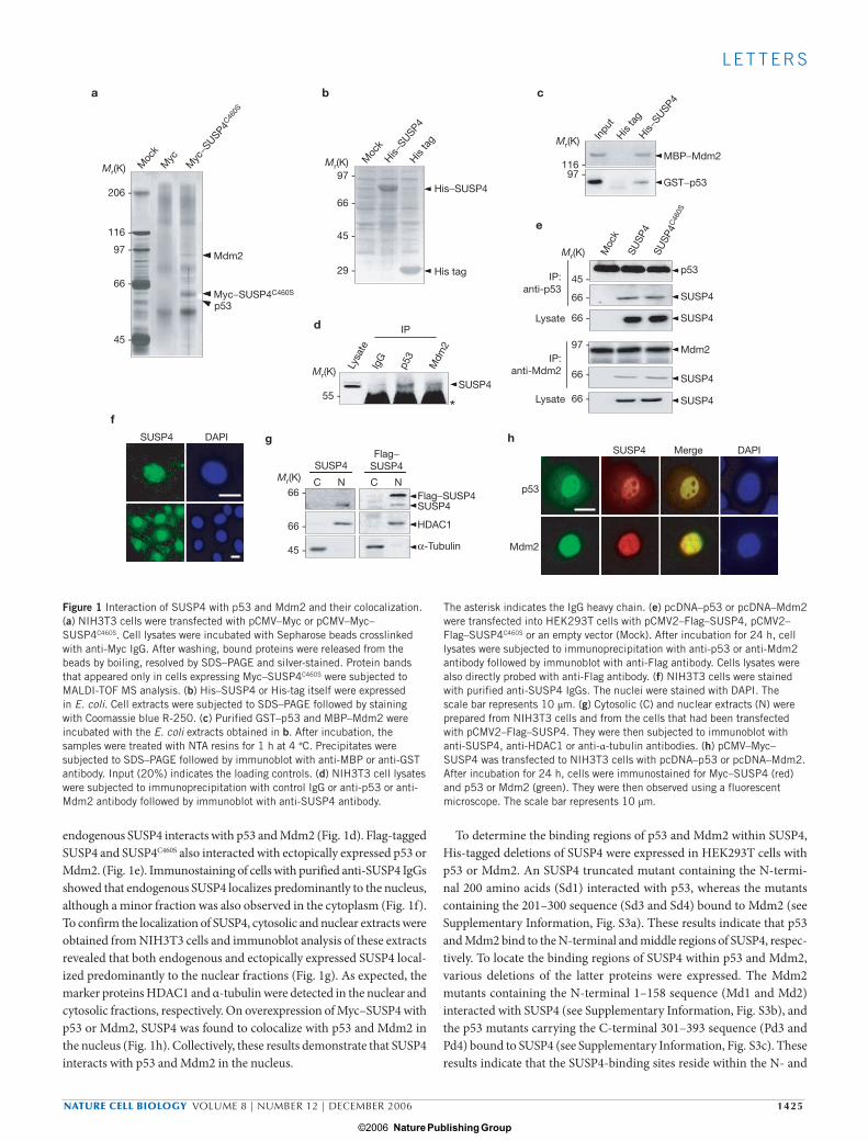
p53 and Mdm2 were found to interact with SUSP4 on MALDI-TOF mass spectrometric analysis of the proteins that bound to Myc–SUSP4C460S (in which the active site Cys 460 was replaced by Ser), but not to the Myc-tag itself (Fig. 1a). To confirm their interaction, His-tagged SUSP4 and a His tag expressed in Escherichia coli (Fig. 1b) were incubated with GST–p53 or MBP–Mdm2. Pulldown assay with NTA resins showed that GST–p53 and MBP–Mdm2 were coprecipitated with His–SUSP4, but not with the His-tag (Fig. 1c), indicating that the proteins directly interact with SUSP4. Immunoprecipitation analysis also revealed that
1NRL of Protein Biochemistry, School of Biological Sciences, Seoul National University, Seoul 151-742, Korea. 2Crystalgenomics Inc., Seoul 138-736, Korea. 3Department of Biological Sciences, Sookmyung Woman’s University, Seoul 140-742, Korea. 4Tokyo Metropolitan Institute of Medical Sciences, Tokyo 113, Japan.5Correspondence should be addressed to C.H.C. or O.S.B. (e-mails: [email protected]; [email protected])
Received 12 July 2006; accepted 9 October 2006; published online 5 November 2006; DOI: 10.1038/ncb1512
print ncb1512.indd 1424print ncb1512.indd 1424 15/11/06 17:02:2015/11/06 17:02:20
Nature Publishing Group ©2006
NATURE CELL BIOLOGY VOLUME 8 | NUMBER 12 | DECEMBER 2006 1425
L E T T E R S
endogenous SUSP4 interacts with p53 and Mdm2 (Fig. 1d). Flag-tagged SUSP4 and SUSP4C460S also interacted with ectopically expressed p53 or Mdm2. (Fig. 1e). Immunostaining of cells with purified anti-SUSP4 IgGs showed that endogenous SUSP4 localizes predominantly to the nucleus, although a minor fraction was also observed in the cytoplasm (Fig. 1f). To confirm the localization of SUSP4, cytosolic and nuclear extracts were obtained from NIH3T3 cells and immunoblot analysis of these extracts revealed that both endogenous and ectopically expressed SUSP4 local-ized predominantly to the nuclear fractions (Fig. 1g). As expected, the marker proteins HDAC1 and α-tubulin were detected in the nuclear and cytosolic fractions, respectively. On overexpression of Myc–SUSP4 with p53 or Mdm2, SUSP4 was found to colocalize with p53 and Mdm2 in the nucleus (Fig. 1h). Collectively, these results demonstrate that SUSP4 interacts with p53 and Mdm2 in the nucleus.
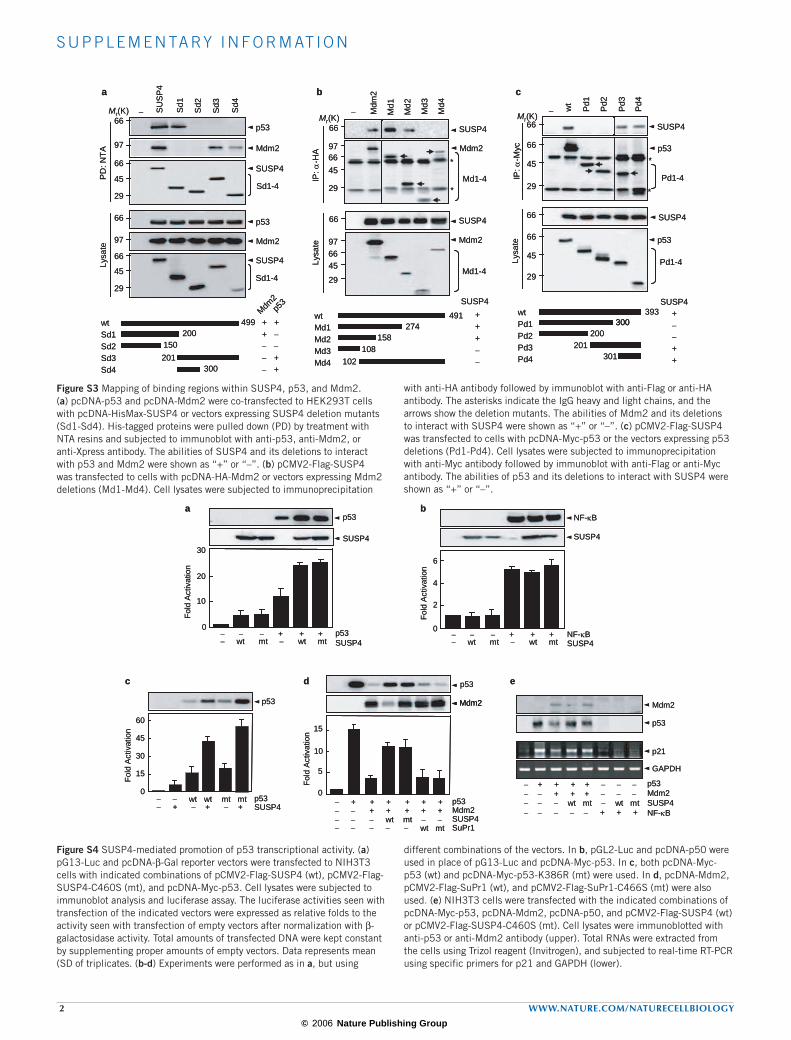
To determine the binding regions of p53 and Mdm2 within SUSP4, His-tagged deletions of SUSP4 were expressed in HEK293T cells with p53 or Mdm2. An SUSP4 truncated mutant containing the N-termi-nal 200 amino acids (Sd1) interacted with p53, whereas the mutants containing the 201–300 sequence (Sd3 and Sd4) bound to Mdm2 (see Supplementary Information, Fig. S3a). These results indicate that p53 and Mdm2 bind to the N-terminal and middle regions of SUSP4, respec-tively. To locate the binding regions of SUSP4 within p53 and Mdm2, various deletions of the latter proteins were expressed. The Mdm2 mutants containing the N-terminal 1–158 sequence (Md1 and Md2) interacted with SUSP4 (see Supplementary Information, Fig. S3b), and the p53 mutants carrying the C-terminal 301–393 sequence (Pd3 and Pd4) bound to SUSP4 (see Supplementary Information, Fig. S3c). These results indicate that the SUSP4-binding sites reside within the N- and
His−SUSP4
His−SUSP4
b
His ta
g His ta
g
Inpu
t
c
116 -97 -
g h
f
Myc
−SUSP4
C460S
206 -
116 -
97 -
p53
Myc
66 -
45 -
a
Mdm2
Myc−SUSP4C460S
Moc
k
Moc
k
d
*
e
Mr(K) Mr(K)
Mr(K)
Mr(K)
Mr(K)
Mr(K)
His−SUSP4
His tag
97 -
66 -
66 -
66 -
45 -
45 -
29 -
MBP−Mdm2
GST−p53
55 -
Lysa
te
Moc
k
SUSP
4SU
SP4
C46
0S
IgG
p53
Mdm
2
IP
SUSP4
IP:anti-p53
IP:anti-Mdm2
45 -
66 -
66 -
97 -
66 -
66 -
Lysate
Lysate
p53
SUSP4
SUSP4
SUSP4
Flag−SUSP4SUSP4
SUSP4
Mdm2
SUSP4
SUSP4
SUSP4 Merge DAPIFlag−SUSP4
DAPI
C N C N
HDAC1
α-Tubulin
p53
Mdm2
Figure 1 Interaction of SUSP4 with p53 and Mdm2 and their colocalization. (a) NIH3T3 cells were transfected with pCMV–Myc or pCMV–Myc–SUSP4C460S. Cell lysates were incubated with Sepharose beads crosslinked with anti-Myc IgG. After washing, bound proteins were released from the beads by boiling, resolved by SDS–PAGE and silver-stained. Protein bands that appeared only in cells expressing Myc–SUSP4C460S were subjected to MALDI-TOF MS analysis. (b) His–SUSP4 or His-tag itself were expressed in E. coli. Cell extracts were subjected to SDS–PAGE followed by staining with Coomassie blue R-250. (c) Purified GST–p53 and MBP–Mdm2 were incubated with the E. coli extracts obtained in b. After incubation, the samples were treated with NTA resins for 1 h at 4 °C. Precipitates were subjected to SDS–PAGE followed by immunoblot with anti-MBP or anti-GST antibody. Input (20%) indicates the loading controls. (d) NIH3T3 cell lysates were subjected to immunoprecipitation with control IgG or anti-p53 or anti-Mdm2 antibody followed by immunoblot with anti-SUSP4 antibody.
The asterisk indicates the IgG heavy chain. (e) pcDNA–p53 or pcDNA–Mdm2 were transfected into HEK293T cells with pCMV2–Flag–SUSP4, pCMV2–Flag–SUSP4C460S or an empty vector (Mock). After incubation for 24 h, cell lysates were subjected to immunoprecipitation with anti-p53 or anti-Mdm2 antibody followed by immunoblot with anti-Flag antibody. Cells lysates were also directly probed with anti-Flag antibody. (f) NIH3T3 cells were stained with purified anti-SUSP4 IgGs. The nuclei were stained with DAPI. The scale bar represents 10 μm. (g) Cytosolic (C) and nuclear extracts (N) were prepared from NIH3T3 cells and from the cells that had been transfected with pCMV2–Flag–SUSP4. They were then subjected to immunoblot with anti-SUSP4, anti-HDAC1 or anti-α-tubulin antibodies. (h) pCMV–Myc–SUSP4 was transfected to NIH3T3 cells with pcDNA–p53 or pcDNA–Mdm2. After incubation for 24 h, cells were immunostained for Myc–SUSP4 (red) and p53 or Mdm2 (green). They were then observed using a fluorescent microscope. The scale bar represents 10 μm.
print ncb1512.indd 1425print ncb1512.indd 1425 15/11/06 17:02:2115/11/06 17:02:21
Nature Publishing Group ©2006
1426 NATURE CELL BIOLOGY VOLUME 8 | NUMBER 12 | DECEMBER 2006
L E T T E R S
C-terminal regions of Mdm2 and p53, respectively. These results suggest that SUSP4 and p53 may compete with each other for binding to the same N-terminal region of Mdm2.
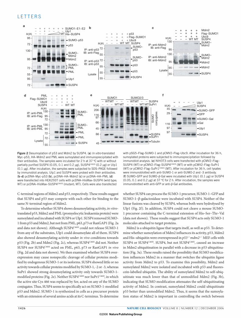
To determine whether SUSP4 shows desumoylating activity, in vitro-translated p53, Mdm2 and PML (promyelocytic leukaemia protein) were sumoylated and incubated with SUSP4 or Ulp1. SUSP4 removed SUMO-1 from p53 and Mdm2, but not from PML, p63, p73 or RanGAP1 (Fig. 2a and data not shown). Although SUSP4C460S could not release SUMO-1 from any of the substrates, Ulp1 could desumoylate all of them. SUSP4 also showed desumoylating activity under in vivo conditions towards p53 (Fig. 2b) and Mdm2 (Fig. 2c), whereas SUSP4C460S did not. Neither SUSP4 nor SUSP4C460S acted on PML, p63, p73 or RanGAP1 in vivo (Fig. 2d and data not shown). We then examined whether SUSP4 over-expression may cause nonspecific cleavage of cellular proteins modi-fied by endogenous SUMO-1 or its isoforms. SUSP4 showed little or no activity towards cellular proteins modified by SUMO-1, -2 or -3, whereas SuPr1 showed strong desumoylating activity only towards SUMO-1-modified proteins (Fig. 2e). Neither SUSP4C460S nor SuPr1C466S, in which the active site Cys 466 was replaced by Ser, acted on any of the SUMO conjugates. Thus, SUSP4 seems to specifically act on SUMO-1-modified p53 and Mdm2. SUMO-1 is synthesized in cells as a precursor protein with an extension of several amino acids at its C-terminus. To determine
whether SUSP4 can process the SUMO-1 precursor, SUMO-1–GFP and SUMO-1–β-galactosidase were incubated with SUSP4. Neither of the linear fusions was cleaved by SUSP4, whereas both were hydrolysed by Ulp1 (Fig. 2f). In addition, SUSP4 could not cleave a mouse SUMO-1 precursor containing the C-terminal extension of His–Ser–Thr–Val (data not shown). These results suggest that SUSP4 acts only SUMO-1 molecules attached to target proteins.
Mdm2 is a ubiquitin ligase that targets itself, as well as p53. To deter-mine whether sumoylation of Mdm2 influences its activity, p53, Mdm2 and His–ubiquitin were overexpressed in p53–/–mdm2–/– MEF cells with SUSP4 or SUSP4C460S. SUSP4, but not SUSP4C460S, caused an increase in Mdm2 ubiquitination in parallel with a decrease in p53 ubiquitina-tion (Fig. 3a). These results raised the possibility that SUMO modifica-tion influences Mdm2 in a manner that switches the ubiquitin ligase activity from Mdm2 to p53. To examine this possibility, Mdm2 and sumoylated Mdm2 were isolated and incubated with p53 and fluores-cein-labelled ubiquitin. The ability of sumoylated Mdm2 to self-ubiq-uitinate was much lower than that of unmodified Mdm2 (Fig. 3b), indicating that SUMO modification attenuates the self-ubiquitinating activity of Mdm2. In contrast, sumoylated Mdm2 could ubiquitinate p53 better than unmodified Mdm2. Thus, it seems that the sumoyla-tion status of Mdm2 is important in controlling the switch between
SUMO1−E1−E2 Ulp1His−SUSP4
p53
Mdm2
PML
Ulp1
SUSP4
SUMO−Mdm2
SUMO−p53
SUMO−PML
−−−
+−−
++−
+−
+−
+−
+−
ed
f
+++
−−−−
+−−−
+++−
+++
+++
−−−−
+−−−
+++− − − −
− − −
+++
WTWT
MT
WT MTWTMT
WT MT − − −− − −WTMT
WT MT
MT
b
IP: anti-p53
IP: anti-Mdm2
IP: anti-Mdm2IB: anti-Mdm2IP: anti-p53
IB: anti-p53
IB: anti-Flag
IB: anti-Flag
c+++
−−−−
+−−−
+++−
+++
+++
−−−−
+
− +− − − −−− −
−−−
+++−
+++
Mr(K)
Mr(K)
Mr(K)Mr(K) Mr(K)
Mr(K)95 -
72 -
130 -
95 -
72 -
55 -
95 -
72 -
MT
a
Lysate
72 -
55 -
55 -
55 -
55 -
72 -
p53
p53
p53
Flag−SUMO1Flag−SUMO1
Ubc9Ubc9
SUSP4SUSP4
SUSP4
SUSP4
Ulp1
SUSP4
SUMO−p53
SUMO−Mdm2
SUMO−GFP
SUMO−GalGal
UIp1
GFP
SUMO−Mdm2
Mdm2
Mdm2
SUMO−p53
130 -
130 -
95 -
95 -
95 -
55 -Lysate
Mdm2
PMLFlag−SUMO1
Ubc9SUSP4
IP: anti-PML
IP: anti-PMLIB: anti-PML
Lysate
IB: anti-Flag
130 - 206 - 45 -
29 -
116 -97 -
97 -
116 -97 -
66 -
45 -
66 -
45 -
95 -72 -
130 -95 -
72 -
72 -
55 -
SUMO−PML
SUMO−PML
PML
PML
SUSP4
SUMO1 SUMO2/3SUSP4
SUSP4
SUSP4
SuPr1
SuPr1
Tubulin
SU
MO
conj
ugat
esFigure 2 Desumoylation of p53 and Mdm2 by SUSP4. (a) In vitro-translated Myc–p53, HA–Mdm2 and PML were sumoylated and immunoprecipitated with their antibodies. The samples were incubated for 2 h at 37 °C with or without partially purified SUSP4 (0.05, 0.1 and 0.2 µg), SUSP4C460S (0.2 µg) or Ulp1 (0.1 µg). After incubation, the samples were subjected to SDS–PAGE followed by immunoblot analysis. Ulp1 and SUSP4 were probed with their antibodies. (b–d) pcDNA–Myc–p53 (b), pcDNA–HA–Mdm2 (c) or pcDNA–HA–PML (d) were transfected into HEK293T cells with pcDNA–HisMax–SUSP4 (wild type; WT) or pcDNA–HisMax–SUSP4C460S (mutant; MT). Cells were also transfected
with pSG5–Flag–SUMO-1 and pCMV2–Flag–Ubc9. After incubation for 36 h, sumoylated proteins were subjected to immunoprecipitation followed by immunoblot analysis. (e) NIH3T3 cells were transfected with pCMV2–Flag–SUSP4 (WT) or pCMV2–Flag–SUSP4C460S (MT) or with pCMV2–Flag–SuPr1 (WT) or pCMV2–Flag–SuPr1C466S (MT). After incubation for 36 h, cell lysates were immunoblotted with anti-SUMO-1 or anti-SUMO-2 and -3 antibody. (f) SUMO–GFP and SUMO–β-Gal were incubated with Ulp1 (0.1 µg) or SUSP4 (0.05, 0.1 and 0.2 µg) at 37 °C for 2 h. After incubation, the samples were immunoblotted with anti-GFP or anti-β-Gal antibodies.
print ncb1512.indd 1426print ncb1512.indd 1426 15/11/06 17:02:2315/11/06 17:02:23
Nature Publishing Group ©2006

NATURE CELL BIOLOGY VOLUME 8 | NUMBER 12 | DECEMBER 2006 1427
L E T T E R S
self-ubiquitination and p53 ubiquitination. Furthermore, pulse–chase analysis using 35S-methionine reveals that SUSP4, but not SUSP4C460S, causes a marked decrease in Mdm2 stability (Fig. 3c). SUSP4 also caused p53 stabi-lization. These results indicate that SUSP4 destabilizes Mdm2 by promoting its self-ubiquitinating activity, thereby leading to p53 stabilization.
Surprisingly, however, expression of SUSP4C460S, which cannot des-umoylate Mdm2, caused a marked decrease in the level of ubiquitinated p53 (Fig. 3a). Moreover, SUSP4C460S, which showed little or no effect
on Mdm2 stability, caused p53 stabilization (Fig. 3c). We showed that SUSP4 binds to the N-terminal region of Mdm2 (see Supplementary Information, Fig. S3). The binding site of p53 has also been identified to reside within the N-terminal region of Mdm2 (ref. 29). Thus, it seemed possible that competition between SUSP4C460S and p53 for binding to Mdm2 results in p53 stabilization. To investigate this possibility, we first examined whether SUSP4C460S can block p53 ubiquitination by Mdm2 in vitro. The level of ubiquitinated p53 was decreased by SUSP4C460S
+−−−
+++−
+++
+++
+++
d
g
+−
+ + +
Mdm2
SUSP4
a
Ubn−p53
Mdm2p53His−UbSUSP4
p53
PD: NTAIB:anti-p53
PD: NTAIB:anti-Mdm2
Lysate
Ubn−Mdm2
c
p53
p53
p530 5 10 15 20 0 5 10 15 20
9 40
30
20
10
00
3
6
b
p53
Ubn−Mdm2 Ubn−p53
−
e f+−−
++−
++
++
++
MT
170 -
130 -
95 -170 -130 -
95 -72 -95 -
55 -
72 -
− − −− − + + + + +− + + + + + +− + + + + + +
Mdm2
Mdm2
Mdm2SUMO−Mdm2
SUMO−Mdm2
SUMO−Mdm2
SUMO−Mdm2
ΔF (×
103
)
ΔF (×
103
)
Mdm2 or SUMO−Mdm2 (μM)
Mr(K)
Mr(K)
170 -
130 -
72 -
Mdm2
Chase:Mock
SUSP4
SUSP4C460S
SUSP4C460S
Mock
SUSP4
SUSP4C460S
0 15 30 45 60 Time (min)Mdm2
Mdm2
Mdm2
SUSP4Lysate
p53
p53
Mdm2E1−E2SUSP4C460S
Mr(K)
Mr(K)
Mr(K)
Mr(K)
Mr(K)
170 -130 -
95 -95 -
130 -
72 -
Ubn−p53
SUSP4C460S
Mdm2
His
−S
US
P4C
460S
His
tag
130 -
72 -
72 -
130 -26 -
72 -
0.05
0.1
0.2
0.5 (μg)
Mdm2
His−SUSP4C460S
GST−p53
Mdm2
His tag
GST−p53
IP: a
nti-
Md
m2
Lysa
te
55 -
170 -130 -
95 -72 -
55 -
55 -95 -
55 -
55 -95 -
Mdm2
Mdm2
Mdm2
p53
p53
p53
SUSP4C460S
SUSP4C460S
SUSP4C460S
IP: a
nti-
p53
IB: a
nti-
Ub
Lysa
te
p53
p53
SUSP4
SUSP4
Ubn−p53
Figure 3 Effect of SUSP4 on ubiquitination and stability of Mdm2 and p53. (a) pcDNA–Myc–p53, pcDNA–HA–Mdm2 and pcDNA–HisMax–ubiquitin (Ub) were transfected to p53–/–mdm2–/– cells with pCMV2–Flag–SUSP4 (2, 4 and 8 μg) or pCMV2–Flag–SUSP4C460S (8 μg). After incubation for 4 h with 10 μM MG132, ubiquitinated proteins were pulled down by NTA resins and subjected to immunoblot analysis with anti-Mdm2 or anti-p53 antibodies. Uncropped images of the upper two panels are shown in the Supplementary Information, Fig. S5a. (b) MBP–Mdm2, Sae1–Sae2 and Ubc9 were incubated with or without SUMO-1. Mdm2 and sumoylated Mdm2 were separated using a DEAE–Sepharose column and each was incubated with p53, fluorescein-N-terminal ubiquitin, Uba1, UbcH5 and an ATP-regenerating system for 1 h. The reaction was stopped by adding 150 mM Tris–HCl (pH 7.6) containing 5% SDS and 30% glycerol. The mixtures were immunoprecipitated by anti-Mdm2 or anti-p53 antibodies and the precipitates were quantified using a fluorometer. The same amounts of the input p53, Mdm2 and SUMO–Mdm2 proteins were subjected to SDS–PAGE and Coomassie stained. The data represent the mean ± s.d. of three experiments. (c) NIH3T3 cells transfected with an empty vector (Mock), pCMV2–Flag–SUSP4 or
pCMV2–Flag–SUSP4C460S were labelled with 35S-methionine followed by treatment with unlabelled methionine. The labelled proteins were subjected to immunoprecipitation with anti-Mdm2 or anti-p53 antibodies followed by SDS–PAGE, and visualized by autoradiography. (d) Mdm2 (0.1 µg) was incubated with SUSP4C460S (0.1, 0.2 and 0.5 µg) for 30 min at 4 °C. The samples were subjected to in vitro ubiquitination followed by immunoprecipitation with anti-p53 antibodies. They were then immunoblotted with anti-ubiquitin antibodies. (e) Mdm2 (0.5 µg) was incubated with His–SUSP4C460S or His-tag itself for 30 min and then with purified GST–p53 (0.2 µg) for 1 h. The samples were subjected to immunoprecipitation with anti-Mdm2 antibodies. (f) pcDNA–Mdm2 (1 μg) and pcDNA–Myc–p53 (1 μg) were transfected to p53–/–mdm2–/– cells with pCMV2–Flag–SUSP4C460S (0.5, 1 and 2 μg). After culturing for 24 h, cell lysates were subjected to immunoprecipitation with anti-Mdm2 antibodies followed by immunoblot with anti-Flag, anti-p53 or anti-Mdm2 antibodies. (g) p53–/–mdm2–/– cells were transfected with pcDNA–Myc–p53 and pCMV2–Flag–SUSP4. After incubation as in a, cell lysates were subjected to immunoprecipitation with anti-p53 antibodies and probed with anti-ubiquitin antibodies.
print ncb1512.indd 1427print ncb1512.indd 1427 15/11/06 17:02:2415/11/06 17:02:24
Nature Publishing Group ©2006
1428 NATURE CELL BIOLOGY VOLUME 8 | NUMBER 12 | DECEMBER 2006
L E T T E R S
treatment in a dose-dependent manner (Fig. 3d), indicating that SUSP4C460S is capable of inhibiting Mdm2-mediated p53 ubiquitina-tion. We then examined whether SUSP4C460S actually competes with p53 for binding to Mdm2. Purified Mdm2 was incubated with increasing amounts of His–SUSP4C460S and then with p53. Immunoprecipitation analysis using anti-Mdm2 antibodies revealed that the amount of SUSP4C460S bound to Mdm2 gradually increased in parallel with a decrease in the amount of p53 bound to Mdm2 (Fig. 3e). In vivo competition experiments were also performed. Mdm2 and p53 were expressed in p53–/–mdm2–/– cells with increasing amounts of SUSP4C460S. The amount of SUSP4C460S coprecipitated with Mdm2 increased in par-allel with a decrease in the amount of p53 coprecipitated with Mdm2 (Fig. 3f). Nearly identical results were obtained with wild-type SUSP4 in both in vivo and in vitro competition experiments (data not shown). These results indicate that both SUSP4 and SUSP4C460S are capable of competing with p53 for binding to Mdm2.
As SUSP4 can also desumoylate p53, we examined the effect of SUSP4 on p53 ubiquitination under conditions where Mdm2 is absent by expressing p53 with increasing amounts of SUSP4 in p53–/–mdm2–/– cells. In contrast with the results obtained in the presence of Mdm2 (see Fig. 3a), SUSP4 showed little or no effect on p53 ubiquitination in the absence of Mdm2 (Fig. 3g). These results indicate that the effects of SUSP4 on the stability of p53, as well as of Mdm2, are mediated by its action on Mdm2 but not on p53. Taken together, these results suggest that SUSP4 can stabilize p53 in two different modes: by interfering with the interaction between p53 and Mdm2; and by promoting self-ubiquitination and destabilization of Mdm2 through desumoylation of Mdm2.
As SUSP4 increased p53 stability, we examined the effect of SUSP4 on p53 transcriptional activity when NIH3T3 cells were transfected with reporter vectors. SUSP4 caused a marked increase in the activity of both endogenous and ectopically expressed p53 (see Supplementary Information, Fig. S4a). Immunoblot analysis of the same cell lysates again showed that SUSP4
70.4%66%51.1% 70.8% 59.2%
b
d
24 48 720
c
a
M
G2
G1M
G2
G1
3 21Ce
f
*
Con
trol
*
h
Control
Control
g
3 21
Con
trol
p-H1/H3 SUSP4 DAPI
SUSP4
Mock 24 36 48 72
Moc
k
24 36 48 72
Time (h)
Time (h)SUSP4
p21
20016012080400
shControl shRNA-3sh
Con
trol
shR
NA-
369.3% 52.9%
24 + 24Time (h)
SUSP4
p21
Cou
nts
20016012080400
Cou
nts
3 H-T
hym
idin
e (×
103
cp
m)
60
4555 -
26 -
30
15
0
Mock
SUSP4
Time (h)
Mr(K)Mr(K)
SUSP4
GFP
(shRNA)
IP: a
nti-
Md
m2
170 -
170 -130 -
95 -
72 -
130 -
95 -
72 -
55 -95 -
55 -
55 -
130 -95 -72 -55 -
170 -130 -
95 -72 -
shRNA-3
Ubn−Mdm2
SUMO−Mdm2
Ubn−p53
SUMO−p53
Mdm2p53
SUSP4
IP: a
nti-
p53
Lysa
te
Fold
act
ivat
ion 1.0
0.5
0
SUSP4 0 24 48 72 0 24 48 72
0 24 48 72 0 24 48 72
Time (h)SUSP4
3 H-T
hym
idin
e (×
103
cp
m)
3 H-T
hym
idin
e (×
103
cp
m)
15
10
5
0
Time (h) Time (h)
20
15
10
5
0
p53+/+ p53−/−
1−3
1−3
Figure 4 SUSP4-mediated inhibition of cell growth. (a) NIH3T3 cells were transiently transfected with pCMV–Myc–SUSP4. After incubation for 24 h, cells were stained with anti-Myc or a mixture of anti-phospho-histone H1 and H3 antibodies. The scale bar represents 10 μm. (b) Cells transfected with pCMV2–Flag–SUSP4 or an empty vector (Mock) were cultured for the indicated times and subjected to FACS analysis to determine their DNA content. They were also subjected to immunoblot analysis with anti-SUSP4 or anti-p21 antibodies. The data represents the mean ± s.d. of three experiments. (c) Cells transfected with pCMV2–Flag–SUSP4 were incubated for 24 h. After incubation, they were transfected with control shRNA (shControl) or shRNA-3 and cultured for a further 24 h. FACS and immunoblot analyses were then performed as in b. (d) Cells cultured as in b were assayed for 3H-thymidine incorporation. (e) Each of three shRNAs (1–3) or control shRNA (C) was transfected for two rounds into cells with a vector encoding GFP as a transfection control. After incubation for 24 h, cells were subjected to immunoblot analysis with anti-SUSP4
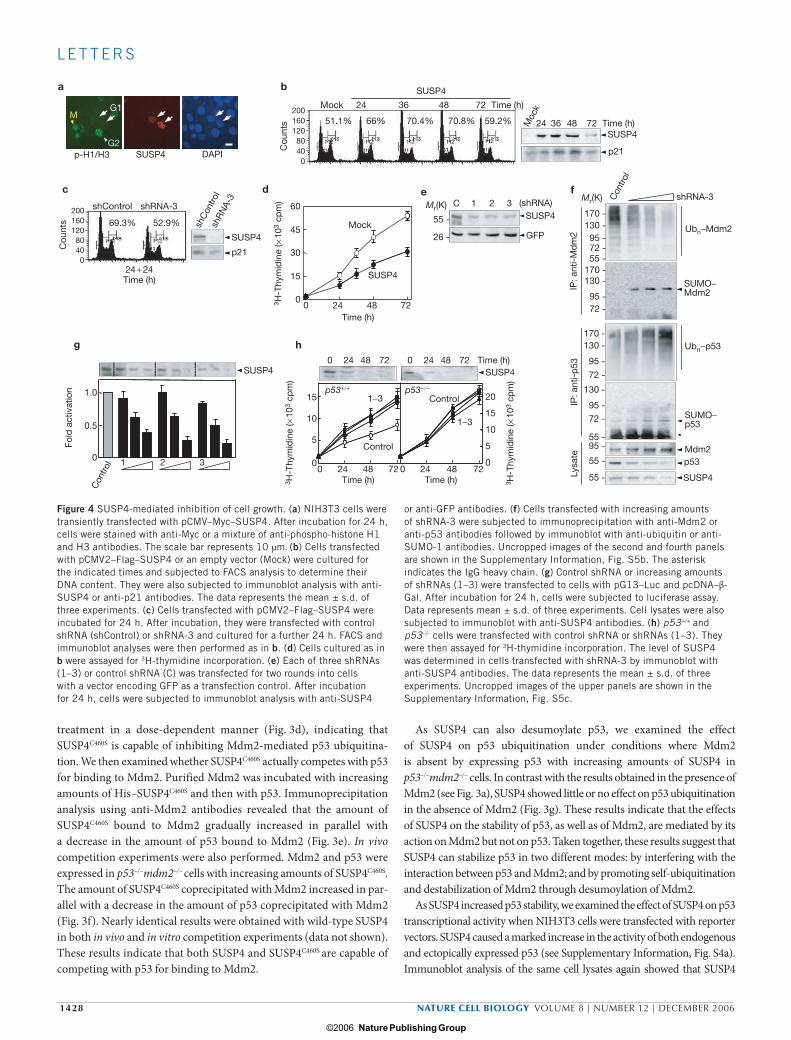
or anti-GFP antibodies. (f) Cells transfected with increasing amounts of shRNA-3 were subjected to immunoprecipitation with anti-Mdm2 or anti-p53 antibodies followed by immunoblot with anti-ubiquitin or anti-SUMO-1 antibodies. Uncropped images of the second and fourth panels are shown in the Supplementary Information, Fig. S5b. The asterisk indicates the IgG heavy chain. (g) Control shRNA or increasing amounts of shRNAs (1–3) were transfected to cells with pG13–Luc and pcDNA–β-Gal. After incubation for 24 h, cells were subjected to luciferase assay. Data represents mean ± s.d. of three experiments. Cell lysates were also subjected to immunoblot with anti-SUSP4 antibodies. (h) p53+/+ and p53–/– cells were transfected with control shRNA or shRNAs (1–3). They were then assayed for 3H-thymidine incorporation. The level of SUSP4 was determined in cells transfected with shRNA-3 by immunoblot with anti-SUSP4 antibodies. The data represents the mean ± s.d. of three experiments. Uncropped images of the upper panels are shown in the Supplementary Information, Fig. S5c.
print ncb1512.indd 1428print ncb1512.indd 1428 15/11/06 17:02:2615/11/06 17:02:26
Nature Publishing Group ©2006

NATURE CELL BIOLOGY VOLUME 8 | NUMBER 12 | DECEMBER 2006 1429
L E T T E R S
stabilizes p53. SUSP4C460S also increased the stability and activity of p53. However, SUSP4 had little or no effect on the stability and activity of p50 (NF-κB; see Supplementary Information, Fig. S4b). These results suggest that both SUSP4 and SUSP4C460S positively regulate p53 activity by antagoniz-ing endogenous Mdm2. These results also suggest that the SUSP4-mediated increase in p53 activity is independent of p53 sumoylation. To confirm this, p53 or p53K386R (in which the SUMO-accepting Lys residue was replaced by Arg) were expressed in cells with SUSP4. SUSP4 caused a marked increase in the stability and activity of both p53 and p53K386R (see Supplementary Information, Fig. S4c). Thus, it seems that p53 sumoylation itself has little or no influence on the SUSP4-mediated increase in p53 activity.
We then examined the effect of SUSP4 on the stability and activity of p53 in conditions where Mdm2 was overproduced. As expected, Mdm2 expression resulted in a dramatic reduction in p53 activity by destabilizing it (see Supplementary Information, Fig. S4d). Coexpression of SUSP4 or SUSP4C460S reversed the Mdm2-mediated decrease in p53 activity. On the other hand, neither SuPr1 nor SuPr1C466S could stabilize p53 or reverse the Mdm2-mediated decrease in p53 activity, indicating that Mdm2 is a spe-cific target of SUSP4. Moreover, Mdm2 expression resulted in a marked reduction in p21 mRNA levels, but coexpression of SUSP4 or SUSP4C460S reversed the inhibitory effect of Mdm2 (see Supplementary Information,
Fig. S4e). Little or no accumulation of p21 mRNA was observed on expression of NF-κB, regardless of whether SUSP4 or SUSP4C460S was coexpressed. These results further demonstrate that SUSP4 positively regulates p53 activity by antagonizing Mdm2 function.
As SUSP4 mediates p53 stabilization, we examined whether SUSP4 is involved in p53-dependent control of cell growth. NIH3T3 cells trans-fected with pCMV–Myc–SUSP4 were strongly stained by anti-Myc anti-bodies (Fig. 4a). However, the same cells were barely stained by a mixture of anti-phospho-histone H1 and H3 antibodies, which is a characteristic feature of G1-phase cells. These results suggest that SUSP4 overexpression leads to cell-cycle arrest at the G1 phase. To confirm this, cells transfected with pCMV2–Flag–SUSP4 were cultured for various times and subjected to FACS analysis to determine their DNA contents. The fraction of cells in G1 phase was significantly increased up to 48 h in culture (Fig. 4b). The level of p21 protein was also gradually increased up to 48 h in culture. Both the fraction of G1-phase cells and p21 levels decreased thereaf-ter, most likely due to an increase in the number of cells that were not transfected with pCMV2–Flag–SUSP4, as SUSP4 levels also decreased at 72 h. Furthermore, cotransfection of a susp4-specific short hairpin RNA (shRNA-3; see below), but not a negative control vector (shControl), resulted in a decrease in both the fraction of cells in the G1 phase and p21
c
ba
d
+ +− −
Mdm2
p53
SUSP4
Mdm2
Mdm2
p53
p53
SUSP4
e
p53
UV: UV:0 0 2 4 8 12 24 0 2 4 8 12 2415 30 60 90 120 180 Time (min) Time (h)
Time (h)
SUSP4
p21
GAPDH
shControl shRNA-3
SUSP4
Mdm2
p53
p21
Etoposide Camptothesin
0 2 4 8 12 24 0 2 4 8 12 24
SUSP4
p53
Anti-SUSP4 Anti-p53
UV:p53
Mdm2
SUSP4
p53
Mdm2
SUSP4
p53
SUMO Ub
Degraded
StabilizedSUSP4
Mdm2
Mdm2
UV
IP:
Figure 5 Induction of SUSP4 expression by UV. (a) NIH3T3 cells were irradiated with UV (50 J m–2) for 30 s and incubated for the indicated times. Total RNAs were prepared from cells using Triazol reagent. RT–PCR was then carried out using specific primers for susp4, p21 and GAPDH. (b) Cells were transfected with control shRNA or shRNA-3 for two rounds. After incubation for 24 h, cells were irradiated with UV. They were then subjected to immunoblot with respective antibodies. (c) Cells were treated with 20 µM etoposide or 10 µg ml–1 of camptothesin, and incubated for
the indicated times. They were then subjected to immunoblot with anti-SUSP4 or anti-p53 antibody. (d) Cells that had been treated without (–) or with UV (+) were incubated for 1 h. After incubation, cells were subjected to immunoprecipitation with anti-SUSP4 or anti-p53 antibody followed by immunoblot with respective antibodies. Uncropped images of the left upper two panels and the right lower two panels are shown in the Supplementary Information, Fig. 5Sd. (e) A schematic representation of a model for SUSP4-mediated control of the p53–Mdm2 pathway by SUSP4
print ncb1512.indd 1429print ncb1512.indd 1429 15/11/06 17:02:2815/11/06 17:02:28
Nature Publishing Group ©2006

1430 NATURE CELL BIOLOGY VOLUME 8 | NUMBER 12 | DECEMBER 2006
L E T T E R S
levels (Fig. 4c), indicating that shRNA-3 can reverse the cell-cycle arrest caused by SUSP4 overexpression. We then examined whether SUSP4-mediated cell-cycle arrest leads to inhibition of cell growth. The rate of 3H-thymidine incorporation was significantly reduced by transfection of cells with pCMV2–Flag–SUSP4 compared with that of an empty vector (Fig. 4d). Taken together, these results suggest that overproduced SUSP4 can downregulate the growth of cells by stabilizing p53.
To clarify the involvement of SUSP4 in p53-mediated control of cell growth, three susp4-specific shRNAs (indicated by 1–3) were transfected into NIH3T3 cells with a vector encoding GFP. All three shRNAs effec-tively reduced endogenous SUSP4 levels (Fig. 4e). Therefore, we exam-ined the effect of shRNA-3 on ubiquitin and SUMO-1 modification of Mdm2. shRNA-mediated knockdown of susp4 caused a decrease in the level of ubiquitinated Mdm2 in parallel with an increase in the level of sumoylated Mdm2, leading to stabilization of endogenous Mdm2 and destabilization of endogenous p53 (Fig. 4f). Consistently, all three shRNAs caused a decrease in the transcriptional activity of endogenous p53 in a dose-dependent manner (Fig. 4g). Moreover, the rate of 3H-thymidine incorporation was significantly increased by transfection of p53+/+ cells with the shRNAs, compared with that observed with the control shRNA (Fig. 4h), indicating that SUSP4 is involved in downregu-lation of cell growth. In contrast, knockdown of susp4 had little or no effect on the proliferation of p53–/– cells (Fig. 4h), demonstrating the link between p53 and SUSP4 in the control of cell growth. Taken together, these results reveal that SUSP4 has an important function in the control of cell growth by p53.
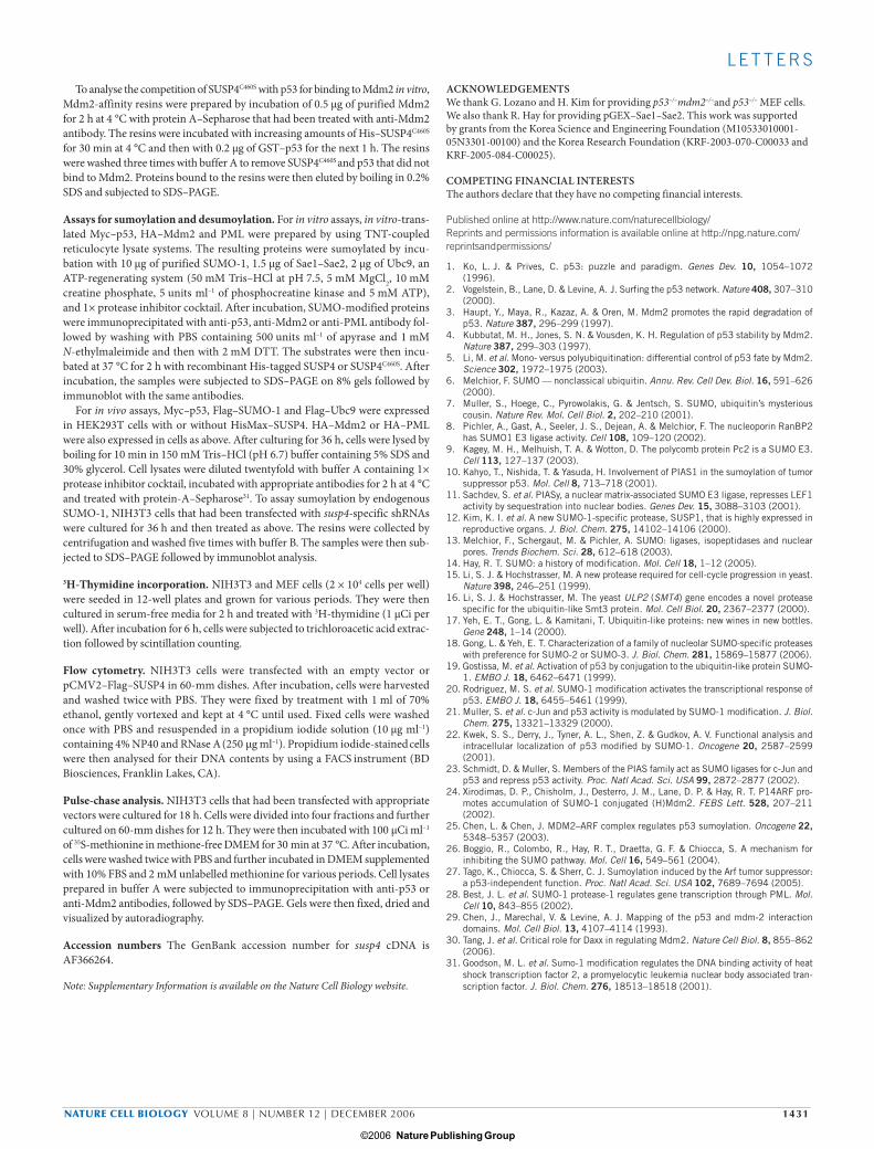
Given that UV damage is one of the major stresses that provokes p53 action, we examined whether UV induces susp4 gene transcription. susp4 mRNA levels dramatically increased as early as 15 min after UV irradiation (Fig. 5a). p21 mRNA level also increased about 1 h after the irradiation. These results suggest that SUSP4 is an UV-inducible protein that stabilizes p53, leading to an increase in p21 mRNA levels. To deter-mine whether UV-induced SUSP4 influences the endogenous levels of Mdm2 and p53 proteins, NIH3T3 cells transfected with control shRNA or shRNA-3 was irradiated with UV. Concurrent with an increase in SUSP4 levels, Mdm2 levels were sharply declined in cells transfected with control shRNA (Fig. 5b). p53 levels then gradually increased, followed by an increase in endogenous p21 levels. On the other hand, shRNA-medi-ated knockdown of susp4 resulted in a marked delay in the accumulation of p53 and p21. These results indicate that SUSP4 has a critical role in the control of the cellular levels of Mdm2 and consequently of p53 and p21, when cells are damaged by UV. However, etoposide and camptothesin, which also cause DNA damage, showed little or no effect on SUSP4 induction (Fig. 5c), suggesting that SUSP4 responds to a limited range of stresses, such as UV.
We then examined whether UV damage causes an alteration in the amounts of p53 and Mdm2 that bind to SUSP4. UV irradiation led to an increase in the amount of Mdm2 bound to SUSP4, with a decrease in the interaction between p53 and SUSP4 (Fig. 5d). UV also caused a decrease in the amounts of SUSP4 and Mdm2 that bind to p53. These results sug-gest that UV damage leads to an increase in relative abundance of the SUSP4–Mdm2 complex over p53–Mdm2 complex through competition of accumulated SUSP4 with p53 for binding to Mdm2.
Mdm2 targets not only p53, but also itself for ubiquitination, and the fate of p53 is determined accordingly. Recently, Daxx (a multifunc-tional protein involved in the control of apoptosis and transcription)
was shown to have a key role in switching the Mdm2 activity from p53 to itself in response to DNA damage30. Here, we have provided an addi-tional mechanism for the control of Mdm2 function and a model for the involvement of SUSP4 in the control of the p53–Mdm2 pathway, based on the results obtained in this study, is shown in Figure 5d. On UV damage, SUSP4 accumulates and replaces p53 for binding to Mdm2, leading to stabilization of p53 for induction of cell-cycle arrest and cell-growth inhibition. Desumoylation by SUSP4 would then promote self-ubiquitination and degradation of Mdm2 for further stabilization of p53. However, under normal conditions (that is, when SUSP4 levels are low), the SUMO-modification system, including Ubc9, may override SUSP4 and sumoylate Mdm2, leading to promotion of p53 ubiquitination. Thus, SUSP4 seems to have a critical role in switching the Mdm2 activity from p53 to itself in response to UV damage.
METHODSPlasmids and antibodies. susp4 cDNA was isolated from a mouse brain cDNA library and cloned into pcDNA–Myc, pcDNA–HisMax, pCMV–Myc and pCMV2–Flag. It was also cloned into pET-32a for bacterial expression. Therefore, His–SUSP4 and His-tag itself contain an extra-amino acid sequence of approxi-mately 20K corresponding to the multicloning site region of the vector. susp4-specific shRNAs were synthesized and cloned into pSilencer 2.0-U6 (Ambion, Austin, TX). Oligonucelotides used were: 1, GATCAGGGATGGTATTTAA; 2, GATGAGGTCATCAATTTCT; 3, GAATGTTTACCTGTAAAT.
Antibodies against Myc (9E10), SUMO-1 (D-11), p53 (DO-1 and N-19), p50 (H-119), p21 (C-19) and Mdm2 (SMT4) were obtained from SantaCruz (Santa Cruz, CA). Peroxidase-conjugated AffiniPure goat anti-rabbit and anti-mouse, and donkey anti-goat IgGs, FITC-conjugated goat anti-rabbit IgG, and TRITC-conjugated goat anti-mouse IgG were purchased from Jackson ImmunoResearch Laboratories (West Glove, PA). Anti-Flag M2, anti-Xpress, anti-Ub (FK1; Affiniti Res, Manhead, UK), and anti-SUMO-2 and -3 (Zymed, San Francisco, CA) antibodies were also used. Rabbit polyclonal anti-SUSP4 antiserum was raised against a recombinant His-tagged N-terminal fragment of SUSP4 (that is, amino acids 1–320). Anti-SUSP4 IgGs were purified by applying the serum onto an affinity column, which had been generated by conjugation of the N-terminal SUSP4 fragments to NHS-activated Sepharose 4 Fast Flow. Anti-SUSP4 IgGs bound to the resins were eluted with 100 mM glycine buffer (pH 3.0).
Cell culture and transfection. HEK293T, NIH3T3, MEF cells (p53+/+, p53–/–, and p53–/–mdm2–/–) were grown at 37 °C in DMEM) supplemented with 100 units ml–1 penicillin, 1 μg ml–1 streptomycin and 10% FBS. Transfections in HEK293T and NIH3T3 cells were carried out using LipofectaminePlus. MEF cells were transfected by electroporation.
Immunocytochemistry. NIH3T3 cells plated on gelatin-coated cover-glasses were fixed with 2% formaldehyde in PBS for 30 min at room temperature and per-meabilized with 0.5% Triton X-100 in PBS. All subsequent dilutions and washes were performed with PBS containing 0.1% Triton X-100 (PBS-T). Nonspecific binding sites were saturated by incubation for 30 min with a blocking solution consisting of 10% goat serum, 1% BSA and 1% gelatin in PBS. Cells were incu-bated with primary antibody for 1 h and washed with PBS-T four times at 10 min intervals. They were then incubated with FITC- or TRITC-conjugated secondary antibody for 1 h and washed four times. DAPI was used for counterstaining the nuclei. The cover glasses were mounted in Vectashield and cells were visualized under a Zeiss Axioplan II microscope.
Assays for protein–protein interaction. Cells were lysed in buffer A consisting of 50 mM Tris–HCl (pH 7.4), 120 mM NaCl, 0.5% NP40 and 1× protease inhibitor cocktail. Cell lysates were incubated with appropriate antibodies for 2 h at 4 °C and then with 50 μl of a 50% slurry of protein A–Sepharose for 1 h. The resins were collected by centrifugation and washed five times with buffer B consisting of 20 mM Tris–HCl (pH 7.4), 500 mM NaCl, 1 mM EDTA and 0.5% NP40. Bound proteins were eluted by boiling in 0.2% SDS and subjected to SDS–PAGE followed by immunoblot with appropriate antibodies.
print ncb1512.indd 1430print ncb1512.indd 1430 15/11/06 17:02:2915/11/06 17:02:29
Nature Publishing Group ©2006
NATURE CELL BIOLOGY VOLUME 8 | NUMBER 12 | DECEMBER 2006 1431
L E T T E R S
To analyse the competition of SUSP4C460S with p53 for binding to Mdm2 in vitro, Mdm2-affinity resins were prepared by incubation of 0.5 μg of purified Mdm2 for 2 h at 4 °C with protein A–Sepharose that had been treated with anti-Mdm2 antibody. The resins were incubated with increasing amounts of His–SUSP4C460S for 30 min at 4 °C and then with 0.2 μg of GST–p53 for the next 1 h. The resins were washed three times with buffer A to remove SUSP4C460S and p53 that did not bind to Mdm2. Proteins bound to the resins were then eluted by boiling in 0.2% SDS and subjected to SDS–PAGE.
Assays for sumoylation and desumoylation. For in vitro assays, in vitro-trans-lated Myc–p53, HA–Mdm2 and PML were prepared by using TNT-coupled reticulocyte lysate systems. The resulting proteins were sumoylated by incu-bation with 10 μg of purified SUMO-1, 1.5 μg of Sae1–Sae2, 2 μg of Ubc9, an ATP-regenerating system (50 mM Tris–HCl at pH 7.5, 5 mM MgCl2, 10 mM creatine phosphate, 5 units ml–1 of phosphocreatine kinase and 5 mM ATP), and 1× protease inhibitor cocktail. After incubation, SUMO-modified proteins were immunoprecipitated with anti-p53, anti-Mdm2 or anti-PML antibody fol-lowed by washing with PBS containing 500 units ml–1 of apyrase and 1 mM N-ethylmaleimide and then with 2 mM DTT. The substrates were then incu-bated at 37 °C for 2 h with recombinant His-tagged SUSP4 or SUSP4C460S. After incubation, the samples were subjected to SDS–PAGE on 8% gels followed by immunoblot with the same antibodies.
For in vivo assays, Myc–p53, Flag–SUMO-1 and Flag–Ubc9 were expressed in HEK293T cells with or without HisMax–SUSP4. HA–Mdm2 or HA–PML were also expressed in cells as above. After culturing for 36 h, cells were lysed by boiling for 10 min in 150 mM Tris–HCl (pH 6.7) buffer containing 5% SDS and 30% glycerol. Cell lysates were diluted twentyfold with buffer A containing 1× protease inhibitor cocktail, incubated with appropriate antibodies for 2 h at 4 °C and treated with protein-A–Sepharose31. To assay sumoylation by endogenous SUMO-1, NIH3T3 cells that had been transfected with susp4-specific shRNAs were cultured for 36 h and then treated as above. The resins were collected by centrifugation and washed five times with buffer B. The samples were then sub-jected to SDS–PAGE followed by immunoblot analysis.
3H-Thymidine incorporation. NIH3T3 and MEF cells (2 × 10⁴ cells per well) were seeded in 12-well plates and grown for various periods. They were then cultured in serum-free media for 2 h and treated with 3H-thymidine (1 μCi per well). After incubation for 6 h, cells were subjected to trichloroacetic acid extrac-tion followed by scintillation counting.
Flow cytometry. NIH3T3 cells were transfected with an empty vector or pCMV2–Flag–SUSP4 in 60-mm dishes. After incubation, cells were harvested and washed twice with PBS. They were fixed by treatment with 1 ml of 70%
ethanol, gently vortexed and kept at 4 °C until used. Fixed cells were washed once with PBS and resuspended in a propidium iodide solution (10 μg ml–1) containing 4% NP40 and RNase A (250 μg ml–1). Propidium iodide-stained cells were then analysed for their DNA contents by using a FACS instrument (BD Biosciences, Franklin Lakes, CA).
Pulse-chase analysis. NIH3T3 cells that had been transfected with appropriate vectors were cultured for 18 h. Cells were divided into four fractions and further cultured on 60-mm dishes for 12 h. They were then incubated with 100 μCi ml–1 of 35S-methionine in methione-free DMEM for 30 min at 37 °C. After incubation, cells were washed twice with PBS and further incubated in DMEM supplemented with 10% FBS and 2 mM unlabelled methionine for various periods. Cell lysates prepared in buffer A were subjected to immunoprecipitation with anti-p53 or anti-Mdm2 antibodies, followed by SDS–PAGE. Gels were then fixed, dried and visualized by autoradiography.
Accession numbers The GenBank accession number for susp4 cDNA is AF366264.
Note: Supplementary Information is available on the Nature Cell Biology website.
ACKNOWLEDGEMENTSWe thank G. Lozano and H. Kim for providing p53–/–mdm2–/–and p53–/– MEF cells. We also thank R. Hay for providing pGEX–Sae1–Sae2. This work was supported by grants from the Korea Science and Engineering Foundation (M10533010001-05N3301-00100) and the Korea Research Foundation (KRF-2003-070-C00033 and KRF-2005-084-C00025).
COMPETING FINANCIAL INTERESTSThe authors declare that they have no competing financial interests.
Published online at http://www.nature.com/naturecellbiology/Reprints and permissions information is available online at http://npg.nature.com/reprintsandpermissions/
1. Ko, L. J. & Prives, C. p53: puzzle and paradigm. Genes Dev. 10, 1054–1072 (1996).
2. Vogelstein, B., Lane, D. & Levine, A. J. Surfing the p53 network. Nature 408, 307–310 (2000).
3. Haupt, Y., Maya, R., Kazaz, A. & Oren, M. Mdm2 promotes the rapid degradation of p53. Nature 387, 296–299 (1997).
4. Kubbutat, M. H., Jones, S. N. & Vousden, K. H. Regulation of p53 stability by Mdm2. Nature 387, 299–303 (1997).
5. Li, M. et al. Mono- versus polyubiquitination: differential control of p53 fate by Mdm2. Science 302, 1972–1975 (2003).
6. Melchior, F. SUMO — nonclassical ubiquitin. Annu. Rev. Cell Dev. Biol. 16, 591–626 (2000).
7. Muller, S., Hoege, C., Pyrowolakis, G. & Jentsch, S. SUMO, ubiquitin’s mysterious cousin. Nature Rev. Mol. Cell Biol. 2, 202–210 (2001).
8. Pichler, A., Gast, A., Seeler, J. S., Dejean, A. & Melchior, F. The nucleoporin RanBP2 has SUMO1 E3 ligase activity. Cell 108, 109–120 (2002).
9. Kagey, M. H., Melhuish, T. A. & Wotton, D. The polycomb protein Pc2 is a SUMO E3. Cell 113, 127–137 (2003).
10. Kahyo, T., Nishida, T. & Yasuda, H. Involvement of PIAS1 in the sumoylation of tumor suppressor p53. Mol. Cell 8, 713–718 (2001).
11. Sachdev, S. et al. PIASy, a nuclear matrix-associated SUMO E3 ligase, represses LEF1 activity by sequestration into nuclear bodies. Genes Dev. 15, 3088–3103 (2001).
12. Kim, K. I. et al. A new SUMO-1-specific protease, SUSP1, that is highly expressed in reproductive organs. J. Biol. Chem. 275, 14102–14106 (2000).
13. Melchior, F., Schergaut, M. & Pichler, A. SUMO: ligases, isopeptidases and nuclear pores. Trends Biochem. Sci. 28, 612–618 (2003).
14. Hay, R. T. SUMO: a history of modification. Mol. Cell 18, 1–12 (2005).15. Li, S. J. & Hochstrasser, M. A new protease required for cell-cycle progression in yeast.
Nature 398, 246–251 (1999).16. Li, S. J. & Hochstrasser, M. The yeast ULP2 (SMT4) gene encodes a novel protease
specific for the ubiquitin-like Smt3 protein. Mol. Cell Biol. 20, 2367–2377 (2000).17. Yeh, E. T., Gong, L. & Kamitani, T. Ubiquitin-like proteins: new wines in new bottles.
Gene 248, 1–14 (2000).18. Gong, L. & Yeh, E. T. Characterization of a family of nucleolar SUMO-specific proteases
with preference for SUMO-2 or SUMO-3. J. Biol. Chem. 281, 15869–15877 (2006).19. Gostissa, M. et al. Activation of p53 by conjugation to the ubiquitin-like protein SUMO-
1. EMBO J. 18, 6462–6471 (1999).20. Rodriguez, M. S. et al. SUMO-1 modification activates the transcriptional response of
p53. EMBO J. 18, 6455–5461 (1999).21. Muller, S. et al. c-Jun and p53 activity is modulated by SUMO-1 modification. J. Biol.
Chem. 275, 13321–13329 (2000).22. Kwek, S. S., Derry, J., Tyner, A. L., Shen, Z. & Gudkov, A. V. Functional analysis and
intracellular localization of p53 modified by SUMO-1. Oncogene 20, 2587–2599 (2001).
23. Schmidt, D. & Muller, S. Members of the PIAS family act as SUMO ligases for c-Jun and p53 and repress p53 activity. Proc. Natl Acad. Sci. USA 99, 2872–2877 (2002).
24. Xirodimas, D. P., Chisholm, J., Desterro, J. M., Lane, D. P. & Hay, R. T. P14ARF pro-motes accumulation of SUMO-1 conjugated (H)Mdm2. FEBS Lett. 528, 207–211 (2002).
25. Chen, L. & Chen, J. MDM2–ARF complex regulates p53 sumoylation. Oncogene 22, 5348–5357 (2003).
26. Boggio, R., Colombo, R., Hay, R. T., Draetta, G. F. & Chiocca, S. A mechanism for inhibiting the SUMO pathway. Mol. Cell 16, 549–561 (2004).
27. Tago, K., Chiocca, S. & Sherr, C. J. Sumoylation induced by the Arf tumor suppressor: a p53-independent function. Proc. Natl Acad. Sci. USA 102, 7689–7694 (2005).
28. Best, J. L. et al. SUMO-1 protease-1 regulates gene transcription through PML. Mol. Cell 10, 843–855 (2002).
29. Chen, J., Marechal, V. & Levine, A. J. Mapping of the p53 and mdm-2 interaction domains. Mol. Cell Biol. 13, 4107–4114 (1993).
30. Tang, J. et al. Critical role for Daxx in regulating Mdm2. Nature Cell Biol. 8, 855–862 (2006).
31. Goodson, M. L. et al. Sumo-1 modification regulates the DNA binding activity of heat shock transcription factor 2, a promyelocytic leukemia nuclear body associated tran-scription factor. J. Biol. Chem. 276, 18513–18518 (2001).
print ncb1512.indd 1431print ncb1512.indd 1431 15/11/06 17:02:3015/11/06 17:02:30
Nature Publishing Group ©2006

S U P P L E M E N TA RY I N F O R M AT I O N
WWW.NATURE.COM/NATURECELLBIOLOGY 1
mmSUSP4mmSuPr1hsSENP1hsSENP2scUlp1
H D C499507643
621589
mmSUSP4mmSuPr1hsSENP1hsSENP2scUlp1
H D C499507643
621589
PPPPP
PPPPP
HWHW
HWHWHW
HWHW
HWHWHW
DCGDCGDCGDCGDCGDCGDCGDCGDCGDCG
DSDS
DSDSDS
DSDS
DSDSDS
LL
LLL
LL
LLL
PPPPP
PPPPP
QQQ
QQQ
NN
NNN
NN
NNN
FFFFFFFFFF
Figure S1 Sequence of SUSP4. Schematic diagrams compares the primary structure of mouse SUSP4 with that of other SUMO-specific proteases. The boxes denote the conserved domains carrying the His-Asp-Cys catalytic triad
(upper). The sequences of conserved active site domains of SUSP4 were compared with those of other SUMO proteases. The catalytic residues were indicated by the triangles, and the invariant residues were shaded (lower).
Raw
264.
7
C2C
12
C3H
10T
GC
-1
Neu
ro2A
NIH
3T3
SUSP4
α-Tubulin
Bra
in
Hea
rt
Kid
ney
Live
r
Lung
Spl
een
Test
is
Raw
264.
7
C2C
12
C3H
10T
GC
-1
Neu
ro2A
NIH
3T3
SUSP4
α-Tubulin
Bra
in
Hea
rt
Kid
ney
Live
r
Lung
Spl
een
Test
is
Figure S2 Expression of SUSP4 in various tissues and cell lines. Extracts were prepared from various mouse tissues and cell lines. Aliquots of them
(50 μg) were then subjected to SDS-PAGE followed by immunoblot with anti-SUSP4 or anti-α-tubulin antibody.
© 2006 Nature Publishing Group
S U P P L E M E N TA RY I N F O R M AT I O N
2 WWW.NATURE.COM/NATURECELLBIOLOGY
p53
Mdm2
+ ++ −− −− +− +
499200
150201
300
wtSd1Sd2Sd3Sd4
PD
:NTA
Lysa
te− S
USP
4
Sd1
Sd2
Sd3 Sd4
SUSP4
Sd1-4
SUSP4
Sd1-4
Mdm2
p53
Mdm2
p53
66
45
29
66
45
29
a
Mr(K)66
97
97
66
p53
Mdm2
+ ++ −− −− +− +
499200
150201
300
wtSd1Sd2Sd3Sd4
PD
:NTA
Lysa
te− S
USP
4
Sd1
Sd2
Sd3 Sd4
SUSP4
Sd1-4
SUSP4
Sd1-4
Mdm2
p53
Mdm2
p53
66
45
29
66
45
29
a
Mr(K)66
97
97
66
SUSP4
SUSP4
SUSP4+++−−
wtMd1Md2Md3Md4
491274
158108
102
Lysa
teIP
:α-H
A
*
− Mdm
2
Md1
Md2
*
Md3
Md4
Mdm2
Mdm2
Md1-4
Md1-4
6697
45
29
6697
45
29
b
Mr(K)
66
66 SUSP4
SUSP4
SUSP4+++−−
wtMd1Md2Md3Md4
491274
158108
102
Lysa
teIP
:α-H
A
*
− Mdm
2
Md1
Md2
*
Md3
Md4
Mdm2
Mdm2
Md1-4
Md1-4
6697
45
29
6697
45
29
b
Mr(K)
66
66
Pd1-4
SUSP4
Lysa
te
− wt
Pd1
Pd2
SUSP4
*
SUSP4
*
IP:α
-Myc
Pd3
Pd4
p53
Pd1-4
+−−++
wtPd1Pd2Pd3Pd4
393
201301
200300
66
45
29
66
45
29
c
Mr(K)
66
66
p53
Pd1-4
SUSP4
Lysa
te
− wt
Pd1
Pd2
SUSP4
*
SUSP4
*
IP:α
-Myc
Pd3
Pd4
p53
Pd1-4
+−−++
wtPd1Pd2Pd3Pd4
393
201301
200300300
66
45
29
66
45
29
c
Mr(K)
66
66
p53
Figure S3 Mapping of binding regions within SUSP4, p53, and Mdm2. (a) pcDNA-p53 and pcDNA-Mdm2 were co-transfected to HEK293T cells with pcDNA-HisMax-SUSP4 or vectors expressing SUSP4 deletion mutants (Sd1-Sd4). His-tagged proteins were pulled down (PD) by treatment with NTA resins and subjected to immunoblot with anti-p53, anti-Mdm2, or anti-Xpress antibody. The abilities of SUSP4 and its deletions to interact with p53 and Mdm2 were shown as “+” or “–”. (b) pCMV2-Flag-SUSP4 was transfected to cells with pcDNA-HA-Mdm2 or vectors expressing Mdm2 deletions (Md1-Md4). Cell lysates were subjected to immunoprecipitation
with anti-HA antibody followed by immunoblot with anti-Flag or anti-HA antibody. The asterisks indicate the IgG heavy and light chains, and the arrows show the deletion mutants. The abilities of Mdm2 and its deletions to interact with SUSP4 were shown as “+” or “–”. (c) pCMV2-Flag-SUSP4 was transfected to cells with pcDNA-Myc-p53 or the vectors expressing p53 deletions (Pd1-Pd4). Cell lysates were subjected to immunoprecipitation with anti-Myc antibody followed by immunoblot with anti-Flag or anti-Myc antibody. The abilities of p53 and its deletions to interact with SUSP4 were shown as “+” or “–”.
NF-κBSUSP4
Fold
Act
ivat
ion
0
2
4
6
− − − + + +− wt mt − wt mt
NF-κB
SUSP4
b
NF-κBSUSP4
Fold
Act
ivat
ion
0
2
4
6
− − − + + +− wt mt − wt mt
NF-κB
SUSP4
b
p53SUSP4
− − wt wt mt mt− + − + − +
0
15
30
45
60
Fold
Act
ivat
ion
p53
c
p53SUSP4
− − wt wt mt mt− + − + − +
0
15
30
45
60
Fold
Act
ivat
ion
p53
c
p53Mdm2SUSP4SuPr1
− + + + + + +− − + + + + +− − − wt mt − −− − − − − wt mt
0
5
10
15
Fold
Act
ivat
ion
p53
Mdm2
d
p53Mdm2SUSP4SuPr1
− + + + + + +− − + + + + +− − − wt mt − −− − − − − wt mt
0
5
10
15
Fold
Act
ivat
ion
p53
Mdm2Mdm2
d
p53SUSP4
− − − + + +− wt mt − wt mt
Fold
Act
ivat
ion
0
10
20
30
p53
SUSP4
a
p53SUSP4
− − − + + +− wt mt − wt mt
Fold
Act
ivat
ion
0
10
20
30
p53
SUSP4
a
− + + + + − − −− − + + + − − −− − − wt mt − wt mt− − − − − + + +
GAPDH
p53Mdm2SUSP4NF-κB
e
p53
Mdm2
p21
− + + + + − − −− − + + + − − −− − − wt mt − wt mt− − − − − + + +
GAPDH
p53Mdm2SUSP4NF-κB
e
p53
Mdm2
p21
Figure S4 SUSP4-mediated promotion of p53 transcriptional activity. (a) pG13-Luc and pcDNA-β-Gal reporter vectors were transfected to NIH3T3 cells with indicated combinations of pCMV2-Flag-SUSP4 (wt), pCMV2-Flag-SUSP4-C460S (mt), and pcDNA-Myc-p53. Cell lysates were subjected to immunoblot analysis and luciferase assay. The luciferase activities seen with transfection of the indicated vectors were expressed as relative folds to the activity seen with transfection of empty vectors after normalization with β-galactosidase activity. Total amounts of transfected DNA were kept constant by supplementing proper amounts of empty vectors. Data represents mean (SD of triplicates. (b-d) Experiments were performed as in a, but using
different combinations of the vectors. In b, pGL2-Luc and pcDNA-p50 were used in place of pG13-Luc and pcDNA-Myc-p53. In c, both pcDNA-Myc-p53 (wt) and pcDNA-Myc-p53-K386R (mt) were used. In d, pcDNA-Mdm2, pCMV2-Flag-SuPr1 (wt), and pCMV2-Flag-SuPr1-C466S (mt) were also used. (e) NIH3T3 cells were transfected with the indicated combinations of pcDNA-Myc-p53, pcDNA-Mdm2, pcDNA-p50, and pCMV2-Flag-SUSP4 (wt) or pCMV2-Flag-SUSP4-C460S (mt). Cell lysates were immunoblotted with anti-p53 or anti-Mdm2 antibody (upper). Total RNAs were extracted from the cells using Trizol reagent (Invitrogen), and subjected to real-time RT-PCR using specific primers for p21 and GAPDH (lower).
© 2006 Nature Publishing Group

S U P P L E M E N TA RY I N F O R M AT I O N
WWW.NATURE.COM/NATURECELLBIOLOGY 3
0 24 48 72 (h)0 24 48 72
p53+/+ p53−/−
SUSP4
95
72
55
40
35
c
Mr(K) 0 24 48 72 (h)0 24 48 72
p53+/+ p53−/−
SUSP4
95
72
55
40
35
c
Mr(K)
Mdm2p53His-UbSUSP4
−−−−
++−−
+++−
+++
+++
+++
+++mt
Ubn-p53
Ubn-Mdm2
PD
:NTA
IB:α
-p53
PD
:NTA
IB:α
-Mdm
2
95
130
170
72
55
95
130
170
72
55
a
Mr(K)
Mdm2p53His-UbSUSP4
−−−−
++−−
+++−
+++
+++
+++
+++mt
Ubn-p53
Ubn-Mdm2
PD
:NTA
IB:α
-p53
PD
:NTA
IB:α
-Mdm
2
95
130
170
72
55
95
130
170
72
55
a
Mr(K)
C shRNA-3
95
130
170
72
55
95
130
170
72
55
SUMO-Mdm2
SUMO-p53
IgG
IgG
bMr(K) C shRNA-3
95
130
170
72
55
95
130
170
72
55
SUMO-Mdm2
SUMO-p53
IgG
IgG
bMr(K)
SUSP4
(UV)
Mdm2
p53
α-p53α-SUSP4IP:– + – +
d
95
130
170
72
55
95
130
170
72
55
Mr(K)
SUSP4
(UV)
Mdm2
p53
α-p53α-SUSP4IP:– + – +
d
95
130
170
72
55
95
130
170
72
55
Mr(K)
Figure S5 (a) Full scans of the upper two panels of Fig. 3a. (b) Full scans of the second and fourth panels of Fig. 4f. (c) Full scans of the upper panels
of Fig. 4h. (d) Full scans of the left upper two panels and the right lower two panels in Fig. 5d.
© 2006 Nature Publishing Group