A SUMO-dependent step during establishment of Sister ...
243
A SUMO-dependent step during establishment of Sister Chromatid Cohesion Seba Almedawar Dipòsit Legal: L.1319-2013 http://hdl.handle.net/10803/123807 ADVERTIMENT. L'accés als continguts d'aquesta tesi doctoral i la seva utilització ha de respectar els drets de la persona autora. Pot ser utilitzada per a consulta o estudi personal, així com en activitats o materials d'investigació i docència en els termes establerts a l'art. 32 del Text Refós de la Llei de Propietat Intel·lectual (RDL 1/1996). Per altres utilitzacions es requereix l'autorització prèvia i expressa de la persona autora. En qualsevol cas, en la utilització dels seus continguts caldrà indicar de forma clara el nom i cognoms de la persona autora i el títol de la tesi doctoral. No s'autoritza la seva reproducció o altres formes d'explotació efectuades amb finalitats de lucre ni la seva comunicació pública des d'un lloc aliè al servei TDX. Tampoc s'autoritza la presentació del seu contingut en una finestra o marc aliè a TDX (framing). Aquesta reserva de drets afecta tant als continguts de la tesi com als seus resums i índexs. ADVERTENCIA. El acceso a los contenidos de esta tesis doctoral y su utilización debe respetar los derechos de la persona autora. Puede ser utilizada para consulta o estudio personal, así como en actividades o materiales de investigación y docencia en los términos establecidos en el art. 32 del Texto Refundido de la Ley de Propiedad Intelectual (RDL 1/1996). Para otros usos se requiere la autorización previa y expresa de la persona autora. En cualquier caso, en la utilización de sus contenidos se deberá indicar de forma clara el nombre y apellidos de la persona autora y el título de la tesis doctoral. No se autoriza su reproducción u otras formas de explotación efectuadas con fines lucrativos ni su comunicación pública desde un sitio ajeno al servicio TDR. Tampoco se autoriza la presentación de su contenido en una ventana o marco ajeno a TDR (framing). Esta reserva de derechos afecta tanto al contenido de la tesis como a sus resúmenes e índices. WARNING. Access to the contents of this doctoral thesis and its use must respect the rights of the author. It can be used for reference or private study, as well as research and learning activities or materials in the terms established by the 32nd article of the Spanish Consolidated Copyright Act (RDL 1/1996). Express and previous authorization of the author is required for any other uses. In any case, when using its content, full name of the author and title of the thesis must be clearly indicated. Reproduction or other forms of for profit use or public communication from outside TDX service is not allowed. Presentation of its content in a window or frame external to TDX (framing) is not authorized either. These rights affect both the content of the thesis and its abstracts and indexes.
-
Upload
khangminh22 -
Category
Documents
-
view
3 -
download
0
Transcript of A SUMO-dependent step during establishment of Sister ...

A SUMO-dependent step during establishment of Sister Chromatid Cohesion
Seba Almedawar
Dipòsit Legal: L.1319-2013 http://hdl.handle.net/10803/123807
Nom/Logotip de la Universitat on s’ha
llegit la tesi
ADVERTIMENT. L'accés als continguts d'aquesta tesi doctoral i la seva utilització ha de respectar els drets de la persona autora. Pot ser utilitzada per a consulta o estudi personal, així com en activitats o materials d'investigació i docència en els termes establerts a l'art. 32 del Text Refós de la Llei de Propietat Intel·lectual (RDL 1/1996). Per altres utilitzacions es requereix l'autorització prèvia i expressa de la persona autora. En qualsevol cas, en la utilització dels seus continguts caldrà indicar de forma clara el nom i cognoms de la persona autora i el títol de la tesi doctoral. No s'autoritza la seva reproducció o altres formes d'explotació efectuades amb finalitats de lucre ni la seva comunicació pública des d'un lloc aliè al servei TDX. Tampoc s'autoritza la presentació del seu contingut en una finestra o marc aliè a TDX (framing). Aquesta reserva de drets afecta tant als continguts de la tesi com als seus resums i índexs.
ADVERTENCIA. El acceso a los contenidos de esta tesis doctoral y su utilización debe respetar los derechos de la persona autora. Puede ser utilizada para consulta o estudio personal, así como en actividades o materiales de investigación y docencia en los términos establecidos en el art. 32 del Texto Refundido de la Ley de Propiedad Intelectual (RDL 1/1996). Para otros usos se requiere la autorización previa y expresa de la persona autora. En cualquier caso, en la utilización de sus contenidos se deberá indicar de forma clara el nombre y apellidos de la persona autora y el título de la tesis doctoral. No se autoriza su reproducción u otras formas de explotación efectuadas con fines lucrativos ni su comunicación pública desde un sitio ajeno al servicio TDR. Tampoco se autoriza la presentación de su contenido en una ventana o marco ajeno a TDR (framing). Esta reserva de derechos afecta tanto al contenido de la tesis como a sus resúmenes e índices.
WARNING. Access to the contents of this doctoral thesis and its use must respect the rights of the author. It can be used for reference or private study, as well as research and learning activities or materials in the terms established by the 32nd article of the Spanish Consolidated Copyright Act (RDL 1/1996). Express and previous authorization of the author is required for any other uses. In any case, when using its content, full name of the author and title of the thesis must be clearly indicated. Reproduction or other forms of for profit use or public communication from outside TDX service is not allowed. Presentation of its content in a window or frame external to TDX (framing) is not authorized either. These rights affect both the content of the thesis and its abstracts and indexes.

A SUMO-dependent step during
establishment of Sister
Chromatid Cohesion
Seba Almedawar
PhD Thesis, UdL
Lleida 2013
PhD thesis supervised by Jordi Torres Rosell and presented by Seba Almedawar to
obtain the doctor title from the University of Lleida (UdL)


I

II

III
List of Tables
Table 1. Components of the cohesin complex in S. cerevisiae and Humans. ............... 19
Table 2. SUMO Proteins and Enzymes in S. cerevisiae and humans. ........................... 36
List of Supplementary Tables
Supplementary Table 1. List of Antibodies used in Western blot analysis and
immunofluorescence. ......................................................................................... 171
Supplementary Table 2. List of yeast strains used in this work. ................................ 173
Supplementary Table 3. List of plasmids used in this work ....................................... 182
Supplementary Table 4. List of primers used in this work ......................................... 186
List of Figures
All the experimental results in this thesis have been obtained in the cell cycle lab
except:
Figure 35,36 was done in CNRS (Montpellier) by Seba Almedawar, Mireille Tittle-Elmer
and Armelle Lengronne
Figure 37 was done in MRC (London) by Alexandra McAleenan and Luis Aragon.
Introduction
Figure 1. SMC Complexes in S. cerevisiae ................................................................. 18
Figure 2. Cohesin models for sister chromatids coentrapment. ................................... 20
Figure 3 Cohesin loading in G1 ................................................................................... 21
Figure 4. The cohesin ring cycle .................................................................................. 23
Figure 5. Cohesin coentrapment of newly replicated sister chromatids ....................... 23
Figure 6. Cohesin release in S. cerevisiae .................................................................. 25
Figure 7. Damage induced cohesion. .......................................................................... 27
Figure 8. Structure of the cohesin ring in humans ....................................................... 29
Figure 9. Cohesin as a key regulator of long range enhancer-promoter interactions. .. 30
Figure 10. Establishment of sister chromatid cohesion in humans, and the two step
release of cohesin.. .............................................................................................. 31

IV
Figure 11. Phenotype of individuals with cohesinopathies.. ......................................... 32
Figure 12. SUMO conjugation and deconjugation pathway ......................................... 38
Figure 13. Structure of Nse2 in complex with the coiled-coil region of Smc5 ............... 39
Materials and Methods
Figure 14. Metaphase cohesion assay ........................................................................ 61
Figure 15. Schematic illustration of the degron system ............................................... 63
Figure 16. Schematic representation of the SUMO pull-down experiment. .................. 67
Results
Figure 17. Cohesin is sumoylated in vivo. ................................................................... 85
Figure 18. Sumoylation of cohesin is up-regulated in response to DNA damage. ........ 86
Figure 19. Cohesin sumoylation peaks during DNA replication. .................................. 88
Figure 20. Sumoylation of different cohesin subunits depends variably on E3 ligases.90
Figure 21. Nse2 binding to Smc5 is required for cohesin sumoylation ........................ 92
Figure 22. Scc1 sumoylation decreases in nse3-2 and to a lesser degree in smc6-9
mutants ................................................................................................................ 94
Figure 23. Sumoylation of the cohesin complex takes place in a window between
cohesin loading and chromatin entrapment .......................................................... 96
Figure 24. UBC9 fusion to SCC1 as a model to up-regulate sumoylation of the whole
complex ............................................................................................................... 99
Figure 25. ULP1 Domain (UD) fusion to SCC1 as a model to down-regulate sumoylation
of the cohesin complex ...................................................................................... 100
Figure 26. SCC1-UD expressed from the SCC1 promoter decreases sumoylation levels
of the cohesin complex ...................................................................................... 102
Figure 27. SCC1-UD overexpression shows growth defects in wild-type cells .......... 103
Figure 28. Wild-type cells that overexpress SCC1-UD are sensitive to HU but not to
MMS or UV. ....................................................................................................... 105
Figure 29. Overexpression of SCC1-UD from the GAL promoter is toxic in mcd1-1
thermosensitive background .............................................................................. 107

V
Figure 30. Cohesin sumoylation is required for viability ............................................. 108
Figure 31. Expression of SCC1-UD as the only copy in the cell in Scc1 degron
background is lethal ........................................................................................... 109
Figure 32. Cells that express SCC1-UD as the only copy accumulate in G2/M. ........ 112
Figure 33. Cohesin rings are properly assembled around Scc1-UD .......................... 114
Figure 34. Cohesin rings assembled around Scc1-UD are efficiently recruited to
chromatin. .......................................................................................................... 116
Figure 35. Cohesin ChIP-on-chip: Scc1-UD and Scc1-UDFA,CS localize to the same
regions as WT Scc1 ........................................................................................... 117
Figure 36. Average ChIP-On-CHIP profiles of the genome-wide localization of Scc1,
Scc1-UD, and Scc1-UDFA,CS ............................................................................... 118
Figure 37. Scc1-Ubc9 and Scc1-UD are efficiently recruited to a DSB. ..................... 120
Figure 38. SCC1-UD overexpression does not affect global levels of sumoylation. ... 121
Figure 39. SCC1-UD expression as the only copy does not affect sumoylation of
condensin. .......................................................................................................... 122
Figure 40. Scc1-UD might slightly affect sumoylation of PCNA without altering its
function. ............................................................................................................. 124
Figure 41. Overexpression of SCC1-UD does not rescue SCC defects of scc1-73. .. 127
Figure 42. SCC1-UD expressed from the SCC promoter does not rescue SCC defects of
scc1-73 at centromere 4 and 5. Overexpression of SCC1-UD does not rescue SCC
in scc1-73 cells at the HMR locus, while smc6-9 and nse2∆C have no defects in
SCC. .................................................................................................................. 128
Figure 43. Smc3 acetylation does not depend on sumoylation. ................................. 130
Figure 44. Smc3 acetylation does not depend on cohesin sumoylation. .................... 131
Figure 45. Unsumoylated cohesin subunits interact efficiently with acetylated Smc3. 131
Figure 46. Inactivation of the anti-establishment activity does not restore growth defects
of cells with unsumoylated cohesin. ................................................................... 133
Figure 47. Cohesin sumoylation does not depend on its acetylation. ......................... 134

VI
Figure 48. Strains that expres scc111KR as the only copy of SCC1 show growth defects..
.......................................................................................................................... 135
Figure 49. Sumoylation of Smc1 and Smc3 require Scc1 dependent ring formation and
Scc1 sumoylation ............................................................................................... 137
Figure 50. Cohesin rings are properly assembled around mRad21-UD. .................... 139
Figure 51. mRad21-UD localizes to the nucleus in vivo ............................................. 140
Figure 52. mRad21-UD fusion localizes to the nucleus (Immunofluorescence) ......... 141
Figure 53. Cells that overexpress mRAD21-UD accumulate in G2/M ........................ 142
Discussion
Figure 54. Model: Cohesin sumoylation might be required during coentrapment of sister
chromatids ......................................................................................................... 158
Figure 55. Sumoylation can promote co-entrapment of sister chromatids through ring
stacking ............................................................................................................. 162
Figure 56. Sumoylation can promote co-entrapment of sister chromatids by promoting/
inhibiting interaction with other proteins ............................................................. 162
Figure 57. Sumoylation can promote co-entrapment of sister chromatids through
promoting a conformational change ................................................................... 163

VII
Abbreviations
ABC: ATP Binding Cassette
APC: Anaphase Promoting Complex
ATP: Adenosine Tri-phosphate
CDK: Cyclin Dependent Kinase
CoIP: Coimmunoprecipitation
DDR: DNA Damage Repair
DI: Damage induced
DSB: Double Strand Break
DNA: Deoxyribonucleic acid
HR: Homologous Recombination
HU: Hydroxyurea
I.P: Immunoprecipitation
IR: Ionizing Irradiation
MMS: methyl methanesulfonate
NBD: Nucleotide Binding Domain
NHEJ: Non Homologous End Joining
P.D: Pull-Down
SCC: Sister Chromatid Cohesion
SCR: Sister Chromatid Recombination
SENP: Sentrin specific Protease
SIM/SBM: SUMO Interacting Motif/ SUMO Binding Motif
SMC: Structural Maintenance of Chromosomes
ssDNA: Single stranded DNA
STUbL: SUMO-Targeted Ubiquitin Ligase
SUMO: Small Ubiquitin Like Modifiers
UD: Ulp Domain
ULP: Ubiquitin like protein protease
UV: Ultra Violet

VIII

IX
Index
................................................................................I
List of Tables ................................................................................. III
List of Supplementary Tables ...................................................... III
List of Figures ............................................................................... III
Abbreviations .............................................................................. VII
I. Summary ......................................................................................3
I.1. Resumen .................................................................................................................5
I.2. Resum .....................................................................................................................7
II. Introduction .............................................................................. 11
II.1. The cell cycle ....................................................................................................... 12
II.2. DNA Damage Response (DDR) ........................................................................... 13
II.3. SMC complexes ................................................................................................... 16
II.4. Cohesin: ............................................................................................................... 18
II.4.1. Structure and composition in S. cerevisiae ........................................................ 18
II.4.2. The cohesin ring cycle in S. cerevisiae .............................................................. 20
II.4.2.1. Loading: .......................................................................................................... 20
II.4.2.2. Establishment of sister chromatid cohesion..................................................... 21
II.4.2.3. Cohesin release and chromosome segregation .............................................. 24
II.4.3. Beyond cohesin: resolution and segregation of chromosomes .......................... 25
II.4.4. Damage induced cohesion ................................................................................ 26
II.4.5. Other roles of cohesin in budding yeast ............................................................. 27
II.4.6. Cohesin in Humans: Structure and regulation .................................................... 28
II.4.7. Involvement in disease: cohesinopathies and cancer ........................................ 31
II.5. Sumoylation ......................................................................................................... 33
II.5.1. The SUMO conjugation pathway ....................................................................... 36
II.5.1.1. Nse2 E3 SUMO ligase and Smc5/6 complex .................................................. 38
II.5.2. SUMO deconjugation ......................................................................................... 40
II.5.3. Functional outcomes of SUMO modification ...................................................... 41
II.5.4. Sumoylation and Genome integrity .................................................................... 42
II.5.5. Sumoylation and SMC complexes ..................................................................... 43
II.5.6. Methods to study sumoylation ............................................................................ 44
III. Objectives ................................................................................ 49

X
IV. Materials and Methods ........................................................... 53
IV.1. Construction of yeast strains .............................................................................. 53
IV.1.1. Yeast competent cells preparation and transformation ..................................... 53
IV.1.2. Colony PCR from yeast .................................................................................... 54
IV.2. Gene cloning and plasmid construction .............................................................. 54
IV.2.1. E.coli competent cells preparation and transformation ..................................... 55
IV.2.2. Colony PCR from E. coli ................................................................................... 56
IV.2.3. Jet preps .......................................................................................................... 56
IV.3. Growth media ..................................................................................................... 57
IV.4. Mating, sporulation, and tetrad dissection ........................................................... 57
IV.5. FACS (Fluorescence activated cell sorting): ....................................................... 58
IV.6. Metaphase cohesion assay ................................................................................ 59
IV.7. Down-regulation of the endogenous copy of SCC1 by the auxin- based induced
degron (AID) system .......................................................................................... 61
IV.8. Serial dilution and replica plating of yeast cells ................................................... 63
IV.9. Generation time measurement ............................................................................ 63
IV.10. Protein Techniques ........................................................................................... 64
IV.10.1. Post-alkaline extraction .................................................................................. 64
IV.10.2. Urea extraction ............................................................................................... 64
IV.10.3. Pull-Down ....................................................................................................... 65
IV.10.4. Protein Immunoprecipitation ........................................................................... 68
IV.10.5. Chromatin Binding Assay ............................................................................... 69
IV.10.6. ChIP-On-CHIP ............................................................................................... 70
IV.10.6.1. Extract preparation ...................................................................................... 70
IV.10.6.2. DNA Amplification ........................................................................................ 71
IV.10.6.3. Array Hybridization, Staining and Scanning: ................................................ 71
IV.10.6.4. Data Analysis ............................................................................................... 71
IV.10.7. Cohesin ChIP-q PCR at DSB ......................................................................... 72
IV.10.7.1. Induction of DSBs. ....................................................................................... 72
IV.10.7.2. Chromatin Immunoprecipitation (ChIP). ....................................................... 72
IV.10.7.3. Real-time PCR. ............................................................................................ 74
IV.10.8. Western Blot ................................................................................................... 74
IV.11. Methods used to transfer the UD fusion model to human cell lines ................... 75
IV.11.1. Gene Cloning and Plasmid construction ......................................................... 75
IV.11.2. Growth media of cell lines and culture conditions ........................................... 76
IV.11.3. Transfection of cell lines ................................................................................. 77

XI
IV.11.4. Live cell imaging and Immunofluorescence of Rad21 in cell lines ................... 79
IV.11.5. Rad21 Immunoprecipitation in HEK293T cells ................................................ 79
IV.11.6. Total protein extraction from human cell lines ................................................. 80
IV.11.7. FACS HEK293Tcells ...................................................................................... 80
V. Results ...................................................................................... 83
V.1. Cohesin sumoylation is required for establishment of SCC in S. cerevisiae ......... 83
V.1.1. Cohesin is sumoylated in vivo............................................................................ 83
V.1.2. Cohesin sumoylation peaks during DNA replication ........................................... 86
V.1.3. Molecular requirements of cohesin sumoylation ................................................ 89
V.1.3.1. Sumoylation of different cohesin subunits depends variably on E3 ligases ... 89
V.1.3.2. Nse2 binding to Smc5 is required for cohesin sumoylation ............................ 91
V.1.3.3. A functional Smc5/6 complex is required for cohesin sumoylation .................. 92
V.1.3.4. Sumoylation of the cohesin complex takes place in a window between cohesin
loading and chromatin entrapment ................................................................ 94
V.1.4. ULP1 Domain (UD)/ UBC9 fusion to SCC1 as a model to down/up- regulate
sumoylation of the cohesin complex ................................................................ 96
V.1.4.1. Overexpression of SCC1-UBC9 up-regulates sumoylation levels of the cohesin
complex ......................................................................................................... 97
V.1.4.2. Scc1-UD down-regulates sumoylation levels of the cohesin complex ............. 99
V.1.4.3. Scc1-UD overexpression affects the growth rate of wild-type cells ................ 103
V.1.4.4. Wild-type cells that overexpress SCC1-UD are sensitive to HU but not to MMS or
UV ............................................................................................................... 104
V.1.4.5. SCC1-UD does not rescue growth defects of scc1-73 at restrictive temperatures
and is toxic at permissive temperatures when overexpressed ..................... 106
V.1.4.6. Cells that express SCC1-UD as the only Scc1 copy accumulate in G2/M ..... 110
V.1.4.7. Cohesin rings are properly assembled around an Scc1-UD fusion ................ 113
V.1.4.8. Cohesin rings assembled around an Scc1-UD fusion are efficiently recruited to
chromatin..................................................................................................... 115
V.1.4.9. Scc1-UD is found at known cohesin binding sites ......................................... 116
V.1.4.10. SCC1-UD overexpression does not affect global levels of sumoylation ....... 120
V.1.4.11. Scc1-UD has minor effects in sumoylation of nearby proteins ..................... 122
V.1.4.12. Scc1-UD does not rescue the SCC defects of scc1-73 cells. ...................... 124
V.1.5. Sumoylation and acetylation are required independently during establishment of
SCC ............................................................................................................... 129
V.1.6. Sumoylation of Smc1 and Smc3 requires Scc1 dependent ring formation and Scc1
sumoylation.................................................................................................... 134
V.2. UD (SENP1) fusion to RAD21 as model to down-regulate cohesin sumoylation in
HEK293T human cell line ................................................................................. 138

XII
V.2.1. Cohesin rings are properly assembled around mRad21-UD ............................ 138
V.2.2. mRad21-UD localizes to the nucleus ............................................................... 139
V.2.3. Cells that overexpress mRAD21-UD accumulate in G2/M ............................... 141
VI. Discussion ............................................................................ 145
VI.1. Cohesin sumoylation is required for establishment of SCC ............................... 145
VI.2. Sumoylation target/s for Sister Chromatid Cohesion ......................................... 151
VI.3. Molecular determinants of cohesin sumoylation ................................................ 155
VI.4. Cohesin sumoylation is required for DI cohesion in yeast and in humans ......... 159
VI.5. How might sumoylation promote co-entrapment of sister chromatids ................ 161
VI.6. Implications ....................................................................................................... 163
VII. Conclusions ......................................................................... 167
VIII. Supplementary Tables ....................................................... 171
IX. References ............................................................................ 197
Supplementary Article ............................................................... 225

1
Summary

2

Summary
3
I. Summary
Cohesin rings composed of the Smc1, Smc3, Scc1 and Scc3 proteins
topologically bind to DNA, keeping pairs of sister chromatids together from the time of
DNA replication until the onset of anaphase. This feature, known as Sister Chromatid
Cohesion (SCC), allows the biorientation of chromosomes on the mitotic spindle, and
their subsequent segregation. Sister Chromatid Cohesion also has other roles, such as
enabling repair of DNA damage through homologous recombination. Thus, it is not
surprising that cohesin is subjected to multiple levels of control during the cell cycle by
different regulatory factors and post-translational modifications. For example, acetylation
of the Smc3 subunit is required to prevent the opening of cohesin rings, keeping them
stably bound to chromatin. Alterations in the cohesin molecule itself and/or its regulation
may lead to the development of serious pathologies and can contribute to tumor
progression.
In this study, we describe the sumoylation of cohesin as a new post-translational
modification required for Sister Chromatid Cohesion in Saccharomyces cerevisiae.
Sumoylation of cohesin is partially dependent on the Nse2 SUMO ligase and the Smc5/6
complex. All subunits of the cohesin complex are sumoylated in vivo during DNA
replication, after the formation of cohesin rings and their recruitment onto chromatin, in a
process dependent on the binding of ATP to the SMC subunits, and independent of
Smc3 acetylation.
In order to alter the sumoylation status of cohesin rings and to identify its
functional relevance, we designed a new approach to remove SUMO from all cohesin
subunits, based on the fusion of the SUMO peptidase domain of Ulp1 (UD) to the Scc1
protein. Scc1-UD fusions are properly incorporated into cohesin rings, loaded onto
chromatin and located along yeast chromosomes. However, desumoylation of cohesin

Summary
4
rings prevents Sister Chromatid Cohesion, arresting cells in G2/M and causing the loss
of cell viability. These effects are due to the activity of the SUMO peptidase domain
rather than structural problems in the Scc1-UD fusion, since mutation of the catalytic site
in the UD restores cohesion and cell viability. Parallel experiments suggest that
sumoylation of cohesin might have similar functions in human cells.
Surprisingly, cohesin rings remain acetylated in the absence of sumoylation.
Current models propose that cohesin rings are stably locked once they are acetylated.
Therefore, it is likely that in the absence of sumoylation cohesin encircles a single
chromatid. Consequently, we propose that sumoylation of cohesin is required during
DNA replication to entrap the two sister chromatids inside its ring structure.

Summary
5
I.1. Resumen
Los anillos de cohesina, formados por las proteínas Smc1, SMC3, Scc1 y Scc3,
se unen topológicamente al DNA, manteniendo las parejas de cromátidas hermanas
unidas desde la duplicación del DNA hasta el comienzo de la anafase. Esta función,
conocida como Cohesión entre Cromátidas Hermanas, permite la biorientación de los
cromosomas en el huso mitótico y, posteriormente, su correcta segregación. Se trata por
lo tanto de una función fundamental para la vida. La cohesión entre cromátidas
hermanas también tiene otras funciones, como favorecer la reparación del daño en el
DNA a través de recombinación homóloga. Es por estos motivos que la cohesina está
sometida a varios niveles de regulación a lo largo del ciclo celular, a través de diferentes
factores reguladores y modificaciones post-traduccionales. Por ejemplo, la acetilación de
la subunidad Smc3 es necesaria para que los anillos se mantengan establemente
unidos a cromatina. Alteraciones en la molécula de cohesina y/o en su regulación
pueden provocar el desarrollo de patologías y contribuir a la progresión tumoral.
En este estudio, describimos la sumoilación de la cohesina como una nueva
modificación post-traduccional necesaria para la cohesión en Saccharomyces
cerevisiae. La sumoilación de la cohesina depende, en parte, de la SUMO ligasa Nse2 y
de un complejo Smc5/6 plenamente funcional. Todas las subunidades del complejo
cohesina se sumoilan in vivo durante la replicación del ADN, después de la formación de
los anillos de cohesina y de su reclutamiento en cromatina, en un proceso dependiente
de la unión de ATP a las subunidades SMC, e independiente de la acetilación de Smc3.
Con el fin de alterar el estado de sumoilación de los anillos de cohesina e
identificar la relevancia funcional de esta modificación, hemos diseñado una nueva
aproximación experimental que permite eliminar SUMO de todas las proteínas del
complejo, basado en la fusión del dominio SUMO peptidasa de Ulp1 (UD) a la proteína
Scc1. Las fusiones Scc1-UD se incorporan a los anillos de cohesina, se cargan en la

Summary
6
cromatina y se localizan adecuadamente sobre los cromosomas de levadura. Sin
embargo, la desumoilación de los anillos de cohesina impide la cohesión entre las
cromátidas hermanas, deteniendo el ciclo celular en G2/M y provocando la pérdida de
viabilidad de las células. Estos efectos son debidos a la actividad del dominio SUMO
peptidasa, y no a problemas estructurales en la proteína de fusión Scc1-UD, ya que la
mutación puntual del centro catalítico de UD restaura la cohesión y la viabilidad celular.
Experimentos en paralelo sugieren que la sumoilació de la cohesina podría tener
funciones similares en células humanas.
Sorprendentemente, los anillos de cohesina continúan acetilados en ausencia de
sumoilación. Dado que los modelos actuales proponen que los anillos se cierran
establemente al ser acetilados, es probable que en ausencia de sumoilación la cohesina
se cierre en torno a una sola cromátida. En consecuencia, proponemos que la
sumoilación de la cohesina sería necesaria durante la replicación del ADN para atrapar
las dos cromátidas hermanas de forma estable en el interior del anillo.

Summary
7
I.2. Resum
Els anells de cohesina, formats per les proteïnes Smc1, Smc3, Scc1 i Scc3,
s’uneixen topològicament al DNA, mantenint les parelles de cromàtides germanes
unides des de la duplicació del DNA fins al començament de l’anafase. Aquesta funció,
coneguda com a Cohesió entre Cromàtides Germanes, permet la biorientació dels
cromosomes en el fus mitòtic i, posteriorment, la seva correcta segregació. Es tracta per
tant d’una funció fonamental per a la vida. La cohesió entre cromàtides germanes també
té altres funcions, com ara afavorir la reparació del dany en el DNA a través de
recombinació homòloga. És per aquests motius que la cohesina està sotmesa a
diferents nivells de regulació al llarg del cicle cel·lular, a través de diversos factors
reguladors i modificacions post-traduccionals. Per exemple, l’acetilació de la subunitat
Smc3 és necessària per a que els anells es mantinguin establement units a cromatina.
Alteracions en la molècula de cohesina o en la seva regulació poden provocar el
desenvolupament de patologies i contribuir en la progressió tumoral.
En aquest estudi, descrivim la sumoilació de la cohesina com una nova
modificació post-traduccional necessària per la cohesió en Saccharomyces cerevisiae.
La sumoilació de la cohesina depèn, en part, de la SUMO lligasa Nse2 i d’un complex
Smc5/6 plenament funcional. Totes les subunitats del complex cohesina es sumoilen in
vivo durant la replicació del DNA, després de la formació dels anells de cohesina i del
seu reclutament a cromatina, en un procés depenent de la unió d’ATP a les subunitats
SMC, i independent de l’acetilació de Smc3.
Per tal d’alterar l’estat de sumoilació dels anells de cohesina i identificar la
rellevància funcional d’aquesta modificació, hem dissenyat un nou sistema experimental
que permet eliminar SUMO de totes les proteïnes del complex, basat en la fusió del
domini SUMO peptidasa de Ulp1 (UD) a la proteïna Scc1. Les fusions Scc1-UD
s’incorporen als anells de cohesina, es carreguen en la cromatina i es localitzen

Summary
8
adequadament sobre els cromosomes de llevat. Tanmateix, la desumoilació dels anells
de cohesina bloqueja la cohesió entre les cromàtides germanes, aturant el cicle cel·lular
en G2/M i provocant la pèrdua de viabilitat de les cèl·lules. Aquests efectes són deguts a
l’activitat del domini SUMO peptidasa, i no a problemes estructurals en la proteïna de
fusió Scc1-UD, ja que la mutació puntual del centre catalític de UD restaura la cohesió i
la viabilitat cel·lular. Experiments en paral·lel suggereixen que la sumoilació de la
cohesina podria tenir funcions similars en cèl·lules humanes.
Sorprenentment, els anells de cohesina continuen acetilats en absència de
sumoilació. Donat que els models actuals proposen que els anells es tanquen de forma
estable en ser acetilats, és probable que en absència de sumoilació la cohesina encercli
una sola cromàtida. Per tant, proposem que la sumoilació de la cohesina seria
necessària durant la replicació del DNA per atrapar les dues cromàtides germanes de
forma estable en l’interior de l’anell.

9
Introduction

10

Introduction
11
II. Introduction
The word “Cell” comes from the latin word “Cella” which means small room
(Simpson 1977). The size of living eukaryotic cells can range from 10 microns, which is
the diameter of budding yeast Saccharomyces cerevisiae (Pelczar, Chan et al. 1993), to
around 30 microns, the diameter of human keratinocytes (Sun, Green 1976), and up to
1300 microns, the diameter of Xenopus laevis frog oocytes (Dumont 1972). Scientists
have always been amazed by the ability of such small compartments to enclose large
molecular structures such as chromosomes (up to 2 meters if extended), which contain
the genetic material that has to be duplicated during DNA replication, and then divided
with high fidelity to daughter cells (Bak, Zeuthen et al. 1977). Chromosomes undergo
abrupt transformations, that range from the “ball of string” appearance to the amazing
emergence of defined duplicated chromosomes (sister chromatids), held together by
some sort of a glue (sister chromatid cohesion), in the middle of the cell. This state is
changed by the sudden loss of this glue, which leads to the proper segregation of
chromosomes towards the opposite poles of the cell. The fidelity of chromosomes
segregation is essential to transfer the genetic material to the daughter cells and to
ensure the continuity of the organism. Any defects in this process are deleterious and
can lead to diseases such as cancer and/or death. The glue that holds sister chromatids
from the time of DNA replication until the time when sister chromatids separate is now
known as cohesin (Michaelis, Ciosk et al. 1997, Guacci, Koshland et al. 1997). This
protein belongs to a recently discovered family of chromosomal enzyme complexes
called structural maintenance of chromosomes (SMC), which also include condensin
(Hirano, Kobayashi et al. 1997), and Smc5/6 complex (Lehmann 2005). These
complexes are remarkably involved in almost all aspects of chromosomal
transformations, and are regulated by the cell cycle so that such transformations are
finely tuned with the rest of events leading to nuclear and cell division.

Introduction
12
II.1. The cell cycle
Nucleated cells destined to grow and reproduce, have to go through a cyclic
process that includes growth, DNA duplication, nuclear division (mitosis) and cellular
division (cytokinesis). During interphase cells take their time to grow (G1 and G2),
replicate their DNA (S phase), repair DNA damage, and make sure that cells are ready
to divide in the proceeding stage (M phase). M phase is composed of two major events
mitosis and cytokinesis. During mitosis sister chromatids are attached to microtubules
coming from opposite poles of the spindle, and are aligned in the middle of the cell
forming the metaphase plate. Once all sister chromatids are correctly bi-oriented, sister
chromatid cohesion is destroyed resulting in their separation and retraction towards the
opposite ends of the cell during anaphase. Mitosis ends with nuclear cleavage
(telophase), and is followed by cytokinesis, which results in two daughter cells each with
identical chromosome number to that of the original cell.
To ensure the correct order of events, the cell contains a complex regulatory
network called the cell cycle control system. Cyclin dependent kinases (Cdks) are the
central components of this system, which catalyze the covalent attachment of phosphate
groups derived from ATP to protein substrates. Cdks are activated by binding to different
cyclins, which trigger different cell cycle events. The different cyclin/Cdk complexes can
be classified into G1/Cdk, G1-S/Cdk, S/Cdk, and M/Cdk. Each cyclin/Cdk complex
promotes the activation of the next in sequence, thus ensuring that the cycle progresses
in an orderly manner. The cell cycle control system drives progression through the cell
cycle at regulatory transitions called checkpoints. The first is called Start or G1/S
checkpoint. When conditions are ideal for cell proliferation, the levels of G1/S cyclin
increase, which promotes the formation of G1-S/Cdk complexes. These complexes
activate S/Cdk, resulting in phosphorylation of proteins that initiate DNA replication.

Introduction
13
Eventually, G1/S and S/Cdk complexes promote the activation of M/Cdk complexes,
which drive progression through the second major checkpoint at the entry into mitosis
(G2/M checkpoint). M/Cdk complexes phosphorylate proteins that promote spindle
assembly, bringing the cell to metaphase. Progression through the last checkpoint
(Spindle assembly checkpoint) at the metaphase to anaphase transition, occurs when
M/Cdk stimulate the anaphase promoting complex (APC) which causes the proteolytic
destruction of cyclins to close the cell cycle. In addition, APC triggers anaphase by
destruction of a protein called securin. If the conditions are not appropriate for cell
proliferation, cells arrest cell cycle progression at these checkpoints until they are
satisfied and the conditions are favorable again to continue. Arrest at the early stages of
the cell cycle occurs at the start checkpoint by inhibiting the activation of S/Cdks.
Similarly, failure to complete DNA replication blocks entry into mitosis by inhibiting M/Cdk
activation. The proteolytic activity of the APC is also inhibited at the metaphase to
anaphase transition when there is a delay in the spindle assembly, which prevents sister
chromatid segregation until the spindle is ready (Morgan 2007, Peters 2006).
II.2. DNA Damage Response (DDR)
Checkpoints act as surveillance mechanisms not only during unperturbed cell
cycle conditions but also under DNA damage conditions where a block in cell cycle
progression is required to avoid replication and segregation of damaged DNA, and to
activate DNA repair mechanisms.
There are two main DNA damage checkpoint kinases, ATM and ATR (Tel1 and
Mec1 in budding yeast), which are activated by the presence of a DSB (Double strand
break) and single stranded DNA (ssDNA) respectively. These kinases initiate signaling
pathways that inhibit cell cycle progression and stimulate the expression of large
numbers of proteins involved in DNA repair and other kinases such as Chk1 (Jazayeri,
Falck et al. 2006).

Introduction
14
Exposure to ionizing irradiation (IR) leads to the most deleterious type of DNA
damage, namely DSB formation. DSB can be repaired by either homologous
recombination (HR) during stages of the cell cycle, when the sister chromatid is
available, and the CDK activity is high (between S phase and mitosis); or by non-
homologous end joining (NHEJ), when CDK activity is low, and the sister chromatid is
still not available for recombinational repair, mainly during G1 (Ira, Pellicioli et al. 2004).
When CDK activity is high, the MRX complex (Mre11/Rad50/Xrs2) is recruited to
DSBs, which facilitates recruitment of the DNA damage checkpoint kinase ATM and
promotes resection leading to ssDNA overhangs. ATM phosphorylates downstream
targets, such as H2A histone, which promotes recruitment of more proteins involved in
the damage repair pathway. Next, replication protein A (RPA) coating of ssDNA
activates the kinase ATR. As a result, RPA is replaced by Rad51 with the help of Rad52
leading to the formation of presynaptic filaments. This initiates strand invasion of the
homologous region forming a D-loop, which is extended by DNA synthesis. Double
holiday junction intermediates arise when the second DSB end is captured. These
intermediates have to be resolved for complete repair by the action of resolvases
(nucleases that cleave and resolve the junctions) or dissolvases (a helicase-
topoisomerase III pair capable to remodel the junction into a substrate for
topoisomerase-mediated disentanglement). ATR and ATM also phosphorylate
downstream targets that block cell cycle progression until damage is repaired (Branzei,
Foiani 2005, Branzei, Foiani 2008, Wu, Hickson 2003).
If DNA damage causes DSB during DNA replication (intra-S phase damage), the
replication fork stalls at the site of the lesion, and thus, these checkpoint kinases are
additionally required to phosphorylate down stream targets that inhibit origin of
replication firing and stabilize the replication fork, which prevents fork collapse and
allows fork restart. Damage repair and fork restart depend on template switch (TS) with

Introduction
15
the sister chromatid in an error free manner similar to homologous recombination
(Branzei, Foiani 2005).
Replication forks can also stall due to natural replication fork barriers or slow
replication zones, a phenomenon known as replicative stress. Replicative stress can be
also provoked externally by adding hydroxyurea (HU), which inhibits ribonucleotide
reductase (RNR), and thus, depletes the dNTP pool (Slater 1973). In this case ATR
checkpoint kinase is activated due to exposure to RPA coated ssDNA, which
phosphorylates downstream targets leading to cell cycle arrest, inhibition of origin firing,
and stabilization of the replication fork until the stress is relieved, and conditions are
available for fork restart (Branzei, Foiani 2005).
Alkylating agents, such as methyl methanesulfonate (MMS), methylate certain
reactive sites on some bases such as guanine, which might mispair with thymine during
DNA synthesis. Repair of such damage requires base excision repair (BER) to remove
the damaged base; the undamaged complementary strand is used as template to restore
the sequence (Wyatt, Pittman 2006). Other sources of DNA damage include exposure to
UV (ultraviolet) light, which causes the covalent crosslinking of adjacent pyrimidine
bases producing dimers. These pyrimidine dimers block DNA replication because the
replication machinery cannot tell which bases to insert opposite the dimer. Repair of
such damage is done by the nucleotide excision repair (NER) pathway, which removes a
short stretch of the damaged strand; the undamaged strand is then used as a template
to restore the sequence (Lehoczky, McHugh et al. 2007). While strong UV damage
induces accumulation of ssDNA and ATR activation, methylated bases do not normally
activate the DNA damage checkpoint kinases, as long as the replication fork does not
encounter these modified bases, or any of the repair intermediates (Wyatt, Pittman
2006).

Introduction
16
II.3. SMC complexes
SMC complexes are highly conserved proteins from bacteria to humans,
highlighting their importance in chromosome organization and dynamics (Cobbe, Heck
2004). In eukaryotes there are three SMC complexes: cohesin, condensin, and Smc5/6
(Figure 1, Table 1). At the heart of each complex there are two Smc subunits that
heterodimerize to form a V shaped molecule, which associates with other regulatory non-
smc subunits to shape the whole complex. Each Smc subunit is formed of self-folded
anti-parallel coiled-coil, that at one end has an heterodimerization domain commonly
referred to as the hinge domain (Figure 1A) (Melby, Ciampaglio et al. 1998). At the other
end, each Smc subunit possesses an ATP binding cassette-like (ABC) head domain.
ATP binding brings the two heads from each subunit together, while its hydrolysis drives
them apart, a feature that has been proposed to allow SMC complexes to dynamically
associate with chromosomes (Hirano, Hirano 2004). The core subunits of cohesin are
Smc1 and Smc3 (Figure 1C, Table 1), which associate with the Scc1/Mcd1/Rad21
kleisin protein (Sonoda, Matsusaka et al. 2001), and to heat repeat-containing Scc3/SA
(Losada, Yokochi et al. 2000, Toth, Ciosk et al. 1999, Kueng, Hegemann et al. 2006) to
form the whole complex. Other less stabely associated, but equally important, subunits
include Pds5 (Hartman, Stead et al. 2000, Losada, Yokochi et al. 2005, Panizza, Tanaka
et al. 2000, Toth, Ciosk et al. 1999), and Rad61/ Wapl (Kueng, Hegemann et al. 2006,
Sutani, Kawaguchi et al. 2009).
In humans there are two condensin complexes, I and II, while in S. cerevisiae
there is only one condensin complex homologous to condensin I. The core subunits of
condensin are Smc2 and Smc4 (Figure 1D), which associate in S. cerevisiae with Brn1
(kleisin), Ycs4 and Ycg1 (Ono, Losada et al. 2003, Freeman, Aragon-Alcaide et al.
2000).

Introduction
17
Smc5 and Smc6 associate with 6 non-SMC elements (Nse1-6), including a kleisin
subunit (Nse4), an heterodimeric ubiquitin ligase (Nse1-Nse3) and a SUMO ligase
(Nse2), to form the more recently described Smc5/6 complex (Figure 1B) (Zhao, Blobel
2005, Lehmann 2005, Pebernard, Wohlschlegel et al. 2006, Pebernard, McDonald et al.
2004, Stephan, Kliszczak et al. 2011).
Although SMC complexes share sequence and structure similarities, probably
because they derive from a common ancestor (Cobbe, Heck 2004), each complex differs
in the way it interacts with DNA and the modifications that it brings upon (Nasmyth,
Haering 2005). Cohesin’s main function is to hold sister chromatids during DNA
replication until the onset of anaphase, which is important for proper chromosome bi-
orientation and segregation (Nasmyth, Haering 2009), while that of condensin is to
condense and resolve chromosomes which prevents them from getting tangled up during
segregation (Renshaw, Ward et al. 2010). Unlike cohesin and condensin, little is known
about the Smc5/6 complex. Clearly however, it is essential for DNA damage repair, and
sister chromatid resolution during anaphase (Bermudez-Lopez, Ceschia et al. 2010).
However different, the collective role of these complexes is to ensure proper replication
of chromosomes, maintenance, and their equal segregation to the new daughter cells
(Nasmyth, Haering 2005).

Introduction
18
Figure 1. SMC Complexes in S. cerevisiae. (A) Each SMC complex is formed of self-folded anti-parallel
coild-coils that contain at one end the ATP-binding cassette (head domain) and a dimerization domain at the
other end (hinge domain). (B) Smc5/6 complex. Nse1-6 subunits, labeled 1 to 6, are shown complexed with
Smc5 and Smc6 (Zhao, Blobel 2005, Lehmann 2005, Pebernard, Wohlschlegel et al. 2006, Pebernard,
McDonald et al. 2004, Stephan, Kliszczak et al. 2011). (C) The core subunits of cohesin are Smc1 and
Smc3, which associate with Scc1 kleisin protein, to heat repeat-containing Scc3, and to other less stabely
associated proteins, which include Pds5, Rad61 (Haering, Lowe et al. 2002, Kulemzina, Schumacher et al.
2012, Chatterjee, Zakian et al. 2013). The complex has been proposed to embrace replicated sister
chromatids. (D) The condensin complex is formed of Smc2 and Smc4, which associate with Brn1 (kleisin),
Ycs4 and Ycg1 (Piazza, Haering et al. 2013). The illustration depicts the open ring structure of cohesin as
opposed to the more closed lollipop structure of condensin as observed in (Anderson, Losada et al. 2002).
II.4. Cohesin:
II.4.1. Structure and composition in S. cerevisiae
Detailed studies of cohesin ring structure have shown that Smc1 binds stably,
through its NBD, to the C- terminal of an essential α-kleisin protein called Scc1/Mcd1.
This interaction is a prerequisite for the binding of its N-terminal domain to the NBD of
Smc3, and thus, for tripartite ring formation.
This sequential interaction with Smc1 first, and then Smc3, ensures that only one
Scc1 molecule can interact with each Smc1/3 heterodimer (Haering, Lowe et al. 2002,
Haering, Schoffnegger et al. 2004). Moreover, it alters the structure of the NBD in
favorable position for ATP binding and hydrolysis, which has been shown to be
sandwiched between the two NBDs of Smc1 and Smc3 (Arumugam, Nishino et al.
2006).
Scc3 is another non-SMC subunit that associates stably with the C-terminal part
of Scc1, while Pds5 is a heat-repeat containing subunit that binds to Scc3, and
associates less stably with the N-terminal part of Scc1 in an Scc3 independent manner
(Kulemzina, Schumacher et al. 2012). Scc3 and Pds5 are both essential and they were
first identified as genes that, when mutated, result in sister chromatid cohesion (SCC)
defects (Toth, Ciosk et al. 1999, Hartman, Stead et al. 2000).

Introduction
19
Table 1. Components of the cohesin complex in S. cerevisiae and Humans.
S. cerevisiae Humans
SMC proteins Smc1 Smc1A (Mitosis) , Smc1B (Meiosis)
Smc3 Smc3
α-kleisin Scc1/Mcd1 (Mitosis), Rec8 (Meiosis) Rad21(Mitosis), Rec8 (Meiosis)
Regulatory Subunits Scc3 SA1/SA2
Rad61 Wapl
Pds5 Pds5A/B
sororin
Sgo1(Meiosis only) Sgo1
Loading Complex Scc2 Nipbl
Scc4 Mau2
Acetyl Transferase Eco1 Esco1/2
Deacetylase Hos1 HDAC8
Two key proteins that do not stably bind to cohesin, but are nevertheless crucial
for its regulation, are the essential acetyl transferase Eco1/Ctf7, and the non-essential
Rad61. Eco1/Ctf7 is responsible for Smc3 and Scc1 acetylation (Ivanov, Schleiffer et al.
2002, Heidinger-Pauli, Unal et al. 2009). Rad61 interacts stably with the N-terminal part
of Pds5 and less stably with Scc3 (Chan, Roig et al. 2012, Gruber, Arumugam et al.
2006, Kulemzina, Schumacher et al. 2012). It also interacts with Smc3 ATPase domain
and may modulate the ATPase activity of the cohesin complex (Chatterjee, Zakian et al.
2013).
Based on electron micrographs of the cohesin complex in humans (Figure 8A),
the ring structure has been shown to have a wide angle at the hinge (around 88°) and a
kink in the coiled coil region of around 102° (Anderson, Losada et al. 2002). Other
micrographs of cohesin in S. cerevisiae have shown that the ring can adapt foldback
structures where the hinge and the head domain might interact (Gruber, Arumugam et al.
2006). The 40 nm diameter of the ring shaped cohesin molecule quickly lead to the
proposal that it traps DNA inside thereby providing SCC (Haering, Lowe et al. 2002,
Introduction
20
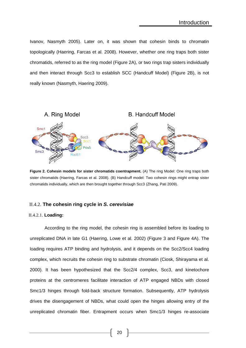
Ivanov, Nasmyth 2005). Later on, it was shown that cohesin binds to chromatin
topologically (Haering, Farcas et al. 2008). However, whether one ring traps both sister
chromatids, referred to as the ring model (Figure 2A), or two rings trap sisters individually
and then interact through Scc3 to establish SCC (Handcuff Model) (Figure 2B), is not
really known (Nasmyth, Haering 2009).
Figure 2. Cohesin models for sister chromatids coentrapment. (A) The ring Model: One ring traps both
sister chromatids (Haering, Farcas et al. 2008). (B) Handcuff model: Two cohesin rings might entrap sister
chromatids individually, which are then brought together through Scc3 (Zhang, Pati 2009).
II.4.2. The cohesin ring cycle in S. cerevisiae
II.4.2.1. Loading:
According to the ring model, the cohesin ring is assembled before its loading to
unreplicated DNA in late G1 (Haering, Lowe et al. 2002) (Figure 3 and Figure 4A). The
loading requires ATP binding and hydrolysis, and it depends on the Scc2/Scc4 loading
complex, which recruits the cohesin ring to substrate chromatin (Ciosk, Shirayama et al.
2000). It has been hypothesized that the Scc2/4 complex, Scc3, and kinetochore
proteins at the centromeres facilitate interaction of ATP engaged NBDs with closed
Smc1/3 hinges through fold-back structure formation. Subsequently, ATP hydrolysis
drives the disengagement of NBDs, what could open the hinges allowing entry of the
unreplicated chromatin fiber. Entrapment occurs when Smc1/3 hinges re-associate

Introduction
21
through the free energy of hinge-dimerization which does not require ATP (Hu, Itoh et al.
2011). After loading, the cohesin ring relocates to places of convergent transcription,
origins of replications, and pericentromeric regions. This relocation is essential to move
cohesin away from the Scc2/4 loading complex, which might destabilize it by stimulating
the NBD ATPase activity (Hu, Itoh et al. 2011, Lengronne, Katou et al. 2004).
Unlike the replication apparatus, the transcription machinery is too large to pass
through the ring. Thus, the relocation of cohesin between converging intergenes could
be attributed to transcription, which pushes cohesin while it translocates along the DNA
until it meets another converging transcription machinery. The topological association
between cohesin and chromatin explains the ability of cohesin to slide along chromatin
and relocate (Ivanov, Nasmyth 2005).
Figure 3 Cohesin loading in G1. Scc2/4 complex, Scc3, and kinetochore proteins facilitate interaction of
ATP engaged NBDs with closed Smc1/3 hinges through fold-back structure formation. Subsequently, ATP
hydrolysis drives the disengagement of NBDs, which opens the hinges allowing entry of the unreplicated
chromatin fiber. Entrapment occurs when Smc1/3 hinges re-associates through the free energy of hinge-
dimerization which does not require ATP (Hu, Itoh et al. 2011). After loading, the cohesin ring relocates to
places of convergent transcription, origins of replications, and pericentromeric regions (Hu, Itoh et al. 2011,
Lengronne, Katou et al. 2004).
II.4.2.2. Establishment of sister chromatid cohesion
It has been proposed that the binding of cohesin to unreplicated chromatin is
unstable due to the antiestablishment complex (described below) (Lopez-Serra,
Lengronne et al. 2013), what leads to its release through the newly described release

Introduction
22
gate present at the Scc1-Smc3 interface (Figure 4A) (Buheitel, Stemmann 2013, Chan,
Roig et al. 2012). During passage of the replication fork cohesin coentraps newly
replicated sister chromatids (Figure 4B), and cohesin complexes with short residence
time on chromatin are converted to ones with higher half life when two lysine residues,
namely K112 and K113, within the Smc3 head domain are acetylated by EcoI
(Lengronne, McIntyre et al. 2006, Rolef Ben-Shahar, Heeger et al. 2008).
Smc3 acetylation is essential for SCC, and thus, eco1∆ is lethal. Certain
mutations in SCC3, PDS5, and RAD61 reverse the lethality of eco1∆, what proves the
existence of an antiestablishment complex, formed by the interaction of Scc3, Pds5 and
Rad61. Upon Eco1 dependent acetylation of Smc3 cohesin rings counteract the
antiestablishment activity thereby becoming cohesive (tightly shut), (Figure 4C). In the
absence of Smc3 acetylation, the antiestablishment complex would release cohesin
through the exit gate at the Scc1-Smc3 interface leading to precocious loss of SCC
(Figure 4D) (Rowland, Roig et al. 2009).

Introduction
23
Figure 4. The cohesin ring cycle. (A) Cohesin is loaded onto chromatin during the G1 phase of the cell
cycle through the entry gate located at the hinge domain. The binding of cohesin to unreplicated chromatin is
unstable due to the antiestablishment complex, what leads to its release through the release gate present at
the Scc1-Smc3 interface. (B) Cohesin co-entraps newly replicated sister chromatid by an unknown
mechanism. (C) Cohesin ring becomes cohesive (tightly shut) upon Eco1 dependent acetylation of Smc3,
which counteracts the antiestablishment activity. (D) In the absence of acetylation, the antiestablishment
complex releases cohesin from sister chromatids leading to precocious loss of SCC.
The mechanism of sister chromatids coentrapment is not really known. However,
It has been suggested that passage of the replisome forces the hinge to reopen, and
once again fold to interact with the NBD of Smc3. This interaction would stimulate
acetylation of Smc3 K112 and K113 by EcoI (Figure 5). After passage the hinge recloses
allowing co-entrapment of the two newly replicated sister chromatids (Lengronne,
McIntyre et al. 2006, Kurze, Michie et al. 2011). It is not known whether reopening and
closing the ring requires a new ATP-binding/hydrolysis cycle, and if so, how is this
coordinated with acetylation, taking into consideration that the acetylation sites are close
to Smc3’s ATP-binding pocket. Alternatively, cohesin can be loaded to already
replicated sister chromatids, a model that is yet to be explored experimentally (Kurze,
Michie et al. 2011).
Figure 5. Cohesin coentrapment of newly replicated sister chromatids. Passage of the replisome forces
the hinge to reopen, and once again fold to interact with the NBD of Smc3. This interaction stimulates
acetylation of Smc3 K112 and K113 by EcoI. After passage the hinge recloses allowing co-entrapment of the
two newly replicated sister chromatids (Lengronne, McIntyre et al. 2006, Kurze, Michie et al. 2011)
Introduction
24
There are many other factors that may be important for establishment of SCC.
One of these factors is Proliferating cell nuclear antigen (PCNA), which is a homotrimeric
protein that acts as a sliding clamp on DNA, and recruits a variety of proteins involved in
replication, recombination, or DNA damage repair (Hoege, Pfander et al. 2002).
RFCctf18/Dcc1/Ctf8 proteins are required for PCNA association with the replication fork and
contribute to SCC. PCNA, in return, binds Eco1 through its N-terminal motif thus
recruiting it to the replication fork, and potentially bringing it in close proximity to cohesin
(Moldovan, Pfander et al. 2006). Other factors involved in replication, such as Ctf4, Tof1,
Csm3, Chl1 and Mrc1 also contribute to cohesion establishment, and facilitate cohesin
acetylation (Borges, Smith et al. 2013).
II.4.2.3. Cohesin release and chromosome segregation
Cohesion established during S phase is maintained during G2 and until the onset
of anaphase to prevent premature separation of sister chromatids due to the pulling
forces of microtubules on kinetochores. The tension created between sister chromatids
by cohesin and the pulling forces of the microtubules enable chromosome biorientation.
This tension will eventually be removed by cleavage and opening of cohesin rings at
anaphase onset. Cleavage of cohesin rings is promoted by activation of the APC/Cd20
ubiquitin ligase once all chromosomes are properly bioriented, a process that is
supervised by the spindle checkpoint.
Cohesin rings are cleaved by a protease known as separase, which recognizes a
specific sequence on the Scc1 subunit (Ciosk, Zachariae et al. 1998). Prior to anaphase,
separase (Esp1), is inhibited by the chaperone securin (Pds1). However, at the onset of
anaphase, and once APC/Cdc20 is activated, securin is targeted for degradation by the
ubiquitin system leading to separase release and activation (Morgan 2007, Peters 2006).
In addition, Scc1 is phosphorylated by the Polo Kinase Plk, which is required for efficient
cleavage by separase (Alexandru, Uhlmann et al. 2001) (Figure 6A).

Introduction
25
Cleavage of the Scc1 subunit triggers Smc3 deacetylation by Hos1 (Figure 6B).
Importantly, Smc3 molecules that fail to be deacetylated in hos1∆ background are not
able to establish SCC during the next cell cycle, which means that de novo acetylation of
Smc3 during S phase is required for establishment of SCC (Beckouet, Hu et al. 2010).
Figure 6. Cohesin release in S. cerevisiae. (A) At the onset of anaphase, APC/Cdc20 targets securin to
degradation by the ubiquitin system leading to separase release and activation. Scc1 phosphorylation by Plk
is required for efficient cleavage by separase. (B) After cleavage, Hos1 deacetylates Smc3 so that it can be
reused in the next cell cycle.
II.4.3. Beyond cohesin: resolution and segregation of chromosomes
Apart from cohesin release, topoisomerase II contributes to SC resolution by
removing most of the tangles between SC that form during DNA replication (catenation)
in a process termed “decatenation”. Topoisomerase II decatenates DNA strands that
cross one another by creating a DSB in one strand, passing the unbroken strand through
this break, and finally resealing it.
The action of topoisomerase II is coordinated and occurs in parallel with
chromosome condensation, carried out mainly by condensin through axial chromosome

Introduction
26
compaction (Carter, Sjogren 2012, Nitiss 2009). In vitro studies suggest that condensin
induces positive supercoiling in the mitotic chromosome, thereby exposing
intermolecular catenanes to Top2 action (Baxter, Sen et al. 2011). An earlier study
showed that chromosome recoiling by condensin induces removal of residual cohesin
during anaphase by reinforcing the physical separation of sister chromatids (Renshaw,
Ward et al. 2010).
II.4.4. Damage induced cohesion
Cohesin rings loaded onto chromatin outside S phase are not acetylated.
Therefore, they are not cohesive, and are thus discharged by the action of the
antiestablishment complex (Chan, Roig et al. 2012). However, the presence of DSBs
during G2/M causes de novo cohesin loading and establishment both at the break site
and genome wide on normal cohesin binding sites, a phenomenon known as damage
induced (DI) cohesion (Figure 7). DI cohesion maintains the physical proximity of the
sister chromatids, and thus, favors repair by sister chromatid recombination (SCR) over
other types of repair (Unal, Arbel-Eden et al. 2004, Unal, Heidinger-Pauli et al. 2007,
Strom, Karlsson et al. 2007).
Mec1 and Tel1 dependent phosphorylation of histone H2A is required for cohesin
binding, which is enabled by Mre11 protein and Scc2/4 loading complex (Unal, Arbel-
Eden et al. 2004). In striking contraposition to the pivotal role played by Smc3
acetylation, it has been suggested that the main acetylation target for establishment
during DNA repair is Scc1. The Chk1 kinase would phosphorylate Scc1 on S83 to allow
Eco1-mediated acetylation of Scc1 at residues K84 and K210 for establishment of DI
cohesion (Heidinger-Pauli, Unal et al. 2009, McAleenan, Cordon-Preciado et al. 2012).
Although mutation of K84 and K210 certainly supports this hypothesis, it has not been
possible to demonstrate their in vivo acetylation yet. While cohesin would be maintained
at proximal regions to ensure recombinational repair with sister chromatid, it has been

Introduction
27
recently shown that separase would mediate dissociation of cohesin at the break site to
allow the access of the resection machinery (McAleenan, Clemente-Blanco et al. 2013).
Cohesin is also required for proper repair of stalled forks and for fork restart.
Replication fork may stall due to natural replication barriers or to external agents such as
replication inhibitors or base modification. It has been shown that cohesin is enriched at
early replication origins after HU treatment in a Rad50 dependent manner, what would
promote recovery by maintaining a conformation that favors recombination dependent
fork restart (Tittel-Elmer, Lengronne et al. 2012).
Figure 7. Damage induced cohesion. Cohesin rings loaded to chromatin outside S phase do not get
acetylated and thus do not establish SCC under physiological conditions due to their release by the action of
the antiestablishment complex through opening of the Smc3/Scc1 interface (Chan, Roig et al. 2012).
However, the presence of DNA DSBs during G2/M causes de novo cohesin loading and establishment not
only at domains flanking the break site, but also across the whole genome, a phenomenon known as
damage induced (DI) cohesion. Phosphorylation of residue S83 of Scc1 by the Chk1 kinase activates Eco1-
mediated acetylation of Scc1 at residues K84 and K210, which is required for establishment of SCC
(Heidinger-Pauli, Unal et al. 2009, McAleenan, Cordon-Preciado et al. 2012).
II.4.5. Other roles of cohesin in budding yeast
Cohesin is responsible for mono-orientation of sister kinetochores during meiosis
I and bi-orientation of dyad chromosomes during meiosis II. These functions are
achieved by replacing Scc1/Rad21 with a meiosis-specific version (Rec8), which is also

Introduction
28
responsible for meiotic DSB repair using non-sister chromatids (Bannister, Reinholdt et
al. 2004, Goldstein 1981). Cohesin is important for some aspects of transcription
regulation too, such as restricting the spread of silencing at the silent mating-type loci
(Gullerova, Proudfoot 2008, Donze, Adams et al. 1999). Finally, cohesin is required for
chromosome condensation, since mcd1-ts and eco1-ts mutants display defects in
chromosome condensation (Guacci, Koshland et al. 1997, Skibbens, Corson et al.
1999). Smc3 acetyl-mimicking mutant smc3R113Q and rad61∆ partially restore
condensation defects seen in eco1-ts mutants, pointing out the importance of cohesin
regulation by Eco1 and Rad61 in chromosome condensation and segregation (Lopez-
Serra, Lengronne et al. 2013, Guacci, Koshland 2011).
II.4.6. Cohesin in Humans: Structure and regulation
The basic composition and structure of the cohesin complex is be conserved in
evolution. Smc1, Smc3, and Rad21 constitute the core structure of the ring in human
cells (Figure 8B and Table 1)(McKay, Troelstra et al. 1996, Schmiesing, Ball et al. 1998).
On the other hand, there are two human homologues of budding yeast Scc3: SA1, which
mediates telomere cohesion, and SA2, which is responsible for centromere cohesion,
and both are required for arm cohesion. Importantly, SA1 and SA2 do not coexist in the
same cohesin complex (Losada, Yokochi et al. 2000, Canudas, Smith 2009). Two Pds5
proteins with overlapping functions have been identified in humans, namely Pds5A and
Pds5B (Losada, Yokochi et al. 2005). Other regulatory proteins of the cohesin complex
include Esco1 and Esco2 (homologues of Eco1), which are both required for Smc3
acetylation. Wapl (homologue of Rad61) is another regulatory protein, which interacts
with Pds5 and SA through its FGF motives (Shintomi, Hirano 2010). Finally, sister
chromatid cohesion in humans requires the participation of sororin, a cohesin factor that
apparently does not have a counterpart in yeast (Whelan, Kreidl et al. 2012).

Introduction
29
Figure 8. Structure of the cohesin ring in humans. (A) Different conformations of the cohesin ring shown
by electron micrographs. Arrows point at the kink seen in this complex (Anderson, Losada et al. 2002). (B)
The structure of cohesin in humans is very similar to that in S. cerevisiae with few differences. Mainly, Scc3
in S. cerevisiae has two homologs in humans SA1and SA2 which do not coexist in the same cohesin
complex. Pds5 has also two homologues in humans, Pds5A and Pds5B. Homologues of regulatory proteins
have also been identified, which include; Esco1 and Esco2 (homologs of Eco1), and Wapl (homolog of
Rad61). Sororin is the only subunit identified in humans that, so far, has no known homolog in S. cerevisiae.
Although cohesin has a fundamental role in mitotic and meiotic cycles, it is also
expressed in non-cycling cells, suggesting it may have other roles beyond SCC. Other
evidence suggests important functions in gene regulation through global organization of
chromatin architecture. For example, cohesin is a key regulator of long-range enhancer-
promoter interactions by formation of chromatin loops (Figure 9) (Hadjur, Williams et al.
2009). Besides, it may facilitate the V(D)J recombination of immunoglobulin genes
(Degner, Wong et al. 2009).

Introduction
30
Figure 9. Cohesin as a key regulator of long range enhancer-promoter interactions. cohesin stabilizes
enhancer-promoter interactions by creating chromatin loops (Hadjur, Williams et al. 2009)
Loading of cohesin depends on NipbI/Mau2 loading complex (homologue of
Scc2/4) (Watrin, Schleiffer et al. 2006, Krantz, McCallum et al. 2004). Smc3 acetylation
is not sufficient for SCC, which also requires recruitment of sororin to counteract the
antiestablishment activity of Wapl (Figure 10A). Sororin contains an FGF motif that
allows its interaction with Pds5, thus competing with Wapl (Remeseiro, Losada 2012,
Nishiyama, Ladurner et al. 2010).
Differently to budding yeast, cohesin release takes place in two steps in
mammalian cells. During the first step, called the “prophase pathway”, SA2 and sororin
are phosphorylated by Plk1 and Cdk1 respectively, what disrupts the Pds5-Soronin
interaction and favors that of Wapl, leading to cohesin removal from chromosome arms
(Shintomi, Hirano 2010) (Figure 10B). At this stage, the centromeric cohesion is still
required for bipolar attachment, and is protected from mitotic kinases by Sgo1-PP2A
complex (Liu, Rankin et al. 2012). Cohesin release during the prophase pathway
involves opening of the Smc3-Scc1 gate (Buheitel, Stemmann 2013). Once the
chromosomes are bi-oriented and the spindle assembly checkpoint is satisfied, separase
cleaves Rad21, thereby removing the remnant centromeric cohesin and triggering
chromosome segregation (Figure 10C). In human cells, separase is additionally inhibited
by Cdk1 cyclin B dependent phosphorylation. Therefore, activation of separase does not

Introduction
31
only depend on securin destruction by the APC/Cdc20, but also on cyclin B destruction
and concomitant Cdk1 inactivation (Huang, Hatcher et al. 2005). Finally, Smc3 is
deacetylated by HDAC8 after Rad21 cleavage, allowing its reuse in the next cell cycle
(Deardorff, Bando et al. 2012).
Figure 10. Establishment of sister chromatid cohesion in humans, and the two step release of
cohesin. (A) Establishment of SCC in humans requires binding of sororin to stabilize cohesin. (B) The
prophase pathway. The antiestablishment activity of Wapl destabilizes the ring causing its release by
opening of the Smc3-Scc1 gate. SA2 and sororin are phosphorylated by Polo/Aurora B and Cdk1
respectively, what disrupts the Pds5-Soronin interaction; this leads to cohesin removal from chromosome
arms. Centromeric cohesion is protected by Sgo1-PP2A complex from mitotic kinases and Wapl(Liu, Rankin
et al. 2012)(Liu, Rankin et al. 2012)(Liu, Rankin et al. 2012)(Liu, Rankin et al. 2012)(Liu, Rankin et al.
2012)(Liu, Rankin et al. 2012)(Liu, Rankin et al. 2012). (C) In anaphase, Rad21 is cleaved by separase
which removes centromeric cohesin for proper segregation.
II.4.7. Involvement in disease: cohesinopathies and cancer
Mutations in cohesin subunits, or its regulators are responsible for two main
developmental syndromes in humans collectively referred to as cohesinopathies (Figure
11).

Introduction
32
Cornelia de Lange Syndrome (CdLS) is the most common cohesinopathy
(1:10,000). 65% of CdLS cases are caused by dominantly inherited mutations in the
NIPBL gene, which causes the more severe phenotype. These mutations, which include
heterozygous truncations or non-sense mutations, seem to be haplo-insufficient with 15-
30% decrease sufficient to give rise to CdLS phenotype. Some mutations interfere with
NipbI interaction with Mau2, or other proteins. More importantly, there are no obvious
chromosome segregation deficiencies, indicating that developmental defects in CdLS
might be attributed to cohesin functions in gene regulation. CdLS patients are normally
small in size and have arched eyebrows, thin upper lip, long eyelashes, hirsutism, an
upturned nose, synophrysptosis, a long philtrum, and micrognathia (Figure 11B).
Additional severe developmental anomalies include upper limb truncations or limb
differences (Jackson et al., 1993). Neurodevelopmental delay and highly variable mental
retardation are also observed in all CDLS patients. A milder phenotype is caused by
heterozygous missense mutations in SMC1 and SMC3 which account for about 5% of
CdLS cases. An even milder phenotype is seen with RAD21 heterozygous deletions and
missense mutations. However, lymphoblastoid cell lines from patients with RAD21
mutations exhibit radiation sensitivity (Horsfield, Print et al. 2012).
Figure 11. Phenotype of individuals with cohesinopathies. (a) A child with CdLS showing characteristic
facial features. (b,c) CDLs patient with upper limb abnormalities and hirsutism of the back. a, b, and c are
taken from (Krantz, McCallum et al. 2004) (d) Photograph of a newborn baby with RBS. The baby died 17
minutes after delivery. This photos is taken from (Mann, Fitzsimmons et al. 1982).
d

Introduction
33
Robert’s syndrome (RBS) (Figure 11A) is another less common, although severe,
cohesinopathy. A less severe variant of this syndrome is SC phocomelia. RBS is
characterized by multiple craniofacial abnormalities, limb malformations, growth
deficiency, and mental retardation. Severely affected fetuses normally do not fully
develop, or dye just after birth. While in the variant type, individuals normally get to
adulthood. The syndrome is caused by homozygous recessive mutations in the ESCO2
gene. Unlike cells from CDLS, cells from RBS patients show precocious loss of SCC,
micronuclei formation, aneuploidy, lagging chromosomes, and mitotic defects (Horsfield,
Print et al. 2012).
Chromosome instability and aneuploidy seen in many cancer types has been
attributed to mutations in cohesin subunits affecting cohesin’s main function in sister
chromatid cohesion. One such example is colorectal cancer, where mutations within
SMC1, SMC3 and NIPBL have been identified. These mutations lead to decreased
cohesin gene expression resulting in premature loss of SCC and increased chromosome
instability, which contribute to the pathogenesis of this cancer. Overexpression of SMC3
and RAD21 in cancer cell lines has also been reported. For example, SMC3 is
overexpressed in human colon cancer cell lines (Xu, Tomaszewski et al. 2011).
Other human cancers can emerge from mutations that affect the role of cohesin
in the regulation of gene transcription, or DNA damage repair through homologous
recombination, which could lead to emergence of oncogenic chromosomal aberrations,
deletions, inversions and translocations (Xu, Tomaszewski et al. 2011).
II.5. Sumoylation
Some subunits of the SMC complexes have been identified as potential targets of
SUMO modification by proteomic screens (Wohlschlegel, Johnson et al. 2004, Denison,
Rudner et al. 2005). Sumoylation is a reversible post-translational modification where

Introduction
34
SUMO proteins (Small Ubiquitin like MOdifiers) are covalently conjugated to lysine
residue(s) within target proteins (Johnson, Schwienhorst et al. 1997, Kamitani, Nguyen
et al. 1997). The name comes from the resemblance seen between the three
dimensional structure and the biochemistry of conjugation in both ubiquitin and
SUMOpathways, which proceed through an enzymatic cascade of E1 activating enzyme,
E2 conjugase, and an E3 ligase (Johnson 2004) (Figure 12, Table 2). However, these
enzymes do not overlap between the two processes. Moreover, although they might
seem similar, SUMO and ubiquitin differ in many aspects, which include amino acid
sequence (less than 20% identity) and surface charge (Bayer, Arndt et al. 1998).
Sumoylation is mostly used to direct localization of its targets (Matunis, Coutavas et al.
1996), modify their interactions with other proteins (Mahajan, Delphin et al. 1997), or
mark them for, or protect them from, degradation by the ubiquitin system (Hoege,
Pfander et al. 2002).
SUMO conjugates are usually low abundant and short-lived. The highly dynamic
nature of this process is due to SUMO-specific proteases, which actively remove SUMO
from the target protein. This has led to the proposal that the rate of desumoylation is
much faster than that of sumoylation. Strikingly, the effect of this modification on cellular
functions seems to be dramatic compared to the levels of sumoylation seen for its
targets (Mukhopadhyay, Dasso 2007).
Proteomic studies have identified more than 1000 proteins as potential SUMO
targets in budding yeast. Known targets localize predominantly in the nucleus. However,
some targets are found in other cellular locations including the cytoplasm, the ER, the
mitochondria, and the plasma membrane. Potential targets identified by these proteomic
studies include proteins involved in DNA metabolism, including replication, repair,
transcriptional activators and repressors, and factors involved in chromatin remodeling.
Other known SUMO targeted processes are metabolic pathways, RNA-related
Introduction
35
processes, protein translation and folding, and cell division (Wohlschlegel, Johnson et al.
2004, Denison, Rudner et al. 2005, Zhao, Kwon et al. 2004, Panse, Hardeland et al.
2004). Many of these proteins have been validated as true targets in vivo, and their
modification by SUMO is relevant in processes such as transcription (Sapetschnig,
Rischitor et al. 2002), DNA repair (Psakhye, Jentsch 2012, Dou, Huang et al. 2011),
mitotic chromosome structure and segregation (Wan, Subramonian et al. 2012,
Alexandru, Uhlmann et al. 2001, Stead, Aguilar et al. 2003, Bachant, Alcasabas et al.
2002).
Given the broad array of cellular functions affected by sumoylation, SUMO
proteins, as well as SUMO enzymes, are highly conserved throughout the eukaryotic
kingdome. Both SUMO and Ubc9 are essential in S. cerevisiae and in mammals
(Johnson, Blobel 1997, Dieckhoff, Bolte et al. 2004, Nacerddine, Lehembre et al. 2005).
In S. cerevisiae, there is only one SUMO protein, which is coded by the SMT3 gene, and
is conjugated either individually or in chains to target proteins (Johnson, Schwienhorst et
al. 1997). In human cells there are four SUMO proteins: SUMO1-4 (Saitoh, Hinchey
2000, Guo, Li et al. 2004). SUMO1-3 are ubiquitously expressed, while SUMO4
expression is restricted to the spleen lymph node and kidney, and it is not clear whether
it is processed to its mature form, or if it participates in formation of SUMO conjugates in
vivo (Owerbach, McKay et al. 2005, Melchior 2000). The mature form of SUMO1 shares
50% identity with SUMO2 and 3. In addition, SUMO1 has non-redundant targets with
SUMO2 and 3. On the other hand, SUMO2 and 3 share 97% similarity (Saitoh, Hinchey
2000). Moreover, they are both involved in forming poly-SUMO chains, and have
redundant functions in the cell (Tatham, Jaffray et al. 2001). The ability to form SUMO
chains is due to the presence of a conserved lysine residue in the N-terminal amino acid
stretch found in SUMO proteins (Tatham, Jaffray et al. 2001). In budding yeast formation
of these chains is not essential for vegetative growth. However, it is required for
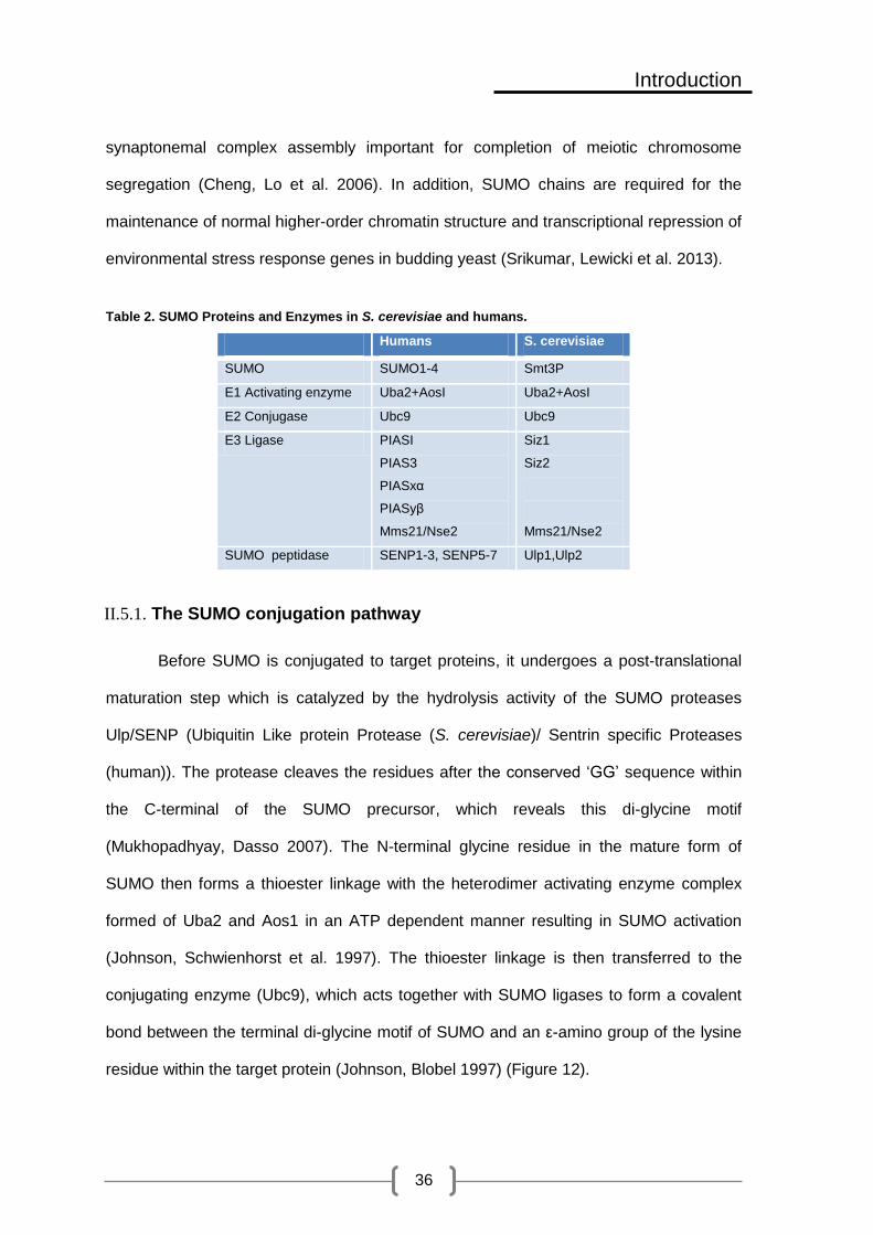
Introduction
36
synaptonemal complex assembly important for completion of meiotic chromosome
segregation (Cheng, Lo et al. 2006). In addition, SUMO chains are required for the
maintenance of normal higher-order chromatin structure and transcriptional repression of
environmental stress response genes in budding yeast (Srikumar, Lewicki et al. 2013).
Table 2. SUMO Proteins and Enzymes in S. cerevisiae and humans.
Humans S. cerevisiae
SUMO SUMO1-4 Smt3P
E1 Activating enzyme Uba2+AosI Uba2+AosI
E2 Conjugase Ubc9 Ubc9
E3 Ligase PIASI
PIAS3
PIASxα
PIASyβ
Mms21/Nse2
Siz1
Siz2
Mms21/Nse2
SUMO peptidase SENP1-3, SENP5-7 Ulp1,Ulp2
II.5.1. The SUMO conjugation pathway
Before SUMO is conjugated to target proteins, it undergoes a post-translational
maturation step which is catalyzed by the hydrolysis activity of the SUMO proteases
Ulp/SENP (Ubiquitin Like protein Protease (S. cerevisiae)/ Sentrin specific Proteases
(human)). The protease cleaves the residues after the conserved ‘GG’ sequence within
the C-terminal of the SUMO precursor, which reveals this di-glycine motif
(Mukhopadhyay, Dasso 2007). The N-terminal glycine residue in the mature form of
SUMO then forms a thioester linkage with the heterodimer activating enzyme complex
formed of Uba2 and Aos1 in an ATP dependent manner resulting in SUMO activation
(Johnson, Schwienhorst et al. 1997). The thioester linkage is then transferred to the
conjugating enzyme (Ubc9), which acts together with SUMO ligases to form a covalent
bond between the terminal di-glycine motif of SUMO and an ε-amino group of the lysine
residue within the target protein (Johnson, Blobel 1997) (Figure 12).

Introduction
37
The majority of SUMO ligases contain an SP-RING motif essential for their
function (Hochstrasser 2001). PIAS (protein inhibitor of activated STAT) proteins in
humans (Palvimo 2007), Siz (SAP and miz-finger domain) proteins in budding yeast
(Johnson, Gupta 2001, Takahashi, Kahyo et al. 2001), and Nse2 (part of the Smc5/6
complex) in both species, contain this SP-RING. Siz1, Siz2 and Nse2 are the only known
mitotic ligases is budding yeast. SP-RING ligases simultaneously bind to Ubc9 and the
target protein, bringing the three players in a favorable position for SUMO transfer
(Hochstrasser 2001, Song, Durrin et al. 2004). In S. cerevisiae Siz1 and Siz2 are
responsible for most SUMO conjugation (Takahashi, Kahyo et al. 2001, Johnson, Gupta
2001), while Nse2 has few specific targets, which include the Smc5/6 complex itself, and
Yku70 (Zhao, Blobel 2005, Potts, Yu 2005). siz1∆siz2∆ double mutants are viable, while
Nse2 is essential for vegetative growth and DNA repair (Zhao, Blobel 2005).
Unlike ubquitylation, where a ubiquitin E3 ligase is always required, SUMO E1
and Ubc9 alone are able to sumoylated many substrates in vitro, without the need for an
E3 SUMO ligase. However, in some cases the presence of an E3 SUMO ligase
increases sumoylation efficiency and substrate specificity (Johnson, Blobel 1997, Zhao,
Blobel 2005).

Introduction
38
Figure 12. SUMO conjugation and deconjugation pathway. Before SUMO is conjugated to target
proteins, it undergoes a post-translational maturation step which is catalyzed by the hydrolysis activity of the
SUMO proteases Ulp/SENP. The protease cleaves the residues after the conserved ‘GG’ region within the
C-terminal of the SUMO precursor, which reveals this diglycine motif. These glycine residues then form a
thioester linkage with heterodimer activating enzyme (Uba2) in an ATP dependent manner resulting in
SUMO activation. The thioester linkage is then transferred to the conjugating enzyme (Ubc9), which acts
together with SUMO ligases to form a covalent bond between the terminal di-glycine residues of SUMO and
ε-amino group of the lysine residue within the target protein. Deconjugation of SUMO by SUMO proteases
(Ulp/SENP) is essential to ensure the reversible nature of SUMO conjugation. Cleavage of SUMO from the
target protein occurs at the scissile isopeptide bond.
II.5.1.1. Nse2 E3 SUMO ligase and Smc5/6 complex
Nse2 is an E3 SUMO ligase that belongs to the Smc5/6 complex, which is
essential in S. cerevisiae. Smc5/6 is recruited to DSBs and is required for HR dependent
DNA damage repair. In addition, Smc5/6 complex is enriched at repetitive rDNA,
telomeric sequences, and at discrete sites along chromosome arms in a length
dependent manner (Kegel, Betts-Lindroos et al. 2011, Lindroos, Strom et al. 2006). It
has been shown that Smc5/6 is required for maintenance and proper segregation of the
rDNA (Torres-Rosell, Machin et al. 2005, Torres-Rosell, Sunjevaric et al. 2007), and for
proper resolution of SC linkages during mitosis (Bermudez-Lopez, Ceschia et al. 2010).
This complex is composed of the core subunits Smc5 and Smc6 (Figure 1A) (Zhao,

Introduction
39
Blobel 2005), and is associated with other regulatory non-SMC subunits Nse1-6. Nse4 is
a kleisin protein that binds to the head domains of Smc5. Nse1 and Nse3 interact with
each other and with Nse4 (Palecek, Vidot et al. 2006). Nse1 has a ubiquitin ligase ring
domain and its ubiquitin ligase activity is only gained when associated with Nse3 (Doyle,
Gao et al. 2010). Nse5 and Nse6 bind to the hinge domain (Duan, Yang et al. 2009,
Palecek, Vidot et al. 2006). Nse2 is formed of two domains; an Smc5 binding domain,
which resides in the N terminal of Nse2, and a SUMO ligase domain in the C-terminal of
Nse2 (Figure 13A). In the absence of DNA damage, the SUMO ligase activity is not
essential for survival. However, deleting the entire NSE2 gene is lethal in S. cerevisiae,
which suggests that Nse2 might have other roles that do not involve its SUMO ligase
activity. Nse2 binds to the coiled-coils of Smc5 in a site where a bend of 165° has been
observed (Figure 13C). Mutations that disrupt binding of Nse2 (Figure 13B) to Smc5 are
inviable (Duan, Sarangi et al. 2009).
Figure 13. Structure of Nse2 in complex with the coiled-coil region of Smc5. (A) Nse2 is shown in gold
and the coiled-coil regions of Smc5 are shown in blue. (B) M1 and M2 mutations that abolish interaction
between Nse2 and Smc5 are colored in red and green respectively. (C) The arm region of Smc5 exhibits a
moderate curvature (Duan, Sarangi et al. 2009).

Introduction
40
II.5.2. SUMO deconjugation
Deconjugation of SUMO by SUMO proteases (Ulp/SENP) is essential to ensure
the reversible nature of SUMO conjugation. Cleavage of SUMO from the target protein
occurs at the scissile isopeptide bond (Figure 12). Ulp/SENP proteases contain a
conserved catalytic domain in the C terminal region referred to as Ulp1 domain (UD),
which is necessary and sufficient to promote SUMO deconjugation in vitro (Mossessova,
Lima 2000). In addition to the catalytic domain, Ulp/SENPs possess an N-terminal
domain that shares no similarity between the different proteases, and is required for
subcellular localization and substrate specificity (Lima, Reverter 2008).
In S. cerevisiae, there are two SUMO proteases; Ulp1 and Ulp2/Smt4. Ulp1 is
associated with the nuclear envelope, while Ulp2 is predominantly nuclear. Ulp1 is
responsible for SUMO maturation, it is essential for cell viability and for cell cycle
progression through G2/M phase (Li, Hochstrasser 1999). On the other hand, Ulp2 is not
essential for vegetative growth, but it is required for disassembly of poly-SUMO chains
(Bylebyl, Belichenko et al. 2003). Although ulp2 mutants are viable, they grow poorly,
and exhibit chromosome segregation defects (Mossessova, Lima 2000).
In humans there are six different SUMO specific proteases; SENP1, SENP2,
SENP3, SENP5, SENP6, and SENP7. SENP1 and SENP2 are most closely related to
each other and are more efficient in catalyzing SUMO deconjugation over processing.
SENP1 has no preference for specific SUMO isoforms. However, SENP2 shows a slight
preference for SUMO2/3. SENP1 and 2 are very similar to the budding yeast Ulp1 and
they both localize to the nuclear envelop (Mukhopadhyay, Dasso 2007). SENP3 and 5
are also similar to Ulp1, but they localize to the nucleolus, and they have a preference for
SUMO2/3 deconjugation and processing (Mukhopadhyay, Dasso 2007). SENP6 and 7

Introduction
41
are similar to budding yeast Ulp2 in that they are found in the nucleoplasm, and have a
strong preference for disassembly of SUMO2/3 polymeric chains (Lima, Reverter 2008).
II.5.3. Functional outcomes of SUMO modification
As with other post-translational modifications, several lysines within the same
protein can be conjugated to SUMO. The multiple sumoylation sites might be functionally
redundant, as occurs in many recombinational proteins (Psakhye, Jentsch 2012), or
might have specific functions, as is the case for PCNA (Hoege, Pfander et al. 2002).
Sometimes, sumoylated lysines can lie within a signature motif, which is useful for
predicting candidate lysines. This motif is defined by the amino acid sequence ψKxE/D,
where x is any amino acid and ψ is a hydrophobic amino acid, which can be valine,
isoleucine, or leucine. During SUMO conjugation, the target lysine is localized to the
catalytic pocket of Ubc9, while the other amino acids interact with the surface of Ubc9
(Bernier-Villamor, Sampson et al. 2002). However, in many other cases, as seen for the
modification of K164 in PCNA, sumoylation can also occur at lysines that are not present
within a Ubc9-consensus sequence (Hoege, Pfander et al. 2002).
The effect of sumoylation on its targets is specific and can influence their
interactions with other proteins or DNA in a positive or negative manner. This can be
because covalent binding of SUMO can mask binding sites within the sumoylated
protein, which will cause decrease in the ability of this protein to interact with other
proteins or DNA, as noted in some transcription repressors (Zheng, Yang 2004). On the
other hand, sumoylation can add an additional binding interface by allowing the target
protein to interact with other proteins, that bind to SUMO non-covalently through their
SUMO interacting/binding motifs SIM/SBM. For example, in S. cerevisiae, sumoylated
PCNA attracts the anti-recombinogenic SIM-containing DNA helicase Srs2 to replication
forks to prevent unnecessary recombination events during S phase by interacting with
Rad51 and disrupting formation of Rad51 filaments. Upon replication fork stalling, due to

Introduction
42
DNA lesions, PCNA is monoubiquitylated at the same lysine residue K164, which is
promoted by it previous sumoylation. Other modifications such as acetylation can
compete with sumoylation for acceptor lysines within target proteins, which might serve
to regulate it (Stankovic-Valentin, Deltour et al. 2007, Hoege, Pfander et al. 2002). In
addition, phosphorylation of the same target can enhance or down-regulate sumoylation
due to enhanced interaction with Ubc9, or masking of sumoylation site respectively
(Hietakangas, Anckar et al. 2006).
A part from promoting new interactions, sumoylation may induce a conformational
change in the target protein. For instance, thymine DNA glycosylase (TDG) is a DNA
mismatch repair enzyme that is sumoylated, but at the same time binds non-covalently to
SUMO, which induces a conformational change. This conformational change is required
for the release of TDG from DNA after base excision (Hardeland, Steinacher et al. 2002).
In other cases, sumoylation can affect the localization of the target protein. For
example, RanGAP1 is localized to the cytoplasm in human cells during interphase.
However, sumoylated RanGAP1 localizes it to the NPC, where it interacts with RanBP2
to form the RRSU complex. This complex is localized to kinetochores and mitotic
spindles during mitosis (Matunis, Coutavas et al. 1996).
II.5.4. Sumoylation and Genome integrity
Mutation or depletion of sumoylation enzymes results in failure to repair damaged
genome, what highlights the importance of SUMO-mediated response in protecting
genome integrity, and in modulating DNA repair. It has been proposed that sumoylation-
based response to DNA damage occurs in parallel to phosphorylation-based damage
response and is required for efficient repair and maintenance (Cremona, Sarangi et al.
2012).

Introduction
43
Many DNA repair proteins become sumoylated in response to DNA damage. For
example, Rad52 and Yku70, which regulate HR and NHEJ respectively, are sumoylated.
Sumoylation of Rad52 protects it from proteasomal degradation, and regulates ribosomal
DNA recombinational repair (Sacher, Pfander et al. 2006, Zhao, Blobel 2005, Torres-
Rosell, Sunjevaric et al. 2007). As mentioned previously, PCNA sumoylation and
ubiquitylation cooperate to control the response to replication-blocking lesions during S
phase (Hoege, Pfander et al. 2002, Parker, Ulrich 2012). Other proteins involved in
homologous recombination repair pathway in response to DNA damage are also
regulated by sumoylation in a Siz2 dependent manner; such as Rad9, Mrc1, Mec1, and
Tel1 (Psakhye, Jentsch 2012, Cremona, Sarangi et al. 2012).
Ubc9 is known to play a key role in genome integrity, specially by preventing the
accumulation of X-shaped DNA at damaged replication forks. This DNA repair function of
Ubc9 requires the Nse2 SUMO ligase (Branzei, Sollier et al. 2006). In fact, nse2 mutant
is the only E3 mutant in budding yeast that is sensitive to DNA damage, indicating that
the Nse2-branch of the SUMO pathway plays a prominent role in DNA repair. Nse2-
dependent sumoylation is most probably required to trigger removal of recombination
intermediates (Bermudez-Lopez, Ceschia et al. 2010, Chavez, George et al. 2010).
However, the real targets at this stage are still not known (Zhao, Blobel 2005).
II.5.5. Sumoylation and SMC complexes
Over the past years, several proteomic studies have identified the Smc
complexes as potential targets of SUMO modification (Wohlschlegel, Johnson et al.
2004, Denison, Rudner et al. 2005). Although in vivo sumoylation was detected for
various subunits of the SMC complexes, the physiological role of this modification is still
not clear. In S. cerevisiae Pds5 is has been proposed to be sumoylated in a Siz1
dependent manner, starting in S phase until the onset of mitosis. Defects seen in pds5
thermosensitive alleles are suppressed by Ulp2 overexpression. Thus, it has been

Introduction
44
proposed that Pds5 sumoylation disrupts the interaction between Pds5 and cohesin,
which leads to cohesin destabilization and release (Stead, Aguilar et al. 2003).
Moreover, Nse2 dependent sumoylation of Smc1, Smc3, Smc2, and Smc5 was shown in
nocodazole arrested S. cerevisiae cells. It has been suggested that sumoylated SMC
complexes cooperate with topoisomerases in rDNA maintenance (Takahashi, Dulev et
al. 2008). In addition, sumoylation of human Rad21 and Sa2, was also shown to depend
on Nse2 in vitro (Potts, Porteus et al. 2006). Furthermore, sumoylation of Ycs4 subunit of
condensin was also detected in vivo in S. cerevisiae. It seems that Cdc14p promotes
sumoylation of Ycs4 during anaphase, which allows condensin’s localization to rDNA
(D'Amours, Stegmeier et al. 2004). Finally, sumoylation of Smc5 and Nse2 was
demonstrated in vivo using S. cerevisiae as a model. It has been suggested that the
Smc5/6 complex facilitates sumoylation of Nse2 targets by localizing it to the target site
(Zhao, Blobel 2005).
II.5.6. Methods to study sumoylation
The study of protein sumoylation faces two main challenges that stem from the
characteristics of the sumoylation process. First, only a small fraction of a given protein
is sumoylated for a short period of time. Second, the highly active peptidases are
normally controlled by the cell by confining them to certain cell compartments, such as;
the nuclear pore. However, upon cell disruption these peptidase are released, and
rapidly desumoylate proteins, which might lead to failure of detection of SUMO
conjugates. In order to solve these problems several approaches can be followed
separately or in combination. First, purification of SUMO conjugates is normally done
under denaturing conditions (such as 8M urea). This helps in various aspects, which
include; restricting the activity of the peptidases; and eliminating proteins that are bound
non-covalently to SUMO through SIM. The use of protease inhibitors, specially cysteine-
dependent proteases like N-ethylmaleimide (NEM), and others like PMSF is also

Introduction
45
recommended. A second approach would be to purify the SUMO conjugates after
conditional inactivation of Ulp peptidases (Sacher, Pfander et al. 2005).

46

47
Objectives

48

Objectives
49
III. Objectives
Determine whether cohesin is sumoylated in vivo under physiological conditions.
Identify the molecular requirements of cohesin sumoylation.
Develop a model, which uses Ulp1 Domain (UD) fusion to Scc1 protein to down-
regulate sumoylation of the whole complex, and thus, characterize the role of
sumoylation in cohesin function.
Study the phenotype of cohesin desumoylation in human cells.

50

51
Materials and Methods

52

Materials and Methods
53
IV. Materials and Methods
IV.1. Construction of yeast strains
Epitope tagging of genes and deletions was performed as described (Goldstein,
McCusker 1999, Janke, Magiera et al. 2004, Alexandru, Uhlmann et al. 2001). Lists of
the strains, plasmids, and primers used in this study are provided in Supplementary
Table 2, Supplementary Table 3, and Supplementary Table 4. All PCRs were done
using GeneAmp PCR system 2700 from Applied BioSystems. The polymerase
“Expand high fidelity” from Roche was used for epitope tagging and deletions of genes.
To check gene integrations in yeast one of these two methods were used; colony PCR
or genomic DNA extraction (Promega) followed by PCR using SupraTherm DNA
polymerase (Gene Craft).
IV.1.1. Yeast competent cells preparation and transformation
Optical density was always measured with Jenway 6300 spectrophotometer at
600 nm (OD600) before collecting cells. For preparation of yeast competent cells, 50 ml
of exponentially growing cells (OD600 0.8-1) were collected and washed first with Milli-Q
water and then with sorbitol buffer (100mM LiOAc, 10mM Tris-HCl pH 8, 1mM
EDTA/NaOH pH 8, 1M Sorbitol). After washing, the pellet was resuspended with 360 µl
sorbitol buffer and 40 µl of denatured carrier DNA (Salmon sperm DNA, Gibco BRL).
Next, 50 µl aliquots were directly used for transformation, or stored at -80°C.
The amount of DNA used for transformation varied depending on the target homology
in the case of PCR transformation, or on the concentration of DNA in the case of
plasmids. Commonly, for centromere or multicopy plasmid transformation 1- 5 µl of
plasmid DNA (100ng- 5µg) was added to 10 µl of competent cells, while for
transformation of integrative vectors 10 µl of digested plasmid DNA (≈5µg) were added
to 50 µl of cells. In the case of PCR transformation, 10µl of DNA (≈50µg) were used if

Materials and Methods
54
the target homology was low (40bp), while 3-4µl were used for targets with higher
homology (>150bp). Next, PEG buffer (100mM LiOAc, 10mM Tris-HCl pH 8, 1mM
EDTA/NaOH pH 8, 40% PEG) was added to the transformation tube and incubated at
room temperature for 30 minutes. Then, heat shock was done at 42°C for 15 minutes
after adding DMSO to 10%. Finally, the transformation buffer was removed after
spinning at 2400 rpm for 5 minutes, and cells were resuspended with Milli-Q water and
plated on the appropriate selection medium.
For transformations that require expression of drug resistance genes before
platting, cells were incubated at room temperature with non-selective medium for 2-4
hours before plating.
IV.1.2. Colony PCR from yeast
A yeast colony was resuspended in 15 µl of Milli-Q (Millipore) water and
patched in appropriate selection plate. Next, a small spoon of glass beads (SIGMA)
was added, and cells were boiled at 95°C for 2 minutes. Then, cells were broken for 30
seconds at power 5.5 using the FastPrep (FP120, Bio101 Thermosavant). Finally, after
spinning at 10 krpm for 1 min, 2 µl of supernatant were taken for the PCR reaction.
IV.2. Gene cloning and plasmid construction
PCR purification was done using QIAquick PCR purification kit from Qiagen.
Dephosphorylation and ligation were done using the Rapid DNA Dephos & Ligation Kit
from Roche. Site-directed mutagenesis was performed using the Quickchange
mutagenesis kit from Agilent Technologies. Plasmid extraction was done using
GenElute Plasmid Miniprep Kit (SIGMA). Newly constructed plasmids were checked by
either plasmid jet preps and restriction digestion, or E.coli colony PCR. Detection of
DNA was done by loading on D-1 low EEO agarose (CONDA) gel prepared with the
running buffer 1XTAE (from 50X; Tris-HCL, EDTA-Na2 pH 8.5). DNA was then stained

Materials and Methods
55
by incubating the agarose gel for 15 minutes in ethidium bromide bath with a final
concentration of 0.5 µg/ml. Detection was done by exposing the gel to UV light source
(Alpha Inno Tech), image capture by Olympus camera, and analysis with AlphaDigiDoc
software.
The Scc1-UD fusions were made by cloning the Ulp1 SUMO-peptidase domain
(amino acids 418 to 621) next to the 3xHA epitope into the multi-copy vector pYES2.
The SCC1 open reading frame was cloned N-terminally of the 3xHA linker. The fusions
were then transferred to the integrative vector YIplac211 by PCR and recombination
cloning in recA+ E. coli cells (MC1061). Replacement of the GAL promoter by the
SCC1 promoter was done recombinational cloning in MC1061 using two PCRs: one
containing the yeast SCC1 promoter (1.1 Kb) flanked by 40 nucleotide sequences
homologous to the ends of a second PCR, performed on the YIplac211 plasmids to
remove the GAL promoter.
NSE2-3HA was amplified from yeast genomic DNA with 5’-HindIII-Nse2
promoter and 3’-SacI -3HA, and cloned in to HindIII-SacI sites in pRS315. M1 and M2
single and double mutations on Nse2 were done by site-directed mutagenesis.
IV.2.1. E.coli competent cells preparation and transformation
DH5α
For plasmid transformation, 50 µl of Subcloning Efficiency™ DH5α™
Competent Cells from Invitrogen were thawed on ice and 1-5 µl of DNA were added
with gentle mixing. Next, they were incubated on ice for 30 minutes, at 42°C for 20
seconds (heat shock) and on ice again for 2 minutes. 1 ml of prewarmed LB was added
to the transformation tube, and cells were then incubated at 37°C for one hour to allow
expression of the antibiotic (ampicillin) resistance gene. After incubation, cells were
pelleted at 6000 rpm for 5 minutes, and 950 µl of the supernatant was removed, while
the pellet was resuspended with the 50 µl left and plated on LB-Ampicillin plate.

Materials and Methods
56
MC1061
For MC1061 competent cells preparation, cells were grown overnight in LB at
37°C. The next day, 1 ml of the culture was diluted in 100 ml of LB and left to reach
OD600 0.2-0.4 at 37°C. Next, cells were collected by centrifugation at 3200 rpm for 10
minutes at 4°C, and resuspended with 50 ml of ice cold 0.1 M CaCl2, and then left on
ice for 2 hours. Then, cells were collected as before and resuspended with 10 ml of ice
cold 0.1 M CaCl2 and 10% glycerol. After leaving on ice for 30 minutes cells were
aliquoted (100 µl/aliquot) and stored at -80°C. MC1061 cells were mainly used for
plasmid construction through recombination. This was done by transforming 10 µl of
vector plus insert DNAs per 100 µl of competent cells. Transformation of the vector
alone or the insert alone was used as background control. Special care was taken that
the molar concentration of the insert was at least twice as that of the vector. The
transformation procedure was the same as that described for DH5α cells, except for
the heat shock step, which was done for 2 minutes at 42°C.
IV.2.2. Colony PCR from E. coli
Colonies were picked with toothpicks and swirled into 25 µl of Milli-Q water. The
same toothpick was used to make a patch on LB-Ampicillin plate. The suspended
colonies were then boiled for 2 minutes, and centrifuged at 13000 rpm for 2 minutes.
20 µl of the supernatant were transferred to a new tube, from which 1 µl was used as
template for the PCR. This procedure allows rapid screening for the presence of the
insert in its correct orientation.
IV.2.3. Jet preps
Colonies that were previously picked on LB-Ampicillin plate were resuspended
in 500 µl of LB-Ampicillin liquid medium and then pelleted by centrifugation for 30
seconds at 13000 rpm. Next, 50 µl of BT (2% Triton X-100/NaOH pH 12.4) were added
to the pellet without resuspension. Then, 50 µl of phenol/chloroform were added and

Materials and Methods
57
the tubes where shaken vigorously for 30 seconds. After centrifugation for 5 minutes at
13000 rpm, 5 µl were combined with loading buffer (5X FLB, 100 µg/ml RNAse). To
analyze with restriction digestion, 40 µl of the supernatant were taken and chromatin
was precipitated by adding 4 µl of 2% acetic acid 4 µl of 3 M NaOAc pH 8, and 100 µl
of ethanol, and centrifugation at 13000 rpm for 5 minutes. The pellet was then washed
with 1 ml 70% ethanol, and resuspended with10 µl Milli-Q water. 5 µl of the
resuspended chromatin was used in 10 µl digestion reaction. The digested plasmids
were combined with loading buffer (5X FLB, 100 µg/ml RNAse) and loaded into an
agarose gel along with the undigested ones.
IV.3. Growth media
Yeast cells were grown in YP (Yeast extract, peptone) supplemented with
different sugars (2%), glucose, galactose and raffinose. A final concentration of 200
µg/ml of genetecin (DUCHEFA), 300 µg/ml of hygromycin B (SIGMA), and 200 µg/ml of
nourseothricin (Werner BioAgents) antibiotics, was used to select for gene tagging and
deletions that confer antibiotic resistance. SC medium (YNB, drop-out) was used to
select for plasmid auxotrophies, which include; Tryptophan (40 µg/ml), Leucine (120
µg/ml), Histidine (20 µg/ml) and Uracil (20 µg/ml). LB was used to grow bacterial E.coli
DH5α and MC1061. A final concentration of 50 µg/ml of Ampicillin (ROCHE) was
added to select for cloned plasmids.
IV.4. Mating, sporulation, and tetrad dissection
MATa and MATα strains were crossed on YPD plates, and the appearance of
zygotes was checked under the light microscope after 4 hours of incubation at 30°C.
Diploids were then streaked and isolated on selective media, and after 2 days, big
colonies were crossed against haploid MATa and MATα strains to exclude haploid
colonies.

Materials and Methods
58
Sporulation was done by growing cells in YPD supplemented with the
auxotrophies; uracil, tryptophan, and adenine for 2-3 days, then the culture was diluted
to OD600 1 in sporulation medium containing potassium acetate (1%) and the
auxotrophies (10X less concentrated). The observation of tetrads under light
microscope was normally seen in 2-3 days.
For tetrad dissection, cells were treated with β-glucoronidase/Arylsulfatase
(Roche) as follows; 1 ml of the culture was collected and resuspended in 12 ml tube
with 0.5 ml Milli-Q water. Then, 12 µl of β-glucoronidase/Arylsulfatase were added to
the resuspended tetrads, and the tube was incubated at 30°C for 30 minutes in
horizontal position. Next, the enzyme was inactivated by diluting the reaction to 5 ml
with Milli-Q water. Tetrads were collected by spinning for 10 minutes at 1000 rpm
without break, and then resuspended with 0.5 ml Milli-Q water. Finally, 10 µl were
added as a drop on YPD plate in an inclined position, so that the drop could extend
vertically. Tetrad dissection was done using the dissection microscope MSM400 from
Singer Instruments.
IV.5. FACS (Fluorescence activated cell sorting):
Propidium iodide (PI) staining
FACS tubes with 1 ml of 100% ethanol were prepared prior to sample
collection. 333 µl of culture were added to the FACS tube and incubated for 1 hour and
up to two weeks at 4°C. After incubation, cells were collected by spinning at 2500 rpm
for 5 minutes, and the pellet was resuspended with 1XSCC buffer from 20X stock (3M
NaCl, 3M Na3citrate pH 7) + RNAse (200 µg/ml), and incubated overnight at 50°C. The
next day 1XSSC+ Proteinase K (100 µg/ml) were added and cells were incubated at
50°Cfor one hour. Finally, 1XSCC+PI (3 µg/ml) were added and cells were incubated at
room temperature for at least 60 minutes.

Materials and Methods
59
FACS reading and analysis
Before reading with BD FACSCanto II, cells were sonicated for 10 seconds at
power 8 with SoniPrep 150. The cytometer was normally set to read 20,000 events at a
slow rate. FSC was set to 357, SSC to 418, and PE to 709. Analysis was done using
the program WinMDITM.
IV.6. Metaphase cohesion assay
Exponentially growing wild- type or scc1-73 cells, carrying a chromosome tag
on centromere 5 (tet operators, tet repressor-YFP), were arrested in G1 using 10-8 M
alpha factor (Genscript). Once arrested, the GAL promoter was switched on to allow
expression of the SCC1 fusions, and the temperature was shifted to 36ºC to inactivate
the scc1-73 allele. After 30 minutes, cells were released into a synchronous cell cycle
at 36ºC by washing twice with YP galactose and resuspended with YP galactose
containing pronase (0.1 mg/ml, SIGMA) and 1% DMSO. Next, budding was monitored
and nocodazole (SIGMA) was added to a final concentration of 7,5 μg/ml once budding
starts (45 minutes later) for metaphase block (Figure 14).
To tag centromere V with tet operators, we transformed a plasmid that contains
tet operators after restriction digestion (SpeI) to a site that is 1.4 Kb away from the
centromere. This plasmid has been previously described in and we have kindly
received it from this lab pT271. pCJ097 plasmid, expressing Tet repressor-YFP, was
received from Elmar Schiebel’s lab and described in (Janke, Ortiz et al. 2002). To
integrate this plasmid in the ADE locus the plasmid was digested with StuI. Wild-type
(Y CCG8946) and scc1-73 (Y CCG9151) strains carrying tet operators next to
centromere IV were obtained from Luis Aragon’s lab.
Similarly, wild-type or scc1-73 cells carrying a chromosome tag on centromere
4 or 5, and expressing SCC1 fusions from the SCC1 promoter, were arrested in G1.
Materials and Methods
60
Cultures were then shifted to 37ºC to inactivate scc1-73 temperature sensitive allele.
30 min later, cells were released into a synchronous cell cycle at 37ºC as described in
the previous paragraph except that the washes were done with YPD instead of YP
galactose. Next, cells were arrested in metaphase with nocodazole and observed
microscopically for centromere cohesion.
Binding sites for Scc1 were found near the silent loci, and coinciding with the
boundary elements in the case of silenced mating-type loci HMR. To check SCC in the
HMR locus when cohesin sumoylation is absent, we transformed SCC1, SCC1-UD to
scc1-73 cells, where the HMR locus, containing an origin of replication, was tagged
with lac-GFP and flanked by loxP sites. Expression of the Cre recombinases from the
GAL promoter leads to excision of the locus producing two chromatin circles that
remain associated with one another in wild-type cells but loose cohesion in scc1-73
cells (Chang, Wu et al. 2005). Cells were arrested in G1, and then, once arrested,
galactose was added to express the SCC1 constructs and the HO recombinase, and
the temperature was shifted to 37ºC to inactivate the scc1-73 allele. 30 min later, cells
were released into a synchronous cell cycle at 37ºC, arrested in metaphase with
nocodazole and observed microscopically for HMR cohesion.
To check whether the same defects in SCC at the HMR locus are also seen in
nse2∆C cells, we transformed this allele to cells that have lac-GFP tag on HMR and we
scored for metaphase cohesion as before. To check whether the same defects in SCC
at the HMR locus are also seen in smc6-9 cells, we transformed this allele to cells that
have lac-GFP tag on HMR and we scored for metaphase cohesion as before.
To monitor microscopically how cells that lack cohesin sumoylation progress
throughout the cell cycle we arrested scc1-73 thermosensitive cells that overexpress
SCC1 and SCC1-UD from the GAL promoter and tet operators on centromere V in G1
by α factor. Then, we released them at non permissive temperatures into a

Materials and Methods
61
synchronous cell cycle and we added galactose to induce the GAL promoter. Then, we
took samples every 15 minutes for analysis by FACS and fluorescence microscopy.
Cells carrying GFP or YFP chromosome tags were analyzed by fluorescence
microscopy after DNA staining. DNA was stained using 4,6,-Diamidino-2-phenylindole
(DAPI) at 1 μg/ml final concentration in the presence of mounting solution and 0,4%
Triton X-100 to permeabilize cells. For fluorescence microscopy, a series of z-focal
plane images were collected with a DP30 monochrome camera mounted on an upright
BX51 Olympus fluorescence microscope (Figure 15). Synchronic cultures were
routinely checked by FACS analysis.
Figure 14. Metaphase cohesion assay. Wild-type or scc1-73 cells carrying a chromosome tag on
centromere 5 were arrested in G1. Once arrested, the GAL promoter was switched on to allow expression
of the indicated SCC1 fusions, and the culture shifted to 36ºC to inactivate the scc1-73 allele. After 30
minutes, cells were released into a synchronous cell cycle at 36ºC, arrested in metaphase with nocodazole
and observed microscopically for centromere cohesion.
IV.7. Down-regulation of the endogenous copy of SCC1 by the auxin-
based induced degron (AID) system
Indole-3-acetic acid (IAA) belongs to a family of plant hormones called auxins,
which control gene expression during many aspects of growth and development. IAA
binds to the F-box transport inhibitor response 1 (TIR1) protein and promotes the
interaction of the E3 ubiquitin ligase SCF-TIR1 and the IAA transcription repressors
(AUX/IAA). SCF-TIR1 recruits an E2 ubiquitin conjugating enzyme that then

Materials and Methods
62
polyubiquitylates AUX/IAAs resulting in rapid degradation of the latter by the
proteasome.
Budding yeast lack the auxin response but share the SCF degradation
pathway. Transforming the AID system to S. cerevisiae enables rapid depletion of a
protein of interest in the presence of auxin, which allows the generation of efficient
conditional mutants of essential proteins (Figure 15). To induce degradation of Scc1,
the ‘aid’ degron IAA17 (Arabidopsis thaliana) was amplified from pSM409 (hygromycin
resistance) or pSM411 (nurseothrycin resistance with Scc1 S2/S3 primers, and
recombined in yeast with the C-terminal of SCC1. In addition, pNC1854 (HIS3 marker)
or pNHK53 (URA3 marker), carrying OsTIR1 (Oryza sativa) expressed under the ADH1
promoter and tagged with 9xmyc, were digested with BsiwI or StuI respectively to be
integrated in the yeast genome (Nishimura, Fukagawa et al. 2009).
The final concentration of IAA in liquid and solid YPD medium was 1000 µM.
For exponentially growing cultures, auxin was added at around OD600 0.4 and cells
were collected 2 hours afterwards. To study cell cycle progression of strains carrying
Scc1-UD fusion in Scc1-AID background by FACS analysis, auxin was added once
cells were arrested in G1 with the alpha factor. Then, 30 minutes after auxin addition,
cells were released by adding pronase, and samples were taken every 10 minutes for
200 minutes. To check acetylation levels of Smc3 in strains carrying Scc1-UD fusion in
Scc1-aid background, auxin was added once cells were arrested in G1 with the alpha
factor. Then, 30 minutes after auxin addition, cells with released by washing the alpha
factor off. Once released (in 30 minutes), nocodazole was added and cells were
collected 90 minutes afterwards.

Materials and Methods
63
Figure 15. Schematic illustration of the degron system. Budding yeast lack the auxin response but
share the SCF degradation pathway (Skp1, Cul1, Rtx1). Auxin binding to TIR1 promotes the interaction
between TIR1 and the aid degron of the target protein. SCF-TIR1 acts as an E3 ubiquitin ligase to recruit
an E2 ligase resulting in polyubiquitylation of the aid degron. Finally, the target is degraded by the
proteasome (Nishimura, Fukagawa et al. 2009).
IV.8. Serial dilution and replica plating of yeast cells
First, around one colony was resuspended in 400 µl of the appropriate growth
medium. Then, the OD600 was measured and cells were diluted in 200 µl to OD600 0.3
inside 96 well plate. Four 10X serial dilutions or five 5X serial dilutions were made by
carrying 20 µl from the first dilution to the next and throwing away the last 20 µl, or by
carrying 40 µl respectively. Replica plating on different media was done by 48 pin
stamp.
IV.9. Generation time measurement
Cells were grown overnight in YP plus raffinose. The next day, cultures were
diluted to the same OD600 and split in to two; in one of the two the sugar raffinose was
substituted with galactose to allow the expression of the fusions under the GAL
promoter. The OD600 was measured every hour for 8 hours. The ln of the OD600 was
calculated in an excel table and plotted on a graph as a function of time. The slope was

Materials and Methods
64
used to calculate the generation time by dividing the natural logarithm of 2 ln(2) over
the slope.
IV.10. Protein Techniques
IV.10.1. Post-alkaline extraction
Post-alkaline extraction was mainly used to check correct tagging of
transformation colonies. Yeast cells, obtained from colony patches on plates, were
resuspended in 200 µl 0.1 M NaOH, and incubated for 5 minutes at room temperature.
After incubation, the alkaline supernatant was removed by spinning at 13.000 rpm for
15 seconds, and the pellets were resuspended with 1XSSR and boiled at 95°C for
protein elution. Next, samples were loaded on SDS-PAGE gels and analyzed by
western blot. 4XSS (20% sucrose, 0.05% bromophenol blue, 0.1% NaAZ) and 4X SR
(8%SDS in 0.5 M Tris-HCl pH 6.8) are used at equal volumes, along with 2% β-
mercaptoethanol, to prepare the loading buffer 2XSSR.
IV.10.2. Urea extraction
Urea extraction was mainly used to quantitatively check the levels of expression
of proteins. 10 ODs of exponentially growing cultures were collected and processed by
adding 30 µl of 5 M urea and boiling at 95°C for 2 minutes. Next, two small spatulefull
of glass beads were added, and cells were broken using the FastPrep for 30 seconds
power 6. Then, 100 µl of 1XSR were added and cell extracts were boiled at 95°C for 2
minutes.
Finally, the protein extracts in the supernatant were obtained after centrifugation
at high speed for 5 minutes. Protein concentration was checked using BIO-RAD Micro
DC protein assay. Equal concentrations of proteins were loaded on SDS-PAGE gels
after boiling at 95°C with loading buffer (1XSS and 2% 2-β-mercaptoethanol) for 2
minutes.

Materials and Methods
65
IV.10.3. Pull-Down
SUMO Pull-Down
SUMO pull-down analysis was done on strains that contain N-terminal 6hi-
FLAG (HF) tag on SMT3 (SUMO). Histidine tags have affinity to nickel, and thus SUMO
conjugated proteins were retained using nickel beads (Figure 16).
Strains with no HF tag on SMT3 were used to discard unspecific binding to
nickel beads. On the other hand, strains with no tag on cohesin subunits were used as
negative controls, to discard unspecific bands that do not correspond to the proteins
under investigation.
200 ODs of exponentially growing cultures were collected and processed either
directly or stored at -80°C. In some cases, pellets were taken from cells that were
arrested in G1, and then released into a synchronous cell cycle to monitor
accumulation of SUMO conjugates throughout the cell cycle. Progression throughout
the cell cycle was monitored by FACS.
Cells were mechanically broken for 40 seconds power 6 at room temperature
with glass beads under denaturing conditions using buffer A (8M Urea, 100mM
NaH2PO4, 10mM Tris-HCl, 0.05% Tween-20, pH 8 with 1X protease inhibitors PIA
100X in Milli-Q (leupeptin, antipain, chymostain, aprotinin), PIB 200X in ethanol (TPCK,
pepstain), PMSF 200X in ethanol (phenyl methyl sulfonyl fluoride)).
The supernatant was separated from the glass beads and cell debris by
centrifugation at 4°C. 17 µl of the protein extract was saved to check protein
concentration and to be used as input, and the rest (1 ml) was combined with 50µl
nickel beads (Ni-NTA Agarose, Qiagen) in the presence of 15 mM imidazole.

Materials and Methods
66
After 2 hours of incubation on the rotor at 4°C, the beads were washed at room
temperature three times (10 minutes each) with buffer A containing 2mM imidazole,
and three times with buffer C (8M Urea, 100mM NaH2PO4, 10mM Tris-HCl, 0.05%
Tween-20 pH 6,3).
For elution, 25 µl and 15 µl of 2XSSR were added to the beads and the protein
extract respectively. SUMO pull downs were loaded in SDS-PAGE gels next to protein
extracts to confirm the slower mobility of SUMO conjugates with respect to the
unmodified protein. The antibodies used for detection of SUMO-conjugated proteins
were anti-HA and anti-myc (Supplementary Table 1). Membranes were reproved with
anti-FLAG or anti-SUMO to control the amount of SUMO pulled down.
PCNA Pull-Down
PCNA pull-down analysis was done on strains that contained 6His-myc tag on
POL30 (PCNA). The pull-down procedure was the same as above. Detection of
sumoylated PCNA was done using anti-myc and anti-SUMO (Supplementary Table 1).

Materials and Methods
67
Figure 16. Schematic representation of the SUMO pull-down experiment. SUMO pull down analysis
was done on strains that contain N-terminal 6HIS-FLAG tag on SMT3 (SUMO), and a myc or HA tag on
the protein under investigation, in this case cohesin subunits. Histidine has affinity to nickel, and thus
SUMO conjugated proteins were retained using nickel beads, and then eluted under high concentrations of
imidazole, which competes with Histidine for nickel binding. HF: 6xHis-Flag, P.D. Pull Down, P.E. Protein
Extract, PAGE: Poly Acrylamide Gel Electrophoresis.

Materials and Methods
68
IV.10.4. Protein Immunoprecipitation
Pellets of 100 ODs were collected and processed either directly or stored at -
80°C. Cells were mechanically broken for 40 seconds power 6 at room temperature
with glass beads under non-denaturing conditions using buffer EBX (50mM
HEPES/KOH pH 7.5, 100 mM KCl, 2.5 mM MgCl2, 0.25% Triton X-100, 0.5 mM DTT,
protease inhibitors PIA, PIB, PMSF, protease cocktail tablet EDTA free from Roche), or
buffer A that has higher salt concentration (50mM HEPES/NaOH, 150 mM KCl, 1.5 mM
MgCl2, 0.5% Triton X-100, 0.5 mM DTT, protease inhibitors PIA, PIB, PMSF, protease
cocktail tablet EDTA free from Roche and 25XPPI (25 mM Sodium Floride, 25 mM β-
glycerophosphate, 25mM EGTA, 125 mM sodium pyrophosphate)). Sodium butyrate
(SIGMA) was added to protein extracts during purification of acetylated Smc3.
17 µl of the protein extract was saved to check protein concentration and to be
used as input, while the rest (0.5 ml) was combined with 50µl beads. myc-tagged
proteins were immunoprecipitated using anti-myc antibodies (9E10;Roche) coupled to
protein G Dynabeads (Invitrogen). Washing of the beads and binding to the antibody
was done using 0.1 M phosphate buffer pH 8 with 0.01% Tween-20. HA-tagged
proteins were immunoprecipitated using anti-HA Affinity matrix (Roche).
After 2 hours of incubation on the rotor at 4°C, the beads were washed five
times for 5 minutes with IPP150 buffer (100mM Tris-HCL, 150 mM NaCl, 0.1% Triton
X-100). Proteins were eluted from HA beads by adding 25 µl 2XSSR and boiling for 5
minutes at 95°C. Elution from the magnetic beads was done by incubating the beads
with 1XSR at 70°C for 10 minutes, and then the supernatant was boiled at 95°C with
loading buffer (1XSS and 2% 2-β-mercaptoethanol) for two minutes.

Materials and Methods
69
The immunoprecipitated proteins were loaded together with the input in SDS-
PAGE, and detected using western blot analysis. The antibodies used for western blot
analysis were anti-HA, anti-myc, and anti-acetyl lysine (Supplementary Table 1).
IV.10.5. Chromatin Binding Assay
Pellets of 15 OD600 were collected and resuspended with 1ml of prespheroplast
buffer (100mM PIPES/KOH pH 9.4, 10mM DTT, 0.1% sodium azide), then incubated at
room temperature for 10 minutes.
After spinning at 2000 rpm for 3 minutes, the supernatant was decanted and
cells were resuspended with the spheroplast buffer (50 mM Kpi, 0.6 M sorbitol, 10 mM
DTT, 50 mm KH2PO4/K2HPO4 pH 7.5, Zymoliase 100T 0.03 mM), and incubated at
37°C with occasional mixing until the OD600 of a 1:100 dilution of the cell suspension (in
water) dropped to <10% of the value before digestion (around 10 minutes).
After centrifugation at 1200 rpm for 5 minutes, sphereoplasts were washed with
1 ml of ice-chilled wash buffer (100 mM KCl, 50 mM HEPES/KOH pH 7.5, 2.5 mM
MgCl2, and 0.4 M Sorbitol), pelleted at 1200 rpm for 5 minutes at 4°C, and
resuspended in an equal pellet volume (∼50 μl) of extraction buffer EB (100 mM KCl,
50 mM HEPES-KOH pH 7.5, 2.5 mM MgCl2, 10 mM DTT, and protease inhibitors
PMSF, PIA, PIB, and protease cocktail tablet EDTA free from Roche).
Next, spheroplasts were lysed by adding Triton X-100 to 0.25% and incubating
on ice for 5 minutes with gentle mixing. 1 µl was saved to measure protein
concentration (BIO-RAD). Then the suspension was split in two tubes (40 µl each) one
corresponding to the whole cell extract (WCE) and the other was underlayered with
100 µl EBXS (EB, Triton X-100 0.25%, 30% sucrose) and spun at 12,000 rpm for 10
min at 4°C to separate the chromatin and supernatant fractions. After centrifugation, 40
µl of the yellowish part of the supernatant were transferred to a new tube (SN), and the

Materials and Methods
70
chromatin pellet was washed with 100 µl EBX, and finally, resuspended with 40 µl EBX
(CP).
For elution, the three fractions (WCE, SN, CP) were boiled at 95°C for 3
minutes with 40 µl of 2XSSR, and centrifuged at 10,000 rpm for 1 min before loading to
SDS-PAGE gels for western blot analysis.
IV.10.6. ChIP-On-CHIP
IV.10.6.1. Extract preparation
Cultures were grown exponentially in YPD. 100 mls of 8x10⁶ cells/ml were
collected after they were fixed overnight with a final concentration of 1% formaldehyde
at 4°C. Extracts were processed for ChIP-on-chip analysis as described in (lengronne
et al., 2004) with some modifications. In brief, after cell breakage using Multi-Beads
Shoker (MB400, YASUI KIKAI, Osaka), and sonication using Bioruptor TM NEXT GEN,
the cell extracts were combined with magnetic beads (protein A Dynabeads) coupled to
anti-HA Mouse monoclonal antibody (HAF7, Santa cruz). A total of 40 μl of the
supernatant was saved to be used as microarray control (Input). After elution, the
immunoprecipitates and the supernatant (saved previously) were incubated for 6 hours
at 65°C to reverse cross-link. For DNA cleanup a Proteinase K solution containing TE
pH 8, and Glycogen were added to the immunoprecipitated chromatin and the
supernatant and kept at 37°C for 2 hours. The clean-up was followed by two
phenol/chloroform DNA extractions using Phase Lock Gel Kit (5 prime). Extracted
chromatin was then precipitated with 200mM NaCl and 100% ethanol overnight at -
20°C. The precipitated chromatin pellet was resuspended in TE with RnaseA and
incubated at 37°C for one hour. Chromatin was purified with Qiagen purification Kit
minelute.

Materials and Methods
71
IV.10.6.2. DNA Amplification
The Input DNA and the Immunoprecipitated DNA were Amplified using the
Genomeplex Whole Genome amplification kit (WGA2). However the first step which is
the Random fragmentation step was skipped. OmniPlex library preparation was done
as described in the Kit. In the last step (Whole Genome Amplification) dUTP was
included in the reaction mixture in order to incorporate dUTP in the Amplified DNA.
This modification is important for the following step, namely “Fragmentation of Double
stranded DNA” which was done according to Affymetrix recommendations using the
GeneChip® WT Double stranded DNA Terminal Labeling Kit (PN900812). The labeling
step was also done using the same kit.
IV.10.6.3. Array Hybridization, Staining and Scanning:
For hybridization of the labeled target on arrays, we prepared the hybridization
cocktail using GeneChip® Hybridization, Wash and Stain Kit (P/N 900720). Then the
cocktails were hybridized to the GeneChip S. cerevisiae Tiling 1.0R Array. The labeled
immunoprecipitated DNA and Input DNA were both hybridized on two separate arrays.
The staining and washing steps were done in the affymetrix service center in
Montpellier using the GeneChip® Fluidics Station following the Affymetrix® Chromatin
Immunoprecipitation Assay Protocol, available on affymetrix website. Array Scanning
was also done in the affymetrix service center using GeneChip® Scanner 3000 7G.
IV.10.6.4. Data Analysis
A two sample comparison analysis was carried out using Affymetrix Tiling
Analysis software version 1.1.02 to obtain signal (log2) and p-values(-10log10). The
signal intensities were normalized on each array separately to 100. The bandwidth for
probe analysis was chosen to be 300. The intensities were calculated from perfect
match probes (PM) only, and a one sided upper test was chosen. P-values greater
than the chosen threshold (1e-005) were used for interval analysis with a maximum

Materials and Methods
72
gap of 80 and a minimum run of 40. Details of the algorithms used for the computation
of these values are available in the affymetrix website.
Graphical maps which show the signal log ratio across S. cerevisiae
chromosomes, were generated using the Integrated Genome Browser software (IGB
version 5.3).
For generating average profiles, the signal files were transformed to text files
and then averages were calculated based on reference bed files for centromeres,
origins of replication, and ORFs using a homemade program.
IV.10.7. Cohesin ChIP-q PCR at DSB
IV.10.7.1. Induction of DSBs.
DSBs were induced in strains containing stably integrated GAL10::HO.
Centromeric plasmids that express SCC1, SCC1-UD and SCC1-UBC9 form the GAL
promoter were transformed to this background. Strains were grown until they reached
mid-log phase in a small volume of appropriate dropout media with glucose, then
diluted into YP lactate or YP raffinose overnight. Once mid-log phase had been
reached again, expression of both HO and SCC1 constructs was induced by adding
galactose to a final concentration of 2% for 6 hours.
IV.10.7.2. Chromatin Immunoprecipitation (ChIP).
For ChIP analysis, 50ml of culture (at OD600~0.5) was fixed with formaldehyde
(final concentration 1.42%) for 15 minutes at room temperature. The formaldehyde was
quenched with glycine (final concentration 125 mM) for 5 minutes at room temperature
and the cells harvested by centrifugation (4000 rpm, 2 minutes). The pellet was
washed with PBS, transferred to a screw cap tube, and snap frozen in liquid nitrogen.
Pellets were stored at -80°C.
Materials and Methods
73
Pellets were resuspended in 100 μl IP buffer (150 mM NaCl, 50 mM Tris-HCl
(pH 7.5), 5 mM EDTA, NP-40 (0.5% v/v), Triton X-100 (1.0% v/v)) containing PMSF
(final concentration 1mM) and Complete protease inhibitor cocktail (Roche), and 500μl
glass beads were added. Cells were lysed by two 20s cycles, power 6.5 in a FastPrep
FP120 (BIO 101) machine, with 5 minutes on ice in between cycles. 300 μl IP buffer
containing PMSF and protease inhibitors was then added. Tubes were pierced with a
hot needle and placed on top of fresh eppendorfs and spun (1000rpm, 2 minutes) to
collect lysate minus glass beads. Samples were pelleted (13000rpm, 1 minute, 4°C).
The nuclear pellet was resuspended thoroughly in 1 ml IP buffer containing PMSF and
protease inhibitors.
The chromatin was sonicated for 1 hour (15s ON 15s OFF at high power at
4°C) (Diagenode Biorupter). At this point, sonicated chromatin could be stored at -
80°C. 120μl (30% of sample) was taken for input and 400μl for IP. After clarification
(13000rpm, 1 minute) input DNA was precipitated from the supernatant by the addition
of KaOAc to a final concentration of 0.3M and 2.75 volumes of ethanol. The mixture
was incubated at -20°C for 2 hours/overnight, then spun (13000rpm, 5 minutes) and
the supernatant discarded. 100μl 10% (w/v) Chelex 100 suspension was added to the
dried pellet and the sample boiled for 20 minutes. The supernatant was cleaned using
the PCR purification kit (Qiagen) according to the manufacturer’s instructions, and the
DNA eluted in 250μl water. DNA was stored at -20°C.
2μg α-HA antibody (Roche) was added to 400μl chromatin for the IP, and the
samples were incubated in an ultrasonic water bath for 30 min at 4°C. After clarification
(13000rpm, 2 minutes, 4°C) the supernatant was added to 100μl of a 50:50 slurry of
Protein A and Protein G beads (Roche), which had been pre-equilibrated in IP buffer.
The sample and beads were incubated for 2 hours/overnight on a rotating platform at
4°C.

Materials and Methods
74
The beads were then washed 3 times in IP buffer, and then 250μl 10% (w/v)
Chelex 100 suspension added and the sample boiled for 20 minutes. After spinning
down (13000rpm, 1 minute) the supernatant was transferred into a fresh tube and
stored at -20°C.
IV.10.7.3. Real-time PCR.
PCR reactions were performed using the Sensimix NoRef Kit (Quantance).
Reactions were carried out according to the manufacturer’s instructions in a total
volume of 20μl containing 3μl Input or IP DNA and oligonucleotide primer pairs (final
concentration 1.5μM). Amplification was performed in a DNA Engine Opticon2 thermal
cycler and analyzed using Opticon software (MJ Research) or in a Bio-Rad C1000
thermal cycler in conjunction with the Bio-Rad CFX96 Real-time-system and analyzed
using CFX manager (Bio-Rad). The primers used for the analysis of the HO-break at
the MAT locus are shown in Supplementary Table 4. The melting curve of each primer
pair was analyzed to confirm the absence of contaminant PCR products.
IV.10.8. Western Blot
The running gel was prepared at different percentages from 30%
acrylamide/Bis-acrylamide with 0.375 M Tris-HCl pH 8.8, 0.1% SDS, 0.08%
Ammonium persulfate, and 0.1% TEMED. 5% stacking gel was prepared from 30%
acrylamide/ Bis-acrylamide, with 0.125 M Tris-HCl pH 6.8, 0.1% SDS, 0.066%
Ammonium persulfate, and 0.1% TEMED. After loading, proteins were allowed to
migrate at 20 mA/gel for around 90 minutes. The Mini-PROTEAN® Tetra cell for
handcast gels and PowerPac™ HC power supply along with casting stands, casting
frames, and glass plates from BIO-RAD were used.
PVDF membrane (Millipore) was prepared by wetting with methanol then
rehydrating with water and then transfer buffer. 20% methanol transfer buffer was used
for transfer small proteins while 10% was used for larger ones. The gel and the

Materials and Methods
75
membrane were assembled with transfer paper (GE Health care) as directed in the
Semi-Dry transfer machine TE 77 (Pharmacia Bioteq), and proteins were transferred at
60 mA/gel for one hour.
Blocking was done with 5% milk TBST (20 mM Tris-HCl, 125 mM NaCl, 0.1%
Tween-20) for one hour at room temperature. The primary antibody was added and left
for 2 hours at room temperature or overnight at 4°C. The antibodies used for western
blot analysis are shown in (Supplementary Table 1). The membrane was washed
three times with TBST for 5 minutes each, and then incubated with the secondary
antibody in 0.025% milk TBST for one hour at room temperature. After washing three
times with TBST, the membrane was incubated with Immobilon Western AP Substrate
from Millipore for 5 minutes. The signal was detected using ChemiDoc™ MP gel
imaging system from BIO-RAD.
IV.11. Methods used to transfer the UD fusion model to human cell
lines
IV.11.1. Gene Cloning and Plasmid construction
4HA-UD from the human SUMO peptidase (SENP1) was cloned in the C-
terminus of human SCC1 (RAD21) found in the vector CMV-FLAG-GFP-RAD21. On
the newly made vector, human RAD21 was substituted by mouse RAD21 using the
restriction sites BsrGI and NotI (pCB2383). To obtain mouse RAD21 vector without UD
(SENP1), UD was removed by digesting the vector with SpeI (pCB2389). SENP1
active site C603 was mutated to Serine by site directed mutagenesis (pCB2411).
Human Rad21 siRNA was designed as described in (Guillou, Ibarra et al. 2010)
and ordered from Thermo scientific as follows;
Target sequence: NNGGUGAAAAUGGCAUUACGG
Sense sequence: GGUGAAAAUGGCAUUACGGdtdt

Materials and Methods
76
Antisense sequence: CCGUAAUGCCAUUUUCACCdtdt
This siRNA is highly specific to human Rad21 as it contains four amino acid
differences with Rad21 from mouse species Mus musculus used in this study.
IV.11.2. Growth media of cell lines and culture conditions
Human Embryonic Kidney 293 cells with T antigen of SV40 (HEK293-T) cells,
were kindly provided by Dr. Joan X. Comella. This cell line has been broadly used to
monitor protein expression and cell cycle events.
Cells were maintained at 37°C in humidified atmosphere of 5% CO2 in
Dulbecco’s modified Eagle medium (DMEM) from PAA Laboratories supplemented with
2 mM L-glutamine, 20 Units/ml penicillin, 20mg/ml streptomycin and 10% (vol/vol) fetal
calf serum from GIBCO. I will refer to supplemented DMEM as DMEM/S.
Culture dishes used to plate cells were precoated with collagen 10 µg/ml (BD
Biosciences) prepared in 0.02 N glacial acetic acid. Every 2-3 days when the
confluence level reaches 80% cells were either transfected and the next day processed
for western blot analysis, immunofluorescence, FACS, or Immunoprecipitation; or
divided again.
To divide the cells, DMEM/S was aspirated and cells were washed with
prewarmed 1XPBS (prepared from 10X PBS; 0.58MNa2HPO4, 0.17 M NaH2PO4.H20,
0.68 M NaCl). Then, cells were left with trypsin for 1-2 minutes and then rapidly
inactivated by adding DMEM/S again. Next, cells were centrifuged at 1000 rpm for 5
minutes and then resuspended with DMEM medium. Cells were counted in a neubauer
chamber and seeded at 40% density.
For example, for transfecting cells used for protein Immunoprecipitation, around
150x 104 cells were seeded in 100 mm/ 60 cm2 dish in a final volume of 10 ml

Materials and Methods
77
DMEM/S. However, if transfected cells were to be used for FACS analysis, 50x 104
cells were seeded in 60 mm/ 20 cm2 dish in a final volume of 4 ml DMEM/S. On the
other hand, 50x 103 cells, were seeded in 16 mm/ 2 cm2 wells in a final volume of 500
µl DMEM/S, for immunofluorescence and western blot analysis. In most cases, cells
were seeded one day before transfection and processed one day afterwards.
IV.11.3. Transfection of cell lines
For transfection of HEK293Tcells with vector DNA, lipofectamine and PEI
(Polyethyleneimine) were used depending on the amount of cells transfected. In
general, PEI was used for experiments that needed bigger amount of cells (FACS and
Immunoprecipitation), while lipofectamine was used for smaller cultures needed for
Immunofluorescence, or western blot analysis. HiPerFect transfection reagent from
QIAGEN was used for transfection of human Rad21 siRNA.
For transfection using lipofectamine, DMEM/S was removed and cells were
washed with prewarmed Opti-MEM (GIBCO). For transfection in 16mm wells 0.5 µg of
DNA was added to 50 µl of Opti-MEM, and 1 µl of lipofectamine was added to 50 µl of
Opti-MEM. Then they were both combined and incubated at room temperature for 20
minutes. The mixture was added to the cells, and 100 µl of Opti-MEM was added on
top, and cells were left in the incubator for 15 minutes. Finally, Opti-MEM was removed
and replaced with DMEM/S and cells were left for expression during 20-24 hours in the
incubator.
For transfection using PEI, DMEM/S was also removed and cells were washed
with prewarmed Opti-MEM (GIBCO). For 60 mm plates, 8 μg total DNA in 500μl Opti-
MEM and 80 μl PEI in 420 μl Opti-MEM were combined, vortexed immediately at half
power (1800 rpm) for 2-5 sec and incubated at room temperature for 10 minutes. For
100 mm plates 24 μg total DNA in 1500 μl Opti-MEM and 240 μl PEI in 1260 μl Opti-

Materials and Methods
78
MEM were mixed. Next, the mixture was added first to the plate, and then 1.5 ml or 4
ml of Opti-MEM were added to 60 mm or 100 mm plates respectively, and were
incubated for 1 hour at 37°C. Finally, Opti-MEM was removed and replaced with
DMEM/S and cells were left for expression during 20-24 hours in the incubator.
Transfection of siRNA was done by reverse transfection. 120 ng of human
RAD21 siRNA was added to 100 µl of DMEM (final concentration of siRNA used was
100 nM in 600 µl), and 3 µl of HiPerFect transfection reagent from QIAGEN was
added. The mixture was incubated for 5-10 minutes at room temperature. Meanwhile,
cells were diluted in 500 µl of DMEM/S to have 60x104 cells, and transferred to 16 mm
dish. The siRNA mixture was added soon afterwards and mixed gently with the cells by
swirling the plate. Finally, cells were left to settle in the incubator for 24-48 hours.
Cotransfection of siRNA and Vector DNA was done by reverse transfection.
DNA-RNAi molecule-Lipofectamine complexes were prepared as follows. First, 0.25 μl
Lipofectamine was diluted in 25 μl Opti-MEM, mixed gently and incubated for 5 minutes
at room temperature. At the same time, 2.5 µl of 10 µM siRNA, and 0.5 µg of vector
DNA were diluted in 25 μl Opti-MEM, mixed gently, and incubated for 5 minutes at
room temperature in the final culture well without the cells. After the 5-minute
incubation, the diluted Lipofectamine was added to the wells with the diluted siRNA and
DNA molecules, mixed gently and incubated for 15 minutes at room temperature to
allow complex formation to occur. Meanwhile, cells were diluted to 20x103 in 100 µl
DMEM and added to the wells containing the siRNA, DNA and lipofectamine mixture
after the 15 minutes incubation. Cells were mixed gently in the wells by swirling and
incubated for 24-48 hours.

Materials and Methods
79
IV.11.4. Live cell imaging and Immunofluorescence of Rad21 in cell lines
HEK293T cells were seeded and transfected as described earlier. Cells were
washed with cold PBS, fixed with 4% paraformaldehyde for 20 minutes at 4°C, and
then washed with cold PBS. Next, cells were treated with cold methanol (-20°C) for 2
minutes, and then washed twice with cold PBS. 5% of BSA (Bovine Serum Albumin) in
1XPBS were added for blocking during 30 minutes, and then cells were incubated with
the primary antibody (anti-GFP, Table 3), diluted with BS (1:200), for 2 hours. The
secondary antibody, also diluted with BS (1:500), was added after washing the excess
primary with PBS three times, and incubated for 1 hour. Finally, cells were washed and
mounted with the solution containing 0.5 µg/ml Hoestch (SIGMA) and slow fade
(Molecular Probes).
All the images were taken using the inverted microscope Olympus IX71 with the
20X and 40X objectives. Images were taken with the DP controller program analyzed
using DP controller software.
IV.11.5. Rad21 Immunoprecipitation in HEK293T cells
HEK293T cells were seeded and transfected as described earlier. Cells were
washed with 10 ml prewarmed PBS (37°C), and then collected with 1 ml PBS using the
cell scraper to lift the cells from the dish. Then, cells were centrifuged in the cold for 2
minutes at 2000 rpm, and the supernatant was discarded and replaced with 500 µl of
breaking buffer (20 mM HEPES, 100 mM KCl, 2 mM MgCl2, 0.2 mM EDTA, 0.1%
Tween-20, PIA, PIB, PMSF, 5 mM sodium orthovanadate). The pellet was
resuspended, and cells were broken with Glass Tenbroek Tissue Grinder. 15 µl of the
whole cell extract were mixed with 2XSSR and boiled at 95°C for 2 minutes. The rest of
the extract was centrifuged at high speed for 5 minutes at 4°C. 15 µl of the supernatant
were saved for western blot analysis, while the rest of the supernatant was combined
with 50 µl of HA beads (previously washed with the breaking buffer). After 2 hours of

Materials and Methods
80
incubation in the orbital roller at 4°C, the beads were washed with the breaking buffer
twice for 5 minutes and twice for 10 minutes, and proteins were finally eluted with 30 µl
2XSSR at 95°C for 5 minutes, and then, centrifuged at full speed for 1 minute before
loading into SDS-PAGE gel.
IV.11.6. Total protein extraction from human cell lines
HEK293T cells were seeded and transfected as described earlier. DMEM was
removed and cells were washed with 500 µl PBS, collected with 75 µl 1XSR using cell
scraper (SIGMA), and boiled at 95°C for 2 minutes. Next, the cell extract was sonicated
for 5 seconds at power 8, and finally, boiled with 1XSS and 1% β-mercaptoethanol at
95°C for 2 minutes.
IV.11.7. FACS HEK293Tcells
Cells were harvested by trypsinization. Next, pellets were washed by spinning
at 500-1000 rpm for 3-2 minutes with ice cold 1ml PBS, and then, transferred to a
FACS tube containing 1 ml of 70% ethanol with vortexing to avoid formation of cell
aggregates. Samples were fixed at -20°C for at least 30 minutes. Then, they were
washed twice with PBS, and incubated at 37°C with PBS plus PI (50 µg/ml), and
RNAse A (50 µg/ml) for 30 minutes. After incubation, samples were vortexed and
analyzed by flow citometry. Analysis of the FACS profile was done using ModFItLT
V3.2 (win32) software.

81
Results

82

Results
83
V. Results
V.1. Cohesin sumoylation is required for establishment of SCC in S.
cerevisiae
Proteomic studies have identified cohesin subunits as potential targets of
sumoylation (Denison, Rudner et al. 2005, (Panse, Hardeland et al. 2004). Moreover,
sumoylation of Smc1 and Smc3 was shown in nocodazole arrested S. cerevisiae cells
(Takahashi, Dulev et al. 2008). In addition, human Rad21 sumoylation has been reported
in vitro (Potts, Yu 2005). However, the physiological significance of this modification in
cohesin regulation has not been elucidated yet. Thus, we started by checking whether
cohesin is sumoylated in vivo under physiological conditions.
V.1.1. Cohesin is sumoylated in vivo
Due to the highly dynamic nature of sumoylation, a small fraction of a given
protein is sumoylated for a short period of time (Johnson 2004), and thus, the chances of
detecting cohesin sumoylation in vivo without further purification measurements are low.
In a first attempt to detect Scc1 sumoylation in vivo, we tagged Scc1 with the
18xmyc epitope in wild-type and ubc9-1 thermosensetive cells. Ubc9 is the only SUMO
conjugase in yeast (Johnson, Blobel 1997), and thus, after heat inactivation of this
thermosensitive allele, bands that correspond to SUMO modified forms of Scc1 should
disappear. Overexposure of western blots from extracts of Scc1-18myc tagged yeast
cells, revelaed a few slow mobility forms of Scc1 that disappear in extracts from ubc9-1
cells, suggesting that they correspond to SUMO conjugated forms of Scc1. These SUMO
conjugates accumulate in a Ubc9 dependent manner after treatment with the DNA-
damaging agent MMS (0,01%) (Figure 17A), suggesting that Scc1 sumoylation might be
required during DNA damage repair.

Results
84
Now that we had seen that cohesin is sumoylated in vivo, we decided to use a
pull down approach to enrich for these modified forms. In order to purify SUMO
conjugated forms of cohesin, pull-down experiments were performed under denaturing
conditions, and in the presence of protease inhibitors to inhibit the highly active SUMO
peptidases. Exponentially growing cells that have a 6xhis-Flag (HF) N-terminal tag on
SMT4 (SUMO gene), and a 9xmyc epitope on cohesin subunits were collected and
processed for pull-down analysis. The protein extracts and the purified extracts were
loaded in parallel in polyacrylamide gel electrophoresis (PAGE) to differentiate the
unmodified form of the protein from the SUMO conjugated forms, which appear as bands
of slower mobility when analyzed by western blot.
Pull-down experiments show that both the core (Scc1, Scc3, Smc1, and Smc3)
and the more loosely associated (Pds5) subunits of the cohesin complex are sumoylated
(Figure 17B). Sumoylation of Pds5 seems to be the highest among the five subunits,
followed by Scc1, while the levels of sumoylation of Smc1, Smc3, and Scc3 are similar.
The pull-down was reprobed with α-Flag as a control for the total amount of SUMO
conjugated proteins pulled down, which seems to be equal in this case.

Results
85
Figure 17. Cohesin is sumoylated in vivo. (A) Protein extracts from wild-type (Y355) and ubc9-1
(YTR692) mutant cells expressing SCC1-18myc were run in SDS-PAGE and probed with anti-myc after
performing a western blot. Note the slower mobility forms of Scc1 in wt extracts, which are absent in extracts
from ubc9-1 cells, and are inducible by the DNA-damaging agent MMS (0,01%). (B) Analysis of
sumoylation of different subunits of the cohesin complex. 6His-Flag (HF) tagged SUMO was pulled
down (P.D.) under denaturing conditions from yeast protein extracts (P.E.). Samples were prepared from
strains expressing the indicated 9xmyc-tagged versions of cohesin subunits at their endogenous locus
(YTR907, YSM1992, YSM1996, YSM1734, YSM1736, and YSM1193). Note the appearance of slower
mobility forms of cohesin subunits in the pull-down, indicative of modification by SUMO. Sumoylation of Pds5
seems to be the highest among the five subunits, followed by Scc1, while the levels of sumoylation of Smc1,
Smc3, and Scc3 are similar. The gray asterisk marks the unsumoylated form. The pull-down is reproved with
α-Flag as a control for the total amount of SUMO conjugated proteins pulled down, which seem to be equal
in this pull-down.
To check whether sumoylation of other cohesin subunits is up-regulated in
response to DNA damage, wild-type cells that express Scc1-18xmyc, Smc1-18xmyc,
Smc3-18xmyc and HF:SUMO were grown exponentially and then MMS was added to a

Results
86
final concentration of 0.01%. Samples were collected before and after MMS addition,
and were processed for pull-down analysis. As seen in Figure 18, similar to Scc1,
sumoylation of Smc1 and Smc3 is also up-regulated in response to DNA damage,
suggesting that sumoylation of the cohesin complex might be required for DNA repair.
Figure 18. Sumoylation of cohesin is up-regulated in response to DNA damage. Exponentially growing
cultures that express HF-SUMO, Scc1-18xmyc (YMB586), Smc1-18xmyc (YSM1992), and Smc3-18xmyc
(YMB748) were treated with 0.01% MMS for two hours, and samples were collected before and after MMS
treatment and processed for pull-down analysis. Note that sumoylation of the three basic subunits of cohesin
(Smc1, Smc3, and Scc1) is up-regulated in response to DNA damage.
V.1.2. Cohesin sumoylation peaks during DNA replication
Cohesin is cell cycle regulated (Nasmyth, Haering 2009). Thus, to check whether
cohesin sumoylation changes during cell cycle progression, we arrested cells that
express Scc1-18xmyc in G1, and then released them in to a synchronous cell cycle at
25°C. Samples were taken for FACS and western blot analysis. Scc1 sumoylation starts
at time 30 minutes after G1 release and is maximal at 45-60 minutes, which corresponds
to replicating cells as seen by the FACS profile. The levels of sumoylation start to decline
again as cells accumulate 2N DNA content, and decrease drastically at 90 minutes,

Results
87
when cells enter anaphase (Figure 19A). The membrane was stained with coomassie,
which serves as a loading control for the total amount of protein loaded. Scc1 is
expressed at the G1/S boundary and is degraded after anaphase onset (Nasmyth,
Haering 2009). The pattern of sumoylation is coincident with the accumulation and
degradation of Scc1 observed during the cell cycle, and thus, changes in Scc1
sumoylation could stem from changes in the levels of Scc1 protein.
To check this possibility, we decided to analyze Smc1 sumoylation, but this time
Smc1 sumo conjugates were purified by SUMO pull-down. To this end, cells expressing
HF-SUMO and Smc1-9xmyc were arrested in G1 and released into a synchronous cell
cycle at 30°C. As seen in Figure 19B Smc1 sumoylation peaks shortly after G1 release
at 30°C, coincident with DNA replication (36-54 minutes), and goes down again when
cells enter anaphase (72 minutes). At 90 minutes sumoylation peaks again as cells enter
the next cell cycle. Thus, both Smc1 and Scc1 become SUMO targets during S phase,
which suggest that cohesin sumoylation is required during DNA replication. The levels of
Smc1 sumoylation in exponentially growing cells are similar to those seen during S
phase. The pull-down was reproved with α-Flag to control the total amount of sumo
conjugated proteins pulled down. However, since sumoylation levels are variable
throughout the cell cycle, reaching maximum levels during mitosis (Wan, Subramonian et
al. 2012), α-Flag blot is not useful in this case as a pull-down control, but it confirms that
at 72 minutes cells were actually in mitosis.

Results
88
Figure 19. Cohesin sumoylation peaks during DNA replication. (A) Exponentially growing wild-type cells
carrying an 18myc C-terminal epitope tag on Scc1 (Y355) were arrested in G1 and released into a
synchronous cell cycle at 25ºC. Samples were taken at the indicated times for FACS and western blot
analysis. Note the appearance of slower mobility forms of Scc1-18myc at times 30-60 minutes (coincident
with DNA replication) and their decrease at times 75-90 minutes (when the anaphase-dependent cleavage of
Scc1 starts to accumulate). The membrane was stained with coomassie as a loading control for the total
amount of proteins. (B) An HF-SUMO Smc1-9myc strain (YSM1994) was arrested in G1 with alpha factor,
and then released into a synchronous cell cycle. Samples were taken for FACS and SUMO pull-down
analysis at regular intervals. Note that Smc1 is not sumoylated in G1, reaches a maximum during DNA
replication, and drops as cells reach 2N DNA content. The levels of Smc1 sumoylation in exponentially
growing cells are similar to those seen during S phase. The pull-down was reprobed with α-Flag to control
the total amount of sumo conjugated proteins pulled down. However, since sumoylation levels are variable
throughout the cell cycle, reaching maximum levels during mitosis (Wan, Subramonian et al. 2012), α-Flag
blot is not useful as a pull-down control, but it confirms that at 72 minutes cells were actually in mitosis. Note
also that the maximum and minimum values for Scc1 sumoylation in (A) are slightly delayed with respect to
those shown for Smc1 in (B) due to the different temperatures used in both experiments. The untagged
strain used in (A) and (B) is YTR907. exp, exponentially growing cells.

Results
89
V.1.3. Molecular requirements of cohesin sumoylation
V.1.3.1. Sumoylation of different cohesin subunits depends variably on E3 ligases
There are three mitotic SUMO E3 ligases in budding yeast: Siz1 and Siz2, which
facilitate sumoylation of most of the target proteins in the cell, and Nse2, which is part of
the Smc5/6 complex, and has few known specific targets. SUMO E1 and Ubc9 alone are
able to sumoylate many substrates in vitro. However, the presence of an E3 ligase
increases sumoylation efficiency and substrate specificity (Johnson, Blobel 1997, Zhao,
Blobel 2005,(Johnson 2004).
To check whether sumoylation of cohesin requires a SUMO ligase, Scc1 was
tagged with 18xmyc in wild-type and E3 ligase mutant backgrounds that also express
HF-SUMO. Since SIZ1 and SIZ2 are not essential (Johnson 2004), we could create both
single and double deletion mutants. Pull-down analysis show that although global
sumoylation levels in the cell decrease when SIZ1 is deleted, alone or in combination
with SIZ2, Scc1 sumoylation levels do not seem to be affected (Figure 20A).
Sumoylation levels of Scc1 in cells that have SIZ2 deletion are also not affected.
However, the sumoylation pattern of Scc1 in siz1,siz2 double mutant cells is slightly
lower than in the wild-type, indicating that Siz1 and Siz2 might functionally contribute to
Scc1 sumoylation.
The NSE2 gene is essential, but its ligase domain, which is present in the C-
terminal domain, is not and is only required during DNA damage (Duan, Sarangi et al.
2009). Cells that express nse2∆C, which lacks the SUMO ligase domain, have increased
global levels of sumoylation, which is indicative of accumulated DNA damage. On the
other hand, sumoylation levels of Scc1 in nse2∆C are lower, compared to those seen in
the wild type or siz1 and siz2 single and double deletions (Figure 20A). This suggests
that sumoylation of Scc1 mostly depends on Nse2, although all three SUMO ligases
contribute to its sumoylation.

Results
90
It might be possible that sumoylation of different subunits of the cohesin complex
depend variably on E3 sumo ligases. Thus, we decided to check sumoylation of Smc1
and Smc3 in nse2∆C cells. To this end, we tagged Smc1 and Smc3 with the 6xHA
epitope in wild-type and nse2∆C cells that also express HF-SUMO. Pull-down analysis
show that sumoylation levels of Smc1 and Smc3 in nse2∆C cells decrease significantly
compared to those seen in wild-type cells (Figure 20B), suggesting that Smc1 and Smc3
require Nse2 SUMO E3 ligase for their efficient sumoylation.
Figure 20. Sumoylation of different cohesin subunits depends variably on E3 ligases. (A) Scc1
sumoylation partially depends on Nse2. Pull-down analysis of exponentially growing wild-type and SUMO
ligase mutant cells (Y557, YMB644, YMB642, YMB648, YMB650, YMB652 ), that express HF-SUMO and
Scc1-18myc, show that nse2∆C and double siz1∆ siz2∆ marginally affect the pattern and levels of
sumoylated Scc1-18myc species. Note that most sumoylation in the cell is dependent on Siz1 and Siz2,
while Nse2 mutation increases the levels of SUMO conjugates. (B) Sumoylation of Smc1 and Smc3
depends on Nse2. Pull-down analysis of exponentially growing wild-type and SUMO ligase mutant cells
(Y557, YMB2214, YTR2373, YTR2374, YMB2164), that have an HF-SUMO and 6xHA tag on Smc1 and
Smc3, shows that nse2∆C drastically decreases sumoylation levels of Smc1 and Smc3. The arrow points at
the unmodified form of the protein, while the straight line marks sumo modified forms of the protein.

Results
91
V.1.3.2. Nse2 binding to Smc5 is required for cohesin sumoylation
Nse2 binds through its N-terminal to the coiled coil domain of Smc5. This binding
is essential for viability, and can be disrupted by several mutations in Nse2 as described
in (Duan, Sarangi et al. 2009). To check whether Nse2 binding to Smc5 is required for
Nse2 dependent sumoylation of cohesin, we replaced the endogenous promoter of
NSE2 with the GAL promoter to be able to conditionally repress the endogenous copy of
NSE2. Then, we tagged Smc1 with the 6xHA epitope, and SUMO with HF. Next,
centromeric plasmids that express Nse2 wild-type or nse25BD, which has 2 mutations that
abolish its binding to Smc5 based on (Duan, Yang et al. 2009), were transformed into
this background.
Cells were grown with galactose overnight, and then, were transferred the next
day to glucose to shut down the GAL promoter, and thus, avoid competition between the
endogenous Nse2, and the ectopically-expressed copy. Next, cells were collected six
hours after they were transferred to glucose, and were processed for SUMO pull-down
analysis.
As seen in Figure 21 sumoylation of Smc1 decreases in glucose, when the GAL
promoter is turned off, and the endogenous Nse2 is no longer present. However, when
wild-type Nse2 is ectopically expressed, sumoylation of Smc1 is restored to wild-type
levels, while ectopic expression of nse25BD shows decreased levels of sumoylation.
Therefore, binding of Nse2 to Smc5 is required for efficient cohesin sumoylation.

Results
92
Figure 21. Nse2 binding to Smc5 is required for cohesin sumoylation. NSE2 wild-type (pTR2395) or
nse2 Smc5 binding deficient mutant allele (nse25BD
) (pSM2401) were expressed ectopically in cells that also
express SMC1-6xHA and HF-SUMO. To conditionally eliminate the endogenous Nse2 protein, its expression
was engineered to be driven by the GAL promoter (YTR2403). Strains were grown overnight in minimum
media supplemented with galactose. The next day, cells were washed and resuspended with YPD. After 6
hours, cells were collected and processed for pull-down analysis. Note that when the GAL promoter is turned
off sumoylation of Smc1 decreases. However, when wild-type Nse2 is ectopically expressed sumoylation of
Smc1 is restored to wild-type levels. On the contrary, ectopic expression of nse25BD
as the only copy leads to
decreased levels of Smc1 sumoylation.
V.1.3.3. A functional Smc5/6 complex is required for cohesin sumoylation
Nse3 is an essential subunit of the Smc5/6 complex, and the Nse2 ligase activity
drops in thermosensitive nse3-2 cells (unpublished data). To check whether inactivation
of nse3-2 also affects cohesin sumoylation, we tagged Scc1 with the 6xHA epitope and
SUMO with HF in nse3-2 and wild-type cells. For pull-down analysis, exponentially
growing cultures (25°C) of nse3-2 cells were split in two. One half was kept at 25°C,
while the other half was shifted to 37°C for 2 hours, and then, both halves were
collected. Wild-type cells were collected only after incubation at 37°C.

Results
93
As seen in Figure 22 nse3-2 cells show lower levels of Scc1 sumoylation, at both
permissive and non-permissive temperatures, than those observed in wild-type cells.
This result suggests that a functional Smc5/6 complex is required for cohesin
sumoylation.
Smc6 is one of the core subunits of the Smc5/6 complex. smc6-9 is a
temperature sensitive allele that leads to inactivation of the Smc5/6 complex but,
contrary to all other smc5/6 mutants tested, displays abnormally high levels of Nse2-
dependent sumoylation (as assayed by testing Smc5 sumoylation; Marcelino Bermúdez
and Jordi Torres-Rosell, unpublished data).
To check cohesin sumoylation, we tagged SUMO with the HF epitope, and SCC1
with the 18xmyc epitope in wild-type, nse2∆C, and smc6-9 cells. Cells were grown
exponentially at permissive temperatures (25°C), and then shifted to restrictive
temperatures (37°C) to inactivate the temperature sensitive allele. Next, cells were
collected and processed for pull-down analysis. As seen in Figure 22B, sumoylation of
Scc1 decreases in nse2∆C cells. On the other hand, Scc1 sumoylation in smc6-9
decreases to a lesser extent, suggesting that inactivation of different subunits of the
Smc5/6 complex affects cohesin sumoylation variably. It is possible that the less drastic
effect observed in the smc6-9 mutant, relative to the nse2DC mutant, is due to the fact
that the Nse2 ligase activity in the former is not impaired.

Results
94
Figure 22. Scc1 sumoylation decreases in nse3-2 and to a lesser degree in smc6-9 mutants. (A) To
check whether inactivation of nse3-2 affects cohesin sumoylation, we tagged Scc1 with the 6xHA epitope
and SUMO with HF in nse3-2 and wild type cells (YTR907, YSM1227, YSM1689). For pull-down analysis,
exponentially growing cultures (25°C) were split in two: one half was kept at 25°C, while the other half was
placed at 37°C for 2 hours to inactivate the thermosensitive allele , and then, both halfs were collected. The
wild-type strain was collected only after incubation at 37°C. Note that nse3-2 cells show lower levels of Scc1
sumoylation, at both permissive and non-permissive temperatures, compared to wild-type cells. The pull-
down was reblotted with α-Flag to check whether the differences seen in Scc1 sumoylation were due to
differences in the total amount of SUMO conjugated proteins pulled down, which was not the case. (B) Wild-
type, nse2∆C, and smc6-9 cells that express HF-SUMO and SCC1-18xmyc were grown exponentially at
permissive temperatures (25°C), and then, temperature was shifted to 37°C to inactivated the
thermosensitive allele. Next, cells were collected and processed for pull-down analysis. Note that
sumoylation of Scc1 decreases in nse2∆C, while sumoylation levels of Scc1 in smc6-9 are decreased but to
a lesser extent, when this allele is inactivated, suggesting that inactivation of different subunits of the Smc5/6
complex affects cohesin sumoylation variably (YMB648, YMB644, YTR746).
V.1.3.4. Sumoylation of the cohesin complex takes place in a window between
cohesin loading and chromatin entrapment
Cohesin loading onto chromatin requires the Scc2/Scc4 loading complex and an
ATP binding and hydrolysis cycle (Ciosk, Shirayama et al. 2000, Arumugam, Gruber et
al. 2003). Cohesin sumoylation can occur before its loading, or afterwards once it is
bound to chromatin. To check these two possibilities, we tagged Scc1 with the 6xHA
epitope and SUMO with HF in wild-type cells, and in cells that express scc2-4
temperature sensitive allele. In scc2-4 cells, cohesin is not properly loaded onto
chromatin at non-permissive temperatures (Ciosk, Shirayama et al. 2000). Cells were

Results
95
exponentially grown at permissive temperatures (25°C), and then shifted to restrictive
temperatures (37°C) for two hours to inactivate the scc2-4 allele. Then, samples were
collected and sumoylation levels were compared by pull-down analysis to similarly
treated wild-type cells. As seen in Figure 23A, sumoylation levels of Scc1 in scc2-4 cells
are much lower than those seen wild type cells. Thus, cohesin loading onto chromatin is
a prerequisite for its sumoylation.
Cohesin is loaded onto chromatin through an entry gate found at the hinge
domain. Mutating the residue F584 to R within Smc1 hinge domain is lethal because it
impairs heterodimerization with Smc3 hinge, and loading onto chromatin (Mishra, Hu et
al. 2010). Wild-type SMC1 and SMC1 (F584R) were tagged with the 9xmyc epitope and
integrated in the URA locus, while keeping the endogenous SMC1 copy. For pull-down
analysis, SUMO was tagged with HF, and exponentially growing cells were collected and
processed. As seen in Figure 23B, bands of slower mobility appeared in wild-type Smc1
but not in Smc1(F584R). These bands correspond to sumoylated forms of Smc1 since
they are not present in cells that have no HF tag on SUMO. Smc1(F584R) shows no
detectable levels of sumoylation compared to that seen in wild-type cells. This further
confirms that cohesin loading onto chromatin is required for its sumoylation.
An ATP binding and hydrolysis cycle is required for initial loading and chromatin
entrapment. Mutating the residue K39 to I within the Smc1 head domain is lethal
because it abrogates ATP binding and interaction with Scc1 (Arumugam, Gruber et al.
2003). While mutating the residue E1158 to Q within the Smc1 head domain is lethal
because it prevents ATP hydrolysis, and locks the NBDs in an engaged conformation,
leading to unstable recruitment of the cohesin complex to chromatin (Hu, Itoh et al.
2011). SMC1 (K39I) or SMC1 (E1158Q) were tagged with the 9xmyc epitope and
integrated in the URA locus, while keeping the endogenous SMC1 copy. Sumoylation
levels of Smc1 (K39I) or Smc1 (E1158Q) were compared to wild-type levels through pull-

Results
96
down analysis of exponentially growing cultures. As seen in Figure 23B, Smc1 (K39I)
shows no detectable levels of sumoylation, while Smc1 (E1158Q) shows detectable,
albeit diminished, levels of sumoylation. Overall, these results suggests that cohesin
sumoylation takes place in a window between initial loading and chromatin entrapment.
Figure 23. Sumoylation of the cohesin complex takes place in a window between cohesin loading
and chromatin entrapment. (A) Cultures of wild-type and scc2-4 mutant cells (YTR907, YTR1600,
YMB1333), expressing HF-SUMO and SCC1-6xHA, were grown exponentially at 25°C, and then, placed at
37°C for two hours before collection to inactivate the thermosensitive allele. Note that sumoylation of Scc1 in
scc2-4 cells is lower than that seen in wild-type cells, indicating that a functional SCC2 gene, and cohesin
loading onto chromatin are required for its sumoylation. (B) Samples of wild-type cells expressing an ectopic
copy of Smc1-9myc (wild-type or the indicated mutations) and HF-SUMO were analyzed by SUMO pull-
down (YTR907,YSM1693, Y1636, YSM1695, YSM1697, YSM1691). Note that Smc1 sumoylation requires
ATP binding (blocked in the K39I mutant) and association with chromatin (impaired in F584R mutant), but
not ATP hydrolysis (blocked in the E1158Q mutant). Arrows point to position of unmodified form, while
vertical lines indicate the position of sumoylated forms of cohesin.
V.1.4. ULP1 Domain (UD)/ UBC9 fusion to SCC1 as a model to down/up-
regulate sumoylation of the cohesin complex
One approach to study the functional role of protein sumoylation is to mutate
target lysines to arginines (Hoege, Pfander et al. 2002). Since we have seen that all
subunits are sumoylated in vivo, we decided to design another approach that can down-

Results
97
regulate cohesin sumoylation levels without the need to simultaneously mutate all the
modifiable lysines in all cohesin subunits. Previously, it was shown that fusing UBC9 to a
target protein increases its sumoylation levels (Jakobs, Koehnke et al. 2007). We
reasoned that fusing one cohesin subunit to a sumo peptidase should be able down-
regulate its sumoylation. To down-regulate/up-regulate cohesin sumoylation, we fused
the C terminus of Scc1 to the SUMO deconjugase domain of Ulp1 or the SUMO
conjugase (Ubc9) respectively.
Ulp1 is a 72 KDa protein, but only the last 200 amino acids of the protein code for
a fully functional Ulp domain (UD), which contains the catalytic residue C580 and other
residues that are responsible for SUMO binding (F474) (Mossessova, Lima 2000). Thus,
we fused the C-terminus of SCC1 to the UD of ULP1, and we engineered the fusion to
be attached by a 3xHA epitope, as a linker to allow the physical separation and proper
folding of the two proteins (Figure 25A). As a control for this fusion, we used the same
one but with the peptidase domain inactivated (C580S, F474A) (Mossessova, Lima
2000). On the other hand, the Ubc9 protein is around 18 KDa. Thus, the entire cDNA of
UBC9 was fused to C-terminal of SCC1 using the 3xHA tag as a linker (Figure 24A).
Both fusions were overexpressed from the GAL promoter in episomal, centromeric and
integrative vectors (URA locus) because we did not know whether they would be toxic for
the cell.
V.1.4.1. Overexpression of SCC1-UBC9 up-regulates sumoylation levels of the
cohesin complex
To check sumoylation levels of different cohesin subunits, when SCC1-UBC9 is
overexpressed, Smc1, Smc3, and Scc1 were tagged with 18xmyc and SUMO with HF.
Then, centromeric vectors expressing SCC1-UBC9 and SCC1 under the GAL promoter
were transformed into this background. Cells were grown exponentially in selective
media supplemented with raffinose, and the next day, they were transferred to selective

Results
98
media supplemented with galactose to induce the expression of the constructs. Two
hours after adding galactose cells were collected for pull-down analysis.
As seen in Figure 24B Scc1-Ubc9 itself shows increased levels of sumoylation,
while modification of overexpressed Scc1 and Scc1-UD was much lower and not
detected in this western blot. Curiously, we observed that inactivation of the SUMO
peptidase domain by mutation of the catalytic cysteine 580 leads to levels comparable to
those of a Scc1-Ubc9 fusion. Overexpression of a SCC1-UBC9 fusion up-regulates
sumoylation of Smc1 and Smc3. The Scc1-Ubc9 fusion affects the sumoylation of Smc1
to a higher degree than Smc3, most probably because of the physical proximity of Smc1
to the fused Ubc9 enzyme. On the other hand, SCC1-UBC9 overexpression leads to a
decrease in endogenous sumoylation levels of Scc1, probably because it competes for
SUMO conjugation (Figure 24C).

Results
99
Figure 24. UBC9 fusion to SCC1 as a model to up-regulate sumoylation of the whole complex. (A) The
C-terminus of Scc1 was fused to Ubc9 using the 3xHA epitope as a linker to allow the physical separation
and proper folding of the two proteins. (B,C) Overexpression of SCC1-UBC9 up-regulates cohesin
sumoylation. To check sumoylation levels of different cohesin subunits when SCC1-UBC9 was
overexpressed, centromeric vectors overexpressing SCC1-UBC9 (pTR918) and SCC1 (pSM957) from the
GAL promoter were transformed to cells that express SMC1, SMC3, and SCC1 tagged with the 18xmyc
epitope and HF-SUMO (YMB586, YSM1992, YMB748). Cells were grown exponentially in selective media
supplemented with raffinose (raffinose: GAL promoter off), and then the next day, cells were transferred to
selective media supplemented with galactose to induce the expression of the constructs. Two hours after
adding galactose cells were collected for pull-down analysis. (B) Scc1-Ubc9 it self shows increased levels of
sumoylation, while sumoylation of overexpressed Scc1 was not detected in this western. Scc1-UD shows no
detectable levels of sumoylation, while inactivating the peptidase domain (Scc1-UDC580S
) restores
sumoylation of Scc1. (C) Overexpression of SCC1-UBC9 up-regulates sumoylation of Smc1 and Smc3.
Sumoylation of Smc1 is more up-regulated with respect to Smc3 because of the proximity of this fusion to
Smc1. Note that sumoyaltion of endogenous Scc1 is down-regulated when SCC1-UBC9 is overexpressed,
probably because it competes for SUMO conjugation.
V.1.4.2. Scc1-UD down-regulates sumoylation levels of the cohesin complex
To check sumoylation levels of different cohesin subunits when SCC1-UD is
overexpressed, SCC1-UD and SCC1-UDC580S were integrated in the URA locus in cells
that express HF-SUMO, and SMC1-9xmyc, PDS5-9xmyc, or SCC3-9xmyc. Cells were
grown exponentially in rich media supplemented with raffinose overnight, and then
transferred to rich media supplemented with galactose to induce the expression of the
constructs. Two hours after adding galactose, cells were collected and processed for
pull-down analysis.
As seen in Figure 25B, overexpression of SCC1-UD lowers sumoylation of Smc1,
Scc3 and Pds5. This decrease is dependent on the catalytic cysteine 580 in the Ulp
domain, since its mutation to serine (Scc1-UDCS) does not only allow recovery of wild-
type cohesin sumoylation levels, but also up-regulates sumoylation of Smc1 and Pds5.
Although sumoylation of Scc3 was detected when SCC1-UDcs was overexpressed, the
levels were slightly lower than in the wild-type. The up-regulation seen in Smc1 and
Pds5 might be due to binding of the inactive domain to cohesin-SUMO conjugates, which
would prevent the access of other SUMO peptidases. The reason why up-regulation of
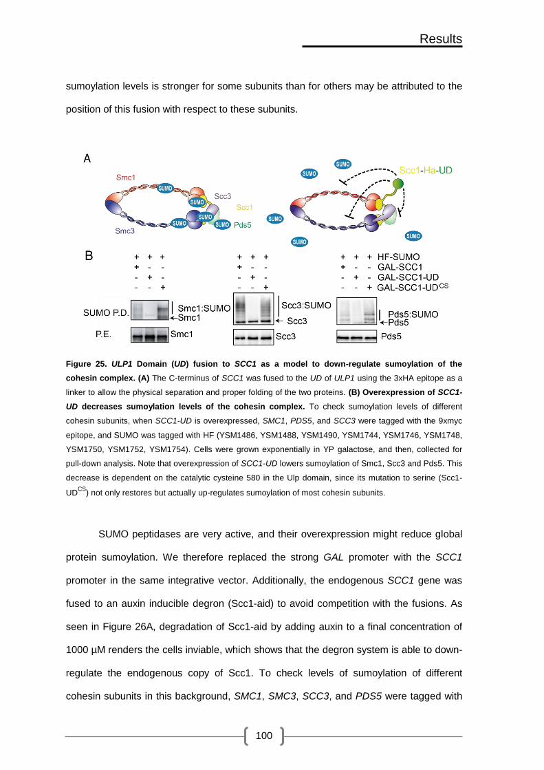
Results
100
sumoylation levels is stronger for some subunits than for others may be attributed to the
position of this fusion with respect to these subunits.
Figure 25. ULP1 Domain (UD) fusion to SCC1 as a model to down-regulate sumoylation of the
cohesin complex. (A) The C-terminus of SCC1 was fused to the UD of ULP1 using the 3xHA epitope as a
linker to allow the physical separation and proper folding of the two proteins. (B) Overexpression of SCC1-
UD decreases sumoylation levels of the cohesin complex. To check sumoylation levels of different
cohesin subunits, when SCC1-UD is overexpressed, SMC1, PDS5, and SCC3 were tagged with the 9xmyc
epitope, and SUMO was tagged with HF (YSM1486, YSM1488, YSM1490, YSM1744, YSM1746, YSM1748,
YSM1750, YSM1752, YSM1754). Cells were grown exponentially in YP galactose, and then, collected for
pull-down analysis. Note that overexpression of SCC1-UD lowers sumoylation of Smc1, Scc3 and Pds5. This
decrease is dependent on the catalytic cysteine 580 in the Ulp domain, since its mutation to serine (Scc1-
UDCS
) not only restores but actually up-regulates sumoylation of most cohesin subunits.
SUMO peptidases are very active, and their overexpression might reduce global
protein sumoylation. We therefore replaced the strong GAL promoter with the SCC1
promoter in the same integrative vector. Additionally, the endogenous SCC1 gene was
fused to an auxin inducible degron (Scc1-aid) to avoid competition with the fusions. As
seen in Figure 26A, degradation of Scc1-aid by adding auxin to a final concentration of
1000 µM renders the cells inviable, which shows that the degron system is able to down-
regulate the endogenous copy of Scc1. To check levels of sumoylation of different
cohesin subunits in this background, SMC1, SMC3, SCC3, and PDS5 were tagged with

Results
101
the 9xmyc epitope and SUMO was tagged with HF. For pull-down analysis cells were
grown exponentially in YPD, and then, auxin was added for two hours to degrade
endogenous Scc1.
As shown in Figure 26B, the Scc1-UD fusion itself shows no detectable levels of
sumoylation when expressed from its own promoter. Bands of slower mobility were
detected above the unmodified form of Smc3-9xmyc in wild-type cells, but not in cells
that express SCC1-UD as the only copy (Figure 26C). These bands correspond to
SUMO modified forms of Smc3, since they are not detected when SUMO is not pulled
down in a strain that lacks the HF tag on SUMO. As previously noted, inactivation of the
catalytic site in the Ulp1 domain lead to abnormally high levels of sumoylation in Scc1-
UD fusions. Similarly, sumoylation of Smc3 is up-regulated in the presence of Scc1-UDCS
(Figure 26C). We therefore created a new Scc1-UD fusion, called Scc1-UDFA,CS, which
contains an F474A mutation on the inactive UDCS domain. This mutation is predicted to
block binding of the Ulp domain to SUMO (Mossessova, Lima 2000). The Scc1-UDFA,CS
fusion restored wild-type levels of Scc1-UD and Smc3 sumoylation. Analogously to what
we have seen when Scc1-UD is overexpressed from the GAL promoter, sumoylation of
Smc1, Scc3, and Pds5 (Figure 26D) is down-regulated when SCC1-UD is expressed
from the SCC1 promoter as the only copy, an effect that is dependent on the catalytic
and SUMO-binding activities of the UD.
Thus, fusing SCC1 to the peptidase domain of Ulp1 (UD) down-regulates
sumoylation of the whole complex whether expressed from the SCC1 promoter as the
only copy or overexpressed from the GAL promoter.

Results
102
Figure 26. SCC1-UD expressed from the SCC1 promoter decreases sumoylation levels of the cohesin
complex. (A) Auxin down-regulates endogenous Scc1-aid. Note that cells that contain Scc1-aid are inviable
in auxin (IAA) plates while wild- type cells are not affected (YMB1804, YSM1907). (B) The Scc1-UD fusion
shows reduced levels of sumoylation when expressed at physiological levels. Sumoylation of Scc1 is
restored to wild-type levels when the F474A mutation, known to prevent Ulp1 binding to SUMO was
introduced in the inactive UDCS
domain (Scc1-UDFA,CS
) (YSM1907, YSM 2254, YSM 2256, YSM 2258). (C)
Scc1 or the indicated Scc1-UD fusions were expressed under the control of the SCC1 promoter from an
ectopic location. The endogenous SCC1 gene was fused to an auxin induced degron (Scc1-aid), and the
protein was degraded before analyzing cohesin sumoylation. To check levels of sumoylation of cohesin
subunits in this background, SCC3, PDS5, SMC1, and SMC3 were tagged with the 9Xmyc epitope and
SUMO with HF. Cells were grown exponentially in YPD, and then, auxin was added for two hours before pull
down of SUMO conjugates. Sumoylation of Smc3 is also down-regulated when Scc1-UD is expressed from
the SCC1 promoter as the only copy. This decrease is dependent on the catalytic cysteine 580 in the UD,
since its mutation to serine (Scc1-UDCS
) allows recovery of cohesin sumoylation. Note that this mutation not
only restores but actually up-regulates sumoylation of Smc3, and that sumoylation of Smc3 is restored to
wild-type levels when the F474A mutation is introduced in the inactive UDCS
domain (Scc1-UDFA,CS
) (YSM
1907, Y357, YSM 2297, YSM 2299, YSM 2301, YSM 2319). (D) sumoylation of Smc1, Scc3, and Pds5 is
down-regulated when Scc1-UD is expressed from the SCC1 promoter as the only copy. However,
sumoylation levels are restored to wild-type levels when the Scc1-UDFA,CS
is expressed (YTR907, YSM
2549, YSM 2550, YSM 2551, YSM 2552, YSM 2553, YSM 2554).

Results
103
V.1.4.3. Scc1-UD overexpression affects the growth rate of wild-type cells
To check the phenotype of cohesin desumoylation we started by overexpressing
SCC1, SCC1-UD, SCC1-UDCS and SCC1-UDFA,CS from the GAL promoter in wild-type
background to avoid possible lethal effects. Serial dilution and replica plating of cells that
express the different chimeras on glucose and galactose plates shows that
overexpression of SCC1-UD is not toxic, but leads to subtle growth defects in wild-type
cells. Cells that overexpress SCC1-UDCS also seemed to interfere with growth of wild
type cells (Figure 27A). To determine more accurately the effect on growth kinetics, we
measured the generation time of cells that overexpress the different chimeras. Cells
were grown overnight in YP raffinose, diluted to the same OD600 and split in two; in one
half, the sugar raffinose was substituted with galactose to allow the expression of the
fusions under the GAL promoter. As seen in Figure 27B wild-type cells that overexpress
SCC1-UD have a slower growth rate, as indicated by the generation time (3,4 hours),
compared to cells that overexpress SCC1 or SCC1-UDFA,CS (2 hours). This result
indicates that overexpression of SCC1-UD might have a dominant negative effect on
cohesin, and suggests that cohesin sumoylation might be required for proper cell growth.
Figure 27. SCC1-UD overexpression shows growth defects in wild-type cells. (A) Overexpression of

Results
104
SCC1-UD (YSM1494) in wild-type background (YSM1193) is toxic compared to that of SCC1 (YSM 1492),
SCC1-UDFA,CS
(YSM 1662) or SCC1-UD
CS(YSM 1496). (B) SCC1-UD overexpression affects growth rate of
wild-type cells. Wild-type cells that overexpress SCC1, SCC1-UD, or SCC1-UDFA,CS
were grown overnight in
YP plus raffinose. The next day cultures were diluted to the same OD600 and split in to two; in one of the two
the sugar raffinose was substituted with galactose to allow the expression of the fusions under the GAL
promoter. The OD600 was measured each hour for 8 hours. Note that wild-type cells that overexpress SCC1-
UD have a slower growth rate, as indicated by the generation time (3,4 hours), compared to cells that
overexpress SCC1 or SCC1-UDFA,CS
(2 hours).
V.1.4.4. Wild-type cells that overexpress SCC1-UD are sensitive to HU but not to
MMS or UV
Cohesin promotes SCR during DSB repair by bringing the two sister chromatids
in close proximity (Heidinger-Pauli, Unal et al. 2008, Strom, Karlsson et al. 2007). In
addition, cohesin is required for replication fork stability and for recovery of stalled
replication forks (Tittel-Elmer, Lengronne et al. 2012). Cohesin mutants such as scc1-73
are sensitive to replicative stress and to DNA damaging agents (Tittel-Elmer, Lengronne
et al. 2012).
Thus, we decided to test whether cohesin sumoylation might also affect these
functions. To this end we checked sensitivity of cells, that express/overexpress the
different constructs, to MMS(0.01%), UV (25 mJ/cm2), and HU (0.1M, 0.2M) by serial
dilution and replica plating on glucose/ galactose plates. We used nse2∆c as a control
because it is known to be sensitive to various types of DNA damage (Zhao, Blobel 2005,
Andrews, Palecek et al. 2005).
As seen in Figure 28, SCC1-UD expressed under the SCC1 promoter does not
alter viability of cells in the presence of HU, MMS, or after exposure to UV light,
compared to wild type cells. On the other hand, overexpression of SCC1-UD renders
cells more sensitive to HU, but not to MMS or UV. This sensitivity depends on the SUMO
peptidase activity of Ulp1, since inactivating this domain restores viability of these cells
when exposed to HU. This suggests that cohesin sumoylation might be required during

Results
105
replicative stress, but not in response to DNA the DNA lesions created by MMS or UV
light.
Figure 28. Wild-type cells that overexpress SCC1-UD are sensitive to HU but not to MMS or UV. Wild-
type cells that express/overexpress SCC1, SCC1-UD, or SCC1-UDFA,CS
were plated by means of serial
dilution and replica plating on their corresponding YPD and YP galactose plates and were exposed to UV
light (25 J/cm2). They were also plated on YPD and YP galactose plates that contained either MMS (0.01%)
or HU (0.1M, 0.2M). Note that SCC1-UD, expressed from the SCC1 promoter, does not alter viability of
these cells in the presence of DNA damage (YSM1193, YSM 1864, YSM1758, YSM1961). On the other
hand, overexpression of SCC1-UD renders cells more sensitive to HU, but not to MMS or UV (YSM1492,
YSM1494, YSM 1662). This sensitivity depends on the SUMO peptidase activity of Ulp1 on cohesin, since
inactivating this domain restores viability of these cells when exposed to HU. nse2∆c strain was used as a
control due to its known sensitivity to DNA damage (YSM2406).

Results
106
V.1.4.5. SCC1-UD does not rescue growth defects of scc1-73 at restrictive
temperatures and is toxic at permissive temperatures when overexpressed
To check the phenotype of cohesin desumoylation when SCC1-UD is expressed
as the only copy in the cell, we transformed episomal vectors that overexpress from the
GAL promoter SCC1-UD, SCC1-UBC9, SCC1, ULP1 and UBC9, into mcd1-1
thermosensitive allele. As seen in Figure 29A, overexpression of SCC1 or SCC1-UBC9
is able to recover the temperature sensitivity of mcd1-1, while overexpression of ULP1,
UBC9, or SCC1-UD does not. This observation suggests that cohesin sumoylation is
required for viability.
It has been shown that over-expression of a nuclear Ulp domain can be toxic
(Mossessova, Lima 2000). Therefore, the phenotype observed in mcd1-1 cells that
overexpress the Scc1-UD fusion could be due to global desumoylation after
accumulation of a SUMO peptidase in the nucleus.
To check this possibility we transformed episomal vectors that overexpress
SMC5-UD, SMC5-Ubc9, SMC5, ULP1 and UBC9, to an smc5-11 thermosensitive allele.
The SMC5-UD fusion is not toxic and is able to rescue growth of a thermosensitive
smc5-11 thermosensitive cells (Figure 29B), strongly suggesting that cohesin
sumoylation is required for viability. We also noted that overexpression of SMC5-UBC9
is toxic at permissive temperatures, suggesting that up-regulation of Smc5 sumoylation
is lethal.

Results
107
Figure 29. Overexpression of SCC1-UD from the GAL promoter is toxic in mcd1-1 thermosensitive
background. (A) mcd1-1 thermosensitive cells (Y758), bearing multicopy vectors (pYES2) that express
SCC1 (pTR 879), SCC1-UBC9 (pTR 871), UBC9 (p827), UD (cUlp1 in the figure) (pTR 865) or SCC1-UD
(pTR TR901) constructs from the GAL promoter, were plated on SC-raffinose (Raf; promoter off) or SC-
galactose (Gal; promoter on) and allowed to grow at the permissive (25ºC) or restrictive (37ºC) temperature
for mcd1-1. Note that SCC1-UD is toxic when overexpressed, and does not complement mcd1-1
thermosensitivity. (B) smc5-11 thermosensitive cells (YTR613), bearing multicopy vectors (pYES2) that
express SMC5 (pTR MB885), SMC5-UBC9 (pTR 882), UBC9 (pTR 827), UD or SMC5-UD (pTR 873) from
the GAL promoter, were treated as in A. Note that both SMC5 and SMC5-UD are able to complement the
smc5-11 thermosensitive phenotype, while overexpression of SMC5-UBC9 does not, and is even toxic at
permissive temperatures.
To check that these effects are dependent on the SCC1 alleles used, we tested
cell growth in scc1-73 thermosensitive cells. Similar to what we have seen with the
episomal vectors in mcd1-1 thermosensitive cells, overexpression of SCC1-UBC9 or
SCC1 is able to rescue temperature sensitivity of scc1-73 cells, indicating that
upregulation of cohesin sumoylation is not lethal (Figure 30A). On the other hand,
overexpression of Scc1-UD does not suppress the temperature sensitivity of scc1-73
cells at restrictive temperatures, and it is even toxic at permissive temperatures. This
effect indicates a dominant gain of function of the Scc1-UD fusion over the endogenous
hypomorphic scc1-73 allele.

Results
108
To avoid secondary effects of overexpression we substituted the GAL promoter
with SCC1 promoter and we integrated these constructs in the URA locus. The lethality
is no longer observed when SCC1-UD is expressed at physiological levels from the
SCC1 promoter (Figure 30B); yet it does not complement the thermosensitive phenotype
of scc1-73 cells. Scc1-UD growth defects are suppressed by inactivation of the SUMO
peptidase domain (Scc1-UDFA,CS), suggesting that the phenotype seen is due to the
specific SUMO peptidase activity on sumoylated cohesin.
Figure 30. Cohesin sumoylation is required for viability. (A) Overexpression of SCC1-UD from the
GAL promoter is toxic in scc1-73 thermosensitive background. scc1-73 thermosensitive cells (Y759)
ectopically expressing SCC1, SCC1-UD, SCC1-UBC9 (pSM957, pTR 918, pTR 921) from the GAL promoter
in a centromeric vector (YCPlac111) were plated on glucose (promoter off) or galactose (promoter on)
containing media, and allowed to grow at the indicated temperatures. Note that SCC1-UD is toxic when
overexpressed in scc1-73 cells, while an analogous fusion to the UBC9 conjugase is not toxic and effectively
complements the thermosensitive allele. (B) SCC1-UD expressed from the SCC1 promoter is not toxic at
the permissive temperature, yet it does not complement the thermosensitive phenotype of scc1-73
cells. scc1-73 thermosensitive cells ectopically expressing the indicated constructs from the SCC1 promoter
(YSM 1040, YSM1860, YSM1832, YSM1857, YSM 2155) were plated on YPD and allowed to grow at the

Results
109
indicated temperatures. Note that expression of SCC1-UD does not complement scc1-73 thermosensitivity,
while expression of SCC1-UDFA,CS
completely rescues this phenotype.
Finally, we designed a different approach to down-regulate the endogenous copy
of Scc1, using the degron system. The different DNA constructs were integrated at the
SCC1 locus by homologous recombination, leaving two copies of the SCC1 gene driven
by their corresponding SCC1 promoters, one fused to the UD (SCC1-UD), and the other
sensitive to auxin (SCC1-aid). As seen in Figure 31, cells are not viable when Scc1-aid is
degraded by growth on auxin-containing plates, while expression of wild type SCC1
restores viability. On the other hand, expression of SCC1-UD as the only copy in the cell
renders cells inviable. This phenotype depends on the SUMO peptidase activity of Ulp1
because Scc1-UD growth defects can be suppressed by inactivation of the SUMO
peptidase domain (Scc1-UDFA,CS).
Figure 31. Expression of SCC1-UD as the only copy in the cell in Scc1 degron background is lethal.
SCC1, SCC1-UD, and SCC1-UDFA,CS
, expressed from the SCC1 promoter, were integrated in the SCC1
locus (YSM2254/2255, YSM2256/2257, YSM2258/2259) in the Scc1-aid background (Y1907). Note that in
the absence of Scc1 cells are not viable. Expression of SCC1 restores viability, while expression of SCC1-
UD as the only copy in the cell renders cells inviable. This phenotype depends on the SUMO peptidase

Results
110
activity of Ulp1 because SCC1-UD growth defects can be suppressed by inactivation of the SUMO peptidase
domain (SCC1-UDFA,CS
).
V.1.4.6. Cells that express SCC1-UD as the only Scc1 copy accumulate in G2/M
scc1-73 thermosensitive cells arrest in G2/M at the restrictive temperature. The
arrest is dependent on activation of the spindle assembly checkpoint because of faulty
cohesion and the inability to biorient chromosomes (Stern, Murray 2001). To understand
the primary defect in cells expressing the Scc1-UD fusions, we studied cell cycle
progression by FACS in synchronous cultures expressing SCC1-UD and the auxin-
sensitive SCC1-AID allele. As seen in Figure 32, cells progress with normal kinetics
through G1 and S phase, but arrest in G2/M when Scc1-aid is degraded. As expected,
this effect is reversed by expression of wild-type SCC1. In contrast, the arrested cells do
not progress beyond G2/M when SCC1-UD is expressed as the only copy. This failure
depends on the SUMO peptidase activity of Ulp1, since cells that express SCC1 fused to
an inactive peptidase show normal cell cycle dynamics.
To monitor microscopically how cells that lack cohesin sumoylation progress
throughout the cell cycle we arrested scc1-73 thermosensitive cells that have the tet
operators(CenV)/tet repressor system and overexpress SCC1, SCC1-UD, SCC1-UDFA,CS
and SCC1-UBC9 from the GAL promoter in G1 by α factor. Then, we released them at
non permissive temperatures into a synchronous cell cycle and we added galactose to
induce the GAL promoter. Next, we took samples every 15 minutes for analysis by FACS
and fluorescence microscopy.
Similarly to what we have seen with the degron system, heat inactivation of scc1
thermosensitive allele leads to cell cycle arrest in the G2/M transition (Figure 32B). In
addition, overexpression of SCC1, SCC1-UDFACS, or SCC1-UBC9 allows cells to
progress to the next cell cycle, while overexpression of SCC1-UD does not, further
confirming our previous results with the degron system.

Results
111
We analyzed the cells microscopically and scored them according to: budding,
nuclear segregation, loss of SCC, and accumulation of mitotic cells with stretched
nucleus in the neck (Figure 32 C,D). Cells that overexpress SCC1 or SCC1-UD start to
have small buds 30 minutes after release, and the number of budded cells reaches a
maximum 105 minutes after release with similar kinetics. As expected, the number of
budded cells in the wild-type culture starts to decrease as cells finish mitosis and start a
new cell cycle. This decrease was not observed in cells that overexpress SCC1-UD,
suggesting an accumulation of budded cells that are not able to finish cell division. The
number of binucleated cells that overexpress wild-type SCC1 peaks 135 minutes after
release, while cells that overexpress SCC1-UD show a 60 minute delay in nuclear
segregation; cells transiently arrest with two centromeric dots as mononucleated or with
a stretched nucleus in the neck. It is worth noting that SCC1-UD overexpressing cells
that eventually manage to finish mitosis have around 60% of missegregation (data not
shown). Finally, we compared the kinetics of sister chromatid separation by counting
budded cells with centromere V loss of cohesion (2 dots). Cells that express wild type
SCC1 start to lose SCC by 90 minutes, which corresponds to the time when cells start
anaphase. In contrast, cells that express SCC1-UD start to show cohesion 60 minutes
after G1 release, which corresponds to the time when cells start to replicate their DNA,
as shown by the FACS profile. These results indicate that cells relying on Scc1-UD for
cohesin function lose cohesion from the time of DNA replication, suggesting a defect in
establishment of SCC.

Results
112
Figure 32. Cells that express SCC1-UD as the only copy accumulate in G2/M. (A) To study cell cycle
progression of strains carrying SCC1-UD fusion in Scc1 degron background by FACS analysis, Auxin (1
mM) was added once cells were arrested in G1 with the alpha factor. Then, 30 minutes after auxin addition,
cells were released by washing plus pronase addition, and samples were taken every 10 minutes for 200
minutes. Note that as Scc1-aid is degraded by auxin, cells accumulate in G2/M. When wild-type SCC1 is
expressed, cells manage to progress to the next cell cycle, while when SCC1-UD is expressed as the only
copy, cells arrest again in G2/M. This arrest depends on the SUMO peptidase activity of Ulp1, as cells that
express SCC1-UDFA,CS
do not arrest in G2/M (YSM1907, YSM 2254, YSM 2256, YSM 2258). (B) FACS
analysis of scc1-73 thermosensitive cells (YSM1040) that have the tet operators(CenV)/tet repressor system
and overexpress SCC1 and SCC1-UD SCC1-UDFA,CS
SCC1-UBC9 from the GAL promoter after arrest in G1
by α factor and release at 37°C in the presence of galactose. Note that overexpression of SCC1 (YSM1860),

Results
113
SCC1-UDFACS
(YSM1478), or SCC1-Ubc9 (YSM1506) allows cells to progress to the next cell cycle, while
overexpression of SCC1-UD does not. (C) Types of cells observed microscopically. a: G1 arrested cells with
1 nucleus and 1 dot, b: mononucleated cells that have a small bud and show one dot, c: mononucleated
cells with separated dots and a small bud, d: mononucleated cells with separated dots and a large bud, e:
mononucleated cells with one dot and a large bud, f: cells with a stretched nucleus in the neck and
separated dots, g: binucleated cells with separated dots. (D) Budding was measured by counting all cells
that have buds (b, c, d, e, f, g). SCC loss was measured by counting cells that show separated dots (c, d, f,
g). The percentage of binucleated cells and mitotic cells with stretched nucleus in the neck was measured by
counting one cell type (f and g respectively). Note that cells that overexpress SCC1-UD start to have
cohesion defects at the onset of DNA replication and accumulate as mononucleated cells with large buds
and separated dots or with stretched nucleus in the neck and fail to finish mitosis.
V.1.4.7. Cohesin rings are properly assembled around an Scc1-UD fusion
A possible explanation for the inviability of Scc1-UD fusions could be their
inability to properly interact with the SMC (Smc1 and Smc3) and/or the non-SMC
subunits (Scc3, Pds5). A different, but not contradictory possibility is that cohesin
sumoylation might be required for cohesin ring formation. To check these possibilities we
tagged SMC3 with the 9xmyc epitope in cells that overexpress HA-tagged SCC1, SCC1-
UD, and SCC1-UBC9 from the GAL promoter. Cells were grown overnight in rich media
supplemented with raffinose, and galactose was then added to induce expression of the
different constructs. Cells were collected two hours after induction, and processed for
anti-myc immunoprecipitation (IP). As seen in Figure 33A Scc1-UD
coimmunoprecipitates (CoIP) with Smc3 to similar levels as Scc1-Ubc9 and Scc1.
Overexpression of cohesin subunits may trigger interaction between different
cohesin complexes, something that is not normally seen under physiological conditions
(Zhang, Kuznetsov et al. 2008). To make sure that the interaction that we see is not
caused by overexpression conditions, we reanalyzed the co-immunoprecipitation in cells
that expressed the different fusions from the SCC1 promoter. Smc3-9xmyc is able to co-
immunoprecipitate similar amounts of Scc1-UD, Scc1 or Scc1-UDCS (Figure 33B). Scc1
binding to Smc1 is upstream to that with Smc3, which suggests that Scc1-UD is also
able to properly interact with Smc1. These findings indicate that fusion of Scc1 to the

Results
114
peptidase domain (UD) does not affect its interaction with the SMC subunits, and further
indicates that sumoylation is not required for cohesin ring formation.
Next, we studied interaction with Scc3 and Pds5. CoIP experiments show that
Scc1-UD interacts with Pds5 (Figure 33C) and Scc3 (Figure 33D) with similar efficiencies
as wild-type Scc1 or Scc1-UDFA,CS, suggesting that neither fusion of Scc1 to the SUMO
peptidase nor decrease in sumoylation levels affect interaction between Scc1 and non-
Smc subunits.
Figure 33. Cohesin rings are properly assembled around Scc1-UD. (A) Scc1-UD overexpressed from
the GAL promoter interacts with Smc3. Smc3 was tagged with the 9xmyc epitope (Y1193) in cells that
overexpress HA tagged SCC1, SCC1-UD, and SCC1-UBC9 from the GAL promoter (pSM957, pTR921, pTR
918). A strain that has HA tag on SCC1 but no myc tag on Smc3 was used as a control to check for
nonspecific binding of Scc1 or HA epitope to the magnetic beads coupled to anti-myc antibody used in the
immunoprecipitation experiment (IP). A strain that has no HA tag on SCC1 was used to discard unspecific
bands recognized by the primary anti-HA antibody. Cells were grown overnight in rich media supplemented
with raffinose. The next day, raffinose was substituted with galactose to induce expression of the different
constructs. Then, cells were collected two hours after induction, and processed for anti-myc IP. Eluted
proteins were loaded in separate gels to check for both IP and coimmunoprecipitation (CoIP). Note that
Scc1-UD coimmunoprecipitates with Smc3 similar to Scc1-Ubc9 and Scc1. (B) Scc1-UD expressed from
the SCC1 promoter interacts with Smc3. Scc1-UD expressed from the SCC1 promoter

Results
115
coimmunoprecipitates with Smc3-9xmyc under physiological conditions with similar efficiencies to Scc1 and
Scc1-UDCS
(YSM1193, YSM1758, YSM1844, YSM1864) . (C and D) Fusion of Scc1 to the SUMO
peptidase domain (UD) does not interfere with its interactions with non-Smc subunits. Pds5 and Scc3
were tagged with the 9xmyc epitope in cells that express SCC1, SCC1-UD , SCC1-UDFA,CS
from the SCC1
promoter. Strains that have myc epitope tag on SCC3/ PDS5, but no HA epitope tag on SCC1, were used as
a control to check for unspecific binding of Scc3/ Pds5 or the myc epitope to HA beads, used for the IP.
Strains that have no epitope tag on any cohesin subunit were used to discard unspecific bands recognized
by either the myc or HA primary antibodies. Exponentially growing cells were collected and processed for
anti-HA IP. The eluted proteins were loaded in separate gels to check for both IP and CoIP. Note that Scc1-
UD is able to interact with Pds5 (C) and Scc3 (D) with similar efficiencies as wild-type Scc1 or Scc1-UDFA,CS
(YSM2121, YSM 2123, YSM 2125, YSM 2126, YSM 2128, YSM 2130, YSM 1734, YSM 1735, YTR907).
V.1.4.8. Cohesin rings assembled around an Scc1-UD fusion are efficiently
recruited to chromatin
Another possible explanation for the Scc1-UD inviability is that down-regulation of
cohesin sumoylation might impair its binding to chromatin. To explore this possibility,
exponentially growing cultures of wild-type strains that express SCC1-UD or SCC1-
UDFA,CS , were collected and processed by chromatin fractionation to separate Triton X-
100 soluble supernatant and chromatin pellet fractions. Histone H3 was used as a
chromatin marker, while hexokinase was used as a cytosolic marker. Additionally, Rpd3
was used as a marker for a nuclear protein that faintly binds to chromatin. As shown in
Figure 34, we detected no difference in chromatin binding between Scc1-UDFA,CS
chimera and the unsumoylated Scc1-UD, suggesting that neither the fusion nor down-
regulation of cohesin sumoylation can affect recruitment of cohesin to chromatin.

Results
116
Figure 34. Cohesin rings assembled around Scc1-UD are efficiently recruited to chromatin. Cells
expressing no HA-tagged protein (YSM1907), SCC1-UD (YSM2256), or SCC1-UDFA,CS
(YSM2258) were
grown to exponential phase and samples were taken for chromatin fractionation. Blots were probed for the
Scc1 fusions with anti-HA. Controls for a chromatin bound protein (histone H3), nuclear soluble (Rpd3), and
cytoplasmic soluble (Hexokinase) proteins are also shown. Note that Scc1-UD and Scc1-UDFA,CS
are
similarly bound to chromatin. WCE, whole-cell extract; SN, soluble supernatant; CP, chromatin pellet.
V.1.4.9. Scc1-UD is found at known cohesin binding sites
Although we have seen that unsumoylated cohesin is recruited to chromatin, it
might not be recruited to the same sites as wild-type Scc1. To explore this possibility,
exponentially growing cultures of wild-type strains that express wild-type SCC1, SCC1-
UD or SCC1-UDFA,CS , were cross linked with formaldehyde, collected and processed for
anti-HA chromatin immunoprecipitation followed by CHIP hybridization (ChIP-on-CHIP).
Analysis of obtained chromatin enrichment profiles showed that Scc1, Scc1-UDFA,CS and
the unsumoylated Scc1-UD bind to the same places on all chromosomes in S.
cerevisiae. An example of the different profiles obtained for the three strains on
chromosome I is seen in Figure 35.
As exemplified for the relatively small chromosome 1, cohesin loading is enriched
at the centromere, convergent sites, and some origins of replication similarly in the wild-
type Scc1, Scc1-UDFA,CS, and the unsumoylated Scc1-UD. In addition, no enrichment is
seen for any of the Scc1 chimeras in sites of divergent genes. We also determined
average profiles for known binding sites including: early origins of replication,
centromeres, and convergent ORFs. As shown in Figure 36, the profiles of the three
strains coincide. Moreover, the average profiles also coincide for divergent ORFs, to
which cohesin does not normally bind. These results support the notion that fusion of
Scc1 to the SUMO peptidase domain (UD) does not affect its localization to known
cohesin binding sites, and that cohesin sumoylation is not required to localize cohesin to
these sites.

Results
117
Figure 35. Cohesin ChIP-on-chip: Scc1-UD and Scc1-UDFA,CS
localize to the same regions as WT
Scc1.. Exponentially growing cultures were cross-linked with formaldehyde (final concentration 1%). After
cell breakage and sonication, chromatin Immunoprecipitation (Anti-HA) was performed. The precipitated
DNA and the cell extract from each strain were reverse cross-linked, amplified, labeled, and hybridized to a
separate GeneChip S. cerevisiae Tiling 1.0R Array. Cohesin Scc1 (YSM2254), Scc1-UD (YSM2256), and
Scc1-UDFACS
(YSM2258) enrichment is shown for chromosome I. The signal log ratio (Log2), represented in
the Y-axis, was generated from the two sample comparison analysis i.e. Scc1 enrichment in the
Immunoprecipitated fraction relative to Input genomic DNA. The position is represented in (Kbs) in the X-
axis. Red Bars represent genes transcribed from left to right and opposite. Pink bars within the red bars
represent some convergent genes. The centromere is represented by a yellow circle. Origins of replication
are depicted by black triangles. Note that cohesin loading is enriched at the centromere, convergent sites,
and some origins of replication similarly in the three backgrounds. An example of 2 convergent genes is
shown by a pink line. Cohesin doesn’t bind to sites of divergent genes. An example of a divergent site is
marked by a dashed black line.

Results
118
Figure 36. Average ChIP-On-CHIP profiles of the genome-wide localization of Scc1 (YSM2254), Scc1-
UD (YSM2256), and Scc1-UDFA,CS
(YSM2258). Since cohesin is known to be enriched at origins of
replication, centromeres, and between genes that are transcribed in converging directions (Conv), but not
genes that are transcribed in diverging orientation (Div), the average profiles were compared in these four
sites. Annotations from the Saccharomyces Genome Database (SGD) were used to determine median
enrichment, which was calculated, in arbitrary units (a.u.), for 20 kb regions centered on these regions with a
2 kb window and a 100 bp step. The X-axis represents the distance in bp and the Y-axis represents Scc1
enrichment in a.u. Note that Scc1, Scc1-UDFA,CS
, and the unsumoylated Scc1-UD are enriched at known
cohesin binding regions, and not between genes that are transcribed in diverging directions.
Cohesin is enriched in the vicinity of double strand breaks (DSB) in response to
DNA damage, and is required to maintain sister chromatids in close proximity to promote
SCR, and thus, damage repair. To see whether cohesin recruitment to DSB is affected
by its sumoylation state, centromeric vectors, expressing Scc1 chimeras under the
conditional GAL promoter, were introduced into wild-type cells that also express the HO
endonuclease from the GAL promoter. Addition of galactose simultaneously triggered
expression of the chimeras and induction of an HO-mediated single irreparable DSB.
The efficiency of DSB formation were monitored microscopically by formation of Ddc2-

Results
119
GFP DNA repair foci. As seen in Figure 37A, 6 hours after adding galactose almost all
the cells had accumulated Ddc2-GFP DNA repair foci, reporting the formation of a DSB.
Overexpression of SCC1, SCC1-UBC9, and SCC1-UD from the GAL promoter was
monitored by western blot (Figure 37B). The presence of an irreparable double strand
break lead to similar enrichment of Scc1, Scc1-UD, and Scc1-Ubc9 in the vicinity of
DSBs (Figure 37D), as tested by quantitative Chromatin immunoprecipitation (ChIPq).
It is worth noting that the Scc1-UD fusion was quantitatively recruited with higher
efficiency to the lesion. Although not quantitative, results from the Chip-on-chip
experiments also suggest higher enrichment of unsumoylated cohesin (figure 36). We
currently do not know the reason for this increase in binding of unsumoylated cohesin.
An intriguing possibility is that cohesin sumoylation might be involved in separase-
dependent removal of cohesin during resection. It has been described that cohesin
needs to be removed by separase at the site of DSB so that resection proteins can
access the damage site (McAleenan, Clemente-Blanco et al. 2013).
Nevertheless, our results show that unsumoylated cohesin is enriched in the
vicinity of DSB and that sumoylation is not required for cohesin loading to DSBs.

Results
120
Figure 37. Scc1-Ubc9 and Scc1-UD are efficiently recruited to a DSB. Centromeric vectors, expressing
SCC1, SCC1-UD, SCC1-UBC9 from the conditional GAL promoter, were introduced in wild-type cells that
also express the HO endonuclease from the GAL promoter. (A) HO induces a single irreparable double
strand break (DSB), and the efficiency can be monitored microscopically by formation of Ddc2-GFP DNA
repair foci. (B) Western blot analysis of protein fusion expression at different times after galactose addition.
(C and D) Chromatin immunoprecipitation (ChIP) of Scc1 fusions. C: time0 before galactose addition. D: 6
hours after galactose addition. Note that both Scc1 and Scc1-Ubc9 are similarly enriched in the around
DSBs, while Scc1-UD seems to be more enriched.
V.1.4.10. SCC1-UD overexpression does not affect global levels of sumoylation
One reason for the lethal phenotype of SCC1-UD expressing cells could be the
desumoylation of other proteins in the cell. To check this possibility, we decided to check
global sumoylation levels in both systems. Exponentially growing cultures of cells that
express SCC1-UD from the SCC1 promoter or from the GAL promoter, were compared

Results
121
to cells that do not express any of the fusions by pull-down analysis. As seen in Figure
38, global sumoylation levels in cells that express SCC1-UD from the GAL promoter are
very similar to those seen in cells that express SCC1-UD from the SCC1 promoter or do
not express any fusion. In addition, the levels of free SUMO, i.e. SUMO not conjugated
to proteins, are also similar, indicating that the decrease in cohesin sumoylation upon
expression/overexpression of SCC1-UD is specific and is not secondary to reduction in
the global amount of sumoylated proteins in the cell.
Figure 38. SCC1-UD overexpression does not affect global levels of sumoylation. Exponentially
growing cultures of cells that express SCC1-UD from the SCC1 promoter (YSM1758) or from the GAL
promoter (YSM1660) were compared to cells that do not express any of the fusions (YSM1193) by pull-down
analysis. Global sumoylation levels in cells that express SCC1-UD from the GAL promoter (fourth lane) are
very similar to those seen in cells that express SCC1-UD from the SCC1 promoter (third lane) or do not
express any fusion (second lane). In addition, the levels of free SUMO, i.e. SUMO not conjugated to
proteins, are also similar. Cells in the first lane (YTR248, control) have no tag on SUMO, and thus SUMO
conjugates are not enriched. No difference is detected in the amount of protein loaded as seen by the
comassie staining.

Results
122
V.1.4.11. Scc1-UD has minor effects in sumoylation of nearby proteins
Next, we tried to check whether the presence of Scc1-UD within the cohesin ring
can affect sumoylation of other nearby proteins. One of the first proteins that we checked
was Ycs4, which belongs to the condensin complex, and has been reported to be
sumoylated (D'Amours, Stegmeier et al. 2004). To this end, we tagged Ycs4 with the
9xmyc epitope in an Scc1 degron background; SCC1-UD or SCC1-UDFA,CS were
expressed from the SCC1 promoter. As seen in Figure 39, there is no decrease in
sumoylation levels of Ycs4 in SCC1-UD expressing cells as compared to SCC1-UDFA,CS,
indicating that the presence of Scc1-UD does not affect sumoylation of nearby condensin
proteins.
Figure 39. SCC1-UD expression as the only copy does not affect sumoylation of condensin. YCS4
was tagged with the 9xmyc epitope in Scc1 degron background (YSM 1907), and SCC1-UD (YSM 2458) or
SCC1-UDFA,CS
(YSM 2459) were expressed from the SCC1 promoter. For pull-down analysis, auxin was

Results
123
added to exponentially growing cultures for two hours to degrade the endogenous Scc1, and use Scc1-UD
or Scc1-UDFA,CS
as the only copy in the cell. Note that there is no decrease in sumoylation levels of Ycs4
when Scc1-UD is expressed as the only copy (second lane) compared to that of Scc1-UDFA,CS
(third lane).
We also wondered whether sumoylation of proteins that are directly involved in
DNA replication, such as PCNA, might be affected in the presence of Scc1-UD within the
cohesin ring, and whether the change in sumoylation levels brought upon can affect their
function during DNA replication. Sumoylation of PCNA at K164 is required during DNA
replication to avoid unnecessary recombination intermediates in the presence of
replicative stress. Siz1 is the SUMO E3 ligase required for PCNA sumoylation. PCNA
monoubiquitylation at the same K164 by Rad18 E3 ligase is required for damage
bypass. It has been described that rad18∆ cells are highly sensitive to MMS. However,
abolishing PCNA sumoylation by siz1∆ can suppress this MMS sensitivity at low
concentrations (0.001%) (Parker, Ulrich 2012).
We reasoned that if Scc1-UD has the same effect on PCNA as siz1∆,
sumoylation levels of PCNA when SCC1-UD is expressed would be as low as in siz1∆.
In addition, Scc1-UD expression might be able to suppress MMS sensitivity at 0.001%.
As seen in Figure 40A, while siz1∆ abolishes sumoylation of PCNA, there is only a small
decrease in sumoylation levels of PCNA when Scc1-UD is expressed compared to that
of wild type Scc1 or Scc1-UDFA,CS expression. In addition and as expected, we have
seen that rad18∆ cells start to be sensitive to MMS at very low concentrations
(0.0003%). Deletion of SIZ1 in rad18∆ cells rescues their MMS sensitivity at 0.0005%,
while expression of SCC1-UD does not (Figure 40B), suggesting that Scc1-UD might
slightly affect PCNA sumoylation levels without affecting its function during replicative
stress.
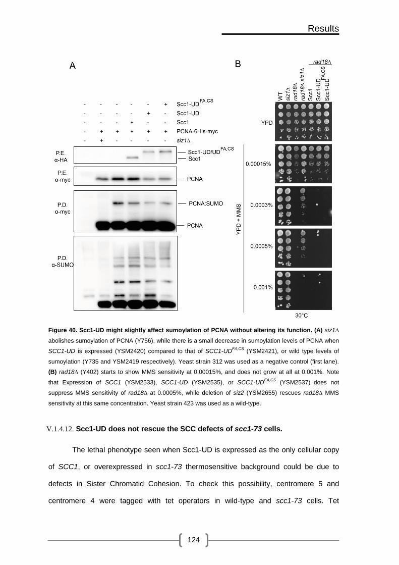
Results
124
Figure 40. Scc1-UD might slightly affect sumoylation of PCNA without altering its function. (A) siz1∆
abolishes sumoylation of PCNA (Y756), while there is a small decrease in sumoylation levels of PCNA when
SCC1-UD is expressed (YSM2420) compared to that of SCC1-UDFA,CS
(YSM2421), or wild type levels of
sumoylation (Y735 and YSM2419 respectively). Yeast strain 312 was used as a negative control (first lane).
(B) rad18∆ (Y402) starts to show MMS sensitivity at 0.00015%, and does not grow at all at 0.001%. Note
that Expression of SCC1 (YSM2533), SCC1-UD (YSM2535), or SCC1-UDFA,CS
(YSM2537) does not
suppress MMS sensitivity of rad18∆ at 0.0005%, while deletion of siz2 (YSM2655) rescues rad18∆ MMS
sensitivity at this same concentration. Yeast strain 423 was used as a wild-type.
V.1.4.12. Scc1-UD does not rescue the SCC defects of scc1-73 cells.
The lethal phenotype seen when Scc1-UD is expressed as the only cellular copy
of SCC1, or overexpressed in scc1-73 thermosensitive background could be due to
defects in Sister Chromatid Cohesion. To check this possibility, centromere 5 and
centromere 4 were tagged with tet operators in wild-type and scc1-73 cells. Tet

Results
125
repressors tagged with YFP bind to tet operators and allow visualization of centromeres
as single dots under the fluorescence microscope. Scc1, Scc1-UD, Scc1-UDFA,CS, Scc1-
UDCS, Scc1-Ubc9 overexpressed from the GAL promoter were integrated in the URA
locus in scc1-73 cells with centromere 5 tags.
To check metaphase cohesion cells were first arrested in G1 by alpha factor at
the permissive temperature (25°C). Once arrested, the temperature was shifted to 37°C
to inactivate scc1-73 allele, and at the same time galactose was added to induce
expression of the different constructs. 30 minutes after adding galactose, cells were
released into a metaphase block by addition of nocodazole. Nocodazole-arrested cells
were scored for metaphase cohesion by counting cells that have one dot, which
represent cells that were able to establish and maintain SCC, and cells that have two
dots, which represent cells with defects in SCC (Figure 41A). scc1-73 allele is not
functional at restrictive temperatures and cells have defects in SCC. Overexpression of
Scc1 or Scc1-Ubc9 restores these defects to wild-type levels (Figure 41B). On the other
hand, overexpression of Scc1-UD does not restore defects in SCC (Figure 41C). This
phenotype is dependent on UD binding to and deconjugating SUMO, since
overexpression of Scc1-UDFA,CS or Scc1-UDCS could revert SCC defects.
The same phenotype was observed when the constructs were expressed from
the SCC1 promoter. As seen in Figure 42A expression of Scc1 or Scc1-UDCS restores
metaphase cohesion defects at centromere 5, while Scc1-UD does not. Similarly,
expression of Scc1, Scc1-UDCS, Scc1-UDFA,CS from the SCC promoter restores defects
in SCC at centromere 4, while Scc1-UD does not (Figure 42B). Thus, in the absence of
cohesin sumoylation cells have defects in SCC, which suggests that cohesin
sumoylation positively regulates SCC.
Binding sites for Scc1 were found near the silent loci, and coinciding with the
boundary elements in the case of silenced mating-type loci HMR. To check SCC in the

Results
126
HMR locus when cohesin sumoylation is absent, we transformed SCC1, SCC1-UD to
scc1-73 cells, where HMR was tagged with lac-GFP and flanked by binding sites for a
GAL-inducible site-specific recombinase (Cre). Addition of galactose leads to excision of
the locus in cells with sister chromatids producing two chromatin circles that remain
associated with each other in wild-type cells but loose cohesion in scc1-73 cells (Chang,
Wu et al. 2005). Cells were arrested in G1, and then, galactose was added to express
the SCC1 constructs and the recombinases. The temperature was then shifted to 37ºC
to inactivate the scc1-73 allele. 30 min later, cells were released into a synchronous cell
cycle at 37ºC, arrested in metaphase with nocodazole and observed microscopically for
HMR cohesion. As seen in Figure 42C scc1-73 cells have cohesion defects at the HMR
locus. Overexpression of wild-type SCC1 rescues the SCC defects in scc1-73 cells,
while overexpression of SCC1 -UD does not, suggesting that cohesin sumoylation is also
required at the HMR locus for SCC.
Intriguingly, we also found that nse2∆C cells do not have gross defects in SCC,
suggesting that the Nse2 SUMO ligase activity is not required for cohesion at the HMR
locus (Figure 42D). As presented in Figure 42E, smc6-9 cells also do not display major
defects in SCC, suggesting that the Smc5/6 complex is also not required for cohesion at
the HMR. Moreover, the minor SCC defects detected in smc6-9 cells seem to be
cohesin-dependent, as double scc1-73 smc6-9 mutants display additive cohesion
defects. These results indicate that the cohesin and Smc5/6 complex act cooperatively at
the mating type locus for sister chromatid cohesion.

Results
127
Figure 41. Overexpression of SCC1-UD does not rescue SCC defects of scc1-73. (A) Wild-type or scc1-
73 cells carrying a chromosome tag on centromere 5 were synchronized in G1 by alpha factor. Once
arrested in G1, the GAL promoter was switched on to allow expression of SCC1, SCC1-UD, SCC1UDCS
,
SCC1-UDFA,CS
, SCC1-UBC9 or SCC1-UBC9CS
in scc1-73 cells, and the temperature was shifted to 36°C to
inactivate scc1-73 thermosensitive allele. After 30 minutes, cells were released into a synchronous cell cycle
at 36°C, arrested in metaphase with nocodazole and observed microscopically for centromere cohesion.
Images of metaphase arrested cells taken with the fluorescence microscope are shown. Yellow arrow points
at a cell with two separated dots, representing cells that failed to establish SCC. White arrow points at a cell
with one dot, representing cells that have established SCC (B) Note that overexpression of SCC1 restores
SCC in a scc1-73 cells, while overexpression of SCC1-UD does not. On the other hand, overexpression of
SCC1-UDFA,CS partially recovers centromeric cohesion (YSM1040, YSM1476, YSM1478, YSM1480,
YSM1614, Y343). Bars indicate mean values for three independent experiments; lines on bars are standard
deviation. (C) Up-regulation of cohesin sumoylation restores SCC defects of scc1-73. Overexpression
of SCC1-UBC9 or SCC1-UBC9CS
restore SCC defects of scc1-73 (YSM1040, YSM1476,YSM1506,
YSM1568).

Results
128
Figure 42. SCC1-UD expressed from the SCC promoter does not rescue SCC defects of scc1-73 at
centromere 4 and 5. Overexpression of SCC1-UD does not rescue SCC in scc1-73 cells at the HMR
locus, while smc6-9 and nse2∆C have no defects in SCC. (A) Wild type or scc1-73 cells carrying a
chromosome tag on centromere 5 and expressing SCC1, SCC1-UD, SCC1UDCS
from the SCC1 promoter,
were treated as in Figure 41 without galactose addition, and observed microscopically for centromere
cohesion. Note that SCC1 expression restores SCC in scc1-73 cells, while SCC1-UD does not. Cells that
express SCC1-UDCS
show partial recovery of centromeric cohesion (YSM1040, YSM1860, YSM1832,
YSM2155, YSM1857, Y343). (B) Wild type or scc1-73 cells carrying a chromosome tag on centromere 4 and
expressing SCC1, SCC1-UD, SCC1UDCS
or SCC1-UDFA,CS
from the SCC1 promoter, were treated as in (A),
and observed microscopically for centromere cohesion. Note that SCC1 expression restores SCC in scc1-73
cells, while SCC1-UD does not. Cells that express SCC1-UDCS
show partial recovery of centromeric
cohesion, while cells that express SCC1-UDFA,CS
show a higher percentage of centromeric cohesion
(YSM1890, YSM1882, YSM1952, YSM1954, YSM1956, YSM1958). Bars indicate mean values for three
independent experiments; lines on bars are standard deviation. (C) To check SCC in the HMR locus when
cohesin sumoylation is absent, we transformed SCC1, SCC1-UD to scc1-73 cells, where HMR was tagged
with lac-GFP and flanked by binding sites for an inducible site-specific recombinase (HO). HO was placed
under the GAL promoter. Addition of galactose leads to excision of the locus in cells with sister chromatids
producing two chromatin circles that remain associated with one another in wild-type cells but loose
cohesion in scc1-73 cells. Cells were treated as in Figure 41 and observed microscopically for HMR
cohesion. Note that scc1-73 cells have defects in SCC. Overexpression of wild-type SCC1 rescues SCC
defects in scc1-73 cells, while overexpression of SCC1-UD does not (Y504, Y503, YSM1514, YSM1516). (D)
To check whether the same defects in SCC at the HMR locus are also seen in nse2∆c cells, we transformed

Results
129
this allele to cells that have lac-GFP tag on HMR and we scored for metaphase cohesion as in (C). Note that
nse2∆C cells do not have defects in SCC compared to those seen in scc1-73 cells, suggesting that Nse2
SUMO ligase activity might not be required for SCC (Y504, Y503, YTR559). (E) To check whether the same
defects in SCC at the HMR locus are also seen in smc6-9 cells, we transformed this allele to cells that have
lac-GFP tag on HMR and we scored for metaphase cohesion as in (C). Note that smc6-9 cells do not have
defects in SCC compared to those seen in scc1-73 cells, suggesting that Smc5/6 complex might not be
required for SCC. Also note that scc1-73, smc6-9 double mutants have sever defects in SCC, suggesting
that these mutations are additive (Y504, Y503, YTR519, YTR535).
V.1.5. Sumoylation and acetylation are required independently during
establishment of SCC
During DNA replication, Eco1-dependent acetylation of Smc3 locks cohesin
around newly replicated sister chromatids. We have seen that cohesin sumoylation is
also required during establishment of SCC. One possibility is that cohesin sumoylation
could be an upstream event to its acetylation. In order to understand the relationship
between these two modifications, we started by checking Smc3 acetylation in the
absence of SUMO conjugation in the cell. Wild-type and ubc9-1 cells expressing SMC3-
3xHA were grown exponentially at 25°C, and one half was shifted to 37°C for two hours.
Both cultures were then collected and processed for anti-HA immunoprecipitation. As
seen in Figure 43A, the acetylation levels of Smc3 are not affected by the absence of
sumoylation in the cell, indicating that sumoylation is not required for Smc3 acetylation.
The anti-acetyl lysine antibody is specific for the acetylated Smc3 since, as expected,
smc3KKRR shows no detectable acetylation (Figure 43B).

Results
130
Figure 43. Smc3 acetylation does not depend on sumoylation. (A) Inactivation of the SUMO pathway
in a ubc9-1 thermosensitive mutant has no effect on Smc3 acetylation. UBC9 wild-type (YSM1710) and
ubc9-1 mutant cells (YSM1756) that have a 3xHA tag on Smc3, were grown at 25ºC (25), and shifted to
37ºC for 2 hours to inactivate ubc9-1 thermosensitive allele. Cells were collected and subjected to anti-HA
IP. After elution the IP was divided in two PAG to probe with anti-HA to detect the total amount of Smc3-3HA
or anti-acetyl lysine antibodies to test the levels of Smc3 acetylation. Note that Smc3 is efficiently acetylated
in the absence of active SUMO conjugase.(B) The anti-acetyl lysine antibody is specific for the
acetylated Smc3. Protein extracts (P.E.) from cells that express a 3HA tagged copy of wild-type Smc3
(YSM1650) or the nonacetylated mutant Smc3 protein (YSM1649, Smc3KKRR
), were subjected to anti-HA IP
similar to (A). Note that the anti-acetyl lysine antibody does not recognize the unacetylated Smc3KKRR
,
although it is immunoprecipitated.
Next, we specifically looked at the effect of cohesin desumoylation in scc1-aid
cells expressing the SCC1, SCC1-UD, SCC1-UDFA,CS constructs under the SCC1
promoter. Degradation of the endogenous Scc1 copy is expected to prevent proper ring
assembly and to lower Smc3 acetylation. Exponentially growing cells were treated with
auxin and samples were taken at times 0, 30, 60, 90, and 120 after activation of the
degron, and processed for anti-myc IP. Figure 44 shows that Smc3 acetylation levels
drop, reaching a minimum 90 min after auxin addition. A similar effect is observed after
inactivation of the scc1-73 allele by shift to the restrictive temperature (Figure 45A).
Scc1-aid cells that express the different chimeras were grown exponentially, and
samples were taken at times 0 and 120 minutes after auxin addition. Cells expressing
any of the fusions, SCC1, SCC1-UD or SCC1-UDFA,CS did not show any change in
acetylation levels 2 hours after triggering degradation of the endogenous copy, indicating
once more that cohesin sumoylation is not required for its acetylation.

Results
131
Figure 44. Smc3 acetylation does not depend on cohesin sumoylation. Scc1-aid, Smc3-9myc
(YSM1965) cells expressing SCC1, SCC1-UD, SCC1-UDFA,CS
from the SCC1 promoter (YSM2025,
YSM2027, YSM2031) were grown to exponential phase, and protein extracts were subjected to anti-myc IP.
Western blots were probed with anti-acetyl lysine antibody to detect levels of acetylation, and reprobed with
anti-myc to detect the levels of immunoprecipitated Smc3. Note that Smc3 acetylation levels drop after
addition of auxin, and this effect can be counteracted by expression of sumoylated (Scc1 and Scc1-UDFA,CS)
or unsumoylated (Scc1-UD) cohesin.
It is possible that unsumoylated cohesin rings (formed around Scc1-UD) fail to
properly interact with acetylated Smc3 leading to loss of SCC. To test this possibility,
scc1-73 cells that express SCC1, SCC1-UD, and SCC1-UDFA,CS were collected, and the
ectopically-expressed Scc1 versions were immunopresipitated with anti-HA antibodies.
As seen in Figure 45B, acetylated Smc3 co-immunoprecipitates with the 3xHA tagged
Scc1-UD with similar efficiency to Scc1 or Scc1-UDFA,CS, demosntrating that cohesin
sumoylation is not required for its interaction with acetylated Smc3.
Figure 45. Unsumoylated cohesin subunits interact efficiently with acetylated Smc3. (A) scc1-73
(Y759) cells that express SMC3-6HA (YSM2225) were grown at 25°C to exponential phase. Half of the
culture was placed at 37°C for 2 hr to inactivate scc1-73 thermosensitive allele. Cells were collected and

Results
132
subjected to anti-HA IP as in Figure 43A. Note that inactivation of Scc1 leads to decrease in Smc3
acetylation levels. (B) scc1-73 (YSM1040) cells expressing SCC1, SCC1-UD, SCC1-UDFA,CS
from the SCC1
promoter (YSM1860, YSM1832, YSM2155) were grown to exponential phase and heat-shocked as in (A).
Scc1-3HA and the Scc1-UD chimeras were immunoprecipitated and analyzed by western blot with anti-HA
and anti-acetyl-lysine antibodies to detect the amount of co-immunoprecipitating acetylated Smc3. Note that
the unsumoylated versions of cohesin coimmunoprecipitate efficiently with acetylated Smc3.
Acetylation of Smc3 counteracts the antiestablishment activity of Rad61/Wapl. It
has been described that rad61∆ makes acetylation dispensable for maintenance of sister
chromatid cohesion. To see whether sumoylation is similarly required to counteract the
antiestablishment activity of Rad61, we deleted RAD61 in scc1-73 cells that express
SCC1 and SCC1-UD from the SCC1 promoter, and we plated the cells by replica plating
at permissive and non-permissive temperatures.
Figure 46 shows that, and as we have seen earlier, expression of wild-type SCC1
in scc1-73 rescues its temperature sensitivity, while expression of SCC1-UD is lethal in
this background at restrictive temperatures. As previously reported (Rowland, Roig et al.
2009), rad61∆cells are slightly thermosensitive, and thus, expression of SCC1 from the
SCC1 promoter in scc1-73/rad61∆ double mutant does not completely suppress
thermosensitivity of these cells. On the other hand, rad61∆ does not recover the growth
defects of scc1-73 SCC1-UD expressing cells at restrictive temperatures. Thus, unlike
acetylation, sumoylation is not required to counteract the antiestablishment activity of
Rad61, although it is required for establishment of SCC.
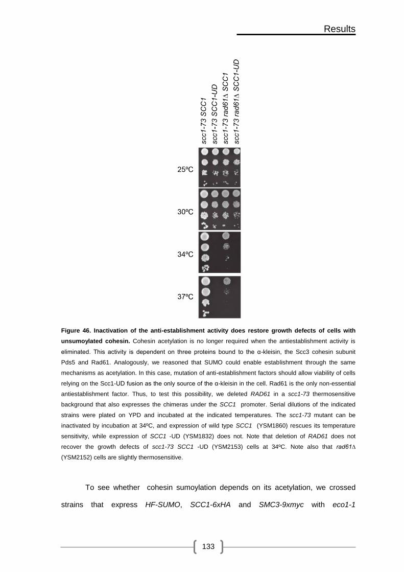
Results
133
Figure 46. Inactivation of the anti-establishment activity does restore growth defects of cells with
unsumoylated cohesin. Cohesin acetylation is no longer required when the antiestablishment activity is
eliminated. This activity is dependent on three proteins bound to the α-kleisin, the Scc3 cohesin subunit
Pds5 and Rad61. Analogously, we reasoned that SUMO could enable establishment through the same
mechanisms as acetylation. In this case, mutation of anti-establishment factors should allow viability of cells
relying on the Scc1-UD fusion as the only source of the α-kleisin in the cell. Rad61 is the only non-essential
antiestablishment factor. Thus, to test this possibility, we deleted RAD61 in a scc1-73 thermosensitive
background that also expresses the chimeras under the SCC1 promoter. Serial dilutions of the indicated
strains were plated on YPD and incubated at the indicated temperatures. The scc1-73 mutant can be
inactivated by incubation at 34ºC, and expression of wild type SCC1 (YSM1860) rescues its temperature
sensitivity, while expression of SCC1 -UD (YSM1832) does not. Note that deletion of RAD61 does not
recover the growth defects of scc1-73 SCC1 -UD (YSM2153) cells at 34ºC. Note also that rad61∆
(YSM2152) cells are slightly thermosensitive.
To see whether cohesin sumoylation depends on its acetylation, we crossed
strains that express HF-SUMO, SCC1-6xHA and SMC3-9xmyc with eco1-1

Results
134
thermosensitive cells, which have no detectable levels of Smc3 acetylation. After
sporulation and tetrad dissection, wild-type ECO1 or eco1-1 thermosensitive allele plus
SCC1-6xHA or SMC3-9myc were grown exponentially at 23°C (permissive temperature)
and then shifted to 37°C for two hours to inactivate eco1-1. Samples were collected
before and after shift to restrictive temperatures (37°C) and were processed for SUMO
pull-down. As seen in Figure 47 sumoylation levels of Scc1 or Smc3 at either the
permissive or restrictive temperature are similar in wild-type and eco1-1 cells, indicating
that cohesin acetylation is not required for its sumoylation.
All together, these results show that both acetylation and sumoylation are
required independently during establishment of SCC.
Figure 47. Cohesin sumoylation does not depend on its acetylation. ECO1 and eco1-1 cells, carrying
HF-SUMO and Scc1-6HA (A)/Smc3-9myc (B) tags, were grown at 23°C (YTR907, YSM1227, YNC2184,
YNC2186, YSM1193, Y1588). The cultures were split in two, and one half was incubated at 37°C to
inactivate the eco1-1 allele. Samples were taken two hours after transfer to the restrictive temperature. Note
that both Scc1(A) and Smc3(B) are sumoylated in an eco1-1 background.
V.1.6. Sumoylation of Smc1 and Smc3 requires Scc1 dependent ring
formation and Scc1 sumoylation
Cohesin subunits might be sumoylated in a sequential manner, i.e. sumoylation
of one subunit might be required for sumoylation of the next. To check this possibility we

Results
135
decided to specifically block sumoylation of Scc1 using the two recently described scc1
alleles (scc111KR and scc13KR). These two alleles bear lysine to arginine mutations in the
primary (scc13KR) or primary plus secondary (scc111KR) SUMO acceptor sites, and have
been shown to be deficient for DNA damage repair (McAleenan, Cordon-Preciado et al.
2012). First, we checked the functionality of these alleles under non-damage conditions.
To this end, we expressed wild-type SCC1, scc111KR, and scc13KR ectopically in an Scc1-
aid background and plated cells on YPD plates with or without auxin (IAA, 1mM) to
degrade the endogenous Scc1. As seen in Figure 48A cells that express scc111KR as the
only SCC1 copy have growth defects even in the absence of DNA damage while those
that express scc13KR or wild-type SCC1 restore growth defects of Scc1-aid cells (Figure
48A). Equivalent results were obtained using thermosensitive scc1-73 cells: the scc13KR
allele fully complements SCC1 function, while the scc111KR allele displays growth defects
at the restrictive temperature (Figure 48B).
Figure 48. Strains that express scc111KR
as the only copy of SCC1 show growth defects. (A) Wild-type
SCC1 (p2015), scc13KR
(p2006), or scc111KR
(p2005) were ectopically expressed in Scc1 degron background

Results
136
(YSM1907). Cells were plated on YPD plates, with or without Auxin (IAA, 1000µM), to degrade the
endogenous SCC1. Note that cells that express scc111KR
as the only SCC1 copy have growth defects under
physiological conditions, and in the absence of DNA damage when compared to those that express scc13KR
or wild-type SCC1. (B) scc111KR
mutant allele was expressed ectopically in scc1-73 thermosensitive cells
(YSM1040) and plated on YPD plates at permissive (25°C) and restrictive temperatures (37°C) to inactivate
scc1-73 thermosensitive allele. Note that scc111KR
shows growth defects at 37°C, suggesting that this allele
is thermosensitive.
Then, we decided to check sumoylation levels of Scc111KR, tagged with the 3HA
epitope, under physiological conditions and in the absence of DNA damage, by
ectopically expressing this allele in the Scc1-aid background. Samples were taken before
and after addition of auxin (1 mM) for pull-down analysis. As seen in Figure 49A,
sumoylation levels of Scc111KR are barely detectable in the presence of the endogenous
copy of Scc1. Surprisingly, some sumoylation of Scc111KR is seen in the absence of the
endogenous copy of Scc1, although sumoylation levels are lower than those seen in the
ectopically expressed wild-type copy of Scc1. These results suggest that in the absence
of DNA damage, there might be additional lysines targeted by SUMO in the Scc1 protein.
Next, we decided to test whether full sumoylation of Scc1 is required for the
subsequent modification of Smc1. For this purpose, we tagged the SMC1 gene with a
9xmyc epitope. Samples were taken for pull-down analysis before and after addition of
auxin (1 mM). As seen in Figure 49B, in the absence of Scc1, Smc1 displays no
detectable levels of sumoylation, showing that tripartite cohesin ring formation is a pre-
requisite for its sumoylation. When wild-type Scc1 is ectopically expressed sumoylation
of Smc1 is restored to wild-type level. In contrast, expression of scc111KR lowered Smc1
sumoylation, even in the presence of the endogenous Scc1. These results indicate that
sumoylation of Scc1 is required for sumoylation of Smc1, and that cohesin might be
sumoylated in a sequential manner.

Results
137
Figure 49. Sumoylation of Smc1 and Smc3 require Scc1 dependent ring formation and Scc1
sumoylation. (A) To check sumoylation levels of Scc111KR
, tagged with the 3HA epitope, under physiological
conditions and in the absence of DNA damage, SCC111KR
was ectopically expressed in Scc1-aid background
(YSM1907). For pull-down analysis, cells were grown exponentially at 25°C and then were split in two
halves. To one half, Auxin was added to a final concentration of 1000 µM and incubated again for two
hours. Both halves were collected, and processed for pull-down analysis. Note that sumoylation levels of
Scc111KR
are barely detectable in the presence of the endogenous copy of Scc1. However, some
sumoylation of Scc111KR
is seen in the absence of the endogenous copy of Scc1, although sumoylation
levels are much lower than those seen in the ectopically expressed wild-type copy of Scc1. (B) Wild-type
SCC1 (p2015), or scc111KR
(p2005) were ectopically expressed in Scc1-aid cells (YSM1963) that express
Smc1-9xmyc and 6xHis-Flag-SUMO. Sumoylation of Smc1 was checked in the presence or absence of
Auxin (IAA) which was used to degrade the endogenous Scc1 and use ectopically expressed wild-type
SCC1 or scc111KR
as the only copy in the cell. Cells were grown and treated as in A and then were collected
for pull-down analysis. Note that when wild-type SCC1 is ectopically expressed sumoylation of Smc1 is
restored to wild type levels. On the other hand, scc111KR
shows decreased levels of sumoylation even in the
presence of the endogenous Scc1 copy. These levels are drastically decreased when scc111KR
is expressed
as the only copy.

Results
138
V.2. UD (SENP1) fusion to RAD21 as model to down-regulate cohesin
sumoylation in HEK293T human cell line
Sumoylation of human cohesin (Rad21) has been detected in vitro, suggesting
that cohesin regulation by SUMO might be conserved in humans. To check this
possibility, we decided to transfer our model to human cell lines and follow the same
approach we used in budding yeast to characterize fusion of cohesin to Ulp domains.
First, we started by constructing the chimeras using mouse RAD21 (mRAD21),
which is the homolog of yeast Scc1, and the UD domain of human SENP1. We chose
mouse Rad21 so that we can down-regulate specifically the endogenous copy of RAD21
(hRAD21) by siRNA, while keeping mRAD21 as the only copy in the cell. Similarly to
what we have done in budding yeast, we used a 3x(4Gly-Ser)-4xHA-6x(Gly-Ala) tag as a
linker between Rad21 and UD (SENP1) to allow separation and proper folding of the two
proteins. In addition, the fusions were cloned in vectors that express GFP to allow in vivo
monitoring by fluorescence microscopy. We also used the same fusion bearing an
inactive peptidase domain (C603S) as a control.
V.2.1. Cohesin rings are properly assembled around mRad21-UD
To check whether these fusions are able to interact with the rest of the complex,
we transfected vectors expressing mRAD21, mRAD21-UD, and mRAD21-UDCS from the
CMV promoter to HEK293T cells. Then, we performed an anti-HA IP on protein extracts
from these cells. The IP product was loaded in two PAGE to check for IP using anti-Scc1
primary antibody, and CoIP of Smc1 and Smc3 using anti-Smc1 and anti-Smc3 primary
antibodies (Supplementary Table 1). We used HEK293T cells that do not carry any
vector as a control for the IP. As seen in Figure 50, Smc1 and Smc3 interact with
mRad21-UD with the same efficiency as wild-type mRad21, and mRad21-UDCS. This
result shows that mRad21 is able to interact with human cohesin subunits, and that
neither the SUMO peptidase domain attached to mRAD21, nor its activity, interfere with

Results
139
cohesin ring formation. Intriguingly, we consistently observed a lower mobility form of
Rad21- UDCS. Expression of Scc1- UDCS in budding yeast leads to the accumulation of a
similarly running band of sumoylation, which is most probably due to binding of the
inactive peptidase domain to sumoylated Scc1. We therefore suspect that the slow
migration band in mRad21- UDCS corresponds to the sumoylated form of this molecule.
Figure 50. Cohesin rings are properly assembled around mRad21-UD fusion. 150x 104 HEK293Tcells
were seeded and transfected using PEI with vectors that overexpress HA tagged mRAD21 (pCB2389),
mRAD21-UD (pCB2383), mRAD21-UDCS
(pCB2411). Cells were processed, as described in section IV.11.5,
for anti-HA immunoprecipitation. Note that Smc1 and Smc3 CoIP with Rad21 chimeras with the same
efficiency. Endogenous Rad21 was seen in the extract but not in the IP. A band of slower mobility is seen in
cells that express RAD21-UDCS.
This band probably corresponds to SUMO conjugated Rad21, which is also
up-regulated in budding yeast in cells expressing this fusion.
V.2.2. mRad21-UD localizes to the nucleus
Next, we checked by in vivo fluorescence and immunofluorescence whether the
UD fusion localizes to the nucleus similarly to wild-type Rad21 in HEK293T cells. As
seen in Figure 51 in vivo fluorescence signal of mRad21-UD localizes to the nucleus.
Similarly, immunofluorescence using anti-GFP antibody shows localization of mRad21-
UD to the nucleus (Figure 52). These observation indicate that the chimeras localize
normally in HEK293T cells.
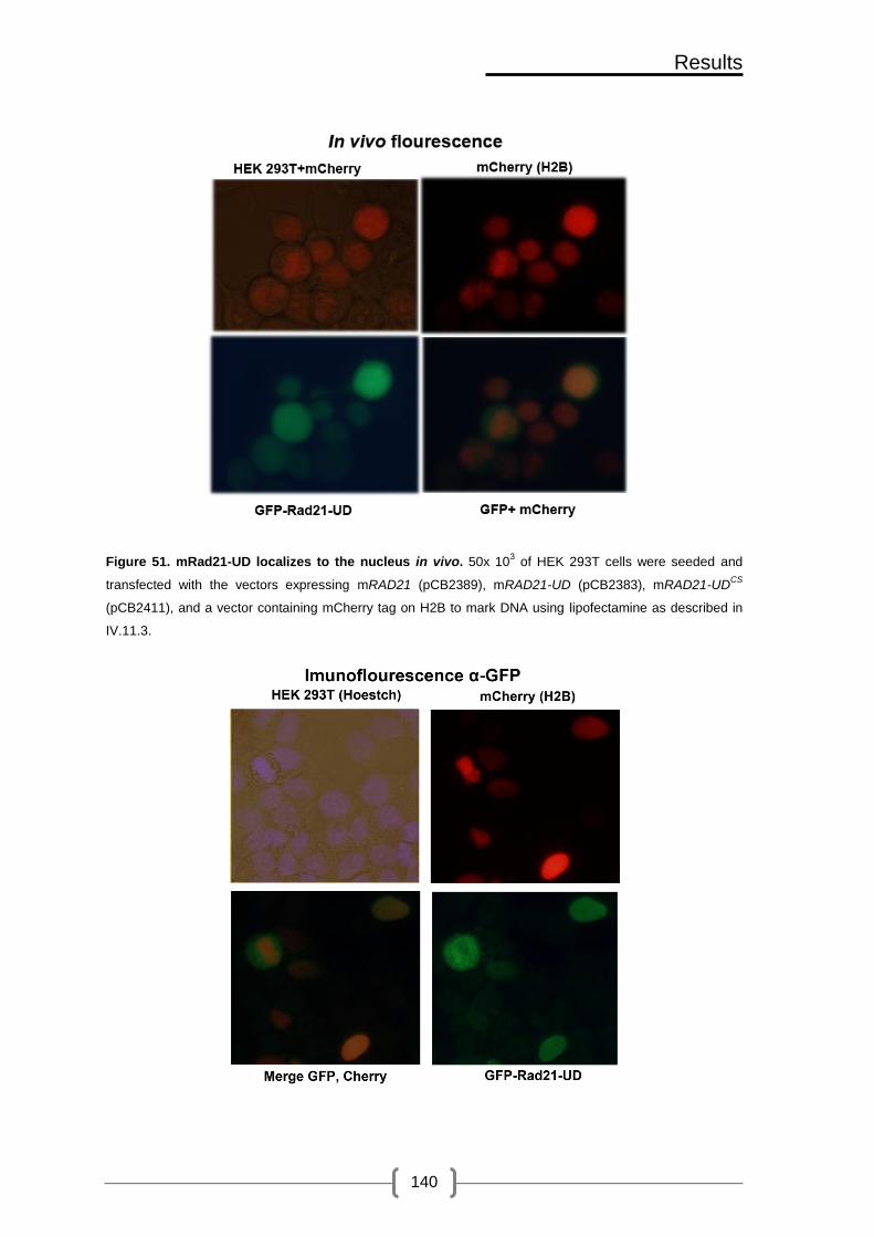
Results
140
Figure 51. mRad21-UD localizes to the nucleus in vivo. 50x 103 of HEK 293T cells were seeded and
transfected with the vectors expressing mRAD21 (pCB2389), mRAD21-UD (pCB2383), mRAD21-UDCS
(pCB2411), and a vector containing mCherry tag on H2B to mark DNA using lipofectamine as described in
IV.11.3.

Results
141
Figure 52. mRad21-UD fusion localizes to the nucleus (Immunofluorescence). 50x 103 of HEK 293T
cells were seeded and transfected with the vectors expressing mRAD21 (pCB2389), mRAD21-UD
(pCB2383), mRAD21-UDCS
(pCB2411), and a vector containing mCherry tag on H2B to mark DNA using
lipofectamine. Cells were washed fixed with 4% paraformaldehyde and processed for immunofluorescence,
as described in section IV.11.4, using anti GFP antibody. Finally, cells were mounted with a solution
containing Hoestch and slow fade.
V.2.3. Cells that overexpress mRAD21-UD accumulate in G2/M
In an attempt to see whether HEK293Tcells expressing mRad21-UD have cell
cycle progression problems, as seen in budding yeast, we collected cells that were
previously transfected with empty vector (GFP-NLS) or with vectors carrying wild-type
Rad21 or Rad21-UD, and we fixed them with ethanol. Then, we processed the samples
for FACS analysis.
As seen in Figure 53, FACS profiles of all fixed cells look similar irrespective to
the presence of mRad21-UD or wild-type mRad21. We noticed that a small population of
cells, presumably the ones expressing the highest levels of the fusions, retains the GFP
fluorescence even after fixation and processing. Gating cells that have retained the GFP
signal, shows that expression of mRad21-UD seem to accumulate in G2/M, as evident
by an increase in the percentage of 4N cells (11.52%) compared to the percentage of 4N
cells that express wild-type mRad21 (6.63%) or cells that express empty vector (6.58%).
These findings suggests that, similar to what we have seen in budding yeast, human
cells that lack cohesin sumoylation might have defects in SCC, what could potentially
lead to activation of the spindle assembly checkpoint and G2/M arrest.

Results
142
Figure 53. Cells that overexpress mRAD21-UD accumulate in G2/M. 50x 104 cells were seeded and
transfected, using PEI, with the vectors expressing mRAD21 (pCB2389), mRAD21-UD (pCB2383), or GFP-
NLS. Next, cells were harvested by trypsinization, and processed for FACS as described in section IV.11.7.
The efficiency of transformation was around 90%. However, GFP signal decreased drastically upon fixation
with ethanol. Note that there is no difference in FACS profile between cells expressing the different vectors
when all cells were included in the analysis. However, by gating cells that have retained the GFP signal we
could see that, while cells that express empty vector (GFP-NLS) or wild-type RAD21 have no problem in
getting to the next cell cycle, cells that express RAD21-UD seem to accumulate in the G2/M transition, as
evident by an increase in the percentage of cells in G2. Thus, indicating that these cells might have problems
in progressing to the next cell cycle as seen in budding yeast.

143
Discussion

144

Discussion
145
VI. Discussion
During DNA replication co-entrapment of sister chromatids by the cohesin
complex is required for the biorientation of chromosomes in the mitotic spindle, which is
essential for correct chromosome segregation. Defects in Sister Chromatid Cohesion
(SCC) have serious consequences which can range from chromosomal translocations
and cancer to cell death. This powerful multi-subunit molecule is now known to be
required for other vital functions, such as holding sister chromatids during DNA
damage repair, and regulating gene expression by stabilizing chromatin loops during
gene expression. But how can one molecule do so many functions? Several proteins
as well as post-translational modifications regulate the different functions of cohesin
throughout the cell cycle. In this study, we describe sumoylation as another post-
translational modification required for SCC.
VI.1. Cohesin sumoylation is required for establishment of SCC
In this work we have set up a novel approach to alter the sumoylation levels of
the cohesin complex. This method is based on fusion of the C-terminal of Scc1 subunit
of the cohesin complex to the SUMO conjugating enzyme or deconjugating domain.
Ulp1 is a 72 KDa protein, but only the last 200 amino acids of the protein code for a
fully functional Ulp domain (UD), which contains the catalytic residue C580 and other
residues that are responsible for SUMO binding (F474) (Mossessova, Lima 2000).
Thus, we fused the C-terminus of SCC1 to the UD of ULP1, and we engineered the
fusion to be connected by a 3xHA tag, as a linker to allow the physical separation and
proper folding of the two proteins. We chose Ulp1 because it has been shown to have
a greater activity than Ulp2 in vitro (Mukhopadhyay, Dasso 2007). To make sure that
the phenotypes that we see are due to the peptidase activity and not because the
fusion itself is affecting Scc1 folding or interaction with other cohesin subunits, we
thought that a good control for this fusion would be to inactivate the peptidase domain

Discussion
146
by point mutation of its catalytic site (C580S) (Mossessova, Lima 2000). Later on, we
saw that by introducing another mutation in the UDCS, which disables binding to SUMO
(F474A) (Mossessova, Lima 2000), we even obtain a better control for this fusion. On
the other hand, the Ubc9 protein is around 18 KDa. Thus, the entire cDNA of UBC9
was fused to the C-terminus of SCC1 using the 3xHA tag as a linker.
Since we did not know whether these fusions are lethal, we started by
overexpressing them from the GAL promoter in wild-type cells. Overexpression of a
Scc1-Ubc9 fusion shows no growth defects in a wild-type background, and
complements the thermosensitive phenotype of mcd1-1 and scc1-73 mutant cells, what
indicates that up-regulation of cohesin sumoylation does not affect viability (Figure 29A,
Figure 30A). On the other hand, overexpression of an Scc1-UD fusion in wild-type cells
leads to decreased growth rate, what suggests a possible toxic effect of depleting
SUMO from cohesin rings (Figure 27). The toxicity is aggravated by overexpression in
a scc1-73 thermosensitive background, and cells become inviable even at permissive
temperatures (Figure 30A). It has been shown that over-expression of a UD is toxic in
budding yeast (Li, Hochstrasser 2003). However, the lethality of Scc1-UD over-
expression cannot be simply due to increased nuclear levels of the Ulp1 domain
because (i) a similarly expressed Smc5-UD fusion is not toxic, but able to rescue
growth of a thermosensitive SMC5 allele (Figure 29B); and (ii) Scc1-UD over-
expression is not lethal in wild type cells. Moreover, expression of the Scc1-UD
chimera from either the GAL or SCC1 promoter does not change global sumoylation
levels, indicating that the lethality is most probably due to a direct effect on cohesin
sumoylation (Figure 38). However, we cannot currently discard that the desumoylation
of other nearby protein/s is causing this lethality.
SUMO pull-down experiments in budding yeast showed that all subunits of the
cohesin complex are sumoylated in vivo (Figure 17). The Scc1-UD fusion, whether
expressed from the strong GAL promoter or from the SCC1 promoter, lowers

Discussion
147
sumoylation of all the cohesin subunits tested (Figure 25, Figure 26). This effect is
remarkable, since it suggests that the chimeras can efficiently interact with the rest of
the cohesin subunits. Moreover, our results indicate that our approach should be
suitable to down-regulate sumoylation of other complexes/pathways. The decrease in
cohesin sumoylation is due to the peptidase activity of Ulp1, since its inactivation
restored wild-type levels of cohesin sumoylation (Scc1-UDFA,CS) (Figure 26). Taken
together these results suggest that the lethality seen in strains expressing Scc1-UD is
due to decreased cohesin sumoylation, which means that cohesin sumoylation is
required for viability.
Cells that can only assemble cohesin rings using Scc1-UD start to have
cohesion defects during DNA replication and accumulate as mononucleated cells with
large buds and separated dots or with stretched nucleus in the neck, transiently
arresting in G2/M as seen by FACS analysis and fluorescence microscopy (Figure 32).
These observations suggest that the presence of unsumoylated cohesin during S
phase might eventually lead to activation of the spindle checkpoint and G2/M arrest,
similarly to cells that lack proper cohesin function (Stern, Murray 2001). A functional
cohesin complex is required to resist pulling by microtubules, what leads to
biorientation of sister chromatids in the metaphase plate. To check metaphase
cohesion, scc1-73 cells were pre-arrested in G1 and released into a metaphase block
after induction of the Scc1-UD chimeras (Figure 41A, Figure 14), and SCC was
measured by evaluating the levels of separated fluorescent chromosome tags inserted
next to centromere 5, centromere 4 or the HMR locus (Figure 41, Figure 42).
Expression of the SCC1-UD does not rescue the SCC defects of scc1-73 mutant cells.
This phenotype is dependent on UD binding to and deconjugating SUMO, since its
inactivation (Scc1-UDFA,CS) restored wild-type levels of SCC. The fact that Scc1-UD
fusions have a specific defect in SCC strongly suggests that its effect on viability is
direct through inactivation of the cohesin complex itself. Although this is the most likely

Discussion
148
explanation, we cannot formally exclude the possibility that desumoylation of other
nearby factors, together with cohesin itself, is the cause of the inviability. The
metaphase SCC failures could arise from defects during establishment or afterwards,
mainly due to the inability to maintain SCC after co-entrapment of sister DNAs. A main
handicap with our UD-fusion approach is that we cannot distinguish between cohesion
establishment and maintenance, because we cannot deplete SUMO from cohesin rings
at a later time point, after establishment. However, we have observed that cohesin
sumoylation takes place during S phase (Figure 19), after its loading onto chromatin
and in a process that requires binding of ATP to the SMC heads, and to a lesser
extent, its hydrolysis (Figure 23). These molecular requirements resemble those of
acetylation, suggesting that sumoylation is required at the time of SCC establishment.
The fundamental regulatory mechanisms of cohesin loading and establishment
of SCC are conserved from yeast to humans (Nasmyth, Haering 2009). Therefore, it is
probable that the SUMO-mediated step for SCC is conserved in humans too. Crucially,
our preliminary results with human cell lines point in this direction. Following the same
approach used in budding yeast, we fused the human SUMO peptidase domain of
SENP1 to the C-terminus of murine SCC1 using the 4HA epitope as a linker. We chose
SENP1 because it has no specific substrate preference for the different SUMO proteins
(SUMO1-4) (Shen, Tatham et al. 2006). In addition, mRad21 was chosen because its
sequence is very similar to human Rad21, but different enough to be resistant to
human-specific siRNA (Guillou, Ibarra et al. 2010). Cells that express the highest levels
of GFP-mRad21-UD partially accumulate in G2/M, suggesting that fusion of cohesin to
a SUMO-peptidase domain might negatively affect cohesin function, as also occurs in
yeast (Figure 53). In this experiment, cells were fixed with ethanol, and only those that
still displayed GFP signal after processing (presumably the ones with the highest
degree of Rad21 over-expression) showed a partial cell cycle arrest phenotype. It is
worth keeping in mind that the endogenous Rad21 was also present in these cells,

Discussion
149
what might explain why the G2/M arrest is not as obvious as that observed in yeast.
This experiment should be repeated including the SENP1CS,BD fusion and using in vivo
FACS analysis instead of ethanol-fixed cells, a process that decreases the GFP signal.
In addition, the competition with the endogenous Rad21 should be removed by
depletion with specific siRNAs. Finally, metaphase spreads can be used to evaluate
the functionality of unsumoylated cohesin during replicative cohesion establishment.
Co-immunoprecipitation experiments indicate that Scc1-UD fusion protein is
able to interact with Smc3 (Figure 33B). Scc1 first binds to Smc1, before interacting
with Smc3, what suggests that the fusions also interact with Smc1 and thus, they do
not interfere with cohesin ring formation. In addition, interaction of Scc3 and Pds5 with
Scc1 is not affected either (Figure 33C,D). This also suggests that sumoylation is not
required for cohesin ring formation or interaction with non-SMC subunits. However, the
opposite is not true, i.e. cohesin ring formation is required for its sumoylation, as Smc1
is not sumoylated when Scc1 is absent (Figure 49), probably because these complexes
are not integer and are never loaded onto chromatin, a prerequisite for cohesin
sumoylation. Analogously, RAD21-UD fusions overexpressed from the CMV promoter
in HEK293T cells interact with the same efficiency as wild-type Rad21 with SMC
subunits of the cohesin complex in human cell lines (Figure 50), what suggests that
cohesin sumoylation is not required for ring formation in humans. We have been
unable to detect sumoylation of human cohesin subunits, as has also been described
in (Wu, Kong et al. 2012). In fact, Scc1 sumoylation has only been detected after
overexpression of SUMO and the Mms21 E3 ligase (Wu, Kong et al. 2012), and we still
do not know whether other cohesin subunits are also sumoylated, as occurs in yeast.
To our surprise, a band of slower mobility was detected above mRad21-UDCS (Figure
50). We suspect that this band corresponds to SUMO-conjugated Rad21 since
inactivation of the peptidase domain in the budding yeast Scc1-UD fusion did not only
restore wild-type levels of cohesin sumoylation, but also led to its hypersumoylation;

Discussion
150
this effect is probably due to hindering the access of other SUMO peptidases, thus
effectively up-regulating sumoylation levels. To confirm this hypothesis in the future, we
should prevent binding of SENP1 to SUMO by combining the SUMO binding-deficient
(BD) and inactive peptidase (CS) mutations. Future pull-down experiments should also
confirm whether other human cohesin subunits are sumoylated and if mRad21-UD
fusion can deplete sumoylation from cohesin rings.
One possible explanation for the Scc1-UD phenotype is that down-regulation of
cohesin sumoylation might impair its binding to chromatin, or relocation at known
cohesin binding sites. In order to explore this possibility, we used chromatin
fractionation in budding yeast to separate Triton X-100 soluble supernatant and
chromatin pellet fractions. Our results indicated that there is no difference in chromatin
binding between the functional Scc1-UDFA,CS chimera and the unsumoylated Scc1-UD
fusion protein (Figure 34). Cohesin ChIP-on-CHIP experiments confirmed this result,
and further showed that unsumoylated cohesin binds to centromeric and
pericentromeric regions, origins of replication, and regions of converging transcription,
which have already been described as main cohesin binding sites (Lengronne, Katou
et al. 2004). In addition, UD-fused Scc1 is depleted from sites of divergent
transcription, similar to what has been described for wild-type cohesin (Figure 35)
(Lengronne, Katou et al. 2004). These findings suggest that, although they are not
functional for SCC, unsumoylated cohesin rings can be pushed by the transcription
machinery, and should therefore interact normally with chromatin. In human cells, the
GFP-mRad21-UD chimera localizes properly to the nucleus (Figure 51, Figure 52).
However, we could not detect its mitotic chromosome localization neither for the wild-
type nor the mRad21-UD chimeras. In fact, metaphase chromosomes showed
exclusion of cohesin staining. It is possible that very little cohesin remains bound to
centromeres at metaphase, making it extremely difficult to detect. Further experiments

Discussion
151
will be required to test its binding to metaphase chromosomes and to determine any
alterations in its binding sites.
As stated above, we have contemplated the possibility that the Scc1-UD fusion
might affect sumoylation of other nearby proteins with potential roles in SCC. As
controls, we have studied condensin and PCNA (Moldovan, Pfander et al. 2006,
Hoege, Pfander et al. 2002). The Ycs4 subunit of the condensin complex is
sumoylated, and it has been proposed that this modification is required for proper
localization to and segregation of the rDNA locus (D'Amours, Stegmeier et al. 2004).
We have observed that sumoylation of condensin is not affected in the presence of
either Scc1-UD or Scc1-UDFA,CS (Figure 39). In contrast, our results also show that the
levels of PCNA sumoylation are slightly decreased in Scc1-UD expressing cells (Figure
40). These results suggest that in some cases, the Scc1-UD chimeras might affect
sumoylation of other proteins/complexes, potentially interfering with other cellular
functions. Siz1-dependent sumoylation of PCNA triggers its monoubiquitylation by the
Rad18 ubiquitin ligase to promote damage bypass. rad18∆ cells are highly sensitive to
MMS due to accumulation of recombination structures and fork collapse (Parker, Ulrich
2012), and this sensitivity can be alleviated by preventing PCNA sumoylation in a siz1∆
mutant. In contrast, expression of Scc1-UD has no effect on the MMS sensitivity of
rad18∆ cells (Figure 46), indicating that Scc1-UD might slightly affect sumoylation of
PCNA without affecting its SUMO-dependent functions.
VI.2. Sumoylation target/s for Sister Chromatid Cohesion
Our results indicate that all the cohesin subunits tested in this study are
sumoylated. Which is then the relevant target for SCC? Or is sumoylation of different
cohesin subunits redundant for its function in SCC?
Two different reports that analyze the role of cohesin sumoylation have been
recently published. These studies are based on the identification and mutation of
Discussion
152
sumoylatable lysine residues in the Scc1 protein. The conclusion from both papers is
that sumoylation of Scc1 is required for DNA damage-induced cohesion in budding
yeast and human cells (Wu, Kong et al. 2012, McAleenan, Cordon-Preciado et al.
2012), what looks strikingly similar to our results using the UD-fusion approach.
Therefore, Scc1 appears to be the most relevant SUMO-target in the cohesin complex
for cohesion-mediated DNA repair, and this role has been probably conserved during
evolution. Scc1 is also the main acetylation target during DNA repair, at least in
budding yeast (Heidinger-Pauli, Unal et al. 2009). It is therefore possible that Scc1
functions as a central element in response to DNA damage, integrating different signals
through various post-translational modifications. However, Scc1 might not be the main
SUMO target for SCC during an unperturbed cell cycle. Interestingly, expression of the
SUMO-impaired scc111KR allele from its own promoter partially complements the
thermosensitivity of scc1-73 cells (McAleenan, Cordon-Preciado et al. 2012). In
accordance with these reports, we have seen that expression of the scc111KR partially
rescues the inviability of auxin-sensitive Scc1-aid cells (Figure 48). Moreover, cells that
ectopically overexpress scc111KR show no defects in SCC in the absence of DNA
damage, while some defects in SCC are detected when expressed from the SCC1
promoter (McAleenan, Cordon-Preciado et al. 2012). In addition, we have seen that
sumoylation of Scc111KR is low in the presence of the endogenous copy of Scc1, yet
some sumoylation was detected when the endogenous copy was depleted using the
degron system (Figure 49A). This observation suggests that the 11KR allele might be
recessive and might not be able to compete with wild-type Scc1 for tripartite ring
formation. The low levels of sumoylation might explain why Scc111KR can partially
rescue the inviability of auxin sensitive Scc1-aid cells. It would be interesting to know if
the same lysine residues are used during a normal S phase and in response to DNA
damage, in order to finally rule out an essential role for Scc1 sumoylation during S
phase. Cells relying only on the scc111KR allele display growth defects (Figure 48).
Currently, it is difficult to ascertain whether the growth defects are due to lower

Discussion
153
sumoylation or to alteration of the structural properties of the Scc1 protein by mutation
of 11 lysine residues.
We have also observed that down-regulation of Scc1 sumoylation in cycling
cells, by expression of the scc111KR allele, decreases sumoylation of other cohesin
subunits (Figure 49B), what suggests that the cohesin complex might be sumoylated in
a sequential manner with Scc1 sumoylation leading to sumoylation of the rest of the
complex. It is also possible that sumoylation of any other subunit might also potentiate
sumoylation of the rest of the complex by attracting more SUMO proteins through
SUMO-interacting motifs (SIMs) present in covalently bound SUMO. However, we
cannot exclude the possibility that the decrease in Smc1 sumoylation levels in scc111KR
are secondary to alterations in Scc1 that do not involve sumoylation, like other post-
translational modifications, protein folding and interactions with other proteins.
Previous reports have described an opposite role for sumoylation during mitosis
for SCC maintenance and cohesin release. ULP2 (SMT4) SUMO peptidase is required
for maintenance of SCC when cells are arrested for prolonged periods in metaphase
(Bylebyl, Belichenko et al. 2003). Two mitotic targets that are involved in SCC have
been described for Ulp2, Pds5 and Top2 (Bachant, Alcasabas et al. 2002, Stead,
Aguilar et al. 2003). Sumoylation enhances Top2 function in centromeric resolution, a
function that could enhance cohesion loss at these loci (Bachant, Alcasabas et al.
2002). SUMO has also been proposed to negatively regulate the Pds5 subunit of the
cohesin complex (Stead, Aguilar et al. 2003). Sumoylation of Pds5 starts during DNA
replication and seems to maintained high until the anaphase onset. Based on genetic
evidence, it has been proposed that Pds5 sumoylation would be detrimental for SCC:
pds5 thermosensitive mutants suffer from precocious loss of SCC and arrest in G2/M,
although they are able to establish SCC during DNA replication; and overexpression of
SMT4 (ULP2) suppresses this phenotype. Pds5 sumoylation has been therefore
proposed to disrupt the interaction between Pds5 and cohesin, leading to cohesin
Discussion
154
destabilization and release (Stead, Aguilar et al. 2003). However, one should take into
account that Pds5 is a subunit that has apparent antagonistic roles in cohesin function.
On one hand, it forms part of the antiestablishment complex that inhibits co-entrapment
of sister chromatids during DNA replication (Rowland, Roig et al. 2009). On the other
hand, it is required for maintenance of SCC after DNA replication (Panizza, Tanaka et
al. 2000). This duality might be easily explained by a putative role of Pds5 in preventing
ring opening, a function that should be inhibited during establishment and co-
entrapment of sister chromatids, and shortly afterwards reactivated to prevent DNA
release. Similarly, sumoylation might promote cohesion through mechanisms that
involve transient opening of the ring during the process of cohesion establishment at
the replication fork. In this scenario, Pds5 sumoylation could aid in opening the ring at
the time of cohesion establishment.
In support for this hypothesis, it is interesting to note that both Pds5 and SUMO
are not essential in the fission yeast Schizoaccharomyces pombe. Although
speculative, these observations suggest that the function of Pds5 and SUMO are
intimately linked (Wang, Read et al. 2002, Tanaka, Nishide et al. 1999). This also
suggests that Pds5 might be the main SUMO target during an unperturbed cell cycle.
In fact, in our hands, Pds5 sumoylation seems to be the highest among the rest of the
subunits of cohesin (Figure 17). However, how this regulation is important for
establishment of sister chromatid cohesion, and how it is coordinated with acetylation
dependent closure of the ring are two interesting issues to be addressed in the future.
One point that should be taken into consideration is that although Pds5 is normally
bound close to the ATPase domains of Smc1 and Smc3, FRET studies have shown
that Pds5 might also interact with their hinge domain (Mc Intyre, Muller et al. 2007).
One can imagine that this interaction could be promoted by sumoylation and might
stabilize foldback structures potentially involved during sister chromatid co-entrapment
(Gruber, Arumugam et al. 2006).

Discussion
155
Our results also suggest that a proper sumoylation-desumoylation cycle of
cohesin might be important for its function. Fusion of Scc1 to an inactive peptidase
domain (UDC580S) up-regulates cohesin sumoylation (Figure 25). This up-regulation
most likely reflects the binding of the inactive domain to cohesin-SUMO conjugates,
and the consequent block in deconjugation by the endogenous Ulp peptidases. The
latter is supported by the finding that sumoylation of cohesin (Figure 26), viability
(Figure 30B) and SCC defects (Figure 41, Figure 42), are all restored to wild-type
levels when the F474A mutation is introduced in the inactive UDCS domain (Scc1-
UDFA,CS). Since a substantial loss of SCC occurs when the Scc1-UDCS fusion is
expressed in scc1-73 cells (Figure 41B), we estimate that the failure to desumoylate
cohesin is detrimental for SCC. In accordance, ulp2 mutants also show SCC defects
(Mossessova, Lima 2000), (Bylebyl, Belichenko et al. 2003). Altogether, these
observations suggest that desumoylation might be required to fine-tune the function of
cohesin, a scenario that would nicely fit with cohesin sumoylation promoting the
opening of the cohesin ring. It is worth noting that the levels of sumoylation are
quantitatively similar in Scc1-UDCS and Scc1-Ubc9 expressing cells. However, and
assuming a higher rate of deconjugation than sumoylation, a fundamental difference
arises between the two fusions: while the half-life of SUMO conjugates is probably very
short in the Ubc9 fusion, it might substantially increase when fused to the inactive
peptidase.
VI.3. Molecular determinants of cohesin sumoylation
Nse2 dependent sumoylation of Smc1 and Smc3 has been previously detected
in nocodazole arrested S. cerevisiae cells but not in cycling cells (Takahashi, Dulev et
al. 2008). In addition, sumoylation of human Scc1 has been shown to be dependent on
Nse2 SUMO ligase in vitro (Potts, Yu 2005). We have seen that sumoylation of Scc1
partially depends on Nse2 in cycling cells. On the other hand, sumoylation of both
Smc1 and Smc3 depends largely on Nse2 (Figure 20). Recently, two concurrent

Discussion
156
studies identified key lysine residues that contribute to most of Scc1 sumoylation in
budding yeast and in humans during DNA damage also in an Nse2 dependent manner
(Wu, Kong et al. 2012, McAleenan, Cordon-Preciado et al. 2012), what suggests that
Nse2 might have different targets during DNA damage and under unperturbed cell
cycle conditions. Interestingly, we have seen that Nse2-dependent sumoylation of
cohesin is much lower when Nse2 is not able to bind to Smc5, which suggests that
Nse2 might use Smc5/6 to reach its targets (Figure 21). If this is the case, when
Smc5/6 complex function is compromised, sumoylation of cohesin should also be
lowered. Indeed, we have seen that in nse3-2 thermosensitive cells sumoylation of
Scc1 is decreased, which confirms our hypothesis (Figure 22). Down-regulation of
Nse2 function disrupts DNA damage-induced cohesion in both budding yeast and
human cells (Andrews, Palecek et al. 2005, Zhao, Blobel 2005). However, we have not
observed SCC defects at the HMR locus in smc5/6 or nse2∆c mutants under
unperturbed conditions. More strikingly, the little cohesion defects we have detected in
smc6-9 cells seems to be cohesin-independent, a phenomenon for which we currently
do not have any explanation (Figure 42C,D,E). It is possible that DNA damage induced
cohesion is more sensitive to cohesin sumoylation levels than normal SCC. In support
for this hypothesis, it has been proposed that the amount of cohesin required for DNA
repair is much larger than the amount required for normal SCC (Heidinger-Pauli, Mert
et al. 2010).
Cohesin loading onto chromatin is required for its sumoylation (Figure 23).
Sumoylation of other proteins such as PCNA also requires their residence on
chromatin (Parker, Ulrich 2012). F584R mutation within the Smc1 hinge domain is
lethal because it impairs heterodimerization with Smc3 hinge and loading onto
chromatin (Mishra, Hu et al. 2010). Smc1 in this case is not sumoylated, suggesting
that not only loading but also heterodimerization of Smc proteins is required for cohesin
sumoylation. Cohesin sumoylation seems to occur in a time space between ATP

Discussion
157
binding and hydrolysis. Smc1 mutants that lack ATP binding and interaction with Scc1
(smc1K39I) are not sumoylated. ATP binding and hydrolysis are required for cohesin
loading to chromatin and relocation away from initial loading sites (Arumugam, Gruber
et al. 2003). This indicates that Scc1-Smc1 interaction and ATP binding are both
required for cohesin sumoylation. ATPase head domains of cohesin rings that are
formed around smc1E1158Q, are locked in an engaged conformation because they
cannot hydrolyze ATP and are not able to move away from initial loading sites. This
mutation also causes unstable recruitment of the cohesin complex to chromatin (Hu,
Itoh et al. 2011) and is inviable. Interestingly, Smc1E1158Q is sumoylated although
sumoylation levels are decreased with respect to wild-type Smc1. This suggests that
cohesin sumoylation somehow depends on its relocalization away from initial Scc2/4
loading sites and on its stable recruitment to chromatin.
During DNA replication, establishment of SCC requires Eco1/Ctf7 dependent
acetylation of two lysine residues (K112 and K113) within Smc3 head domain (Rolef
Ben-Shahar, Heeger et al. 2008). Certain mutations in Scc3, Pds5, and Rad61 reverse
the lethality of eco1∆ and have lead to the proposal of the antiestablishment complex,
which is composed by these three proteins. Upon Eco1 dependent acetylation of Smc3
cohesin rings become cohesive (tightly shut), which counteracts the antiestablishment
activity (Rowland, Roig et al. 2009). Scc1 sumoylation levels are not affected by the
presence of the eco1-1 mutation, neither at the permissive (23°C) nor after shift to the
restrictive (37°C) temperatures. Sumoylation of the Smc3 subunit is also not affected
by inactivation of Eco1, indicating that sumoylation is required before, or in parallel to,
acetylation, for the establishment of SCC (Figure 47). To test the acetylation state of
Smc3 in cells that are impaired in cohesin sumoylation, we used anti-acetyl lysine
antibodies. Smc3 is efficiently acetylated in a ubc9-1 thermosensitive background, both
at the permissive and restrictive temperatures (Figure 43A). Furthermore, cells that
express the Scc1 or Scc1-UD as the only copy of Scc1 in the cell do not show
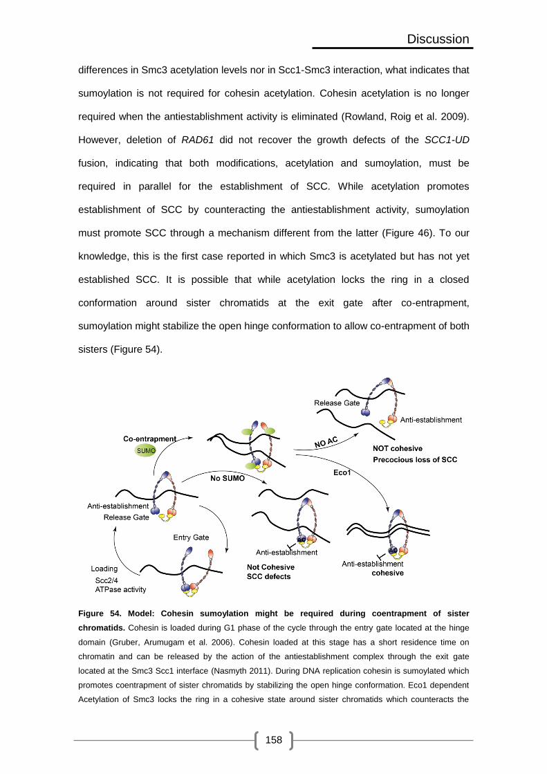
Discussion
158
differences in Smc3 acetylation levels nor in Scc1-Smc3 interaction, what indicates that
sumoylation is not required for cohesin acetylation. Cohesin acetylation is no longer
required when the antiestablishment activity is eliminated (Rowland, Roig et al. 2009).
However, deletion of RAD61 did not recover the growth defects of the SCC1-UD
fusion, indicating that both modifications, acetylation and sumoylation, must be
required in parallel for the establishment of SCC. While acetylation promotes
establishment of SCC by counteracting the antiestablishment activity, sumoylation
must promote SCC through a mechanism different from the latter (Figure 46). To our
knowledge, this is the first case reported in which Smc3 is acetylated but has not yet
established SCC. It is possible that while acetylation locks the ring in a closed
conformation around sister chromatids at the exit gate after co-entrapment,
sumoylation might stabilize the open hinge conformation to allow co-entrapment of both
sisters (Figure 54).
Figure 54. Model: Cohesin sumoylation might be required during coentrapment of sister
chromatids. Cohesin is loaded during G1 phase of the cycle through the entry gate located at the hinge
domain (Gruber, Arumugam et al. 2006). Cohesin loaded at this stage has a short residence time on
chromatin and can be released by the action of the antiestablishment complex through the exit gate
located at the Smc3 Scc1 interface (Nasmyth 2011). During DNA replication cohesin is sumoylated which
promotes coentrapment of sister chromatids by stabilizing the open hinge conformation. Eco1 dependent
Acetylation of Smc3 locks the ring in a cohesive state around sister chromatids which counteracts the

Discussion
159
antiestablishment activity and allows establishment of sister chromatid cohesion. In the absence of
acetylation the antiestablishment activity releases cohesin from sister chromatids leading to precocious
loss of SCC. On the other hand, in the absence of sumoylation cohesin rings are efficiently acetylated but
they are not able to establish SCC. This is probably because they are locked in a closed conformation
around one sister chromatid and they are not able to embrace both sisters.
VI.4. Cohesin sumoylation is required for DI cohesion in yeast and in
humans
DSBs are one of the most dangerous forms of DNA damage because they
might lead to chromosome translocations and cancer. Cells repair DSBs by HR when
CDK activity is high, and when an undamaged DNA template is available, which can be
either the sister chromatid or the homologous chromosome. However, DSB repair with
HR between sister chromatids (SCR) is preferable during the mitotic cell cycle because
HR between homologues can lead to loss of heterozygosity (LOH) (Branzei, Foiani
2008). Cohesin promotes SCR during DSB repair by bringing the two sister chromatids
in close proximity (Heidinger-Pauli, Unal et al. 2008, Strom, Karlsson et al. 2007).
Different cohesin subunits are further sumoylated by various DNA damaging agents in
budding yeast (Figure 18) (McAleenan, Cordon-Preciado et al. 2012). In contrast, it
does not seem to be up-regulated by DNA damage in human cells (Wu, Kong et al.
2012).
Although we have seen that cohesin sumoylation promotes SCC through a
mechanism different from counteracting the antiestablishment activity in yeast, we still
do not know whether this is also the case in response to DNA damage. In accordance
with our results, the absence of Scc1 phosphorylation by the Chk1 checkpoint kinase,
which promotes acetylation, does not affect cohesin sumoylation. In fact, these results
suggest that acetylation might be a downstream event (McAleenan, Cordon-Preciado
et al. 2012). In striking contrast, sumoylation of human Scc1 has been proposed to
counteract the antiestablishment activity of Wapl (Rad61 homolog) in response to
DSBs, thereby enabling SCC and sister chromatid exchange (Wu, Kong et al. 2012). In

Discussion
160
spite of the apparent differences, the mechanism of SUMO-mediated cohesion might
not be so different, as it also does not seem to depend on cohesin acetylation in human
cells.
Cohesin sumoylation does not seem to be required for loading of cohesin to
DSBs; rather it is necessary for transforming unstably bound cohesin complexes to
cohesive ones to promote sister chromatid recombination (Wu, Kong et al. 2012,
McAleenan, Cordon-Preciado et al. 2012). In accordance with these observations, we
have seen by ChIP-qPCR that Scc1-UD is efficiently loaded in the vicinity of DSBs in
budding yeast (Figure 37). However, it seems to be more enriched than Scc1 wild type
or Scc1-Ubc9. These results suggest that unsumoylated cohesin is recruited more
efficiently to damaged sites or is more resistant to removal from chromatin. If the latter
case was correct, it could be speculated that sumoylation might be required for opening
of pre-loaded cohesin rings, maybe to allow the capture and co-entrapment of the
sister chromatid.
Cohesin is not only required for homologous-recombination DSB-repair, but
also for replication fork stability and the recovery of stalled replication forks (Tittel-
Elmer, Lengronne et al. 2012). Replication forks can stall due to natural replication fork
barriers or slow replication zones (replicative stress) (Branzei, Foiani 2005). Replicative
stress can be also provoked externally by adding hydroxyurea (HU) (Slater 1973).
Interestingly, we have seen that wild-type cells that overexpress Scc1-UD are sensitive
to hydroxyurea (Figure 28), which suggests that cohesin sumoylation might also be
required during replicative stress. In the future, it would be interesting to test replication
fork progression in the absence of cohesin sumoylation by DNA combing.
Discussion
161
VI.5. How might sumoylation promote co-entrapment of sister
chromatids
Since all subunits are conjugated to SUMO, sumoylation of different subunits of
the cohesin complex might be redundant during unperturbed cell cycle, and might be
required as a sum to stabilize certain interactions between the same complex, with
other cohesin complexes, or with other proteins involved in SCC. In accordance with
this hypothesis, it has been recently shown that sumoylation of many proteins involved
in the DNA damage repair is up-regulated in response to DNA damage. Individual
sumoylation seems to stabilize physical interactions between these proteins adding up
for efficient repair (Psakhye, Jentsch 2012). Our results show that, in the case of
cohesin, sumoylation is not required for interactions between cohesin subunits within
the same complex, since we were able to detect such interactions in the absence of
sumoylation (Figure 33).
Thus, sumoylation might potentiate interaction between different cohesin
complexes in places where cohesin is extensively required, like in centromeres, and
thus, cohesin sumoylation might promote cohesion through ring stacking which could
facilitate re-capture of escaping chromatin fibers inside an adjacent ring (Figure 55).
However, interactions between different cohesin complexes have never been detected
under physiological conditions. If sumoylation is required for these interactions, then
these interactions might have been missed due to the short lived nature of sumoylation.

Discussion
162
Figure 55. Sumoylation can promote co-entrapment of sister chromatids through ring stacking.
Sumoylation might potentiate interaction between different cohesin complexes in places where cohesin is
extensively required like in centromeres, and thus, cohesin sumoylation might promote cohesion through
ring stacking which could facilitate re-capture of escaping chromatin fibers inside an adjacent ring
Moreover, cohesin sumoylation might act as a platform to recruit new cohesion
factors (Figure 56), like in the case of PCNA were sumoylation recruits Srs2 (Pfander,
Moldovan et al. 2005). Alternatively, cohesin sumoylation might mask sites important
for cohesin interaction with other proteins that would otherwise regulate SCC
negatively (Figure 56), like in the case of transcription repressors (Zheng, Yang 2004).
In the future it would be important to identify suppressors of the growth defects of
SUMO-impaired cohesin mutants to confirm such hypothesis.
Figure 56. Sumoylation can promote co-entrapment of sister chromatids by promoting/ inhibiting
interaction with other proteins. Cohesin sumoylation might act as a platform to recruit new cohesion

Discussion
163
factors. Alternatively, cohesin sumoylation might mask sites important for cohesin interaction with other
proteins that would otherwise regulate SCC negatively.
It is becoming more obvious nowadays that the structure of cohesin is far more
flexible than the typical ring structure that we have drawn in our heads. This flexibility is
highly attributed to the presence of breaks in the coiled coil probability, which allow
cohesin to adapt different conformations including the recently proposed foldback
structures (Gruber, Arumugam et al. 2006, Hu, Itoh et al. 2011). It has been proposed
that folding of cohesin rings could be required during chromatin entrapment to allow
ATPase mediated opening of the entry gate (Kurze, Michie et al. 2011). Cohesin
sumoylation might cause a conformational change in the cohesin ring structure similar
to what has been described for TDG (Hardeland, Steinacher et al. 2002)(Figure 57).
Figure 57. Sumoylation can promote co-entrapment of sister chromatids through promoting a
conformational change. Cohesin sumoylation might cause a conformational change in the cohesin ring
structure which can be stabilized by SUMO-SIM intra-complex interactions.
VI.6. Implications
Defects in cohesin establishment due to a homozygous mutation in Esco2 allele
lead to a developmental disease in humans called Roberts Syndrome (RBS) (Horsfield,
Print et al. 2012). If cohesin sumoylation is also required for establishment of SCC, one
cannot help but wonder whether defects in cohesin sumoylation might lead to similar
phenotypes in humans.

Discussion
164
In humans, cohesin is additionally required for other vital functions that also
involve chromatin coentrapment, such as holding sister chromatids during DNA
damage repair (Heidinger-Pauli, Unal et al. 2008), and stabilizing chromatin loops
during gene expression (Guillou, Ibarra et al. 2010). It would be tempting to think that
the mechanism of coentrapment is universal for all these functions. If cohesin
sumoylation has a role in co-entrapment, it might be similarly required during cohesin
dependent regulation of gene expression or during DI cohesion establishment.
Cornelia de Lange syndrome (CDLS) is a developmental disease with 60% of
the cases resulting from haploinsufficient mutations within the loading factor Nipbl, and
5-10% due to other mutations in Smc1, Smc3, Scc1, and HDAC8. Around 30% of the
cases reported are of unknown reason. Importantly, unlike cells from RBS, cells from
patients with CDLS have no apparent defects in SCC, which suggests that the
developmental defects seen in these patients are due to defects in cohesin function in
regulating gene expression (Horsfield, Print et al. 2012). If sumoylation is required for
cohesin dependent chromatin loops formation during gene expression, it is possible
that mutations in the SUMO pathway or in the mechanism of sumo-dependent
coentrapment might be one of the reasons leading to the pathology of this disease.
Defects in cohesin itself or its regulators can lead to chromosomal
translocations and cancer (Xu, Tomaszewski et al. 2011). Whether defects in
sumoylation dependent regulation of cohesin lead to pathologies such cancer is an
interesting issue that should be addressed in the future.

165
Conclusions

166

Conclusions
167
VII. Conclusions
All subunits of the cohesin complex are sumoylated in vivo under
physiological conditions in S. cerevisiae.
Cohesin sumoylation occurs during S phase and is up-regulated by DNA
damage.
Cohesin sumoylation is mostly dependent on Nse2 E3 SUMO ligase and
the Smc5/6 complex.
Cohesin sumoylation occurs at the time of cohesion establishment, after
cohesin loading and ATP binding, and independently from Eco1-mediated
cohesin acetylation.
Fusion of the Scc1 subunit to a SUMO peptidase Ulp domain (UD) is a
novel and efficient approach to specifically down-regulate sumoylation of
all cohesin subunits.
SUMO-depleted cohesin rings are properly formed and efficiently
recruited to known cohesin binding sites on chromatin.
Cells that lack cohesin sumoylation arrest in G2/M.
Cohesin sumoylation is essential for Sister Chromatid Cohesion and cell
viability.
Sumoylation does not enable SCC by counteracting Rad61-dependent
antiestablishment activity.
Human cell lines that overexpress mRad21-UD accumulate in G2/M.

168

169
Supplementary Tables

170

Supplementary Tables
171
VIII. Supplementary Tables
Supplementary Table 1. List of Antibodies used in Western blot analysis and immunofluorescence.
Primary Antibody Source Conditions Secondary
Antibody Source Conditions
3F10 Rat
Monoclonal
(α-HA)
Roche
2hours at room
temperature, or
overnight at 4°C
1:5000, 5%
TBST Milk
AP136P
Goat α-Rat
IgG horse
reddish
peroxidase
linked
Chemicon
International
1 hour at room
temperature,
1:10,000, TBST
9E10 Mouse
Monoclonal
(α-MYC)
Roche
2hours at room
temperature, or
overnight at 4°C
1:5000, 5%
TBST Milk
NXA931
Sheep
α-Mouse IgG
horse
reddish
peroxidase
linked
ECL
1 hour at room
temperature,
1:10,000, TBST
F3165 M2 Mouse
Monoclonal
(α-FLAG)
SIGMA
2hours at room
temperature, or
overnight at 4°C
1:5000, 5%
TBST Milk
NXA931
α-Mouse ECL
1 hour at room
temperature,
1:10,000, TBST
ST1027 Rabbit
Monoclonal
(α-acetylated
lysine)
Millipore
2hours at room
temperature, or
overnight at 4°C
1:5000, 0.025%
TBST Milk
NA934V
Donkey
α-Rabbit IgG
horse
reddish
peroxidase
linked
ECL
1 hour at room
temperature,
1:10,000, TBST
AT14405 Rabbit
Polyclonal
(α-SUMO)
abcam
2hours at room
temperature, or
overnight at 4°C
1:5000, 5%
TBST Milk
NA934V
α-Rabbit ECL
1 hour at room
temperature,
1:10,000, TBST

Supplementary Tables
172
A21311 Rabbit IgG
Alexa Flour 488
conjugate (α-GFP)
(IF)
Molecular
Probes
2hours at room
temperature,
1:200, PBS
A11008
Alexa Flour
488 goat α-
rabbit IgG
Molecular
Probes
1 hour at room
temperature,
1:500, BS
Ab1791 Rabbit
Polyclonal
(α-Histone H3)
abcam
2hours at room
temperature, or
overnight at 4°C
1:1000, 5%
TBST
NA934V
α-Rabbit ECL
1 hour at room
temperature,
1:10,000, TBST
H2035-01 Rabbit
Monoclonal
(α-Hexokinase)
US
Biological
2hours at room
temperature, or
overnight at 4°C
1:5000, 5%
TBST Milk
NA934V
α-Rabbit ECL
1 hour at room
temperature,
1:10,000, TBST
Ab18085 Rabbit
polyclonal
(α-rpd3)
abcam
2hours at room
temperature, or
overnight at 4°C
1:1000, 5%
TBST
NA934V
α-Rabbit ECL
1 hour at room
temperature,
1:10,000, TBST
AfRS115-4
(α-Smc1A)
Ana
Losada
2hours at room
temperature, or
overnight at 4°C
1:100, 5% TBST
Milk
NA934V
α-Rabbit ECL
1 hour at room
temperature,
1:10,000, TBST
AfR24-3
(α-Smc3A)
Ana
Losada
2hours at room
temperature, or
overnight at 4°C
1:300, 5% TBST
Milk
NA934V
α-Rabbit ECL
1 hour at room
temperature,
1:10,000, TBST
AfRS55-3
(α-Rad21)
Ana
Losada
2hours at room
temperature, or
overnight at 4°C
1:100, 5% TBST
Milk
NA934V
α-Rabbit ECL
1 hour at room
temperature,
1:10,000, TBST

Supplementary Tables
173
Supplementary Table 2. List of yeast strains used in this work.
Strain Genotype Procedure
Y759 MATa scc1-73 ade2-1 TRP+ can1-100 leu2-3,112
his3-11,15 GAL, psi
The strain 759 (W303 MATa
scc1-73) and strain 758
(BY4741 MATα mcd1-1) were
obtained from Luis Aragon’s
laboratory. Scc1 constructs that
were cloned into YIPlac211
plasmid and expressed from the
GAL promoter were cut with
StuI for integration in the URA
locus in 759 background.
YSM1498 759+ ura3:Gal-Scc1-3HA
YSM1500 759+ ura3:Gal-Scc1-3HA-UD
YSM1502 759+ ura3:Gal-Scc1-3HA-UD(C580S)
Y758 MATalpha ade2 ade3 his7 leu2 trp1-1 ura3 mcd1-1
YSM2225 759+ Smc36HA (hph)
759 was transformed with a
PCR to tag Smc3 with 6HA
using S2 S3 primers and pYM
16 (Janke, Magiera et al. 2004).
YSM1040 759+ TetR-YFP:ADE2 CEN5::tetO2x112::HIS3
Centromere V in 759 was
tagged with 112 tet operators
(1.4Kbs away), and tet repressor
fused to YFP was integrated in
the ADE2 locus to generate
strain 1040. This back ground
was transformed with Scc1
constructs expressed from the
GAL promoter and the SCC1
promoter to study SCC.
YSM1476 1040+ ura3:Gal-Scc1-3HA
YSM1478 1040+ ura3:Gal-SCC1 -3HA-UD
YSM1480 1040+ ura3:Gal-Scc1-3HA-UD(C580S)
YSM1506 1040+ ura3:Gal-Scc1-3HA-Ubc9
YSM1614 1040+ ura3:Gal-Scc1-3HA-UD(C580S, F474A)
YSM1832 1040+ ura3:SCC1 p-Scc1-3HA-UD
YSM1857 1040+ ura3:SCC1 p-Scc1-3HA-UD(C580S)
YSM1860 1040+ ura3:SCC1 p-Scc1-3HA
YSM2155 1040+ ura3:SCC1 p-Scc1-3HA-UD(C580S, F474A)

Supplementary Tables
174
Y343 MATa leu2::LEU2-tetR-GFP 1.4kb left of
CEN5::tetO2x112::HIS3
The strain 343 (W303) that has
tet operators on centromere V
and tet repressor integrated in
the LEU2 locus was obtained
from Tomoyuki Tanaka’s lab.
Y1882
Mata scc1-73 ade2-1 TRP1+ can1-100 leu2-3,
112 his3-11,15 ura3 GAL psi+ 3x backcrossed
tetR-YFP tetO 620K ChrIV HIS
WT strain Y1890 (original name
CCG8946) and scc1-73 strain
Y1882 (original name CCG
9151) were both received from
Luis Aragon’s lab. These strains
contain tet operators (HIS) that
are 620 bp away from
centromere IV and tet repressor
fused to GFP (ADE2 locus).
1882 was transformed with Scc1
constructs expressed from the
SCC1 promoter to study sister
chromatid cohesion.
Y1890 MATa ade2::ADE2 tetR-GFP, leu2 ura3 trp1 his3
his::HIS TetO ChrIV:620
YSM1952 1882+ ura3: SCC1 p-Scc1-3HA-UD
YSM1954 1882+ ura3: SCC1 p-Scc1-3HA-UD(C580S)
YSM1956 1882+ ura3: SCC1 p-Scc1-3HA-UD(C580S, F474A)
YSM1958 1882+ ura3: SCC1 p-Scc1-3HA
Y503
MATa Dbar1::hisG can1-100 his3-11,15 trp1-1
ura3-1 ∆lys2 ∆hml::loxP-(kanMX-loxP) 2
(LEU2::GAL1-R)2::leu2-3,112 RS::HMR-tRNA-
GIT1-TRP1-256lacop::RS ADE2::HIS3P-
lacGFP::ade2-1
Strains 503 (WT) and 504 (scc1-
73) (W303 MATa bar1∆) were
obtained from Marc
Gartenberg’s lab. These strains
have 256 lac operators
integrated close to the HMR
locus and the lac repressor
fused to GFP. This background
was transformed with Scc1
constructs expressed from the
GAL promoter to study SCC.
Y504
MATa Dbar1::hisG can1-100 his3-11,15 trp1-1
ura3-1 ∆lys2 scc1-73 ∆hml::loxP-(kanMX-loxP) 2
(LEU2::GAL1-R)2::leu2-3,112 RS::HMR-tRNA-
GIT1-TRP1-256lacop::RS ADE2::HIS3P-
lacGFP::ade2-1
YSM1514 504+ ura3:Gal-Scc1-3HA
YSM1516 504+ ura3:Gal-Scc1-3HA-UD
YTR559 504+ nse2∆C (hph)
YTR519 503+ smc6-9 (NAT)
YTR535 504+ smc6-9 (NAT)

Supplementary Tables
175
YTR907 MATa ade2-1 trp1D2 can1-100 leu2-3,112 his3-
11,15 ura3-52 6his-Flag-SMT3:KanMX4 WT W303 strain
YSM1486 1191+ ura3:Gal-Scc1-3HA
The strain 1191 was obtained
from transforming 907 with
Smc1-18myc PCR. This strain
was transformed later on with
Scc1 constructs expressed from
the GAL promoter and
integrated in the URA locus.
YSM1488 1191+ ura3:Gal-Scc1-3HA-UD
YSM1490 1191+ ura3:Gal-Scc1-3HA-UD(C580S)
YSM1492 1193+ ura3:Gal-Scc1-3HA
The strain 1193 was obtained
from transforming 907 with
Smc3-18MYC PCR. This strain
was transformed later on with
Scc1 constructs expressed from
the GAL promoter /SCC1
promoter and integrated in the
URA locus.
YSM1494 1193+ ura3:Gal-Scc1-3HA-UD
YSM1496 1193+ ura3:Gal-Scc1-3HA-UD(C580S)
YSM1660 1193+ ura3:Gal-Scc1-3HA-UD(F474A)
YSM1662 1193+ ura3:Gal-Scc1-3HA-UD(C580S, F474A)
YSM1864 1193+ ura3:SCC1 p-Scc1-3HA
YSM1758 1193+ ura3:SCC1 p-Scc1-3HA-UD
YSM1844 1193+ ura3:SCC1 p-Scc1-3HA-UD(C580S)
YSM1961 1193+ ura3:SCC1 p-Scc1-3HA-UD(C580S, F474A)
YSM1907
MATa ade2-1 trp1D2 can1-100 leu2-3,112 his3-
11,15 ura3-52 6his-Flag-SMT3:KanMX4 + Tir1-
9myc (His)+ Scc1-AID (hph)
The strain 1907 was obtained by
integrating Tir1-9MYC in the HIS
locus (BsiwI4) in 907 which was
then transformed with Scc1-AID
(hph) PCR using Scc1 S2/S3
primers on p1596. This strain
was transformed with
Smc1/Smc3-18MYC PCR and
then with Scc1 chimeras under
the SCC1 promoter integrated
in the URA3 locus (StuI
restriction).
YSM1963 1907+ Smc1-myc18::TRP
YSM1965 1907+ Smc3-myc9::TRP
YSM2017 1963+ ura3:SCC1 p-SCC1 -3HA
YSM2019 1963+ ura3:SCC1 p-SCC1 -3HA-UD
YSM2021 1963+ ura3:SCC1 p-SCC1 -3HA-UD(C580S)
YSM2023 1963+ ura3:SCC1 p-SCC1 -3HA-UD(C580S,
F474A)
YSM2025 1965+ ura3:SCC1 p-SCC1 -3HA
YSM2027 1965+ ura3:SCC1 p-SCC1 -3HA-UD
YSM2029 1965+ ura3:SCC1 p-SCC1 -3HA-UD(C580S)

Supplementary Tables
176
YSM2031 1965+ ura3:SCC1 p-SCC1 -3HA-UD(C580S,
F474A)
YSM2254 1907+ Scc1-3HA-CYCt:URA3:SCC1 p-Scc1-AID
(hph)
The strain 1907 was also
transformed with Scc1
constructs expressed from the
SCC1 promoter, but this time
integrated in the Scc1 locus (by
restriction digestion of the same
vectors with BsptI). Then, Smc1,
Smc3, Ycs4, Scc3, or Pds5
PCRs all tagged with the 9xmyc
epitope were transformed to
these backgrounds.
YSM2256 1907+ Scc1-3HA-UD-CYCt:URA3:SCC1 p-Scc1-
AID (hph)
YSM2258 1907+ Scc1-3HA-UD(C580S-F474A)-
CYCt:URA3:SCC1 p-Scc1-AID (hph)
YSM2297 2254+ Smc3-9myc (TRP)
YSM2299 2256+ Smc3-9myc (TRP)
YSM2301 2258+ Smc3-9myc (TRP)
YSM2319 1965+ + Scc1-3HA-UD(C580S)-CYCt:URA3:SCC1
p-Scc1-AID (hph)
YSM2456 2254+ Ycs4-9myc (NAT)
YSM2458 2256+ Ycs4-9myc (NAT)
YSM2459 2258+ Ycs4-9myc (NAT)
YSM2468 1907+ Ycs4-9myc (NAT)
YSM2549 2256+ Pds5-9myc
YSM2550 2258+ Pds5-9myc
YSM2551 2256+ Scc3-9myc
YSM2552 2258+ Scc3-9myc
YSM2553 2256+ Smc1-9myc
YSM2554 2258+ Smc1-9myc
Y355
MATa ade2-1 trp1-1 can1-100 leu2-3,112 his3-
11,15 ura3 GAL psi+ pep4::URA3 bar1::hisG Scc1-
myc18 HIS
Original name K10611.
Obtained from Frank Uhlmann’s
lab.
Y356
MATa ade2-1 trp1-1 can1-100 leu2-3,112 his3-
11,15 ura3 GAL psi+ pep4::URA3 bar1::hisG
Smc1-myc18::TRP
Original name K1300. Obtained
from Frank Uhlmann’s lab.
Y357
MATalpha ade2-1 trp1-1 can1-100 leu2-3,112 his3-
11,15 ura3 GAL psi+ pep4::URA3 bar1::hisG
Smc3-9myc (Trp)
Original name K911. Obtained
from Frank Uhlmann’s lab.
Y586 355+ 6xHIS-FLAG-SMT3

Supplementary Tables
177
Y784 357+ 6xHIS-FLAG-SMT3
Y644 MATa his3D1 leu2D0 met15D0 ura3D0 6HisFLAG-
smt3::kanMX6 Scc1-myc18 HIS
Y746 MATa his3D1 leu2D0 met15D0 ura3D0 6HisFLAG-
smt3::kanMX6 Scc1-myc18 HIS smc6-9 (NAT)
Y1992 356+ 6xHIS-FLAG-SMT3
YSM1227 907+ Scc1-6HA:hphNT1
YSM1994
MATa ade2-1 trp1D2 can1-100 leu2-3,112 his3-
11,15 ura3-52 6his-Flag-SMT3:KanMX4 smc1-
9myc(hph)
YSM1996
MATa ade2-1 trp1D2 can1-100 leu2-3,112 his3-
11,15 ura3-52 6his-Flag-SMT3:KanMX4 scc1-
9myc(hph)
YSM1418 907+ ura3:Gal-Scc1-3HA
The strain 907 was transformed
with Scc1 chimeras, which were
expressed from the GAL
promoter and integrated in the
URA locus.
YSM1472 907+ ura3:Gal-Scc1-3HA-UD
YSM1474 907+ ura3::Gal-Scc1-3HA-UD(C580S)
YSM1520 907+ ura3:Gal-Scc1-3HA-Ubc9
YSM1522 907+ ura3:Gal-Scc1-3HA-ubc9(c93s)
YSM2033
1907+ pSCC1 :SCC1
(K165,290,252,345,391,392,394,460,500,509,521R)
-3HA 1907 was transformed with the
centromeric plasmids pRS415
expressing the different
Scc1(KR) alleles under the
SCC1 promoter.
YSM2034 1907+ pSCC1 :SCC1 -3HA
YSM2035 1907+pSCC1 :SCC1 -3HA K165_290_460R
(K165,290,460R)
YSM2036 1907+pRS415
YSM1734 907+ Pds5-9myc (hyg) 907 was transformed with
Pds5/Scc3-9MYC to obtain
strains 1734 and 1736
respectively. These strains were
then transformed with the Scc1
contructs expressed from the e
YSM1736 907+ Scc3-9myc(hyg)
YSM1744 1734+ ura3:Gal-Scc1-3HA
YSM1746 1734+ ura3::Gal-Scc1-3HA-UD
YSM1748 1734+ ura3:Gal-Scc1-3HA-UD(C580S)

Supplementary Tables
178
YSM2121 1734+ ura3:SCC1 p-SCC1 -3HA GAL/SCC1 promoter
(integrated in the URA locus).
Scc1 chimeras under the SCC1
promoter were also integrated in
the Scc1 locus.
YSM2123 1734+ ura3:SCC1 p-SCC1 -3HA-UD
YSM2125 1734+ ura3:SCC1 p-SCC1 -3HA-UD(C580S,
F474A)
YSM1750 1736+ ura3:Gal-Scc1-3HA
YSM1752 1736+ ura3::Gal-Scc1-3HA-UD
YSM1754 1736+ ura3:Gal-Scc1-3HA-UD(C580S)
YSM2126 1736+ ura3:SCC1 p-SCC1 -3HA
YSM2128 1736+ ura3:SCC1 p-SCC1 -3HA-UD
YSM2130 1736+ ura3:SCC1 p-SCC1 -3HA-UD(C580S,
F474A)
YSM2329 1736+ Scc1: Scc1p-Scc1-3HA-UD:URA3
YSM2331 1736+ Scc1:Scc1p-Scc1-3HA-UD(C580S,
F474A):URA3
YSM2333 1734+ Scc1: Scc1p-Scc1-3HA-UD:URA3
YSM2335 1734+ Scc1: Scc1p-Scc1-3HA-UD(C580S,
F474A):URA3
YSM2337 1994+ Scc1: Scc1p-Scc1-3HA-UD:URA3
YSM2339 1994+ Scc1: Scc1p-Scc1-3HA-UD(C580S,
F474A):URA3
1589
MATalpha ade2-1 trp1D2 can1-100 leu2-3,112
his3-11,15 ura3-52 eco1-1 ura3::3XURA3 tetO112
his3::HIS3tetR-GFP
The strain 1589 was obtained
from Armelle Lengronne’s lab
(original name FU78). This
strain is W303 MATalpha and
eco1-1. We crossed 1589 with
1227and 1193 strains and then
sporulated to obtain the
following strains.
YNC2236 MAT a Smc1-myc18::TRP eco1-16His-FLAG-Smt3
(GEN)
YNC2208 MATa 6his-Flag-SMT3:KanMX4,Smc3-myc18::TRP
YNC2184 MAT a 6his-Flag-SMT3:KanMX4, eco1-1Scc1-
6HA:hphNT1
Y898 MATa ade2-1 trp1-1 can1-100 leu2-3,112 his3-
11,15 GAL psi+ scc2-4
The strain 898 was obtained
from Ethel Queralt’s lab. This

Supplementary Tables
179
YMB1600 898+ 6HisFlag-SMT3:KanMX4, Scc1-6HA:hphNT
strain is W303 and scc2-4. 898
was transformed with Smt3-
6HIS-FLAG and Scc1-6HA to
obtain strain 1600
Y680
MATa trp1-1 his3-11,15 leu2-3,112-ubc9-1-LEU2
ura3-1 ade2-1 can1-100 ubc9::TRP1 pJBN214
(pCEN ARS URA3 TOP2-HA3x-KanMX)
The strain 680 was obtained
from Stephen J. Elledge’s lab.
This strain is W303 and ubc9-1.
680 was transformed with Scc1-
18xmyc (HIS) PCR to obtain
strain 692. 680 was also
transformed with Smt3-6HIS-
FLAG to obtain strain 1249,
which was then transformed with
Smc3-3HA PCR.
YTR692 680+ Scc1-18myc (HIS)
YSM1756
MATa trp1-1 his3-11,15 leu2-3,112-ubc9-1-LEU2
ura3-1 ade2-1 can1-100 ubc9::TRP1 6his-Flag-
SMT3:KanMX4+ SMC3-3HA(his)
YSM1249
MATa trp1-1 his3-11,15 leu2-3,112-ubc9-1-LEU2
ura3-1 ade2-1 can1-100 ubc9::TRP1 6his-Flag-
SMT3:KanMX4
YSM1308
MATa ade2-1 trp1D2 can1-100 leu2-3,112 his3-
11,15 ura3-52 6his-Flag-SMT3:KanMX4 NSE2-
TEV-3HA:HIS 10x(GAL p-NLS-myc9-TEVprotease-
NLS2)::TRP1+ Scc1-6HA(hphNT1)
YSM1689
Mata bar1D leu2-3,112 ura3-52 his3-D200 trp1-D63
ade2-1 lys2-801 pep4;nse3-ts2-9myc (TRP) +
6HisFlag SMT3 (GEN) Scc1-6HA
YSM2152 1860+ rad61::kanMx4 DR014W (4,1941)::KanMX4
PCR was transformed to 1860
and 1832 strains. YSM2153 1832+ rad61::kanMx4
Y1649 Smc3:URA3:Smc3-3HA-HIS3MX leu2-3, 112 his3-
11, 15 lys2-801 trp1-1 bar1 GAL + The following strains were
obtained from Elcin Unal’s lab.
They were then transformed
with Smt3-6HIS-FLAG.
Y1650 Smc3:URA3:Smc3(K112RK113R)-3HA-HIS3MX
leu2-3, 112 his3-11, 15 lys2-801 trp1-1 bar1 GAL +
YSM1701 1650+ Smt3-6his-Flag (GEN)
YSM1714 1649+ Smt3-6his-Flag (GEN)
Y1636 ura3:Smc1.myc9 The following strains were
obtained from Douglas Y1637 ura3:Smc1(K39I)-myc9

Supplementary Tables
180
Y1638 ura3:Smc1(E1158Q)-myc9 Koshland’s lab. They were then
transformed with Smt3-6HIS-
FLAG. Y1639 ura3:Smc1(F584R)-myc9 leu2:Smc3-HA3
Y1640 Scc1-PK9:KanMX4 smc1::kanMX4
ura3:Smc1(K554D, K661D)-myc9
Y1641 Smc3(R665A, R668A, K669A) ura3:Smc1(K554D,
K661D)-myc9
YSM1691 1636+ Smt3-6his-Flag (GEN)
YSM1693 1637+ Smt3-6his-Flag (GEN)
YSM1695 1638+ Smt3-6his-Flag (GEN)
YSM1697 1639+ Smt3-6his-Flag (GEN)
YSM1699 1640+ Smt3-6his-Flag (HYG)
YSM1710 1641 + Smt3-6his-Flag (GEN)
Y312 MATa his3-D200 leu2-3, 112 lys2-801 trp1-1 (am)
ura3-52
The strain 312 was obtained
from Marco Foiani (original
name CCG3144, DF5a
background). We transformed
this strain with Pol30-myc-
7his:KanMX4 PCR done with
Pol30 S2/S3 primers on pYM46
to obtain the strain 735. 735 was
transformed with siz1 deletion
PCRs to obtain strain 756. 735
was also transformed with Scc1
chimeras expressed from the
SCC1 promoter.
Y735 MATa his3-D200 leu2-3, 112 lys2-801 trp1-1 (am)
ura3-52 Pol30-myc-7his:KanMX4
Y756 735+ siz1::hphMX4
YSM2419 735+ Scc1-3HA-CYCt:URA3:SCC1 p
YSM2420 735+ Scc1-3HA-UD-CYCt:URA3:SCC1 p
YSM2421 735+ Scc1-3HA-UD(fa,cs)-CYCt:URA3:SCC1 p
Y402 Mat a his3∆1 leu2∆0 met15∆0 ura3∆0
rad18::kanMX4 The strain 423, 402 were
obtained from EUROSCARF
and have BY4741 background.
These strains were transformed
with Scc1 chimeras expressed
from the SCC1 promoter and
integrated in the SCC1 locus.
Y423 MATa his3Δ1 leu2Δ0 met15Δ0 ura3Δ0
YSM2533 402+ Scc1-3HA-CYCt:URA3:SCC1 p
YSM2535 402+ Scc1-3HA-UD-CYCt:URA3:SCC1 p
YSM2537 402+ Scc1-3HA-UD(fa,cs)-CYCt:URA3:SCC1 p
YSM2655 402+ siz2∆::HYG

Supplementary Tables
181
Y557 MATa his3∆1 leu2∆0 met15∆0 ura3∆0 6HisFLAG-
smt3::kanMX6
The strain 557 was obtained
from Xiolan Zhao’s lab (original
name HZY1017, BY4741). 557
was transformed with nse2dc
PCR to obtain strain 570. 557
and 570 were transformed with
Scc1-18MYC and Smc1/Smc3-
6HA PCRs.
Y570 557+ nse2Dc::hphMX4
YMB648 570+Scc1-18myc
YMB644 557+ Scc1-18myc
YSM1959 644+ ura3: SCC1 p-Scc1-3HA-UD
Y569 MATa his3∆1 leu2∆0 met15∆0 ura3∆0 6HisFLAG-
smt3::kanMX6 siz1::hphMX4
569 (siz1::hphMX4), 571
(siz2::hphMX4), 572
(siz1::natMX4 siz2::hphMX4) are
derivatives of HZY1017 and
were obtained from Luis
Aragon’s lab. These strains
were transformed with Scc1-
18xmyc.
Y571 MATa his3∆1 leu2∆0 met15∆0 ura3∆0 6HisFLAG-
smt3::kanMX6 siz2::natMX4
Y572 MATa his3∆1 leu2∆0 met15∆0 ura3∆0 6HisFLAG-
smt3::kanMX6 siz1::natMX4
YMB650 569+ Scc1-18myc HIS
YMB642 571+ Scc1-18myc HIS
YMB652 572+ Scc1-18myc HIS
YMB2373 570+Smc1-6HA
YMB2214 557+Smc1-6HA
YMB2374 570+Smc3-6HA
YMB2164 557+Smc3-6HA
YTR2403 557+ GAL S-3HA-NSE2:natNT2 The strain 2403 was obtained by
replacing the endogenous
promoter of Nse2 in 557 with the
GAL promoter which was done
by transforming Nse2 S1/S4
PCR on pYM-N32. 2403 was
transformed with Smc1/Smc3-
6HA S2/S3 PCR from pYM15.
YSM2465 2403+ Smc1-6HA(HIS)
YSM2467 2403+ Smc3-6HA(HIS)

Supplementary Tables
182
Supplementary Table 3. List of plasmids used in this work
Plasmid Genotype Procedure
pSM957 YCplac111-[LEU2]-[(SalI)-Gal-(KpnI-XhoI)-
SCC1 -(SphI)-3HA-CYCt]
SCC1 constructs were first made in
pYES2 vector then they were
transformed to YCplac111 and finally
they were transferred to YIplac211.
First the 3HA tag was cloned to pYES2
by PCR using 1274/5 primers from
pYMN24 and then ligation with pYES2.
Then the cDNA of UBC9 was cloned
after the 3HA tag by PCR using 1282/3
primers (pTR827). Next, SCC1 was
cloned before the HA tag in p827 using
primers1288/9 from genomic DNA
(pTR871). UD (c-terminal Ulp1 domain)
PCR using primers 1206/7 was cloned
after the 3HA in pYES2 (p865). Then
SCC1 was cloned before the HA tag in
p865 to get p881. SMC5 was also
cloned before the 3HA tag using
primers 1290/1 (p873). To get pYES2
SCC1 -3HA without Ubc9 fusion, Ubc9
was removed by restriction with XbaI in
p871 and then relegation (p901). To
transform SCC1 constructs in to
YCplac111 was transformed with GAL -
3HA PCR using primers 1325/6, and
then SCC1 , SCC1 -UD, and SCC1 -
Ubc9 were amplified from p871, p881,
p901 respectively and cloned into this
vector. Mutations on the UD and Ubc9
were done by SDM. Recombinations
were done in MC1061 E.coli strain then
transferred in to Dh5α. The gal
promoter was replaced by the SCC1
promoter in 1624 by PCR from 1624
(primers1288/1790) and recombination
with PCR SCC1 promoter
(primers1782/1783). SCC1 -3HA alone
was obtained by Sph1 restriction to
pTR918 YCplac111-[LEU2]-[(SalI)-Gal-(KpnI-XhoI)-
SCC1 -(SphI)-3HA-(XbaI)-UBC9-CYCt]
pTR920 YCplac33-[URA3]-[(SalI)-Gal-(KpnI-XhoI)-
SMC5-(SphI)-3HA-(XbaI)-UD-CYCt]
pTR1124 YCplac111-[LEU2]-[(SalI)-Gal-(KpnI-XhoI)-
SCC1 -(SphI)-3HA-(XbaI)-UD(C580S)-CYCt]
pTR873 pYES2-[URA3]-(KpnI-XhoI)-SMC5-(SphI)-
3HA-(XbaI)-UD
pTR882 pYES2-[URA3]-(KpnI-XhoI)-SMC5-(SphI)-
3HA-(XbaI)-UBC9
pSM1718 YIplac211-[URA3]-(SalI, EcorI)-Gal-(KpnI-
XhoI)-SCC1 -(SphI)-3HA-CYCt
pSM1526 YIplac211-[URA3]-Gal-(KpnI-XhoI)-SCC1 -
(SphI)-3HA-(XbaI)-UBC9-CYCt
pSM1527 YIplac211-[URA3]-Gal-(KpnI-XhoI)-SCC1 -
(SphI)-3HA-(XbaI)-UBC9(c93s)i-CYCt
pSM1624 YIplac211-[URA3]-Gal-(KpnI-XhoI)-SCC1 -
(SphI)-3HA-(XbaI)-UD-CYCt
pSM1720 YIplac211-[URA3]-Gal-(KpnI-XhoI)-SCC1 -
(SphI)-3HA-(XbaI)-UD(C580S)-CYCt
pSM1570 YIplac211-[URA3]-Gal-(KpnI-XhoI)-SCC1 -
(SphI)-3HA-(XbaI)-UD(D451N)-CYCt
pSM1572 YIplac211-[URA3]-Gal-(KpnI-XhoI)-SCC1 -
(SphI)-3HA-(XbaI)-UD(F474A)-CYCt
pSM1612 YIplac211-[URA3]-Gal-(KpnI-XhoI)-SCC1 -
(SphI)-3HA-(XbaI)-UD(F474A)(C580S)-CYCt

Supplementary Tables
183
pSM1653 YIplac211-[URA3]-SCC1 p-SCC1 -(SphI)-
3HA-(XbaI)-UD-CYCt
remove the UD and religation.
PSM1842 YIplac211-[URA3]-SCC1 p-SCC1 -(SphI)-
3HA-(XbaI)-UD(C580S)-CYCt
pSM1846 YIplac211-[URA3]-(SalI, EcorI)-SCC1
promoter-SCC1 -(SphI)-3HA-CYCt
pSM1909 YIplac211-[URA3]-SCC1 p-SCC1 -(SphI)-
3HA-(XbaI)-UD(C580S, F474A)-CYCt
pTR2395 pRS315-NSE2promoter-NSE2-3HA (Leu2,
Amp)
NSE2-3HA was amplified with 5’-
HindIII-Nse2promoter and 3’-SacI -
3HA, and cloned in to HindIII-SacI sites
in pRS315. M1 and M2 single and
double mutations on Nse2 were done
by site-directed mutagenesis.
pTR2397 pRS315-NSE2promoter-nse2-3HA M2
(Leu2, Amp)
pSM2399 pRS315-NSE2promoter-nse2-3HA M1
(Leu2, Amp)
pSM2400 pRS315-NSE2promoter-nse2-3HA M1
(Leu2, Amp)
pSM2401 pRS315-NSE2promoter-nse2-3HA M2+M1
(Leu2, Amp)
pSM2402 pRS315-NSE2promoter-nse2-3HA M2+M1
(Leu2, Amp)
p2006
pSCC1 :SCC1 -3HA K2ndR+K460R
(K165,290,252,345,391,392,394,460,500,50
9,521R)
The following plasmids were provided
by Luis Aragon’s lab.
p2007 pSCC1 :SCC1 -3HA K165_290_460R
(K165,290,460R)
p2015 pSCC1 :SCC1 -3HA
p2016 pRS415

Supplementary Tables
184
pCJ097 tetR-YFP (Ade) in pRS402
Received from Elmar Schiebel lab. Cut
with StuI and integrated in the ADE
locus
pTR 600
pRS303 (YIpHIS3) (BamHI/BglII) tetO2x112
[5,6kb] (BamHI/BamHI)900bpDNA"TO2"[1,4-
0,5kb left of CEN5; AACGTAACGTCTTG---
CAACAAAAAC](XbaI/XbaI)
Original name pT271. Received from T.
Tanaka. Cut with SpeI to integrate 1.4
Kb away from CenV.
p1596
plinker-IAA17:hphNT (PCR template for C-
aid tagging, IAA17 from Arabidopsis
thaliana)
The following plasmids were obtained
from Yeast Genetic Resource Center
p1598 plinker-IAA17:natNT (PCR template for C-aid
tagging, IAA17 from Arabidopsis thaliana)
p1361 pADH11 promoter-OsTIR1-9myc (Digest with
StuI for integration at the URA3 locus.
pNC1854 pADH11 promoter-OsTIR1-9myc (Digest with
BsiwI 4 for integration at the HIS locus.
pCB2408 CMVp-FLAG-GFP-RAD21-4HA-UD C603S
Human full length 4HA-SENP1 was
cloned in the c-terminus of human Scc1
found in the commercial vector CMV
FLAG-GFP-SCC1 . On the newly made
vector, human Scc1 was substituted by
mouse Scc1 using the restriction sites
BsrGI and NotI (pCB2383). To obtain
mouse Scc1 vector without SENP1,
SENP1 was removed by digesting the
vector with SpeI (pCB2389). SENP1
active site C603 was mutated to Serine
by site directed mutagenesis
(pCB2411).
pCB2411 CMVp-FLAG-GFP-MmRAD21-4HA-UD
C603S
pCB2389 CMVp-FLAG-GFP-MmRAD21-4HA
pCB2383 CMVp-FLAG-GFP-MmRAD21-4HA-UD
pTR2328 pUC57-4HA-SENP1
p2320 pET28-SENP1-Kan David Reverter UAB

Supplementary Tables
185
p2321 pmSCC1 -AMP Ana Losada CNIO
p2322 pEGFP-hSCC1 (KAN) EUROSCARF
p2323 pH2B-mCherry (KAN) EUROSCARF
p2326 pYM1 3HA-KAN
pSR2377 pTR2328-p2322 (kan)
pSR2376 pTR2328-p2322 (Kan)
p2327 pYM4 3myc-Kan

Supplementary Tables
186
Supplementary Table 4. List of primers used in this work
Primer Description SEQUENCE Procedure
CYO1554 Nse2 (134) AAACAAATTGATGAGACCAT
Primers used to
clone Nse2, or
check deletion of
c terminal of Nse
CYO1920
5'-HindIII-NSE2p (to clone
Nse2 promoter plus NSE2
ORF
AATAAAGCTTCTATGCGTTCCTTTG
ATTCTGGTTACTC
CYO1921 3' SacI-pYM (to clone Nse2-
3HA)
AGGAATGAGCTCCTAGCACTGAGC
AGCGTAATCTGG
CYO1936
Reverse primer to create M1
mutations on 5BD domain of
NSE2 by ExSite method (up
to ATG). ApaLI site for
diagnosis
P-
CACTCTTAGCGATCGGATTATCGT
TCAAGGCCAT
CYO1699
Forward primer to create M1
mutations on 5BD domain of
NSE2 by ExSite method.
ApaLI site for diagnosis
P-
CACCTGCAGCTCCAAAATCAGGTA
AGTACTTCC
CYO1701
Forward primer to create M2
mutations on 5BD domain of
NSE2 by ExSite method.
XhoI site for diagnosis
P-
GAGACGCTAGCAATATATATCAAC
AATGCTAC
CYO1702
Reverse primer to create M2
mutations on 5BD domain of
NSE2 by ExSite method.
XhoI site for diagnosis
P-
GAGCATGAGCATTATGGAAGTACT
TACCTG
CYO1179 KanB CTGCAGCGAGGAGCCGTAAT
Kan B was used
to check gene
deletions marked
by antibiotics
resistance genes
CYO1274 5'-SphI-3HA GAGTGCATGCATGGGTTACCCATA
CGAT
Primers used to
create Scc1

Supplementary Tables
187
CYO1275 3'-SphI-3HA GAGCGCATGCAGGGAGACCGGCA
GATCC
constructs
CYO1277 5'-SCC1 (834) TGAATCAATCATGTCTGAGGAG
CYO1282 5'-XbaI-Ubc9(+intron) GTAAGTCTAGAATGAGTAGTTTGT
GTCTACAGC
CYO1283 3-XbaI-STOP-Ubc9 AGGAATTCTAGACTATTTAGAGTA
CTGTTTAGCT
CYO1288 5'-KpnI-Scc1 GTAAGGGTACCATGGTTACAGAAA
ATCCTCAACG
CYO1289 3'-XhoI-Scc1 AGAAGCTCGAGCAGCATTGATAAA
CCTTTCAAATA
CYO1306 3'-XbaI-ULP1 AGGAATTCTAGATTTTAAAGCGTC
GGTTAAAATC
CYO1307 5'-XbaI-ULP1C GTAAGTCTAGAATGGCTTTGGCAA
GTAGAGAAAATACTCAGTTAATG
CYO1325 5'-GAL (pYES2)-SalI GTAAGGTCGACCGGATTAGAAGC
CGCCGAGCGGG
CYO1326 3'-CYCt(pYES2)-SalI AGAAGGTCGACAAAGCCTTCGAG
CGTCCCAAAA
CYO1516 5'-ulp1SDMC580S GCAACCAAATGGCTACGACTCCG
GAATATATGTTTGTATGAATAC
CYO1517 3'-ulp1SDMC580S GTATTCATACAAACATATATTCCGG
AGTCGTAGCCATTTGGTTGC
CYO1541 Gal 3'. To create gap in
pTR899 or pTR900 CATGTAATTAGTTATGTCACGC
CYO1542 CYCt 5'. To create gap in
pTR899 or pTR900 TTTTTTCTCCTTGACGTTAAAG
CYO1766 SDM oligo for F474A on
Ulp1
CAGTGGCGTTTAATTCAGCTTTCT
ATACCAATTTATC
CYO1767 SDM oligo for F474A on
Ulp1
GATAAATTGGTATAGAAAGCTGAA
TTAAACGCCACTG

Supplementary Tables
188
CYO1782
5'-SCC1 promoter to
recombine in YIP (PstI
digested) vectors
ATGATTACGCCAAGCTTGCATGCA
GAGAAACCGTTTATTAGCGT
CYO1783 3'-SCC1 (125) TGTGTTTGGATAACCGATCCTC
CYO1790
3'-SCC1 promoter to
recombine in YIP. Anneals
on Yips
ACGCTAATAAACGGTTTCTCTGCA
TGCAAGCTTGGCGTAATCAT
CYO1816
5'-Ubc9/cUlp1-Marker fusion.
Use for amplification from
pYM plasmids
CATGTAATTAGTTATGTCACGTAAA
AAGTGGAACGATCATTCAAG
CYO1817
3'-Ubc9/cUlp1-Marker fusion.
Use for amplification from
pTR1138 or pTR1140
CTTGAATGATCGTTCCACTTTTTAC
GTGACATAACTAATTACATG
CYO1837 5' Ubc9/cUlp1 deletion HpaI AACGCATGCAGGGAGACCGGCAG
CYO1838 3' Ubc9/cUlp1 deletion HpaI AACCATCATGTAATTAGTTATGTCA
CG
CYO1885
3'-SacI-6HA. To clone any
gene tagged with pYM in
YC/Iplac series.
AGGAATGAGCTCAAGACTGTCAAG
GAGGGTATTCTGG
CYO1871 3' SCC1 -457 (promoter
region) CACATCCAGTCTACAGGGATC
CYO1872 3' SCC1 (340) CTGTAGAAGTCATCTTCTGGCTTG
CYO1859 5' 6HA-cUlp1 fusion GTTCCAGATTACGCTTCTAGCATG
GCTTTGGCAAGTAGAGAA
CYO1860 3' 6HA-cUlp1 fusion TTCTCTACTTGCCAAAGCCATGCT
AGAAGCGTAATCTGGAAC
CYO1861
S2-Scc1 (only works for
recombination in the
integrative Scc1 vectors)
GTAAGCGTGACATAACTAATTACA
TGATGCGGCCCTCTAGAATCGATG
AATTCGAGCTCG
CYO1060 SCC1 (1463) GGCGAAGATTTTAAGGAAGGAA Primers to amplify
cohesin subunits
or check there
integration in the
CYO1061 SCC1 +288 TGAGAAAATTTCGGCTTCACC
CYO1207 SMC3(3394) CTGTATGTGCCATTGCTTTGATTC
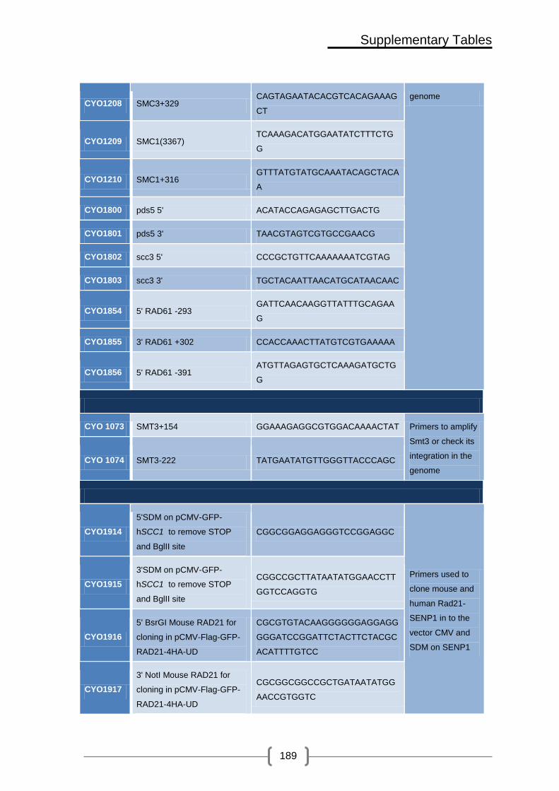
Supplementary Tables
189
CYO1208 SMC3+329 CAGTAGAATACACGTCACAGAAAG
CT
genome
CYO1209 SMC1(3367) TCAAAGACATGGAATATCTTTCTG
G
CYO1210 SMC1+316 GTTTATGTATGCAAATACAGCTACA
A
CYO1800 pds5 5' ACATACCAGAGAGCTTGACTG
CYO1801 pds5 3' TAACGTAGTCGTGCCGAACG
CYO1802 scc3 5' CCCGCTGTTCAAAAAAATCGTAG
CYO1803 scc3 3' TGCTACAATTAACATGCATAACAAC
CYO1854 5' RAD61 -293 GATTCAACAAGGTTATTTGCAGAA
G
CYO1855 3' RAD61 +302 CCACCAAACTTATGTCGTGAAAAA
CYO1856 5' RAD61 -391 ATGTTAGAGTGCTCAAAGATGCTG
G
CYO 1073 SMT3+154 GGAAAGAGGCGTGGACAAAACTAT Primers to amplify
Smt3 or check its
integration in the
genome
CYO 1074 SMT3-222 TATGAATATGTTGGGTTACCCAGC
CYO1914
5'SDM on pCMV-GFP-
hSCC1 to remove STOP
and BglII site
CGGCGGAGGAGGGTCCGGAGGC
Primers used to
clone mouse and
human Rad21-
SENP1 in to the
vector CMV and
SDM on SENP1
CYO1915
3'SDM on pCMV-GFP-
hSCC1 to remove STOP
and BglII site
CGGCCGCTTATAATATGGAACCTT
GGTCCAGGTG
CYO1916
5' BsrGI Mouse RAD21 for
cloning in pCMV-Flag-GFP-
RAD21-4HA-UD
CGCGTGTACAAGGGGGGAGGAGG
GGGATCCGGATTCTACTTCTACGC
ACATTTTGTCC
CYO1917
3' NotI Mouse RAD21 for
cloning in pCMV-Flag-GFP-
RAD21-4HA-UD
CGCGGCGGCCGCTGATAATATGG
AACCGTGGTC

Supplementary Tables
190
CYO1931
Forward primer for
sequencing inserts
downstream EGFP (original
vector from Euroscarf)
TGGTCCTGCTGGAGTTCGTG
CYO1932 Reverse primer for
sequencing Hs or Mm Scc1 CTCCTGTCTCTTTCCACATC
CYO1933
duplex siRNA to inhibit only
Hs Scc1 (Ana Losada
Rad21-2)
GGUGAAAAUGGCAUUACGGDTDT
CYO1933B
CCGUAAUGCCAUUUUCACCDTDT
CYO1934 Forward primer for C602S
exsite on HsSENP1
5'P-
GGAATGTTTGCCTGCAAATATGC
CYO1935 Reverse primer for C602S
exsite on HsSENP1 5'P-GGAGTCACTTCCATTCATCTGC
CYO1936
Reverse primer to create M1
mutations on 5BD domain of
NSE2 by ExSite method (up
to ATG). ApaLI site for
diagnosis
P-
CACTCTTAGCGATCGGATTATCGT
TCAAGGCCAT
CYO1949 Forward primer to sequence
Hs or Mm Scc1 GCCAAGAGGAAGAGGAAG
CYO1950
Reverse primer to sequence
SENP1 on pGFP-RAD21-UD
constructs
ATTCGAAGCTTGAGCTCG
CYO1261 S3-SMT3
CCCAGTTCAGTTCTAGTTTTACAAA
TAAATACACGAGCGATGCGTACGC
TGCAGGTCGAC
S2/S3 primers CYO1918 S1-NSE2 (5' for GalNSE2)
CCAAGGCAAGACTATATAAAAAAA
GAATAACTTTAAAAATGCGTACGC
TGCAGGTCGAC
CYO1919 S4-NSE2 (3' for GalNSE2)
GTAGAGGAACTGACTTGGGTATAG
GATTATCGTTCAAGGCCATCGATG
AATTCTCTGTCG

Supplementary Tables
191
CYO1862 S3-Nse2
CAGGAACAGGATAAAAGAAGTAGT
CAAGCCATCGATGTTTTACGTACG
CTGCAGGTCGAC
CYO1863 S2-Nse2
CGGGCCGAAGGGCTCGGATAAGA
GAAACAATAATTTTGTTTATCGATG
AATTCGAGCTCG
CYO1430 S3-nse2dc
ACGAAGACGATCTACAAATAGAAG
GTGGTAAAATTGAATTGACTCGTA
CGCTGCAGGTCGAC
CYO1818 S2-SMC3
CAAAACTGATATTTTTATATACAAA
TCGTTTCAAATATCTCATCGATGAA
TTCGAGCTCG
CYO1819 S3-SMC3
GCAATCGGATTCATTAGAGGTAGC
AATAAATTCGCTGAAGTCCGTACG
CTGCAGGTCGAC
CYO1834 S2-Smc1
TTAGTTATTTGACGGGTTATAGCA
GAGGTTGGTTTCATAGAATCGATG
AATTCGAGCTCG
CYO1835 S3-Smc1
TCGTCGAAGATCATAACTTTGGAC
TTGAGCAATTACGCAGAACGTACG
CTGCAGGTCGAC
CYO1639 S3-SCC1
AAAATAGACGCCAAACCTGCACTA
TTTGAAAGGTTTATCAATGCTCGTA
CGCTGCAGGTCGAC
-46kb
F:
GTATCACGAGCCCATCTCCAAT
AG
R:
CATCCCTCACCAAGGAAGAAA
GAG
Primer used for
real time PCR:
HMR DSB
-30Kb
F: TCGTCGTCGCCATCATTTTC
R:
GCCCAAGTTTGAGAGAGGTTG
C

Supplementary Tables
192
-9.3kb
F:
TCAGGGTCTGGTGGAAGGAAT
G
R:
CAAAGGTGGCAGTTGTTGAAC
C
-5kb
F: ATTGCGACAAGGCTTCACCC
R:
CACATCACAGGTTTATTGGTTC
CC
-3.4kb
F:
ATTCTGCCATTCAGGGACAGC
G
R:
CGTGGGAAAAGTAATCCGATG
C
-0.7kb
F:
CCACATTAAATACCAACCCATC
CG
R:
TAGTGATGAGGAGAAGAAGTT
GTTGC
+0.5kb
F:
CATGCGGTTCACATGACTTTTG
AC
R:
GGAAGTAACCTCTACTGTGGA
GGCAC
+2.2kb
F:
AACGCTCGTCGATCGCCGTTC
TAA
R:
AATGGATTTGCCAAATGCACAT

Supplementary Tables
193
+5.6kb
F:
CAGGTTTATATCCACCTTCATC
GG
R:
TTTGGGGCAACAGTAGGCAGT
G
+7.7kb
F:
GCAATCGTGTCAATGTGGTCAT
C
R:
GTTTCAGGAGCCCCATAATCAA
C
+12.8kb
F:
CGTTGTCTTTTCGTTTGGTGTC
TG
R:
GCTCTTTGCCCCTGTCTTTGAC
+30kb
F:
TCCAGGCGGGTGTGAAAAAC
R:
ATGGGGAATACGGAAGTGGGT
C

194

195
References

196

References
197
IX. References
ALEXANDRU, G., UHLMANN, F., MECHTLER, K., POUPART, M.A. and NASMYTH,
K., 2001. Phosphorylation of the cohesin subunit Scc1 by Polo/Cdc5 kinase
regulates sister chromatid separation in yeast. Cell, 105(4), pp. 459-472.
ANDERSON, D.E., LOSADA, A., ERICKSON, H.P. and HIRANO, T., 2002. Condensin
and cohesin display different arm conformations with characteristic hinge angles.
The Journal of cell biology, 156(3), pp. 419-424.
ANDREWS, E.A., PALECEK, J., SERGEANT, J., TAYLOR, E., LEHMANN, A.R. and
WATTS, F.Z., 2005. Nse2, a component of the Smc5-6 complex, is a SUMO
ligase required for the response to DNA damage. Molecular and cellular biology,
25(1), pp. 185-196.
ARUMUGAM, P., GRUBER, S., TANAKA, K., HAERING, C.H., MECHTLER, K. and
NASMYTH, K., 2003. ATP hydrolysis is required for cohesin's association with
chromosomes. Current biology : CB, 13(22), pp. 1941-1953.
ARUMUGAM, P., NISHINO, T., HAERING, C.H., GRUBER, S. and NASMYTH, K.,
2006. Cohesin's ATPase activity is stimulated by the C-terminal Winged-Helix
domain of its kleisin subunit. Current biology : CB, 16(20), pp. 1998-2008.
BACHANT, J., ALCASABAS, A., BLAT, Y., KLECKNER, N. and ELLEDGE, S.J.,
2002. The SUMO-1 isopeptidase Smt4 is linked to centromeric cohesion through
SUMO-1 modification of DNA topoisomerase II. Molecular cell, 9(6), pp. 1169-
1182.

References
198
BAK, A.L., ZEUTHEN, J. and CRICK, F.H., 1977. Higher-order structure of human
mitotic chromosomes. Proceedings of the National Academy of Sciences of the
United States of America, 74(4), pp. 1595-1599.
BANNISTER, L.A., REINHOLDT, L.G., MUNROE, R.J. and SCHIMENTI, J.C., 2004.
Positional cloning and characterization of mouse mei8, a disrupted allelle of the
meiotic cohesin Rec8. Genesis (New York, N.Y.: 2000), 40(3), pp. 184-194.
BAXTER, J., SEN, N., MARTINEZ, V.L., DE CARANDINI, M.E., SCHVARTZMAN, J.B.,
DIFFLEY, J.F. and ARAGON, L., 2011. Positive supercoiling of mitotic DNA
drives decatenation by topoisomerase II in eukaryotes. Science (New York,
N.Y.), 331(6022), pp. 1328-1332.
BAYER, P., ARNDT, A., METZGER, S., MAHAJAN, R., MELCHIOR, F., JAENICKE, R.
and BECKER, J., 1998. Structure determination of the small ubiquitin-related
modifier SUMO-1. Journal of Molecular Biology, 280(2), pp. 275-286.
BECKOUET, F., HU, B., ROIG, M.B., SUTANI, T., KOMATA, M., ULUOCAK, P.,
KATIS, V.L., SHIRAHIGE, K. and NASMYTH, K., 2010. An Smc3 acetylation
cycle is essential for establishment of sister chromatid cohesion. Molecular cell,
39(5), pp. 689-699.
BERMUDEZ-LOPEZ, M., CESCHIA, A., DE PICCOLI, G., COLOMINA, N., PASERO,
P., ARAGON, L. and TORRES-ROSELL, J., 2010. The Smc5/6 complex is
required for dissolution of DNA-mediated sister chromatid linkages. Nucleic
acids research, 38(19), pp. 6502-6512.
BERNIER-VILLAMOR, V., SAMPSON, D.A., MATUNIS, M.J. and LIMA, C.D., 2002.
Structural basis for E2-mediated SUMO conjugation revealed by a complex

References
199
between ubiquitin-conjugating enzyme Ubc9 and RanGAP1. Cell, 108(3), pp.
345-356.
BORGES, V., SMITH, D.J., WHITEHOUSE, I. and UHLMANN, F., 2013. An Eco1-
independent sister chromatid cohesion establishment pathway in S. cerevisiae.
Chromosoma, 122 (1-2), pp. 121-34.
BRANZEI, D. and FOIANI, M., 2008. Regulation of DNA repair throughout the cell
cycle. Nature reviews.Molecular cell biology, 9(4), pp. 297-308.
BRANZEI, D. and FOIANI, M., 2005. The DNA damage response during DNA
replication. Current opinion in cell biology, 17(6), pp. 568-575.
BRANZEI, D., SOLLIER, J., LIBERI, G., ZHAO, X., MAEDA, D., SEKI, M., ENOMOTO,
T., OHTA, K. and FOIANI, M., 2006. Ubc9- and mms21-mediated sumoylation
counteracts recombinogenic events at damaged replication forks. Cell, 127(3),
pp. 509-522.
BUHEITEL, J. and STEMMANN, O., 2013. Prophase pathway-dependent removal of
cohesin from human chromosomes requires opening of the Smc3-Scc1 gate.
The EMBO journal, 32(5); pp. 666-76.
BYLEBYL, G.R., BELICHENKO, I. and JOHNSON, E.S., 2003. The SUMO
isopeptidase Ulp2 prevents accumulation of SUMO chains in yeast. The Journal
of biological chemistry, 278(45), pp. 44113-44120.
CANUDAS, S. and SMITH, S., 2009. Differential regulation of telomere and
centromere cohesion by the Scc3 homologues SA1 and SA2, respectively, in
human cells. The Journal of cell biology, 187(2), pp. 165-173.

References
200
CARTER, S.D. and SJOGREN, C., 2012. The SMC complexes, DNA and chromosome
topology: right or knot? Critical reviews in biochemistry and molecular biology,
47(1), pp. 1-16.
CHAN, K.L., ROIG, M.B., HU, B., BECKOUET, F., METSON, J. and NASMYTH, K.,
2012. Cohesin's DNA exit gate is distinct from its entrance gate and is regulated
by acetylation. Cell, 150(5), pp. 961-974.
CHANG, C.R., WU, C.S., HOM, Y. and GARTENBERG, M.R., 2005. Targeting of
cohesin by transcriptionally silent chromatin. Genes & development, 19(24), pp.
3031-3042.
CHATTERJEE, A., ZAKIAN, S., HU, X.W. and SINGLETON, M.R., 2013. Structural
insights into the regulation of cohesion establishment by Wpl1. The EMBO
journal, 6; 32(5), pp. 677-87.
CHAVEZ, A., GEORGE, V., AGRAWAL, V. and JOHNSON, F.B., 2010. Sumoylation
and the structural maintenance of chromosomes (Smc) 5/6 complex slow
senescence through recombination intermediate resolution. The Journal of
biological chemistry, 285(16), pp. 11922-11930.
CHENG, C.H., LO, Y.H., LIANG, S.S., TI, S.C., LIN, F.M., YEH, C.H., HUANG, H.Y.
and WANG, T.F., 2006. SUMO modifications control assembly of synaptonemal
complex and polycomplex in meiosis of Saccharomyces cerevisiae. Genes &
development, 20(15), pp. 2067-2081.
CIOSK, R., SHIRAYAMA, M., SHEVCHENKO, A., TANAKA, T., TOTH, A.,
SHEVCHENKO, A. and NASMYTH, K., 2000. Cohesin's binding to
chromosomes depends on a separate complex consisting of Scc2 and Scc4
proteins. Molecular cell, 5(2), pp. 243-254.

References
201
CIOSK, R., ZACHARIAE, W., MICHAELIS, C., SHEVCHENKO, A., MANN, M. and
NASMYTH, K., 1998. An ESP1/PDS1 complex regulates loss of sister chromatid
cohesion at the metaphase to anaphase transition in yeast. Cell, 93(6), pp.
1067-1076.
COBBE, N. and HECK, M.M., 2004. The evolution of SMC proteins: phylogenetic
analysis and structural implications. Molecular biology and evolution, 21(2), pp.
332-347.
CREMONA, C.A., SARANGI, P., YANG, Y., HANG, L.E., RAHMAN, S. and ZHAO, X.,
2012. Extensive DNA Damage-Induced Sumoylation Contributes to Replication
and Repair and Acts in Addition to the Mec1 Checkpoint. Molecular cell, 45(3);
pp. 422-32.
D'AMOURS, D., STEGMEIER, F. and AMON, A., 2004. Cdc14 and condensin control
the dissolution of cohesin-independent chromosome linkages at repeated DNA.
Cell, 117(4), pp. 455-469.
DEARDORFF, M.A., BANDO, M., NAKATO, R., WATRIN, E., ITOH, T., MINAMINO,
M., SAITOH, K., KOMATA, M., KATOU, Y., CLARK, D., COLE, K.E., DE
BAERE, E., DECROOS, C., DI DONATO, N., ERNST, S., FRANCEY, L.J.,
GYFTODIMOU, Y., HIRASHIMA, K., HULLINGS, M., ISHIKAWA, Y., JAULIN,
C., KAUR, M., KIYONO, T., LOMBARDI, P.M., MAGNAGHI-JAULIN, L.,
MORTIER, G.R., NOZAKI, N., PETERSEN, M.B., SEIMIYA, H., SIU, V.M.,
SUZUKI, Y., TAKAGAKI, K., WILDE, J.J., WILLEMS, P.J., PRIGENT, C.,
GILLESSEN-KAESBACH, G., CHRISTIANSON, D.W., KAISER, F.J.,
JACKSON, L.G., HIROTA, T., KRANTZ, I.D. and SHIRAHIGE, K., 2012. HDAC8
mutations in Cornelia de Lange syndrome affect the cohesin acetylation cycle.
Nature, 489(7415), pp. 313-317.

References
202
DEGNER, S.C., WONG, T.P., JANKEVICIUS, G. and FEENEY, A.J., 2009. Cutting
edge: developmental stage-specific recruitment of cohesin to CTCF sites
throughout immunoglobulin loci during B lymphocyte development. Journal of
immunology (Baltimore, Md.: 1950), 182(1), pp. 44-48.
DENISON, C., RUDNER, A.D., GERBER, S.A., BAKALARSKI, C.E., MOAZED, D. and
GYGI, S.P., 2005. A proteomic strategy for gaining insights into protein
sumoylation in yeast. Molecular & cellular proteomics : MCP, 4(3), pp. 246-254.
DIECKHOFF, P., BOLTE, M., SANCAK, Y., BRAUS, G.H. and IRNIGER, S., 2004.
Smt3/SUMO and Ubc9 are required for efficient APC/C-mediated proteolysis in
budding yeast. Molecular microbiology, 51(5), pp. 1375-1387.
DONZE, D., ADAMS, C.R., RINE, J. and KAMAKAKA, R.T., 1999. The boundaries of
the silenced HMR domain in Saccharomyces cerevisiae. Genes & development,
13(6), pp. 698-708.
DOU, H., HUANG, C., VAN NGUYEN, T., LU, L.S. and YEH, E.T., 2011. SUMOylation
and de-SUMOylation in response to DNA damage. FEBS letters, 585(18), pp.
2891-2896.
DOYLE, J.M., GAO, J., WANG, J., YANG, M. and POTTS, P.R., 2010. MAGE-RING
protein complexes comprise a family of E3 ubiquitin ligases. Molecular cell,
39(6), pp. 963-974.
DUAN, X., SARANGI, P., LIU, X., RANGI, G.K., ZHAO, X. and YE, H., 2009. Structural
and functional insights into the roles of the Mms21 subunit of the Smc5/6
complex. Molecular cell, 35(5), pp. 657-668.
DUAN, X., YANG, Y., CHEN, Y.H., ARENZ, J., RANGI, G.K., ZHAO, X. and YE, H.,
2009. Architecture of the Smc5/6 Complex of Saccharomyces cerevisiae

References
203
Reveals a Unique Interaction between the Nse5-6 Subcomplex and the Hinge
Regions of Smc5 and Smc6. The Journal of biological chemistry, 284(13), pp.
8507-8515.
DUMONT, J.N., 1972. Oogenesis in Xenopus laevis (Daudin). I. Stages of oocyte
development in laboratory maintained animals. Journal of Morphology, 136(2),
pp. 153-179.
FREEMAN, L., ARAGON-ALCAIDE, L. and STRUNNIKOV, A., 2000. The condensin
complex governs chromosome condensation and mitotic transmission of rDNA.
The Journal of cell biology, 149(4), pp. 811-824.
GOLDSTEIN, A.L. and MCCUSKER, J.H., 1999. Three new dominant drug resistance
cassettes for gene disruption in Saccharomyces cerevisiae. Yeast (Chichester,
England), 15(14), pp. 1541-1553.
GOLDSTEIN, L.S., 1981. Kinetochore structure and its role in chromosome orientation
during the first meiotic division in male D. melanogaster. Cell, 25(3), pp. 591-
602.
GRUBER, S., ARUMUGAM, P., KATOU, Y., KUGLITSCH, D., HELMHART, W.,
SHIRAHIGE, K. and NASMYTH, K., 2006. Evidence that loading of cohesin onto
chromosomes involves opening of its SMC hinge. Cell, 127(3), pp. 523-537.
GUACCI, V. and KOSHLAND, D., 2011. Cohesin-independent segregation of sister
chromatids in budding yeast. Molecular biology of the cell, 23(4); pp.729-39.

References
204
GUACCI, V., KOSHLAND, D. and STRUNNIKOV, A., 1997. A direct link between sister
chromatid cohesion and chromosome condensation revealed through the
analysis of MCD1 in S. cerevisiae. Cell, 91(1), pp. 47-57.
GUILLOU, E., IBARRA, A., COULON, V., CASADO-VELA, J., RICO, D., CASAL, I.,
SCHWOB, E., LOSADA, A. and MENDEZ, J., 2010. Cohesin organizes
chromatin loops at DNA replication factories. Genes & development, 24(24), pp.
2812-2822.
GULLEROVA, M. and PROUDFOOT, N.J., 2008. Cohesin complex promotes
transcriptional termination between convergent genes in S. pombe. Cell, 132(6),
pp. 983-995.
GUO, D., LI, M., ZHANG, Y., YANG, P., ECKENRODE, S., HOPKINS, D., ZHENG, W.,
PUROHIT, S., PODOLSKY, R.H., MUIR, A., WANG, J., DONG, Z., BRUSKO,
T., ATKINSON, M., POZZILLI, P., ZEIDLER, A., RAFFEL, L.J., JACOB, C.O.,
PARK, Y., SERRANO-RIOS, M., LARRAD, M.T., ZHANG, Z., GARCHON, H.J.,
BACH, J.F., ROTTER, J.I., SHE, J.X. and WANG, C.Y., 2004. A functional
variant of SUMO4, a new I kappa B alpha modifier, is associated with type 1
diabetes. Nature genetics, 36(8), pp. 837-841.
HADJUR, S., WILLIAMS, L.M., RYAN, N.K., COBB, B.S., SEXTON, T., FRASER, P.,
FISHER, A.G. and MERKENSCHLAGER, M., 2009. Cohesins form
chromosomal cis-interactions at the developmentally regulated IFNG locus.
Nature, 460(7253), pp. 410-413.
HAERING, C.H., FARCAS, A.M., ARUMUGAM, P., METSON, J. and NASMYTH, K.,
2008. The cohesin ring concatenates sister DNA molecules. Nature, 454(7202),
pp. 297-301.

References
205
HAERING, C.H., LOWE, J., HOCHWAGEN, A. and NASMYTH, K., 2002. Molecular
architecture of SMC proteins and the yeast cohesin complex. Molecular cell,
9(4), pp. 773-788.
HAERING, C.H., SCHOFFNEGGER, D., NISHINO, T., HELMHART, W., NASMYTH, K.
and LOWE, J., 2004. Structure and stability of cohesin's Smc1-kleisin
interaction. Molecular cell, 15(6), pp. 951-964.
HARDELAND, U., STEINACHER, R., JIRICNY, J. and SCHAR, P., 2002. Modification
of the human thymine-DNA glycosylase by ubiquitin-like proteins facilitates
enzymatic turnover. The EMBO journal, 21(6), pp. 1456-1464.
HARTMAN, T., STEAD, K., KOSHLAND, D. and GUACCI, V., 2000. Pds5p is an
essential chromosomal protein required for both sister chromatid cohesion and
condensation in Saccharomyces cerevisiae. The Journal of cell biology, 151(3),
pp. 613-626.
HEIDINGER-PAULI, J.M., MERT, O., DAVENPORT, C., GUACCI, V. and KOSHLAND,
D., 2010. Systematic reduction of cohesin differentially affects chromosome
segregation, condensation, and DNA repair. Current biology : CB, 20(10), pp.
957-963.
HEIDINGER-PAULI, J.M., UNAL, E., GUACCI, V. and KOSHLAND, D., 2008. The
kleisin subunit of cohesin dictates damage-induced cohesion. Molecular cell,
31(1), pp. 47-56.
HEIDINGER-PAULI, J.M., UNAL, E. and KOSHLAND, D., 2009. Distinct targets of the
Eco1 acetyltransferase modulate cohesion in S phase and in response to DNA
damage. Molecular cell, 34(3), pp. 311-321.

References
206
HIETAKANGAS, V., ANCKAR, J., BLOMSTER, H.A., FUJIMOTO, M., PALVIMO, J.J.,
NAKAI, A. and SISTONEN, L., 2006. PDSM, a motif for phosphorylation-
dependent SUMO modification. Proceedings of the National Academy of
Sciences of the United States of America, 103(1), pp. 45-50.
HIRANO, M. and HIRANO, T., 2004. Positive and negative regulation of SMC-DNA
interactions by ATP and accessory proteins. The EMBO journal, 23(13), pp.
2664-2673.
HIRANO, T., KOBAYASHI, R. and HIRANO, M., 1997. Condensins, chromosome
condensation protein complexes containing XCAP-C, XCAP-E and a Xenopus
homolog of the Drosophila Barren protein. Cell, 89(4), pp. 511-521.
HOCHSTRASSER, M., 2001. SP-RING for SUMO: new functions bloom for a ubiquitin-
like protein. Cell, 107(1), pp. 5-8.
HOEGE, C., PFANDER, B., MOLDOVAN, G.L., PYROWOLAKIS, G. and JENTSCH,
S., 2002. RAD6-dependent DNA repair is linked to modification of PCNA by
ubiquitin and SUMO. Nature, 419(6903), pp. 135-141.
HORSFIELD, J.A., PRINT, C.G. and MONNICH, M., 2012. Diverse developmental
disorders from the one ring: distinct molecular pathways underlie the
cohesinopathies. Frontiers in genetics, 3, pp. 171.
HU, B., ITOH, T., MISHRA, A., KATOH, Y., CHAN, K.L., UPCHER, W., GODLEE, C.,
ROIG, M.B., SHIRAHIGE, K. and NASMYTH, K., 2011. ATP hydrolysis is
required for relocating cohesin from sites occupied by its Scc2/4 loading
complex. Current biology : CB, 21(1), pp. 12-24.

References
207
HUANG, X., HATCHER, R., YORK, J.P. and ZHANG, P., 2005. Securin and separase
phosphorylation act redundantly to maintain sister chromatid cohesion in
mammalian cells. Molecular biology of the cell, 16(10), pp. 4725-4732.
IRA, G., PELLICIOLI, A., BALIJJA, A., WANG, X., FIORANI, S., CAROTENUTO, W.,
LIBERI, G., BRESSAN, D., WAN, L., HOLLINGSWORTH, N.M., HABER, J.E.
and FOIANI, M., 2004. DNA end resection, homologous recombination and DNA
damage checkpoint activation require CDK1. Nature, 431(7011), pp. 1011-1017.
IVANOV, D. and NASMYTH, K., 2005. A topological interaction between cohesin rings
and a circular minichromosome. Cell, 122(6), pp. 849-860.
IVANOV, D., SCHLEIFFER, A., EISENHABER, F., MECHTLER, K., HAERING, C.H.
and NASMYTH, K., 2002. Eco1 is a novel acetyltransferase that can acetylate
proteins involved in cohesion. Current biology : CB, 12(4), pp. 323-328.
JANKE, C., MAGIERA, M.M., RATHFELDER, N., TAXIS, C., REBER, S., MAEKAWA,
H., MORENO-BORCHART, A., DOENGES, G., SCHWOB, E., SCHIEBEL, E.
and KNOP, M., 2004. A versatile toolbox for PCR-based tagging of yeast genes:
new fluorescent proteins, more markers and promoter substitution cassettes.
Yeast (Chichester, England), 21(11), pp. 947-962.
JANKE, C., ORTIZ, J., TANAKA, T.U., LECHNER, J. and SCHIEBEL, E., 2002. Four
new subunits of the Dam1-Duo1 complex reveal novel functions in sister
kinetochore biorientation. The EMBO journal, 21(1-2), pp. 181-193.
JAZAYERI, A., FALCK, J., LUKAS, C., BARTEK, J., SMITH, G.C., LUKAS, J. and
JACKSON, S.P., 2006. ATM- and cell cycle-dependent regulation of ATR in
response to DNA double-strand breaks. Nature cell biology, 8(1), pp. 37-45.

References
208
JOHNSON, E.S., 2004. Protein modification by SUMO. Annual Review of
Biochemistry, 73, pp. 355-382.
JOHNSON, E.S. and BLOBEL, G., 1997. Ubc9p is the conjugating enzyme for the
ubiquitin-like protein Smt3p. The Journal of biological chemistry, 272(43), pp.
26799-26802.
JOHNSON, E.S. and GUPTA, A.A., 2001. An E3-like factor that promotes SUMO
conjugation to the yeast septins. Cell, 106(6), pp. 735-744.
JOHNSON, E.S., SCHWIENHORST, I., DOHMEN, R.J. and BLOBEL, G., 1997. The
ubiquitin-like protein Smt3p is activated for conjugation to other proteins by an
Aos1p/Uba2p heterodimer. The EMBO journal, 16(18), pp. 5509-5519.
KAMITANI, T., NGUYEN, H.P. and YEH, E.T., 1997. Preferential modification of
nuclear proteins by a novel ubiquitin-like molecule. The Journal of biological
chemistry, 272(22), pp. 14001-14004.
KEGEL, A., BETTS-LINDROOS, H., KANNO, T., JEPPSSON, K., STROM, L., KATOU,
Y., ITOH, T., SHIRAHIGE, K. and SJOGREN, C., 2011. Chromosome length
influences replication-induced topological stress. Nature, 471(7338), pp. 392-
396.
KRANTZ, I.D., MCCALLUM, J., DESCIPIO, C., KAUR, M., GILLIS, L.A., YAEGER, D.,
JUKOFSKY, L., WASSERMAN, N., BOTTANI, A., MORRIS, C.A., NOWACZYK,
M.J., TORIELLO, H., BAMSHAD, M.J., CAREY, J.C., RAPPAPORT, E.,
KAWAUCHI, S., LANDER, A.D., CALOF, A.L., LI, H.H., DEVOTO, M. and
JACKSON, L.G., 2004. Cornelia de Lange syndrome is caused by mutations in
NIPBL, the human homolog of Drosophila melanogaster Nipped-B. Nature
genetics, 36(6), pp. 631-635.

References
209
KUENG, S., HEGEMANN, B., PETERS, B.H., LIPP, J.J., SCHLEIFFER, A.,
MECHTLER, K. and PETERS, J.M., 2006. Wapl controls the dynamic
association of cohesin with chromatin. Cell, 127(5), pp. 955-967.
KULEMZINA, I., SCHUMACHER, M.R., VERMA, V., REITER, J., METZLER, J.,
FAILLA, A.V., LANZ, C., SREEDHARAN, V.T., RATSCH, G. and IVANOV, D.,
2012. Cohesin rings devoid of Scc3 and Pds5 maintain their stable association
with the DNA. PLoS genetics, 8(8), pp. e1002856.
KURZE, A., MICHIE, K.A., DIXON, S.E., MISHRA, A., ITOH, T., KHALID, S.,
STRMECKI, L., SHIRAHIGE, K., HAERING, C.H., LOWE, J. and NASMYTH, K.,
2011. A positively charged channel within the Smc1/Smc3 hinge required for
sister chromatid cohesion. The EMBO journal, 30(2), pp. 364-378.
LEHMANN, A.R., 2005. The role of SMC proteins in the responses to DNA damage.
DNA repair, 4(3), pp. 309-314.
LEHOCZKY, P., MCHUGH, P.J. and CHOVANEC, M., 2007. DNA interstrand cross-
link repair in Saccharomyces cerevisiae. FEMS microbiology reviews, 31(2), pp.
109-133.
LENGRONNE, A., KATOU, Y., MORI, S., YOKOBAYASHI, S., KELLY, G.P., ITOH, T.,
WATANABE, Y., SHIRAHIGE, K. and UHLMANN, F., 2004. Cohesin relocation
from sites of chromosomal loading to places of convergent transcription. Nature,
430(6999), pp. 573-578.
LENGRONNE, A., MCINTYRE, J., KATOU, Y., KANOH, Y., HOPFNER, K.P.,
SHIRAHIGE, K. and UHLMANN, F., 2006. Establishment of sister chromatid
cohesion at the S. cerevisiae replication fork. Molecular cell, 23(6), pp. 787-799.

References
210
LI, S.J. and HOCHSTRASSER, M., 2003. The Ulp1 SUMO isopeptidase: distinct
domains required for viability, nuclear envelope localization, and substrate
specificity. The Journal of cell biology, 160(7), pp. 1069-1081.
LI, S.J. and HOCHSTRASSER, M., 1999. A new protease required for cell-cycle
progression in yeast. Nature, 398(6724), pp. 246-251.
LIMA, C.D. and REVERTER, D., 2008. Structure of the human SENP7 catalytic domain
and poly-SUMO deconjugation activities for SENP6 and SENP7. The Journal of
biological chemistry, 283(46), pp. 32045-32055.
LINDROOS, H.B., STROM, L., ITOH, T., KATOU, Y., SHIRAHIGE, K. and SJOGREN,
C., 2006. Chromosomal association of the Smc5/6 complex reveals that it
functions in differently regulated pathways. Molecular cell, 22(6), pp. 755-767.
LIU, H., RANKIN, S. and YU, H., 2012. Phosphorylation-enabled binding of SGO1-
PP2A to cohesin protects sororin and centromeric cohesion during mitosis.
Nature cell biology, 15(1), pp. 40-49.
LOPEZ-SERRA, L., LENGRONNE, A., BORGES, V., KELLY, G. and UHLMANN, F.,
2013. Budding yeast wapl controls sister chromatid cohesion maintenance and
chromosome condensation. Current biology : CB, 23(1), pp. 64-69.
LOSADA, A., YOKOCHI, T. and HIRANO, T., 2005. Functional contribution of Pds5 to
cohesin-mediated cohesion in human cells and Xenopus egg extracts. Journal of
Cell Science, 15(118(Pt 10)), pp. 2133-41.
LOSADA, A., YOKOCHI, T., KOBAYASHI, R. and HIRANO, T., 2000. Identification and
characterization of SA/Scc3p subunits in the Xenopus and human cohesin
complexes. The Journal of cell biology, 150(3), pp. 405-416.

References
211
MAHAJAN, R., DELPHIN, C., GUAN, T., GERACE, L. and MELCHIOR, F., 1997. A
small ubiquitin-related polypeptide involved in targeting RanGAP1 to nuclear
pore complex protein RanBP2. Cell, 88(1), pp. 97-107.
MANN, N.P., FITZSIMMONS, J., FITZSIMMONS, E. and COOKE, P., 1982. Roberts
syndrome: clinical and cytogenetic aspects. Journal of medical genetics, 19(2),
pp. 116-119.
MATUNIS, M.J., COUTAVAS, E. and BLOBEL, G., 1996. A novel ubiquitin-like
modification modulates the partitioning of the Ran-GTPase-activating protein
RanGAP1 between the cytosol and the nuclear pore complex. The Journal of
cell biology, 135(6 Pt 1), pp. 1457-1470.
MC INTYRE, J., MULLER, E.G., WEITZER, S., SNYDSMAN, B.E., DAVIS, T.N. and
UHLMANN, F., 2007. In vivo analysis of cohesin architecture using FRET in the
budding yeast Saccharomyces cerevisiae. The EMBO journal, 26(16), pp. 3783-
3793.
MCALEENAN, A., CLEMENTE-BLANCO, A., CORDON-PRECIADO, V., SEN, N.,
ESTERAS, M., JARMUZ, A. and ARAGON, L., 2013. Post-replicative repair
involves separase-dependent removal of the kleisin subunit of cohesin. Nature,
493(7431), pp. 250-254.
MCALEENAN, A., CORDON-PRECIADO, V., CLEMENTE-BLANCO, A., LIU, I., SEN,
N., LEONARD, J., JARMUZ, A. and ARAGÓN, L., 2012. SUMOylation of the α-
Kleisin Subunit of Cohesin Is Required for DNA Damage-Induced Cohesion.
Current Biology, 22(17), pp. 1564-1575.
MCKAY, M.J., TROELSTRA, C., VAN DER SPEK, P., KANAAR, R., SMIT, B.,
HAGEMEIJER, A., BOOTSMA, D. and HOEIJMAKERS, J.H., 1996. Sequence

References
212
conservation of the rad21 Schizosaccharomyces pombe DNA double-strand
break repair gene in human and mouse. Genomics, 36(2), pp. 305-315.
MELBY, T.E., CIAMPAGLIO, C.N., BRISCOE, G. and ERICKSON, H.P., 1998. The
symmetrical structure of structural maintenance of chromosomes (SMC) and
MukB proteins: long, antiparallel coiled coils, folded at a flexible hinge. The
Journal of cell biology, 142(6), pp. 1595-1604.
MELCHIOR, F., 2000. SUMO--nonclassical ubiquitin. Annual Review of Cell and
Developmental Biology, 16, pp. 591-626.
MICHAELIS, C., CIOSK, R. and NASMYTH, K., 1997. Cohesins: Chromosomal
Proteins that Prevent Premature Separation of Sister Chromatids. Cell, 91(1),
pp. 35 - 45.
MISHRA, A., HU, B., KURZE, A., BECKOUET, F., FARCAS, A.M., DIXON, S.E.,
KATOU, Y., KHALID, S., SHIRAHIGE, K. and NASMYTH, K., 2010. Both
interaction surfaces within cohesin's hinge domain are essential for its stable
chromosomal association. Current biology : CB, 20(4), pp. 279-289.
MOLDOVAN, G.L., PFANDER, B. and JENTSCH, S., 2006. PCNA controls
establishment of sister chromatid cohesion during S phase. Molecular cell,
23(5), pp. 723-732.
MORGAN, D.O., 2007. Cell cycle: Priciples of Control. New Science Press.
MOSSESSOVA, E. and LIMA, C.D., 2000. Ulp1-SUMO crystal structure and genetic
analysis reveal conserved interactions and a regulatory element essential for cell
growth in yeast. Molecular cell, 5(5), pp. 865-876.

References
213
MUKHOPADHYAY, D. and DASSO, M., 2007. Modification in reverse: the SUMO
proteases. Trends in biochemical sciences, 32(6), pp. 286-295.
NACERDDINE, K., LEHEMBRE, F., BHAUMIK, M., ARTUS, J., COHEN-
TANNOUDJI, M., BABINET, C., PANDOLFI, P.P. and DEJEAN, A., 2005. The
SUMO pathway is essential for nuclear integrity and chromosome segregation in
mice. Developmental cell, 9(6), pp. 769-779.
NASMYTH, K., 2011. Cohesin: a catenase with separate entry and exit gates? Nature
cell biology, 13(10), pp. 1170-1177.
NASMYTH, K. and HAERING, C.H., 2009. Cohesin: its roles and mechanisms. Annual
Review of Genetics, 43, pp. 525-558.
NASMYTH, K. and HAERING, C.H., 2005. The structure and function of SMC and
kleisin complexes. Annual Review of Biochemistry, 74, pp. 595-648.
NISHIMURA, K., FUKAGAWA, T., TAKISAWA, H., KAKIMOTO, T. and KANEMAKI,
M., 2009. An auxin-based degron system for the rapid depletion of proteins in
nonplant cells. Nature methods, 6(12), pp. 917-922.
NISHIYAMA, T., LADURNER, R., SCHMITZ, J., KREIDL, E., SCHLEIFFER, A.,
BHASKARA, V., BANDO, M., SHIRAHIGE, K., HYMAN, A.A., MECHTLER, K.
and PETERS, J.M., 2010. Sororin mediates sister chromatid cohesion by
antagonizing Wapl. Cell, 143(5), pp. 737-749.
NITISS, J.L., 2009. DNA topoisomerase II and its growing repertoire of biological
functions. Nature reviews.Cancer, 9(5), pp. 327-337.

References
214
ONO, T., LOSADA, A., HIRANO, M., MYERS, M.P., NEUWALD, A.F. and HIRANO,
T., 2003. Differential contributions of condensin I and condensin II to mitotic
chromosome architecture in vertebrate cells. Cell, 115(1), pp. 109-121.
OWERBACH, D., MCKAY, E.M., YEH, E.T., GABBAY, K.H. and BOHREN, K.M., 2005.
A proline-90 residue unique to SUMO-4 prevents maturation and sumoylation.
Biochemical and biophysical research communications, 337(2), pp. 517-520.
PALECEK, J., VIDOT, S., FENG, M., DOHERTY, A.J. and LEHMANN, A.R., 2006.
The Smc5-Smc6 DNA repair complex. bridging of the Smc5-Smc6 heads by the
KLEISIN, Nse4, and non-Kleisin subunits. The Journal of biological chemistry,
281(48), pp. 36952-36959.
PALVIMO, J.J., 2007. PIAS proteins as regulators of small ubiquitin-related modifier
(SUMO) modifications and transcription. Biochemical Society transactions, 35(Pt
6), pp. 1405-1408.
PANIZZA, S., TANAKA, T., HOCHWAGEN, A., EISENHABER, F. and NASMYTH, K.,
2000. Pds5 cooperates with cohesin in maintaining sister chromatid cohesion.
Current biology : CB, 10(24), pp. 1557-1564.
PANSE, V.G., HARDELAND, U., WERNER, T., KUSTER, B. and HURT, E., 2004. A
proteome-wide approach identifies sumoylated substrate proteins in yeast. The
Journal of biological chemistry, 279(40), pp. 41346-41351.
PARKER, J.L. and ULRICH, H.D., 2012. A SUMO-interacting motif activates budding
yeast ubiquitin ligase Rad18 towards SUMO-modified PCNA. Nucleic acids
research, 40(22), pp. 11380-11388.

References
215
PEBERNARD, S., MCDONALD, W.H., PAVLOVA, Y., YATES, J.R.,3rd and BODDY,
M.N., 2004. Nse1, Nse2, and a novel subunit of the Smc5-Smc6 complex, Nse3,
play a crucial role in meiosis. Molecular biology of the cell, 15(11), pp. 4866-
4876.
PEBERNARD, S., WOHLSCHLEGEL, J., MCDONALD, W.H., YATES, J.R.,3rd and
BODDY, M.N., 2006. The Nse5-Nse6 dimer mediates DNA repair roles of the
Smc5-Smc6 complex. Molecular and cellular biology, 26(5), pp. 1617-1630.
PELCZAR, M.J., CHAN, E.C.S. and KRIEG, N.R., 1993. Microbiology: Concepts and
applications. McGRAW-HILL, INC., pp. 283.
PETERS, J.M., 2006. The anaphase promoting complex/cyclosome: a machine
designed to destroy. Nature reviews.Molecular cell biology, 7(9), pp. 644-656.
PFANDER, B., MOLDOVAN, G.L., SACHER, M., HOEGE, C. and JENTSCH, S., 2005.
SUMO-modified PCNA recruits Srs2 to prevent recombination during S phase.
Nature, 436(7049), pp. 428-433.
PIAZZA, I., HAERING, C.H. and RUTKOWSKA, A., 2013. Condensin: crafting the
chromosome landscape. Chromosoma, 122(3), pp 175-90.
POTTS, P.R., PORTEUS, M.H. and YU, H., 2006. Human SMC5/6 complex promotes
sister chromatid homologous recombination by recruiting the SMC1/3 cohesin
complex to double-strand breaks. The EMBO journal, 25(14), pp. 3377-3388.
POTTS, P.R. and YU, H., 2005. Human MMS21/NSE2 is a SUMO ligase required for
DNA repair. Molecular and cellular biology, 25(16), pp. 7021-7032.
PSAKHYE, I. and JENTSCH, S., 2012. Protein group modification and synergy in the
SUMO pathway as exemplified in DNA repair. Cell, 151(4), pp. 807-820.
References
216
REMESEIRO, S. and LOSADA, A., 2012. Cohesin, a chromatin engagement ring.
Current opinion in cell biology, 25(1), pp. 63-71.
RENSHAW, M.J., WARD, J.J., KANEMAKI, M., NATSUME, K., NEDELEC, F.J. and
TANAKA, T.U., 2010. Condensins promote chromosome recoiling during early
anaphase to complete sister chromatid separation. Developmental cell, 19(2),
pp. 232-244.
ROLEF BEN-SHAHAR, T., HEEGER, S., LEHANE, C., EAST, P., FLYNN, H.,
SKEHEL, M. and UHLMANN, F., 2008. Eco1-dependent cohesin acetylation
during establishment of sister chromatid cohesion. Science (New York, N.Y.),
321(5888), pp. 563-566.
ROWLAND, B.D., ROIG, M.B., NISHINO, T., KURZE, A., ULUOCAK, P., MISHRA, A.,
BECKOUET, F., UNDERWOOD, P., METSON, J., IMRE, R., MECHTLER, K.,
KATIS, V.L. and NASMYTH, K., 2009. Building sister chromatid cohesion: smc3
acetylation counteracts an antiestablishment activity. Molecular cell, 33(6), pp.
763-774.
SACHER, M., PFANDER, B., HOEGE, C. and JENTSCH, S., 2006. Control of Rad52
recombination activity by double-strand break-induced SUMO modification.
Nature cell biology, 8(11), pp. 1284-1290.
SACHER, M., PFANDER, B. and JENTSCH, S., 2005. Identification of SUMO-protein
conjugates. Methods in enzymology, 399, pp. 392-404.
SAITOH, H. and HINCHEY, J., 2000. Functional heterogeneity of small ubiquitin-
related protein modifiers SUMO-1 versus SUMO-2/3. The Journal of biological
chemistry, 275(9), pp. 6252-6258.

References
217
SAPETSCHNIG, A., RISCHITOR, G., BRAUN, H., DOLL, A., SCHERGAUT, M.,
MELCHIOR, F. and SUSKE, G., 2002. Transcription factor Sp3 is silenced
through SUMO modification by PIAS1. The EMBO journal, 21(19), pp. 5206-
5215.
SCHMIESING, J.A., BALL, A.R.,Jr, GREGSON, H.C., ALDERTON, J.M., ZHOU, S.
and YOKOMORI, K., 1998. Identification of two distinct human SMC protein
complexes involved in mitotic chromosome dynamics. Proceedings of the
National Academy of Sciences of the United States of America, 95(22), pp.
12906-12911.
SHEN, L., TATHAM, M.H., DONG, C., ZAGORSKA, A., NAISMITH, J.H. and HAY,
R.T., 2006. SUMO protease SENP1 induces isomerization of the scissile peptide
bond. Nature structural & molecular biology, 13(12), pp. 1069-1077.
SHINTOMI, K. and HIRANO, T., 2010. Sister chromatid resolution: a cohesin releasing
network and beyond. Chromosoma, 119(5), pp. 459-467.
SIMPSON, D.P., 1977. Cassell's Latin Dictionary: Latin-English, English-Latin.
MACMILLAN, pp. 840.
SKIBBENS, R.V., CORSON, L.B., KOSHLAND, D. and HIETER, P., 1999. Ctf7p is
essential for sister chromatid cohesion and links mitotic chromosome structure
to the DNA replication machinery. Genes & development, 13(3), pp. 307-319.
SLATER, M.L., 1973. Effect of reversible inhibition of deoxyribonucleic acid synthesis
on the yeast cell cycle. Journal of Bacteriology, 113(1), pp. 263-270.
SONG, J., DURRIN, L.K., WILKINSON, T.A., KRONTIRIS, T.G. and CHEN, Y., 2004.
Identification of a SUMO-binding motif that recognizes SUMO-modified proteins.

References
218
Proceedings of the National Academy of Sciences of the United States of
America, 101(40), pp. 14373-14378.
SONODA, E., MATSUSAKA, T., MORRISON, C., VAGNARELLI, P., HOSHI, O.,
USHIKI, T., NOJIMA, K., FUKAGAWA, T., WAIZENEGGER, I.C., PETERS,
J.M., EARNSHAW, W.C. and TAKEDA, S., 2001. Scc1/Rad21/Mcd1 is required
for sister chromatid cohesion and kinetochore function in vertebrate cells.
Developmental cell, 1(6), pp. 759-770.
SRIKUMAR, T., LEWICKI, M.C., COSTANZO, M., TKACH, J.M., VAN BAKEL, H.,
TSUI, K., JOHNSON, E.S., BROWN, G.W., ANDREWS, B.J., BOONE, C.,
GIAEVER, G., NISLOW, C. and RAUGHT, B., 2013. Global analysis of SUMO
chain function reveals multiple roles in chromatin regulation. The Journal of cell
biology, 201(1), pp. 145-163.
STANKOVIC-VALENTIN, N., DELTOUR, S., SEELER, J., PINTE, S., VERGOTEN, G.,
GUERARDEL, C., DEJEAN, A. and LEPRINCE, D., 2007. An
acetylation/deacetylation-SUMOylation switch through a phylogenetically
conserved psiKXEP motif in the tumor suppressor HIC1 regulates transcriptional
repression activity. Molecular and cellular biology, 27(7), pp. 2661-2675.
STEAD, K., AGUILAR, C., HARTMAN, T., DREXEL, M., MELUH, P. and GUACCI, V.,
2003. Pds5p regulates the maintenance of sister chromatid cohesion and is
sumoylated to promote the dissolution of cohesion. The Journal of cell biology,
163(4), pp. 729-741.
STEPHAN, A.K., KLISZCZAK, M., DODSON, H., COOLEY, C. and MORRISON, C.G.,
2011. Roles of vertebrate Smc5 in sister chromatid cohesion and homologous
recombinational repair. Molecular and cellular biology, 31(7), pp. 1369-1381.

References
219
STERN, B.M. and MURRAY, A.W., 2001. Lack of tension at kinetochores activates the
spindle checkpoint in budding yeast. Current biology : CB, 11(18), pp. 1462-
1467.
STROM, L., KARLSSON, C., LINDROOS, H.B., WEDAHL, S., KATOU, Y.,
SHIRAHIGE, K. and SJOGREN, C., 2007. Postreplicative formation of cohesion
is required for repair and induced by a single DNA break. Science (New York,
N.Y.), 317(5835), pp. 242-245.
SUN, T.T. and GREEN, H., 1976. Differentiation of the epidermal keratinocyte in cell
culture: formation of the cornified envelope. Cell, 9(4 Pt 1), pp. 511-521.
SUTANI, T., KAWAGUCHI, T., KANNO, R., ITOH, T. and SHIRAHIGE, K., 2009.
Budding yeast Wpl1(Rad61)-Pds5 complex counteracts sister chromatid
cohesion-establishing reaction. Current biology : CB, 19(6), pp. 492-497.
TAKAHASHI, Y., DULEV, S., LIU, X., HILLER, N.J., ZHAO, X. and STRUNNIKOV, A.,
2008. Cooperation of sumoylated chromosomal proteins in rDNA maintenance.
PLoS genetics, 4(10), pp. e1000215.
TAKAHASHI, Y., KAHYO, T., TOH-E, A., YASUDA, H. and KIKUCHI, Y., 2001. Yeast
Ull1/Siz1 is a novel SUMO1/Smt3 ligase for septin components and functions as
an adaptor between conjugating enzyme and substrates. The Journal of
biological chemistry, 276(52), pp. 48973-48977.
TANAKA, K., NISHIDE, J., OKAZAKI, K., KATO, H., NIWA, O., NAKAGAWA, T.,
MATSUDA, H., KAWAMUKAI, M. and MURAKAMI, Y., 1999. Characterization of
a fission yeast SUMO-1 homologue, pmt3p, required for multiple nuclear events,
including the control of telomere length and chromosome segregation. Molecular
and cellular biology, 19(12), pp. 8660-8672.

References
220
TATHAM, M.H., JAFFRAY, E., VAUGHAN, O.A., DESTERRO, J.M., BOTTING, C.H.,
NAISMITH, J.H. and HAY, R.T., 2001. Polymeric chains of SUMO-2 and SUMO-
3 are conjugated to protein substrates by SAE1/SAE2 and Ubc9. The Journal of
biological chemistry, 276(38), pp. 35368-35374.
TITTEL-ELMER, M., LENGRONNE, A., DAVIDSON, M.B., BACAL, J., FRANCOIS, P.,
HOHL, M., PETRINI, J.H., PASERO, P. and COBB, J.A., 2012. Cohesin
association to replication sites depends on rad50 and promotes fork restart.
Molecular cell, 48(1), pp. 98-108.
TORRES-ROSELL, J., MACHIN, F., FARMER, S., JARMUZ, A., EYDMANN, T.,
DALGAARD, J.Z. and ARAGON, L., 2005. SMC5 and SMC6 genes are required
for the segregation of repetitive chromosome regions. Nature cell biology, 7(4),
pp. 412-419.
TORRES-ROSELL, J., SUNJEVARIC, I., DE PICCOLI, G., SACHER, M., ECKERT-
BOULET, N., REID, R., JENTSCH, S., ROTHSTEIN, R., ARAGON, L. and
LISBY, M., 2007. The Smc5-Smc6 complex and SUMO modification of Rad52
regulates recombinational repair at the ribosomal gene locus. Nature cell
biology, 9(8), pp. 923-931.
TOTH, A., CIOSK, R., UHLMANN, F., GALOVA, M., SCHLEIFFER, A. and NASMYTH,
K., 1999. Yeast cohesin complex requires a conserved protein, Eco1p(Ctf7), to
establish cohesion between sister chromatids during DNA replication. Genes &
development, 13(3), pp. 320-333.
UNAL, E., ARBEL-EDEN, A., SATTLER, U., SHROFF, R., LICHTEN, M., HABER,
J.E. and KOSHLAND, D., 2004. DNA damage response pathway uses histone

References
221
modification to assemble a double-strand break-specific cohesin domain.
Molecular cell, 16(6), pp. 991-1002.
UNAL, E., HEIDINGER-PAULI, J.M. and KOSHLAND, D., 2007. DNA double-strand
breaks trigger genome-wide sister-chromatid cohesion through Eco1 (Ctf7).
Science (New York, N.Y.), 317(5835), pp. 245-248.
WAN, J., SUBRAMONIAN, D. and ZHANG, X., 2012. SUMOylation in Control of
Accurate Chromosome Segregation during Mitosis. Current Protein & Peptide
Science, 13(5), pp. 467 <last_page> 481.
WANG, S.W., READ, R.L. and NORBURY, C.J., 2002. Fission yeast Pds5 is required
for accurate chromosome segregation and for survival after DNA damage or
metaphase arrest. Journal of cell science, 115(Pt 3), pp. 587-598.
WATRIN, E., SCHLEIFFER, A., TANAKA, K., EISENHABER, F., NASMYTH, K. and
PETERS, J.M., 2006. Human Scc4 is required for cohesin binding to chromatin,
sister-chromatid cohesion, and mitotic progression. Current biology : CB, 16(9),
pp. 863-874.
WHELAN, G., KREIDL, E., PETERS, J.M. and EICHELE, G., 2012. The non-redundant
function of cohesin acetyltransferase Esco2: some answers and new questions.
Nucleus (Austin, Tex.), 3(4), pp. 330-334.
WOHLSCHLEGEL, J.A., JOHNSON, E.S., REED, S.I. and YATES, J.R.,3rd, 2004.
Global analysis of protein sumoylation in Saccharomyces cerevisiae. The
Journal of biological chemistry, 279(44), pp. 45662-45668.
References
222
WU, L. and HICKSON, I.D., 2003. The Bloom's syndrome helicase suppresses
crossing over during homologous recombination. Nature, 426(6968), pp. 870-
874.
WU, N., KONG, X., JI, Z., ZENG, W., POTTS, P.R., YOKOMORI, K. and YU, H., 2012.
Scc1 sumoylation by Mms21 promotes sister chromatid recombination through
counteracting Wapl. Genes & development, 26(13), pp. 1473-1485.
WYATT, M.D. and PITTMAN, D.L., 2006. Methylating agents and DNA repair
responses: Methylated bases and sources of strand breaks. Chemical research
in toxicology, 19(12), pp. 1580-1594.
XU, H., TOMASZEWSKI, J.M. and MCKAY, M.J., 2011. Can corruption of
chromosome cohesion create a conduit to cancer? Nature reviews.Cancer,
11(3), pp. 199-210.
ZHANG, N., KUZNETSOV, S.G., SHARAN, S.K., LI, K., RAO, P.H. and PATI, D.,
2008. A handcuff model for the cohesin complex. The Journal of cell biology,
183(6), pp. 1019-1031.
ZHANG, N. and PATI, D., 2009. Handcuff for sisters: a new model for sister chromatid
cohesion. Cell cycle (Georgetown, Tex.), 8(3), pp. 399-402.
ZHAO, X. and BLOBEL, G., 2005. A SUMO ligase is part of a nuclear multiprotein
complex that affects DNA repair and chromosomal organization. Proceedings of
the National Academy of Sciences of the United States of America, 102(13), pp.
4777-4782.

References
223
ZHAO, Y., KWON, S.W., ANSELMO, A., KAUR, K. and WHITE, M.A., 2004. Broad
spectrum identification of cellular small ubiquitin-related modifier (SUMO)
substrate proteins. The Journal of biological chemistry, 279(20), pp. 20999-
21002.
ZHENG, G. and YANG, Y.C., 2004. ZNF76, a novel transcriptional repressor targeting
TATA-binding protein, is modulated by sumoylation. The Journal of biological
chemistry, 279(41), pp. 42410-42421.

224

225
Supplementary Article

226