Direct Ubiquitination of Pattern Recognition Receptor FLS2 Attenuates Plant Innate Immunity
Upload
independentCategory
view
3download
0
Lysine 63-linked ubiquitination promotes the formation and autophagic
clearance of protein inclusions associated with neurodegenerative diseases.
Jeanne M. M. Tan1,2,3#, Esther S.P. Wong1,9#, Donald S. Kirkpatrick6, Olga Pletnikova5, Han Seok Ko2, Shiam-Peng Tay1, Michelle W.L. Ho1 , Juan Troncoso3,5, Steven P.
Gygi6, Michael K. Lee5, Valina L. Dawson2,3,4 , Ted M. Dawson2,3,4, Kah-Leong Lim1,7,8*
1Neurodegeneration Research Lab, National Neuroscience Institute, Singapore. 2Institute for Cell Engineering, Departments of 3Neurology, 4Neuroscience, Physiology and 5Pathology, Johns Hopkins University School of Medicine, Baltimore, MD, USA.
6Department of Cell Biology, Harvard Medical School, Boston, MA, USA. 7Duke-NUS Graduate Medical School, 8Department of Biological Sciences, National
University of Singapore, Singapore. 9Current address: Department of Anatomy and Structural Biology, Marion Bessin Liver
Research Center, Albert Einstein College of Medicine # Authors contributed equally * To whom all correspondence should be addressed: Kah-Leong Lim, Ph.D. Neurodegeneration Research Laboratory, National Neuroscience Institute, 11 Jalan Tan Tock Seng, Singapore 308433 Tel: (65)-6357-7520 Fax: (65)-6256-9178 E-mail: [email protected] © The Author 2007. Published by Oxford University Press. All rights reserved. For Permissions, please e-mail: [email protected]
1
HMG Advance Access published November 1, 2007 by guest on February 2, 2015
http://hmg.oxfordjournals.org/
Dow
nloaded from
ABSTRACT
Although ubiquitin-enriched protein inclusions represent an almost invariant feature of
neurodegenerative diseases, the mechanism underlying their biogenesis remains unclear.
In particular, whether the topology of ubiquitin linkages influences the dynamics of
inclusions is not well explored. Here we report that lysine 48 (K48) and lysine 63 (K63)-
linked polyubiquitination, as well as monoubiquitin modification contribute to the
biogenesis of inclusions. K63-linked polyubiquitin is the most consistent enhancer of
inclusions formation. Under basal conditions, ectopic expression of K63 mutant ubiquitin
in cultured cells promotes the accumulation of proteins and the formation of intracellular
inclusions in the apparent absence of proteasome impairment. When co-expressed with
disease-associated tau and SOD1 mutants, K63 ubiquitin mutant facilitates the formation
of tau- and SOD-1–positive inclusions. Moreover, K63-linked ubiquitination was found
to selectively facilitate the clearance of inclusions via autophagy. These data indicate that
K63-linked ubiquitin chains may represent a common denominator underlying inclusions
biogenesis, as well as a general cellular strategy for defining cargo destined for the
autophagic system. Collectively, our results provide a novel mechanistic route that
underlies the life cycle of an inclusion body. Harnessing this pathway may offer
innovative approaches in the treatment of neurodegenerative disorders.
2
by guest on February 2, 2015http://hm
g.oxfordjournals.org/D
ownloaded from

INTRODUCTION
Most, if not all, neurodegenerative diseases are marked by the presence of
ubiquitin-positive protein aggregates. Indeed, the accumulation of insoluble
proteinaceous deposits enriched with ubiquitin and components of the ubiquitin-
proteasome system (UPS) occur in a broad spectrum of human neurodegenerative
disorders, such as the neurofibrillary tangles in Alzheimer’s disease (AD) and
Frontotemporal Dementia, Lewy bodies (LBs) in Parkinson’s disease (PD) and Bunina
bodies in Amyotrophic Lateral Sclerosis (ALS) (1). This general phenomenon that
typifies an otherwise diverse array of diseases suggests a common mechanism in the
formation of protein inclusions associated with these varied disorders. Supporting this,
several studies collectively point toward an intimate relationship between UPS
aberrations and the biogenesis of neurodegenerative disease-linked protein inclusions (1-
3).
The UPS is an intracellular protein degradation system that is responsible for the
majority of protein turnover within the cell (4). In this system, proteins destined for
degradation are covalently tagged with a polymeric ubiquitin chain in which the terminal
residue (G76) of one ubiquitin molecule is linked through an isopeptide bond to a lysine
(K) residue (most commonly K48) within another. The G76-K48 polyubiquitinated
substrate is then targeted for degradation by the 26S proteasome, a large protease
complex consisting of a barrel-shaped 20S proteolytic core in association with two 19S
regulatory caps, one on each side of the barrel’s openings. It is important to mention that
the ubiquitin sequence contains seven lysine residues (at positions 6, 11, 27, 29, 33, 48
and 63) and polyubiquitin chain assembly can occur at any of these lysine residues (5, 6).
3
by guest on February 2, 2015http://hm
g.oxfordjournals.org/D
ownloaded from

In addition, proteins can also be monoubiquitinated (5, 6). Notably, both K63-linked
polyubiquitination and monoubiquitination of proteins are not typically associated with
their degradation (6-8).
Currently, it is unclear whether different ubiquitin topologies contribute to the
formation of an inclusion body. However, we have recently demonstrated that K63-linked
ubiquitination facilitates the formation of LB-like inclusions in cultured cells (9). This
observation, together with the recognition that ubiquitin-enriched protein aggregates are
seemingly stabilized structures, prompted us to investigate the likely generic role of non-
proteolytic ubiquitin modifications in the formation of inclusions associated with
neurodegenerative diseases. We show here in the setting of ectopic overexpression of
different ubiquitin species that lysine 48 (K48)-linked polyubiquitination, as well as non-
proteolytic monoubiquitination or lysine 63 (K63)-linked ubiquitin modification
contribute to the biogenesis of inclusions. K63-linked polyubiquitin is the most consistent
enhancer of inclusions formation. Moreover, K63 ubiquitin-modified inclusions are
preferentially cleared by autophagy. Taken together, these data point toward a potential
mechanism of protein inclusion biogenesis and clearance that may be important in
neurodegenerative diseases.
4
by guest on February 2, 2015http://hm
g.oxfordjournals.org/D
ownloaded from
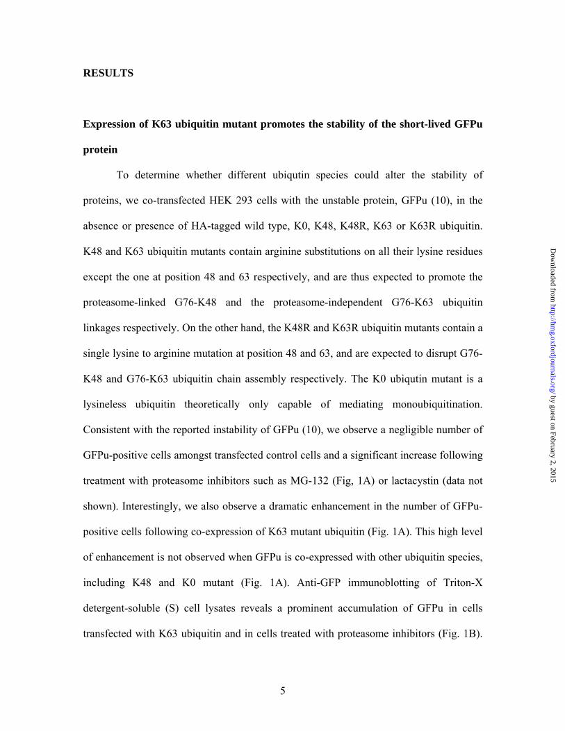
RESULTS
Expression of K63 ubiquitin mutant promotes the stability of the short-lived GFPu
protein
To determine whether different ubiqutin species could alter the stability of
proteins, we co-transfected HEK 293 cells with the unstable protein, GFPu (10), in the
absence or presence of HA-tagged wild type, K0, K48, K48R, K63 or K63R ubiquitin.
K48 and K63 ubiquitin mutants contain arginine substitutions on all their lysine residues
except the one at position 48 and 63 respectively, and are thus expected to promote the
proteasome-linked G76-K48 and the proteasome-independent G76-K63 ubiquitin
linkages respectively. On the other hand, the K48R and K63R ubiquitin mutants contain a
single lysine to arginine mutation at position 48 and 63, and are expected to disrupt G76-
K48 and G76-K63 ubiquitin chain assembly respectively. The K0 ubiqutin mutant is a
lysineless ubiquitin theoretically only capable of mediating monoubiquitination.
Consistent with the reported instability of GFPu (10), we observe a negligible number of
GFPu-positive cells amongst transfected control cells and a significant increase following
treatment with proteasome inhibitors such as MG-132 (Fig, 1A) or lactacystin (data not
shown). Interestingly, we also observe a dramatic enhancement in the number of GFPu-
positive cells following co-expression of K63 mutant ubiquitin (Fig. 1A). This high level
of enhancement is not observed when GFPu is co-expressed with other ubiquitin species,
including K48 and K0 mutant (Fig. 1A). Anti-GFP immunoblotting of Triton-X
detergent-soluble (S) cell lysates reveals a prominent accumulation of GFPu in cells
transfected with K63 ubiquitin and in cells treated with proteasome inhibitors (Fig. 1B).
5
by guest on February 2, 2015http://hm
g.oxfordjournals.org/D
ownloaded from
This accumulation is also observed in the Triton-X detergent-insoluble (P) fraction (Fig.
1B). In contrast, the co-expression of GFPu with ubiquitin species other than K63
ubiquitin produce substantially smaller effects, suggesting that GFPu accumulation is
preferentially enhanced in the presence of K63-linked ubiquitination (Fig. 1B). Next, we
examined whether GFPu is ubiquitinated following over expression of K63 ubiquitin.
GFPu precipitated from cells transfected with HA-tagged wild type ubiquitin or K63
mutant ubiquitin show a robust ladder of anti-HA immunoreactivity in both cases,
indicating competent polyubiquitination of GFPu by both ubiquitin species (Fig 1C).
Interestingly, we also observe robust ubiquitination associated with the co-expression of
GFPu and K0 ubiquitin, suggesting that GFPu can be multi-monoubiquitinated (Fig 1C).
Although GFPu appears capable of being modified by ubiquitin signals of various
topologies, K63-linked polyubiquitination appears to preferentially promote its
accumulation (Fig. 1B). To rule out possible impairment of the proteasome by K63
ubiquitin over expression, we performed in vitro 20S proteasome activity assays with
lysates prepared from cells transfected with wild type, K48, K48R, K63 or K63R
ubiquitin. Whereas the proteasome activity in MG-132-treated control cell lysates
decreases by ~60% relative to control cells, we did not observe any proteasomal
impairment associated with the over expression of the various ubiquitin species
examined, including K63 ubiquitin (Fig. 1C). Similar results were obtained with lysates
prepared from cells stably expressing wild type, K63 or K63R ubiquitin species (data not
shown). Our results therefore suggest that over expression of mutant ubiquitin species
does not overtly impair the catalytic activity of the proteasome core particle (20S).
However, these data do not exclude the possibility that different ubiquitin linked chains,
6
by guest on February 2, 2015http://hm
g.oxfordjournals.org/D
ownloaded from
including K63-linked ubiquitin chains, may exert inhibitory effects through interactions
with regulatory components present within the 19S cap.
Expression of K63 ubiquitin mutant promotes the formation of intracellular
aggresome-like aggregates
To determine the relative contribution of various ubiquitin topologies towards
inclusions formation, SH-SY5Y neuroblastoma cells were transfected with wild type, K0,
K48, K48R, K63 or K63R ubiquitin. Examination of transfected cells by confocal
microscopy reveals the occasional occurrence of anti-HA-immunoreactive inclusions in
about 16% of control cells transfected with wild type ubiquitin (Fig. 2A & B). When cells
were transfected with K48 mutant ubiquitin in place of wild type ubiquitin, the number of
inclusion-positive cells increases slightly but did not approach statistical significance
(Fig. 2B). Similarly, the number of the inclusion-positive cells is comparable between
wild type and K48R or K0 mutant ubiquitin-expressing cells (Fig. 2B). However, when
cells were transfected with K63 mutant ubiquitin in place of wild type ubiquitin, the
number of the inclusion-positive cells increases significantly by >2-fold (Fig. 2A & B).
These inclusion bodies, which are often peri-nuclear in location, commonly co-localize
with the centrosome marker γ-tubulin and resemble aggresome structures (Fig. 2A).
Treatment with the microtubule depolymerizing agent nocodozole led to dispersion of
these perinuclear aggregates into multiple smaller aggregates scattered throughout the
cytoplasm and confirmed their aggresome-like property (Fig. 2C). Substituting K63 with
K63R in transfected cells resulted in a significant reduction in their propensity to generate
aggresome-like inclusions (Fig. 2A & B). Interestingly, anti-HA immunoblotting of the
7
by guest on February 2, 2015http://hm
g.oxfordjournals.org/D
ownloaded from

Triton-X detergent-soluble (S) and -insoluble (P) fractions (SDS-soluble) prepared from
these transfected cells reveals a global accumulation of anti-HA positive, high molecular
weight proteins in the P fractions prepared from cells over expressing K0, K48 or K63,
but not wild type, K48R or K63R ubiquitin (Fig. 2D). In contrast, wild type-ubiquitinated
proteins accumulate mainly in the Triton-X detergent-soluble (S) fractions (Fig. 2D). Our
results suggest that excessive monoubiquitination, as well as K48- or K63-linked
polyubiquitination, could influence the cellular distribution of proteins, but only K63-
linked polyubiquitination significantly promotes the formation of aggresome-like
aggregates.
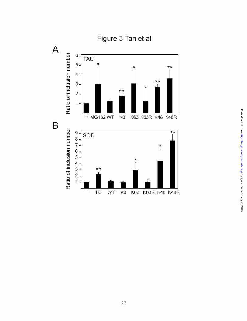
Both K63- and K48-linked ubiquitination promote mutant tau and SOD1
accumulation and inclusion formation
Next, we examined the effects of over expression of different ubiquitin mutants
on the propensity of a disease-associated tau mutant to form inclusions (Fig. 3 & S1).
Examination of mutant (P301L) tau-transfected SH-SY5Y cells under confocal
microscopy reveals a generally uniform cytoplasmic staining of the exogenous tau
protein, although a subset of cells (about 19% of transfected cells) expressing the mutant
tau produce inclusions (Fig. 3A & S1A). Upon MG-132 treatment, the number of
inclusion-positive cells associated with mutant tau expression increases by >3-fold
relative to control cells (Fig. 3A & S1A). This observation is consistent with a recent
report showing that proteasomal inhibition stabilizes tau inclusions (11). Co-expression
of mutant tau with K63 ubiquitin, but not wild type or K63R ubiquitin, in the absence of
MG-132 treatment resulted in a significant enhancement of tau-positive inclusion
8
by guest on February 2, 2015http://hm
g.oxfordjournals.org/D
ownloaded from

formation (Fig. 3A & S1A). The number of tau-positive inclusion also increases
significantly in the presence of K48 and K48R ubiquitin over expression, but more
modestly so in the presence of K0 ubiquitin over expression (Fig. 3A & S1A). Similar
observations are made when we substituted tau mutant with ALS-associated mutant
SOD1, except that in this case, K0 ubiquitin, along with wild type or K63R ubiquitin
species, have no significant effects on the propensity of mutant SOD to form inclusions
when co-expressed (Fig. 3B & S1B). It thus appears that in the presence of disease-
associated proteins, both K48- and K63-linked ubiquitin modifications (and to a lesser
extent, monoubiquitination) could participate in the formation of inclusions mediated by
the pathogenic proteins. Notably, sequential detergent fractionation of lysates prepared
from these transfected cells reveals a selective accumulation of mutant P301L (Fig. S1C)
and mutant A4V SOD (Fig. S1D) protein in the detergent-insoluble fraction with K63
ubiquitin co-expression.
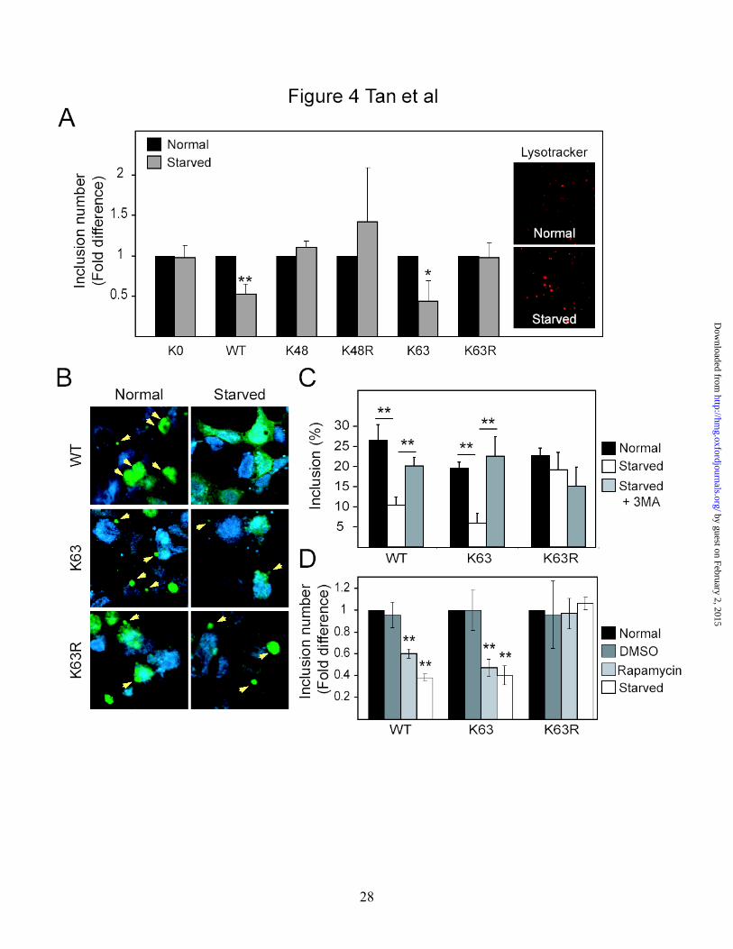
Inclusions enriched with K63-linked polyubiquitin chains are preferentially cleared
by autophagy
Emerging evidence implicates autophagy, a lysosome-mediated bulk degradation
system, as a key regulator of inclusion dynamics (12-14). We wondered whether
ubiquitin linkages on protein inclusions influenced their degradation through autophagy.
Accordingly, autophagic clearance of inclusions formed under conditions of proteasomal
impairment was investigated using a method recently described by Fortun et al (13). SH-
SY5Y cells transiently transfected with wild type, K0, K48, K48R, K63 and K63R
ubiquitin species were initially treated with lactacystin for 16 h to facilitate protein
9
by guest on February 2, 2015http://hm
g.oxfordjournals.org/D
ownloaded from

inclusion formation before a recovery period (24 h) in normal culture media
supplemented either with normal (10%) or low (1%) serum. Cells recovering in low
serum conditions undergo stimulated autophagy (13). Cells were initially visualized using
the lysosomal stain, lysotracker. Consistent with activation of the autophagic system in
response to serum starvation, we observed an apparent increase in the size and number of
lysosomal vacuoles in control cells recovering from proteasomal inhibition in low serum-
containing media compared to those recovering in normal serum-containing media (Fig.
4A). In cells ectopically expressing either wild type or K63 mutant ubiquitin, we
observed a comparable reduction in the number of ubiquitin-positive inclusions when
they were recovering in low serum conditions compared to those that recover in normal
serum (Fig. 4A), suggesting the removal of inclusions via the autophagic pathway.
However, this phenomenon is not observed in cells ectopically expressing all other forms
of ubiquitin species examined, including K48 and K0 ubiquitin mutant (Fig. 4A). To
extend these findings, experiments were conducted with SH-SY5Y cells stably
expressing wild type, K63 or K63R ubiquitin. Similarly, there is a reduction in the size as
well as the number of ubiquitin-positive inclusions in cells stably expressing either wild
type or K63 mutant ubiquitin when these cultures recover in low serum conditions
compared to cultures that recover in normal serum (Fig. 4B & C). Again, the average
reduction in the number of wild type ubiquitin-positive and K63-positive inclusions
associated with serum-starved induced autophagy, i.e. 60% and 70% respectively, are
comparable (Fig. 4B & C). In contrast, the number of inclusions in cells recovering in
low or normal serum remains essentially unchanged in the presence of K63R mutant
ubiquitin over expression (Fig. 4B & C). To exclude the possibility that the observed
10
by guest on February 2, 2015http://hm
g.oxfordjournals.org/D
ownloaded from
reduction of wild type or K63 ubiquitin-positive inclusions in cells undergoing stimulated
autophagy is due to a decrease in their formation rather than an increase in their
clearance, we treated cells recovering in low serum with a widely-used autophagy
inhibitor, 3-methyladenine (3-MA) to block lysosomal degradation. No reduction in the
number of ubiquitin-positive inclusions is observed in either wild type or K63 ubiquitin-
expressing cells recovering in low serum condition in the presence of 3-MA, suggesting
that inhibition of autophagy prevents the removal of inclusions in these cell types (Fig.
4C). On the other hand, 3-MA treatment does not appear to have a significant effect on
the population of inclusions generated in cells expressing K63R ubiquitin (Fig. 4C). This
is consistent with an impaired clearance of inclusions associated with non-K63-linked
ubiquitination (Fig. 4C). Conversely, direct stimulation of autophagy in these cell lines
via pharmacological activation of mTOR by rapamycin essentially reproduces the
phenomenon brought about by serum starvation, thus supporting the role of
macroautophagy in the clearance of K63 ubiquitin-enriched inclusions (Fig. 4D). Similar
observations were made when we repeated our above experiments in the presence of tau
P301L mutant (Fig. S2). We found that tau-positive inclusions formed in cells co-
expressing mutant tau and wild type or K63 ubiquitin are preferentially cleared under
conditions of stimulated autophagy (Fig. S2). On the other hand, co-expression of mutant
tau with K63R ubiquitin leads to impaired autophagic clearance of tau-positive inclusions
(Fig. S2). Collectively, our results suggest that K63-linked ubiquitination facilitates the
autophagic clearance of inclusions.
Lending support to the above suggestion, examination of the different exogenous
ubiquitin-expressing cell lines under electron microscopy reveals the frequent presence of
11
by guest on February 2, 2015http://hm
g.oxfordjournals.org/D
ownloaded from
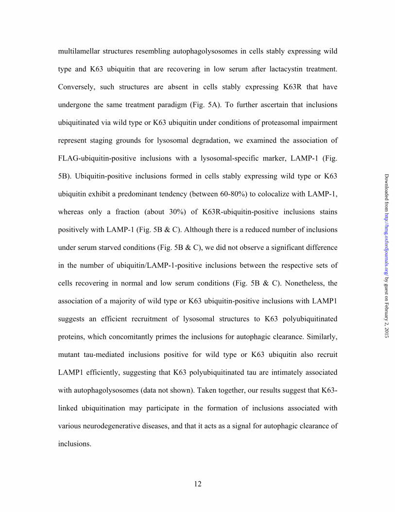
multilamellar structures resembling autophagolysosomes in cells stably expressing wild
type and K63 ubiquitin that are recovering in low serum after lactacystin treatment.
Conversely, such structures are absent in cells stably expressing K63R that have
undergone the same treatment paradigm (Fig. 5A). To further ascertain that inclusions
ubiquitinated via wild type or K63 ubiquitin under conditions of proteasomal impairment
represent staging grounds for lysosomal degradation, we examined the association of
FLAG-ubiquitin-positive inclusions with a lysosomal-specific marker, LAMP-1 (Fig.
5B). Ubiquitin-positive inclusions formed in cells stably expressing wild type or K63
ubiquitin exhibit a predominant tendency (between 60-80%) to colocalize with LAMP-1,
whereas only a fraction (about 30%) of K63R-ubiquitin-positive inclusions stains
positively with LAMP-1 (Fig. 5B & C). Although there is a reduced number of inclusions
under serum starved conditions (Fig. 5B & C), we did not observe a significant difference
in the number of ubiquitin/LAMP-1-positive inclusions between the respective sets of
cells recovering in normal and low serum conditions (Fig. 5B & C). Nonetheless, the
association of a majority of wild type or K63 ubiquitin-positive inclusions with LAMP1
suggests an efficient recruitment of lysosomal structures to K63 polyubiquitinated
proteins, which concomitantly primes the inclusions for autophagic clearance. Similarly,
mutant tau-mediated inclusions positive for wild type or K63 ubiquitin also recruit
LAMP1 efficiently, suggesting that K63 polyubiquitinated tau are intimately associated
with autophagolysosomes (data not shown). Taken together, our results suggest that K63-
linked ubiquitination may participate in the formation of inclusions associated with
various neurodegenerative diseases, and that it acts as a signal for autophagic clearance of
inclusions.
12
by guest on February 2, 2015http://hm
g.oxfordjournals.org/D
ownloaded from

DISCUSSION
The major finding of this study is that non-classic, proteasomal-independent, K63
ubiquitin chain assembly, through its influence on the dynamics of protein inclusions,
represents a novel strategy utilized by the cell to regulate protein homeostasis. By
promoting the formation of protein inclusions as well as tagging them for subsequent
clearance via autophagy, K63-linked polyubiquitination appears to control the life cycle
of an inclusion body.
As protein inclusions are a consistent hallmark of neurodegenerative diseases,
much attention is focussed on their roles in disease pathogenesis and the mechanisms
underlying their biogenesis. Interestingly, several reports (15-18), including an elegant
live-imaging study conducted by Arrasate et al (19), suggest a neuroprotective function
for inclusion formation. This is in agreement with emerging evidence implicating a
pathogenic role of soluble disease-associated protein intermediates (1, 20-23). It is thus
tempting to think that the cell has evolved a defence mechanism against a build up of a
soluble toxic load that it cannot otherwise degrade through normal means by channelling
this load to an inert location for subsequent handling by autophagy. In this way, a
susceptible neuron may prolong its survival and limit neurodegeneration. It is
conceivable, as we and others have recently speculated (8, 24), that non-proteolytic
ubiquitination of proteins could participate in this process, potentially by helping to divert
proteasomal load away from an otherwise overloaded machinery. Supporting this, we
have recently demonstrated that proteasome-independent K63-linked ubiquitination
facilitates the formation of LB-like inclusions in cultured cells (9). Further, a very recent
mass spectrometry-based study demonstrated the accumulation of K63-linked
13
by guest on February 2, 2015http://hm
g.oxfordjournals.org/D
ownloaded from
polyubiquitin in a mouse model of HD (25). In this study, we observed that ectopic
expression of K0, K48 or K63 ubiquitin promotes the formation of tau-positive inclusions
mediated by a co-expressing pathogenic tau mutant. Our results suggest that multiple
ubiquitin topologies, may be involved in inclusions formation. However, these different
types of ubiquitin modification appear to have varying effects on the formation of
intracellular inclusions under different conditions. For example, when mutant tau is
replaced by a SOD mutant, K0 ubiquitin co-expression no longer have an effect on the
propensity of the SOD mutant to generate inclusions. On the other hand, when pathogenic
proteins are absent altogether, only K63 ubiquitin, following its over expression, seems to
be the only one capable of significantly enhancing the formation of inclusions in cultured
cells. These repeated observations, together with our previous study (9), suggest that K63
ubiquitination may be a common denominator underlying protein inclusion formation.
The failure of K48R ubiquitin over expression in mimicking faithfully the effects brought
about by K63 over expression may be due to other ubiquitin linkages promoted by the
K48R mutant, such as the degradation-associated K29-linked polyubiquitination (7).
Likewise, we observe dissimilar effects between K63R and K48 overexpression on
inclusions formation. Notwithstanding this, the precise role of each of the above-
discussed ubiqiuitin species in the biogenesis of disease-related protein inclusions
remains to be further clarified through techniques capable of examining the relative
contribution of endogenous ubiquitin species to inclusion formation.
An interesting recent development pertaining to inclusion dynamics is the
suggestion that inclusion bodies act as staging areas for the disposal of protein aggregates
resistant to proteasomal degradation via the autophagic route (13, 26). Morphological
14
by guest on February 2, 2015http://hm
g.oxfordjournals.org/D
ownloaded from
evidence of autophagy is present in several neurodegenerative disorders, including AD,
PD and HD (12, 27, 28). Moreover, two recent independent reports clearly indicates the
importance of autophagy in neurodegeneration and inclusion formation (29, 30).
However, it remains controversial whether autophagy is clearing monomeric and
oligomeric precursors of aggregates, or inclusions themselves. Unlike the UPS, where the
mechanism of cargo selection is well defined, an important mystery that remains to be
solved is how cargo is selected for autophagic degradation. At least with inclusion bodies,
we have provided a mechanism that underlies cargo recognition by the autophagic
system. Given the similar positive effects elicited by both wild-type and K63, but not
K63R or other forms of ubiquitin examined on the autophagic clearance of inclusions
formed in the presence of proteasomal inhibition, our results suggest that K63 ubiquitin
chain assembly is potentially an important form of ubiquitin modification of intracellular
inclusions that is responsible for subsequently targeting them for autophagy.
In conclusion, we have provided a mechanism that underlies cargo recognition by
the autophagic system as our results suggest that K63 ubiquitin chain assembly is
potentially an important form of ubiquitin modification of intracellular inclusions that is
responsible for subsequently targeting them for autophagy. Harnessing this pathway may
offer innovative approaches in the treatment of neurodegenerative disorders.
15
by guest on February 2, 2015http://hm
g.oxfordjournals.org/D
ownloaded from

MATERIALS AND METHODS
Plasmids, antibodies and reagents. Plasmids expressing HA-tagged ubiquitin and
various ubiquitin mutants have been previously described (9). pCDNA3-FLAG-tagged
wild-type and mutant ubiquitin constructs were derived from their corresponding HA-
tagged counterparts by means of PCR-mediated sub-cloning methodologies. The myc-
tagged tau P301L and FLAG-tagged SOD A4V plasmids were kindly provided by Dr. L.
Petrucelli (Mayo Clinic, USA) and Dr. R. Takahashi (Kyoto University, Japan)
respectively. The following antibodies were used: Monoclonal anti-[c-myc]-peroxidase,
anti-HA-peroxidase, rabbit anti-HA (All from Roche Diagnostics), monoclonal anti-
FLAG-peroxidase, anti-β actin, rabbit anti-γ-tubulin (All from Sigma), rabbit anti-
ubiquitin (DAKO), monoclonal anti-LAMP1 (H5G11) (Santa Cruz Biotechnology Inc),
Rhodamine-conjugated anti-mouse IgG (Molecular Probes), FITC-conjugated anti-mouse
IgG (Jackson ImmunoResearch Laboratories Inc.), FITC-conjugated anti-rabbit IgG (BD
Pharmingen), Rhodamine-conjugated anti-rabbit IgG (Molecular Probes), TritC-
conjugated anti-goat (Santa Cruz) and FITC-conjugated anti-guinea pig IgG (Sigma).
Stock solutions of the following chemicals were prepared and stored at -20oC: MG-132
(1M in DMSO), 3-methyladenine (10 mM in DMEM), Rapamycin (0.2 mg/ml in DMSO)
and Nocodozole (2 mg/ml in DMSO) (All from Sigma), Clasto-lactacystin-β-lactone
(Affiniti Research) (5 mM in dH2O) and LysoTracker® Red DND-99 (Molecular Probes)
(1 mM in DMSO).
16
by guest on February 2, 2015http://hm
g.oxfordjournals.org/D
ownloaded from

Cell culture and transfection. SH-SY5Y cells were grown in DMEM containing 10%
FBS in a 5% CO2 atmosphere. Cells were transiently transfected with various expression
vectors using the LipofectAMINE 2000 reagent (Invitrogen) according to manufacturer’s
instructions. Two days later, sequential fractionation of transfected cell lysates into
Triton-X-soluble (S) and SDS-soluble (P) fractions was performed and analyzed as
previously described (31). To facilitate our study on inclusions clearance, we generated
SH-SY5Y cells stably expressing WT, K63 and K63R ubiquitin using previously
described methods (31). All positive stable cell lines used for the experiments described
here were maintained in serum-containing DMEM supplemented with 200 µg/ml
Geneticin (Invitrogen) to prevent extrusion of the integrated constructs.
Immunocytochemistry, proteasome assay and lysotracking. Cellular localization and
confocal microscopy was carried out as previously described (9, 31). A minimum of 50
cells were counted from each of the three independent wells per experiment. Cells were
counted in a blinded manner and quantitative results reported are an average of at least 3
experiments. Transfected cells are scored as positive when they contain ubiquitin-positive
spherical bodies that are visible at 40X magnification. More often than not, a single
perinuclear inclusion body per cell is observed. Proteasome activities were determined by
incubating lysates (10 µg of protein) with substrates Suc-LLVY-AMC for 1.5 hr at 37°C.
The relative amount of AMC released was measured using a fluorometer equipped with a
380/460 nm filter set (TECAN). Lysosome tracking was carried by incubating cells with
50 nM LysoTracker for 30 min at 37 °C followed by 3 washes in PBS before fixing in
17
by guest on February 2, 2015http://hm
g.oxfordjournals.org/D
ownloaded from

4% paraformaldehyde and mounting with FluorSave reagent (Calbiochem) before
viewing.
Inclusion formation and autophagic removal. The autophagic clearance of inclusions
formed under conditions of proteasomal impairment was investigated using a method
recently described by Fortun et al (13). Cells were first treated with 5 μM lactacystin to
facilitate inclusion formation. After 16 hr incubation, the treated cells were allowed to
recover in normal media for 24 hr. Concurrently, a parallel set of treated cells were
incubated with starvation media (1% serum) to stimulate autophagy either in the presence
or absence of 10 mM 3-methyladenine, an autophagy inhibitor. Thereafter, cells were
processed for immunocytochemical staining for blinded evaluation of inclusions.
Ultrastructural analysis. Cells were washed with PBS prior to fixing in 2.5%
glutaldehyde at 4 °C for 4 hours. After which, cells were washed twice with PBS before
treatment with 2% osmium tetroxide for 2 hours at room temperature. Cells were then
packed in 5% w/v gelatin and fixed with 2.5% glutaldehyde for 10 min before sequential
dehydration and subsequent embedding in aradite resin. Semithin and ultrathin sections
were cut in an ultrotome (Leica UCT) and post-stained with lead citrate prior to viewing
Images were acquired with Ultrathin sections (<100 nm) viewed with Phillips EM208
100kV transmission electron microscope.
18
by guest on February 2, 2015http://hm
g.oxfordjournals.org/D
ownloaded from

Statistical Analysis. Statistical significance for all the quantitative data obtained was
analyzed using Student’s t-test (*P <0.05, **P < 0.001) unless otherwise stated.
ACKNOWLEDGEMENTS
We thank Dr. Mary Ng and Lucas Lu (EM unit, National University of Singapore) for
their help with the electron microscopy studies, and Shaun Mok for technical assistance.
The authors wish it to be known that both the senior authors of this paper, T.M.D. and
L.K.L., contributed equally to the work. This work was supported by grants from
SingHealth Group, Singapore National Medical Research Council NMRC/0776/2003 and
Biomedical Research Council 0613319483 (LKL), Singapore Millennium Foundation
(JT), NIGMS GM67945 (SG) and NINDS Grants NS38377 and NS48206 (TMD). TMD
is the Leonard and Madlyn Abramson Professor in Neurodegenerative Diseases.
CONFLICT OF INTEREST
The authors declare no conflict of interest
19
by guest on February 2, 2015http://hm
g.oxfordjournals.org/D
ownloaded from
REFERENCE
1. Ross, C.A. and Poirier, M.A. (2004) Protein aggregation and neurodegenerative disease. Nat. Med., 10 Suppl, S10-17.
2. Miller, R.J. and Wilson, S.M. (2003) Neurological disease: UPS stops delivering! Trends Pharmacol. Sci., 24, 18-23.
3. Chung, K.K., Dawson, V.L. and Dawson, T.M. (2001) The role of the ubiquitin-proteasomal pathway in Parkinson's disease and other neurodegenerative disorders. Trends Neurosci., 24, S7-14.
4. Pickart, C.M. (2001) Mechanisms underlying ubiquitination. Annu. Rev. Biochem., 70, 503-533. 5. Peng, J., Schwartz, D., Elias, J.E., Thoreen, C.C., Cheng, D., Marsischky, G., Roelofs, J., Finley,
D. and Gygi, S.P. (2003) A proteomics approach to understanding protein ubiquitination. Nat. Biotechnol., 21, 921-926.
6. Hicke, L., Schubert, H.L. and Hill, C.P. (2005) Ubiquitin-binding domains. Nat. Rev. Mol. Cell. Biol., 6, 610-621.
7. Pickart, C.M. (2000) Ubiquitin in chains. Trends Biochem. Sci., 25, 544-548. 8. Mukhopadhyay, D. and Riezman, H. (2007) Proteasome-independent functions of ubiquitin in
endocytosis and signaling. Science, 315, 201-205. 9. Lim, K.L., Chew, K.C., Tan, J.M., Wang, C., Chung, K.K., Zhang, Y., Tanaka, Y., Smith, W.,
Engelender, S., Ross, C.A. et al. (2005) Parkin mediates nonclassical, proteasomal-independent ubiquitination of synphilin-1: implications for Lewy body formation. J. Neurosci., 25, 2002-2009.
10. Bence, N.F., Sampat, R.M. and Kopito, R.R. (2001) Impairment of the ubiquitin-proteasome system by protein aggregation. Science, 292, 1552-1555.
11. Goldbaum, O., Oppermann, M., Handschuh, M., Dabir, D., Zhang, B., Forman, M.S., Trojanowski, J.Q., Lee, V.M. and Richter-Landsberg, C. (2003) Proteasome inhibition stabilizes tau inclusions in oligodendroglial cells that occur after treatment with okadaic acid. J. Neurosci., 23, 8872-8880.
12. Cuervo, A.M. (2006) Autophagy in neurons: it is not all about food. Trends Mol. Med., 12, 461-464.
13. Fortun, J., Dunn, W.A., Jr., Joy, S., Li, J. and Notterpek, L. (2003) Emerging role for autophagy in the removal of aggresomes in Schwann cells. J. Neurosci., 23, 10672-10680.
14. Iwata, A., Riley, B.E., Johnston, J.A. and Kopito, R.R. (2005) HDAC6 and microtubules are required for autophagic degradation of aggregated huntingtin. J. Biol. Chem., 280, 40282-40292.
15. Bowman, A.B., Yoo, S.Y., Dantuma, N.P. and Zoghbi, H.Y. (2005) Neuronal dysfunction in a polyglutamine disease model occurs in the absence of ubiquitin-proteasome system impairment and inversely correlates with the degree of nuclear inclusion formation. Hum. Mol. Genet., 14, 679-691.
16. Cummings, C.J., Reinstein, E., Sun, Y., Antalffy, B., Jiang, Y., Ciechanover, A., Orr, H.T., Beaudet, A.L. and Zoghbi, H.Y. (1999) Mutation of the E6-AP ubiquitin ligase reduces nuclear inclusion frequency while accelerating polyglutamine-induced pathology in SCA1 mice. Neuron, 24, 879-892.
17. Klement, I.A., Skinner, P.J., Kaytor, M.D., Yi, H., Hersch, S.M., Clark, H.B., Zoghbi, H.Y. and Orr, H.T. (1998) Ataxin-1 nuclear localization and aggregation: role in polyglutamine-induced disease in SCA1 transgenic mice. Cell, 95, 41-53.
18. Saudou, F., Finkbeiner, S., Devys, D. and Greenberg, M.E. (1998) Huntingtin acts in the nucleus to induce apoptosis but death does not correlate with the formation of intranuclear inclusions. Cell, 95, 55-66.
19. Arrasate, M., Mitra, S., Schweitzer, E.S., Segal, M.R. and Finkbeiner, S. (2004) Inclusion body formation reduces levels of mutant huntingtin and the risk of neuronal death. Nature, 431, 805-810.
20. Ross, C.A. and Pickart, C.M. (2004) The ubiquitin-proteasome pathway in Parkinson's disease and other neurodegenerative diseases. Trends Cell Biol., 14, 703-711.
21. Walsh, D.M., Klyubin, I., Fadeeva, J.V., Rowan, M.J. and Selkoe, D.J. (2002) Amyloid-beta oligomers: their production, toxicity and therapeutic inhibition. Biochem. Soc. Trans., 30, 552-557.
20
by guest on February 2, 2015http://hm
g.oxfordjournals.org/D
ownloaded from
22. Sanchez, I., Mahlke, C. and Yuan, J. (2003) Pivotal role of oligomerization in expanded polyglutamine neurodegenerative disorders. Nature, 421, 373-379.
23. Volles, M.J. and Lansbury, P.T., Jr. (2003) Zeroing in on the pathogenic form of alpha-synuclein and its mechanism of neurotoxicity in Parkinson's disease. Biochemistry, 42, 7871-7878.
24. Lim, K.L., Dawson, V.L. and Dawson, T.M. (2006) Parkin-mediated lysine 63-linked polyubiquitination: a link to protein inclusions formation in Parkinson's and other conformational diseases? Neurobiol. Aging, 27, 524-529.
25. Bennett, E.J., Shaler, T.A., Woodman, B., Ryu, K.Y., Zaitseva, T.S., Becker, C.H., Bates, G.P., Schulman, H. and Kopito, R.R. (2007) Global changes to the ubiquitin system in Huntington's disease. Nature, 448, 704-708.
26. Kopito, R.R. (2000) Aggresomes, inclusion bodies and protein aggregation. Trends Cell Biol., 10, 524-530.
27. Levine, B. and Yuan, J. (2005) Autophagy in cell death: an innocent convict? J. Clin. Invest., 115, 2679-2688.
28. Rubinsztein, D.C., Difiglia, M., Heintz, N., Nixon, R.A., Qin, Z.H., Ravikumar, B., Stefanis, L. and Tolkovsky, A. (2005) Autophagy and its possible roles in nervous system diseases, damage and repair. Autophagy, 1, 11-22.
29. Komatsu, M., Waguri, S., Chiba, T., Murata, S., Iwata, J.I., Tanida, I., Ueno, T., Koike, M., Uchiyama, Y., Kominami, E. et al. (2006) Loss of autophagy in the central nervous system causes neurodegeneration in mice. Nature, 441, 880-884.
30. Hara, T., Nakamura, K., Matsui, M., Yamamoto, A., Nakahara, Y., Suzuki-Migishima, R., Yokoyama, M., Mishima, K., Saito, I., Okano, H. et al. (2006) Suppression of basal autophagy in neural cells causes neurodegenerative disease in mice. Nature, 441, 885-889.
31. Wang, C., Ko, H.S., Thomas, B., Tsang, F., Chew, K.C., Tay, S.P., Ho, M.W., Lim, T.M., Soong, T.W., Pletnikova, O. et al. (2005) Stress-induced Alterations in Parkin Solubility Promote Parkin Aggregation and Compromise Parkin's Protective Function. Hum. Mol. Genet., 14, 3885-3897
21
by guest on February 2, 2015http://hm
g.oxfordjournals.org/D
ownloaded from

LEGEND TO FIGURES Figure 1. K63-linked ubiquitination promotes the stability of the short-lived GFPu
protein (A) Fluorescence microscopy of HEK 293 cells transfected with GFPu in the
presence and absence of proteasome inhibitor, MG-132 or in the presence of wild type or
mutant ubiquitin co-expression. A carefully titrated amount of GFPu was used in our
transfection paradigm such that GFPu expression is negligible in untreated control cells.
(B) Anti-GFP immunoblots of cell extracts sequentially prepared with Triton X-100 (S)
and SDS (P) from HEK 293 cells transfected with GFPu alone or with various HA-tagged
ubiquitin species. Membranes were stripped and re-probed for β-actin to show equivalent
loading of the different lysates. (C) Anti-GFP and anti-HA immunoblots of GFPu
immunoprecipitates (IPGFP) prepared from HEK 293 cells transfected with GFPu alone or
with wild type K63 or K0 ubiquitin. Each of these experiments was repeated at least three
times. (D) Bar graph showing the chymotrypsin-like proteasome activities of lysates
prepared from cells transfected with various ubiquitin species, as indicated. Control cells
treated with MG-132 is indicated. (*P < 0.05, **P < 0.001 vs untreated control group).
Figure 2. K63-linked ubiquitination enhances the formation of protein aggregates
(A) Representative confocal images of SH-SY5Y cells ectopically expressing WT, K63
or K63R ubiquitin immunostained with anti-ubiquitin (green) or anti-γ-tubulin (red).
Arrows in merge pictures show the co-localization of K63 (but not WT or K63R)
ubiquitin-positive inclusions with γ-tubulin (yellow). (B) Bar graph showing the
percentage of cells containing anti-HA-ubiquitin-positive inclusions in cells ectopically
expressing various ubiquitin species, as indicated. (*P < 0.05, **P < 0.001 vs control
22
by guest on February 2, 2015http://hm
g.oxfordjournals.org/D
ownloaded from

group) (C) Dispersion of anti-FLAG (Ub)-positive inclusions (green) occurs in
nocodozole-treated K63 ubiquitin-expressing cells (D) Anti-HA immunoblots of cell
extracts sequentially prepared with Triton X-100 (S) and SDS (P) from SH-SY5Y cells
transfected with various ubiquitin species, as indicated. Each of these experiments was
repeated at least three times.
Figure 3. Both K48- and K63-linked ubiquitination promotes the formation of tau-
and SOD-positive inclusions. Bar graph showing the ratio of inclusion number relative
to untreated cells (-) observed in (A) P301L mutant tau-transfected cells or (B) SOD-A4V
mutant-transfected cells under various conditions (*P < 0.05, **P < 0.001 vs control
group). LC refers to lactacystin-treated cells. Each of these experiments was repeated at
least three times.
Figure 4. Inclusions enriched with K63 ubiquitin are preferentially degraded by the
autophagic system (A) Bar graph showing the relative fold difference in the number of
inclusions observed in SH-SY5Y cells transfected with HA-tagged WT, K0, K63, K63R,
K48 and K48R ubiquitin recovering in low (1%) or normal (10%) serum after treatment
with lactacystin (*P < 0.05, **P < 0.001 vs control group). Note the increased number
and size of lysotracker-stained lysosomes in starved cells (inset). (B) Representative
confocal images of anti-FLAG (ubiquitin)-stained SH-SY5Y cells stably expressing WT,
K63 or K63R ubiquitin that have undergone similar treatment. The relative difference in
number of inclusions observed among these cells, as well as with those treated with 3-
MA or rapamycin in the presence of low serum conditions, is depicted by the bar graph
23
by guest on February 2, 2015http://hm
g.oxfordjournals.org/D
ownloaded from

shown in (C) and (D), as indicated (*P < 0.05, **P < 0.001). Each of these experiments
was repeated at least three times.
Figure 5. K63 ubiquitin-positive inclusions are enriched with LAMP1 (A)
Representative electron micrographs showing multi-lamellar structures resembling
autophagolysosome in SH-SY5Y cells stably expressing wild type and K63 ubiquitin (but
not K63R) that have undergone a similar treatment regime as in described in Fig. 4A. (B)
Representative confocal images of SH-SY5Y cells stably expressing WT, K63 or K63R
ubiquitin that have undergone the above treatment regime immunostained with anti-
FLAG (ubiquitin) (green) or anti-LAMP1 (red). Arrows in merge pictures show the co-
localization of LAMP1 with wild type and K63 (but not K63R) ubiquitin-positive
inclusions (yellow). (C) Bar graph showing the percentage of LAMP-1/ubiquitin-positive
inclusions in these cells (*P < 0.05, K63 or K63R vs WT). Each of these experiments was
repeated at least three times.
24
by guest on February 2, 2015http://hm
g.oxfordjournals.org/D
ownloaded from

25
by guest on February 2, 2015http://hm
g.oxfordjournals.org/D
ownloaded from

26
by guest on February 2, 2015http://hm
g.oxfordjournals.org/D
ownloaded from
27
by guest on February 2, 2015http://hm
g.oxfordjournals.org/D
ownloaded from
28
by guest on February 2, 2015http://hm
g.oxfordjournals.org/D
ownloaded from

29
by guest on February 2, 2015http://hm
g.oxfordjournals.org/D
ownloaded from
Abbreviations
AD, Alzheimer’s disease; ALS, Amyotrophic Lateral Sclerosis; LB, Lewy body; PD,
Parkinson’s disease; UPS, Ubiquitin-proteasome system
30
by guest on February 2, 2015http://hm
g.oxfordjournals.org/D
ownloaded from
Copyright © 2022 FDOKUMEN


![[Current Topics in Microbiology and Immunology] || Modulation of the Ubiquitination Machinery by Legionella](https://static.fdokumen.com/doc/165x107/63328412f0080405510482a9/current-topics-in-microbiology-and-immunology-modulation-of-the-ubiquitination.jpg)