JNK: A Stress-Activated Protein Kinase Therapeutic Strategies and Involvement in Alzheimer's and...
15
JNK: A Stress-Activated Protein Kinase Therapeutic Strategies and Involvement in Alzheimer’ s and Various Neurodegenerative Abnormalities Sidharth Mehan & Harikesh Meena & Deepak Sharma & Rameshwar Sankhla Received: 16 August 2010 / Accepted: 16 September 2010 # Springer Science+Business Media, LLC 2010 Abstract The c-Jun N-terminal kinase (JNKs), also known as stress-activated protein kinase (SAPK), is one such family of multifunctional-signaling molecules, activated in response to wide range of cellular stresses as well as in response to inflammatory mediators. JNKs regulate various processes such as brain development, repair, and memory formation; but on the other hand, JNKs are potent effectors of neuroinflammation and neuronal death. A large body of evidence indicates that JNK activity is critical for normal immune and inflammatory response. Indeed, aberrant activation of JNK has been implicated in the pathogenesis of Alzheimer’ s disease. Moreover, the JNK pathway is considered to be a key regulator of various inflammatory pathways which are activated during normal aging and Alzheimer’ s disease therapy as well as key regulator of pro- inflammatory cytokines biosynthesis at the transcriptional and translational levels, which makes different components of these pathway potential targets for the treatment of autoimmune and inflammatory diseases. Pharmacological inhibition of JNK has been demonstrated to attenuate microglial activation and the release of neurotoxic chemicals including pro-inflammatory cytokines. In this review, we provide an overview on implications and therapeutic strategies of JNK in neurodegenerative disorders. Keywords Alzheimer’ s disease . Neuroinflammation . c-Jun N-terminal kinase Alzheimer’s Disease: A Review Alzheimer’ s disease (AD) is a late onset, age-related neuro- degenerative disorder (Jellinger 2006) clinically characterized by progressive deterioration of cognitive functions (Chong et al. 2005). Epidemiologically, AD is the sixth leading cause of death (Zhao and Townsend 2009). At cellular level, major neuropathological lesions of AD include extracellular deposits of amyloid beta (Aβ) peptides leading to formation of senile/neuritic plaques and intracellular neurofibrillary tangles (NFTs), which are paired helical filaments of hyper- phosphorylated tau proteins (Maiese and Chong 2004; Chong et al. 2005). Aβ plaques and tau tangles are present and show their neurotoxic effects mainly in entorhinal cortex, hippocampus, nucleus of basalis Meynert, basal forebrain, and amygdale–brain regions playing major role in learning and memory (Buee et al. 2000; Katsuno et al. 2005). At the molecular, cellular, and biochemical levels, cerebroenergetic decline (Blass et al. 2000; Moreira et al. 2005) and oxidative stress (Smith et al. 2002; Moreira et al. 2005) with mitochondrial abnormalities have been reported very early in the initial stages of AD and detected all across the brain regions (Kidd 2005; Moreira et al. 2009). Decline S. Mehan (*) Department of Pharmacology, GD Memorial College of Pharmacy, Jodhpur 342005 Rajasthan, India e-mail: [email protected] H. Meena Department of Phamaceutics, GD Memorial College of Pharmacy, Jodhpur 342005 Rajasthan, India e-mail: [email protected] D. Sharma : R. Sankhla Department of Pharmaceutical Chemistry, GD Memorial College of Pharmacy, Jodhpur 342005 Rajasthan, India D. Sharma e-mail: [email protected] R. Sankhla e-mail: [email protected] J Mol Neurosci DOI 10.1007/s12031-010-9454-6
-
Upload
independent -
Category
Documents
-
view
0 -
download
0
Transcript of JNK: A Stress-Activated Protein Kinase Therapeutic Strategies and Involvement in Alzheimer's and...

JNK: A Stress-Activated Protein Kinase TherapeuticStrategies and Involvement in Alzheimer’sand Various Neurodegenerative Abnormalities
Sidharth Mehan & Harikesh Meena & Deepak Sharma &
Rameshwar Sankhla
Received: 16 August 2010 /Accepted: 16 September 2010# Springer Science+Business Media, LLC 2010
Abstract The c-Jun N-terminal kinase (JNKs), also knownas stress-activated protein kinase (SAPK), is one suchfamily of multifunctional-signaling molecules, activated inresponse to wide range of cellular stresses as well as inresponse to inflammatory mediators. JNKs regulate variousprocesses such as brain development, repair, and memoryformation; but on the other hand, JNKs are potent effectorsof neuroinflammation and neuronal death. A large body ofevidence indicates that JNK activity is critical for normalimmune and inflammatory response. Indeed, aberrantactivation of JNK has been implicated in the pathogenesisof Alzheimer’s disease. Moreover, the JNK pathway isconsidered to be a key regulator of various inflammatorypathways which are activated during normal aging andAlzheimer’s disease therapy as well as key regulator of pro-inflammatory cytokines biosynthesis at the transcriptionaland translational levels, which makes different components
of these pathway potential targets for the treatment ofautoimmune and inflammatory diseases. Pharmacologicalinhibition of JNK has been demonstrated to attenuatemicroglial activation and the release of neurotoxic chemicalsincluding pro-inflammatory cytokines. In this review, weprovide an overview on implications and therapeuticstrategies of JNK in neurodegenerative disorders.
Keywords Alzheimer’s disease . Neuroinflammation .
c-Jun N-terminal kinase
Alzheimer’s Disease: A Review
Alzheimer’s disease (AD) is a late onset, age-related neuro-degenerative disorder (Jellinger 2006) clinically characterizedby progressive deterioration of cognitive functions (Chong etal. 2005). Epidemiologically, AD is the sixth leading causeof death (Zhao and Townsend 2009). At cellular level,major neuropathological lesions of AD include extracellulardeposits of amyloid beta (Aβ) peptides leading to formationof senile/neuritic plaques and intracellular neurofibrillarytangles (NFTs), which are paired helical filaments of hyper-phosphorylated tau proteins (Maiese and Chong 2004;Chong et al. 2005). Aβ plaques and tau tangles are presentand show their neurotoxic effects mainly in entorhinalcortex, hippocampus, nucleus of basalis Meynert, basalforebrain, and amygdale–brain regions playing major rolein learning and memory (Buee et al. 2000; Katsuno et al.2005). At the molecular, cellular, and biochemical levels,cerebroenergetic decline (Blass et al. 2000; Moreira et al.2005) and oxidative stress (Smith et al. 2002; Moreira et al.2005) with mitochondrial abnormalities have been reportedvery early in the initial stages of AD and detected all acrossthe brain regions (Kidd 2005; Moreira et al. 2009). Decline
S. Mehan (*)Department of Pharmacology,GD Memorial College of Pharmacy,Jodhpur 342005 Rajasthan, Indiae-mail: [email protected]
H. MeenaDepartment of Phamaceutics, GD Memorial College of Pharmacy,Jodhpur 342005 Rajasthan, Indiae-mail: [email protected]
D. Sharma : R. SankhlaDepartment of Pharmaceutical Chemistry,GD Memorial College of Pharmacy,Jodhpur 342005 Rajasthan, India
D. Sharmae-mail: [email protected]
R. Sankhlae-mail: [email protected]
J Mol NeurosciDOI 10.1007/s12031-010-9454-6

in mitochondrial enzyme activities such as pyruvate dehy-drogenase (PDH) and desensitization of insulin receptors(IR) have been shown to play major role in cerebroenergeticfailure in AD (Sorbi et al. 1983; Gibson et al. 1998; Kidd2005; Steen et al. 2005; Parihar and Brewer 2007).Furthermore, mitoenergetic failure has been linked toneurotransmitter deficit (Gibson and Blass 1976; Shohamet al. 2003), excitotoxicity (Mason et al. 1999; Sonkusare etal. 2005), oxidative stress (Sharma and Gupta 2002),amyloid beta deposists, and tau hyperphosphorylation(Grunblatt et al. 2007) in AD brain. All these stressconditions have been reported to cause neuronal deathleading to cognitive abnormalities including memory loss(Gibson and Blass 1976; Mason et al. 1999; Shoham et al.2003; Sonkusare et al. 2005; Fig. 1).
But, recently it has been shown that stress-activatedprotein kinases (SAPKs) play a major role in neuronaldegeneration under stress conditions (Davis 2000; Bendottiet al. 2006). There are two well-characterized subfamiliesof SAPKs viz., c-Jun-N-terminal kinase (JNK) and p38protein kinase (Xia et al. 1995; Yang et al. 2007b; Mehan etal. 2010), generally activated by stress stimuli (Kyriakisand Avruch 1996; Davis 1999; Sugino et al. 2000). Theyare all activated by dual phosphorylation of both tyrosineand threonine residues (Derijard et al. 1994).
JNKs belong to the family of mitogen-activated proteinkinases (MAPKs) particularly to serine/threonine class ofprotein kinases (Force et al. 2004). The enzyme has beenreported to phosphorylate N-terminal of its substrate, i.e.,c-Jun (Derijard et al. 1994; Kallunki et al. 1994; Guan et
al. 2005). Three genes of JNKs have been identified asJNK-1, JNK-2, and JNK-3, encoding for ten differentisoforms (Gupta et al. 1996; Coffey et al. 2002). All theisoforms are activated by various extracellular stimuli andtheir expression has been reported to be increased in ADbrain (Zhu et al. 2001b; Yoshida et al. 2004). JNK 1 andJNK 2 are constitutively expressed in a large variety oftissues where as JNK 3 isoform seems primarily localizedto neuronal tissues, cardiac myocytes, and testis (Gupta etal. 1996; Johnson and Nakamura 2007). JNKs have beenshown to regulate the levels of functional proteins both attranslational and post-transcriptional levels (Pulverer et al.1991). The major transcriptional factor involved in mediatingvarious activities is activator protein-1 (AP-1) which iscomposed of c-Jun and Fos family of hetrodimers. Both c-Jun and Fos are immediate early genes, i.e., their transcrip-tion is rapidly induced by variety of stimuli (Raivich andBehrens 2006). JNKs-mediated activation of AP-1 isreported to be involved in various activities such asinflammatory and apoptotic activities (Behrens et al. 1999;Raivich et al. 1999; Herdegen and Waetzig 2001).
As mentioned above, pyruvate dehydrogenase plays fun-damental role in energy homeostasis (Patel and Korotchkina2006). The activities of mitochondrial enzymes, particularlyPDH, have been reported to be declined in AD brain(Gibson et al. 1998; Kidd 2005; Parihar and Brewer 2007).The decreased PDH enzyme activity has been suggested tobe due to prolonged activation of JNKs (SAPKs; Zhou et al.2008, 2009). It is further well known that IR signaling playsa major role in energy homeostasis, neuronal survival, and
TAU Tangle FormationAβ-Aggregation
Glial Cell Activation Insulin receptor
Desensitization
Glutamate Release Oxidative Stress
Ca2+Overload
ROS
COX/LOX & Energy FailureRNS
Cytokine ExcitotoxictySignaling
NEURONALDEATH
CholinergicDysfunctionNeuroinflammaton
ALZHEIMER’S DISEASES
Fig. 1 Schematic presentationof neuronal death via variouspathways in Alzheimer’s disease
J Mol Neurosci

cognitive functions. Impaired IR signaling has been reportedduring aging as well as in AD brain (Steen et al. 2005; Coleand Frautschy 2007; Planel et al. 2007). Prolonged activationof JNKs has been demonstrated to cause IR desensitizationby negative regulation of insulin receptor substrate-1 (IRS-1) and thus implicated in impaired energy homeostasis(Levkovitz et al. 2007). Furthermore, overactivation ofJNKs has been considered as apoptotic signal that leads toinactivation of BCl-2 protein while the latter promotescytochrome-c release from mitochondria (Tournier et al.2000; Waetzig and Herdegen 2004; Hui et al. 2005). JNKsare also reported to be functionally active in human cellsparticularly microglia and involved in enlargement ofmicroglia (Waetzig and Herdegen 2004), induction of pro-inflammatory enzymes, and the release of proinflammatorymediators (Wang et al. 2004). Recently, activated JNKs havebeen shown to be associated with hippocampal NFTsformation (Yoshida et al. 2004; Lagalwar et al. 2006) andalso that amyloid beta may produce its neurotoxic effectsthrough JNKs activation (Hashimoto et al. 2003).
Stress-Activated Protein Kinases in Neurodegeneration
There are two well-characterized subfamilies of SAPKsviz., JNK and p38 protein kinase (Xia et al. 1995; Yang etal. 2007a), generally activated by stress stimuli (Kyriakisand Avruch 1996; Sugino et al. 2000). They are allactivated by dual phosphorylation of both tyrosine andthreonine residues (Derijard et al. 1994).
JNK
JNKs (SAPKs) belong to the family of MAPKs, particu-larly serine/threonine class of protein kinases. They havebeen reported to phosphorylate N-terminal of its substrate,i.e., c-Jun (Derijard et al. 1994; Kallunki et al. 1994; Guanet al. 2005). Each JNK is expressed as a short form(46 kDa) and long form (54 kDa) (Pulverer et al. 1991).Three genes of JNK have been identified viz., JNK-1, JNK-2, and JNK-3 which encode for 10 different isoforms. Theyare activated by various extracellular stimuli and theirexpression has been reported to be increased in AD brain(Zhu et al. 2001a; Zhu et al. 2001b). JNK-1 and JNK-2 areconstitutively expressed in a large variety of tissues whereas JNK-3 isoform seems primarily localized to neuronaltissues, cardiac myocytes and testis (Gupta et al. 1996;Johnson and Nakamura 2007).
JNK Structure All 10 splice variants of the three humanJNK genes are sequenced and structurally analyzed.Irrespective of the high level of homology (80%), theisoforms show some differences in their domain assemblysuch as C-terminal extension or different exon usage
between sub-domains which can be associated withfunctional specificity (Gupta et al. 1996). JNK3 isoform,for example, are characterized by an N-terminal extension,which contains the binding domain for the scaffold proteinβ-arrestin-2 (Guo and Whtimarsh 2008). For the majorityof JNK substrates and binding partners, however, little isknown about isoform-specific affinities to their substrates invivo, particularly in the brain. Apart from amino acidsequences and transcript modifications, the crystal struc-tures of JNK1 (Heo et al. 2004) and JNK3 (Xie et al. 1998)have been published. Very recently, the crystal structure ofJNK2 was determined with a peculiar conformation of theactivation loop; when JNK2 adopts this conformation itcannot be phosphorylated by upstream kinases (Shaw et al.2008). Indeed, studies on epithelial morphogenesis revealedthat differences in the variable region (amino acids 173–190) of JNK1 and JNK2 determine their structuralconformation and in consequence the status of phosphory-lation (Takatori et al. 2008; Fig. 2).
Thus, it is not the mere presence of activated upstreamkinases, but the protein structure and conformation, too,which determine the activity of JNKs.
JNK/SAPK Regulatory Network
The JNKs (JNK1, 2, and 3) are known as regulators of genetranscription playing a role in neuronal death associatedwith neurodegenerative diseases (Bendotti et al. 2006). TheJNKs respond to a variety of stimuli collectively designatedas stress-signals [ultraviolet (UV) and gamma irradiation,osmotic or oxidative stress, heat shock proteins, inflamma-tory cytokines, growth factors, and excitotoxicity], leadingto phosphorylation of its substrates (Bogoyevitch and Kobe2006).
Upstream Kinases JNKs, like all MAPKs, are part of themodular structure of a three kinase signaling cascade withmitogen-activated protein kinase kinase (MKK4) and(MKK7) as direct activators and at least 14 mitogen-activated protein kinase kinase kinases (MAP3K; Johnsonand Nakamura 2007). In addition, the cellular distributionof upstream kinases affects JNK activities. In cerebellargranule neurons, for example, MKK4 is present in thesoma, dendrites, and axons, while MKK7 is almostexclusively restricted to the nucleus (Coffey et al. 2000).The Thr-X-Tyr motif in the activation loop of each JNK isdually phosphorylated at threonine 183 (Thr183) andtyrosine 185 (Tyr185) by specific MAPK kinases (MKKs)[MKK4 and MKK7]. MKKs (MKK4 and MKK7) are inturn phosphorylated at specific serine (Ser) or threonine(Thr) residues within their activation loop (Ser 257 andThr 265 for MKK4; Ser 271 and Thr 275 for MKK7) by
J Mol Neurosci

MAPKKKs that includes apoptosis-signal regulatingkinase 1 (ASK1), transforming growth factor-beta-activatedprotein kinase 1 (TAK1), dual leucine zipper-bearing kinase(DLK), leucine zipper bearing-kinase (LZK), tumor progres-sion locus-2 (TPL2), mixed lineage kinase (MLK)-likemitogen-activated protein triple kinase, MEKK1-5, andmixed lineage kinase (MLK1-3, MLK7), etc. (Raivich andBehrens 2006; Manning and Davis 2003). Many MKKKsare activated by GTPases such as Ras (the Raf MKKKs) orRho family GTPases, cell division cycle-42 (Cdc42), andRac1 (Gallo and Johnson 2002; Gallagher et al. 2004; Chenand Cobb 2006).
Scaffold Proteins and Other Modulators Scaffold proteinsor repeat-motif extended proteins, capable of simultaneous
interaction with numerous proteins, function as couplersbetween specific subgroups of MAPKs and their activators(e.g., mitogen-activated protein kinase kinases (MAP2Ks orMKKs), substrates (e.g., transcription factors) or inactiva-tors (e.g., MKPs). Scaffolds of JNK signaling includemembers of the JNK-interacting protein family (c-Jun N-terminal kinase interacting proteins, JIPs; Whitmarsh 2006),β-arrestin-2, plenty of SH3, filamin B, stress-activatedprotein kinase pathway-regulating phosphatase 1, GRIP1-associated protein 1, or IκB kinase complex-associatedprotein (Waetzig et al. 2006). Importantly, some scaffoldssuch as β-arrestin-2 have an isoform specificity whichexclusively binds JNK3 at its N-terminal elongation whileothers such as JIP1 bind to all three JNKs (Dickens et al.1997; Guo and Whtimarsh 2008). Next, scaffolds modulate
JNK-1 JNK-2
JNK-3
Fig. 2 Structure of C-Jun-N-terminal kinases (JNKs); JNK-1 (Heo et al. 2004) JNK-2 (Shaw et al. 2008) and JNK-3 (Bogoyevitch and Kobe 2006)
J Mol Neurosci
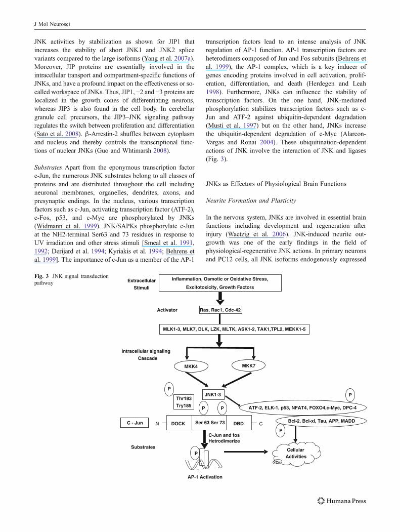
JNK activities by stabilization as shown for JIP1 thatincreases the stability of short JNK1 and JNK2 splicevariants compared to the large isoforms (Yang et al. 2007a).Moreover, JIP proteins are essentially involved in theintracellular transport and compartment-specific functions ofJNKs, and have a profound impact on the effectiveness or so-called workspace of JNKs. Thus, JIP1, −2 and −3 proteins arelocalized in the growth cones of differentiating neurons,whereas JIP3 is also found in the cell body. In cerebellargranule cell precursors, the JIP3–JNK signaling pathwayregulates the switch between proliferation and differentiation(Sato et al. 2008). β-Arrestin-2 shuffles between cytoplasmand nucleus and thereby controls the transcriptional func-tions of nuclear JNKs (Guo and Whtimarsh 2008).
Substrates Apart from the eponymous transcription factorc-Jun, the numerous JNK substrates belong to all classes ofproteins and are distributed throughout the cell includingneuronal membranes, organelles, dendrites, axons, andpresynaptic endings. In the nucleus, various transcriptionfactors such as c-Jun, activating transcription factor (ATF-2),c-Fos, p53, and c-Myc are phosphorylated by JNKs(Widmann et al. 1999). JNK/SAPKs phosphorylate c-Junat the NH2-terminal Ser63 and 73 residues in response toUV irradiation and other stress stimuli [Smeal et al. 1991,1992; Derijard et al. 1994; Kyriakis et al. 1994; Behrens etal. 1999]. The importance of c-Jun as a member of the AP-1
transcription factors lead to an intense analysis of JNKregulation of AP-1 function. AP-1 transcription factors areheterodimers composed of Jun and Fos subunits (Behrens etal. 1999), the AP-1 complex, which is a key inducer ofgenes encoding proteins involved in cell activation, prolif-eration, differentiation, and death (Herdegen and Leah1998). Furthermore, JNKs can influence the stability oftranscription factors. On the one hand, JNK-mediatedphosphorylation stabilizes transcription factors such as c-Jun and ATF-2 against ubiquitin-dependent degradation(Musti et al. 1997) but on the other hand, JNKs increasethe ubiquitin-dependent degradation of c-Myc (Alarcon-Vargas and Ronai 2004). These ubiquitination-dependentactions of JNK involve the interaction of JNK and ligases(Fig. 3).
JNKs as Effectors of Physiological Brain Functions
Neurite Formation and Plasticity
In the nervous system, JNKs are involved in essential brainfunctions including development and regeneration afterinjury (Waetzig et al. 2006). JNK-induced neurite out-growth was one of the early findings in the field ofphysiological-regenerative JNK actions. In primary neuronsand PC12 cells, all JNK isoforms endogenously expressed
Inflammation, Osmotic or Oxidative Stress,
Excitotoxicity, Growth Factors
Ras, Rac1, Cdc-42
MLK1-3, MLK7, DLK, LZK, MLTK, ASK1-2, TAK1,TPL2, MEKK1-5
JNK1-3
DOCK Ser 63 Ser 73 DBD CN
Thr183
Try185
C - Jun
-
AP-1 Activation
CellularActivities
Extracellular
Stimuli
Activator
Intracellular signaling
Cascade
Substrates
C-Jun and fosHetrodimerize
Bcl-2, Bcl-xl, Tau, APP, MADD
MKK4 MKK7
ATF-2, ELK-1, p53, NFAT4, FOXO4,c-Myc, DPC-4
P
P
PP
P
P
Fig. 3 JNK signal transductionpathway
J Mol Neurosci

or transfected could induce sprouting (Waetzig and Herdegen2003; Riese et al. 2004; Eminel et al. 2008). Overall,however, specific functions of individual JNK isoforms inthe brain remain elusive, and the only outcome of thiscomplex task is the finding of cytoplasmic JNK1 beingessential for microtubule stability and microtubule-associatedprotein phosphorylation (Chang et al. 2003; Bjorkblom et al.2005). The analysis of the nerve growth factor-dependentJNK signaling cascade during cellular differentiation is evenmore difficult. Among the group of MAP3Ks, only p21-activated kinase 1 (PAK1) has a significant effect onsprouting (Daniels et al. 1998), and the question for theunderlying scaffold proteins has hardly been addressed.Cellular localization and movement kinetics suggest a roleof JIP1 and JIP3 in the organization of JNK-mediatedmicrotubule dynamics underlying neurite outgrowth and cellpolarity (Sato et al. 2004; Dajas-Bailador et al. 2008). Othercytoplasmic JNK substrates such as doublecortin (DCX),SCG10, or microtubule-associated protein (MAP)2 canpromote neurite extension and stability (Chang et al. 2003;Bjorkblom et al. 2005; Tararuk et al. 2006). Despite theiraccepted importance in neurite formation, JNKs are onlypart of the overall cellular response. In addition to the well-established parallel signaling of JNKs and phosphoinositide-3-kinase or extracellular signal-regulated kinase 1/2 (Kita etal. 1998; Waetzig and Herdegen 2003), new evidencesuggests crosstalks, e.g., between Wnt signaling and JNKs,whereby JNKs stabilize microtubules (Rosso et al. 2005;Ciani and Salinas 2007).
Brain and Development
Kuan et al. (1999) were the first to show that JNK1 andJNK2 are indispensable for the intact development of thenervous system. Similarly, the upstream activators MKK4and MKK7 are both essential, since early embryonic deathoccurs in mice lacking either mkk4 or mkk7 genes (Wanget al. 2007a). Specific actions of MKK4 and MKK7 havebeen attributed to their distinct tissue distribution andcellular localization (Coffey et al. 2000; Wang et al.2007b). The overall importance of JNK signaling in braindevelopment results from the multitude of basal functionssuch as the regulation of region-specific neuronal death(Kuan et al. 1999) or migration and neuronal polarity (Satoet al. 2004; Dajas-Bailador et al. 2008). Interestingly, JNKactivity is enhanced in cortical layers or areas that mostlyconsist of migrating neurons (Kawauchi et al. 2003;Gdalyahu et al. 2004). By phosphorylating cytoskeletalproteins like DCX (Gdalyahu et al. 2004) and therebychanging microtubule dynamics (Kawauchi et al. 2003),JNKs facilitate radial migration in the cortex downstreamof DLK (Hirari et al. 2006). Likewise, crucial is theinvolvement of JNKs in axonogenesis (Oliva et al. 2006)
and dendrite formation, with SCG10 and its relatedstathmins as targets for axonal path-finding (Bjorkblom etal. 2005; Tararuk et al. 2006). The specific signalosome forJNK activation in axon guidance is most likely assembledvia JIP3 (Ha et al. 2005). JNKs also support thedevelopment of the peripheral nervous system. In murinedorsal root ganglia, JNKs mediate cell death of redundantneurons between embryonic days 12 and 14 withsemaphorin-3A as death-inducing molecule for the deter-mination of the number of dorsal root ganglia neurons(Ben-Zvi et al. 2006).
Neuronal Regeneration
Traumatic injury of peripheral neurons is followed by rapidand persistent JNK activation with concomitant transient c-Jun phosphorylation (Eminel et al. 2008; Waetzig et al.2006). So far, it has not been satisfactorily determined towhich extent JNKs mediate regenerative efforts of theneurons and/or mark the neurons that are prone to die. Onthe one hand, local application of JNK inhibitors blocked theregeneration of explanted neurons from dorsal root ganglia,nodose ganglia, and sympathetic ganglia (Lindwall andKanje 2005). Inhibition of JNK prevented axotomy-inducedcell death of dopaminergic neurons of substantia nigra(Brecht et al. 2005) and central extrinsic motoneurons—butthe surviving motoneurons could no longer regenerate theiraxons (Newbern et al. 2007). Similarly, the JNK target c-Jun mediates the regeneration of facial nerve fibers byregulating the expression of molecules like CD44, galanin,or alpha/beta 1 integrins (Raivich et al. 2004; Raivich andBehrens 2006).
Learning and Memory
Studies in JNK knockout mice or mice that were treatedwith the JNK inhibitor SP600125 demonstrated that JNKsare involved in various aspects of neuronal excitation,learning, and memory formation. At first, deletion of JNK3enhanced the excitation threshold of kainic acid (Brecht etal. 2005). Quite undisputed is the supportive role of JNKsin hippocampal long-term potentiation (LTP) in adult mice(Bevilaqua et al. 2003). In contrast, JNK inhibition does notaffect young mice (Costello and Herron 2004; Li et al.2007). The most prominent effects are observed in JNK1-deficient mice (Li et al. 2007). On the other hand, JNKs arealso involved in the inhibition of LTP by Aβ (Costello andHerron 2004) or by lipopolysaccharide-induced cytokines(Barry et al. 2005). These complex findings emphasizeagain the need to analyze the isoform specific or signal somedependent contributions to these processes. Beyond LTP,JNKs are involved in long-term depression (LTD). TheJNK inhibitor SP600125 significantly attenuated N-methyl
J Mol Neurosci

D-aspartate-induced LTD (Curran et al. 2003) whilemGluR-mediated LTD was impaired in JNK1-deficientmice (Li et al. 2007). Thus, lasting JNK inhibition mightbe critical to physiological processes underlying cognitivefunctions. For short-term synaptic plasticity, data arecontroversial. SP600125 enhances the formation of short-term memory (Bevilaqua et al. 2003), whereas JNK1-deficiency blocks it (Li et al. 2007). Discussing thesediscrepancies, we should be aware that inhibition of allJNK isoforms by SP600125 easily masks isoform-specificactions. Novel findings on glutamate receptor signalingindicate a relevant role of JNKs in membrane receptormobility. Thus, JNKs are either observed to remove AMPAreceptors (Zhu et al. 2005) or promote their re-insertion inthe membrane (Thomas et al. 2008).
The Initial Concept: JNKs as Effectorsof Neurodegeneration and Neuronal Cell Death
JNKs have held a central position in apoptosis. Deletion orinhibition of JNKs has been reported to substantially limitthe cellular potential to undergo caspase-dependent death inneuronal and non neuronal cells (Weston and Davis 2007).Mechanistically, JNKs regulate the functions of members ofthe pro-apoptotic BH3-only subfamily, which are criticalactivators of the mitochondrial cell death program. JNKsphosphorylate BimEL and Bcl2-associated agonist of celldeath (Bad) at distinct serine residues on the post-translational level (Donovan et al. 2002; Putcha et al.2003). Additionally, JNKs enhance Bcl2-interacting medi-ator (bim) mediator of cell death gene expression throughactivation of the transcription factor c-Jun (Harris andJohnson 2001). In spite of this well-established JNKfunction, new components of the death cascade arecontinually discovered. Recently, the isomerase peptidyl-prolyl cis/trans isomerase 1 (Pin1) was defined as a novelplayer of neuronal JNK-mediated apoptosis (Becker andBonni 2006). At the transcriptional level, activatingtranscription factor 3 (ATF-3) acts as dimerization partnerof c-Jun and JNK substrate which contribute respectively,to c-Jun/AP-1 and JNK-triggered death (Lindwall et al.2004; Mei et al. 2008).
Neural Tube Birth Defects
Concurrent loss of JNK1 and JNK2 in the JNK1−/− andJNK2−/− double knockout, respectively results in defectiveneural tube development manifested primarily as exencephaly(Kuan et al. 1999; Sabapathy et al. 1999). The neurotubedefect is not seen in either JNK1−/− or JNK2−/− animalsindicating a compensatory function in neural tube develop-ment for the two JNK/SAPKs. Exencephaly in the JNK1/2double knockout embryos was due to enhanced apoptosis in
the forebrain and decreased apoptosis in the hindbrain(Kuan et al. 1999). Further, examination of the JNK1−/−and JNK2−/− mice demonstrated that the anterior commis-sure axons are absent in JNK1 but not JNK2 knockoutanimals. This is due to a diminished MAP1 and 2phosphorylation, where MAP1 and MAP2 are preferentialJNK1 substrates relative to JNK2 (Kuan et al. 1999;Sabapathy et al. 1999). Phosphorylated MAP1 and MAP2promote microtubule polymerization. Thus, loss of theirphosphorylation causes shortening of neuronal microtubules.
In AD
AD is characterized by the accumulation of amyloid-betaand hyperphosphorylated tau. Recent data have supportedthe notion that JNK activation is associated with tau-induced neurodegeneration (Yoshida et al. 2004; Lagalwaret al. 2006; Dias-Santagata et al. 2007) and with Aβpathology (Hashimoto et al. 2003; Colombo et al. 2007).Furthermore, JNKs were involved in Aβ triggered downregulation of the anti-apoptotic Bcl-w (Yao et al. 2005) andactivation of Toll-like receptor 4 (TLR4) signaling. Neuronsfrom TLR4 mutant mice exhibit reduced JNK and caspase-3 activation and are protected against Aβ induced apoptosis(Tang et al. 2008). Similarly, the D-JNK1-I peptideinhibitor could efficiently diminish the production of Aβprecursor protein and Aβ fragments (Colombo et al.2007).
In Parkinson’s Disease
Animal or cell culture models provided early evidence for arole of JNK in Parkinson’s pathology (Brecht et al. 2005).Murine knockouts of JNK2 and/or JNK3 and JNKsmediated COX-2 transcriptional induction have shown thatthese isoforms are essentially involved in the death ofdopaminergic substantia nigra neurons (Hunot et al. 2004;Brecht et al. 2005). JNKs-induced COX expression wasreported to be specific to COX-2 isoforms, as brain COX-1expression was not altered in JNK-deficient mice (Hunot etal. 2004). This was the rationale for the clinical studies onthe indirect JNK inhibitor CEP-1347, an antagonist of theMAP3K mixed-lineage kinase (MLK) family. Interestingly,CEP-1347 also protected against early events of Parkinson’sdisease (PD) such as neurite degeneration (Lotharius et al.2005). Unfortunately, treatment with CEP-1347 could notconfer neuroprotection in PD patients (Precept 2007).Conditioned media from thrombin-stimulated microglialcultures substantially reduced dopaminergic mesencephalicneurons in culture. Neuronal survival was significantlyenhanced by inhibitors of JNK and p38 (Lee et al. 2005)indicating that JNKs realize degeneration in context withother degenerative MAPK players.
J Mol Neurosci

In Excitotoxicity
Kainate-evoked neuronal death in the hippocampus wasreduced in JNK3-deficient mice (Yang et al. 1997), the firstreport on neuroprotection by JNK deficiency. Numerousstudies followed that also showed an effect of JNKinhibition on neuronal excitation (Brecht et al. 2005), butonly recently was glutamate receptor signaling that mightunderlie excitotoxicity analyzed in more detail. Intracerebro-ventricular (ICV) injection of Tat protein transductionsequence, Tat-GluR6-9c, disrupted the assembly of theGluR6-PSD95-MLK3 module, suppressed the activation ofMLK3, MKK7, and JNK and in consequence diminishedkainate-induced neuronal cell death in adult rats (Liu et al.2006). Similarly, JIP1 has a pro-apoptotic role in thiscontext (Zhang et al. 2007). However, JNKs are not onlyinvolved in excitotoxicity but also in the related plasticityprovoked by neuronal depolarization. The JNK-catalyzedphosphorylation of the focal adhesion protein paxillinmediates the differentiation of N1E-115 neuroblastoma cells(Yamauchi et al. 2007). Subsequent studies demonstratedthat the anti-epileptic drug valproic acid (VPA) inducesdifferentiation by upregulation of the neurofibromatosis type2 tumor suppressors, merlin, which binds to paxillin andmediates VPA-stimulated neurite outgrowth. Mutation of theJNK phosphorylation site abrogates neurite outgrowth(Yamauchi et al. 2007). Hippocampus kindling caused theactivation of JNKs, but not of p38 with downstreamphosphorylation of paxillin (Cole-Edwards et al. 2006) asan effector of neuronal plasticity following epileptic damagein the hippocampus.
In Stroke
Several pharmacological studies with JNK antagonistscould demonstrate the neuroprotective potential of JNKinhibitors such as SP600125 or AS601245 in stroke withdownregulation of apoptotic features such as translocationof Bax to the mitochondria or release of activated caspase-3(Gao et al. 2005). More important and of major clinicalrelevance are the neuroprotective actions of the JNKinhibitory peptide D-JNK1-I (XG-102) which can beapplied several hours after ischemia (Esneault et al. 2008;Repici et al. 2009).
Role of JNK in Brain Mitochondria
JNKs (SAPKs) are multifunctional signaling moleculesinvolved in mitoenergetic failure, neuroinflammation, andneuronal death (Haeusgen et al. 2009; Zhou et al. 2009).JNKs have been reported to have a role in Alzheimer’sdisease, Parkinson’s disease, stroke, Huntington’s disease,and amyotrophic lateral sclerosis. Mitochondria have been
reported to contribute 90% of the required energy forcellular functions (Parihar and Brewer 2007). Recently,JNKs have been considered as mitochondrial metabolicsignaling molecules and prolonged activation of JNKsreported to mediate changes in mitochondrial bioenergeticsby inactivating PDH, thereby diminishing mitochondrialrespiration and energy transduction (ABC transporter;ATP). Oxidative inactivation of mitochondrial matrixenzymes such as PDH have been implicated in metabolicfailure (Tretter and Adam-Vizi 2000; Soane et al. 2007).Thus, JNKs have been implicated in shift from aerobicgylcolysis (mitochondrial pyruvate dehydrogenase depen-dent) to anaerobic glycolysis (cytosolic lactate dehydroge-nase dependent). In addition, by modulating PDH activity,JNKs may also be regulating mitochondrial electron flow aswell as reactive oxygen species (ROS; superoxide anionand hydrogenperoxide) from the respiratory chain. JNKshave also been implicated in IR desensitization that alsocontributes to bioenergetic failure. Moreover, recently it hasbeen reported that antidepressants belonging to selectiveserotonin reuptake inhibitor family such as paroxetine andsertraline in Fao hepatoma cells mediate activation of JNKsleading to IR desensitization by negative regulation ofIRS-1 and thus implicated in impaired energy homeostasis(Levkovitz et al. 2007). Furthermore, it has been reportedthat JNKs activity is abnormally elevated in liver, muscle,and adipose tissue in obese type II diabetic mice, leading toIR desensitization by inhibitory serine phosphorylation(S307) of IRS-1, thereby decreasing glucose utilization(Kaneto et al. 2005). These findings suggest that JNKs maybe directly involved in IR desensitization and then lead toimpaired energy homeostasis associated with AD.
In Neuroinflammation
In all organs, JNKs are not only expressed in parenchymacells but also in connective tissue and, importantly, incirculating and resident immune cells. This is also true forthe brain. JNKs are expressed in microglia, astrocytes, andoligodendrocytes. As are neurons, JNKs are involved inboth pathological events such as neuroinflammation bytransactivation of AP-1 (Waetzig et al. 2005; Oh et al.2006; Jang et al. 2008; Pocivavsek et al. 2009) andphysiological–regenerative events (Raivich et al. 2004;Figueiredo et al. 2008). JNKs are reported to be involvedin the enlargement of microglia as well as induction ofproinflammatory cytokine genes coding for TNF-α, IL-6,or MCP-1 in addition to COX-2 (Waetzig and Herdegen2004), suggesting that JNKs are relevant co-mediators ofthe activation of microglia. Activation of JNKs in braintriggers the inflammatory processes and enhances theexpression of iNOS in microglia. However, this may beresult of the promoter regions of the genes that encodes
J Mol Neurosci

iNOS having binding sites for the JNK-dependent tran-scription factor, AP-1 (Hidding et al. 2002). Inhibitions ofJNKs counteract inflammation by attenuation of basiccellular features of activated microglia, i.e., morphologicalenlargement, metabolic activity/proliferation, and c-jun/AP-1 mediated expression of pro-inflammatory mediators(Waetzig et al. 2005).
In vivo, it is difficult to discern that to which extent neuro-degeneration or neurogeneration depends on neuronal and/orextra-neuronal actions of JNKs. In any case, pathophysio-logical functions of JNKs have to be considered as systemicactions that involve numerous cell types. Summarizing, thepast years have substantiated the role of JNKs in neuro-degeneration and have defined novel substrates and patho-logical processes. However, at the same time, several reportshave demonstrated that inhibition of JNKs attenuatesreactive–regenerative or plastic responses and might neutral-ize the useful anti-apoptotic effects (Haeusgen et al. 2009).
Neuronal Cell Death
JNKs hold a central position in apoptosis. Deletion orinhibition of JNKs substantially limits the cellular potentialto undergo caspase-dependent death in neuronal and non-neuronal cells (Weston and Davis 2007). Mechanistically,JNKs regulate the functions of members of the pro-apoptotic BH3-only subfamily, which are critical activatorsof the mitochondrial cell death program. On the post-translational level, JNKs phosphorylate BimEL and Bcl2-associated agonist of Bad at distinct serine residues(Donovan et al. 2002; Putcha et al. 2003). Additionally,JNKs enhance bim of cell death gene expression throughactivation of the transcription factor c-Jun (Harris andJohnson 2001). In spite of this well-established JNKfunction, new components of the death cascade arecontinually discovered. Recently, the Pin1 was defined asa novel player of neuronal JNK-mediated apoptosis (Beckerand Bonni 2006). At the transcriptional level, ATF-3 acts asdimerization partner of c-Jun and JNK substrate whichcontributes to c-Jun/AP-1 and JNK, respectively, triggereddeath (Lindwall et al. 2004; Mei et al. 2008).
Strategies to Block JNK Activity
Beneficial effects of JNK inhibition have been suggested forneurological disorders and these data mainly derive fromknockout studies and the application of JNK inhibitors. JNKinhibitors are characterized as: Chemical compounds(SP600125 and AS601245) and cell permeable peptideinhibitors [XG-102 (D-JNK1-I)].
Knockout Techniques JNK knockout mice are a valuabletool to elucidate the specific functions of JNK isoforms.
However, the constitutive knockout techniques are handi-capped by compensation of the lacking protein by func-tionally and/or structurally related molecules. For example,the knockout of JNK isoforms does not result in asubstantial decrease in c-Jun phosphorylation (Brecht etal. 2005). Regarding the brain, constitutive knockouts arecompromised by severe metabolic changes which mightinfluence the outcome of studies on acute or chronicneurodegeneration. For example, JNK1 knockout mice areprotected against diabetes mellitus type 2 while JNK2knockout mice are resistant against hypercholesterolemia-induced endothelial dysfunction. JNK1 can accelerate thedeath of pancreatic β cells (Varona-Santos et al. 2008).These were first studies on cells and mice transfected withinactive scaffolds or substrates, but so far, JNK researchstill suffers from the lack of inducible JNK knockouts(Harding et al. 2001; Suto et al. 2004).
Cell Permeable Peptide Inhibitors: XG-102 D-JNK-1 Thecell-permeable JNK inhibitor is an efficient and specificinhibitor of JNK action. D-JNK-1 does not inhibit JNK’senzymatic activity, but selectively blocks protein–proteininteractions. Furthermore, D-JNK-1 does not interfere withthe activities of the other kinases. A recent study hasreported that the D-JNK-1 peptide completely inhibitedexcitotoxicity in primary cultures and prevented neuronalloss in transient and permanent middle cerebral arteryocclusion. Beside cerebral ischemia, D-JNK-1 has demon-strated its effects on hearing loss, paracrine effects on brainmetabolism, and nerve ligation (Bendotti et al. 2006;Haeusgen et al. 2009).
Chemical Compounds SP600125 and AS601245 are com-petitive inhibitors of the ATP-binding site of the kinase andfor this reason they have only moderate specificity.
AS601245 Serono’s (Geneva, Switzerland) lead JNK inhibit-ing compound AS601245, another ATP-binding site inhibitor,has been reported to prevent neuronal cell death in variousexperimental setups such as focal and global ischemia in ratsand gerbils, respectively (Carboni et al. 2004, 2008).
SP600125 Celgene’s (Summit, New Jersey, USA) JNKinhibitor SP600125, a competitive inhibitor of the highlyconserved ATP-binding pocket of all JNKs, is widelyemployed in vitro and in vivo. Inhibition of JNKs bySP600125 in adult animals has been reported to decreaseAβ production in Alzheimer’s disease (Shen et al. 2008),reduce neuroepigenetic changes in a model of trigeminusneuralgia (Wu et al. 2008), or diminish vasospasms in amodel of experimental subarachnoid hemorrhage (Yatsushigeet al. 2008). Furthermore, β-secretase activity has beenreported to be completely suppressed by SP600125 to the
J Mol Neurosci

basal level (Liao et al. 2004). SP600125 has been shown toabolish the inhibition of pyruvate dehydrogenase, preventsmitochondrial bioenergetic failure (Zhou et al. 2008, 2009).JNK inhibition with SP600125 has been reported tosignificantly attenuate the synaptic depression induced byadenosine in hippocampal CA1 pyramidal cells (Brust et al.2007). SP600125 has further been shown to prevent thephosphorylation of c-jun and blocks the expression ofproinflammatory cytokines (IL-6, TNF-α), COX-2, andIFN-γ and inhibits the expression of iNOS and ROS,thereby reducing neuroinflammation (Wang et al. 2004).SP600125 has been reported to inhibit the activation of non-nuclear substrates such as Bcl-2, Bax, and Bim and preventneuronal death. It has also been shown that this compoundincreases the number of surviving hippocampal CA1pyramidal cells in transient ischemic/reperfusion injury(Guan et al. 2005). Therefore, inhibition of JNK signalingpathway by SP600125 in early stages of AD may be aplausible strategy, to explore the development of sometherapeutic agents.
Clinical Trials
Several small molecule and peptide inhibitors of JNK areunder current clinical investigation. Celgene has completeda phase I study (myeloid leukemia) on their compound CC-401 and is running preclinical studies on CC-359 (ischemia/reperfusion damage) and CC-930 (fibrotic diseases). Bothcompounds are derivates of SP600125, the first publiclyavailable ATP-competitive small-molecule antagonist.Xigen Pharm. (Lausanne, Switzerland) currently is runninga phase I study with XG-102 in elderly patients with recentstroke and reports about successful treatment of traumatichearing loss (Suckfuell et al. 2007).
The loss of neurons is a hallmark of neurodegenerativedisorders and evidence suggests that this occurs through anapoptotic mechanism. Following an insult, neuronal cellsactivate signal transduction pathways that lead to cell deathand the establishment of the pathological state. Themechanisms underlying the cell death response involveprotein kinases, which phosphorylate many substrates andculminate in changes in gene expression. Traditionally,attempts at blocking such signaling targeted the phosphor-ylation of the substrates. A signal transduction cascade isdefined by the biochemical events that are mobilized by acell to convey and interpret a given signal, such as ligandbinding to specific receptors, as well as the consequentoutput from the cell. Accumulating evidence has indicatedthat the recruitment of the JNK signaling pathway has animportant role in the development of different neuronalpathologies. As do all signal-transduction processes, themobilization of the JNK cascade involves both theactivation of modifying enzymes, such as kinases, and the
recruitment of non-enzymatic components, including adap-tors and scaffold proteins.
Future Prospective
After going through all the information given above, it canbe seen very clearly that the role of JNK has been reportedto propagate neuroinflammatory process by activatingmicroglial cells and activation of deleterious signalingpathways including induction of iNOS and COX2. It hasfurther been reported to enhance the synthesis and releaseof pro-inflammatory cytokines (Waetzig and Herdegen2004). While, pharmacological inhibition of JNK has beendemonstrated to attenuate microglial activation and therelease of neurotoxic chemicals including pro-inflammatorycytokines, ROS and RNS, and induction of iNOS andCOX2 (Wang et al. 2004). Blockade of JNK has also beenreported to have disease modifying potential in experimen-tal models of Alzheimer’s disease (Yoshida et al. 2004;Lagalwar et al. 2006). ICV streptozotocin administration inexperimental animals is the most widely used model forexperimental dementia and is considered to be a validmodel for studying early pathophysiological changessimilar to Alzheimer’s disease. It was therefore consideredto be appropriate to investigate the possible role, if any ofJNK in this model for development of experimentaldementia (Deshmukh et al. 2009).
Acknowledgement Authors are thankful to Mr. S.N.Kachhwaha, theChairman, G.D.Memorial College of Pharmacy, Jodhpur (Rajasthan)and Mr. Manish Kachhwaha, Director, G.D.Memorial College ofPharmacy, Jodhpur (Rajasthan) for invaluable support and encourage-ment. Authors also express their thankfulness to his late guide Prof.Manjeet Singh, Director Academics, ISF College of Pharmacy, Moga(Punjab) for always being with us.
References
Alarcon-Vargas D, Ronai Z (2004) C-Jun-NH2 kinase (JNK) contributesto the regulation of c-My protein stability. J Biol Chem 279:5008–5016
Barry CE, Nolan Y, Clarke RM, Lynch A, Lynch MA (2005)Activation of c-Jun-terminal kinase is critical in mediatinglipopolysachharide-induced changes in the rat hippocampus. JNeurochem 93:221–231
Becker EB, Bonni A (2006) Pinl mrdiates neural-specific activation ofthe mitochondrial apoptotic machinery. Neuron 49:655–662
Behrens A, Sibilia M, Wagner EF (1999) Amino-terminal phosphor-ylation of c-Jun regulates stress-induced apoptosis and cellularproliferation. Nat Genet 21:326–329
Bendotti C, Tortarolo M, Borsello T (2006) Targeting stress activatedprotein kinases, JNK and p38, as new therapeutic apporoach forneurodegenerative diseases. Cent Nerv Syst AgentsMed Chem 6:1–9
Ben-Zvi A, Yagil Z, Hagalili Y, Klein H, Lerman O, Behre O (2006)Semaphorin 3A and neurotrophins: a balance between apoptosis
J Mol Neurosci

and survival signalling in embryonic DRG neurons. J Neurochem96:585–597
Bevilaqua LR, Kerr DS, Medina JH, Izquierdo I, Cammarota M (2003)Inhibition of hippocampal jun N-terminal kinase enhances short-term memory but blocks long-term memory formation and retrievalof an inhibitory avoidance task. Eur J Neurosci 17:897–902
Bjorkblom B, Ostman N, Hongisto V, Komarovski V, Filen JJ, NymanTA, Kallunki T, Courtney MJ, Coffey ET (2005) Constitutivelyactive cytoplasmic c-Jun-N-terminal kinase 1 is a dominantregulator of dendritic architecture: role of microtubule-associatedprotein 2 as an effector. J Neurosci 25:6350–6361
Blass JP, Gibson GE, Sheu RK (2000) Inherent abnormalities inenergy metabolism in Alzheimer disease. Interaction withcerebrovascular compromise. Ann NY Acad Sci 903:204–221
Bogoyevitch MA, Kobe B (2006) Uses for JNK: the many and variedsubstrates of the c-Jun-N-terminal kinases. Microbiol Mol BiolRev 70:1061–1095
Brecht S, Kirchhof R, Chromik A, Willesen M, Nicolaus T, RaivichG, Wessig J, Waetzig V, Goetz M, Claussen M, Pearse D, KuanCY, Vaudano E, Behrens A, Wagner E, Flavell RA, Davis RJ,Herdegen T (2005) Specific pathophysiological functions of JNKisoforms in the brain. Eur J Neurosci 21:363–377
Brust TB, Cayabyab FS, Mac Vicar BA (2007) C-Jun-N-terminalkinase regulates A1 receptor-mediated synaptic depression in therat hippocampus. Neuropharmacol 53:906–917
Buee L, Bussiere T, Buee-Scherrer V, Delacourte A, Hof PR (2000)Tau protein isoforms, phospho-rylation and role in neuro-degenerative disorders. Brain Res Rev 33:95–130
Carboni S, Hiver A, Szyndralewiez C, Gaillard P, Gotteland JP, VittePA (2004) AS601245 (1, 3-benzothiazol-2-yl (2-[[2-(3-pyridinyl)ethyl] amino[4 pyrimidinyl) acetonitrile): a c-Jun NH2-terminalprotein kinase inhibitor with neuroprotective properties. JPharmacol Exp Ther 310:25–32
Carboni S, Boschert U, Gaillard P, Gotteland JP, Gillon JY, Vitte PA(2008) AS601245, a c-Jun NH2-terminal kinase (JNK) inhibitor,reduces axon/dendrite damage and cognitive deficits after globalcerebral ischaemia in gerbils. Br J Pharmacol 153:157–163
Chang L, Jones Y, Ellisman MH, Goldstein LS, Karin M (2003) JNK1is required for maintenance of neuronal microtubules andcontrols phosphorylation of microtubule-associated proteins.Dev Cell 4:521–533
Chen Z, Cobb MH (2006) Activation of MEKK1 by Rho GTPases.Method in Enzymology 406:468–478
Chong ZZ, Li F, Maiese K (2005) Stress in the brain: novel cellularmechanisms of injury linked to Alzheimer’s disease. Brain ResRev 49:1–21
Ciani L, Salinas PC (2007) c-Jun N-terminal kinase (JNK) cooperateswith Gsk3-beta to regulate disheveled-mediated microtubulestability. BMC Cell Biol 8:27
Coffey ET, Hongisto V, Dickens M, Davis RJ, CourtneyMJ (2000) Dualroles for c-Jun-N-terminal kinase in developmental and stressresponses in cerebellar granule neurons. J Neurosci 20:7602–7613
Coffey ET, Smiciene G, Hongisto V, Cao J, Brecht S, Herdegen T,Courtney MJ (2002) C-Jun-N-terminal protein kinase (JNK2/3)is specifically activated by stress, mediating c-Jun activation, inthe presence of constitutive JNK1 activity in cerebral neurons. JNeurosci 22:4335–4345
Cole GM, Frautschy SA (2007) The role of insulin and neurotrophicfactor signaling in brain aging and Alzheimer’s disease. ExpGerontol 42:10–21
Cole-Edwards KK, Musto AE, Bazan NG (2006) c-Jun N-terminalkinase activation responses induced by hippocampal kindling aremediated by reactive astrocytes. J Neurosci 26:8295–8304
Colombo A, Repici M, Pesaresi M, Santambrogio S, Forloni G,Borsello T (2007) The TAT-JNK inhibitor peptide interferes withbeta amyloid protein stability. Cell Death Differ 14:1845–1848
Costello DA, Herron CE (2004) The role of c-Jun N-terminal kinasein the A beta-mediated impair-ment of LTP and regulation ofsynaptic transmission in the hippocampus. Neuropharmacology46:655–662
Curran BP, Murray HJ, O’Connor JJ (2003) A role for c-Jun N-terminal kinase in the inhibition of long-term potentiation byinterleukin-1beta and long-term depression in the rat dentategyrus in vitro. Neuroscience 118:347–357
Dajas-Bailador F, Jones EV, Whitmarsh AJ (2008) The JIP1 scaffoldprotein regulates axonal develop-ment in cortical neurons. CurrBiol 18:221–226
Daniels RH, Hall PS, Bokoch GM (1998) Membrane targeting of p21-activated kinase1 (PAK1) induces neurite outgrowth from PC12cells. EMBO J 17:754–764
Davis RJ (1999) Signal transduction by the C-jun-N-terminal kinase.Biochem Soc Symp 64:1–12
Davis RJ (2000) Signal transduction by the JNK group of MAPkinases. Cell 103:239–252
Derijard B, Hibi M, Wu IH, Barrett T, Su B, Deng T, Karin M, DavisRJ (1994) JNK1: a protein kinase stimulated by uv light and Ha-Ras that binds and phosphorylates the c-jun activation domain.Cell 76:1025–1037
Deshmukh R, Sharma V, Mehan S, Sharma N, Bedi KL (2009)Amelioration of intracerebroventricular streptozotocin inducedcognitive dysfunction and oxidative stress by vinpocetine—aPDE1 inhibitor. Eur J Pharmacol 620:49–56
Dias-Santagata D, Fulga TA, Duttaroy A, Feany MB (2007) Oxidativestress mediates tau-induced neurodegeneration in Drosophila. JClin Invest 117:236–245
Dickens M, Rogers JS, Cavanagh J, Raitano A, Xia Z, Halpern JR,Greenberg ME, Sawyers CL, Davis RJ (1997) A cytoplasmicinhibitor of the JNK signal transduction pathway. Science277:693–696
Donovan N, Becker EB, Konishi Y, Bonni A (2002) JNK phosphor-ylation and activation of BAD couples the stress-activatedsignaling pathway to the cell death machinery. J Biol Chem277:40944–40949
Eminel S, Roemer L, Waetzig V, Herdegen T (2008) C-Jun-N-terminalkinases trigger both degeneration and neurite outgrowth inprimary hippocampal and cortical neurons. J Neurochem104:957–969
Esneault E, Castagne V, Moser P, Bonny C, Bernaudin M (2008) D-JNKi, a peptide inhibitor of c-Jun N-terminal kinase, promotesfunctional recovery after transient focal cerebral ischemia in rats.Neuroscience 152:308–320
Figueiredo C, Pais TF, Gomes JR, Chatterjee S (2008) Neuron-microglia crosstalk up-regulates neuronal FGF-2 expressionwhich mediates neuroprotection against excitotoxicity via JNK1/2.J Neurochem 107:73–85
Force T, Kuida K, Namchuk M, Parang K, Kyriakis JM (2004)Inhibitors of protein kinase signaling pathways. Circulation 109:1196–1205
Gallagher ED, Gutowski S, Sternweis PC, Cobb MH (2004) RhoAbinds to the amino terminus of MEKK1 and regulates its kinaseactivity. J Biol Chem 279:1872–1877
Gallo KA, Johnson GL (2002) Mixed-lineage kinase control of JNKand p38 MAPK pathway. Cell Molecular Nature Reviews 3:663–672
Gao Y, Signore AP, Yin W, Cao G, Yin XM, Sun F, Luo Y, GrahamSH, Chen J (2005) Neuroprotection against focal ischemia braininjury by inhibition of C-Jun-N-terminal kinase and attenuationof the mitochondrial apoptosis-signaling pathway. J Metab25:694–712
Gdalyahu A, Ghosh I, Levy T, Sapir T, Sapoznik S, Fishler Y, AzoulaiD, Reiner O (2004) DCX, a new mediator of the JNK pathway.EMBO J 23:823–832
J Mol Neurosci

Gibson GE, Blass JP (1976) Impaired synthesis of acetylcholine in brainaccompanying mild hypoxia and hypoglycaemia. J Neurochem27:37–42
Gibson GE, Sheu KF, Blass JP (1998) Abnormalities of mitochondrialenzymes in Alzheimer Disease. J Neural Transm 105:855–870
Grunblatt E, Petrisic MS, Osmanovic J, Riederer P, Hoyer S (2007)Brain insulin system dysfunction in streptozotocin intracerebro-ventricularly treated rats generates hyperphosphorylated tauprotein. J Neurochem 101:757–770
Guan QH, Pie DS, Zhang QG, Hao ZB, Xu TL, Zhang GY (2005)The neuroprotective action of SP600125, a new inhibitor of JNK,on transient brain ischemia/reperfusion-induced neuronal death inrat hippocampal CA1 via nuclear and non-neuclear pathways.Brain Res 1035:51–59
Guo C, Whtimarsh AJ (2008) The beta-arrestin-2 scaffold proteinpromotes c-jun-N-terminal kinase-3 activation by binding to itsnonconserved N terminus. J Biol Chem 283:15903–15911
Gupta S, Barrett T, Whitmarsh AJ, Cavanagh J, Sluss HK, Derijard B,Davis RJ (1996) Selective interaction of JNK protein kinaseisoforms with transcription factors. EMBO J 15:2760–2770
Ha HY, Cho IH, Lee KW, Song JY, Kim KS, Yu YM, Lee JK (2005)The axon guidance defect of the telencephalic commissures ofthe JSAP1-deficient brain was partially rescued by the transgenicexpression of JIP1. Dev Biol 277:184–199
Haeusgen W, Boehm R, Zhao Y, Herdegen T, Waetzig V (2009)Specific activities of individual C-Jun N-terminal kinase in thebrain. Neurosci 161:951–959
Harding TC, Xue L, Bienemann A, Haywood D, Dickens M,Tolkovsky AM, Uney JB (2001) Inhibition of JNK by over-expression of the JNL binding domain of JIP-1 preventsapoptosis in sympathetic neurons. J Biol Chem 276:4531–4534
Harris CA, Johnson EM Jr (2001) BH3-only Bcl-2 family membersare coordinately regulated by the JNK pathway and require Baxto induce apoptosis in neurons. J Biol Chem 276:377754–377760
Hashimoto Y, Tsuji O, Niikura T, Yamagishi Y, Ishizaka M,Kawasumi M, Chiba T, Kanekura K, Yamada M, Tsukamoto E,Kouyama K, Terashita K, Aiso S, Lin A, Nishimoto I (2003)Involvement of c-Jun N-Terminal kinase in amyloid precursorprotein-meiated neuronal cell death. J Neurochem 84:864–877
Heo YS, Kim SK, Seo CI, Kim YK, Sung BJ, Lee JI, Park SY, KimJH, Hwang KY, Hyun YL, Jeon YH, Ro S, Cho JM, Lee TG,Yang CH (2004) Structural basis for the selective inhibition ofJNK1 by the scaffolding protein JIP1 and SP600125. EMBO J23:2185–2195
Herdegen T, Leah JD (1998) Inducible and constitutive transcriptionfactors in the mammalian nervous system: control of geneexpression by Jun, Fos and Krox and CREB/ATF proteins. BrainRes Brain Res Rev 28:370–390
Herdegen T, Waetzig V (2001) The JNK and p38 signal transductionfollowing axotomy, restor. Neurol Neurosci 19:29–39
Hidding U, Mielke K, Waetzig V, Brecht S, Hanisch U, Behrens A,Wagner E, Herdegen T (2002) The c-Jun N-terminal kinases incerebral microglia: immunological functions in the brain.Biochem Pharmacol 64:781–788
Hirari S, Cuide F, Miyata T, Ogawa M, Kiyonari H, Suda Y, Aizawa S,Banba Y, Ohno S (2006) The c-Jun N-terminal kinase activator dualleucine zipper kinase regulates axon growth and neuronal migrationin the developing cerebral cortex. J Neurosci 26:11992–12002
Hui L, Pei DS, Zhang QG, Guan QH, Zhang GY (2005) Theneuroprotection of insulin on ischemic brain injury in rathippocampus through negative regulation of JNK signalingpathway by PI3K/AKT activation. Brain Res 1052:1–9
Hunot S, Vila M, Teismann P, Davis RJ, Hirsch EC, Przedborski S,Rakic P, Flavell RA (2004) JNK-mediated induction of cyclo-oxygenase 2 is required for neurodegeneration in a mouse modelof Parkinson’s disease. Proc Natl Acad Sci USA 101:665–670
Jang S, Kelley KW, Johnson RW (2008) Luteolin reduces IL-6production in microglia by inhibiting JNK phosphorylation andactivation of AP-1. Proc Natl Acad Sci USA 105:7534–7539
Jellinger KA (2006) Alzheimer 100-highlights in the history ofAlzheimer research. J Neural Transm 113:1603–1623
Johnson GL, Nakamura K (2007) The c-Jun kinase/stress activatedpathway: regulation, function and role in human disease.Biochem Biophys Acta 1773:1341–1348
Kallunki T, Su B, Tsigelny I, Sluss H, Derijard B, Moore G, Dasvis R,Karin M (1994) JNK 2 contains a specificity-determined regionresponsible for efficient c-Jun binding and phosphorylation.Genes Dev 8:2996–3007
Kaneto H, Matsuoka TA, Nakatani Y, Kawamori D, Matsuhisa M,Yamasaki Y (2005) Oxidative stress and the JNK pathway indiabetes. Curr Diab Rev 1:65–72
Katsuno M, Morishima-Kawashima M, Saito Y, Yamanouchi H,Ishiura S, Murayama S, Ihara Y (2005) Independent accumu-lations of tau and amyloid beta-protein in the human entorhinalcortex. Neurolo 64:687–692
Kawauchi T, Chihama K, Nabeshima Y, Hoshino M (2003) The invivo roles of STEF/Tiam1, Rac1 and JNK in cortical neuronalmigration. EMBO J 22:4190–5001
Kidd PM (2005) Neurodegeneration from mitochondrial insufficiency:nutrients, stem cells, growth factors and prospects for brainrebuilding using integrative management. Altern Med Rev10:268–293
Kita Y, Kimura KD, Kobayashi M, Ihara S, Kaibuchi K, Kuroda S, UiM, Iba H, Konishi H, Kikkawa U, Nagata S, Fukui Y (1998)Microinjection of activated phosphatidylinositol-3 kinase inducesprocess outgrowth in rat PC12 cells through the Rac-JNK signaltransduction pathway. J Cell Sci 11:907–1115
Kuan CY, Yang DD, Samanta-Roy DR, Davis RJ, Rakic P, Flavell RA(1999) The JNK1 and JNK2 protein kinas are required forregional specific apoptosis during early brain development.Neuron 22:667–676
Kyriakis JM, Avruch J (1996) Protein kinase cascades activated bystress and inflammatiory cytokines. Bioessays 18:567–577
Kyriakis JM, Banerjee P, Nikolakaki E, Dai T, Rubie EA, Ahmad MF,Avruch MF, Woodgett JR (1994) The stress-activated proteinkinase subfamily of c-Jun kinases. Nature 369:156–160
Lagalwar S, Angela L, Bongaarts G, Berry WR, Binder LI (2006)Formation of phospho-SAPK/JNK granules in the hippocampusis an early event in Alzheimer’s disease. J Neuropathol ExpNeurol 65:455–464
Lee DY, Oh YJ, Jin BK (2005) Thrombin-activated microgliacontribute to death of dopaminergic neurons in rat mesencephaliccultures: dual roles of mitogen-activated protein kinase signalingpathways. Glia 51:98–110
Levkovitz Y, Ben-Shushan G, Hershkovitz A, Isaac R, Gil-Ad I,Shvartman D, Ronen D,Weizman A, Zick Y (2007) Antidepressantinduces cellular insulin resistance by activation of IRS-1 kinases.Mol Cell Neurosci 37:305–312
Li XM, Li CC, Yu SS, Chen JT, Sabapathy K, Ruan DY (2007) JNK1contributes to metabotropic glutamate receptor-dependent long-term depression and short-term synaptic plasticity in the micearea hippocampal CA1. Eur J Pharmacol 25:391–396
Liao YF, Wang BJ, Cheng HT, Kuo LH, Wolfe MS (2004) Tumournecrosis factor-a, interleukin-1b, and interferone-g stimulate g-secretase mediated cleavage of Amyloid Precursor Proteinthrough a JNK-dependent MAPK pathway. J Biol Chem 279:49523–49532
Lindwall C, Kanje M (2005) The Janus role of c-Jun: cell death versussurvival and regeneration of neonatal sympathetic and sensoryneurons. Exp Neurol 196:184–194
Lindwall C, Dahlin L, Lundborg G, Kanje M (2004) Inhibition of c-Jun phosphorylation reduces axonal outgrowth of adult rat
J Mol Neurosci

nodose ganglia and dorsal root ganglia sensory neurons. Mol CellNeurosci 27:267–279
Liu F, Liang Z, Gong CX (2006) Hyperphosphorylation of tau andprotein phosphatase in Alzheimer’s disease. Panminerva Med48:97–108
Lotharius J, Falsig J, van Beek J, Payne S, Dringen R, Brundin P,Leist M (2005) Progressive degeneration of human mesence-phalic neuron-derived cells triggered by dopamine-dependentoxidative stress is dependent on the mixed-lineage kinasepathway. J Neurosci 25:6329–6342
Maiese K, Chong ZZ (2004) Insight into oxidative stress and potentialnovel therapeutic target for Alzhiemer’s disease. Restor NeurolNeurosci 22:87–104
Manning AM, Davis RJ (2003) Targeting JNK for therapeutic benefit:from junk to gold. Nature Reviews 2:554–565
Mason RP, Leeds PR, Jacob RF, Hough CJ, Zhang KG, Mason PE,Chuang DM (1999) Inhibition of excessive neuronal apoptosis bythe calcium antagonist amlodipine and antioxidants in cerebellargranule cells. J Neurochem 72:1448–1456
Mei Y, Yuan Z, Song B, Li D, Ma C, Hu C, Ching YP, Li M (2008)Activating transcription factor 3 up-regulated by c-Jun NH(2)-terminal kinase/c-Jun contributes to apoptosis induced by potas-sium deprivation in cerebellar granule neurons. Neuroscience151:771–779
Mehan S, Miishra D, Sankhla R, Singh M (2010) Mitogen activatedprotein kinase at the crossroads of Alzheimer’s diseases. Inter JPharma Prof Res 1:52–60
Moreira PI, Honda K, Liu Q, Alviev G, Oliveira CR, Santos MS, ZhuX, Smith A, Perry G (2005) Alzheimer’s disease and oxidativestress: the old problem remains unsolved. Curr MedChem–CentNerv Sys Agents 5:51–62
Moreira PI, Duarte AI, Santos MS, Rego AC, Oliveria CR (2009) Anintegrative review of the role of oxidative stress, mitochondriaand insulin in Alzheimer’s disease. J Alz Dis 16:741–761
Musti AM, Taylor M, Bohmann D (1997) Reduced ubiquitin-dependent degradation of c-Jun after phosphorylation by MAPkinases. Science 275:400–402
Newbern J, Taylor A, Robinson M, Lively MO, Miligan CE (2007) c-JunN-terminal kinase signaling regulates events associated with bothhealth and degeneration in motoneurons. Neurosci 147:680–692
Oh HL, Seok JY, Kwon CH, Kang SK, Kim YK (2006) Role ofMAPK in ceramide-induced cell death in primary culturedastrocytes from mouse embryonic brain. Neurotoxicol 27:31–38
Oliva AA, Atkins CM, Copenagle L, Banker GA (2006) Activated c-Jun N-terminal kinase is required for axon formation. J Neurosci26:9462–9470
Parihar MS, Brewer GJ (2007) Mitoenergetic failure in Alzheimerdisease. Am J Physiol Cell Physiol 292:8–23
Patel MS, Korotchkina LG (2006) Regulation of pyruvate dehydro-genase complex. Biochem Soc Trans 34:217–222
Planel E, Tatebayashi Y, Miyasaka T, Liu L, Wang L, Herman M, YuWH, Luchsinger JA, Wadzinski B, Duff KE, Takashima A (2007)Insulin dysfunction induces in vivo tau hyperphosphorylationthrough distinct mechanisms. J Neurosci 27:13635–13648
Pocivavsek A, Burns MP, Rebeck GW (2009) Low-density lipoproteinreceptors regulate microglial inflammation through c-Jun N-terminal kinase. Glia 57:444–453
Precept (2007) Mixed lineage kinase inhibitor CEP-1347 fails to delaydisability in early Parkinson disease. Neurology 69:1480–1490
Pulverer BJ, Kyriakis JM, Avruch J, Nikolakaki E, Woodgett JR(1991) Phosphorylation of C-jun mediated by MAP kinases.Nature 353:670–674
Putcha GV, Le S, Frank S, Besirli CG, Clark K, Chu B, Alix S, YouleRJ, LaMarche A, Maroney AC, Johnson EM Jr (2003) JNK-mediated BIM phosphorylation potentiates BAX-dependentapoptosis. Neuron 38:899–914
Raivich G, Behrens A (2006) Role of the AP-1 transcription factor c-Junin developing, adult and injured brain. Prog Neurobiol 78:347–363
Raivich G, Bohatschek M, Kloss CU, Werner A, Jones LL,Kreutzberg GW (1999) Neuroglial activation repertoire in theinjured brain: graded response, molecular mechanisms and cuesto physiological function. Brain Res Brain Res Rev 30:77–105
Raivich G, Bohatschek M, Da Costa C, Iwata O, Galiano M, HristovaM, Nateri AS, Makwana M, Riera-Sans L, Wolfer DP, Lipp HP,Aguzzi A, Wagner EF, Behrens A (2004) The AP-1 transcriptionfactor c-Jun is required for efficient axonal regeneration. Neuron43:57–67
Repici M, Mare L, Colombo A, Ploia C, Sclip C, Bonny C, Nicod P,Salmona M, Borsello T (2009) C-Jun-N-terminal kinase-bibdingdomain-dependent phosphorylation of mitogen-actvated proteinkinase kinase 7 and balancing cross-talk between C-Jun N-terminal kinase and extracellular signal-regulated kinase path-ways in cortical neurons. Neurosci 159:94–103
Riese U, Ziegler E, Hamburger M (2004) Militarinone A inducesdifferentiation in PC12 cells via MAP and AKT Kinase signaltransduction pathways. FEBS Lett 577:455–459
Rosso SB, Sussman D, Wynshaw-Boris A, Salinas PC (2005) Wntsignaling through disheveled, Rac and JNK regulates dendriticdevelopment. Nat Neurosci 8:34–42
Sabapathy K, Joohum W, Hochedlinger K, Chang L, Karin M,Wagner EF (1999) Defective neural tube morphogenesis andaltered apoptosis in the absence of both JNK1 and JNK2. Mechof Devel 89:115–124
Sato S, Ito M, Ito T, Yoshioka K (2004) Scaffold protein JSAP1 istransported to growth cones of neuritis independent of JNKsignaling pathways in PC12h cells. Genes 329:51–60
Sato T, Torashima T, Sugihara K, Hirai H, Asano M, Yoshioka K(2008) The scaffold protein JSAP1 regulates proliferation anddifferentiation of cerebellar granule cell precursors by modulatingJNK signaling. Mol Cell Neurosci 39:569–578
Sharma M, Gupta YK (2002) Chronic treatment with resveratrolprevents intracerebro-ventricular streptozotocin induced cognitiveimpairment and oxidative stress in rats. Life Sci 71:2489–2498
Shaw D, Wang SM, Villasenor AG, Tsing S, Walter D, Browner MF,Barnett J, Kuglstatter A (2008) The crystal structure of JNK2reveals conformational flexibility in the MAP kinase insert andindicates its involvement in the regulation of catalytic activity. JMol Biol 383:885–893
Shen C, Chen Y, Liu H, Zhang K, Zhang T, Lin A, Jing N (2008)Hydrogen peroxide promotes Abeta production through JNK-dependent activation of gamma-secretase. J Biol Chem 283:17721–17730
Shoham S, Bejar C, Kovalev E, Weinstock M (2003) Intracerebroven-tricular injection of streptozotocin causes neurotoxicity to myelinthat contributes to spatial memory deficits in rats. Exp Neurol184:1043–1052
Smeal T, Binetruy B, Mercola DA, Birrer M, Karin M (1991) Oncogenicand transcriptional cooperation with Ha-Ras requires phosphoryla-tion of c-Jun on serine 63 and 73. Nature 354:494–496
Smeal T, Binetruy B, Mercola D, Grover-Bardwick A, Heidecker G,Rapp UR, Karin M (1992) Oncoprotein mediated signalingcascade stimulates c-Jun activity by phosphorylation of serine 63and 73. Mol And Cell Biol 12:3507–3513
Smith MA, Nunomura A, Zhu X, Takeda A, Perry G (2002)Metabolic, metallic and mitotic sources of oxidative stress inAlzheimer disease. Antioxid Redox Signal 2:413–420
Soane L, Kahraman S, Kristian T, Fiskum G (2007) Mechanism ofimpaired mitochondrial energy metabolism in acute and chronicneurodegenerative disorders. J Neurosci Res 85:3407–3415
Sonkusare S, Srinivasan K, Kaul C, Ramarao P (2005) Effect ofDonepezil and lercanidipine on memory impairment induced byintracerebroventricular streptozotocin in rats. Life Sci 77:1–14
J Mol Neurosci

Sorbi S, Bird ED, Blass JP (1983) Decreased pyruvate dehydrogenasecomplex activity in Huntington and Alzheimer brain. Ann Neurol13:72–78
Steen E, Terry BM, Rivera EJ, Cannon JL, Neely TR, Tavares R, XuXJ, Wands JR, de la Monte SM (2005) Impaired insulin andinsulin-like growth factor expression and signaling mechanismsin lzheimer’s disease-is this type 3 diabetes? J Alz Dis 7:63–80
Suckfuell M, Canis M, Strieth S, Scherer H, Haisch A (2007)Intratympanic treatment of acute acoustic trauma with a cell-permeable JNK ligand: a prospective randomized phase I/IIstudy. Acta Otolaryngol 127:938–942
Sugino T, Nozaki K, Takagi Y, Hattori I, Hashimoto N, Moriguchi T,Nishida E (2000) Activation of mitogen-activated protein kinasesafter transient forebrain ischemia in gerbil hippocampus. JNeurosci 20:4506–4514
Suto R, Tominaga K, Mizuguchi H, Sasaki E, Higuchi K, Kim S, IwaoH, Arakawa T (2004) Dominant-negative mutant of c-jun genetransfer: a novel therapeutic strategy for colorectal cancer. GeneTher 11:187–193
Takatori A, Geth E, Chen L, Zhang L, Meller J, Xia Y (2008)Differential transmission of MEKK1 morphogenetic signals byJNK1 and JNK2. Development 135:23–32
Tang SC, Lathia JD, Selvaraj PK, Jo DG, Mughal MR, Cheng A, SilerDA, Markesbery WR, Arumugam TV, Mattson MP (2008) Toll-like receptor-4 mediates neuronal apoptosis induced by amyloidbetapeptide and the membrane lipid peroxidation product 4-hydroxynonenal. Exp Neurol 213:114–121
Tararuk T, Ostman N, Li W, Bjorkblom B, Padzik A, Zdrojewska J,Hongisto V, Herdegen T, Konopka W, Courtney MJ, Coffey ET(2006) JNK1 phosphorylation of SCG10 determines microtubuledynamics and axodendritic length. J Cell Biol 173:265–277
Thomas GM, Lin DT, Nuriya M, Huganir RL (2008) Rapid andbidirectional regulation of AMPA receptor phosphorylation andtrafficking by JNK. EMBO J 27:361–372
Tournier C, Hess P, Yang DD, Xu J, Turner TK, Nimnual A, Bar-SagiD, Jones SN, Flavell RA, Davis RJ (2000) Requirement of JNKfor stress-induced activation of the cytochrome c-mediated deathpathway. Sience 288:870–874
Tretter L, Adam-Vizi V (2000) Inhibition of Krebs cycle by hydrogenperoxide: a key role of a-ketoglutarate dehydrogenase in limitingNADH production under oxidative stress. J Neurosci 20:8972–8979
Varona-Santos JL, Pileggi A, Molano RD, Sanabria NY, Ijaz A,Atsushi M, Ichii H, Pastori RL, Inverardi L, Ricordi C, FornoniA (2008) C-J-un-N-terminal kinase 1 is deleterious to the functionand survival of murine islets. Diabetologia 51:2271–2280
Waetzig V, Herdegen T (2003) The concerted signaling of ERK1/2and JNKs is essential for PC12 cell neuritogenesis and convergesat the level of target proteins. Mol Cell Neurosci 24:238–249
Waetzig V, Zhao Y, Herdegen T (2006) The bright side of JNKs:multitalented mediators in neuronal sprouting, brain developmentand nerve fiber regeneration. Prog Neurobiol 80:84–97
Waetzig V, Herdegen T (2004) Neurodegenerative and physiologicalactions of c-Jun N-terminal kinases in the mammalian brain.Neurosci Letters 361:64–67
Waetzig V, Czeloth K, Hidding U, Mielke K, Kanzow M, Brecht S,Goetz M, Lucius R, Herdegen T, Hanisch UK (2005) C-Jun N-terminal kinases (JNKs) mediate pro-inflammatory actions ofmicroglia. Glia 50:235–346
Wang MJ, Jeng KCG, Kuo JS, Chen HL, Huang HY, Chen WF, LinSZ (2004) C-Jun-N-terminal kinase and, to lesser extent, p38mitogen-activated protein kinase regulate inducible nitric oxidesynthase expression in hyaluronan fragments-stimulated BV-2microglia. J Neuroimmunol 146:50–62
Wang X, Nadarajah B, Robinson AC, McColl BW, Jin JW, Dajas-Bailador F, Boot-Handford RP, Tournier C (2007a) Targeteddeletion of the mitogen-activated protein kinase kinase 4 gene in
the nervous system causes severe brain developmental defectsand premature death. Mol Cell Biol 27:7935–7946
Wang X, Destrument A, Tournier C (2007b) Physiological roles ofMKK4 and MKK7: insight from animal models. BiochemBiophys Acta 1773:1349–1357
Weston CR, Davis RJ (2007) The JNK signal transduction pathway.Curr Opin Cell Biol 19:142–149
Whitmarsh AJ (2006) The JIP family of MAPK scaffold proteins.Biochem Sco Trans 34:828–832
Widmann C, Gibson S, Jarpe MB, Johnson GL (1999) Mitogen-activated protein kinase: conservation of a three-kinase modulefrom yeast to human. Physiol Rev 79:143–180
Wu J, Zhang X, Nauta HJ, Lin Q, Li J, Fang L (2008) JNK1 regulateshistone acetylation in trigeminal neurons following chemicalstimulation. Biochem Biophys Res Commun 376:781–786
Xia Z, Dickens M, Raingeaud J, Davis RJ, Greenberg ME (1995)Opposing effects of ERK and JNK-p38 MAPK kinase onapoptosis. Science 24:1326–1331
Xie X, Gu Y, Fox T, Coll JT, Fleming MA, Markland W, Caron PR,Wilson KP, Su MS (1998) Crystal structure of JNK3: a kinaseimplicated in neuronal apoptosis. Structure 6:983–991
Yamauchi J, Miyamoto Y, Murabe M, Fujiwara Y, Sanbe A, Fujita Y,Murase S, Tanoue A (2007) Gadd45a, the gene induced by themood stabilizer valproic acid, regulates neurite outgrowththrough JNK and the substrate paxillin in N1E-115 neuroblastomacells. Exp Cell Res 313:1886–1896
Yang DD, Kuan CY, Whitmarsh AJ, Rincon M, Zheng TS, Davis RJ,Rakic P, Flavell RA (1997) Absence of excitotoxicity-inducedapoptosis in the hippocampus of mice lacking the Jnk3 gene.Nature 389:865–70
Yang JY, Moulin N, Van Bemmelen MX, Dubuis G, Tawadros T,Haefliger JA, Waeber G, Widmann C (2007a) Splice variant-specific stabilization of JNKs by IB1/JIP1. Cell Signal 19:2201–2207
Yang Y, Zhu X, Chen Y, Wang X, Chen R (2007b) p38 and JNKMAPK, but not ERK1/2 MAPK, play important role incolchicines-induced cortical neurons apoptosis. Eur J Pharmacol576:26–33
Yao M, Nguyen TV, Pike CJ (2005) Beta-amyloid-induced neuronalapoptosis involves c-Jun N-terminal kinase-dependent down-regulation of Bcl-w. J Neurosci 25:1149–1158
Yatsushige H, Yamaguchi-Okada M, Zhou C, Calvert JW, Cahill J,Colohan ART, Zhang JH (2008) Inhibition of C-Jun-N-terminalkinase pathway attenuates cerebral vasospasm after experimentalsubarachnoid hemorrhage through the suppression of apoptosis.Acta Neurochir Suppl 104:27–31
Yoshida H, Hastie CJ, Mclauchlan H, Cohen P, Goedert M(2004) Phosphorylation of micro- tubule associated protein tau byisoforms of c-Jun-N-teminal kinase (JNK). J Neurochem 90:352–358
Zhang QX, Pei DS, Guan QH, Sun YF, Liu XM, Zhang GY (2007)Crosstalk between PSD-95 and JIP1-mediated signaling modules:the mechanism of MLK3 activation in cerebral ischemia.Biochemistry 46:4006–4016
Zhao WQ, Townsend M (2009) Insulin resistance and amyloido-genesis as common molecular foundation for type 2 diabetesand Alzheimer’s disease. Biochim Biophys Acta 1792:482–496
Zhou Q, Lam PY, Han D, Cadenas E (2008) C-Jun-N-terminal kinaseregulates mitochondrial bioenergetics by modulating pyruvatedehydrogenase activity in primary cortical neurons. J Neurochem104:325–335
Zhou Q, Lam PY, Han D, Cadenas E (2009) Activation of c-Jun-N-terminal kinase and decline of mitochondrial pyruvatedehydrogenase activity during brain aging. FEBS Lett583:1132–1140
J Mol Neurosci

Zhu X, Castellani RJ, Takeda A, Nunomura A, Atwood CS, Perry G,Smith MA (2001a) Differential activation of neuronal ERK,JNK/SAPK and p38 in Alzheimer disease: the two hit hypothesis.Mech Ageing Dev 123:39–46
Zhu X, Raina AK, Rottkamp CA, Aliev G, Perry G, Boux H, SmithMA (2001b) Activation and redistribution of c-jun N-terminal
kinase/stress activated protein kinase in degenerating neurons inAlzheimer’s disease. J Neurochem 76:435–441
Zhu Y, Pak D, Qin Y, McCormack SG, Kim MJ, Baumgart JP,Velamoor V, Auberson YP, Osten P, van Aelst L, Sheng M, ZhuJJ (2005) Rap2-JNK removes synaptic AMPA receptors duringdepotentiation. Neuron 46:905–16
J Mol Neurosci