TRAP1 and the proteasome regulatory particle TBP7/Rpt3 interact in the endoplasmic reticulum and...
13
TRAP1 and the proteasome regulatory particle TBP7/Rpt3 interact in the endoplasmic reticulum and control cellular ubiquitination of specific mitochondrial proteins MR Amoroso 1 , DS Matassa 1 , G Laudiero 1 , AV Egorova 2 , RS Polishchuk 2 , F Maddalena 3 , A Piscazzi 4 , S Paladino 5,6 , D Sarnataro 5,6 , C Garbi 5 , M Landriscina* ,4 and F Esposito* ,1 Tumor necrosis factor receptor-associated protein-1 (TRAP1) is a mitochondrial (MITO) antiapoptotic heat-shock protein. The information available on the TRAP1 pathway describes just a few well-characterized functions of this protein in mitochondria. However, our group’s use of mass-spectrometric analysis identified TBP7, an AAA-ATPase of the 19S proteasomal subunit, as a putative TRAP1-interacting protein. Surprisingly, TRAP1 and TBP7 colocalize in the endoplasmic reticulum (ER), as demonstrated by biochemical and confocal/electron microscopic analyses, and interact directly, as confirmed by fluorescence resonance energy transfer analysis. This is the first demonstration of TRAP1’s presence in this cellular compartment. TRAP1 silencing by short-hairpin RNAs, in cells exposed to thapsigargin-induced ER stress, correlates with upregulation of BiP/Grp78, thus suggesting a role of TRAP1 in the refolding of damaged proteins and in ER stress protection. Consistently, TRAP1 and/or TBP7 interference enhanced stress-induced cell death and increased intracellular protein ubiquitination. These experiments led us to hypothesize an involvement of TRAP1 in protein quality control for mistargeted/misfolded mitochondria-destined proteins, through interaction with the regulatory proteasome protein TBP7. Remarkably, expression of specific MITO proteins decreased upon TRAP1 interference as a consequence of increased ubiquitination. The proposed TRAP1 network has an impact in vivo, as it is conserved in human colorectal cancers, is controlled by ER-localized TRAP1 interacting with TBP7 and provides a novel model of the ER–mitochondria crosstalk. Cell Death and Differentiation advance online publication, 7 October 2011; doi:10.1038/cdd.2011.128 Tumor necrosis factor receptor-associated protein-1 (TRAP1) was initially identified as a TNF-receptor-associated protein and is a member of the heat-shock protein-90 (HSP90) chaperone family. 1,2 Through an mRNA-differential display analysis between oxidant-adapted and control osteosarcoma cells, our group identified, among other proteins, TRAP1, whose expression was highly induced upon oxidant adapta- tion. 3 Furthermore, TRAP1 showed antioxidant and antia- poptotic functions, 4 while an involvement of this mitochondrial (MITO) chaperone in the multi-drug resistance of human colorectal carcinoma (CRC) cells was also established. 5 Little is known about TRAP1 signal transduction: the first most important finding on TRAP1 function came from studies by the Altieri’s group, which identified TRAP1 as a member of a cytoprotective network selectively active in the mitochondria of tumor tissues. 6 The same group has recently proposed TRAP1 as a novel molecular target in localized and metastatic prostate cancer, 7 and is now involved in a promising preclinical characterization of mitochondria-targeted small- molecule HSP90 inhibitors. 8,9 Besides some well-character- ized TRAP1 functions in mitochondria, during preparation of this manuscript it was reported that interference by HSP90 chaperones triggers an unfolded protein response (UPR) and activates autophagy in the mitochondria of tumor cells. 10 A putative role of TRAP1 in endoplasmic reticulum (ER) stress control was concomitantly suggested by Takemoto et al., 11 even though no evidence regarding the mechanisms involved was provided in this study. A proteomic analysis of TRAP1 co-immunoprecipitation (co-IP) complexes was performed in our laboratory, in order to Received 20.4.11; revised 18.8.11; accepted 22.8.11; Edited by N Chandel 1 Department of Biochemistry and Medical Biotechnologies, University of Naples Federico II, Via Pansini 5, Naples 80131, Italy; 2 Telethon Institute of Genetics and Medicine (TIGEM), Via Pietro Castellino 111, Naples 80131, Italy; 3 IRCCS CROB, Rionero in Vulture, Italy; 4 Clinical Oncology Unit, Department of Medical Sciences, University of Foggia, Foggia, Italy; 5 Department of Biology and Molecular and Cellular Pathology, University of Naples Federico II, Naples, Italy and 6 CEINGE Biotecnologie Avanzate SCARL, Naples, Italy *Corresponding authors: F Esposito, Dipartimento di Biochimica e Biotecnologie Mediche, Universita ` di Napoli Federico II, Via Pansini 5, Napoli 80131, Italy. Tel: þ 39 081 746 3145; Fax: þ 39 081 746 4359; E-mail: [email protected] or M Landriscina, Dipartimento di Scienze Mediche e del Lavoro, Universita ` degli Studi di Foggia, Viale Pinto, 1, Foggia 71100, Italy. Tel: þ 39 0881 736241; Fax: þ 39 0881 733614; E-mail: [email protected] Keywords: TRAP1; TBP7; mitochondria/ER crosstalk; protein quality control; ubiquitination; apoptosis Abbreviations: TRAP1, tumor necrosis factor receptor-associated protein-1; UPR, unfolded protein response; ER, endoplasmic reticulum; MS, mass spectrometry; FRET, fluorescence resonance energy transfer; EM, electron microscopy; CRC, colorectal carcinoma; TG, thapsigargin; Ub, ubiquitin; MTS, mitochondrial targeting sequence; UPS, ubiquitin–proteasome system; shRNA, short-hairpin RNA; GAPDH, glyceraldehyde-3-phosphate dehydrogenase; FITC, fluorescein isothiocyanate; MITO, mitochondrial; CYTO, cytosolic; S, supernatant; P, pellet; PM, post-mitochondrial Cell Death and Differentiation (2011), 1–13 & 2011 Macmillan Publishers Limited All rights reserved 1350-9047/11 www.nature.com/cdd
-
Upload
independent -
Category
Documents
-
view
0 -
download
0
Transcript of TRAP1 and the proteasome regulatory particle TBP7/Rpt3 interact in the endoplasmic reticulum and...
TRAP1 and the proteasome regulatory particleTBP7/Rpt3 interact in the endoplasmic reticulum andcontrol cellular ubiquitination of specificmitochondrial proteins
MR Amoroso1, DS Matassa1, G Laudiero1, AV Egorova2, RS Polishchuk2, F Maddalena3, A Piscazzi4, S Paladino5,6, D Sarnataro5,6,
C Garbi5, M Landriscina*,4 and F Esposito*,1
Tumor necrosis factor receptor-associated protein-1 (TRAP1) is a mitochondrial (MITO) antiapoptotic heat-shock protein. Theinformation available on the TRAP1 pathway describes just a few well-characterized functions of this protein in mitochondria.However, our group’s use of mass-spectrometric analysis identified TBP7, an AAA-ATPase of the 19S proteasomal subunit,as a putative TRAP1-interacting protein. Surprisingly, TRAP1 and TBP7 colocalize in the endoplasmic reticulum (ER), asdemonstrated by biochemical and confocal/electron microscopic analyses, and interact directly, as confirmed by fluorescenceresonance energy transfer analysis. This is the first demonstration of TRAP1’s presence in this cellular compartment. TRAP1silencing by short-hairpin RNAs, in cells exposed to thapsigargin-induced ER stress, correlates with upregulation of BiP/Grp78,thus suggesting a role of TRAP1 in the refolding of damaged proteins and in ER stress protection. Consistently, TRAP1 and/orTBP7 interference enhanced stress-induced cell death and increased intracellular protein ubiquitination. These experiments ledus to hypothesize an involvement of TRAP1 in protein quality control for mistargeted/misfolded mitochondria-destined proteins,through interaction with the regulatory proteasome protein TBP7. Remarkably, expression of specific MITO proteins decreasedupon TRAP1 interference as a consequence of increased ubiquitination. The proposed TRAP1 network has an impact in vivo, asit is conserved in human colorectal cancers, is controlled by ER-localized TRAP1 interacting with TBP7 and provides a novelmodel of the ER–mitochondria crosstalk.Cell Death and Differentiation advance online publication, 7 October 2011; doi:10.1038/cdd.2011.128
Tumor necrosis factor receptor-associated protein-1 (TRAP1)was initially identified as a TNF-receptor-associated proteinand is a member of the heat-shock protein-90 (HSP90)chaperone family.1,2 Through an mRNA-differential displayanalysis between oxidant-adapted and control osteosarcomacells, our group identified, among other proteins, TRAP1,whose expression was highly induced upon oxidant adapta-tion.3 Furthermore, TRAP1 showed antioxidant and antia-poptotic functions,4 while an involvement of this mitochondrial(MITO) chaperone in the multi-drug resistance of humancolorectal carcinoma (CRC) cells was also established.5
Little is known about TRAP1 signal transduction: the firstmost important finding on TRAP1 function came from studiesby the Altieri’s group, which identified TRAP1 as a member ofa cytoprotective network selectively active in the mitochondria
of tumor tissues.6 The same group has recently proposedTRAP1 as a novel molecular target in localized and metastaticprostate cancer,7 and is now involved in a promisingpreclinical characterization of mitochondria-targeted small-molecule HSP90 inhibitors.8,9 Besides some well-character-ized TRAP1 functions in mitochondria, during preparation ofthis manuscript it was reported that interference by HSP90chaperones triggers an unfolded protein response (UPR) andactivates autophagy in the mitochondria of tumor cells.10 Aputative role of TRAP1 in endoplasmic reticulum (ER) stresscontrol was concomitantly suggested by Takemoto et al.,11
even though no evidence regarding the mechanisms involvedwas provided in this study.
A proteomic analysis of TRAP1 co-immunoprecipitation(co-IP) complexes was performed in our laboratory, in order to
Received 20.4.11; revised 18.8.11; accepted 22.8.11; Edited by N Chandel
1Department of Biochemistry and Medical Biotechnologies, University of Naples Federico II, Via Pansini 5, Naples 80131, Italy; 2Telethon Institute of Genetics andMedicine (TIGEM), Via Pietro Castellino 111, Naples 80131, Italy; 3IRCCS CROB, Rionero in Vulture, Italy; 4Clinical Oncology Unit, Department of Medical Sciences,University of Foggia, Foggia, Italy; 5Department of Biology and Molecular and Cellular Pathology, University of Naples Federico II, Naples, Italy and 6CEINGEBiotecnologie Avanzate SCARL, Naples, Italy*Corresponding authors: F Esposito, Dipartimento di Biochimica e Biotecnologie Mediche, Universita di Napoli Federico II, Via Pansini 5, Napoli 80131, Italy.Tel: þ 39 081 746 3145; Fax: þ 39 081 746 4359; E-mail: [email protected] M Landriscina, Dipartimento di Scienze Mediche e del Lavoro, Universita degli Studi di Foggia, Viale Pinto, 1, Foggia 71100, Italy. Tel: þ 39 0881 736241;Fax: þ 39 0881 733614; E-mail: [email protected]: TRAP1; TBP7; mitochondria/ER crosstalk; protein quality control; ubiquitination; apoptosisAbbreviations: TRAP1, tumor necrosis factor receptor-associated protein-1; UPR, unfolded protein response; ER, endoplasmic reticulum; MS, mass spectrometry;FRET, fluorescence resonance energy transfer; EM, electron microscopy; CRC, colorectal carcinoma; TG, thapsigargin; Ub, ubiquitin; MTS, mitochondrial targetingsequence; UPS, ubiquitin–proteasome system; shRNA, short-hairpin RNA; GAPDH, glyceraldehyde-3-phosphate dehydrogenase; FITC, fluorescein isothiocyanate;MITO, mitochondrial; CYTO, cytosolic; S, supernatant; P, pellet; PM, post-mitochondrial
Cell Death and Differentiation (2011), 1–13& 2011 Macmillan Publishers Limited All rights reserved 1350-9047/11
www.nature.com/cdd
Matteo
Highlight
Matteo
Highlight
Matteo
Highlight
further characterize the TRAP1 network and evaluate proteininteractors relevant for its roles. Among several otherproteins, a novel MITO isoform of Sorcin, a calcium-bindingprotein, was identified as a new TRAP1 ‘ligand’ and acytoprotective function against apoptosis induced by anti-blastic agents was recently demonstrated for this protein byour group.12 In the present paper, we characterize anothernew interaction of TRAP1 with TBP7/ATPase-4/Rpt3, an S6ATPase protein of the proteasome regulatory subunit.13,14
TBP7 was first identified as a novel synphilin-1-interactingprotein,13 so a functional role in Parkinson’s disease wasproposed for this protein. However, not many novel resultsbecame available subsequently on TBP7 function.
Altogether, (i) the absence of TBP7 in mitochondria; (ii) itsstill uncharacterized function as a regulatory protein; and (iii)its association with TRAP1 prompted us to analyze the sub-cellular localization of TRAP1/TBP7 interaction and toinvestigate its functional role. Several studies have describeda function of HSP in the control of gene expression,15,16 andrecent evidence demonstrated the importance of 19SATPases in the transcription machinery, as well as theiradditional regulatory mechanisms in mammalian transcrip-tion.17 Therefore, we hypothesized that the TRAP1/TBP7interaction might have a role in protein quality control andcellular ubiquitination. Moreover, the finding that the twoproteins directly interact in the ER further supports ourhypothesis, as it is known that misfolding of proteins is tightlycontrolled by a large number of molecular chaperones and, ifthe quality control fails, they are ubiquitinated and degradedby the proteasome.18 This paper, for the first time, describesthe presence of TRAP1 on the outer side of the ER and showsthe functional role that TRAP1 has in the quality controlof proteins destined to the mitochondria, and in the regulationof intracellular protein ubiquitination, through interaction withTBP7.
Results
TRAP1 and TBP7 colocalize and directly interact in theER. The ‘fishing for partners’ strategy combined with mass
spectrometric procedures performed to identify TRAP1protein partners has already been described elsewhere.12
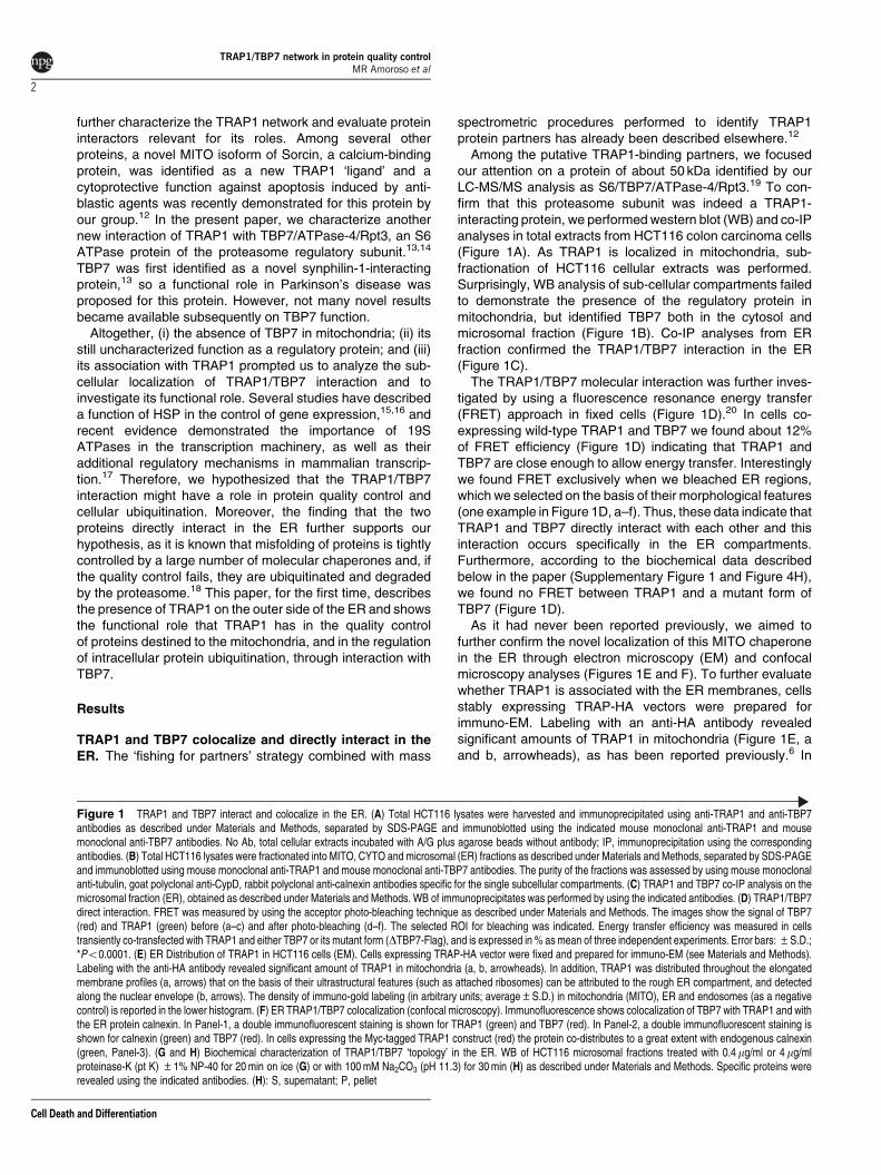
Among the putative TRAP1-binding partners, we focusedour attention on a protein of about 50 kDa identified by ourLC-MS/MS analysis as S6/TBP7/ATPase-4/Rpt3.19 To con-firm that this proteasome subunit was indeed a TRAP1-interacting protein, we performed western blot (WB) and co-IPanalyses in total extracts from HCT116 colon carcinoma cells(Figure 1A). As TRAP1 is localized in mitochondria, sub-fractionation of HCT116 cellular extracts was performed.Surprisingly, WB analysis of sub-cellular compartments failedto demonstrate the presence of the regulatory protein inmitochondria, but identified TBP7 both in the cytosol andmicrosomal fraction (Figure 1B). Co-IP analyses from ERfraction confirmed the TRAP1/TBP7 interaction in the ER(Figure 1C).
The TRAP1/TBP7 molecular interaction was further inves-tigated by using a fluorescence resonance energy transfer(FRET) approach in fixed cells (Figure 1D).20 In cells co-expressing wild-type TRAP1 and TBP7 we found about 12%of FRET efficiency (Figure 1D) indicating that TRAP1 andTBP7 are close enough to allow energy transfer. Interestinglywe found FRET exclusively when we bleached ER regions,which we selected on the basis of their morphological features(one example in Figure 1D, a–f). Thus, these data indicate thatTRAP1 and TBP7 directly interact with each other and thisinteraction occurs specifically in the ER compartments.Furthermore, according to the biochemical data describedbelow in the paper (Supplementary Figure 1 and Figure 4H),we found no FRET between TRAP1 and a mutant form ofTBP7 (Figure 1D).
As it had never been reported previously, we aimed tofurther confirm the novel localization of this MITO chaperonein the ER through electron microscopy (EM) and confocalmicroscopy analyses (Figures 1E and F). To further evaluatewhether TRAP1 is associated with the ER membranes, cellsstably expressing TRAP-HA vectors were prepared forimmuno-EM. Labeling with an anti-HA antibody revealedsignificant amounts of TRAP1 in mitochondria (Figure 1E, aand b, arrowheads), as has been reported previously.6 In
Figure 1 TRAP1 and TBP7 interact and colocalize in the ER. (A) Total HCT116 lysates were harvested and immunoprecipitated using anti-TRAP1 and anti-TBP7antibodies as described under Materials and Methods, separated by SDS-PAGE and immunoblotted using the indicated mouse monoclonal anti-TRAP1 and mousemonoclonal anti-TBP7 antibodies. No Ab, total cellular extracts incubated with A/G plus agarose beads without antibody; IP, immunoprecipitation using the correspondingantibodies. (B) Total HCT116 lysates were fractionated into MITO, CYTO and microsomal (ER) fractions as described under Materials and Methods, separated by SDS-PAGEand immunoblotted using mouse monoclonal anti-TRAP1 and mouse monoclonal anti-TBP7 antibodies. The purity of the fractions was assessed by using mouse monoclonalanti-tubulin, goat polyclonal anti-CypD, rabbit polyclonal anti-calnexin antibodies specific for the single subcellular compartments. (C) TRAP1 and TBP7 co-IP analysis on themicrosomal fraction (ER), obtained as described under Materials and Methods. WB of immunoprecipitates was performed by using the indicated antibodies. (D) TRAP1/TBP7direct interaction. FRET was measured by using the acceptor photo-bleaching technique as described under Materials and Methods. The images show the signal of TBP7(red) and TRAP1 (green) before (a–c) and after photo-bleaching (d–f). The selected ROI for bleaching was indicated. Energy transfer efficiency was measured in cellstransiently co-transfected with TRAP1 and either TBP7 or its mutant form (DTBP7-Flag), and is expressed in % as mean of three independent experiments. Error bars: ±S.D.;*Po0.0001. (E) ER Distribution of TRAP1 in HCT116 cells (EM). Cells expressing TRAP-HA vector were fixed and prepared for immuno-EM (see Materials and Methods).Labeling with the anti-HA antibody revealed significant amount of TRAP1 in mitochondria (a, b, arrowheads). In addition, TRAP1 was distributed throughout the elongatedmembrane profiles (a, arrows) that on the basis of their ultrastructural features (such as attached ribosomes) can be attributed to the rough ER compartment, and detectedalong the nuclear envelope (b, arrows). The density of immuno-gold labeling (in arbitrary units; average±S.D.) in mitochondria (MITO), ER and endosomes (as a negativecontrol) is reported in the lower histogram. (F) ER TRAP1/TBP7 colocalization (confocal microscopy). Immunofluorescence shows colocalization of TBP7 with TRAP1 and withthe ER protein calnexin. In Panel-1, a double immunofluorescent staining is shown for TRAP1 (green) and TBP7 (red). In Panel-2, a double immunofluorescent staining isshown for calnexin (green) and TBP7 (red). In cells expressing the Myc-tagged TRAP1 construct (red) the protein co-distributes to a great extent with endogenous calnexin(green, Panel-3). (G and H) Biochemical characterization of TRAP1/TBP7 ‘topology’ in the ER. WB of HCT116 microsomal fractions treated with 0.4 mg/ml or 4 mg/mlproteinase-K (pt K) ±1% NP-40 for 20 min on ice (G) or with 100 mM Na2CO3 (pH 11.3) for 30 min (H) as described under Materials and Methods. Specific proteins wererevealed using the indicated antibodies. (H): S, supernatant; P, pellet
TRAP1/TBP7 network in protein quality controlMR Amoroso et al
2
Cell Death and Differentiation
�-TRAP1 75 kDa
No Ab
IP
TRAP1 LYSATE
LYSATE
TBP7
�-TRAP1 75 kDa
90 kDa�-CALNEXIN
CYTO ER
�-TBP7 47 kDa
�-TUBULIN
�-TBP7
�-CypD
55 kDa
47 kDa
20 kDa
ERNo AbTRAP1ER
TBP7ER
IP
�-TRAP1 75 kDa
�-TBP7 47 kDa
16
8101214
TBP7/TRAP1�TBP7/TRAP1
0246
% o
f FR
ET
effi
cien
cy
beforephotobleaching
afterphotobleaching
TBP7 TRAP1 Overlay
FRET assay
*
1.5
2
2.5
3
A.U
.
0
0.5
1
MITO
1
2
3
0.4 �g/ml
pt K
NP40 0.1%-- + +4 �g/ml
++-
�-TRAP1
�-CALNEXIN
75 kDa
90 kDa
+ +++
�-TBP7 47 kDa
Na2CO3 pH 11.3+SP
�-CALNEXIN
�-TRAP1
�-TBP7
90 kDa
75 kDa
47 kDa
ENDOSOMESER
-+
MITO
*
TRAP1/TBP7 network in protein quality controlMR Amoroso et al
3
Cell Death and Differentiation
addition, we found that TRAP1 was distributed throughout theelongated membrane profiles (Figure 1E, a, arrows) that onthe basis of their ultrastructural features (such as attachedribosomes) can be attributed to the rough ER compartment.Moreover, gold particles – indicating TRAP1 molecules – weredetected along the nuclear envelope (Figure 1E, b, arrows),considered to be part of the ER membrane network. A carefulexamination of the density of immuno-gold labeling (inarbitrary units; average±S.D.) over different intracellularmembranes allowed us to demonstrate that indeed TRAP1was enriched in mitochondria (2.53±0.34) as it has alreadybeen reported. However, significant labeling density was alsocalculated for ER membranes (1.02±0.22) as shown inFigure 1E. Taken together, the EM observations are in linewith the above results showing association of TRAP1 with theER (Figures 1A–C). Accordingly, immunofluorescence con-focal microscopy analysis showed colocalization of TBP7 withTRAP1 and with the ER protein calnexin, thus confirming ERlocalization further (Figure 1F).
Altogether, these findings demonstrate that the interactionbetween TRAP1 and TBP7 occurs in the ER, but are unable toreveal a detailed localization in this context. To furtherevaluate the ‘topology’ of TRAP1 and TBP7 in the ER, abiochemical assay based on protease digestion was per-formed. Figure 1G shows that both proteins are sensitive to aproteinase-K digestion, whereas calnexin, a well-known ER-resident protein, is undigested. These approaches allowed usto demonstrate that TRAP1 and TBP7 are located on theoutside of the ER. Moreover, alkaline treatment of ERfractions to remove peripheral membrane proteins allowedus to demonstrate that both TRAP1 and TBP7 are looselyassociated to ER membranes (Figure 1H).
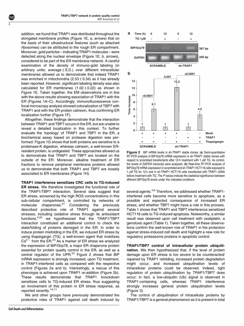
TRAP1 interference sensitizes CRC cells to TG-inducedER stress. We therefore investigated the functional role ofthe TRAP1/TBP7 interaction. Several data suggest thatER stress, worsened by the high ROS concentrations in thissub-cellular compartment, is controlled by networks ofmolecular chaperones.21 Considering the previouslydescribed protective role of TRAP1 against severalstresses, including oxidative stress through its antioxidantfunctions,4,22 we hypothesized that the TRAP1/TBP7interaction constituted an additional control to check thestate/folding of proteins damaged in the ER. In order toinduce protein misfolding in the ER, we induced ER stress byusing thapsigargin (TG), a well-known agent that mobilizesCa2þ from the ER.23 As a marker of ER stress we analyzedthe expression of BiP/Grp78, a major ER chaperone proteinessential for protein quality control in the ER, as well as acentral regulator of the UPR.24 Figure 2 shows that BiPmRNA expression is strongly increased, upon TG treatment,in TRAP1-interfered cells as compared with the respectivecontrol (Figures 2a and b). Interestingly, a rescue of thisphenotype is achieved upon TRAP1 re-addition (Figure 2b).These results demonstrate that TRAP1 knockdownsensitizes cells to TG-induced ER stress, thus suggestingan involvement of this protein in ER stress response, asreported recently.10,11
We and other groups have previously demonstrated theprotective roles of TRAP1 against cell death induced by
several agents.4,6 Therefore, we addressed whether TRAP1-interfered cells become more sensitive to apoptosis, as apossible and expected consequence of increased ERstress, and whether TBP7 might have a role in this process.Table 1 shows that TRAP1 and TBP7 interference sensitizedHCT116 cells to TG-induced apoptosis. Noteworthy, a similarresult was observed upon cell treatment with oxaliplatin, agenotoxic agent (Table 1). Taken together, all these observa-tions confirm the well-known role of TRAP1 in the protectionagainst stress-induced cell death and highlight a new role forregulatory proteasome proteins in apoptotic control.
TRAP1/TBP7 control of intracellular protein ubiquiti-nation. We then hypothesized that, if the level of proteindamage upon ER stress is too severe to be counteracted/repaired by TRAP1 refolding, increased protein degradationmight occur, and increased ubiquitination levels ofintracellular proteins could be observed. Indeed, tightregulation of protein ubiquitination by TRAP1/TBP7 doesoccur: in fact, a low-ubiquitin (Ub) signal is observed inTRAP1-containing cells, whereas TRAP1 interferencestrongly increases general protein ubiquitination levels(Figure 3).
The control of ubiquitination of intracellular proteins byTRAP1/TBP7 is a general phenomenon as it is present in total
TG 1�M
Time (h) 0
- + -
012
BiP/Grp78
GAPDH
387 bp
200 bp
<0.00010.001
sh-TRAP1SCRAMBLE
<0.000110
12
14 12.2
Tim
e In
crea
se
6
86.1
4.5
2
4
1 1 1
ThapsigarginTRAP1
SCRAMBLE
0Mock-
----- -
-++
+
++++
++ +
12
+
sh-TRAP1
Figure 2 BiP mRNA levels in sh-TRAP1 stable clones. (a) Semi-quantitativeRT-PCR analysis of BiP/Grp78 mRNA expression in sh-TRAP1 stable clones withrespect to scrambled transfectants after 12-h treatment with 1mM TG. As control,the levels of GAPDH transcript were analyzed. (b) Real-time RT-PCR analysis ofBiP/Grp78 mRNA expression in scrambled and sh-TRAP1 HCT116 cells exposed to1mM TG for 12 h and in sh-TRAP1 HCT116 cells transfected with TRAP1 cDNAbefore treatment with TG. The P-values indicate the statistical significance betweendifferent BiP/Grp78 levels under the indicated conditions
TRAP1/TBP7 network in protein quality controlMR Amoroso et al
4
Cell Death and Differentiation
extracts (Figure 3a). Interestingly, both TRAP1 and TBP7seem to have a critical role in the regulation of proteinubiquitination. In fact, upon transfection of TRAP1 expressionvectors in sh-TRAP1 stable transfectants a rescue of the high-Ub ‘phenotype’ is observed (Figure 3a). Sub-cellular fractio-nation allowed us to demonstrate that this regulatory roleis more evident in the post-mitochondrial (PM) fraction(cytosolþmicrosomes), where TRAP1/TBP7 control is parti-cularly necessary given the abundance of proteins translated,but often damaged. Conversely, the same regulation is notobserved in MITO extracts even after re-transfection ofTRAP1, likely because TBP7 is absent in these organelles(Figure 3b). These results confirm that TRAP1 function in theregulation of protein ubiquitination requires the presence ofTBP7. Additionally, increased cellular levels of ubiquitinatedproteins, very similar to those obtained in cells transientlytransfected with TRAP1 siRNAs, were observed upon TBP7siRNA transfection (Figure 3c), thus confirming the role ofTBP7 in this regulation. Altogether, these results demonstratethat the TRAP1/TBP7 interaction is a useful and importantcheckpoint in which these two proteins concomitantly work tojudge whether a protein can be repaired and reach the finaldestination or, if the damage is too severe, it needs to bedegraded. Furthermore, despite the block of proteasomeactivity by MG132 treatment, the experiments shown inFigure 3 demonstrate that the regulation of protein ubiquitina-tion by TRAP1 is not due to inhibition of proteasome function,as it is observed also in the absence of the inhibitory drug(Figure 3a). Indeed, this finding was confirmed by assayingthe proteasome’s activity in vitro using fluorescent substratesand extracts from scrambled- and TRAP1- or TBP7-interferedcells. The results shown in Figure 3d demonstrate that neitherTRAP1 nor TBP7 interference inhibits the proteasome’sfunction.
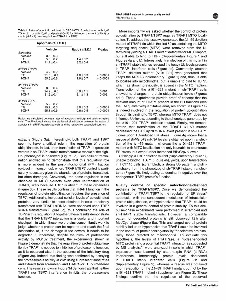
More importantly we asked whether the control of proteinubiquitination by TRAP1/TBP7 requires TRAP1 MITO locali-zation. To address this issue we generated theD1–59 deletionmutant of TRAP1 (in which the first 59 aa containing the MITOtargeting sequences (MTS2) were removed from the N-terminus) yielding a TRAP1 mutant defective for MITO import,but still able to bind to TBP7 (Supplementary Figure 1 andFigures 4a and b). Interestingly, transfection of this mutant insh-TRAP1 stable clones rescued the heavy Ub levels presentin TRAP1-interfered cells (Figure 4c). Conversely, anotherTRAP1 deletion mutant (D101–221) was generated thatkeeps the MTS (Supplementary Figure 1) and, thus, is ableto localize into mitochondria, but is unable to bind to TBP7,which, as shown previously, is absent in the MITO fraction.Transfection of the D101–221 mutant in sh-TRAP1 cellsshowed no changes in protein ubiquitination levels (Figures4d–f). These experiments provide proof of concept that therelevant amount of TRAP1 present in the ER fractions (seethe EM qualitative/quantitative analyses shown in Figure 1e)is indeed involved in the regulation of protein ubiquitinationthrough its binding to TBP7, whereas MITO TRAP1 does notinfluence Ub levels, according to the phenotype generated bythe D101–221 TRAP1 deletion mutant. Finally, we demon-strated that transfection of the TRAP1 D1–59 mutantdecreased the BiP/Grp78 mRNA levels present in sh-TRAP1clones upon TG-induced ER stress. Figure 4g shows that arescue of BiP/Grp78 mRNA levels is obtained upon transfec-tion of the D1–59 mutant, whereas the D101–221 TRAP1mutant with MITO localization not only is unable to counteractER stress, but even further increased BiP/Grp78 levels.
Strikingly, a TBP7 deletion mutant (Supplementary Figure 1),unable to bind to TRAP1 (Figure 4h), yields, upon transfectionin HCT116 cells (scrambled), a strong Ub pattern undistin-guishable from the phenotype of sh-TRAP1 stable transfec-tants (Figure 4i), likely acting as dominant negative over theendogenous TBP7 protein’s function.
Quality control of specific mitochondria-destinedproteins by TRAP1/TBP7. Once we demonstrated thecontribution of TRAP1/TBP7 to the regulation of ER stress/apoptosis, with the consequent modulation of intracellularprotein ubiquitination, we hypothesized that TRAP1 could beinvolved in a general control of protein stability. To this aim,pulse–chase experiments were performed in scrambled andsh-TRAP1 stable transfectants. However, a comparablepattern of degraded proteins is still observed 72 h afterMet/Cys chase (Figure 5a). This unchanged overall proteinstability led us to hypothesize that TRAP1 could be involvedin the control of protein folding/stability for selective proteins,likely those directed to mitochondria. To evaluate thishypothesis, the levels of F1ATPase, a nuclear-encodedMITO protein and a potential TRAP1 interactor as suggestedby MS analysis,12 were analyzed in cells in which TRAP1expression was lowered by short-hairpin RNA (shRNA)interference. Interestingly, protein levels decreasedin TRAP1 stably interfered cells (Figure 5b andSupplementary Figure 2), whereas a rescue was obtainedupon re-addition of the D1–59 TRAP1 mutant but not by theD101–221 TRAP1 mutant (Supplementary Figure 3). Thesefindings confirm that the regulation of the observed
Table 1 Rates of apoptotic cell death in CRC HCT116 cells treated with 1mMTG for 24 h or with 10mM oxaliplatin (l-OHP) for 48 h upon transient (siRNA) orstable (shRNA) downregulation of TRAP1 or TBP7
Apoptosis (%±S.D.)
Ratio (±S.D.) P-valueVehicleScramble
Vehicle 3.5±0.3TG 5.0±0.2 1.4±0.2l-OHP 11.2±0.4 3.2±0.4
siRNA TRAP1Vehicle 4.7±0.2TG 21.5±.0.4 4.6±0.3 o0.0001l-OHP 55.5±0.6 11.8±0.7 o0.0001
shRNA TRAP1Vehicle 3.5±0.4TG 24.2±.0.5 6.9±1.1 0.001l-OHP 31.8±0.4 9.1±1.3 0.002
siRNA TBP7Vehicle 5.2±0.2TG 15.7±0.3 3.0±0.2 o0.0001l-OHP 55.1±0.6 10.6±0.5 o0.0001
Ratios are calculated between rates of apoptosis in drug- and vehicle-treatedcells. The P-values indicate the statistical significance between the ratios ofapoptosis in siRNA-transfected cells and the respective scrambled controls.
TRAP1/TBP7 network in protein quality controlMR Amoroso et al
5
Cell Death and Differentiation
Ub-HA++++++TRAP1----++
SCRAMBLE
sh-T
RAP1
sh-T
RAP1
siRNA S
CRAMBLE
siRNA T
RAP1/TBP7
siRNA T
RAP1
siRNA T
BP7
+++ - MG 132 1�M
�-HA
�-TRAP1
�-GAPDH75 kDa34 kDa
MITO
TRAP1-
POST-MITOCHONDRIAL FRACTION
TRAP1
Ub-HA
SCRAMBLE
sh-T
RAP1
sh-T
RAP1
SCRAMBLE
sh-T
RAP1
sh-T
RAP1
+++ Ub-HA+
+
+
--
+
�-HA �-HA
�-COX IV 14 kDa �-GAPDH 34 kDa
++++ Ub-HA
�-HA
�-TBP7�-TRAP1
47 kDa75 kDa
�-GAPDH 34 kDa
500
600
700
300
400
100
200
Flu
ore
scen
ce In
ten
sity
0siRNA
SCRAMBLE +MG132
siRNASCRAMBLE
siRNATBP7
--
+-
siRNASORCIN
siRNATRAP1
Figure 3 Ub levels in HCT116 cells. (a) Total cell lysates from sh-TRAP1 and scrambled HCT116 stable clones were transfected with either an HA-tagged Ub vector(Ub-HA) or with TRAP1 expression vectors; treated with 1mM MG132 for 24 h; harvested 48 h after transfection; and subjected to immunoblot using rabbit polyclonal anti-HAantibodies. The same filter was re-probed using mouse monoclonal anti-GAPDH antibodies for normalization of cell lysates. Three independent experiments were performed, withsimilar results. (b) Sub-cellular fractionation was obtained from sh-TRAP1 and scrambled HCT116 stable transfectants treated as described in panel a. The extracts from the PMfraction (microsomesþCYTO fraction) and mitochondria (MITO, see Materials and Methods) were separated by SDS-PAGE and immunoblotted using a rabbit polyclonal anti-HAantibody to detect Ub levels. The purity of fractions was verified by using mouse monoclonal anti-COX IV or mouse monoclonal anti-GAPDH antibodies. Three independentexperiments were performed, with similar results. (c) HCT116 cells were co-transfected with a Ub-HA vector and an siRNA negative control (scramble), or with siRNAs specific forTRAP1, TBP7, or both (as indicated) and total cell lysates were harvested after 48 h from transfection. Total lysates were subjected to SDS-PAGE and immunoblotted using rabbitpolyclonal anti-HA antibodies to detect total Ub levels. The same filter was re-probed using mouse monoclonal anti-GAPDH antibodies for normalization of cell lysates, and usingmouse monoclonal anti-TRAP1 and mouse monoclonal anti-TBP7 antibodies. (d) Proteasome activity is not affected by TRAP1 and TBP7 silencing. Total cellular extracts wereprepared after 48 h of transfection with specific siRNA for TRAP1, TBP7 or Sorcin, as control, or with an siRNA negative control (scramble), and incubated in the presence of assaybuffer and the fluorogenic substrate Suc-LLVY-AMC, as described under Materials and Methods. Samples were analyzed in triplicate using an excitation wavelength of 360 nm andan emission wavelength of 450 nm to detect chymotryptic proteasome activity. The data represent the mean of three independent experiments
Figure 4 The TRAP1/TBP7 interaction in the ER is required for control of protein ubiquitination and ER stress. (a and d) Sub-cellular localization of D1–59-Myc/D101–221-HAmutants. HCT116 cells were transfected with theD1–59-Myc (a) orD101–221-HA (d) TRAP1 mutants; sub-fractionated into MITO, CYTO and microsomal (ER) fractions (a), or MITOand PM (cytosolþmicrosomes) fractions (d), as described under Materials and Methods; separated by SDS-PAGE; and immunoblotted using the indicated antibodies to verify theexpression of mutants and the purity of fractions. For details on procedures for generation of the mutants see Materials and Methods. (b, e) Interaction between D1–59-Myc/D101–221-HA mutants and TBP7. HCT116 cells were transfected with D1–59-Myc (b) or D101–221-HA (e) TRAP1 mutants, harvested and immunoprecipitated using anti-Myc or anti-HAantibodies as described under Materials and Methods. Immunoprecipitates were separated by SDS-PAGE and immunoblotted using the indicated antibodies. No Ab, total cellularextracts incubated with A/G plus agarose beads without antibody; IP, immunoprecipitation with the corresponding antibodies. Three independent experiments were performed, withsimilar results. (c) Ubiquitination levels upon transfection of the D1–59-Myc TRAP1 deletion mutant. Total lysates from HCT116 scrambled, sh-TRAP1 stable clones and sh-TRAP1cells transfected with theD1–59-Myc TRAP1 mutant were subjected to immunoblot analysis using mouse monoclonal anti-Ub antibodies to detect total ubiquitination levels and with ananti-GAPDH antibody for normalization of cell lysates. Three independent experiments were performed, with similar results. (f) Ubiquitination levels upon transfection of theD101–221-HA TRAP1 deletion mutant. HCT116 scramble, sh-TRAP1 and sh-TRAP1 cells transfected with theD101–221-HA TRAP1 mutant were sub-fractionated in PM (microsomesþCYTOfraction) and MITO fractions as described under Materials and Methods. Total lysates from the same cells were used as controls (left panel). Protein lysates were subjected toimmunoblot analysis using mouse monoclonal anti-Ub antibodies to detect total ubiquitination levels. The purity of fractions was verified using mouse monoclonal anti-GAPDH (left andright panels) and mouse-monoclonal anti-COX IV (middle panel) antibodies. Three independent experiments were performed, with similar results. (g) Real-time RT-PCR analysis ofBiP/Grp78 mRNA expression in scrambled and sh-TRAP1 HCT116 cells exposed to 1mM TG for 12 h (same as in Figure 2b) and in sh-TRAP1 HCT116 cells transfected with theD1–59-Myc or D101–221-HA TRAP1 mutant, as indicated, before treatment with TG. The P-values indicate the statistical significance between the BiP/Grp78 levels under theindicated conditions. (h) Interaction between TRAP1 and the DTBP7-Flag deletion mutant. HCT116 cells were transfected with the DTBP7-Flag deletion mutant, harvested andimmunoprecipitated using anti-TRAP1 antibodies as described under Materials and Methods. Immunoprecipitates were separated by SDS-PAGE and immunoblotted using theindicated antibodies. No Ab, total cellular extracts incubated with A/G plus agarose beads without antibody; IP, immunoprecipitation with the corresponding antibodies. Threeindependent experiments were performed, with similar results. The arrow indicates theDTBP7-Flag mutant band. (i) Ubiquitination levels upon transfection of theDTBP7-Flag deletionmutant. Total lysates from HCT116 scrambled cells transfected withDTBP7-Flag mutant were subjected to immunoblot analysis using mouse monoclonal anti-Ub antibodies to detecttotal ubiquitination levels and with mouse monoclonal anti-HSP60 antibodies for normalization of cell lysates. Three independent experiments were performed, with similar results
TRAP1/TBP7 network in protein quality controlMR Amoroso et al
6
Cell Death and Differentiation
phenomena occurs in the cytosolic (CYTO) compartmentand support our model. Accordingly, the protein levels of p18Sorcin, another MITO protein, recently identified by our groupas a novel MITO Sorcin isoform interacting with TRAP1,12
decreased upon TRAP1 interference (Figure 5b, arrow). Ofnote, under the same experimental conditions, no differenceswere observed in the protein levels of the higher mobility p22Sorcin isoform, which shares high homology with p18 Sorcin,
�1-59-Myc
90kDa
ER MITO
SCRAMBLE
sh-T
RAP1
sh-T
RAP1
SCRAMBLE
sh-T
RAP1
sh-T
RAP1
SCRAMBLE
sh-T
RAP1
sh-T
RAP1
SCRAMBLE
SCRAMBLE
sh-T
RAP1
SCRAMBLE
sh-T
RAP1
sh-T
RAP1
CYTO�1-59-Myc+--
�-TUBULIN
�-MYC
55 kDa
72 kDa
�-CALNEXIN
�-MYC 72 kDa
�-COX IV 14 kDa
�-UBIQUITIN
No Ab LYSATES
IP
MYC
�1-59-Myc+++
34 kDa
�-MYC
�-TBP7
72 kDa
47 kDa �-GAPDH
�101-221-HA
MITO�101-221-HA+++
�-CALNEXIN
�-HA 71 kDa
34 kDa
�-TUBULIN 55 kDa
90 kDaNo Ab
IP
HA
�-TBP7
�-HA 71 kDa
47 kDa�-VDAC1
POST-MITOCHONDRIAL FRACTIONMITO+--
TOTAL LYSATE
�101-221�101-221 +--+--
71 kDa
�101-221
�-HA
�-U
BIQ
UIT
IN
�-U
BIQ
UIT
IN
�-U
BIQ
UIT
IN
34 Dka�-HSP60 60 kDa�-GAPDH �-GAPDH
<0.0001
32.235
40
20
25
30
0.006
Tim
e In
crea
se
1 1 1
12.2
6.9
0
5
10
15
1
6.1
Thapsigargin-
sh-T
RA
P1
Mo
ck
sh-T
RA
P1
�1-
59
sh-T
RA
P1
�10
1-22
2
-
Scr
amb
leM
ock
�TBP7-Flag-+-
�TBP7-Flag- ++-+
�-TBP7 37 kDa
�-TRAP147 kDa
75 kDaNo Ab
IP
TR
AP
1
TR
AP
1
�-TBP737 kDa
�-UBIQUITIN
60 kDa�-HSP60
-
PM-
LYSATES
34 kDa
+ - + - + +
LYSATES
TRAP1/TBP7 network in protein quality controlMR Amoroso et al
7
Cell Death and Differentiation
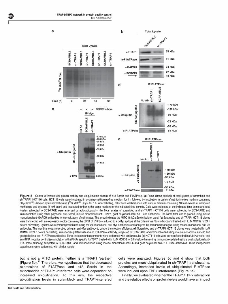
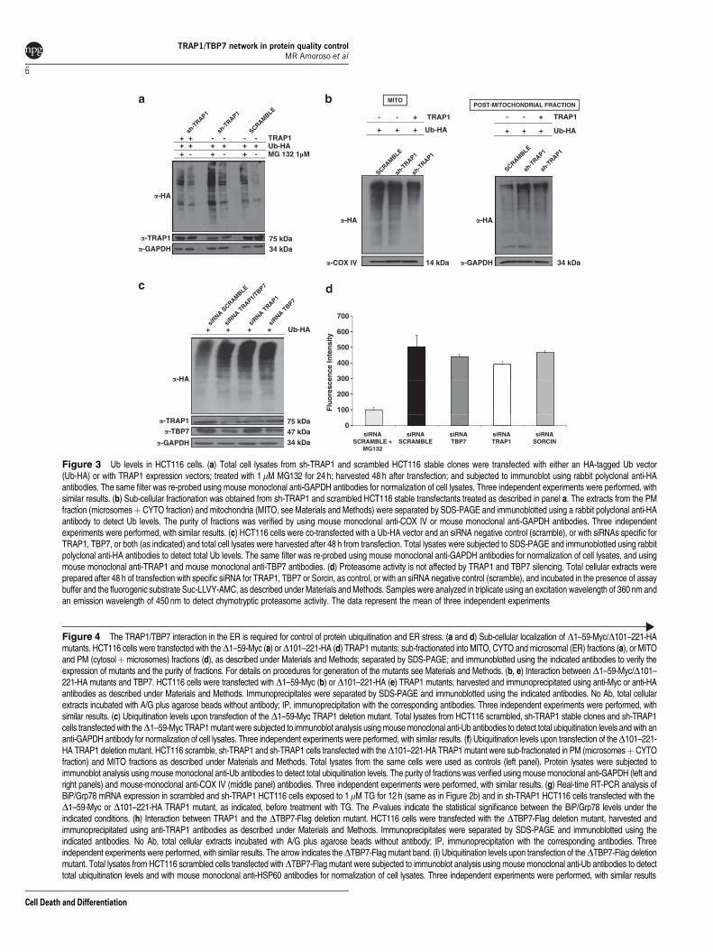
but is not a MITO protein, neither is a TRAP1 ‘partner’(Figure 5b).12 Therefore, we hypothesize that the decreasedexpressions of F1ATPase and p18 Sorcin in themitochondria of TRAP1-interferred cells were dependent onincreased ubiquitination. To this aim, the respectiveubiquitination levels in scrambled- and TRAP1-interfered
cells were analyzed. Figures 5c and d show that bothproteins are more ubiquitinated in sh-TRAP1 transfectants.Accordingly, increased levels of ubiquitinated F1ATPasewere induced upon TBP7 interference (Figure 5e).
Finally, we evaluated whether the TRAP1/TBP7 interactionand the relative effects on protein levels would have an impact
Total Lysate
Total Lysate
SC
RA
MB
LE
sh-T
RA
P1
SC
RA
MB
LE
SC
RA
MB
LE
SC
RA
MB
LE
sh-T
RA
P1
sh-T
RA
P1
sh-T
RA
P1 �-TRAP1 75 kDa
35S
Met
/35S
Cys
SCRAMBLE
sh-T
RAP1
�-GAPDH
�-F1ATPase
34 kDa
51 kDa
Time (h) 48
�-SORCIN18 kDa22 kDa
IP F1ATPase
IP Myc
+ + + SORCIN-Myc
SC
RA
MB
LE
sh-T
RA
P1
No Ab
-170 kDa
-130 kDa
SC
RA
MB
LE
sh-T
RA
P1
No
Ab
�-Ubiquitin
Ub
-co
nju
gat
ed
-95 kDa
-55 kDa
-72 kDa
-170 kDa-130 kDa-95 kDa
-55 kDa
-72 kDa
�-F1ATPase 51 kDa
IP F1ATPase
�-Ubiquitin
Ub
-co
nju
gat
ed
-43 kDa
-34 kDa
si-T
BP
7
si-S
CR
AM
BL
E
No Ab
�-Myc 24 kDa �-Ubiquitin
-170 kDa-130 kDa
Ub
-co
nju
gat
ed
�-F1ATPase 51 kDa
-95 kDa
-55 kDa
-72 kDa
0 24 72
Figure 5 Control of intracellular protein stability and ubiquitination pattern of p18 Sorcin and F1ATPase. (a) Pulse–chase analysis of total lysates of scrambled andsh-TRAP1 HCT116 cells. HCT116 cells were incubated in cysteine/methionine-free medium for 1 h followed by incubation in cysteine/methionine-free medium containing50mCi/ml 35S-labeled cysteine/methionine (35S Met/35S Cys) for 1 h. After labeling, cells were washed once with culture medium containing 10-fold excess of unlabeledmethionine and cysteine (5 mM each) and incubated further in the same medium for the indicated time periods. Cells were collected at the indicated time points and totallysates subjected to SDS-PAGE were analyzed by autoradiography. (b) Total lysates of scrambled and sh-TRAP1 HCT116 cells were subjected to SDS-PAGE andimmunoblotted using rabbit polyclonal anti-Sorcin, mouse monoclonal anti-TRAP1, goat polyclonal anti-F1ATPase antibodies. The same filter was re-probed using mousemonoclonal anti-GAPDH antibodies for normalization of cell lysates. The arrow indicates the MITO 18-kDa Sorcin isoform band. (c) Scrambled and sh-TRAP1 HCT116 cloneswere transfected with an expression vector containing the cDNA of p18 Sorcin fused to a c-Myc epitope at the C-terminus (Sorcin-Myc) and treated with 1 mM MG132 for 24 hbefore harvesting. Lysates were immunoprecipitated using mouse monoclonal anti-Myc antibodies and analyzed by immunoblot analysis using mouse monoclonal anti-Ubantibodies. The membrane was re-probed using an anti-Myc antibody to control transfection efficiency. (d) Scrambled and sh-TRAP1 HCT116 clones were treated with 1mMMG132 for 24 h before harvesting, immunoprecipitated with an-anti F1ATPase antibody, subjected to SDS-PAGE and immunoblotted using mouse monoclonal anti-Ub andgoat polyclonal anti-F1ATPase antibodies. Three independent experiments were performed with similar results. (e) HCT116 cells were co-transfected with a Ub-HA vector andan siRNA negative control (scramble), or with siRNAs specific for TBP7; treated with 1 mM MG132 for 24 h before harvesting; immunoprecipitated using a goat polyclonal anti-F1ATPase antibody; subjected to SDS-PAGE; and immunoblotted using mouse monoclonal anti-Ub and goat polyclonal anti-F1ATPase antibodies. Three independentexperiments were performed, with similar results
TRAP1/TBP7 network in protein quality controlMR Amoroso et al
8
Cell Death and Differentiation
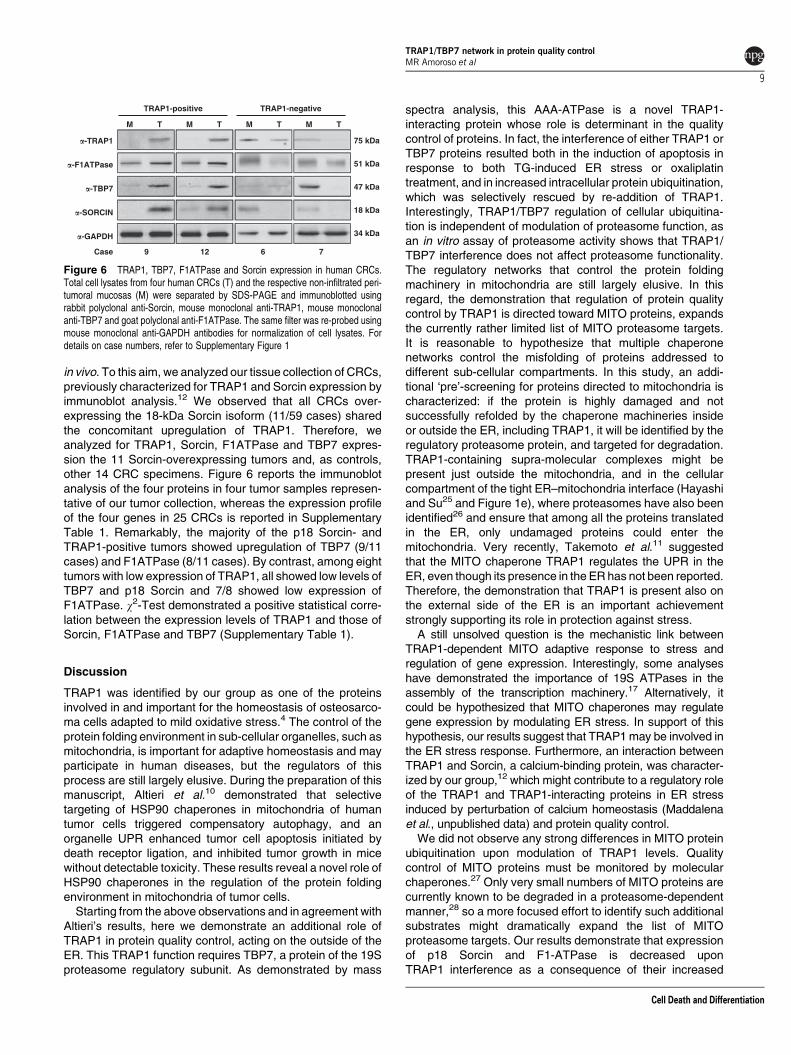
in vivo. To this aim, we analyzed our tissue collection of CRCs,previously characterized for TRAP1 and Sorcin expression byimmunoblot analysis.12 We observed that all CRCs over-expressing the 18-kDa Sorcin isoform (11/59 cases) sharedthe concomitant upregulation of TRAP1. Therefore, weanalyzed for TRAP1, Sorcin, F1ATPase and TBP7 expres-sion the 11 Sorcin-overexpressing tumors and, as controls,other 14 CRC specimens. Figure 6 reports the immunoblotanalysis of the four proteins in four tumor samples represen-tative of our tumor collection, whereas the expression profileof the four genes in 25 CRCs is reported in SupplementaryTable 1. Remarkably, the majority of the p18 Sorcin- andTRAP1-positive tumors showed upregulation of TBP7 (9/11cases) and F1ATPase (8/11 cases). By contrast, among eighttumors with low expression of TRAP1, all showed low levels ofTBP7 and p18 Sorcin and 7/8 showed low expression ofF1ATPase. w2-Test demonstrated a positive statistical corre-lation between the expression levels of TRAP1 and those ofSorcin, F1ATPase and TBP7 (Supplementary Table 1).
Discussion
TRAP1 was identified by our group as one of the proteinsinvolved in and important for the homeostasis of osteosarco-ma cells adapted to mild oxidative stress.4 The control of theprotein folding environment in sub-cellular organelles, such asmitochondria, is important for adaptive homeostasis and mayparticipate in human diseases, but the regulators of thisprocess are still largely elusive. During the preparation of thismanuscript, Altieri et al.10 demonstrated that selectivetargeting of HSP90 chaperones in mitochondria of humantumor cells triggered compensatory autophagy, and anorganelle UPR enhanced tumor cell apoptosis initiated bydeath receptor ligation, and inhibited tumor growth in micewithout detectable toxicity. These results reveal a novel role ofHSP90 chaperones in the regulation of the protein foldingenvironment in mitochondria of tumor cells.
Starting from the above observations and in agreement withAltieri’s results, here we demonstrate an additional role ofTRAP1 in protein quality control, acting on the outside of theER. This TRAP1 function requires TBP7, a protein of the 19Sproteasome regulatory subunit. As demonstrated by mass
spectra analysis, this AAA-ATPase is a novel TRAP1-interacting protein whose role is determinant in the qualitycontrol of proteins. In fact, the interference of either TRAP1 orTBP7 proteins resulted both in the induction of apoptosis inresponse to both TG-induced ER stress or oxaliplatintreatment, and in increased intracellular protein ubiquitination,which was selectively rescued by re-addition of TRAP1.Interestingly, TRAP1/TBP7 regulation of cellular ubiquitina-tion is independent of modulation of proteasome function, asan in vitro assay of proteasome activity shows that TRAP1/TBP7 interference does not affect proteasome functionality.The regulatory networks that control the protein foldingmachinery in mitochondria are still largely elusive. In thisregard, the demonstration that regulation of protein qualitycontrol by TRAP1 is directed toward MITO proteins, expandsthe currently rather limited list of MITO proteasome targets.It is reasonable to hypothesize that multiple chaperonenetworks control the misfolding of proteins addressed todifferent sub-cellular compartments. In this study, an addi-tional ‘pre’-screening for proteins directed to mitochondria ischaracterized: if the protein is highly damaged and notsuccessfully refolded by the chaperone machineries insideor outside the ER, including TRAP1, it will be identified by theregulatory proteasome protein, and targeted for degradation.TRAP1-containing supra-molecular complexes might bepresent just outside the mitochondria, and in the cellularcompartment of the tight ER–mitochondria interface (Hayashiand Su25 and Figure 1e), where proteasomes have also beenidentified26 and ensure that among all the proteins translatedin the ER, only undamaged proteins could enter themitochondria. Very recently, Takemoto et al.11 suggestedthat the MITO chaperone TRAP1 regulates the UPR in theER, even though its presence in the ER has not been reported.Therefore, the demonstration that TRAP1 is present also onthe external side of the ER is an important achievementstrongly supporting its role in protection against stress.
A still unsolved question is the mechanistic link betweenTRAP1-dependent MITO adaptive response to stress andregulation of gene expression. Interestingly, some analyseshave demonstrated the importance of 19S ATPases in theassembly of the transcription machinery.17 Alternatively, itcould be hypothesized that MITO chaperones may regulategene expression by modulating ER stress. In support of thishypothesis, our results suggest that TRAP1 may be involved inthe ER stress response. Furthermore, an interaction betweenTRAP1 and Sorcin, a calcium-binding protein, was character-ized by our group,12 which might contribute to a regulatory roleof the TRAP1 and TRAP1-interacting proteins in ER stressinduced by perturbation of calcium homeostasis (Maddalenaet al., unpublished data) and protein quality control.
We did not observe any strong differences in MITO proteinubiquitination upon modulation of TRAP1 levels. Qualitycontrol of MITO proteins must be monitored by molecularchaperones.27 Only very small numbers of MITO proteins arecurrently known to be degraded in a proteasome-dependentmanner,28 so a more focused effort to identify such additionalsubstrates might dramatically expand the list of MITOproteasome targets. Our results demonstrate that expressionof p18 Sorcin and F1-ATPase is decreased uponTRAP1 interference as a consequence of their increased
TRAP1-positive TRAP1-negative
M
�-F1ATPase 51 kDa
�-TRAP1 75 kDa
�-TBP7 47 kDa
18 kDa�-SORCIN
�-GAPDH 34 kDa
Case
T M T M T M T
9 12 6 7
Figure 6 TRAP1, TBP7, F1ATPase and Sorcin expression in human CRCs.Total cell lysates from four human CRCs (T) and the respective non-infiltrated peri-tumoral mucosas (M) were separated by SDS-PAGE and immunoblotted usingrabbit polyclonal anti-Sorcin, mouse monoclonal anti-TRAP1, mouse monoclonalanti-TBP7 and goat polyclonal anti-F1ATPase. The same filter was re-probed usingmouse monoclonal anti-GAPDH antibodies for normalization of cell lysates. Fordetails on case numbers, refer to Supplementary Figure 1
TRAP1/TBP7 network in protein quality controlMR Amoroso et al
9
Cell Death and Differentiation
ubiquitination. The identification of TRAP1/TBP7-specific‘substrates’ strongly contributes to the complex study of MITOprotein quality control. One reason for ubiquitination of MITOproteins may be that when mitochondria-destined proteins aremistargeted or misfolded, they are identified as aberrant andrecognized by the Ub–proteasome system (UPS) for removal.This may even support a role of the CYTO UPS in controllingthe levels and/or the quality of proteins destined formitochondria. In addition, the presence of TRAP1 on theouter side of ER and the absence of TBP7 in mitochondriasuggest that TRAP1 functions in these latter organelles arenot directly linked to ubiquitination control, whereas thiscontrol is present in a different compartment and requiresinteraction with TBP7. In support of this model are results oftransfection experiments using newly generated TRAP1mutants, either able to interact with TBP7 and localized inthe ER/cytosol fractions (D1–59) or unable to bind to TBP7 butimported into mitochondria (D101–221). Indeed, only theMITO import-defective D1–59 mutant, and not the MITOD101–221 deletion mutant, rescues the strong Ub levels insh-TRAP1-interfered cells. These findings provide proof ofconcept that protein quality control depends on the CYTOinteraction between TRAP1 and TBP7. Consistent with theseresults is the observation that again only the D1–59 TRAP1mutant rescues the decreased levels of TRAP1-regulatedproteins (Supplementary Figure 3), as well as the levels ofBiP/Grp78 mRNA upon TG-induced ER stress (Figure 4g). All
these observations are in agreement with still unidentifiedproteasome members in mitochondria, even though severalproteases, ATPases and Ub ligases have been identified.29
Furthermore, a functional interplay between MITO andproteasome activity has been demonstrated, thus suggestingthat both systems are interdependent.30
Remarkably, the finding that the proposed TRAP1 networkis conserved in CRCs is consistent with our model andprovides new insights into the quality control/stability/ubiqui-tination of proteins in human cancer, a still highly debatedissue. Indeed, the proteotoxic stress generated by accumula-tion of misfolded proteins and the consequent heat-shockresponse is currently under evaluation as a potential antic-ancer treatment target, as many tumor cells show constitutiveproteotoxic stress and dependence on heat-shock responsebecause of their rapid rates of proliferation and translation.31
Interestingly, bortezomib, a reversible inhibitor of the 26Sproteasome, is at present a valuable option for the first-linetreatment of multiple myeloma.32 Thus, characterization ofTRAP1, a chaperone upregulated in about 60% of humanCRCs,5 as a protein involved in quality control and inprotection against apoptosis in cancer cells provides a strongrationale for considering this network as a novel moleculartarget for treatment of human CRC.
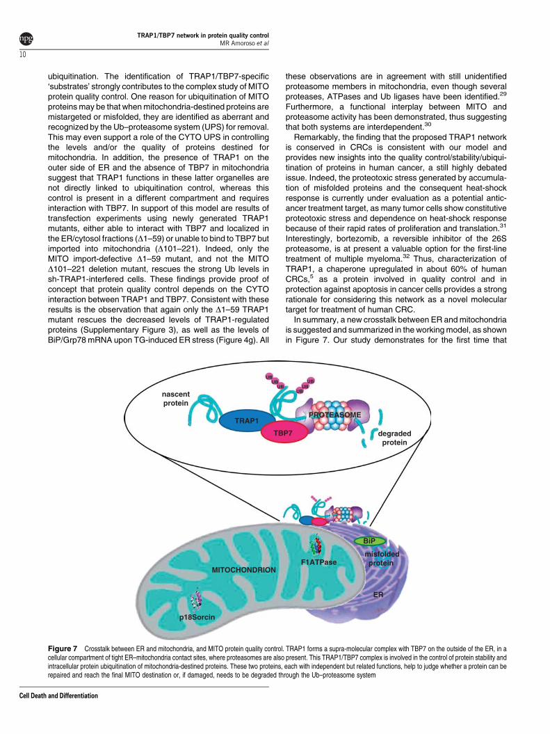
In summary, a new crosstalk between ER and mitochondriais suggested and summarized in the working model, as shownin Figure 7. Our study demonstrates for the first time that
TRAP1
nascentprotein
degradedprotein
PROTEASOME
TBP7
UB
UBUB
UBUB
UB
ER
BiP
misfoldedprotein
MITOCHONDRION
p18Sorcin
F1ATPase
Figure 7 Crosstalk between ER and mitochondria, and MITO protein quality control. TRAP1 forms a supra-molecular complex with TBP7 on the outside of the ER, in acellular compartment of tight ER–mitochondria contact sites, where proteasomes are also present. This TRAP1/TBP7 complex is involved in the control of protein stability andintracellular protein ubiquitination of mitochondria-destined proteins. These two proteins, each with independent but related functions, help to judge whether a protein can berepaired and reach the final MITO destination or, if damaged, needs to be degraded through the Ub–proteasome system
TRAP1/TBP7 network in protein quality controlMR Amoroso et al
10
Cell Death and Differentiation
TRAP1 is also present in the ER of cancer cells where it isinvolved in the quality control and intracellular proteinubiquitination of mitochondria-destined proteins, throughdirect interaction (as demonstrated by the FRET analysisshown in Figure 1d) with TBP7, one of the proteins present inthe regulatory proteasome subunit. Thus, a ‘customs office’could be hypothesized at the ER/mitochondria interface, withTRAP1 and TBP7 being the officers at this importantcheckpoint. These two officers, each with independent butrelated functions, help to judge whether a protein can berepaired and reach its final MITO destination or, if the damageis too severe, it needs to be degraded.
Materials and MethodsCell culture, plasmid generation and transfection procedures.HCT116 cells were cultured in DMEM supplemented with 10% fetal bovine serum(FBS) under standard conditions. Full-length TRAP1 and Sorcin expression vectorswere obtained as described previously.12
Mutant D1–59-Myc was generated by using the following primers: D1–59-myc,forward: 50-ATTAGAATTCATGAGCACGCAGACCGCCGAGG-30, reverse: 30-AT-TACTCGAGGTGTCGCTCCAGGGCCTTGA-50. PCR-amplified fragments weregel-purified and cloned in-frame into the pcDNA 3.1 plasmid (Invitrogen, SanGiuliano Milanese, Italy) at the EcoRI and XhoI restriction sites.
Mutant D101–221-HA was generated by using the following primers: TRAP1-HA, forward: 50-attaGCGGCCGCGCAGCCAACATGGCGCGCGAGCCTGCGGG-30, reverse: 50-attaTCTAGATTAAGCGTAATCTGGAACATCATATGGGTATCAGTGTCGCTCCAGGGCCTTGA-30; and D101–221-HA, forward: 50-attaCCGCGGTCGGCAGCCCCGGGGAGCCT-30, reverse: 50-attaCCGCGGAAACACCTCTTTTTCTGAGT-30. The PCR products obtained with the primers TRAP1-HA forward andD101–221-HA reverse were cloned in the pRc-CMV vector (Invitrogen); the PCRproduct obtained with the primers DATPase-HA forward and TRAP1-HA reversewas subcloned in the same plasmid. All clones were sequenced to confirm identityand PCR fidelity. The plasmid pCMV5L/S6 (TBP7-HA) was a gift from Dr SimonDawson (University of Nottingham).
Mutant DTBP7-FLAG was generated by excising a fragment from the full-lengthTBP7 expression vector by using EcoRI and BamHI restriction endonucleases. Thefragment was gel-purified and cloned into the corresponding sites of the expressionvector p3x-FLAG.
Transient transfection of DNA plasmids was performed with the PolyfectTransfection reagent (Qiagen, Milan, Italy). siRNAs of TRAP1 and TBP7 werepurchased from Qiagen (cat. no. S100301469 for TBP7 and cat. no. SI00115150 forTRAP1). For knockdown experiments, siRNAs were diluted to a final concentrationof 20 nmol/l and transfected according to the manufacturer’s protocol. For controlexperiments, cells were transfected with a similar amount of scrambled siRNA(Qiagen; cat. no. SI03650318). Transient transfections of siRNAs were performedby using the HiPerFect Transfection Reagent (Qiagen). TRAP1-stable interferencewas achieved by transfecting HCT116 cells with TRAP1 (TGCTGTTGACAGTGAGCGACCCGGTCCCTGTACTCAGAAATAGTGAAGCCACAGATGTATTTCTGAGTACAGGGACCGGGCTGCCTACTGCCTCGGA) or scrambled (sequence con-taining no homology to known mammalian genes) shRNAs (Open Biosystems,Huntsville, AL, USA).
Cell extracts, purification and treatments. Total cell lysates wereobtained by homogenization of cell pellets and tumor specimens in cold lysis buffer(20 mM Tris (pH 7.5), containing 300 mM sucrose, 60 mM KCl, 15 mM NaCl, 5% (v/v) glycerol, 2 mM EDTA, 1% (v/v) Triton X-100, 1 mM PMSF, 2 mg/ml aprotinin,2 mg/ml leupeptin and 0.2% (w/v) deoxycholate) for 1 min at 4 1C and furthersonication for 30 s at 4 1C. For ER stress induction, cells were treated overnight with1mM TG (Sigma-Aldrich, Milan, Italy) before harvesting.
Mitochondria and ER were purified by using the Qproteome MitochondriaIsolation kit (Qiagen) according to the manufacturer’s protocol and as describedelsewhere.12 Briefly, HCT116 cells were washed and suspended in lysis buffer,which selectively disrupts the plasma membrane without solubilizing it, resulting inthe isolation of CYTO proteins. Plasma membranes and compartmentalizedorganelles, such as nuclei, mitochondria and ER, remained intact and were pelletedby centrifugation. The resulting pellet was resuspended in disruption buffer,repeatedly passed through a narrow-gauge needle (to ensure complete cell
disruption) and centrifuged to pellet nuclei, cell debris and unbroken cells. Thesupernatant (containing mitochondria and the microsomal fraction) was re-centrifuged to pellet mitochondria. The resulting supernatant (microsomal fraction)was treated with proteinase-K for 20 min on ice±NP-40 (Igepal; Sigma-Aldrich)according to Hassink et al.33 or with 0.1 M Na2CO3 (pH 11.3) for 30 min to removeperipheral ER membrane proteins.34
WB analysis and antibodies. Equal amounts of protein from cell lysatesand tumor specimens were subjected to 10% (v/v) SDS-PAGE and transferred to aPVDF membrane (Millipore, Temecula, CA, USA). The membrane was blockedwith 5% (w/v) skimmed milk and incubated with the primary antibody, followed byincubation with an HRP-conjugated secondary antibody. Proteins were visualizedwith an ECL detection system (GE Healthcare, Waukesha, WI, USA). The followingantibodies from Santa Cruz Biotechnology (Segrate, Italy) were used for WBanalysis and immunoprecipitation: anti-TRAP1 (sc-13557), anti-Sorcin (sc-100859),anti-TBP7 (sc-166003), anti-cMyc (sc-40), anti-CypD (sc-82570), anti-VDAC1(sc-8830), anti-HSP60 (sc-1052), anti-Ub (sc-8017), anti-COX4 (sc-58348),anti-F1ATPase (ATP5B subunit; sc-58619), anti-tubulin (sc-8035), anti-HA(sc-805) and anti-glyceraldehyde-3-phosphate dehydrogenase (GAPDH; sc-69778). A rabbit polyclonal anti-calnexin antibody (BD Biosciences, Milan, Italy)was also used.
RNA extraction and semi-quantitative and real-time RT-PCRanalysis. Total RNA from cell pellets and tumor specimens was extracted byusing the TRIzol Reagent (Invitrogen). For first-strand synthesis of cDNA, 3 mg ofRNA were used in a 20-ml reaction mixture by using a cDNA Superscript II(Invitrogen). For real-time PCR analysis, 1 ml of cDNA sample was amplified byusing the Platinum SYBR Green qPCR Supermix UDG (Invitrogen) in an iCycler iQReal-Time Detection System (Bio-Rad Laboratories GmbH, Segrate, Italy). Thefollowing primers were used: BiP/Grp78, forward: 50-CGTGGATGACCCGTCTGTG-30, reverse: 50-cTGCCGTAGGCTCGTTGATG-30 (PCR product 308 bp); and GAPDH, forward: 50-CAAGGCTGAGAACGGGAA-30, reverse: 50-GCATCGCCCCACTTGATTTT-30 (PCR product 90 bp). Primers were designed to be intron-spanning. The reaction conditions were 50 1C for 2 min; 95 1C for 2 min; followed by45 cycles of 15 s at 95 1C, 30 s at 60 1C and 30 s at 72 1C. GAPDH was chosen asan internal control.
For semi-quantitative RT-PCR, the RNA obtained by scrambled and sh-TRAP1HCT116 cells was retro-transcribed and amplified using specific primers for BiP/Grp78 and GAPDH by using the Superscript III-One STEP kit (Invitrogen),according to the manufacturer’s instructions. The following primers were used toamplify the corresponding transcripts: GAPDH, forward: 50-GAAGGTGAAGGTCGGAGTC-30, reverse 50-GAAGATGGTGATGGGATTTC-30; and BiP/Grp78, forward:50-CTGGGTACATTTGATCTGACTGG-30, reverse: 50-GCATCCTGGTGGCTTTCCAGCCATTC-30. The primers for BiP/Grp78 were a gift from Professor P Remondelli(University of Salerno, Italy).
Apoptosis assay. HCT116 cells were subjected to downregulation of TRAP1and TBP7 expression by siRNA transfection. Apoptosis was evaluated bycytofluorimetric analysis of Annexin-V and 7-amino-actinomycin-D (7-AAD)-positive cells using the fluorescein isothiocyanate (FITC)–Annexin-V/7-AAD kit(Beckman Coulter, Milan, Italy). Stained cells were analyzed by using the ‘EPICSXL’ Flow Cytometer (Beckman Coulter). Ten thousand events were collected persample. Positive staining for Annexin-V as well as double staining for Annexin-V and7-AAD were interpreted as signs of, respectively, early and late phases ofapoptosis.35 Experiments were performed three times using three replicates foreach experimental condition.
Immunofluorescence, confocal microscopy and EM analysis.HCT116 cells were fixed with 0.1 M phosphate buffer containing 4% (w/v)paraformaldehyde for 15 min, then blocked and permeabilized with 5% (w/v) BSA,0.1% (v/v) Triton X-100, 10% (v/v) FBS in PBS for 20 min at RT before staining withprimary antibodies (for TRAP1, CALNEXIN and TBP7) and the correspondingsecondary TEXAS RED/FITC-conjugated antibodies. Immunofluorescence wasanalyzed by confocal laser-scanning microscopy using Zeiss 510 LSM (Carl ZeissMicroimaging, Gottingen, Germany), equipped with an Argon ionic laser (Carl ZeissMicroimaging) whose wavelength was set up to 488 nm; a He–Ne laser whosewavelength was set up to 546 nm; and an oil-immersion � 63/1.4 f objective. Forimmuno-EM analysis, cells were fixed with a mixture of 4% (v/v) paraformaldehydeand 0.05% (v/v) glutaraldehyde; labeled with a monoclonal antibody against HA by
TRAP1/TBP7 network in protein quality controlMR Amoroso et al
11
Cell Death and Differentiation
using the gold-enhance protocol; embedded in Epon-812; and cut as describedpreviously.36 EM images were acquired from thin sections by using an FEI Tecnai-12 electron microscope equipped with an ULTRA VIEW CCD digital camera (FEI,Eindhoven, The Netherlands). Thin sections were also used for quantification ofgold particles residing within mitochondria by using the AnalySIS software (SoftImaging Systems GmbH, Munster, Germany).
FRET experiments. FRET was measured by using the acceptor photo-bleaching technique,20 where, upon irreversible photo-bleaching, the donorfluorescence increase was recorded. Cells on coverslips were fixed;immunostained with specific anti-TBP7 and anti-TRAP1 antibodies, andsecondary antibodies conjugated, respectively, to Cy3 and Cy5; and mounted inPBS/glycerol (1 : 1). Images were collected using a laser-scanning confocalmicroscope (Zeiss LSM 510 Meta) equipped with a planapo � 63 oil-immersion(NA 1.4) objective lens. Laser lines at 543 and 633 nm were used to excite,respectively, the fluorophores Cy3 and Cy5. For Cy5 bleaching, the 633-nm He–Nelaser light with 100% output power was used and pinhole diameters were set tohave 1.0-mm optical slices.
FRET measurements were performed by using the LSM software (LSM Zeiss,Gottingen, Germany) after photo-bleaching of a selected squared ROI of 6mm2. Wecalculated the FRET efficiency on the basis of the following equation:E¼ (Fluorescence intensity of Cy3 after bleaching�Fluorescence intensity ofCy3 before bleaching)/Fluorescence intensity of Cy3 after bleaching.20
As control we measured FRET on cells expressing TBP7 alone labeled with Cy3in order to ensure that photo-bleaching per se does not affect the fluorescence ofthe donor and that photo-conversion does not occur during the photo-bleachinganalysis. We calculated the background raised by the photo-bleaching per se bybleaching Cy5 in cells negative for this fluorophore. The background value wassubtracted from all samples.
Pulse–chase assay. Pulse–chase analysis was performed as describedelsewhere.37 In brief, HCT116 cells were incubated in cysteine/methionine-freemedium (Sigma-Aldrich) for 1 h followed by incubation in cysteine/methionine-freemedium containing 50mCi/ml 35S-labeled cysteine/methionine (GE Healthcare) for1 h. After labeling, cells were washed once with culture medium containing 10-foldexcess of unlabeled methionine and cysteine (5 mM each) and incubated further inthe same medium for the indicated time periods. Cells were collected at theindicated time points and separated by 10% SDS-PAGE. Proteins were transferredonto a PVDF membrane (Millipore) and analyzed by autoradiography. The samefilters were then probed by WB analysis.
Patients. Between May 2008 and May 2011, specimens from both tumorand normal, non-infiltrated peri-tumoral mucosa were obtained from 59 patientswith CRC during surgical removal of the neoplasm. Samples were divided into125-mm3 pieces; one specimen was fixed in formalin and used for thehistopathological diagnosis, whereas the others were immediately frozen in liquidnitrogen and stored at �80 1C for immunoblot analysis. Samples were analyzedwithin 30 days after collection and were thawed only once. Express written informedconsent to use biological specimens for investigational procedures was obtainedfrom all patients.
Statistical analysis. w2-Test was used to establish statistical correlationbetween the expression levels of TRAP1 and those of Sorcin, F1ATPase and TBP7in human CRCs. Statistically significant values (Po0.05) are reported undersection Results.
Conflict of Interest
The authors declare no conflict of interest.
Acknowledgements. This work was supported by the Associazione Italianaper la Ricerca sul Cancro (AIRC) (Grant IG8780), Ministero dell’Istruzionedell’Universita e della Ricerca (PRIN 2008) and Fondazione Berlucchi to ML and FE.Our special thanks to Anthony Green for proofreading the manuscript andsuggesting stylistic improvements, as well as to the Mass Spectrometry Unit(CEINGE Biotecnologie Avanzate, Naples, Italy), the Telethon Electron MicroscopyCore Facility (TeEMCoF; IBP, CNR, Naples) and the Integrated Microscopy Facility(IGB, CNR, Naples) for EM assistance.
1. Song HY, Dunbar JD, Zhang YX, Guo D, Donner DB. Identification of a protein withhomology to HSP90 that binds the type 1 tumor necrosis factor receptor. J Biol Chem 1995;270: 3574–3581.
2. Felts SJ, Owen BA, Nguyen P, Trepel J, Donner DB, Toft DO. The HSP90-related proteinTRAP1 is a mitochondrial protein with distinct functional properties. J Biol Chem 2000; 275:3305–3312.
3. Montesano Gesualdi N, Chirico G, Catanese MT, Pirozzi G, Esposito F. AROS-29 isinvolved in adaptive response to oxidative stress. Free Radic Res 2006; 40: 467–476.
4. Montesano Gesualdi N, Chirico G, Pirozzi G, Costantino E, Landriscina M, Esposito F.Tumor necrosis factor-associated protein 1 (TRAP-1) protects cells from oxidative stressand apoptosis. Stress 2007; 10: 342–350.
5. Costantino E, Maddalena F, Calise S, Piscazzi A, Tirino V, Fersini A et al. TRAP1, a novelmitochondrial chaperone responsible for multi-drug resistance and protection fromapoptosis in human colorectal carcinoma cells. Cancer Lett 2009; 279: 39–46.
6. Kang BH, Plescia J, Dohi T, Rosa J, Doxsey SJ, Altieri DC. Regulation of tumorcell mitochondrial homeostasis by an organelle-specific HSP90 chaperone network.Cell 2007; 131: 257–270.
7. Leav I, Plescia J, Goel HL, Li J, Jiang Z, Cohen RJ et al. Cytoprotective mitochondrialchaperone TRAP-1 as a novel molecular target in localized and metastatic prostate cancer.Am J Pathol 2010; 176: 393–401.
8. Kang BH, Siegelin MD, Plescia J, Raskett CM, Garlick DS, Dohi T et al. Preclinicalcharacterization of mitochondria-targeted small molecule HSP90 inhibitors, gamitrinibs, inadvanced prostate cancer. Clin Cancer Res 2010; 16: 4779–4788.
9. Kang BH, Tavecchio M, Goel HL, Hsieh CC, Garlick DS, Raskett CM et al.Targeted inhibition of mitochondrial HSP90 suppresses localised and metastaticprostate cancer growth in a genetic mouse model of disease. Br J Cancer 2011; 104:629–634.
10. Siegelin MD, Dohi T, Raskett CM, Orlowski GM, Powers CM, Gilbert CA et al. Exploiting themitochondrial unfolded protein response for cancer therapy in mice and human cells. J ClinInvest 2011; 121: 1349–1360.
11. Takemoto K, Miyata S, Takamura H, Katayama T, Tohyama M. Mitochondrial TRAP1regulates the unfolded protein response in the endoplasmic reticulum. Neurochem Int2011; 58: 880–887.
12. Landriscina M, Laudiero G, Maddalena F, Amoroso MR, Piscazzi A, Cozzolino F et al.Mitochondrial chaperone TRAP1 and the calcium binding protein Sorcin interactand protect cells against apoptosis induced by antiblastic agents. Cancer Res 2010; 70:6577–6586.
13. Marx FP, Soehn AS, Berg D, Melle C, Schiesling C, Lang M et al. The proteasomal subunitS6 ATPase is a novel synphilin-1 interacting protein – implications for Parkinson’s disease.FASEB J 2007; 21: 1759–1767.
14. Kaneko T, Hamazaki J, Iemura S, Sasaki K, Furuyama K, Natsume T et al. Assemblypathway of the mammalian proteasome base subcomplex is mediated by multiple specificchaperones. Cell 2009; 137: 914–925.
15. Landriscina M, Amoroso MR, Piscazzi A, Esposito F. Heat shock proteins, cell survival anddrug resistance: the mitochondrial chaperone TRAP1, a potential novel target for ovariancancer therapy. Gynecol Oncol 2010; 117: 177–182.
16. Landriscina M, Maddalena F, Laudiero G, Esposito F. Adaptation to oxidative stress,chemoresistance, and cell survival. Antioxid Redox Signal 2009; 11: 2701–2716.
17. Truax AD, Koues OI, Mentel MK, Greer SF. The 19S ATPase S6a (S60 /TBP1)regulates the transcription initiation of class II transactivator. J Mol Biol 2010; 395:254–269.
18. Tsai YC, Weissman AM. The unfolded protein response, degradation from endoplasmicreticulum and cancer. Genes Cancer 2010; 1: 764–778.
19. Ohana B, Moore PA, Ruben SM, Southgate CD, Green MR, Rosen CA. The type 1 humanimmunodeficiency virus Tat binding protein is a transcriptional activator belongingto an additional family of evolutionarily conserved genes. Proc Natl Acad Sci USA 1993; 90:138–142.
20. Kenworthy AK, Edidin M. Imaging fluorescence resonance energy transfer as probe ofmembrane organization and molecular associations of GPI-anchored proteins. MethodsMol Biol 1999; 116: 37–49.
21. Enyedi B, Varnai P, Geiszt M. Redox state of the endoplasmic reticulum is controlled byEro1L-alpha and intraluminal calcium. Antioxid Redox Signal 2010; 13: 721–729.
22. Masuda Y, Shima G, Aiuchi T, Horie M, Hori K, Nakajo S et al. Involvement of tumornecrosis factor receptor-associated protein 1 (TRAP1) in apoptosis induced by beta-hydroxyisovalerylshikonin. J Biol Chem 2004; 279: 42503–42515.
23. Morales AP, Carvalho AC, Monteforte PT, Hirata H, Han SW, Hsu YT et al. Endoplasmicreticulum calcium release engages Bax translocation in cortical astrocytes. NeurochemRes 2011; 36: 829–838.
24. Chen WT, Lee AS. Measurement and modification of the expression level of the chaperoneprotein and signaling regulator GRP78/BiP in mammalian cells. Methods Enzymol 2011;490: 217–233.
25. Hayashi T, Su TP. Sigma-1 receptor chaperones at the ER–mitochondrion interfaceregulate Ca(2+) signaling and cell survival. Cell 2007; 131: 596–610.
26. Azzu V, Brand MD. Degradation of an intramitochondrial protein by the cytosolicproteasome. J Cell Sci 2010; 123 (Part 4): 578–585.
27. Baker BM, Haynes CM. Mitochondrial protein quality control during biogenesis and aging.Trends Biochem Sci 2011; 36: 254–261.
TRAP1/TBP7 network in protein quality controlMR Amoroso et al
12
Cell Death and Differentiation
28. Livnat-Levanon N, Glickman MH. Ubiquitin–proteasome system and mitochondria –reciprocity. Biochim Biophys Acta 2011; 1809: 80–87.
29. Germain D. Ubiquitin-dependent and -independent mitochondrial protein qualitycontrols: implications in ageing and neurodegenerative diseases. Mol Microbiol 2008;70: 1334–1341.
30. Kozieł R, Greussing R, Maier AB, Declercq L, Jansen-Durr P. Functional interplaybetween mitochondrial and proteasome activity in skin aging. J Invest Dermatol 2011; 131:594–603.
31. Neznanov N, Komarov AP, Neznanova L, Stanhope-Baker P, Gudkov A.Proteotoxic stress targeted therapy (PSTT): induction of protein misfolding enhancesthe antitumor effect of the proteasome inhibitor bortezomib. Oncotarget 2011; 2:209–221.
32. Ludwig H, Beksac M, Blade J, Cavenagh J, Cavo M, Delforge M et al. Multiple myelomatreatment strategies with novel agents in 2011: a European perspective. Oncologist 2011;16: 388–403.
33. Hassink GC, Zhao B, Sompallae R, Altum M, Gastaldello S, Zinin NV et al. The ER-residentubiquitin-specific protease19 participates in the UPR and rescues ERAD substrates.EMBO Rep 2009; 10: 755–761.
34. Fujiki Y, Hubbard AL, Fowler S, Lazarow PB. Isolation of intracellular membranes bymeans of sodium carbonate treatment: application to endoplasmic reticulum. J Cell Biol1982; 93: 97–102.
35. George TC, Basiji DA, Hall BE, Lynch DH, Ortyn WE, Perry DJ et al. Distinguishing modesof cell death using the ImageStream multispectral imaging flow cytometer. Cytometry A2004; 59: 237–245.
36. Polishchuk EV, Di Pentima A, Luini A, Polishchuk RS. Mechanism of constitutive exportfrom the Golgi: bulk flow via the formation, protrusion, and en bloc cleavage of large trans-Golgi network tubular domains. Mol Biol Cell 2003; 14: 4470–4485.
37. Lieberman AP, Harmison G, Strand AD, Olson JM, Fischbeck KH. Altered transcriptionalregulation in cells expressing the expanded polyglutamine androgen receptor. Hum MolGenet 2002; 11: 1967–1976.
Supplementary Information accompanies the paper on Cell Death and Differentiation website (http://www.nature.com/cdd)
TRAP1/TBP7 network in protein quality controlMR Amoroso et al
13
Cell Death and Differentiation