An unstructured initiation site is required for efficient proteasome-mediated degradation
8
ARTICLES 830 VOLUME 11 NUMBER 9 SEPTEMBER 2004 NATURE STRUCTURAL & MOLECULAR BIOLOGY Energy-dependent proteolysis is central to the regulation of many cel- lular processes, including the cell cycle, signal transduction and the immune response 1 . In eukaryotes, this activity is carried out mostly by a large multi-component protease called the 26S proteasome 1 . The proteasome is composed of two subcomplexes, the 20S core particle and the 19S regulatory particle 2 . The 20S core particle consists of two sets of seven different α and β subunits, which assemble as four heptameric rings. The rings are stacked on top of each other to form a hollow cylindrical structure 3 . Proteolytic sites are located on β subunits in the middle of the com- plex and, by themselves, show little specificity for their substrates. Selectivity in degradation is achieved by sequestering the proteolytic sites within a central chamber inside the core particle and tightly controlling access. Substrates have to reach the proteolytic chamber through a gated channel that runs along the long axis of the core particle 4 . Several constrictions along the length of the channel prevent folded proteins from entering the proteolytic chamber. Therefore, folded proteins must be at least partially unfolded during degradation 5,6 . The 19S regulatory particle is a multi-subunit complex composed of ∼18 polypeptides, 6 of which are ATPases. It associates with the small ends of the core particle at the entrance to the degradation channel. The regulatory particle is involved in substrate recognition, unfolding and translocation into the core particle 7–9 , and it controls the gating to the degradation channel 4 . The mechanism by which the proteasome unfolds its substrates is not fully understood. The protease degrades substrates by sequentially unraveling them from their degradation tag, and in doing so it destabi- lizes folded proteins by disrupting the local structure first encountered by the protease 10,11 . In a plausible mechanism, the proteasome may trap local unfolding fluctuations within the substrate in an ATP- dependent manner, which would lead to the cooperative collapse of the remaining protein structure 10 . ATP is also required for protein translocation, raising the possibility that unfolding and translocation are coupled. In this model, the proteasome may exert a force to pull the bound substrate into the degradation channel, mechanically causing the folded domain to unravel. Most protein substrates are specifically targeted to the proteasome through the covalent attachment of polyubiquitin chains. Ubiquitination is catalyzed by the successive action of three enzymes or enzyme complexes 12,13 . Ubiquitin-activating enzymes (E1) form a thiol linkage with ubiquitin and transfer ubiquitin to conjugating enzymes (E2). The E2 enzymes then collaborate with substrate-spe- cific ubiquitin protein ligases (E3) to transfer the ubiquitin to the sub- strate protein. The polyubiquitin chain mediates the productive binding of the substrate to the proteasome 7,14 . During degradation, the ubiquitin moieties are removed by deubiquitinating activities while the substrate remains bound to the proteasome 14,15 . Although ubiquitination of the substrate leads to recognition by the proteasome, it does not ensure rapid degradation of all proteins 14,16 . We have inves- tigated the mechanism by which ubiquitinated substrates are unfolded and degraded by the proteasome. We find that a tightly folded protein is degraded only very slowly even when ubiquitinated unless it con- tains an unstructured region. This unstructured region, when present, serves as the degradation initiation site and leads to the sequential degradation of the substrate. We conclude that efficient degradation of folded proteins requires two steps: binding to the proteasome through the polyubiquitin tag and, subsequently, engaging of the proteasomal Department of Biochemistry, Molecular Biology and Cell Biology, Northwestern University, 2153 Sheridan Road, Evanston, Illinois 60208, USA, and Robert H. Lurie Comprehensive Cancer Center, Northwestern University, Chicago, Illinois 60611, USA. Correspondence should be addressed to A.M. ([email protected]). Published online 15 August 2004; doi:10.1038/nsmb814 An unstructured initiation site is required for efficient proteasome-mediated degradation Sumit Prakash, Lin Tian, Kevin S Ratliff, Rebecca E Lehotzky & Andreas Matouschek The proteasome is the main ATP-dependent protease in eukaryotic cells and controls the concentration of many regulatory proteins in the cytosol and nucleus. Proteins are targeted to the proteasome by the covalent attachment of polyubiquitin chains. The ubiquitin modification serves as the proteasome recognition element but by itself is not sufficient for efficient degradation of folded proteins. We report that proteolysis of tightly folded proteins is accelerated greatly when an unstructured region is attached to the substrate. The unstructured region serves as the initiation site for degradation and is hydrolyzed first, after which the rest of the protein is digested sequentially. These results identify the initiation site as a novel component of the targeting signal, which is required to engage the proteasome unfolding machinery efficiently. The proteasome degrades a substrate by first binding to its ubiquitin modification and then initiating unfolding at an unstructured region. © 2004 Nature Publishing Group http://www.nature.com/natstructmolbiol
-
Upload
independent -
Category
Documents
-
view
0 -
download
0
Transcript of An unstructured initiation site is required for efficient proteasome-mediated degradation

A R T I C L E S
830 VOLUME 11 NUMBER 9 SEPTEMBER 2004 NATURE STRUCTURAL & MOLECULAR BIOLOGY
Energy-dependent proteolysis is central to the regulation of many cel-lular processes, including the cell cycle, signal transduction and theimmune response1. In eukaryotes, this activity is carried out mostly bya large multi-component protease called the 26S proteasome1. Theproteasome is composed of two subcomplexes, the 20S core particleand the 19S regulatory particle2.
The 20S core particle consists of two sets of seven different α and βsubunits, which assemble as four heptameric rings. The rings arestacked on top of each other to form a hollow cylindrical structure3.Proteolytic sites are located on β subunits in the middle of the com-plex and, by themselves, show little specificity for their substrates.Selectivity in degradation is achieved by sequestering the proteolytic sites within a central chamber inside the core particle andtightly controlling access. Substrates have to reach the proteolyticchamber through a gated channel that runs along the long axis of thecore particle4. Several constrictions along the length of the channelprevent folded proteins from entering the proteolytic chamber.Therefore, folded proteins must be at least partially unfolded duringdegradation5,6.
The 19S regulatory particle is a multi-subunit complex composed of∼ 18 polypeptides, 6 of which are ATPases. It associates with the smallends of the core particle at the entrance to the degradation channel.The regulatory particle is involved in substrate recognition, unfoldingand translocation into the core particle7–9, and it controls the gating tothe degradation channel4.
The mechanism by which the proteasome unfolds its substrates isnot fully understood. The protease degrades substrates by sequentiallyunraveling them from their degradation tag, and in doing so it destabi-lizes folded proteins by disrupting the local structure first encountered
by the protease10,11. In a plausible mechanism, the proteasome maytrap local unfolding fluctuations within the substrate in an ATP-dependent manner, which would lead to the cooperative collapse ofthe remaining protein structure10. ATP is also required for proteintranslocation, raising the possibility that unfolding and translocationare coupled. In this model, the proteasome may exert a force to pull thebound substrate into the degradation channel, mechanically causingthe folded domain to unravel.
Most protein substrates are specifically targeted to the proteasomethrough the covalent attachment of polyubiquitin chains.Ubiquitination is catalyzed by the successive action of three enzymesor enzyme complexes12,13. Ubiquitin-activating enzymes (E1) form athiol linkage with ubiquitin and transfer ubiquitin to conjugatingenzymes (E2). The E2 enzymes then collaborate with substrate-spe-cific ubiquitin protein ligases (E3) to transfer the ubiquitin to the sub-strate protein. The polyubiquitin chain mediates the productivebinding of the substrate to the proteasome7,14. During degradation,the ubiquitin moieties are removed by deubiquitinating activitieswhile the substrate remains bound to the proteasome14,15. Althoughubiquitination of the substrate leads to recognition by the proteasome,it does not ensure rapid degradation of all proteins14,16. We have inves-tigated the mechanism by which ubiquitinated substrates are unfoldedand degraded by the proteasome. We find that a tightly folded proteinis degraded only very slowly even when ubiquitinated unless it con-tains an unstructured region. This unstructured region, when present,serves as the degradation initiation site and leads to the sequentialdegradation of the substrate. We conclude that efficient degradation offolded proteins requires two steps: binding to the proteasome throughthe polyubiquitin tag and, subsequently, engaging of the proteasomal
Department of Biochemistry, Molecular Biology and Cell Biology, Northwestern University, 2153 Sheridan Road, Evanston, Illinois 60208, USA, and Robert H. LurieComprehensive Cancer Center, Northwestern University, Chicago, Illinois 60611, USA. Correspondence should be addressed to A.M.([email protected]).
Published online 15 August 2004; doi:10.1038/nsmb814
An unstructured initiation site is required for efficientproteasome-mediated degradationSumit Prakash, Lin Tian, Kevin S Ratliff, Rebecca E Lehotzky & Andreas Matouschek
The proteasome is the main ATP-dependent protease in eukaryotic cells and controls the concentration of many regulatoryproteins in the cytosol and nucleus. Proteins are targeted to the proteasome by the covalent attachment of polyubiquitin chains.The ubiquitin modification serves as the proteasome recognition element but by itself is not sufficient for efficient degradation offolded proteins. We report that proteolysis of tightly folded proteins is accelerated greatly when an unstructured region isattached to the substrate. The unstructured region serves as the initiation site for degradation and is hydrolyzed first, after whichthe rest of the protein is digested sequentially. These results identify the initiation site as a novel component of the targetingsignal, which is required to engage the proteasome unfolding machinery efficiently. The proteasome degrades a substrate by firstbinding to its ubiquitin modification and then initiating unfolding at an unstructured region.
©20
04 N
atur
e P
ublis
hing
Gro
up
http
://w
ww
.nat
ure.
com
/nat
stru
ctm
olbi
ol
A R T I C L E S
NATURE STRUCTURAL & MOLECULAR BIOLOGY VOLUME 11 NUMBER 9 SEPTEMBER 2004 831
unfolding activity through an unstructured region in the substrateprotein, which then serves as the initiation site.
RESULTSBidirectional proteasome degradationATP-dependent proteases degrade their substrates by processivelyunraveling them from the substrates’ degradation signal10. In a mul-tidomain protein, the domain adjacent to the degradation signal isdegraded first, followed by domains that are downstream. The func-tional analogs of the proteasome in bacteria seem to be able to degradesubstrates in either the N- to C-terminal10,17,18 or the C- to N-terminaldirection19,20, depending on the position of the degradation signal.Here we present evidence that the proteasome can do so also.
To investigate the direction of proteasomal degradation, we fusedubiquitination signals to the N or C termini of two-domain modelproteins. The ubiquitination signals were derived from the well-characterized N-end rule tag21 for targeting from the N terminus orfrom the ubiquitination domain of the NFκB precursor protein p105(ref. 22) for targeting from the C terminus. The two-domain proteinsconsisted of Escherichia coli dihydrofolate reductase (DHFR) and theribonuclease barnase fused in two orientations to yield N-DHFR-barnase-C or N-barnase-DHFR-C. The resulting proteasome sub-strates were degraded efficiently (Fig. 1a–d).
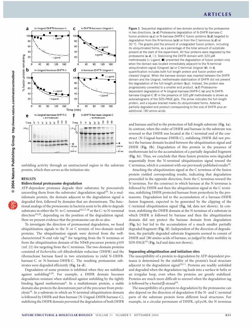
Degradation of some proteins is inhibited when they are stabilizedagainst unfolding6,10. For example, a DHFR domain becomes degradation-resistant when stabilized against unfolding by the tightlybinding ligand methotrexate6. In a multidomain protein, a stabledomain also protects the downstream part of the precursor from prote-olysis10. In a substrate in which an N-terminal ubiquitination domainis followed by DHFR and then barnase (N-Usignal-DHFR-barnase-C),stabilizing the DHFR domain prevented the degradation of both DHFR
and barnase and led to the protection of full-length substrate (Fig. 1a).In contrast, when the order of DHFR and barnase in the substrate wasreversed so that DHFR was located at the C-terminal end of the con-struct (N-Usignal-barnase-DHFR-C), stabilizing DHFR did not pro-tect the barnase domain located between the ubiquitination signal andDHFR (Fig. 1b). Degradation of this protein in the presence ofmethotrexate led to the accumulation of a partially degraded fragment(Fig. 1e). Thus, we conclude that these fusion proteins were degradedsequentially from the N-terminal ubiquitination signal toward the C terminus, which is consistent with our previously published results10.
Attaching the ubiquitination signal at the C terminus of the fusionprotein yielded corresponding results, indicating that degradationproceeded in the opposite direction, from the C terminus toward theN terminus. For the construct in which barnase at the N terminus isfollowed by DHFR and then the ubiquitination signal at the C termi-nus, stabilizing DHFR protected barnase from proteolysis by the pro-teasome. Degradation led to the accumulation of a barnase-DHFRfusion fragment, expected to be generated by the clipping of the C-terminal ubiquitination signal (Fig. 1d, data not shown). In con-trast, stabilizing the DHFR domain at the N terminus of a substrate inwhich DHFR is followed by barnase and then the ubiquitinationdomain did not protect the barnase domain from degradation(Fig. 1c) but led to the accumulation of a much smaller partiallydegraded fragment (Fig. 1f). Independent of the direction of degrada-tion, the partially degraded substrate fragments seemed to consist ofDHFR and ∼ 90 amino acids of barnase, as judged by their mobility inSDS-PAGE10 (Fig. 1e,f and data not shown).
Separating ubiquitination and initiation sitesThe susceptibility of a protein to degradation by ATP-dependent pro-teases is determined by the stability of the protein’s local structureadjacent to the degradation signal10,11. Proteins are readily unfoldedand degraded when the degradation tag leads into a surface α-helix oran irregular loop, even when the proteins are greatly stabilized.Proteins are much more difficult to unravel when the degradation tagis followed by a buried β-strand10.
The susceptibility of a protein to degradation by the proteasome canalso depend on the direction of degradation if the N- and C-terminalparts of the substrate protein form different local structures. Forexample, in a circular permutant of DHFR, cpLys38, the N terminus
Figure 1 Sequential degradation of two-domain proteins by the proteasomein two directions. (a–d) Proteasome degradation of N-DHFR-barnase-Cfusion proteins (a,c) or N-barnase-DHFR-C fusion proteins (b,d) targeted todegradation from the N terminus (a,b) or from the C terminus (c,d) at 30 °C. The graphs plot the amount of undegraded fusion protein, includingits ubiquitinated forms, as a percentage of the total amount of substratepresent at the start of the experiment. All four proteins were degraded by theproteasome (a–d, �). Stabilizing the DHFR domain with 320 µMmethotrexate (+ Ligand, �) prevented the degradation of fusion protein onlywhen the domain was located immediately adjacent to the N-terminalubiquitination signal (Usignal) (a) or C-terminal Usignal (d). In d,quantification includes both full-length protein and fusion protein withcleaved Usignal. When the barnase domain was inserted between the DHFRdomain and the Usignal, methotrexate stabilization of DHFR did not preventthe degradation of the full-length protein (b,c). Instead, the protein wasprogressively converted to a smaller end product. (e,f) Proteasome-dependent degradation of N-Usignal-barnase-DHFR-C (e) and N-DHFR-barnase-Usignal-C (f) in the presence of 320 µM methotrexate is shown asautoradiograms of the SDS-PAGE gels. The arrow indicates the full-lengthprotein, and a square bracket marks its ubiquitinated forms. Asterisk,partially degraded end product corresponding to the size of DHFR plus anadditional ∼ 90 amino acids.
©20
04 N
atur
e P
ublis
hing
Gro
up
http
://w
ww
.nat
ure.
com
/nat
stru
ctm
olbi
ol

A R T I C L E S
832 VOLUME 11 NUMBER 9 SEPTEMBER 2004 NATURE STRUCTURAL & MOLECULAR BIOLOGY
leads into a buried β-sheet whereas the C terminus leads into a surfaceα-helix10. This protein was degraded efficiently when the ubiquitina-tion signal was fused to its N terminus (N-Usignal-cpLys38-C, Fig. 2a)or its C terminus (N-cpLys38-Usignal-C, Fig. 2b). Degradation fromthe N terminus was completely inhibited when the protein was stabi-lized against global unfolding by methotrexate (Fig. 2a), whereasdegradation from the C-terminal ubiquitination signal was unaffectedby methotrexate binding (Fig. 2b). Thus, the fate of cpLys38 dependson the site at which the proteasome initiates degradation.
This conclusion raises the question whether the site of ubiquitina-tion is always the site at which proteasome degradation initiates. The19S regulatory particle can bind to proteins that lack a ubiquitin mod-ification if these proteins are unfolded or misfolded9,23, and degrada-tion signals targeting proteins to prokaryotic ATP-dependentproteases are unstructured peptide sequences24. Therefore, we specu-lated that an unstructured region in a proteasome substrate couldserve as the degradation initiation site. To test this hypothesis, weattached a lysine-less tail to the C terminus of a protein consisting ofan N-terminal ubiquitination signal followed by cpLys38 (yielding N-Usignal-cpLys38∼∼∼∼ -C). Notably, this protein was degraded effi-ciently even when stabilized by methotrexate (compare Fig. 2a toFig. 2d). The C-terminal tail seems to be disordered in the fusion protein, as it was highly sensitive to trypsin, proteinase K and chymotrypsin digestion (Fig. 2c) and showed a characteristicallyincreased hydrodynamic radius compared with a natively folded
protein as measured by gel filtration experiments25 (data not shown).The unstructured tail did not simply destabilize cpLys38 against spon-taneous global unfolding. The stabilities of cpLys38 with and withoutthe unstructured region are similar as assessed by a GroEL bindingassay26 (Fig. 2e). Also, addition of the unstructured region did notchange the position of the ubiquitination site. Ubiquitination of thesubstrate with the C-terminal unstructured region still occurred at theN-terminal ubiquitination signal. The protein was stabilized when theN-end rule tag was inactivated by the mutation of the N-end residuefrom arginine to methionine27, by mutating the ubiquitin acceptorlysines, Lys15 and Lys17, in the linker region to arginine27, or by deleting the N-terminal ubiquitination site entirely (Fig. 2f).
The N-terminal and C-terminal regions of wild-type DHFR bothform buried β-strands. This protein is efficiently degraded when targeted to the proteasome by an N-terminal degradation tag, andmethotrexate stabilization prevents its degradation6 (data not shown).Attaching an unstructured region to the C terminus did not release themethotrexate-induced block to degradation but instead led to clippingof the protein (Fig. 3a,b,d). To determine the site of the clipping, we attached a His6 sequence to the C terminus of the protein as an epitope tag. An immunoblot against the C-terminal tag showed thatthe protein is clipped at the C terminus (Fig. 3c,d). Together, these results suggest that degradation of methotrexate-stabilized N-Usignal-cpLys38∼∼∼∼ -C and N-Usignal-DHFR∼∼∼∼ -C initiates atthe C-terminal unstructured region and not at the ubiquitination site.
Figure 2 Proteasome degradation is accelerated by an unstructured region in the substrate protein. (a,b) N-terminally tagged (a) and C-terminally tagged (b)DHFR mutant cpLys38 were incubated in a proteasome degradation assay at 25 °C in the absence of ligand (�). Stabilization of cpLys38 with 320 µMmethotrexate (+ Ligand, �) prevented its degradation only when targeted from the N-terminal tag (a), and the C-terminally tagged protein was degraded rapidly(b). (c) The N-terminal ubiquitination signal in N-Usignal-cpLys38 (lanes 1–4) as well as the C-terminal region in N-Usignal-cpLys38∼∼∼∼ -C (lanes 5–8)appear disordered and are sensitive to digestion by nonspecific proteases. Protease sensitivity was assayed by incubating the protein with the indicatedprotease at 4 °C for 1 min. (d) N-terminally tagged cpLys38 containing a C-terminal unstructured region was degraded efficiently by the proteasome in theabsence (�) or presence of 320 µM methotrexate (�). Thus, the addition of a C-terminal unstructured region significantly accelerated the degradation of N-terminally tagged cpLys38 and completely reversed its methotrexate-induced degradation block. (e) An unstructured region does not affect spontaneousunfolding of cpLys38. The rate of spontaneous unfolding of N-Usignal-cpLys38-C (�) and N-Usignal-cpLys38∼∼∼∼ -C (�) was followed in the presence of100 µM methotrexate as measured by GroEL binding assay at 15 °C. (f) Degradation of cpLys38 containing a C-terminal unstructured region depends onubiquitination at Lys15 and Lys17. N-Usignal-cpLys38∼∼∼∼ -C (�), N-cpLys38∼∼∼∼ -C (�), N-Usignal(Met)-cpLys38∼∼∼∼ -C (�) in which the N-end residue has been mutated from arginine to methionine, and N-Usignal(Lys-less)-cpLys38∼∼∼∼ -C (�) in which Lys15 and Lys17 in the N-end rule tag were mutated toarginine, were degraded by the proteasome at 25 °C in the presence of 320 µM methotrexate.
©20
04 N
atur
e P
ublis
hing
Gro
up
http
://w
ww
.nat
ure.
com
/nat
stru
ctm
olbi
ol

A R T I C L E S
NATURE STRUCTURAL & MOLECULAR BIOLOGY VOLUME 11 NUMBER 9 SEPTEMBER 2004 833
Requirement for unstructured initiation sitesThe results described above suggest that degradation can begin at anunstructured region in a substrate that is separated from the ubiquiti-nation site in primary structure. This finding raises the questionwhether an unstructured region is always required for the efficientdegradation of stably folded substrate proteins. A DHFR constructwith the N-end rule degradation signal becomes ubiquitinated onLys15 or Lys17 of the 40-residue tag27, and mutating the two lysines toarginine seemed to abolish ubiquitination (data not shown). The tagitself is largely unstructured (Fig. 2c), and either the first 14 aminoacids preceding the lysines or the 22 amino acids following the lysinescould serve as the site at which the proteasome engages the substrateprotein. Truncating the N-end rule tag by deleting residues 25–40 ofthe tag considerably stabilized the DHFR construct against protea-some degradation both in vitro (compare N-Usignal-DHFR-C and N-Usignal∆15-DHFR-C, Fig. 4a) and in vivo27. The slower degrada-tion of the truncated substrate, however, was not caused by reducedubiquitination, and the truncated substrate was ubiquitinated at thesame rate and to the same extent as the full-length substrate(Fig. 4b–d). Also, degradation of the truncated substrate was com-pletely restored when either of two unrelated unstructured tails lack-
ing any lysine residues were attached at its C terminus (N-Usignal∆15-DHFR∼∼∼∼ -Cand N-Usignal∆15-DHFR-̂ ^̂ -C) (Fig. 4a).The three proteins N-Usignal-DHFR-C, N-Usignal∆15-DHFR-C and N-Usignal∆15-DHFR∼∼∼∼ -C have similar stabilities againstglobal unfolding as measured by GroEL bind-ing assay26 (data not shown). Together, theseresults suggest that the unstructured region inthe N-end rule degradation tag is required forefficient degradation by the proteasome.
To design a substrate that lacks an unstruc-tured region completely, we constructed aprotein that is targeted to the proteasome by aconcatamer of four ubiquitin moieties fusedto each other in frame14,28. When thistetraubiquitin tag was fused directly toDHFR, the resulting fusion protein (N-Ub4-DHFR-C) was degraded only inefficiently(Fig. 4e). Attaching either of two unstruc-tured extensions to the C terminus of the substrate (N-Ub4-DHFR∼∼∼∼ -C and N-Ub4-DHFR-̂ ^̂ -C) led to its rapid degradation(Fig. 4e). Unfolding of DHFR was stillrequired, as methotrexate binding preventedthe degradation of N-Ub4-DHFR∼∼∼∼ -C(data not shown). The amino acid sequencesof the two degradation initiation sites testedhere were unrelated. Therefore, it is improba-ble that degradation initiation requires thepresence of a specific sequence motif that isrecognized by the proteasome. Additionalexperiments, in which parts of the initiationsites were deleted, suggested that stretches ofas few as 20 amino acids can serve as degrada-tion initiation sites (data not shown). Fromthese results, we conclude that rapid degrada-tion of a folded protein by the proteasomerequires the presence of an unstructuredregion in addition to a ubiquitin modifica-
tion. Degradation initiates at the unstructured region, and the regionpresumably serves to engage the unfolding activity of the proteasome.
Internal initiation sitesNext we wanted to determine any restrictions on the location of theunstructured region in the substrate protein and whether free N or C termini were required. We constructed a fusion protein consisting ofa short N-terminal ubiquitination signal followed by cpLys38 and thenby barnase (N-Usignal∆15-cpLys38-barnase-C). Stabilizing thecpLys38 domain by methotrexate prevented its N- to C-terminaldegradation and hence protected both cpLys38 and barnase from pro-teolysis (Fig. 5). When the unstructured region was placed betweenthe two domains (N-Usignal∆15-cpLys38∼∼∼∼ barnase-C), the proteinwas degraded rapidly even when cpLys38 was stabilized withmethotrexate (Fig. 5). Presumably, degradation of this precursor initi-ated at the internal unstructured segment. In agreement with an ear-lier study29, this result suggests that a free N or C terminus is notrequired for proteasome degradation. Attaching an unstructuredregion to the C terminus of the two-domain protein (N-Usignal∆15-cpLys38-barnase∼∼∼∼ -C) stimulated degradation only mildly. A possi-ble explanation could be that the unstructured region in a substrate
Figure 3 Proteasome degradation initiates at an unstructured region in the substrate protein. (a,b)Proteasome degradation of N-Usignal-DHFR∼∼∼∼ His6-C at 25 °C in the absence (a) or presence (b) of320 µM methotrexate (MTX) is shown as autoradiograms of SDS-PAGE gels separating the reactionproducts. Each lane represents the amount of protein remaining at the indicated time points. Arrow,bands representing full-length substrate protein; square bracket, ubiquitinated species. Stabilization ofDHFR with MTX prevented the complete degradation of the full-length protein and led to the clipping ofthe substrate. The asterisk in b indicates the truncated degradation end product that forms when DHFRis stabilized with 320 µM MTX. (c) Western blot, using antibody to His-tag, of the degradation of N-Usignal-DHFR∼∼∼∼ His6-C at 25 °C in the presence of 320 µM MTX. Lane E contains reticulocytelysate as background control. The degradation end product is not recognized by antibodies specific toHis-tag, demonstrating that the full-length protein was truncated at its C terminus. The asterisk in cindicates the expected position of the degradation end product as seen by autoradiography in b. (d)Quantification of autoradiograms from a (�) and b (�) and western blot from c (�). In the presence ofMTX, N-Usignal-DHFR∼∼∼∼ His6-C is rapidly clipped at the C-terminal unstructured region without thedegradation of the DHFR domain. Undegraded (�, �) or unclipped (�) substrate, includingubiquitinated forms, at each time point are plotted as a percentage of the total substrate present at thebeginning of the time course.
©20
04 N
atur
e P
ublis
hing
Gro
up
http
://w
ww
.nat
ure.
com
/nat
stru
ctm
olbi
ol

A R T I C L E S
834 VOLUME 11 NUMBER 9 SEPTEMBER 2004 NATURE STRUCTURAL & MOLECULAR BIOLOGY
region. This requirement for an unstructured initiation site is some-what reminiscent of the mechanism by which helicases engage theirsubstrates. Helicases are multidomain ATPases that are also sometimesmultimeric and that separate the strands of nucleic acid double helicesprocessively. Helicases require a single-stranded region in their sub-strates as the initiation site to unwind the double-stranded remainderof the nucleic acid35. In at least one helicase, the initiation site can beclearly separated from the substrate recognition site36. The mechanismwe propose for the proteasome is also reminiscent of the way in whichsome prokaryotic ATP-dependent proteases engage their substratesthrough adaptor proteins such as SspB37 or UmuD38. For example, theprotease ClpXP binds its substrate UmuD′ indirectly by using UmuDas an adaptor and then engages the substrate directly through a definedinitiation site38. In other protease substrates, such as SsrA-tagged pro-teins, the substrate recognition sites and initiation sites seem to beeither overlapping or close to each other38. The latter scenario is similarto the way precursor proteins engage the mitochondrial unfolding andimport machinery, where the recognition and initiation sites in themitochondrial targeting sequence are not easily separated.
The proteasome can actively unfold proteins9,10 and degrade themfaster than they would unfold spontaneously by trapping local unfold-ing fluctuations in the substrate10,11. We propose that the proteasomeis only able to trap these local fluctuations efficiently after it hasengaged its substrate fully through an unstructured region, possibly bythreading it into the degradation channel. In the absence of anunstructured region, much larger and therefore rarer unfolding
protein has to be located near to the ubiquitination site to serve as anefficient initiation site for degradation.
DISCUSSIONThe eukaryotic proteasome degrades a wide range of cellular proteinsthat differ greatly in their structures, stabilities and functions. Most ofthese proteins are targeted for degradation by ubiquitination, and theubiquitination sites30 can be found near the substrates’ N termini31,32,near their C termini33 or internally22,34. We reported previously thatthe proteasome can degrade substrate proteins sequentially by engag-ing a substrate near an N-terminal ubiquitination site and runningalong the polypeptide chain toward the C terminus. Here we show thatthe proteasome can also degrade substrates in the opposite direction,from the C terminus toward the N terminus. The direction of degrada-tion is not strictly determined by the location of the ubiquitin modifi-cation on the substrate, and degradation does not necessarily initiate atthe ubiquitination site. For example, proteolysis of a substrate that isubiquitinated at its N terminus can begin at an unstructured regionprovided at the C terminus. Degradation then proceeds from the C terminus toward the N-terminal ubiquitination site (Fig. 2).
The presence of an unstructured region in a folded proteasome sub-strate greatly enhances the efficiency with which the protein isdegraded (Fig. 4). Our results suggest that ubiquitination alone is notsufficient for the efficient degradation of tightly folded proteins.Instead, it seems that the proteasome binds its substrate through theubiquitin chain and then engages the protein at an unstructured
Figure 4 Tightly folded substrates must contain an unstructured region for efficient proteasome degradation. (a) Degradation of N-Usignal-DHFR-C requiresthe unstructured region of the Usignal. Deletion of the portion of unstructured Usignal following the ubiquitin acceptor lysines, N-Usignal∆15-DHFR-C,substantially stabilized the protein against degradation compared with N-terminally tagged DHFR at 25 °C. This stabilization was overcome when anunstructured region was added to the C terminus of the protein carrying the deletion to create N-Usignal∆15-DHFR∼∼∼∼ -C or N-Usignal∆15-DHFR-̂ ^̂ -C. (b,c) The truncation of the Usignal did not alter the ubiquitination of the substrate. The ubiquitination rate of N-Usignal-DHFR-C (b) and N-Usignal∆15-DHFR-C (c) in reticulocyte lysate was measured in a ubiquitination assay in the presence of 320 µM methotrexate. The SDS-PAGE gels resolve theubiquitinated forms (square bracket) from the nonubiquitinated protein (arrow), and the lanes represent the progress of the ubiquitination reaction.(d) Quantification of autoradiograms from b and c. The amount of ubiquitinated species at the indicated times was quantified and plotted as a percentage of the initial nonubiquitinated protein. (e) Degradation of N-Ub4–tagged DHFR-C by proteasome at 25 °C. N-Ub4-DHFR-C lacks any unstructured region and was degraded very inefficiently. Degradation was accelerated several-fold when an unstructured region was added to the C terminus of the protein, N-Ub4-DHFR∼∼∼∼ -C and N-Ub4-DHFR-̂ ^̂ -C.
©20
04 N
atur
e P
ublis
hing
Gro
up
http
://w
ww
.nat
ure.
com
/nat
stru
ctm
olbi
ol

A R T I C L E S
NATURE STRUCTURAL & MOLECULAR BIOLOGY VOLUME 11 NUMBER 9 SEPTEMBER 2004 835
fluctuations may have to occur in a folded substrate for it to fullyengage the proteasome. Thus, the requirement for an unstructuredregion should become greater when a substrate is more stable againstglobal unfolding.
The requirement of an unfolded region for efficient proteasomaldegradation of tightly folded proteins may explain some unexpectedexperimental observations. For example, a chain of four ubiquitinmoieties linked through Lys48 is a sufficient targeting signal for degra-dation by the proteasome14. However, attachment of tetraubiquitin toa ubiquitin-DHFR fusion protein led to notably slow degradation ofthis protein in an in vitro assay14. The explanation for this observationcould be that the substrate lacked an appropriate unstructured regionto serve as the degradation initiation site. In addition, there are severalexamples of authentic cellular proteins that associate with the protea-some but escape degradation. For example, the cell division cycle regu-lator Cdc34 and the Hsc70 cochaperone BAG-1 are extensivelyubiquitinated and recognized by the proteasome but are notdegraded15,39,40. Similarly, the adaptor proteins Rad23 and Dsk2 bindboth proteasome and substrates but avoid degradation them-selves41–43. A possible explanation of these observations is that theproteins lack appropriate degradation initiation sites. However,inspection of the domain structure of the proteins in the Pfam data-base44 suggests that they contain extensive unstructured regions.Three different explanations are possible. First, the unstructuredregions may be located too far away from the ubiquitination sites orproteasome interaction domains to function as effective initiationsites. This situation would be analogous to that observed with themodel protein in which the ubiquitination site and unstructuredregion were separated by two folded domains (N-Usignal∆15-cpLys38-barnase∼∼∼∼ -C). This protein was degraded only slowly, pre-sumably because of the large physical distance between proteasomebinding and degradation initiation sites (Fig. 5). A second potentialexplanation has been proposed in the context of the degradation of thecyclin-dependent kinase inhibitor Sic1 (ref. 16). Sic1 can becomeubiquitinated at any of its 20 lysine residues, but only ubiquitinationnear the N terminus leads to its rapid degradation16. It is possible thatthe proteasome can engage the substrate at several unstructuredregions but that the structure of the folded domain of Sic1 is more
easily unraveled from the initiation site close to the N-terminallysines16, just as the DHFR mutant cpLys38 is more easily unfoldedfrom the C terminus than the N terminus (Fig. 2). This scenario couldalso explain the behavior of the transcription factor inhibitor IκBα.The protein is ubiquitinated on Lys21 and Lys22 of a ∼ 70-residue N-terminal region, but degradation requires the presence of a 40-residue tail at its C terminus45. The C-terminal tail is not involvedin the ubiquitination step but has a second, unknown function, possi-bly to serve as a degradation initiation site. Finally, a third possibleexplanation for the experimental observations listed earlier is that theproteasome may exhibit sequence specificity such that the effective-ness of degradation initiation sites depends on their amino acid composition. For example, the proteasome might be unable to initiatedegradation of IκBα at its N terminus because the amino acidsequence does not interact well with the proteasome.
In summary, we demonstrate that polyubiquitination of proteins,though necessary, is not sufficient for rapid degradation of tightlyfolded proteins. Their efficient degradation requires an additionalunstructured region that serves as the initiation site to engage the pro-teasomal unfolding machinery (Fig. 6). Once the substrate has fullyengaged the proteasome through its unstructured region, the substrateis unfolded and degraded. In a competing process, the proteasome-associated deubiquitinating enzymes continuously remove the ubiqui-tin moieties from the bound substrate, releasing the substrate from thecomplex46,47. Therefore, failure to successfully engage the proteasomeunfolding machinery would eventually lead to substrate dissociation.Even a substrate that fully engages the proteasome can be released ifthe degradation is blocked or slowed down considerably by a particu-larly stable domain. This dissociation would result in a partiallydegraded substrate and seems to be the mechanism for the processingof some transcription factors, such as NFκB and Spt23p10. Our resultsindicate that differences in substrate structure can have important
Figure 5 Characterization of degradation initiation sites. Degradation of N-terminally tagged cpLys38-barnase fusion protein by the proteasome at25 °C in the presence of 320 µM methotrexate. Stabilizing cpLys38 withmethotrexate prevents the N- to C-terminal sequential degradation of N-Usignal∆15-cpLys38-barnase-C (�). Under the same conditions, thefusion protein containing an unstructured degradation initiation site betweencpLys38 and barnase domain, N-Usignal∆15-cpLys38∼∼∼∼ barnase-C, wasdegraded efficiently in the presence of methotrexate (�). However, addingthe unstructured region to the C terminus of the fusion protein to create N-Usignal∆15-cpLys38-barnase∼∼∼∼ -C did not accelerate degradation (�).
Figure 6 Schematic representation of the proposed proteasomedegradation cycle. Polyubiquitinated proteins bind to the proteasomethrough the ubiquitin chain. Unfolding and degradation, however, ensueonly after the substrate has engaged the proteasome through anunstructured domain or the degradation initiation site. Once the substrateis engaged, it is degraded sequentially along the polypeptide chain from thedegradation initiation site. The EM reconstruction of the proteasome isreproduced from Larsen and Finley54.
©20
04 N
atur
e P
ublis
hing
Gro
up
http
://w
ww
.nat
ure.
com
/nat
stru
ctm
olbi
ol

A R T I C L E S
836 VOLUME 11 NUMBER 9 SEPTEMBER 2004 NATURE STRUCTURAL & MOLECULAR BIOLOGY
consequences in determining fate, providing another mechanism bywhich the proteasome can achieve specificity of degradation.
METHODSSubstrate proteins. Protein substrates were derived either from barnase, aribonuclease from Bacillus amyloliquefaciens48 or from E. coli DHFR49. The cir-cular permutant of DHFR cpLys38 was constructed by connecting the N and C termini of E. coli DHFR with a linker consisting of six glycine residues andrearranging the gene so that the resulting protein started with what was for-merly Lys38 and ended with Asn37 (ref. 50). Ubiquitination at the N terminusof substrate proteins occurred through the N-end rule pathway51. In these con-structs, an ubiquitin moiety was attached to the N terminus of the substrateprotein via a 40-residue linker derived from lac repressor6. In reticulocytelysate, the ubiquitin moiety is rapidly cleaved, and a new N terminus is formedat the amino acid that follows the last residue of the ubiquitin sequence.Depending on the identity of the new N-terminal amino acid, the substrates arethen ubiquitinated at two lysine residues in the linker region27. The C-terminalubiquitination signal consisted of amino acids 352–646 of the NFκB precursorprotein, p105 (ref. 22). The Ub4 degradation signal was constructed by con-necting four ubiquitins through their N and C termini. The signal was thenattached directly to the N terminus of DHFR. The ubiquitin moieties in theconcatamer contain G76V mutations to prevent their deconjugation. Theunstructured regions were derived from residues 1–95 of cytochrome b2 (∼∼∼∼ )or residues 1–40 of lac repressor (^̂ )̂. These regions seem to be disordered, asjudged by their increased sensitivity to unspecific proteases and by theirincreased hydrodynamic volume25. All proteins were constructed using stan-dard molecular biology techniques in pGEM-3Zf(+) vectors (Promega) andverified by DNA sequencing.
Radioactive proteins were expressed from a T7 promoter by in vitro tran-scription and translation in reticulocyte lysate (Promega), supplemented with[35S]methionine. Arg-Ala dipeptide (12 mM) was included during the transla-tion of N-end rule substrates to prevent their ubiquitination and degradationin the translation mix. Proteins were then partially purified by high-speed cen-trifugation and ammonium sulfate precipitation as described52. N-Usignal-DHFR∼∼∼∼ His6-C protein for western blot analysis was expressed from thepGEM-3Zf(+) T7 promoter in E. coli strain BL21 (DE3) pLysS and purifiedfrom clarified cell lysate by affinity chromatography using a 1-ml Ni2+-NTAagarose column (Qiagen) according to the supplier’s protocol.
Proteasome degradation assays. Degradation by the proteasome was followedin rabbit reticulocyte lysate (Green Hectares) that had been ATP-depleted asdescribed53. In vitro–translated protein was precipitated by ammonium sulfate,resuspended in one-fifth reaction volume of resuspension buffer (25% (v/v)glycerol, 25 mM MgCl2, 0.25 M Tris-HCl, pH 7.4) and diluted five-fold intoATP-depleted reticulocyte lysate supplemented with 1 mM DTT, 5 mMmethionine and 25 µM ubiquitin. Methotrexate was included in the reactionwherever mentioned, and the control reactions contained an equal volume ofDMSO. The reaction was prewarmed at 25 °C for 3 min, and ubiquitinationand degradation were initiated by addition of ATP and an ATP regenerationsystem (0.5 mM ATP, 10 mM creatine phosphate, 0.1 mg ml–1 creatine phosphokinase). Incubation was continued at 25 °C, and at the indicated timesequal-volume samples were withdrawn from the reaction mix and added toSDS-PAGE sample buffer to stop the reaction. Samples were resolved by SDS-PAGE, and the radioactive proteins were analyzed and quantified by electronic autoradiography (Instant Imager; Packard). The His6-tagged proteins were analyzed by immunoblotting using rabbit antibodies to His6
(Bethyl Labs) and Alexa Fluor 680–labeled secondary antibody specific to rab-bit (Molecular Probes) and quantified using the Odyssey infrared imaging system (LI-COR Biosciences).
Protease sensitivity assay. 35S-labeled DHFR proteins were incubated at 4 °C in50 mM Tris-Cl, pH 7.4, containing 100 µM methotrexate, 5 mM MgCl2, 5%(v/v) glycerol and 0.1 mg ml–1 trypsin or proteinase K or chymotrypsin. Thereaction was stopped at the indicated times with two volumes of 10% (w/v)TCA, and the TCA-insoluble precipitate was solubilized in SDS-PAGE samplebuffer. The products were analyzed by denaturing PAGE and electronic autora-diography.
Ubiquitination assays. The condition for the ubiquitination assay of N-Usignal-DHFR-C and N-Usignal∆15-DHFR-C was similar to the protea-some degradation assay except that 2 µM Ubal (Calbiochem) was included toinhibit the deubiquitinating enzymes. To prevent the degradation of the protein, 320 µM methotrexate was included in the reaction. The ubiquitinationrate was measured by quantifying the amount of ubiquitinated protein as a percentage of the total amount of nonubiquitinated substrate at the start of the experiment.
Stability assays. The stability of substrate proteins against global unfolding wasassessed using a GroEL binding assay26. Substrate proteins were resuspended inGroEL buffer (50 mM Tris-Cl, pH 7.5, 0.5 mM EDTA, 10 mM KCl, 100 µMmethotrexate, 5 mM MgCl2) and incubated at the indicated temperature. Thereaction was started by adding 1 µM GroEL14 to the reaction mix. At differenttime intervals 10-µl samples were withdrawn and added to protease buffer(GroEL buffer supplemented with 0.1 mg ml–1 proteinase K, 100 µMmethotrexate and 1 mM DTT) kept at 4 °C. After 10 min the reaction wasstopped by TCA precipitation, and the samples were analyzed by SDS-PAGEand electronic autoradiography (Instant Imager; Packard).
ACKNOWLEDGMENTSWe thank C. Pickart, L. Hicke, J. Widom, C. Holmberg and O. Uhlenbeck foradvice and helpful comments on the manuscript. We also thank P. Bellare, J. Schnell, N. Jaffe and A. Wilcox for carefully reading and correcting themanuscript. M. Fisher (University of Kansas Medical Center) provided purifiedGroEL and advice. We acknowledge the use of the instruments at the KeckBiophysics Facility of the Robert H. Lurie Comprehensive Cancer Center atNorthwestern University. The work was supported by US National Institutes ofHealth grant R01GM63004, by a Scholar Award to A.M. from the Leukemia andLymphoma Society and by a Gramm Travel Fellowship Award to S.P. from theRobert H. Lurie Comprehensive Cancer Center at Northwestern University.
COMPETING INTERESTS STATEMENTThe authors declare that they have no competing financial interests.
Received 17 February; accepted 30 June 2004Published online at http://www.nature.com/nsmb/
1. Glickman, M.H. & Ciechanover, A. The ubiquitin-proteasome proteolytic pathway:destruction for the sake of construction. Physiol. Rev. 82, 373–428 (2002).
2. Baumeister, W., Walz, J., Zühl, F. & Seemüller, E. The proteasome: paradigm of a self-compartmentalizing protease. Cell 92, 367–380 (1998).
3. Groll, M. et al. Structure of 20S proteasome from yeast at 2.4 Å resolution. Nature386, 463–471 (1997).
4. Groll, M. et al. A gated channel into the proteasome core particle. Nat. Struct. Biol. 7,1062–1067 (2000).
5. Wenzel, T. & Baumeister, W. Conformational constraints in protein degradation by the20S proteasome. Nat. Struct. Biol. 2, 199–204 (1995).
6. Johnston, J.A., Johnson, E.S., Waller, P.R.H. & Varshavsky, A. Methotrexate inhibitsproteolysis of dihydrofolate reductase by the N-end rule pathway. J. Biol. Chem. 270,8172–8178 (1995).
7. Deveraux, Q., Ustrell, V., Pickart, C. & Rechsteiner, M. A 26S protease subunit thatbinds ubiquitin conjugates. J. Biol. Chem. 269, 7059–7061 (1994).
8. Lam, Y.A., Lawson, T.G., Velayutham, M., Zweier, J.L. & Pickart, C.M. A proteasomalATPase subunit recognizes the polyubiquitin degradation signal. Nature 416,763–767 (2002).
9. Braun, B.C. et al. The base of the proteasome regulatory particle exhibits chaperone-like activity. Nat. Cell Biol. 1, 221–226 (1999).
10. Lee, C., Schwartz, M.P., Prakash, S., Iwakura, M. & Matouschek, A. ATP-dependentproteases degrade their substrates by processively unraveling them from the degrada-tion signal. Mol. Cell 7, 627–637 (2001).
11. Kenniston, J.A., Baker, T.A., Fernandez, J.M. & Sauer, R.T. Linkage between ATP con-sumption and mechanical unfolding during the protein processing reactions of anAAA+ degradation machine. Cell 114, 511–520 (2003).
12. Breitschopf, K., Bengal, E., Ziv, T., Admon, A. & Ciechanover, A. A novel site forubiquitination: the N-terminal residue, and not internal lysines of MyoD, is essen-tial for conjugation and degradation of the protein. EMBO J. 17, 5964–5973(1998).
13. Pickart, C.M. Mechanisms underlying ubiquitination. Annu. Rev. Biochem. 70,503–533 (2001).
14. Thrower, J.S., Hoffman, L., Rechsteiner, M. & Pickart, C.M. Recognition of the polyu-biquitin proteolytic signal. EMBO J. 19, 94–102 (2000).
15. Verma, R. et al. Role of Rpn11 metalloprotease in deubiquitination and degradationby the 26S proteasome. Science 298, 611–615 (2002).
16. Petroski, M.D. & Deshaies, R.J. Context of multiubiquitin chain attachment influ-ences the rate of Sic1 degradation. Mol. Cell 11, 1435–1444 (2003).
©20
04 N
atur
e P
ublis
hing
Gro
up
http
://w
ww
.nat
ure.
com
/nat
stru
ctm
olbi
ol

A R T I C L E S
NATURE STRUCTURAL & MOLECULAR BIOLOGY VOLUME 11 NUMBER 9 SEPTEMBER 2004 837
17. Hoskins, J.R., Yanagihara, K., Mizuuchi, K. & Wickner, S. ClpAP and ClpXP degradeproteins with tags located in the interior of the primary sequence. Proc. Natl. Acad.Sci. USA 99, 11037–11042 (2002).
18. Kihara, A., Akiyama, Y. & Ito, K. Dislocation of membrane proteins in FtsH-mediatedproteolysis. EMBO J. 18, 2970–2981 (1999).
19. Reid, B.G., Fenton, W.A., Horwich, A.L. & Weber-Ban, E.U. ClpA mediates directionaltranslocation of the substrate proteins into the ClpP protease. Proc. Natl. Acad. Sci.USA 98, 3768–3772 (2001).
20. Herman, C., Prakash, S., Lu, C.Z., Matouschek, A. & Gross, C.A. Lack of a robustunfoldase activity confers a unique level of substrate specificity to the universal AAAprotease FtsH. Mol. Cell 11, 659–669 (2003).
21. Bachmair, A., Finley, D. & Varshavsky, A. In vivo half-life of a protein is a function ofits amino-terminal residue. Science 234, 179–186 (1986).
22. Orian, A. et al. Structural motifs involved in ubiquitin-mediated processing of the NF-κB precursor p105: roles of the glycine-rich region and a downstream ubiquitinationdomain. Mol. Cell Biol. 19, 3664–3673 (1999).
23. Orlowski, M. & Wilk, S. Ubiquitin-independent proteolytic functions of the protea-some. Arch. Biochem. Biophys. 415, 1–5 (2003).
24. Flynn, J.M., Neher, S.B., Kim, Y.I., Sauer, R.T. & Baker, T.A. Proteomic discovery ofcellular substrates of the ClpXP protease reveals five classes of ClpX-recognition sig-nals. Mol. Cell 11, 671–683 (2003).
25. Uversky, V.N. What does it mean to be natively unfolded? Eur. J. Biochem. 269, 2–12(2002).
26. Viitanen, P.V., Donaldson, G.K., Lorimer, G.H., Lubben, T.H. & Gatenby, A.A. Complexinteractions between the chaperonin 60 molecular chaperone and dihydrofolatereductase. Biochemistry 30, 9716–9723 (1991).
27. Bachmair, A. & Varshavsky, A. The degradation signal in a short-lived protein. Cell 56,1019–1032 (1989).
28. Stack, J.H., Whitney, M., Rodems, S.M. & Pollok, B.A. A ubiquitin-based tagging sys-tem for controlled modulation of protein stability. Nat. Biotechnol. 18, 1298–1302(2000).
29. Liu, C.W., Corboy, M.J., DeMartino, G.N. & Thomas, P.J. Endoproteolytic activity ofthe proteasome. Science 299, 408–411 (2003).
30. Peng, J. et al. A proteomics approach to understanding protein ubiquitination. Nat.Biotechnol. 21, 921–926 (2003).
31. Scherer, D.C., Brockman, J.A., Chen, Z., Maniatis, T. & Ballard, D.W. Signal-induceddegradation of IkBa requires site-specific ubiquitination. Proc. Natl. Acad. Sci. USA92, 11259–11263 (1995).
32. Glotzer, M., Murray, A.W. & Kirschner, M.W. Cyclin is degraded by the ubiquitin path-way. Nature 349, 132–138 (1991).
33. Rodriguez, M.S., Desterro, J.M., Lain, S., Lane, D.P. & Hay, R.T. Multiple C-terminallysine residues target p53 for ubiquitin-proteasome–mediated degradation. Mol. CellBiol. 20, 8458–8467 (2000).
34. Hoppe, T. et al. Activation of a membrane-bound transcription factor by regulatedubiquitin/proteasome-dependent processing. Cell 102, 577–586 (2000).
35. Delagoutte, E. & von Hippel, P.H. Helicase mechanisms and the coupling of helicaseswithin macromolecular machines. Part I: Structures and properties of isolated heli-
cases. Q. Rev. Biophys. 35, 431–478 (2002).36. Tsu, C.A., Kossen, K. & Uhlenbeck, O.C. The Escherichia coli DEAD protein DbpA
recognizes a small RNA hairpin in 23S rRNA. RNA 7, 702–709 (2001).37. Levchenko, I., Seidel, M., Sauer, R.T. & Baker, T.A. A specificity-enhancing factor for
the ClpXP degradation machine. Science 289, 2354–2356 (2000).38. Neher, S.B., Sauer, R.T. & Baker, T.A. Distinct peptide signals in the UmuD and
UmuD’ subunits of UmuD/D’ mediate tethering and substrate processing by theClpXP protease. Proc. Natl. Acad. Sci. USA 100, 13219–13224 (2003).
39. Elsasser, S. et al. Proteasome subunit Rpn1 binds ubiquitin-like protein domains.Nat. Cell Biol. 4, 725–730 (2002).
40. Alberti, S. et al. Ubiquitylation of BAG-1 suggests a novel regulatory mechanism dur-ing the sorting of chaperone substrates to the proteasome. J. Biol. Chem. 277,45920–45927 (2002).
41. Rao, H. & Sastry, A. Recognition of specific ubiquitin conjugates is important for theproteolytic functions of the ubiquitin-associated domain proteins Dsk2 and Rad23. J. Biol. Chem. 277, 11691–11695 (2002).
42. Raasi, S. & Pickart, C.M. Rad23 ubiquitin-associated domains (UBA) inhibit 26Sproteasome-catalyzed proteolysis by sequestering lysine 48–linked polyubiquitinchains. J. Biol. Chem. 278, 8951–8959 (2003).
43. Kleijnen, M.F., Alarcon, R.M. & Howley, P.M. The ubiquitin-associated domain ofhPLIC-2 interacts with the proteasome. Mol. Biol. Cell 14, 3868–3875 (2003).
44. Bateman, A. et al. The Pfam protein families database. Nucleic Acids Res. 32(Database issue), D138–D141 (2004).
45. Dai, R.M., Chen, E., Longo, D.L., Gorbea, C.M. & Li, C.C. Involvement of valosin-containing protein, an ATPase co-purified with IκBα and 26 S proteasome, in ubiq-uitin-proteasome–mediated degradation of IκBα. J. Biol. Chem. 273, 3562–3573(1998).
46. Lam, Y.A., Xu, W., DeMartino, G.N. & Cohen, R.E. Editing of ubiquitin conjugates byan isopeptidase in the 26S proteasome. Nature 385, 737–740 (1997).
47. Leggett, D.S. et al. Multiple associated proteins regulate proteasome structure andfunction. Mol. Cell 10, 495–507 (2002).
48. Hartley, R.W. A two state conformational transition of the extracellular ribonuclease ofBacillus amyloliquefaciens (barnase) induced by sodium dodecyl sulfate.Biochemistry 14, 2367–2370 (1975).
49. Rood, J.I., Laird, A.J. & Williams, J.W. Cloning of the Escherichia coli K-12 dihydro-folate reductase gene following mu-mediated transposition. Gene 8, 255–265(1980).
50. Iwakura, M., Nakamura, T., Yamane, C. & Maki, K. Systematic circular permutation ofan entire protein reveals essential folding elements. Nat. Struct. Biol. 7, 580–585(2000).
51. Varshavsky, A. The N-end rule. Cell 69, 725–735 (1992).52. Matouschek, A. et al. Active unfolding of precursor proteins during mitochondrial pro-
tein import. EMBO J. 16, 6727–6736 (1997).53. Gonda, D.K. et al. Universality and structure of the N-end rule. J. Biol. Chem. 264,
16700–16712 (1989).54. Larsen, C.N. & Finley, D. Protein translocation channels in the proteasome and other
proteases. Cell 91, 431–434 (1997).
©20
04 N
atur
e P
ublis
hing
Gro
up
http
://w
ww
.nat
ure.
com
/nat
stru
ctm
olbi
ol