Structural Basis for the Inhibition of Host Protein Ubiquitination by Shigella Effector Kinase OspG
Upload
independentCategory
view
2download
0
Accelerated Dephosphorylation of the �2-Adrenergic Receptor by Mutation of theC-Terminal Lysines: Effects on Ubiquitination, Intracellular Trafficking, and
Degradation†
Wei Liang,‡ Quang Hoang,‡ Richard B. Clark,§ and Peter H. Fishman*,‡
Membrane Biochemistry Section, Laboratory of Molecular and Cellular Neurobiology, National Institute of NeurologicalDisorders and Stroke, National Institutes of Health, Bethesda, Maryland 20892, and Department of IntegratiVe Biology and
Pharmacology, UniVersity of Texas Health Sciences Center, Medical School, Houston, Texas 77030
ReceiVed February 6, 2008; ReVised Manuscript ReceiVed August 17, 2008
ABSTRACT: Agonist-mediated ubiquitination regulates some G protein-coupled receptors by targeting themto lysosomes for degradation. Phosphorylation also regulates receptor endocytosis and trafficking tolysosomes. To explore the roles of the two post-translational modifications, we mutated the three C-terminallysines to arginines in the human �2-adrenergic receptor (�2AR) (K348/372/375R). The level of agonist-mediated ubiquitination of the mutant (3K/R) was greatly reduced compared to that of wild-type (WT)�2AR in whole cells and in cell-free assays. Downregulation of 3K/R also was attenuated compared tothat of the WT, whereas internalization and recycling were more similar. During endocytosis, WT and3K/R appeared in different vesicles and WT, but not 3K/R, was transported to lysosomes. Both wererapidly phosphorylated in agonist-stimulated cells, but upon agonist removal, the rate of dephosphorylationof 3K/R initially was ∼5 times faster than that of WT. The increased rate also was observed in a cell-free, soluble assay and, thus, was not due to differences in receptor trafficking. Okadaic acid, a potentphosphatase inhibitor, reduced the level of dephosphorylation and increased the levels of lysosomal targetingand degradation of 3K/R. The reduced level of ubiquitination and rapid dephosphorylation of 3K/R appearto prevent it from being sorted to lysosomes in contrast to the phosphorylated and ubiquitinated WT�2AR. Our findings indicate that both phosphorylation and ubiquitination are involved in the intracellularsorting of �2AR between pathways of recycling to the plasma membrane and degradation in lysosomes,and that the rate of dephosphorylation may be another mechanism of regulating the sorting.
Phosphorylation of G protein-coupled receptors (GPCRs)1
by second-messenger and GPCR kinases (GRKs) modulatesreceptor function, endocytosis, intracellular trafficking, anddegradation (1-4). Some of the molecular mechanismsinvolved in the earlier processes such as desensitization andinternalization have been identified. Phosphorylation ofagonist-occupied receptors by GRKs promotes high-affinitybinding of arrestins which uncouple the receptors from theirG proteins and attenuate signaling. Arrestins also bindclathrin and adapter protein-2 and recruit many GPCRs toclathrin-coated pits where they are internalized. Less under-stood are the mechanisms for the intracellular sorting and
degradation of GPCRs (4-6). Most models envision interac-tions between sorting motifs on the receptors and sortingproteins which target the receptors to different endosomalcompartments and fates such as being recycled or degraded.
Recently, ubiquitination has been found to regulate severalmammalian GPCRs by targeting them to lysosomes (7-11;reviewed in ref 12). Ubiquitin, a 76-amino acid residuepolypeptide, is conjugated to acceptor proteins in threeenzymatic steps: activation of the terminal carboxyl groupof ubiquitin (E1), transfer of the activated polypeptide tothe conjugating enzyme (E2), and ligation of the ubiquitinto ε-amino groups of lysines on the acceptor protein (E3)(13). Originally, ubiquitination was identified as a mechanismfor directing proteins to proteasomes for degradation (13, 14).Subsequent studies revealed that many plasma membranereceptors are sorted to lysosomes by ligand-inducedubiquitination (14-16).
A relationship between phosphorylation and ubiquitinationin the intracellular sorting and trafficking of GPCRs has yetto be established (17). For the mammalian GPCRs studiedso far, phosphorylation is required for ubiquitination andendocytosis, whereas ubiquitination is not required for eitherphosphorylation or endocytosis (7, 8). Recently, human �1ARin contrast to �2AR was shown not to undergo agonist-mediated ubiquitination (18) which may explain the resis-
† Supported in part by the Intramural Research Program of theNational Institute of Neurological Disorders and Stroke, NationalInstitutes of Health (W.L., Q.H., and P.H.F.), and National Institutesof Health Grant GM031208 (R.B.C.).
* To whom correspondence should be addressed. Telephone: (301)496-1325. Fax: (301) 480-3734. E-mail: [email protected].
‡ National Institutes of Health.§ University of Texas Health Sciences Center, Medical School.1 Abbreviations: �AR, �-adrenergic receptor; DMEM, Dulbecco’s
modified essential medium; DPBS, Dulbecco’s phosphate-bufferedsaline; DUB, deubiquitinating enzyme; EEDQ, N-ethoxycarbonyl-2-ethoxy-1,2-dihydroquinoline; GPCR, G protein-coupled receptor; GRK,GPCR kinase; GRP, GPCR phosphatase; 125ICYP, [125I]iodocyanopin-dolol; ISO, isoproterenol; PNS, postnuclear supernatant(s); PP1, proteinphosphatase 1; PP2A, protein phosphatase 2A; pNPP, p-nitrophenylphosphate; SEM, standard error of the mean.
Biochemistry 2008, 47, 11750–1176211750
10.1021/bi800219q CCC: $40.75 2008 American Chemical SocietyPublished on Web 10/09/2008

tance of �1AR to agonist-mediated degradation and down-regulation (19). �AR chimeras in which the cytoplasmictermini are exchanged were used to establish that the �2ARC-tail contains the major sites for ubiquitination (18). Thus,�1AR with a �2AR C-tail is ubiquitinated and degraded,whereas �2AR with a �1AR C-tail is not. Previously, it wasfound that the �1AR C-tail undergoes less agonist-stimulatedphosphorylation than the �2AR C-tail does (19, 20), and�1AR is more resistant to agonist-mediated internalizationthan �2AR (see ref 20 and references cited therein). Thesefindings suggested to us that a mutant �2AR resistant toubiquitination but still able to undergo phosphorylation andendocytosis may be useful in deciphering the relative rolesof phosphorylation and ubiquitination in receptor trafficking.
In this study, we demonstrate that a mutant �2AR withthe three C-tail lysines replaced with arginines (K348/372/375R or 3K/R) is more resistant to agonist-mediated ubiq-uitination, sorting to lysosomes, and downregulation. Uponendocytosis, WT and mutant �2ARs appeared in differentpopulations of cytoplasmic vesicles and WT but not 3K/Rappeared in lysosomes. Furthermore, while the C-tail GRKsites of both were rapidly phosphorylated in agonist-stimulated cells, the sites on 3K/R were more rapidlydephosphorylated than those on WT �2AR, both in wholecells and under cell-free conditions. Inhibition of dephos-phorylation by okadaic acid increased the levels of lysosomaltargeting and degradation of 3K/R. Thus, both ubiquitinationand phosphorylation have roles in the sorting of �2ARbetween recycling and degradative pathways.
MATERIALS AND METHODS
Materials. (-)-Isoproterenol (ISO), (-)-propranolol, N-ethylmaleimide, N-ethoxycarbonyl-2-ethoxy-1,2-dihydro-quinoline (EEDQ), and p-nitrophenol phosphate (pNPP) werefrom Sigma. Okadaic acid, biotinylated ubiquitin, andlactacystin were from BioMol. Protease inhibitor cocktailwas from Roche Applied Science. Peptide N-glycosidase Fand protein phosphatase 1 (PP1) were from New EnglandBiolabs. PP2A and rabbit anti-PP1R IgG were from Upstate.Rabbit anti-�2AR and -�2AR pS(355/356) IgGs, goat anti-PP2A-AR IgG, mouse anti-ubiquitin IgG1, and protein A/GPlus agarose were from Santa Cruz Biotechnology. Uncon-jugated and Alexa Fluor 488-conjugated mouse anti-HA.11IgG1 were from Covance. HRP-conjugated streptavidin andIgGs were from Zymed. Cy3-conjugated donkey anti-mouseIgG was from Jackson ImmunoResearch. LysoTracker Redand ProLong were from Molecular Probes. Other reagentswere obtained as described previously (18-21). PlasmidpcDNA3.1-HA-h�2AR (19) was used to generate mutantswith either one (K348R), two (K372/375R), or three (K348/372/375R) lysines replaced with arginines by site-directedmutagenesis. All the mutants were sequenced.
Cell Culture and Transfection. HEK 293 cells from theAmerican type Culture Collection were grown in Dulbecco’smodified Eagle’s medium (DMEM) with 10% fetal bovineserum. Cells were transfected using LipofectAMINE Plusand selected with G418 to produce resistant, unclonedcultures that expressed fairly constant �2AR levels (3.11 (0.22 and 2.55 ( 0.25 pmol/mg of protein, respectively, forWT and 3K/R HA-�2AR) during the experimental period(18). All stably transfected cells were maintained in medium
containing 0.2 mg/mL G418. For colocalization of receptors,cells stably expressing 3K/R HA-�2AR were transientlytransfected with pcDNA3.1-�2AR-GFP (20) and used 24-36h after transfection. Cells used for experiments were grownon polylysine-coated plastic culture ware or glass coverslips.
�2AR Binding Assays. The assays for determining changesin surface and total cellular receptor levels have beendescribed previously (21). For internalization of surface�2AR, cells grown in 24-well plates were stimulated with 1µM ISO in serum-free medium containing 25 mM Hepes at37 °C for up to 30 min, washed twice with ice-coldDulbecco’s phosphate-buffered saline (DPBS), exposed to5 nM (-)-[3H]CGP-12177 at 4 °C for 1 h, washed asdescribed above, and assayed for 3H and protein. To assess�2AR recycling, the cells were stimulated with ISO usuallyfor 30 min, rapidly washed twice with warm medium,incubated for increasing times of up to 1 h, and then assayedfor surface binding as described above. For downregulationof total �2AR, cells grown in six-well plates were incubatedwith 10 µM ISO at 37 °C for up to 24 h, rinsed with ice-cold Ca2+- and Mg2+-free DPBS, and lysed in 1 mM Tris-HCl (pH 7.4), 2 mM EDTA, 1 mM EGTA, and proteaseinhibitors at 4 °C (lysis buffer). Portions of the lysates wereassayed for protein and total binding activity with 250 pM(-)-[125I]iodocyanopindolol (125ICYP) for 1 h at 37 °C. Forboth binding assays, 10 µM propranolol was used to definenonspecific binding.
�2AR Binding Properties, Coupling to Adenylyl Cyclase,and Desensitization. The methods for determining thecoupling efficiency for activation of adenylyl cyclase byagonist stimulation of �2AR have been described previously(22, 23). Membranes were prepared from the cells, andportions (e1 µg of membrane protein) were used insaturation and competition binding assays to determine themaximum receptor levels (Bmax) and the Kd values for125ICYP and ISO. For the former, a range of 5-200 pM125ICYP was used, and at each concentration, the amount ofnonspecific binding was determined. The values werecalculated by fitting the data to a one-site hyperbolic curve.For the latter, the assays included 30 pM 125ICYP, 100 µMGTP, and from 0.1 nM to 100 µM ISO. The values werecalculated by fitting the data to a one-site competition curve.Membranes (5-10 µg) were assayed for adenylyl cyclaseactivity with six to eight concentrations of ISO, and EC50
and Vmax for ISO were determined by fitting the data to asigmoidal concentration-response curve (22). The param-eters determined above were used to calculate the couplingefficiency of the mutant and WT �2ARs by eq 1:
k1 ⁄ k-1 ) (Kd -EC50) ⁄ (EC50r) (1)
where k1 and k-1 are the rate constants for activation andinactivation of adenylyl cyclase, respectively, and r is Bmax.The ratio of k1/k-1 represents the coupling efficiency.Desensitization was assessed by eq 2:
(k1r)D ⁄ (k1r)N ) (Vmax)D(EC50)N ⁄ (Vmax)N(EC50)D (2)
The term (k1r)D/(k1r)N is defined as the fractional activityremaining and is calculated from the EC50 and Vmax valuesfor ISO-stimulated adenylyl cyclase in membranes preparedfrom naı̈ve (N) and desensitized (D) cells (1 µM ISO for 15min).
�2AR Dephosphorylation and Ubiquitination Biochemistry, Vol. 47, No. 45, 2008 11751

Immunoprecipitations and Immunoblotting. Control andtreated cells were washed and lysed in lysis buffer. Whenubiquitinated �2AR was being detected, the lysis buffercontained 10 mM N-ethylmaleimide to inhibit deubiquiti-nating enzymes (DUBs). The lysates were mixed with 5×RIPA buffer, gently rotated for 45 min at 4 °C, andcentrifuged at 17000g for 20 min. Proteins were immuno-precipitated from the soluble extracts using protein A-agarosebeads coated with anti-�2AR or protein A/G Plus-agarosebeads coated with anti-HA antibodies. The beads were rotatedovernight at 4 °C, collected by centrifugation, washed fourtimes with RIPA buffer, and heated in SDS sample bufferat 60 °C for 15 min. Proteins from the immunoprecipitatesas well as lysates and soluble extracts were separated bySDS-PAGE, transferred to Immobilon-p (Millipore), andprobed with HRP-conjugated streptavidin (1:10000) to detectbiotinylated �2AR, mouse anti-HA (1:5000) or anti-ubiquitin(1:2000), or rabbit anti-pS(355/356) (1:5000) or anti-�2AR(1:80000) followed by HRP-conjugated goat anti-mouse or-rabbit IgG (1:10000), respectively. Proteins were detectedby enhanced chemiluminescence and, following exposure toBio-Max MR or Lite 2 film (Kodak), quantified by scanningdensitometry and NIH Image J (21). Where indicated, blotsprobed with anti-pS(355/356) were stripped and probed withanti-�2AR.
�2AR Phosphorylation and Dephosphorylation Assays.Cells in six-well plates were stimulated with 1 µM ISO fordifferent periods of time, washed, and lysed as describedabove except the buffer included phosphatase inhibitors (10mM NaF, 20 mM sodium pyrophosphate, and 0.1 µMokadaic acid). Portions of the lysates were mixed with 5×solubilization buffer [1× ) 0.25% dodecyl �-maltoside, 20mM Hepes (pH 7.5), 150 mM NaCl, 5 mM EDTA, and 2mM EGTA], gently rotated for 45 min, and centrifuged at17000g for 20 min. The soluble supernatants (50 µg ofprotein) were treated with peptide N-glycosidase F (500 units)for 2 h at 37 °C (22, 24), diluted with 4× SDS sample buffer,heated for 15 min at 60 °C, and subjected to Western blotting(21) as described above. The deglycosylation step wasomitted in later experiments.
For dephosphorylation, the cells were stimulated for 10min with 1 µM ISO, rinsed once with warm medium,exposed to 10 µM propranolol for increasing amounts oftime, and then processed as described above. For cell-freedephosphorylation (25), the stimulated cells were washed,scraped into phosphatase assay buffer [50 mM Tris-HCl (pH7.4), 0.1 mM EDTA, 0.1% �-mercaptoethanol, and proteaseinhibitors], and homogenized with seven strokes in a Douncehomogenizer. Portions were incubated at 37 °C for up to1 h, diluted with 4× SDS sample buffer, and analyzed forphosphorylated and total �2AR by Western blotting asdescribed above. For some experiments, the homogenateswere centrifuged at 600g for 5 min to remove cell debrisand nuclei. The postnuclear supernatants (PNS) were directlyassayed or solubilized in 0.5% Triton X-100, centrifuged forclarification, and then assayed.
Dephosphorylation of Immunoprecipitated �2ARs by Puri-fied Phosphatases. �2ARs were extracted in RIPA bufferfrom cells stimulated with 1 µM ISO for 10 min andimmunoprecipitated with anti-HA antibody-coated proteinA/G Plus-agarose beads. The receptors were eluted from thebeads with 50 mM glycine and 100 mM NaCl (pH 2.5) which
was neutralized with 1 M Tris-HCl (pH 8.0). Portions (16fmol) of the eluted receptors were incubated at 30 °C in 24µL reaction mixtures containing 50 mM Tris-HCl (pH 7.4),0.1 mM EDTA, 0.1% �-ME, 1 mM PMSF, 0.1% NP-40, 40µg/mL BSA, and 100 milliunits of PP2A or 50 mM Hepes(pH 7.0), 1 mM MnCl2, 0.1 mM EDTA, 5 mM DTT, 1 mMPMSF, 0.025% Tween 20, and 100 milliunits of PP1. Thereactions were stopped by adding 8 µL of 4× SDS samplebuffer and the mixtures heated at 60 °C for 15 min. Portionsof 12 and 2 fmol were analyzed by Western blotting forphosphorylated and total �2AR, respectively.
Cell-Free Ubiquitination Assay. Portions of PNS (180 µgof cell protein) from cells stimulated with ISO for 5 minwere incubated in 150 µL reaction mixtures containing 50mM Tris-HCl (pH 7.4), 5 mM MgCl2, 0.2 mM dithiothreitol,10 mM ATP, 2 µM biotinylated ubiquitin, and 10 µM ISOfor 60 min at 37 °C. Then, 40 µL of 5× RIPA buffer and10 µL of 0.2 M N-ethylmaleimide were added. The sampleswere immunoprecipitated with anti-HA antibodies and blot-ted with HRP-conjugated streptavidin or anti-�2AR antibod-ies as described above.
�2AR Degradation Assay. A biotinylation method was usedto assess basal and agonist-mediated �2AR turnover aspreviously described (19). Briefly, cells grown in six-wellplates were rinsed with ice-cold buffer B [40 mM NaHCO3
and 100 mM NaCl (pH 8.6)], incubated with 1 mM sulfo-NHS-LC-biotin in buffer B at 4 °C for 30 min, and rinsedwith ice-cold 20 mM glutamine in DPBS. Cells in some ofthe wells were lysed as described above and the rest warmedto 37 °C in fresh medium in the absence or presence of 0.2µM okadaic acid for 30 min and 10 µM ISO for an additional4 h. The lysates were extracted in RIPA buffer, and �ARswere immunoprecipitated, detected with HRP-conjugatedstreptavidin, and quantified as described above.
Confocal Fluorescence Microscopy. Cells were visualizedusing a Zeiss LSM 510 laser confocal microscope aspreviously described with minor modifications (20). Briefly,cells expressing WT or mutant HA-�2AR grown on glasscoverslips were stained at 4 °C with Alexa Fluor 488-conjugated anti-HA antibody (1 µg/mL), washed with DPBS,and incubated at 37 °C in medium with or without 10 µMISO for up to 4 h. When used, LysoTracker Red (75 nM)was added 30 min prior to the end of the ISO treatment.Cells were washed and fixed with 4% paraformaldehyde inPBS. The coverslips were mounted on slides with ProLong,and the cells were scanned. The scanned images wereprocessed using Adobe Photoshop 7.0. For cells coexpressingWT �2AR-GFP and 3K/R HA-�2AR, the fixed cells werepermeabilized and stained with anti-HA and then Cy3-conjugated donkey anti-mouse antibody (20).
Data Analysis. Unless otherwise indicated, each experi-ment was repeated at least three times and each data pointwithin an experiment was determined in triplicate. Data werefitted to curves by nonlinear regression analysis and analyzedfor statistical significance with a two-tailed t test using Prism4 or 5 (GraphPad Software). Receptor phosphorylation wasexpressed directly by the ratio of densitometric units ofphosphorylated to that of total receptors or by normalizingthe ratios to the percentage of initial phosphorylation asindicated in the figures.
11752 Biochemistry, Vol. 47, No. 45, 2008 Liang et al.

RESULTS
Agonist-Mediated Ubiquitination and Downregulation ofWT and Mutant �2ARs. In a previous study, it was shownthat �2AR and a �1AR-�2AR C-tail chimera undergoagonist-stimulated ubiquitination whereas �1AR and a�2AR-�1AR C-tail chimera do not (18). The human �2ARhas 16 lysines, three of which are located in the C-tail (K348,K372, and K375). We constructed the �2AR mutant 3K/R(K348/372/375R) to investigate the role of the C-tail lysinesin ubiquitination. HEK 293 cells stably expressing WT andmutant HA-�2ARs were stimulated with ISO for increasingtimes, and the receptors were mmunoprecipitated with anti-�2AR antibodies and probed with anti-ubiquitin antibodies(18). Agonist-stimulated ubiquitination of WT �2AR wasreadily observed compared to that of 3K/R (Figure 1A).
Quantification of the blots by scanning densitometry showedthat the level of ubiquitination of WT �2AR increasedsteadily for up to 4 h and then declined slowly (Figure 1B).A small peak of ubiquitinated 3K/R was observed at 2 h.On the basis of analysis by area under the curve, the levelof ubiquitination of 3K/R was 16 ( 4.5% of that of WT.While we determined the level of ubiquitination of �2ARover a 24 h period, it occurred very quickly and wasenhanced by proteasomal inhibitors such as lactacystin(Figure 1C) as reported previously (7, 18). Although themechanism is not known, it indicates that deubiquitinationis constantly occurring.
We also succeeded in ubiquitinating �2AR in a cell-freeassay containing biotinylated ubiquitin, ATP, ISO, and PNSfrom cells stimulated with ISO for 5 min (Figure 1D). �2ARfrom the reactions was immunoprecipitated with anti-HAantibody and detected on blots with HRP-conjugated strepta-vidin. PNS from unstimulated cells did not work (data notshown) which is consistent with ubiquitination requiringphosphorylated receptors (7). In the absence of ATP and ISO,a faint band was detected in the area where �2AR is found(lanes 1 and 5). In the presence of ATP, the intensity of thisband greatly increased in the samples containing WT �2AR(lane 2) and even more in samples containing ATP and ISO(lane 4). In contrast, ATP had only a slight effect on thelabeling of 3K/R (lane 6) which actually weakened insamples containing ATP and ISO (lane 8). No labeling wasdetected in the absence of biotinylated ubiquitin (lanes 3 and7).
It is important to point out that in the experimentsdescribed above, only a small fraction of WT �2AR wasubiquitinated based on the minimal reduction in unmodifiedreceptors detected by anti-�2AR antibodies (see the bottompanels of Figure 1C,D). In our previous study, we had tooverexpose the blots to visualize the ubiquitinated receptorswith anti-�2AR antibodies (18). Similar results were obtainedin another study on �2AR (7) as well as other GPCRs (9, 11).
We also noticed that with time of agonist exposure,immune reactive WT receptor proteins disappeared morerapidly than the 3K/R mutant (Figure 1A, bottom panel).The differences in receptor loss were confirmed by determin-ing the levels of receptor proteins and binding activity incell lysates (Figure 2). In cells expressing WT �2AR,prolonged agonist treatment decreased the number of bindingsites by 40 ( 1.6% compared to only 12 ( 2.5% for 3K/R-expressing cells (Figure 2B,C). The loss of C-tail immunereactivity paralleled that of binding activity, and bothoccurred in WT-expressing cells after a distinct lag of 4 h.Due to the small changes in 3K/R-expressing cells, delayswere not apparent (Figure 2C). In contrast, the disappearanceof HA-tagged receptors was more rapid than loss of bindingor the C-tails in WT- and 3K/R-expressing cells. Others hadobserved that during agonist-mediated downregulation ofhuman HA-�2AR in HEK cells, anti-HA immunofluores-cence disappeared more rapidly than did binding activity(26). Loss of an N-terminal FLAG epitope from the deltaopioid receptor expressed in HEK cells was 2.5-fold greaterthan loss of binding activity after agonist treatment for 3 h(27). Such results suggest that the N-terminus of a GPCR ismore susceptible to proteolysis than the rest of the receptor.Taken together, our data support the thesis that the lysine
FIGURE 1: Agonist-stimulated ubiquitination of WT and mutant�2ARs. (A) Western blots of ubiquitinated HA-tagged �2ARs stablyexpressed in HEK 293 cells. Cells were stimulated at 37 °C with10 µM ISO for the indicated times, washed, lysed, and solubilizedin RIPA buffer as described in Materials and Methods. �2ARs wereimmunoprecipitated using anti-�2AR antibodies, and the immuno-precipitates were resolved by SDS-PAGE and detected by blottingwith mouse monoclonal anti-ubiquitin (top panel) or anti-ΗΑantibodies (bottom panel). The same amount of receptors from eachcontrol cell sample and the equivalent amount of protein from thecorresponding stimulated cell samples were loaded on the gels. Theblot shown is one of three similar blots from separate experiments.(B) Quantification of ubiquitination of WT (b) and 3K/R (9) �2ARsby scanning densitometry of the blots. Values are expressed asarbitrary densitometric units and are the means ( SEM from threeseparate experiments. (C) Rapid ubiquitination of WT �2AR. Detailsare the same as those described for panel A except cells wereincubated in the absence and presence of 10 µM lactacystin for 30min and then stimulated with 10 µM ISO for the indicated periodsof time. (D) Cell-free ubiquitination of WT and 3K/R. Portions ofPNS prepared from ISO-stimulated cells were incubated with ISO,ATP, and biotinylated ubiquitin as indicated. �2AR was immuno-precipitated using anti-HA antibody and subjected to blotting withHRP-conjugated streptavidin (top panel) or anti-�2AR antibodies(bottom panel). Shown is one of three similar blots.
�2AR Dephosphorylation and Ubiquitination Biochemistry, Vol. 47, No. 45, 2008 11753

residues in the cytoplasmic tail of �2AR are the sites ofubiquitination involved in receptor degradation.
Properties of 3K/R Mutant �2AR. We next showed thatthe mutations did not impair receptor functions such asligand binding, adenylyl cyclase activation, and desensi-tization. The parameters for 3K/R were similar to thosefor WT �2AR (Table 1 and Figure S1 of the SupportingInformation). These included the binding constants or Kd
values for 125ICYP and ISO, the EC50 and Vmax values forISO-stimulated adenylyl cyclase activity, and the coupling
efficiencies calculated from these values. In addition,exposing the cells to 1 µM ISO for 15 min resulted indesensitization of 3K/R (Figure S1C,D of the SupportingInformation) as evidenced by a 90% reduction in thefractional activity remaining and an almost 8-fold increasein the EC50 for ISO-stimulated activity which are similarto the changes observed for WT �2AR (Table 1).
Endocytosis, Intracellular Trafficking, and Recycling ofWT and Mutant �2ARs. While �2AR ubiquitination is notrequired for endocytosis, it is necessary for degradation (dataabove and ref 7) and may affect the intracellular traffickingof the receptor. To explore this possibility, HEK 293 cellsexpressing HA-tagged �2ARs were stained at 4 °C withAlexa Fluor 488-conjugated anti-HA antibody and incubatedat 37 °C for increasing amounts of time with ISO (Figure3A). Initially, most of the receptors were on the plasmamembrane and, upon agonist stimulation, rapidly internalized.WT accumulated in larger, perinuclear vesicles as previouslyreported (18, 20), whereas 3K/R �2AR appeared in small,peripheral, cytoplasmic vesicles that became more punctateand intense with time. We directly compare the distributionof the two receptors by transiently expressing WT �2AR-GFP in cells stably expressing HA-3K/R (Figure 3B). We
FIGURE 2: Agonist-mediated downregulation of WT and 3K/Rreceptor binding activity and proteins. Cells were incubated at 37°C in the absence and presence of 10 µM ISO for the indicatedtimes, washed, and lysed. Portions were assayed for total �2ARbinding sites using 125ICYP or for receptor proteins by Westernblotting with anti-HA and anti-�2AR antibodies as described inMaterials and Methods. The blots were quantified by scanningdensitometry. (A) Western blots of receptor proteins from ISO-treated cells expressing WT or 3K/R. (B) Effect of agonist treatmenton WT �2AR binding sites (O) and receptor proteins detected byanti-C-tail (0) or anti-HA (4) antibodies. (C) Same as panel Bexcept cells expressing 3K/R were used: binding sites (b), anti-C-tail (9), or anti-HA (2). Values are the means ( SEM fromfour to six independent experiments.
Table 1: Properties of WT and 3K/R Mutant �2ARsa
WT(mean ( SEM)
3K/R(mean ( SEM) n
naı̈ve cellsEC50 for ISO (nM) 0.647 ( 0.19 0.411 ( 0.091 4Vmax for ISO (pmol min-1 mg-1) 420 ( 39 347 ( 38 4Kd for 125ICYP (pM) 8.14 ( 0.80 8.50 ( 2.2 4Kd for ISO (nM) 238 ( 21 215 ( 32 5Bmax (fmol/mg of protein) 6850 ( 562 6840 ( 792 4coupling efficiency 0.070 ( 0.01 0.087 ( 0.021 4
desensitized cellsshift in EC50 (x-fold) 9.0 ( 2.1 7.8 ( 1.7 4Vmax for ISO (% of control) 62.5 ( 9.5 59.3 ( 4.9 4fractional activity remaining 0.092 ( 0.01 0.087 ( 0.02 4a Membranes were prepared from naı̈ve and agonist-stimulated (1 µM
ISO for 15 min) cells and assayed for binding and adenylyl cyclaseactivity as described in Materials and Methods. The Kd and Bmax valuesfor 125ICYP were determined by saturation binding. The Kd for ISO wasmeasured by competition binding in the presence of 100 µM GTP. TheEC50 and Vmax values for ISO were determined by stimulation ofadenylyl cyclase activity with increasing concentrations of ISO. For allproperties, differences between WT and 3K/R receptors were notsignificant (P > 0.05; two-tailed t test).
FIGURE 3: Visualization of agonist-stimulated internalization, traf-ficking, and recycling of WT �2AR and the 3K/R mutant. (A)Divergence of intracellular trafficking of WT and 3K/R. HEK 293cells stably expressing WT or 3K/R HA-�2AR were stained at 4°C with Alexa Fluor 488-conjugated mouse anti-HA antibody,washed, heated to 37 °C, and stimulated with 1 µM ISO for theindicated periods of time. The cells then were washed, fixed, andvisualized by confocal fluorescence microscopy. Arrows point tovesicles containing WT (dashed arrows) and 3K/R (solid arrows).(B) Minimal colocalization of WT and 3K/R during intracellulartrafficking. HEK 293 cells stably expressing 3K/R HA-�2AR andtransiently expressing WT �2AR-GFP were stimulated with 1 µMISO for the indicated periods of time. The cells were washed, fixed,permeabilized, and stained with mouse anti-HA antibody followedby Cy3-conjugated donkey anti-mouse IgG. The cells then werevisualized for WT �2AR-GFP (green), 3K/R HA-�2AR (red), andcolocalization (yellow). Selected areas were magnified 2-fold (2×).(C) Visualization of �2AR recycling. Details are the same as thosein panel A except after stimulation with 1 µM ISO for 30 min, thecells were washed, incubated in fresh medium for the indicatedperiods of time, fixed, and visualized. The bar is 5 µm. For eachpanel, images are from the same experiment and are representativeof at least three separate experiments.
11754 Biochemistry, Vol. 47, No. 45, 2008 Liang et al.

observed some colocalization (yellow) of the immunostainedHA-tagged mutant (red) and the GFP-tagged WT (green) atthe plasma membrane. After agonist stimulation of the cells,however, both receptors mostly appeared in different vesiclesby 30 min and the WT in larger aggregates by 60 min (seethe 2× magnification images in Figure 3B). The traffickingof 3K/R is very reminiscent of that of �1AR which does notundergo agonist-induced ubiquitination, appears in small,peripheral, punctate endosomes after endocytosis, and isresistant to downregulation (18-20). Using a radioligandbinding assay, we quantified the agonist-mediated internal-ization of WT and 3K/R and found that the rates for bothwere very similar but 3K/R internalized more than WT(Figure 4A).
When the agonist was removed after stimulation for 30min, the internalized �2ARs recycled back to the plasmamembrane (Figure 3C). The recycled 3K/R and to a lesserextent WT �2AR appeared in punctate structures. This maybe a consequence of the prebound anti-HA antibody ag-gregating during endocytosis and recycling. After internaliza-tion for 30 min, the recycling of WT and that of 3K/R �2ARswere not significantly different (Figure 4B). When weincreased the internalization time to 4 h, the level of recyclingof WT decreased significantly with time from 73.3 ( 2.8 to59.9 ( 2.6% (F < 0.02), whereas the level of recycling of3K/R decreased from 79.6 ( 2.8 to 74.8 ( 4.0% which wasnot significant (Figure 4C).
To determine whether newly synthesized receptors werecontributing to the recovery of surface receptors, we mea-sured the rate of receptor synthesis (Figure S2 of theSupporting Information). The cells were treated with EEDQwhich irreversibly inactivated �2AR by >95% at 300 µM(panel A). The treated cells were washed and placed in freshmedium. After a 2 h delay, the number of binding sites beganto increase at near-linear rates of 120 ( 6 and 114 ( 9 fmolh-1 (mg of protein)-1 for WT and 3K/R, respectively (panelB). These values represent ∼4% of the initial receptors, andthus, total recovery would require 25 h after the 2 h lag. Incontrast, after agonist treatment for 2 h, 63.3 ( 3.5 and 77.9( 3.1% of the original surface receptors, respectively, were
recovered during the 1 h recycling period. At most, the newlysynthesized �2AR represented less than 6% of the recycledreceptors.
Effect of the 3K/R Mutations on Phosphorylation of �2AR.As ubiquitination of �2AR requires GRK-catalyzed phos-phorylation (7), we compared the phosphorylation of the WTand 3K/R receptors using antibodies to phosphoserines 355and 356 in the C-tail (24). Although the human �2AR C-tailhas 11 serine/threonine residues that are potential GRK sites,only serines 355, 356, 364, and 411 have been implicatedin whole cell phosphorylation and receptor regulationstudies (22, 28, 29). Mass spectrometric analysis indicatedthat agonist-mediated phosphorylation of the �2AR C-tailoccurs only in the proximal region (residues 339s369) (30).Vaughan et al. (29) showed that phosphorylation of the threeserines in that sequence, 355, 356, and 364, is necessary fornormal desensitization, arrestin binding, and internalization.
We confirmed the specificity of the anti-phosphoserineantibodies by comparing their reactivity to those of bothglycosylated and deglycosylated �2AR from control andagonist-stimulated cells (22, 24) with antibodies to the �2ARC-terminal peptide (Figure S3 of the Supporting Information).The latter recognized glycosylated (64-91 kDa; lanes 1 and2) and deglycosylated (45 and 42 kDa; lanes 3 and 4) formsof �2AR from control and agonist-stimulated cells. Anti-pS(355,356) detected the same proteins but only fromagonist-stimulated cells (lanes 6 and 8). The levels detectedin control cells (lanes 5 and 7) ranged from 0 to 2% of thosein agonist-stimulated cells. Agonist-stimulated phosphory-lation of S(355,356) in both WT and 3K/R �2ARs was veryrapid and similar up to 2 min, after which the levels ofphosphorylated WT remained stable even at 2 h and that of3K/R decreased over the same time period (Figure 5).
Effect of the 3K/R Mutations on Dephosphorylation of�2AR. The decrease in the level of phosphorylated 3K/R inthe continued presence of agonist may be due to the increasedlevel of dephosphorylation which is believed to be necessaryfor the recovery of phosphorylated and desensitized GPCRs.After dephosphorylation, the resensitized receptors arerecycled back to the plasma membrane (31-34). �2AR is
FIGURE 4: Quantification of agonist-stimulated internalization and recycling of WT �2AR and the 3K/R mutant. (A) Time course ofinternalization. Cells stably expressing WT (b) or 3K/R (9) �2AR were treated with 1 µM ISO at 37 °C for the indicated periods of timeand assayed for cell surface �2AR sites using [3H]CGP12177 as described in Materials and Methods. Data represent the means ( SEM ofthree independent experiments. Maximum levels of internalization of WT and 3K/R were 59.6 ( 2.3 and 71.3 ( 3.0%, respectively, withrate constants (k) of 0.215 ( 0.024 and 0.245 ( 0.032, respectively. (B) Time course of recycling of internalized surface receptors. Cellsstably expressing WT (b) or 3K/R (9) were stimulated with 1 µM ISO at 37 °C for 30 min, washed, incubated in fresh medium forincreasing periods of time, and assayed for surface binding sites using [3H]CGP12177. Data are expressed as the percent of internalizedreceptors that were recycled and are the means ( SEM of three independent experiments. The maximum levels of recycling of WT and3K/R were 73.4 ( 2.8 and 79.6 ( 2.8%, respectively, with rate constants (k) of 0.072 ( 0.008 and 0.077 ( 0.008, respectively. (C) Effectof internalization time on the extent of recycling of internalized receptors. Cells stably expressing WT (white bars) or 3K/R (black bars)were stimulated with 1 µM ISO for the indicated periods of time, washed, incubated for 1 h in fresh medium, and assayed for surfacereceptors. Values are expressed as the percent of internalized receptors that were recycled and are the means ( SEM of three to fiveindependent experiments. Significance determined by a two-tailed t test: ns, not significant; *, P < 0.05; **, P < 0.01.
�2AR Dephosphorylation and Ubiquitination Biochemistry, Vol. 47, No. 45, 2008 11755

reported to be dephosphorylated in acidic endosomes en-riched in GRP, a PP2A-like GPCR phosphatase (33-35).Recently, however, dephosphorylation of �2AR by a PP1-like phosphatase has been observed in the plasma membranein the absence of internalization (25, 36). Thus, the distinctintracellular trafficking of the 3K/R mutant possibly mayinfluence its dephosphorylation. The latter was determinedby stimulating the cells with ISO for 10 min, washing themto remove the agonist, and incubating them for increasingperiods of time in the presence of the antagonist propranolol.Phosphoserines 355 and 356 initially were dephosphorylatedalmost 5-fold faster in 3K/R than in WT �2AR (Figure 6A,B).
Okadaic acid, a potent inhibitor of protein phosphatasesPP2A and GRP (33) as well as PP1, blocked the dephos-phorylation of WT and 3K/R (Figure 6C). While treatmentwith okadaic acid had no effect on the initial (0 min) levelof phosphorylated WT, it increased the initial level ofphosphorylated 3K/R to that of WT which is consistent withthe dephosphorylation of 3K/R observed in continuouslystimulated cells.
An Increased LeVel of Dephosphorylation of 3K/R IsAssociated with the Receptor and Not Its Trafficking. Thedata so far are consistent with a model in which the divergentintracellular trafficking of WT �2AR and the 3K/R mutantaccounted for their different rates of dephosphorylation. Wealso considered the possibilities that replacing the C-terminallysines with arginines causes a conformational change thatmakes the phosphoserines more susceptible to phosphatasesor that the lysines are involved in regulating the dephos-phorylation of the phosphoserines through either the conju-gated ubiquitins or a scaffolding protein (37). To eliminatesome of these possibilities, we performed cell-free dephos-phorylation assays. Portions of homogenates or PNS fromISO-stimulated cells were incubated for increasing periodsof time at 37 °C and then assayed for phosphorylated �2AR.Cell-free dephosphorylation of 3K/R was more rapid thanthat of WT �2AR (Figure 7A), and the initial increase inrate was similar to that observed in intact cells. Okadaic acidinhibited cell-free dephosphorylation of both receptors withsimilar potency [IC50 ∼ 50 nM (Figure 7B)].
To rule out the possibility that during the 10 min periodof agonist stimulation, the receptors become compartmental-ized and remain so during the cell-free assay, we treated thePNS with Triton X-100 and used the soluble extracts forcell-free dephosphorylation (Figure 7C). Again, 3K/R wasdephosphorylated more rapidly than WT �2AR was. Thisexperiment indicated that the disparity in dephosphorylationis due to differences in the receptors and not in theircompartmentalization. As an increased rate of dephospho-rylation may be due to changes in protein phosphataseactivity, we compared control and agonist-stimulated cellsfor phosphatase activity and levels of PP1 and PP2A (FigureS4 of the Supporting Information). The total phosphataseactivity was not significantly different in WT- and 3K/R-expressing cells with or without ISO stimulation (panel A).Western blotting with antibodies specific for PP1 or PP2Aalso revealed no major differences in their levels in controland stimulated cells (panels B-D). We directly compare thedephosphorylation of WT and 3K/R by PP1 and PP2A byimmunoprecipitating phosphorylated receptors from stimu-lated cells, eluting the receptors, and incubating them with
FIGURE 5: Time course of phosphorylation of GRK-specific serines355 and 356 in WT and 3K/R �2ARs. Cells stably expressing WTor 3K/R �2AR were stimulated with 1 µM ISO for the indicatedperiods of time, washed, and lysed in ice-cold buffer containingprotease and phosphatase inhibitors as described in Materials andMethods. The lysates were solubilized, resolved by SDS-PAGE,and immunoblotted with anti-pS(355,356) antibodies. The blotswere stripped and probed with anti-�2AR antibodies. (A) Arepresentative blot is shown. (B) The blots were analyzed byscanning densitometry to quantify the phosphorylation of S(355,356)in WT (b) or 3K/R (9). Each data point represents the ratio ofdensitometric units of phosphorylated to that of total receptors.Values are the means ( SEM of three separate experiments.
FIGURE 6: Differences in dephosphorylation of WT and 3K/R mutant�2ARs. (A) Time course of dephosphorylation of pS(355,356) in�2AR in intact cells. Cells were stimulated with 1 µM ISO for 10min, washed, exposed to 10 µM propranolol (PRO) for the indicatedperiods of time, and lysed. Phosphorylated and total receptors weredetected by Western blotting as described in the legend of Figure5. A representative blot is shown. (B) Quantification of dephos-phorylation of WT (b) and 3K/R (9) �2ARs by scanning densi-tometry of the blots. Values are normalized for total receptors, areexpressed as a percentage of initial phosphorylation (---), andare the means ( SEM of seven separate experiments. (C) Effectof a phosphatase inhibitor on dephosphorylation. Cells wereincubated for 1 h in the absence (white bars) and presence (blackbars) of 0.8 µM okadaic acid and stimulated for 10 min with ISO.Cells were either collected (0 min) or washed, exposed topropranolol with and without 0.8 µM okadaic acid for 60 (WT) or20 (3K/R) min, and analyzed for phosphorylated and total receptorsby Western blotting. Values are the means ( SEM of three separateexperiments and are presented as a percentage of initial phospho-rylation of WT.
11756 Biochemistry, Vol. 47, No. 45, 2008 Liang et al.

purified phosphatases. 3K/R was dephosphorylated moreextensively than WT �2AR was by both phosphatases (Figure7D,E).
Role for Lysines 372 and 375 but Not Lysine 348. Wegenerated the single (K348R) and double mutants (K372/375R) to determine whether the mutation of all three lysineresidues in the C-tail of �2AR is required for the differences.We chose the two mutations as the lysines bracket the clusterof three serines involved in GRK-catalyzed phosphorylationand subsequent receptor regulation (22, 29). When cellsexpressing the mutants were assayed for dephosphorylationand agonist-mediated downregulation, we found that thesingle mutant K348R which is proximal to the serine clusterbehaved like WT �2AR whereas the double mutant K372/375R which is distal to the cluster was similar to 3K/R(Figure 8).
Inhibition of Dephosphorylation Increases the LeVels ofDegradation and Lysosomal Targeting of 3K/R. To determinewhether the increased rate of dephosphorylation of 3K/Rcontributed to the reduction in the level of downregulation,we used a biotinylation procedure to assess receptor turnover.Receptor degradation is detected earlier by this method than
by binding activity or Western blotting as any internal ornewly synthesized receptors are not detected. The cells werebiotinylated, incubated in the absence and presence of 200nM okadaic acid for 30 min, and then incubated in theabsence and presence of ISO for 4 h. The receptors wereimmunoprecipitated, and the biotinylated receptors weredetected with a streptavidin overlay (Figure 9). The basalturnover was 31% for WT compared to 20% for 3K/R,whereas the turnover in the agonist-stimulated cells was 74%compared to 37%. Thus, 43 and 17%, respectively, can beattributed to the effect of the agonist. In the cells treatedwith okadaic acid and agonist, the turnover of WT was notsignificantly changed whereas the turnover of 3K/R increasedto that of WT �2AR. Okadaic acid even had a significanteffect on the basal turnover of 3K/R. It appears thatdephosphorylation impairs the degradation of the receptorand conversely phosphorylation enhances it.
The increased rate of turnover of biotin-labeled �2AR incells exposed to agonist for 4 h appears to be inconsistentwith absence of downregulation at this time. Most likely,the biotin group is being removed prior to further degradationof the receptor protein. The biotinylation reagent labels cell
FIGURE 7: Dephosphorylation of �2ARs in a cell-free system. (A) Time course of cell-free dephosphorylation of WT (b) and 3K/R (9)�2ARs. Cells stimulated with 1 µM ISO for 10 min were homogenized, and portions were incubated at 37 °C for the indicated periods oftime and analyzed sequentially for pS(355,356) and total �2AR by Western blotting and densitometry as described in Materials and Methods.Values are normalized for total receptors, are expressed as a percentage of initial phosphorylation, and are the means ( SEM of threeseparate experiments. (B) Effect of okadaic acid on cell-free dephosphorylation. Experiments were performed as described for panel Aexcept the indicated concentrations of okadaic acid were present and the reaction mixtures containing WT (white bars) were incubated for60 min and those containing 3K/R (black bars) for 20 min. Values are the means ( SEM of three separate experiments. (C) Dephosphorylationof soluble receptors. Experiments were performed as described for panel A except each homogenate was centrifuged at 600g for 5 min toobtain a PNS. The latter was solubilized with 0.5% Triton X-100, and the soluble receptors were assayed. Values are the means ( SEMof two separate experiments, each conducted in duplicate. (D) Dephosphorylation of immunoprecipitated �2ARs by purified phosphatases.Cells expressing WT or 3K/R �2AR were stimulated with 1 µM ISO for 10 min, washed, and lysed. The receptors were extracted from thelysates, immunoprecipitated using anti-HA antibodies, and eluted from the immunoprecipitates as described in Materials and Methods.Portions of the eluted receptors were incubated with 100 milliunits of the catalytic subunits of purified PP1 or PP2A at 30 °C for the timesindicated and were analyzed for phosphorylated (0, 10, and 30 min) and total receptors by Western blotting. Shown is one of three similarblots. (E) The blot shown in panel D and two similar blots were quantified by scanning densitometry. The values (means ( SEM) representthe percentage of dephosphorylation normalized for total receptors after incubation for 30 min with the purified phosphatases. Significancedetermined by a two-tailed t test: **, P < 0.01; ***, P < 0.001.
�2AR Dephosphorylation and Ubiquitination Biochemistry, Vol. 47, No. 45, 2008 11757

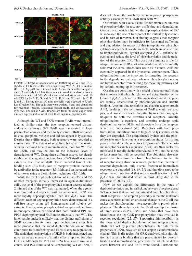
surface amino groups such as the ε-amino of lysine and theN-terminus of �2AR. Although the first and third extracellularloops of �2AR each have a lysine, it is within one residueof a transmembrane region and unlikely to be available tothe reagent. As described above, the N-terminus of �2ARappears to be more susceptible than the C-terminus toproteases that may be present in a late endosomal orprelysosomal compartment to which WT and 3K/R bothtraffic. The additional loss of the N-terminus of WT as wellas the initiation of loss of its C-tail and binding activity maybe a result of its trafficking to lysosomes. Using confocalfluorescence microscopy and the lysosomal marker LysoTrack-er, we observed the colocalization of the marker (red) andWT �2AR (green) with the time of agonist treatment (Figure10, without okadaic acid, panels A-C), whereas colocal-ization of the marker and 3K/R was substantially less evenat 4 h (panels D-F). If phosphorylation of 3K/R influencesits degradation, then it may affect its trafficking. Theinhibition of dephosphorylation by okadaic acid (withokadaic acid) did not appear to change the trafficking of WT(panels G–I). Initially, 3K/R appeared to traffic the same inthe okadaic acid-treated cells as (panels J–L); however, by1 h, some receptors were located in lysosomes (panel K),and by 4 h, there was extensive colocalization of 3K/R andthe lysosomal marker in the okadaic acid-treated cells (panelL).
DISCUSSION
On the basis of a previous study indicating that the �2ARubiquitination sites resided in the C-tail (18), we replacedthe three C-tail lysines with arginines to produce a mutant,3K/R. Our intention was to further explore the roles ofubiquitination and phosphorylation in receptor regulation. Wefound that 3K/R underwent less agonist-mediated ubiquiti-nation, lysosomal targeting, and degradation compared to the
WT receptor. Agonist-stimulated ubiquitination, however,was not totally eliminated in the 3K/R mutant, whereas it isnot detected in a mutant with all 16 lysines replaced witharginines (7) or in a chimera of �2AR with a �1AR C-tail(18). One explanation is the presence of endogenous �2ARin HEK 293 cells which could represent up to ∼5% of thereceptors in 3K/R-expressing cells. It also may be explainedby differences between �1AR and �2AR in interacting witharrestins. In addition to their role in desensitization andendocytosis, arrestins are involved in ubiquitination of �2ARby recruiting ubiquitin ligases to the receptor complex (7).The latter requires high-affinity binding of arrestin to GRK-phosphorylated �2AR and is not observed in cells lackingarrestins or expressing a phosphorylation-defective �2AR thatneither binds arrestins nor is internalized. The �2AR-�1ARchimera undergoes less agonist-stimulated phosphorylationand internalization than WT �2AR (19) and interacts moreweakly with arrestins (38). In contrast, 3K/R was rapidlyphosphorylated by GRKs and was desensitized and internal-ized like WT in agonist-stimulated cells. It is possible thatthe arrestin-associated ligases conjugate ubiquitin to othercytoplasmic lysines in �2AR which serve some unknownfunction. We also observed agonist-dependent ubiquitinationof WT, but not 3K/R, in a cell-free assay using biotinylatedubiquitin.
FIGURE 8: Agonist-mediated dephosphorylation and downregulationof mutant �2ARs with one, two, or three C-tail lysines replacedwith arginine. (A) HEK 293 cells stably expressing HA-�2AR (WT)and K348R HA-�2AR (1K/R), K372/375R HA-�2AR (2K/R), andK348/372/375R HA-�2AR (3K/R) were stimulated with ISO for10 min and then with propranolol for 10 min and assayed fordephosphorylation of pS(355,356) as described in the legend ofFigure 5. Data are normalized for total receptors, are expressed asthe percentage of initial phosphorylation, and represent the means( SEM of three to eight separate experiments. (B) Cells wereexposed to 10 µM ISO for 24 h and assayed for total binding siteswith 125ICYP as described in the legend of Figure 2. Values areexpressed as the percentage of binding sites lost and are the means( SEM of four to six separate experiments.
FIGURE 9: Effect of inhibiting receptor dephosphorylation ondegradation of WT and 3K/R �2ARs. (A) Detection of degradationof biotinylated �2AR with a streptavidin overlay. Cells were surfacebiotinylated as described in Materials and Methods and incubatedfor 30 min in the absence and presence of 200 nM okadaic acid.The cells were collected (0 h) in one instance; in another instance,10 µM ISO was added where indicated and the cells were incubatedfor an additional 4 h. The cells were washed, lysed, and solubilizedin RIPA buffer. �2ARs were immunoprecipitated with anti-�2ARantibodies, resolved by SDS-PAGE, transferred to Immobilon-pmembranes (Millipore), and detected by being blotted with HRP-conjugated streptavidin. (B) Quantification of WT (white bars) or3K/R (black bars) �2AR degradation by scanning densitometry ofstreptavidin overlays. Values are the means ( SEM of three separateexperiments.
11758 Biochemistry, Vol. 47, No. 45, 2008 Liang et al.
Although the WT and 3K/R mutant �2ARs were internal-ized at similar rates, the two receptors entered distinctendocytic pathways. WT �2AR was transported to largeperinuclear vesicles and then to lysosomes. 3K/R remainedin small peripheral vesicles and did not appear in lysosomes.Despite these differences, both receptors were recycled atsimilar rates. The extent of recycling, however, decreasedwith an increased time of internalization, more for WT thanfor 3K/R, and may be due to the increased level ofdegradation of WT. Using three different assays, we clearlyestablished that agonist-mediated loss of WT �2AR was moreextensive than that of 3K/R. These included loss of totalbinding sites (3.3-fold), loss of receptor proteins detectedby antibodies to the receptor (4.3-fold), and an increased rateof turnover using a biotinylation technique (2.5-fold).
While the level of phosphorylation of serines 355 and 356of both receptors initially increased in agonist-stimulatedcells, the level of the phosphorylated mutant decreased after2 min and that of the WT was maintained. When the agonistwas removed and replaced with an antagonist, 3K/R wasmore rapidly dephosphorylated than WT �2AR was. Thedifferent rates of dephosphorylation were demonstrated in acell-free assay using cell homogenates and soluble cellextracts. Finally, using phosphorylated receptors eluted fromimmunoprecipitates, we found that both purified PP1 andPP2A dephosphorylated 3K/R more effectively than WT. Thelatter results make it unlikely that the distinct trafficking of3K/R accounts for its more rapid dephosphorylation. It ismore probable that the rapid dephosphorylation of 3K/Rcontributes to its trafficking and its resistance to degradation.The rapid dephosphorylation of 3K/R is both unexpected andnovel as we are unaware of similar effects reported for otherGPCRs. Although the PP1 and PP2A levels were similar incontrol and ISO-stimulated cells expressing WT or 3K/R, it
does not rule out the possibility that more protein phosphataseactivity associates with 3K/R than with WT.
Our results with okadaic acid further emphasize the roleof phosphorylation in receptor trafficking and degradation.Okadaic acid, which inhibited the dephosphorylation of 3K/R, increased the rate of transport of the mutant to lysosomesand its rate of turnover. Our finding suggests that receptorphosphorylation may be sufficient for lysosomal traffickingand degradation. In support of this interpretation, phospho-rylation-independent arrestin mutants, which are able to bindto unphosphorylated, agonist-occupied �2AR, enhance re-cycling and reduce the level of phosphorylation and degrada-tion of the receptor (39). This does not eliminate a role forubiquitination as 3K/R in okadaic acid-treated cells initiallyfollowed the same intracellular route as in control cells buteventually appeared in the lysosomal compartment. Thus,ubiquitination may be important for targeting the receptorto the degradation pathway, whereas phosphorylation mayinhibit the receptor from entering the recycling pathway and,by default, ending up in lysosomes.
Our data are consistent with a model of receptor traffickingthat involves both phosphorylation and ubiquitination of theC-tail of �2AR (Scheme 1). The agonist-activated receptorsare rapidly desensitized by phosphorylation and arrestinbinding. Arrestins bind to clathrin and clathrin adapter proteinAP-2, resulting in the recruitment of �2AR to clathrin-coatedpits, and arrestins attract ubiquitin ligases that conjugateubiquitin to both the arrestins and receptors. Arrestinubiquitination is transient, and arrestins undergo rapiddeubiquitination by DUBs and dissociate from the receptorswhich are endocytosed (40). Receptors with both post-translational modifications are targeted to lysosomes wherethey are degraded. The ubiquitinated lysines and the phos-phoserines may function as a recognition signal for sortingproteins that direct the receptors to lysosomes. The chemok-ine receptor has such a sequence (8, 41). As 3K/R lacks thismotif and is rapidly dephosphorylated, it will not be sortedto lysosomes but will be recycled. The ubiquitins also mayprotect the phosphoserines from phosphatases. As the rateof receptor internalization is much greater than the rate ofreceptor degradation, only a small fraction of internalizedreceptors are degraded (18, 19, 21) and therefore need to beubiquitinated. We found that only a small fraction of WT�2AR was ubiquitinated which is most likely due to thepresence of DUBs (42).
How do we explain the differences in the rates ofdephosphorylation and in trafficking between phosphorylatedWT receptors that are not ubiquitinated and phosphorylated3K/R receptors? The simplest possibility is that the mutationscause a conformational or structural change in the C-tail thatmakes the phosphoserines more accessible to protein phos-phatases. The three lysines in the C-tail overlap the clusterof three serines (S355, S356, and S364) that have beenidentified as the key GRK phosphorylation sites involved inreceptor regulation (22, 27). Supporting this possibility isthe more rapid dephosphorylation of immunoprecipitated3K/R than WT by purified phosphatases. Our data for theproperties of 3K/R, however, do not support a conformationalchange. This is the region for GRK-catalyzed phosphoryla-tion and arrestin binding that are prerequisites for desensi-tization and internalization, processes for which no differ-ences between WT and 3K/R were found. Furthermore,
FIGURE 10: Effect of okadaic acid on trafficking of WT and 3K/R�2ARs in HEK 293 cells. Cells expressing WT (A-C) or mutant(D-F) HA-�2AR were treated with Alexa Fluor 488-conjugatedanti-HA antibody for 1 h in the absence (-okadaic acid) or presence(+okadaic acid) of 500 nM okadaic acid and stimulated with 10µM ISO for 0 (A, D, G, and J), 2 (B, E, H, and K), and 4 h (C, F,I, and L). During the last 30 min, the cells were exposed to 75 nMLysoTracker Red. The cells then were washed, fixed, and visualizedfor receptors (green), lysosomal marker (red), and colocalization(yellow). The bar is 5 µm. Images are from the same experimentand are representative of at least three separate experiments.
�2AR Dephosphorylation and Ubiquitination Biochemistry, Vol. 47, No. 45, 2008 11759

agonist-mediated activation of Erk1/2 was similar in cellsexpressing WT or 3K/R �2ARs.2 However, a �2AR with all16 lysine residues mutated to arginines was reported to bindligands, stimulate adenylyl cyclase, interact with arrestins,and internalize in a manner similar to that of WT (7). Thus,a conformational change may not necessarily affect theseproperties. To further define the identity of the lysinesinvolved, we separately mutated lysine 348 and lysines 372and 375 which are proximal and distal, respectively, to theserine cluster. The dephosphorylation and downregulationof K372/375R were similar to those of 3K/R, while thoseof K348R were similar to those of WT. Elimination of thelysine proximal to the serine cluster indicates that the effectis more localized.
As for differences in intracellular trafficking betweenWT and 3K/R, sorting may occur early in the internaliza-tion process as suggested in Scheme 1B, where WT and3K/R enter two distinct populations of clathrin-coated pits.Several studies have shown that these distinct populationsexist and that different GPCRs including �2AR enter onetype of coated pit and are excluded from the others (43, 44).The limited colocalization of the two receptors is consis-tent with this possibility (Figure 3B). The pre-endocyticsorting of WT and 3K/R �2ARs may be due to differencesin their phosphorylation. Phosphorylated �2ARs (mainlyWT) enter one population of coated pits, whereas dephos-phorylated �2ARs (mostly 3K/R) enter a different popula-tion. When the rates of dephosphorylation and internal-ization are compared, they initially are more similar for3K/R than for WT. Thus, it is more probable that 3K/Ris dephosphorylated before endocytosis, whereas WTdephosphorylation occurs after internalization. Recently,the dephosphorylation of �2AR at the plasma membranehasbeenshownincellswhereinternalizationisblocked(24,34).It is also possible but less likely that ubiquitination
determines which receptors enter which population ofcoated pits. After entering the coated pit, the WT receptorsmay be susceptible to DUBs (42) as are arrestins, thuskeeping the fraction of ubiquitinated receptors low.Following internalization, the vesicles containing WTcargo will enter the endocytic pathway leading to earlysorting endosomes where any ubiquitinated receptorsremaining are targeted to lysosomes. The nonubiquitinatedWT receptors are dephosphorylated and recycled. Thevesicles containing 3K/R cargo are derived from adifferent population of coated pits, enter a differentendocytic pathway, and are directed to the recyclingpathway (6), a process enhanced by the lack of ubiquiti-nation (42). Phosphorylation and ubiquitination of �2ARappear to reinforce each other to promote the sorting ofthe receptor to lysosomes and degradation. It is importantto point out that not all GPCRs undergo agonist-mediatedubiquitination (18, 45) and other lysosomal sorting signalshave been identified (26, 46). Nevertheless, ubiquitinationof GPCRs such as the �2AR in consort with phosphory-lation appears to be an important mechanism for regulatingtheir degradation.
ACKNOWLEDGMENT
We thank S. Wu, J. Kang, J. Friedman, and T. Tran forexcellent technical assistance and R. V. Rebois (NationalInstitutes of Health) for critically reading the manuscript.
SUPPORTING INFORMATION AVAILABLE
Method for the phosphatase assay, effects of agonistconcentration on binding to WT and 3K/R �2ARs andadenylyl cyclase stimulation (Figure S1), specificity ofantiphosphorylated �2AR antibodies (Figure S2), rates ofsynthesis of �2ARs (Figure S3), and levels of phosphataseactivity, PP1, and PP2A in control and agonist-stimulatedHEK 293 cells expressing WT and 3K/R �2ARs (Figure S4).2 Unpublished data.
Scheme 1: Model of the Role of Phosphorylation and Ubiquitination in the Regulation of the �2ARa
a (A) Pathway of �2AR activation, desensitization, internalization, and sorting between recycling and degradation. The receptors are activated byagonist binding and then are phosphorylated by GRKs and PKA. Arrestins bind with high affinity to the GRK sites, and the receptors becomedesensitized. Arrestins attract ubiquitin ligases to the receptor complexes which ubiquitinate the arrestins and receptors. Arrestins also recruit �2ARto clathrin-coated pits where the arrestins are deubiquitinated by DUBs and dissociate from the receptors which undergo endocytosis. The fate ofWT �2AR will depend on DUBs and PPases. The receptors that remain phosphorylated and ubiquitinated will be transported through the degradationpathway to lysosomes, while the dephosphorylated and deubiquitinated ones will follow the recycling pathway back to the plasma membrane. Themutant 3K/R receptors lacking the C-tail-conjugated ubiquitins enter the recycling pathway possibly at a different site than the WT receptors(dotted arrow). (B) WT and mutant 3K/R receptors undergo early sorting by entering distinct populations of coated pits. See the text in the Discussionfor details.
11760 Biochemistry, Vol. 47, No. 45, 2008 Liang et al.

This material is available free of charge via the Internet athttp://pubs.acs.org.
REFERENCES
1. Clark, R. B., Knoll, B. J., and Barber, R. (1999) Partial agonistsand G protein-coupled receptor desensitization. Trends Pharmacol.Sci. 20, 279–286.
2. Ferguson, S. S. G. (2001) Evolving concepts in G protein-coupledreceptor endocytosis: The role in receptor desensitization andsignaling. Pharmacol. ReV. 53, 1–24.
3. Marchese, A., Chen, C., Kim, Y. M., and Benovic, J. L. (2003)The ins and outs of G protein-coupled receptor trafficking. TrendsBiochem. Sci. 28, 369–376.
4. Drake, M. T., Shenoy, S. K., and Lefkowitz, R. J. (2006)Trafficking of G protein-coupled receptors. Circ. Res. 99, 570–582.
5. Gaborik, Z., and Hunyady, L. (2004) Intracellular trafficking ofhormone receptors. Trends Endocrinol. Metab. 15, 286–293.
6. Tsao, P., Cao, T., and von Zastrow, M. (2001) Role of endocytosisin mediating downregulation of G-protein-coupled receptors. TrendsPharmacol. Sci. 22, 91–96.
7. Shenoy, S. K., McDonald, P. H., Kohout, T. A., and Lefkowitz,R. J. (2001) Regulation of receptor fate by ubiquitination ofactivated �2-adrenergic receptor and �-arrestin. Science 294, 1307–1313.
8. Marchese, A., and Benovic, J. L. (2001) Agonist-promotedubiquitination of the G protein-coupled receptor CXCR4 mediateslysosomal sorting. J. Biol. Chem. 276, 45509–45512.
9. Martin, N. P., Lefkowitz, R. J., and Shenoy, S. K. (2003) Regulationof V2 vasopressin receptor degradation by agonist-promotedubiquitination. J. Biol. Chem. 278, 45954–45959.
10. Cottrell, G. S., Padilla, B., Pikios, S., Roosterman, D., Steinhoff,M., Gehringer, D., Grady, E. F., and Bunnett, N. W. (2006)Ubiquitin-dependent down-regulation of the neurokinin-1 receptor.J. Biol. Chem. 281, 27773–27783.
11. Gonzalez-Cabrera, P. J., Hla, T., and Rosen, H. (2007) Mappingpathways downstream of sphingosine 1-phosphate subtype 1 bydifferential chemical perturbation and proteomics. J. Biol. Chem.282, 7254–7264.
12. Shenoy, S. K. (2007) Seven-transmembrane receptors and ubiq-uitination. Circ. Res. 100, 1142–1154.
13. Hershko, A., and Ciechanover, A. (1998) The ubiquitin system.Annu. ReV. Biochem. 67, 425–479.
14. Bonifacino, J. S., and Weissman, A. M. (1998) Ubiquitin and thecontrol of protein fate in the secretory and endocytic pathways.Annu. ReV. Cell DeV. Biol. 14, 19–57.
15. Hicke, L. (1999) Gettin’ down with ubiquitin: Turning off cell-surface receptors, transporters and channels. Trends Cell Biol. 9,107–112.
16. Hicke, L., and Dunn, R. (2003) Regulation of membrane proteintransport by ubiquitin and ubiquitin-binding proteins. Annu. ReV.Cell DeV. Biol. 19, 141–172.
17. Torrecilla, I., and Tobin, A. B. (2006) Co-ordinated covalentmodification of G-protein coupled receptors. Curr. Pharm. Des.12, 1797–1808.
18. Liang, W., and Fishman, P. H. (2004) Resistance of the human�1-adrenergic receptor to agonist-induced ubiquitination: A mech-anism for impaired receptor degradation. J. Biol. Chem. 279,46882–46889.
19. Liang, W., Austin, S., Hoang, Q., and Fishman, P. H. (2003)Resistance of the human �1-adrenergic receptor to agonist-mediateddown-regulation. Role of the C terminus in determining �-subtypedegradation. J. Biol. Chem. 278, 39773–39781.
20. Liang, W., Curran, P. K., Hoang, Q., Moreland, R. T., and Fishman,P. H. (2004) Differences in endosomal targeting of human �1- and�2-adrenergic receptors following clathrin-mediated endocytosis.J. Cell Sci. 117, 723–734.
21. Dunigan, C. D., Hoang, Q., Curran, P. K., and Fishman, P. H.(2002) Complexity of agonist- and cyclic AMP-mediated down-regulation of the human �1-adrenergic receptor: Role of internaliza-tion, degradation, and mRNA destabilization. Biochemistry 41,8019–8030.
22. Seibold, A., Williams, B., Huang, Z. F., Friedman, J., Moore, R. H.,Knoll, B. J., and Clark, R. B. (2000) Localization of the sitesmediating desensitization of the �2-adrenergic receptor by the GRKpathway. Mol. Pharmacol. 58, 1162–1173.
23. Whaley, B. S., Yuan, N., Birnbaumer, L., Clark, R. B., and Barber,R. (1994) Differential expression of the �-adrenergic receptormodifies agonist stimulation of adenylyl cyclase: A quantitativeevaluation. Mol. Pharmacol. 45, 481–489.
24. Tran, T. M., Friedman, J., Qunaibi, E., Baameur, F., Moore,R. H., and Clark, R. B. (2004) Characterization of agoniststimulation of cAMP-dependent protein kinase and G protein-coupled receptor kinase phosphorylation of the �2-adrenergicreceptor using phosphoserine-specific antibodies. Mol. Pharma-col. 65, 196–206.
25. Tran, T. M., Friedman, J., Baameur, F., Knoll, B. J., Moore, R. H.,and Clark, R. B. (2007) Characterization of �2-adrenergic receptordephosphorylation: Comparison with the rate of resensitization.Mol. Pharmacol. 71, 47–60.
26. Moore, R. H., Tuffaha, A., Millman, E. E., Dai, W., Hall, H. S.,Dickey, B. F., and Knoll, B. J. (1999) Agonist-induced sorting ofhuman �2-adrenergic receptors to lysosomes during downregulation.J. Cell Sci. 112, 329–338.
27. Tsao, P. I., and von Zastrow, M. (2000) Type specific sorting ofG protein-coupled receptors after endocytosis. J. Biol. Chem. 275,11130–11140.
28. Cao, T. T., Deacon, H. W., Reczek, D., Bretscher, A., and vonZastrow, M. (1999) A kinase-regulated PDZ-domain interactioncontrols endocytic sorting of the �2-adrenergic receptor. Nature401, 286–290.
29. Vaughan, D. J., Millman, E. E., Godines, V., Friedman, J., Tran,T. M., Dai, W., Knoll, B. J., Clark, R. B., and Moore, R. H. (2006)Role of the G protein-coupled receptor kinase site serine clusterin �2-adrenergic receptor internalization, desensitization, and �-ar-restin translocation. J. Biol. Chem. 281, 7684–7692.
30. Trester-Zedlitz, M., Burlingame, A., Kobilka, B., and von Zastrow,M. (2005) Mass spectrometric analysis of agonist effects onposttranslational modifications of the �2 adrenoceptor in mam-malian cells. Biochemistry 44, 6133–6143.
31. Yu, S. S., Lefkowitz, R. J., and Hausdorff, W. P. (1993)�-Adrenergic receptor sequestration. A potential mechanism ofreceptor resensitization. J. Biol. Chem. 268, 337–341.
32. Pippig, S., Andexinger, S., and Lohse, M. J. (1995) Sequestrationand recycling of �2-adrenergic receptors permit receptor resensi-tization. Mol. Pharmacol. 47, 666–676.
33. Krueger, K. M., Daaka, Y., Pitcher, J. A., and Lefkowitz, R. J.(1997) The role of sequestration in G protein-coupled receptorresensitization. Regulation of �2-adrenergic receptor dephos-phorylation by vesicular acidification. J. Biol. Chem. 272, 5–8.
34. Seachrist, J. L., Anborgh, P. H., and Ferguson, S. S. (2000) �2-Adrenergic receptor internalization, endosomal sorting, and plasmamembrane recycling are regulated by rab GTPases. J. Biol. Chem.275, 27221–27228.
35. Pitcher, J. A., Payne, E. S., Csortos, C., DePaoli-Roach, A. A.,and Lefkowitz, R. J. (1995) The G-protein-coupled receptorphosphatase: A protein phosphatase type 2A with a distinctsubcellular distribution and substrate specificity. Proc. Natl. Acad.Sci. U.S.A. 92, 8343–8347.
36. Iyer, V., Tran, T. M., Foster, E., Dai, W., Clark, R. B., and Knoll,B. J. (2006) Differential phosphorylation and dephosphorylationof �2-adrenoceptor sites Ser262 and Ser355,356. Br. J. Pharmacol.147, 249–259.
37. Shih, M., Lin, F., Scott, J. D., Wang, H. Y., and Malbon, C. C.(1999) Dynamic complexes of �2-adrenergic receptors with proteinkinases and phosphatases and the role of gravin. J. Biol. Chem.274, 1588–1595.
38. Shiina, T., Kawasakii, A., Nagao, T., and Kurose, H. (2000)Interaction with �-arrestin determines the difference in internaliza-tion behavior between �1- and �2-adrenergic receptors. J. Biol.Chem. 275, 29082–29090.
39. Pan, L., Gurevich, E. V., and Gurevich, V. V. (2003) The natureof the arrestin · receptor complex determines the ultimate fateof the internalized receptor. J. Biol. Chem. 278, 11623–11632.
40. Shenoy, S. K., and Lefkowitz, R. J. (2003) Trafficking patterns of�-arrestin and G protein-coupled receptors determined by thekinetics of �-arrestin deubiquitination. J. Biol. Chem. 278, 14498–14506.
41. Marchese, A., Raiborg, C., Santini, F., Keen, J. H., Stenmark, H.,and Benovic, J. L. (2003) The E3 ubiquitin ligase AIP4 mediatesubiquitination and sorting of the G protein-coupled receptorCXCR4. DeV. Cell 5, 709–722.
42. Millard, S. M., and Wood, S. A. (2006) Riding the DUBway:Regulation of protein trafficking by deubiquitylating enzymes.J. Cell Biol. 173, 463–468.
�2AR Dephosphorylation and Ubiquitination Biochemistry, Vol. 47, No. 45, 2008 11761

43. Mundell, S. J., Luo, J., Benovic, J. L., Conley, P. B., and Poole,A. W. (2006) Distinct clathrin-coated pits sort different G protein-coupled receptor cargo. Traffic 7, 1420–1431.
44. Puthenveedu, M. A., and von Zastrow, M. (2006) Cargo regulatesclathrin-coated pit dynamics. Cell 127, 113–124.
45. Tanowitz, M., and von Zastrow, M. (2002) Ubiquitination-independent trafficking of G protein-coupled receptors to lysos-omes. J. Biol. Chem. 277, 50219–50222.
46. Whistler, J. L., Enquist, J., Marley, A., Fong, J., Gladher, F.,Tsuruda, P., Murray, S. R., and von Zastrow, M. (2002) Modulationof postendocytic sorting of G protein-coupled receptors. Science297, 615–620.
BI800219Q
11762 Biochemistry, Vol. 47, No. 45, 2008 Liang et al.
Copyright © 2022 FDOKUMEN