Ensemble-based virtual screening reveals dual-inhibitors for the p53–MDM2/MDMX interactions
14
Ensemble-based virtual screening reveals dual-inhibitors for the p53–MDM2/ MDMX interactions Khaled Barakat a , Jonathan Mane b , Douglas Friesen b , Jack Tuszynski a,b, * a Department of Physics, University of Alberta, Edmonton, AB, Canada b Department of Oncology, University of Alberta, Edmonton, AB, Canada 1. Introduction Tumor suppressor protein p53 is a key regulator of cell cycle, apoptosis, DNA repair and senescence [1–4]. In these processes, p53 responds to cellular stress, such as hypoxia and DNA damage, by accumulating in the nucleus and activating various pathways to maintain the cell’s functional normality [5]. Because of its vital role as a guardian of the genome, tumor cells have developed numerous ways to disable its function. Indeed, the Tp53 gene is mutated or deleted in 50% of human cancers [6]. In the rest of human cancers, although p53 retains its wild-type form, the p53 activity is eradicated by its main cellular inhibitor, murine double minute 2 protein (MDM2) [7,8]. MDM2 is the main regulator for p53. In fact, MDM2 and p53 regulate each other through a feedback loop [7]. In this mechanism, p53 transcribes for MDM2, while MDM2 acts as an E3 ubiquitin ligase that exports p53 out of the nucleus and promotes its degradation. Moreover, by binding to the transactivation domain of p53 within the nucleus, MDM2 inhibits p53 function as a transcription factor for other proteins. Consequently, MDM2 is envisaged as an effectual inhibitor for p53. Certainly, over- expression of MDM2 reduces the cellular ability to activate the p53 pathway under stress conditions. This abnormality of p53 regulation was initially discovered in sarcomas retaining wild-type p53, and it was later observed in several cancers as a common mechanism to disable p53 activity [9–11]. Structurally related to MDM2, MDMX (also known as MDM4) is a second cellular regulator of p53 [12]. Although MDMX lacks the intrinsic E3 ligase activity of MDM2 [13], current models suggest that it functions as a major p53 transcriptional antagonist independent of MDM2 [14]. The two proteins form a heterodimeric complex through their C-terminal RING domain interaction which, in turn, increases the ability of MDM2 to promote p53 degradation [15]. In fact, the MDMX–MDM2 interaction can also lead to ubiquitination and degradation of MDMX leading to the elimina- tion of MDMX during DNA damage response [16]. The binding domains of p53 within MDM2 and MDMX are very similar (80% sequence homology) [17], offering promise for the discovery of new small-molecule compounds that can target the two proteins simultaneously. Recent studies proved that activation of the p53 pathway through a disruption of its interaction with MDM2 is a promising therapeutic strategy for cancers that retain the wild-type p53 [18– 21]. In particular, the last decade has witnessed the identification of an increasing number of non-peptide, small-molecule MDM2- inhibitors with promising binding affinities [21]. These are analogs of cis-imidazoline (Nutlins) [22], spiro-oxindole (MI-63 and MI- Journal of Molecular Graphics and Modelling 28 (2010) 555–568 ARTICLE INFO Article history: Received 22 October 2009 Received in revised form 4 December 2009 Accepted 8 December 2009 Available online 14 December 2009 Keywords: p53 MDM2 MDMX Virtual screening Clustering NCI database DrugBank Dual-inhibitor ABSTRACT The p53 protein, a guardian of the genome, is inactivated by mutations or deletions in approximately half of human tumors. While in the rest of human tumors, p53 is expressed in wild-type form, yet it is inhibited by over-expression of its cellular regulators MDM2 and MDMX proteins. Although the p53- binding sites within the MDMX and MDM2 proteins are closely related, known MDM2 small-molecule inhibitors have been shown experimentally not to bind to its homolog, MDMX. As a result, the activity of these inhibitors including Nutlin3 is compromised in tumor cells over-expressing MDMX, preventing these compounds from fully activating the p53 protein. Here, we applied the relaxed complex scheme (RCS) to allow for the full receptor flexibility in screening for dual-inhibitors that can mutually antagonize the two p53-regulator proteins. First, we filtered the NCI diversity set, DrugBank compounds and a derivative library for MDM2-inhibitors against 28 dominant MDM2-conformations. Then, we screened the MDM2 top hits against the binding site of p53 within the MDMX target. Results described herein identify a set of compounds that have been computationally predicted to ultimately activate the p53 pathway in tumor cells retaining the wild-type protein. Crown Copyright ß 2009 Published by Elsevier Inc. All rights reserved. * Corresponding author at: Department of Physics, University of Alberta, Edmonton, AB, Canada. E-mail address: [email protected] (J. Tuszynski). Contents lists available at ScienceDirect Journal of Molecular Graphics and Modelling journal homepage: www.elsevier.com/locate/JMGM 1093-3263/$ – see front matter . Crown Copyright ß 2009 Published by Elsevier Inc. All rights reserved. doi:10.1016/j.jmgm.2009.12.003
-
Upload
independent -
Category
Documents
-
view
2 -
download
0
Transcript of Ensemble-based virtual screening reveals dual-inhibitors for the p53–MDM2/MDMX interactions

Journal of Molecular Graphics and Modelling 28 (2010) 555–568
Ensemble-based virtual screening reveals dual-inhibitors for the p53–MDM2/MDMX interactions
Khaled Barakat a, Jonathan Mane b, Douglas Friesen b, Jack Tuszynski a,b,*a Department of Physics, University of Alberta, Edmonton, AB, Canadab Department of Oncology, University of Alberta, Edmonton, AB, Canada
A R T I C L E I N F O
Article history:
Received 22 October 2009
Received in revised form 4 December 2009
Accepted 8 December 2009
Available online 14 December 2009
Keywords:
p53
MDM2
MDMX
Virtual screening
Clustering
NCI database
DrugBank
Dual-inhibitor
A B S T R A C T
The p53 protein, a guardian of the genome, is inactivated by mutations or deletions in approximately half
of human tumors. While in the rest of human tumors, p53 is expressed in wild-type form, yet it is
inhibited by over-expression of its cellular regulators MDM2 and MDMX proteins. Although the p53-
binding sites within the MDMX and MDM2 proteins are closely related, known MDM2 small-molecule
inhibitors have been shown experimentally not to bind to its homolog, MDMX. As a result, the activity of
these inhibitors including Nutlin3 is compromised in tumor cells over-expressing MDMX, preventing
these compounds from fully activating the p53 protein. Here, we applied the relaxed complex scheme
(RCS) to allow for the full receptor flexibility in screening for dual-inhibitors that can mutually
antagonize the two p53-regulator proteins. First, we filtered the NCI diversity set, DrugBank compounds
and a derivative library for MDM2-inhibitors against 28 dominant MDM2-conformations. Then, we
screened the MDM2 top hits against the binding site of p53 within the MDMX target. Results described
herein identify a set of compounds that have been computationally predicted to ultimately activate the
p53 pathway in tumor cells retaining the wild-type protein.
Crown Copyright � 2009 Published by Elsevier Inc. All rights reserved.
Contents lists available at ScienceDirect
Journal of Molecular Graphics and Modelling
journal homepage: www.e lsev ier .com/ locate /JMGM
1. Introduction
Tumor suppressor protein p53 is a key regulator of cell cycle,apoptosis, DNA repair and senescence [1–4]. In these processes,p53 responds to cellular stress, such as hypoxia and DNA damage,by accumulating in the nucleus and activating various pathways tomaintain the cell’s functional normality [5]. Because of its vital roleas a guardian of the genome, tumor cells have developed numerousways to disable its function. Indeed, the Tp53 gene is mutated ordeleted in�50% of human cancers [6]. In the rest of human cancers,although p53 retains its wild-type form, the p53 activity iseradicated by its main cellular inhibitor, murine double minute 2protein (MDM2) [7,8].
MDM2 is the main regulator for p53. In fact, MDM2 and p53regulate each other through a feedback loop [7]. In this mechanism,p53 transcribes for MDM2, while MDM2 acts as an E3 ubiquitinligase that exports p53 out of the nucleus and promotes itsdegradation. Moreover, by binding to the transactivation domainof p53 within the nucleus, MDM2 inhibits p53 function as atranscription factor for other proteins. Consequently, MDM2 isenvisaged as an effectual inhibitor for p53. Certainly, over-
* Corresponding author at: Department of Physics, University of Alberta,
Edmonton, AB, Canada.
E-mail address: [email protected] (J. Tuszynski).
1093-3263/$ – see front matter . Crown Copyright � 2009 Published by Elsevier Inc. A
doi:10.1016/j.jmgm.2009.12.003
expression of MDM2 reduces the cellular ability to activate thep53 pathway under stress conditions. This abnormality of p53regulation was initially discovered in sarcomas retaining wild-typep53, and it was later observed in several cancers as a commonmechanism to disable p53 activity [9–11].
Structurally related to MDM2, MDMX (also known as MDM4) isa second cellular regulator of p53 [12]. Although MDMX lacks theintrinsic E3 ligase activity of MDM2 [13], current models suggestthat it functions as a major p53 transcriptional antagonistindependent of MDM2 [14]. The two proteins form a heterodimericcomplex through their C-terminal RING domain interaction which,in turn, increases the ability of MDM2 to promote p53 degradation[15]. In fact, the MDMX–MDM2 interaction can also lead toubiquitination and degradation of MDMX leading to the elimina-tion of MDMX during DNA damage response [16]. The bindingdomains of p53 within MDM2 and MDMX are very similar (�80%sequence homology) [17], offering promise for the discovery ofnew small-molecule compounds that can target the two proteinssimultaneously.
Recent studies proved that activation of the p53 pathwaythrough a disruption of its interaction with MDM2 is a promisingtherapeutic strategy for cancers that retain the wild-type p53 [18–21]. In particular, the last decade has witnessed the identificationof an increasing number of non-peptide, small-molecule MDM2-inhibitors with promising binding affinities [21]. These are analogsof cis-imidazoline (Nutlins) [22], spiro-oxindole (MI-63 and MI-
ll rights reserved.
K. Barakat et al. / Journal of Molecular Graphics and Modelling 28 (2010) 555–568556
219) [23–26], benzodiazepinedione (TDP665759) [27–29], terphe-nyl [30], quilinol [31], chalcone [32] and sulfonamide [33]. Of thesemolecules only three compounds, namely, Nutlin3, MI-219 andTDP665759 showed sufficiently high binding affinity, and desir-able pharmacokinetic profiles in cells [18]. However, thesecompounds are more highly selective for MDM2 than for itshomolog MDMX. In particular, MI-219 showed a greaterthan10,000-fold selectivity for MDM2 relative to MDMX [18]which is undesirable since p53 activation by Nutlin3 is compro-mised in cells over-expressing MDMX [34]. These results suggestthat development of novel compounds that are MDMX-specific oroptimized for dual-inhibition of MDM2 and MDMX is a necessarystep to achieve full activation of p53 in tumor cells. Recently,Pazgier et al. [35] reported the development of a potent peptideinhibitor, termed PMI (p53–MDM2/MDMX-inhibitor) that cantarget the interactions of p53 with both MDM2 and MDMX. Thispeptide inhibitor provides the proof of concept for this strategy andopens the door for the discovery of novel small-molecule inhibitorsthat can mimic its function.
Receptor-based virtual screening (VS) is a well-establishedtechnique to uncover novel molecular inhibitors that complementa target protein in terms of shape, charge and several additionalbiophysical or biochemical properties [36]. Although VS appliedagainst a crystal or relaxed receptor structure is a commonly usedapproach in structure-based drug design, the dynamic changes dueto receptor flexibility are always neglected to the detriment of thepredictive success of these methods. For instance, a ligand caninduce significant conformational changes to its target, rangingfrom local reorganization of side-chains to hinge movement ofdomains [37]. Sampling these conformational changes duringdocking is impractical, as they involve too large a number ofdegrees of freedom.
One successful approach that has been reported by Bowman etal. in designing novel inhibitors for MDM2, is to build a dynamicreceptor-based pharmacophore by probing conserved interactionspots in an ensemble of bound target structures [38]. Anothertechnique to account for receptor flexibility is the relaxed complexscheme (RCS). In this method, molecular dynamics (MD) simula-tions are applied to explore the conformational space of the proteinreceptor, while docking is subsequently used for the fast screeningof drug libraries against an ensemble of receptor conformations.This methodology has been successfully applied to a number ofcases [39,40]. An excellent example is an HIV inhibitor, raltegravir[40], which became the first FDA-approved drug targeting HIVintegrase. MD simulations played a significant role in discovering anovel binding site, and compounds that can exchange between thetwo binding sites have formed a new generation of HIV integraseinhibitors.
Here, we build upon these achievements and apply the RCStechnique to account for the full receptor flexibility in screening forMDM2/MDMX dual-inhibitors. Our approach involved two majorsteps. First, we filtered a compound library of 6617 smallmolecules for novel MDM2-inhibitors. This library was comprisedof the National Cancer Institute (NCI) diversity set, DrugBank smallmolecules and a set of �3000 derivative structures based onNutlin3a, MI-219 and TDP665759. It should be mentioned that,although MDM2 has been co-crystallized with Nutlin2 (PDB entry1RV1) [41], the binding affinity of Nutlin2 is very low compared toNutlin3a, compelling us to use Nutlin3a as a positive control in thisstudy. The full set of compounds has been screened against anensemble of MDM2 structures. The ensemble incorporated theMDM2 crystal structure in addition to the 27 principal conforma-tions extracted from MD simulations. Finally, the top 300 hits thatresulted from this screening exercise were filtered for compoundsthat can bind to MDMX with a sufficiently high affinity. Theranking of compounds involved two successive iterations. First, we
used AUTODOCK version 4.0 [42] to place the compounds withinthe binding sites of MDM2 and MDMX proteins and to search fortheir minimal energy conformations. Then, the irredundant top300 hits from AUTODOCK screening were rescored using themolecular mechanics Poisson–Boltzmann surface area (MM–PBSA)method [43]. It is hoped that our results will eventually be used inthe design of more potent and particularly specific inhibitors of theMDM2/MDMX–p53 interactions suitable for pre-clinical andclinical development.
2. Results and discussion
MDM2 is the major cellular inhibitor of p53 and its gene issignificantly amplified in �50% of cancers that retain wild-typep53 [7,8]. Consequently, targeting the p53–MDM2 protein–proteininteraction has been a promising but very challenging cancertherapeutic approach for the last couple of years [18,19]. Geneticand biochemical studies limited the interaction between the twoproteins to the 106-N-terminal domain of MDM2 and the N-terminus of the transactivation domain of p53. These studies havebeen based on a high-resolution crystal structure that demon-strates the essential interacting regions located in the MDM2–p53interface. Essentially, p53 forms an amphipathic a-helix peptide(residues 15–29) that is partly buried inside a small but deep,hydrophobic groove on the surface of the MDM2 N-terminaldomain (residues 19–102). This interaction involves four keyresidues from p53, namely F19, L22, W23 and L26 and at least 13residues from MDM2 (L54, L57, I61, M62, Y67, Q72, V75, F86, F91,V93, I99, Y100 and I103) [65,66]. Thus, a small-molecule inhibitorthat mimics the p53-hot spot residues would ultimately disruptthis interaction and is assumed to completely re-activate the p53pathway and restore the cell’s functional normality. Unexpectedly,recent studies revealed that targeting the p53–MDM2 interactionis not sufficient to restore p53 activity, as there are other regulatorsthat have been recently discovered and may play significant rolesin controlling the p53 pathway. Of these regulators, MDMX, whichis a protein that shares structural homology with MDM2, has beenidentified as an inhibitor for p53 [67]. Intriguingly, 10 out of the 13most important MDM2 residues described above are conserved inMDMX, which indicates that the binding site of p53 within thesurface of MDMX is similar to, but not identical with, that ofMDM2. Based on these findings, it was assumed that inhibitors thattarget the MDM2–p53 interaction should function in the same wayto disrupt the MDMX–p53 binding. Surprisingly, this was not thecase, as most of MDM2-inhibitors, including Nutlin3 have beenshown to be inactive in cancer cells over-expressing MDMX,opening a new avenue in p53 research and requiring a newgeneration of MDM2-inhibitors that can target its homolog,MDMX, as well.
To this end, most of the current efforts have been exclusivelyfocused on uncovering small-molecule MDM2-inhibitors and littlework has been done on targeting MDMX. These efforts incorporatecomputational three-dimensional database screening of largechemical libraries [33,31,38], experimental screening of chemicallibraries [68,32,22,28], and structure-based de novo design [23,25].Although the flexibility of the p53-binding site within the MDM2target has been shown to be important [24], most of the non-peptide, small-molecule MDM2-inhibitors discovered computa-tionally targeted a single rigid conformation extracted from theMDM2–p53 crystal structure. Conversely, Bowman et al. carriedout the first study that incorporated receptor flexibility indesigning a dynamic receptor-based pharmacophore model ofMDM2 [38]. In designing this model Bowman et al. employed amultiple protein structures (MPSs) technique that probes con-served interaction regions within the MDM2-pocket. A conservedinteraction was defined as the one that is present in most of the
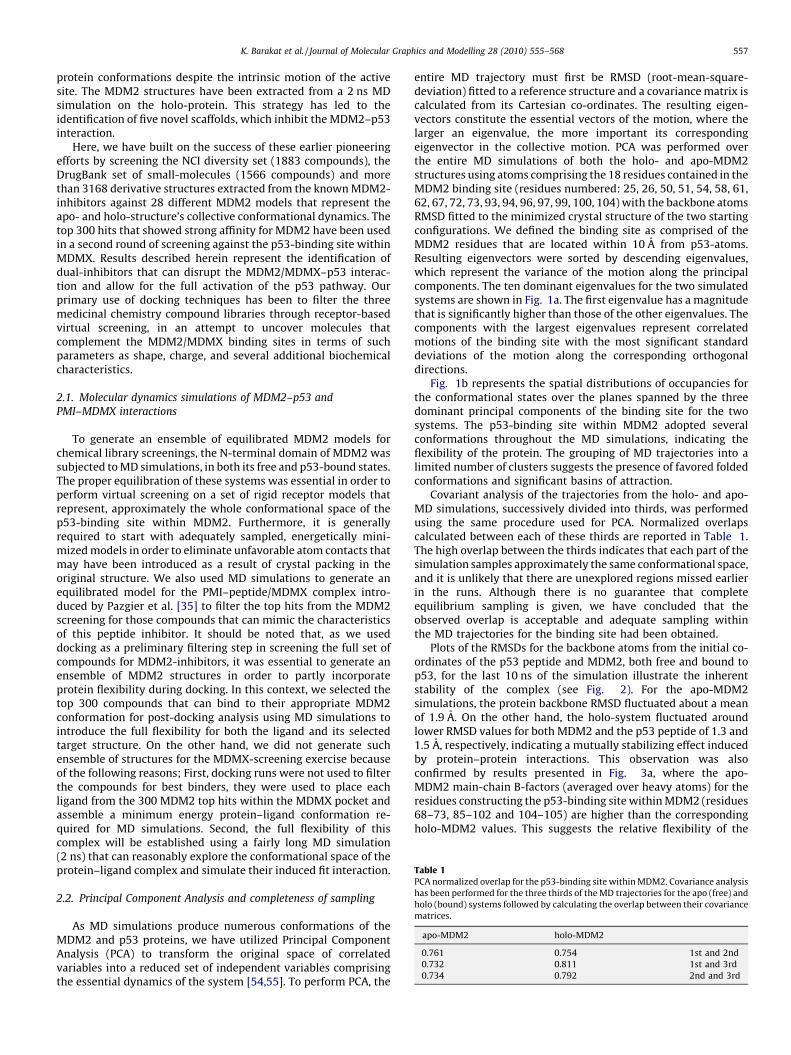
Table 1PCA normalized overlap for the p53-binding site within MDM2. Covariance analysis
has been performed for the three thirds of the MD trajectories for the apo (free) and
holo (bound) systems followed by calculating the overlap between their covariance
matrices.
apo-MDM2 holo-MDM2
0.761 0.754 1st and 2nd
0.732 0.811 1st and 3rd
0.734 0.792 2nd and 3rd
K. Barakat et al. / Journal of Molecular Graphics and Modelling 28 (2010) 555–568 557
protein conformations despite the intrinsic motion of the activesite. The MDM2 structures have been extracted from a 2 ns MDsimulation on the holo-protein. This strategy has led to theidentification of five novel scaffolds, which inhibit the MDM2–p53interaction.
Here, we have built on the success of these earlier pioneeringefforts by screening the NCI diversity set (1883 compounds), theDrugBank set of small-molecules (1566 compounds) and morethan 3168 derivative structures extracted from the known MDM2-inhibitors against 28 different MDM2 models that represent theapo- and holo-structure’s collective conformational dynamics. Thetop 300 hits that showed strong affinity for MDM2 have been usedin a second round of screening against the p53-binding site withinMDMX. Results described herein represent the identification ofdual-inhibitors that can disrupt the MDM2/MDMX–p53 interac-tion and allow for the full activation of the p53 pathway. Ourprimary use of docking techniques has been to filter the threemedicinal chemistry compound libraries through receptor-basedvirtual screening, in an attempt to uncover molecules thatcomplement the MDM2/MDMX binding sites in terms of suchparameters as shape, charge, and several additional biochemicalcharacteristics.
2.1. Molecular dynamics simulations of MDM2–p53 and
PMI–MDMX interactions
To generate an ensemble of equilibrated MDM2 models forchemical library screenings, the N-terminal domain of MDM2 wassubjected to MD simulations, in both its free and p53-bound states.The proper equilibration of these systems was essential in order toperform virtual screening on a set of rigid receptor models thatrepresent, approximately the whole conformational space of thep53-binding site within MDM2. Furthermore, it is generallyrequired to start with adequately sampled, energetically mini-mized models in order to eliminate unfavorable atom contacts thatmay have been introduced as a result of crystal packing in theoriginal structure. We also used MD simulations to generate anequilibrated model for the PMI–peptide/MDMX complex intro-duced by Pazgier et al. [35] to filter the top hits from the MDM2screening for those compounds that can mimic the characteristicsof this peptide inhibitor. It should be noted that, as we useddocking as a preliminary filtering step in screening the full set ofcompounds for MDM2-inhibitors, it was essential to generate anensemble of MDM2 structures in order to partly incorporateprotein flexibility during docking. In this context, we selected thetop 300 compounds that can bind to their appropriate MDM2conformation for post-docking analysis using MD simulations tointroduce the full flexibility for both the ligand and its selectedtarget structure. On the other hand, we did not generate suchensemble of structures for the MDMX-screening exercise becauseof the following reasons; First, docking runs were not used to filterthe compounds for best binders, they were used to place eachligand from the 300 MDM2 top hits within the MDMX pocket andassemble a minimum energy protein–ligand conformation re-quired for MD simulations. Second, the full flexibility of thiscomplex will be established using a fairly long MD simulation(2 ns) that can reasonably explore the conformational space of theprotein–ligand complex and simulate their induced fit interaction.
2.2. Principal Component Analysis and completeness of sampling
As MD simulations produce numerous conformations of theMDM2 and p53 proteins, we have utilized Principal ComponentAnalysis (PCA) to transform the original space of correlatedvariables into a reduced set of independent variables comprisingthe essential dynamics of the system [54,55]. To perform PCA, the
entire MD trajectory must first be RMSD (root-mean-square-deviation) fitted to a reference structure and a covariance matrix iscalculated from its Cartesian co-ordinates. The resulting eigen-vectors constitute the essential vectors of the motion, where thelarger an eigenvalue, the more important its correspondingeigenvector in the collective motion. PCA was performed overthe entire MD simulations of both the holo- and apo-MDM2structures using atoms comprising the 18 residues contained in theMDM2 binding site (residues numbered: 25, 26, 50, 51, 54, 58, 61,62, 67, 72, 73, 93, 94, 96, 97, 99, 100, 104) with the backbone atomsRMSD fitted to the minimized crystal structure of the two startingconfigurations. We defined the binding site as comprised of theMDM2 residues that are located within 10 A from p53-atoms.Resulting eigenvectors were sorted by descending eigenvalues,which represent the variance of the motion along the principalcomponents. The ten dominant eigenvalues for the two simulatedsystems are shown in Fig. 1a. The first eigenvalue has a magnitudethat is significantly higher than those of the other eigenvalues. Thecomponents with the largest eigenvalues represent correlatedmotions of the binding site with the most significant standarddeviations of the motion along the corresponding orthogonaldirections.
Fig. 1b represents the spatial distributions of occupancies forthe conformational states over the planes spanned by the threedominant principal components of the binding site for the twosystems. The p53-binding site within MDM2 adopted severalconformations throughout the MD simulations, indicating theflexibility of the protein. The grouping of MD trajectories into alimited number of clusters suggests the presence of favored foldedconformations and significant basins of attraction.
Covariant analysis of the trajectories from the holo- and apo-MD simulations, successively divided into thirds, was performedusing the same procedure used for PCA. Normalized overlapscalculated between each of these thirds are reported in Table 1.The high overlap between the thirds indicates that each part of thesimulation samples approximately the same conformational space,and it is unlikely that there are unexplored regions missed earlierin the runs. Although there is no guarantee that completeequilibrium sampling is given, we have concluded that theobserved overlap is acceptable and adequate sampling withinthe MD trajectories for the binding site had been obtained.
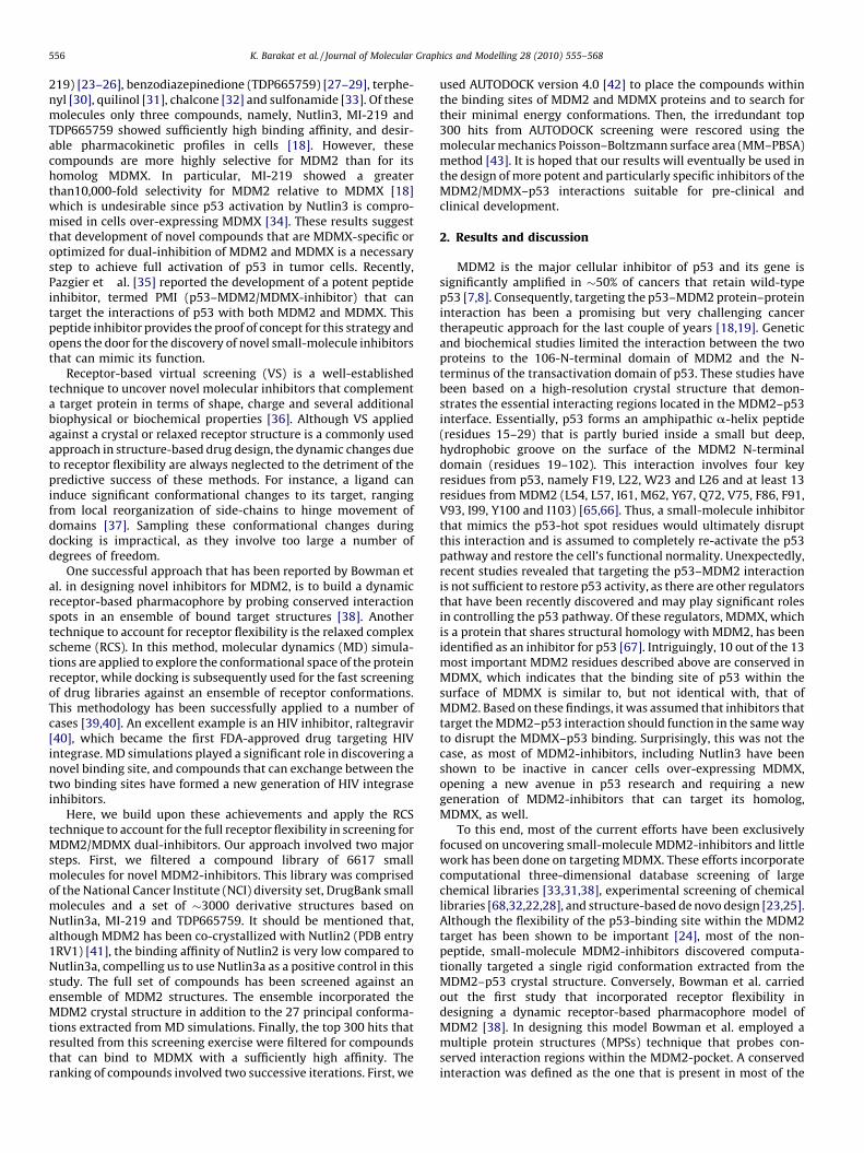
Plots of the RMSDs for the backbone atoms from the initial co-ordinates of the p53 peptide and MDM2, both free and bound top53, for the last 10 ns of the simulation illustrate the inherentstability of the complex (see Fig. 2). For the apo-MDM2simulations, the protein backbone RMSD fluctuated about a meanof 1.9 A. On the other hand, the holo-system fluctuated aroundlower RMSD values for both MDM2 and the p53 peptide of 1.3 and1.5 A, respectively, indicating a mutually stabilizing effect inducedby protein–protein interactions. This observation was alsoconfirmed by results presented in Fig. 3a, where the apo-MDM2 main-chain B-factors (averaged over heavy atoms) for theresidues constructing the p53-binding site within MDM2 (residues68–73, 85–102 and 104–105) are higher than the correspondingholo-MDM2 values. This suggests the relative flexibility of the

Fig. 1. PCA for the MDM2 binding site. (a) The dominant ten eigenvalues for the apo and holo trajectories. (b) Projections of the ensemble of conformations onto the planes of
the three most important principal components. The first and second, the first and third and the second and third principal components are plotted on the x and y axes,
respectively, for the two systems. The histograms represent the occupancies of the corresponding conformation states, with lighter colors indicating more frequently visited
areas.
Fig. 2. Plot of the RMSD of the backbone atoms from the reference structure as a
function of simulation time in p53–peptide, MDM2-free and MDM–p53 complex.
K. Barakat et al. / Journal of Molecular Graphics and Modelling 28 (2010) 555–568558
model in this region where the 18 residues defining the bindingsite seem to be relatively rigid during the MD simulation in thep53–MDM2 models. On the p53 side, residues 19–26 (see Fig. 2b)are more rigid than other p53 residues, suggesting their criticalparticipation in binding to MDM2.
2.3. Ensemble-based virtual screening
While integrating receptor flexibility into docking reduces therisk of unfavorable ligand–target interactions, accommodating fullreceptor flexibility during a docking experiment is impractical[27]. One solution for this problem is to allow only those parts ofthe receptor that affect the protein–ligand interactions to beflexible during docking. Due to the increased sampling require-ments and limited computational resources, flexible parts aregenerally restricted to the principal side chains that are believed tobe involved in binding interactions. This procedure allows for
Fig. 3. Plot of the B-factors averaged over the protein backbone atoms as a function
of residue number in the simulations of (a) MDM2-free and MDM2-bound and (b)
p53 peptide. The solid and dotted lines correspond to MDM2-bound and MDM2-
free, respectively.
Fig. 4. Clustering analysis for the two MDM2 trajectories. A high-quality clustering
is obtained when a local minimum in DBI correlates with saturation in the SSR/SST
ratio. This is clear at cluster count of 60 for the apo-structure and 30 clusters for the
holo-structure.
K. Barakat et al. / Journal of Molecular Graphics and Modelling 28 (2010) 555–568 559
localized protein movement, resulting in improved fit of the ligand.However, an obvious drawback when considering only theflexibility of restricted protein fragments is that the collectivemotion of the complete receptor backbone is neglected. Toovercome this deficiency we have used an ensemble of proteinconformations for docking as an alternative approach to introducea feature of global protein flexibility. This ensemble could describethe entire conformational space of the binding site, yet must still berepresented by a set of limited conformations in order to savecomputational screening time.
To generate a reduced set of representative models of theMDM2 binding site, we applied the RMSD conformationalclustering to the apo-MDM2 and holo-MDM2 binding-sitetrajectories. Fig. 4 shows the evaluation of the Davies–BouldinIndex (DBI) and the percentage of variance explained by the data(SSR/SST) for different cluster counts (see Section 3). DBI for theapo-system exhibited local minima at cluster counts of 10, 20 and60. However, as the percentage of variance explained by the datastarted to plateau after 45 clusters for the apo-system, weconcluded that 60 clusters is a reasonable cut-off for the free-MDM2 structures. On the other hand, the correlation betweenthese two criteria nicely occurred at a cluster count of 30 for theholo-structure.
In this study, we constructed an ensemble of 28 distinctconformations to perform ensemble-based virtual screening onMDM2 against the full set of ligand compounds. This ensembleincorporated the most dominant 22 structures that comprised�75% of apo-trajectory, the most dominant five holo-structuresthat represented �80% of the bound conformations (data not
shown) and finally the MDM2 conformation extracted from thep53-bound crystal structure. The ultimate goal was to reduce thenumber of representative structures included in the ensemble-based screening and concurrently comprise most of the confor-mational space of the binding site. Fig. 5 represents the 28structures used in this work. The MDM2 protein adopted diverseconformational changes demonstrating the significance of intro-ducing receptor flexibility during the docking procedure.
2.4. Pose clustering
As mentioned above, 28 independent virtual screening experi-ments were performed against the full set of database compounds.Screening of the full set of compounds contained in the NCIDS,DrugBank and the inhibitor-derivatives databases (more than 6000molecules), against the 28 target structures, produced a total of�19 million distinct poses that required classification. WhileAUTODOCK is capable of clustering these poses into subgroupsdepending on RMSD, the total number of clusters and thepopulation of each cluster is mostly dependent on the RMSDcut-off that is initially chosen. As such, there is no adequate meansto anticipate an optimum cut-off for the RMSD to produce the bestquality result. Since we are dealing with a diverse set of inputligands, this clustering method does not provide an accurate means
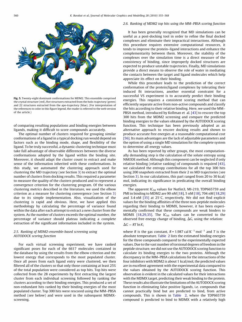
Fig. 5. Twenty-eight dominant conformations for MDM2. This ensemble comprised
the crystal structure (red), five structures extracted from the holo-trajectory (green)
and 22 structures extracted from the apo-trajectory (blue). (For interpretation of
the references to color in this figure legend, the reader is referred to the web version
of the article.)
K. Barakat et al. / Journal of Molecular Graphics and Modelling 28 (2010) 555–568560
of comparing resulting populations and binding energies betweenligands, making it difficult to score compounds accurately.
The optimal number of clusters required for grouping similarconformations of a ligand in a typical docking run would depend onfactors such as the binding mode, shape, and flexibility of theligand. To be truly successful, a dynamic clustering technique musttake full advantage of observable differences between the diverseconformations adopted by the ligand within the binding site.Moreover, it should adapt the cluster count to extract and makesense of the information inherited with these conformations. Inthis study, we automated the clustering approach used forclustering the MD trajectory (see Section 3) to extract the optimalnumber of clusters from docking results. This required a parameterto measure the quality of the clusters produced and to represent aconvergence criterion for the clustering program. Of the variousclustering metrics described in the literature, we used the elbowcriterion as a measure for clustering convergence (see Section 3)due to its simple implementation. Also, visualization of theclustering is rapid and obvious. Here, we have applied thismethodology by calculating the percentage of variance foundwithin the data after each attempt to extract a new cluster from thesystem. As the number of clusters exceeds the optimal number, thepercentage of variance should plateau indicating a completeextraction of the significant information included in the system.
2.5. Ranking of MDM2-ensemble-based screening using
AUTODOCK scoring function
For each virtual screening experiment, we have rankedsignificant poses for each of the 6617 molecules contained inthe database by using the results from the elbow criterion and thelowest energy that corresponds to the most populated cluster.Once all poses from each ligand entry were clustered, we thenfiltered all of the clusters so that only those containing at least 25%of the total population were considered as top hits. Top hits werecollected from the 28 experiments by first extracting the largestcluster from each individual screening followed by ranking theclusters according to their binding energies. This produced a set ofnon-redundant hits ranked by their binding energies of the mostpopulated cluster. Top 300 hits were rescored using the MM–PBSAmethod (see below) and were used in the subsequent MDMX-screening.
2.6. Ranking of MDM2 top hits using the MM–PBSA scoring function
It has been generally recognized that MD simulations can beuseful as a post-docking tool in order to refine the final dockedcomplexes and eliminate their impractical interactions. Althoughthis procedure requires extensive computational resources, ittends to improve the protein–ligand interactions and enhance thecomplementarity between them. Moreover, the stability of thecomplexes over the simulation time is a direct measure of theconsistency of binding, since improperly docked structures areexpected to produce unstable trajectories. Finally, MD simulationsprovide a direct means to observe the role of water in mediatingthe contacts between the target and ligand molecules which helpappreciate its effect on their binding.
While this procedure leads to the prediction of the correctconformation of the protein/ligand complexes by tolerating theirinduced fit interactions, another essential constraint for asuccessful VS experiment is to accurately predict their bindingenergies. This requires a consistent scoring method that canefficiently separate active from non-active compounds and classifythe hits according to their relative binding. Here, we used the MM–PBSA method, introduced by Kollman et al. [43] to rescore the top300 hits from the MDM2 screening and compare the predictedbinding energies to the values obtained by the AUTODOCK scoringfunction. This technique has been previously adopted as analternative approach to rescore docking results and shown toproduce accurate free energies at a reasonable computational cost[56]. Its main advantages are the lack of adjustable parameters andthe option of using a single MD simulation for the complete systemto determine all energy values.
As has been reported by other groups, the most computation-ally demanding step is the calculation of the solute entropy usingNMODE method. Although this component can be neglected if onlyrelative binding (relative ranking) of compounds is required [43],we calculated the entropy contributions for all the top 300 hitsusing 200 snapshots extracted from their 2 ns MD trajectories (seeSection 3). In our calculations, this part ranged from 20 to 30 kcal/mol, indicating its significance in predicating the overall bindingenergies.
The apparent IC50 values for Nutlin3, MI-219, TDP665759 andPMI in binding to MDM2 are 90 nM [18], 5 nM [18], 704 nM [18,29]and 3.4 nM [35] at 258C, respectively. We did not find explicitvalues for the binding affinities of the three non-peptide moleculesregarding their binding to MDMX, however, it has been experi-mentally confirmed that these compounds are weak binders toMDMX [18,29,35]. The IC50 values can be converted to theobserved free energy change of binding, DG, using the relation:
DG ¼ RT ln Ki
where R is the gas constant, R = 1.987 cal K�1 mol�1 and T is theabsolute temperature. Table 2 lists the estimated binding energiesfor the three compounds compared to the experimentally expectedvalues. Due to the vast number of torsional degrees of freedom in thepeptide structure, we did not use the AUTODOCK scoring function tocalculate its binding energies to the two proteins. Although thediscrepancy in the MM–PBSA calculations for the interactions of thefour inhibitors with MDM2 is about 1 kcal/mol, the predicted valuesare in excellent agreement with the experimental data compared tothe values obtained by the AUTODOCK scoring function. Thisobservation is evident in the calculated values for their interactionswith the MDMX target, predicting their weak binding to the protein.These results also illustrate the limitations of the AUTODOCK scoringfunction in eliminating false positive ligands, i.e. compounds thatcannot practically bind but are predicted to bind, from activecompounds. This is shown in Table 2, where the TDP665759compound is predicted to bind to MDMX with a relatively high
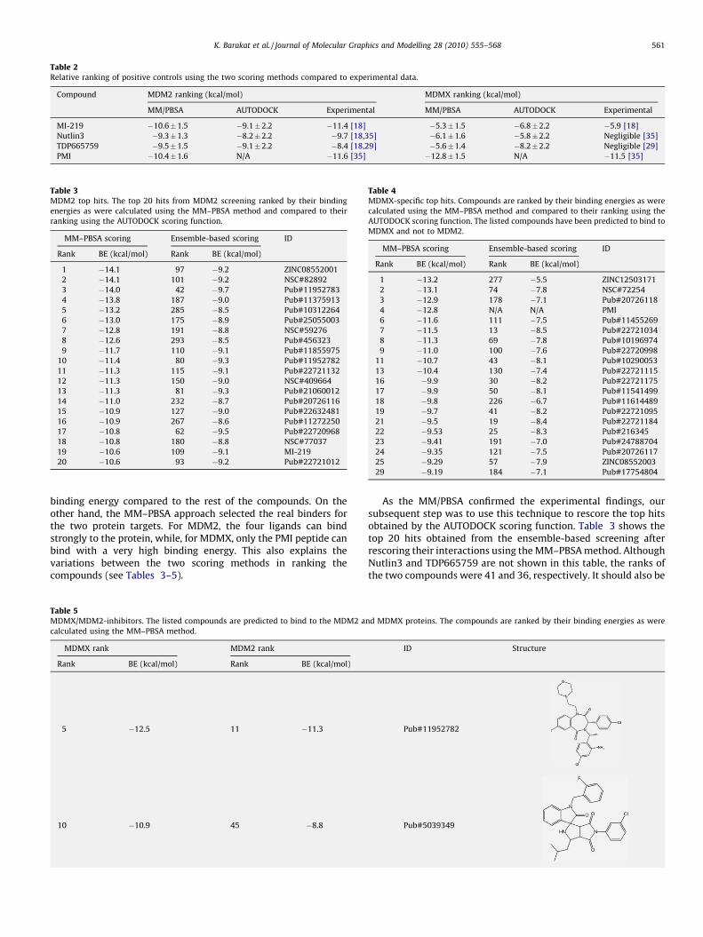
Table 2Relative ranking of positive controls using the two scoring methods compared to experimental data.
Compound MDM2 ranking (kcal/mol) MDMX ranking (kcal/mol)
MM/PBSA AUTODOCK Experimental MM/PBSA AUTODOCK Experimental
MI-219 �10.6�1.5 �9.1�2.2 �11.4 [18] �5.3�1.5 �6.8�2.2 �5.9 [18]
Nutlin3 �9.3�1.3 �8.2�2.2 �9.7 [18,35] �6.1�1.6 �5.8�2.2 Negligible [35]
TDP665759 �9.5�1.5 �9.1�2.2 �8.4 [18,29] �5.6�1.4 �8.2�2.2 Negligible [29]
PMI �10.4�1.6 N/A �11.6 [35] �12.8�1.5 N/A �11.5 [35]
Table 3MDM2 top hits. The top 20 hits from MDM2 screening ranked by their binding
energies as were calculated using the MM–PBSA method and compared to their
ranking using the AUTODOCK scoring function.
MM–PBSA scoring Ensemble-based scoring ID
Rank BE (kcal/mol) Rank BE (kcal/mol)
1 �14.1 97 �9.2 ZINC08552001
2 �14.1 101 �9.2 NSC#82892
3 �14.0 42 �9.7 Pub#11952783
4 �13.8 187 �9.0 Pub#11375913
5 �13.2 285 �8.5 Pub#10312264
6 �13.0 175 �8.9 Pub#25055003
7 �12.8 191 �8.8 NSC#59276
8 �12.6 293 �8.5 Pub#456323
9 �11.7 110 �9.1 Pub#11855975
10 �11.4 80 �9.3 Pub#11952782
11 �11.3 115 �9.1 Pub#22721132
12 �11.3 150 �9.0 NSC#409664
13 �11.3 81 �9.3 Pub#21060012
14 �11.0 232 �8.7 Pub#20726116
15 �10.9 127 �9.0 Pub#22632481
16 �10.9 267 �8.6 Pub#11272250
17 �10.8 62 �9.5 Pub#22720968
18 �10.8 180 �8.8 NSC#77037
19 �10.6 109 �9.1 MI-219
20 �10.6 93 �9.2 Pub#22721012
Table 4MDMX-specific top hits. Compounds are ranked by their binding energies as were
calculated using the MM–PBSA method and compared to their ranking using the
AUTODOCK scoring function. The listed compounds have been predicted to bind to
MDMX and not to MDM2.
MM–PBSA scoring Ensemble-based scoring ID
Rank BE (kcal/mol) Rank BE (kcal/mol)
1 �13.2 277 �5.5 ZINC12503171
2 �13.1 74 �7.8 NSC#72254
3 �12.9 178 �7.1 Pub#20726118
4 �12.8 N/A N/A PMI
6 �11.6 111 �7.5 Pub#11455269
7 �11.5 13 �8.5 Pub#22721034
8 �11.3 69 �7.8 Pub#10196974
9 �11.0 100 �7.6 Pub#22720998
11 �10.7 43 �8.1 Pub#10290053
13 �10.4 130 �7.4 Pub#22721115
16 �9.9 30 �8.2 Pub#22721175
17 �9.9 50 �8.1 Pub#11541499
18 �9.8 226 �6.7 Pub#11614489
19 �9.7 41 �8.2 Pub#22721095
21 �9.5 19 �8.4 Pub#22721184
22 �9.53 25 �8.3 Pub#216345
23 �9.41 191 �7.0 Pub#24788704
24 �9.35 121 �7.5 Pub#20726117
25 �9.29 57 �7.9 ZINC08552003
29 �9.19 184 �7.1 Pub#17754804
K. Barakat et al. / Journal of Molecular Graphics and Modelling 28 (2010) 555–568 561
binding energy compared to the rest of the compounds. On theother hand, the MM–PBSA approach selected the real binders forthe two protein targets. For MDM2, the four ligands can bindstrongly to the protein, while, for MDMX, only the PMI peptide canbind with a very high binding energy. This also explains thevariations between the two scoring methods in ranking thecompounds (see Tables 3–5).
Table 5MDMX/MDM2-inhibitors. The listed compounds are predicted to bind to the MDM2 a
calculated using the MM–PBSA method.
MDMX rank MDM2 rank
Rank BE (kcal/mol) Rank BE (kcal/mol)
5 �12.5 11 �11.3
10 �10.9 45 �8.8
As the MM/PBSA confirmed the experimental findings, oursubsequent step was to use this technique to rescore the top hitsobtained by the AUTODOCK scoring function. Table 3 shows thetop 20 hits obtained from the ensemble-based screening afterrescoring their interactions using the MM–PBSA method. AlthoughNutlin3 and TDP665759 are not shown in this table, the ranks ofthe two compounds were 41 and 36, respectively. It should also be
nd MDMX proteins. The compounds are ranked by their binding energies as were
ID Structure
Pub#11952782
Pub#5039349
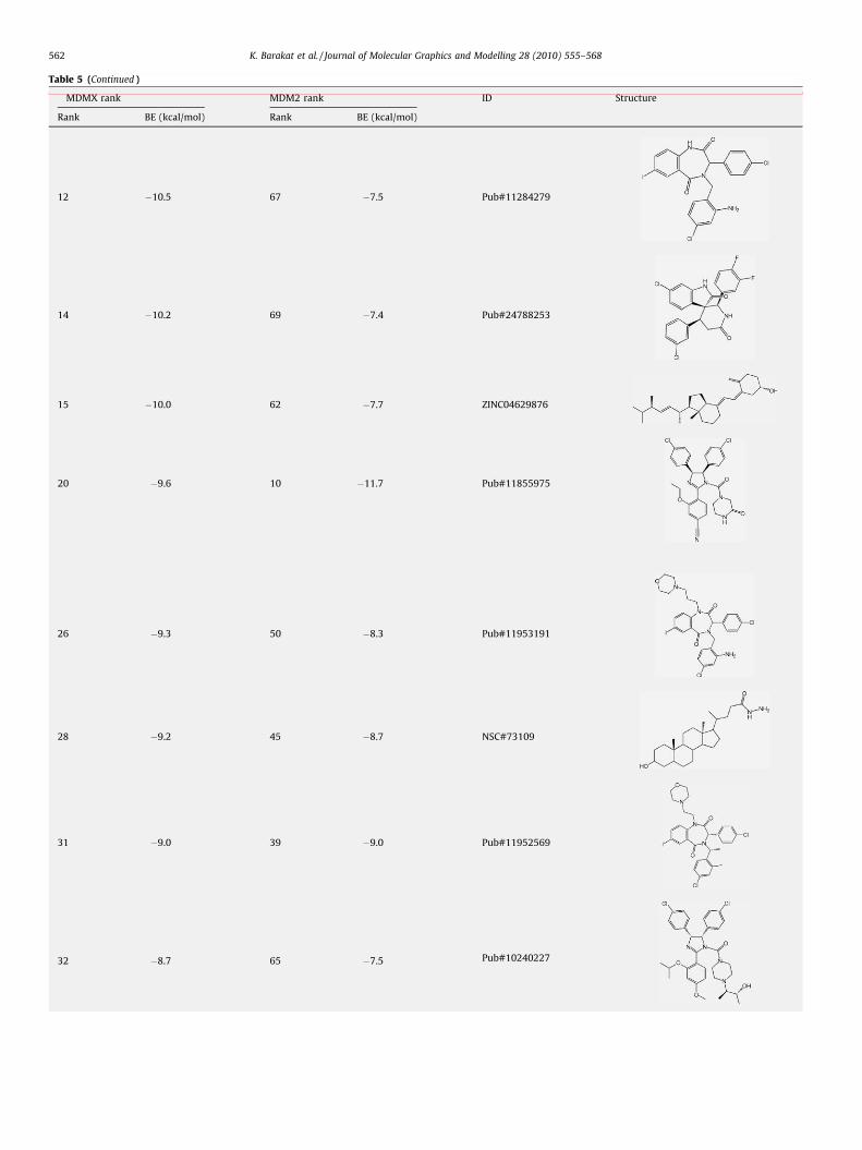
Table 5 (Continued )
MDMX rank MDM2 rank ID Structure
Rank BE (kcal/mol) Rank BE (kcal/mol)
12 �10.5 67 �7.5 Pub#11284279
14 �10.2 69 �7.4 Pub#24788253
15 �10.0 62 �7.7 ZINC04629876
20 �9.6 10 �11.7 Pub#11855975
26 �9.3 50 �8.3 Pub#11953191
28 �9.2 45 �8.7 NSC#73109
31 �9.0 39 �9.0 Pub#11952569
32 �8.7 65 �7.5 Pub#10240227
K. Barakat et al. / Journal of Molecular Graphics and Modelling 28 (2010) 555–568562

Table 5 (Continued )
MDMX rank MDM2 rank ID Structure
Rank BE (kcal/mol) Rank BE (kcal/mol)
38 �8.2 49 �8.4 NSC#179187
43 �8.0 7 �13.0 Pub#25055003
45 �8.0 44 �7.2 Pub#11753378
47 �7.7 17 �10.9 Pub#11272250
49 �7.6 25 �10.3 Pub#21934780
60 �7.0 4 �13.8 Pub#11375913
K. Barakat et al. / Journal of Molecular Graphics and Modelling 28 (2010) 555–568 563
mentioned that a considerable number of the compounds showedpositive binding energies after rescoring them using the MM/PBSAmethod (data not shown), supporting the ability of this techniqueto discriminate inactive compounds from the AUTODOCK sug-gested hits. Although most of the top 20 hits are derivatives of thethree positive controls, in particular the benzodiazepinedionescaffold (TDP665759), 5 compounds from both the NCI diversityset and DrugBank libraries showed strong binding energiescompared to the positive controls.
2.7. Screening of MDM2 top hits against MDMX
The top 300 hits which resulted from the ensemble-basedscreening were then docked to an equilibrated MDMX structure thatwas extracted from the PMI/MDMX complex (see Section 3). The
docking step was essential in order to place the compounds withinthe p53-binding site with their minimum energy conformation.Following the procedure described above, we used the MM/PBSAmethod to predict the absolute binding energies for each compound.Reassuringly, our calculations confirmed the experimental findings,where the three non-peptidic positive controls showed weakbinding to MDMX compared to MDM2 while the PMI peptideshowed very high binding energy (see Table 2). Analogously to theexperimental results concerning the high specificity of thesemolecules to the MDM2 target, our calculations predict that anumber of compounds can also bind more strongly to MDMX than toMDM2. The top 20 hits selected from these compounds are expectedto be specific MDMX-inhibitors and are shown in Table 4.
Table 5 lists the compounds that are suggested to function asdual-MDM2/MDMX-inhibitors obtained from screening the top

Fig. 6. Structural variations between MDM2 (yellow) and MDMX (red) and their effect
on the binding modes of Nutlin3 (a) and two selected hits form the predicted MDM2/
MDMX-inhibitors (b and c). Tyr100 and Leu99 of MDM2 and the same residues in
MDMX are shown in Licorice representations with the same color as that of the two
proteins. For each compound, the binding mode within MDM2 is shown in green and
within MDMX is shown in gray. Tyr99 and Leu98 prevent Nutlin3 from binding to
MDMX with the same binding conformation adopted by Nutlin2 within the MDM2-
pocket (blue). The conformation of Nutlin2 was extracted from the MDM2–Nutlin
crystal structure 1RV1. On the other hand, compounds Pub#11952782 (b) and
ZINC04629876 (c) from the suggested MDM2/MDMX-inhibitor list can tolerate the
structural variations in the two binding sites in order to maximize their interactions
with the proteins. (For interpretation of the references to color in this figure legend,
the reader is referred to the web version of the article.)
K. Barakat et al. / Journal of Molecular Graphics and Modelling 28 (2010) 555–568564
MDM2-hits against the p53-binding site within the MDMXtarget. Here, we used MM–PBSA energies to compare thebinding of these hits to the two target proteins. As we are onlyinterested in compounds that can bind to MDM2 with affinitiesas good as those of the known MDM2-inhibitors, we limited ourselection to the 16 compounds shown below (see Table 5).Although the binding sites are fairly similar, the MDMX pocketseems to be more compact than that of MDM2. This is mainlydue to the three residues Pro95, Ser96 and Pro97 in MDMX thathave been replaced by His96, Arg97 and Lys98 in MDM2. Thesesubstitutions are located on one of the alpha helices thatcomprise the p53-binding site within the two proteins.Consequently, the proline residues (Pro95 and Pro97) in MDMXshift this helical domain in MDMX relative to MDM2 and causeLys98 and Tyr99 to protrude into the p53-binding cleft withinMDMX, making it shallower and less accessible to many of theMDM2 top hits we found. Moreover, we noticed very minordifferences in the electrostatic potential distributions aroundthe surfaces of the two proteins (data not shown), where MDM2is more positively charged in certain regions deeply locatedwithin the binding site. These slight variations in both shape andelectrical properties of the two proteins played a considerablerole in governing the final conformation adopted by the ligands.This observation is clear when comparing the binding modes ofNutlin within the two pockets (see Fig. 6a). While Tyr100 andLeu99 of MDM2 extend the binding site allowing Nutlin tointimately bind to MDM2, the same residues in MDMX clashwith the drug preventing it from taking the normal conforma-tion that was adopted within MDM2. On the other hand, Fig. 6band c show how two compounds from the list of proposedMDM2/MDMX-inhibitors were able to tolerate the structuralvariations between the two binding sites. This is apparent inFig. 6, where the compounds took on different conformationswithin the two binding pockets in order to maximize theirinteractions with the proteins.
3. Methods and materials
3.1. Molecular dynamics simulations
The amino-terminal domain of MDM2 (residues 25–109) boundto a 13-residue transactivation domain peptide of p53 (residues17–29) was taken from PDB entry 1YCR [8]. MD simulations werecarried out using the NAMD program [44] at a mean temperatureof 310 K and physiological pH (pH 7) using the all-hydrogenAMBER99SB force field [45]. Protonation states of all ionizableresidues were calculated using the program PDB2PQR [46].Following parameterization, the MDM2 protein alone (subsequentto removing the p53 peptide from the p53–MDM2 crystalstructure) or in complex with the p53 peptide was immersed inthe center of TIP3P water cube after adding hydrogen atoms to theinitial protein structure. The cube dimensions were chosen toprovide at least a 20 A buffer of 12,724 (12,653) water moleculesaround the systems. To neutralize and prepare the p53-bound or(free) systems under a physiological ionic concentration, 30 (28)chloride and 23 (23) sodium ions were respectively added byreplacing water molecules having the highest electrostaticenergies on their oxygen atoms. The number of counter ions foreach case was calculated by first estimating the amount of ions thatis needed to set up the system under normal physiologicalconditions (pH 7), followed by adding the number of chloride ionsrequired to bring its charge to zero. The fully solvated protein wasthen minimized and subsequently heated to the simulationtemperature with heavy restraints placed on all backbone atoms.Following heating, the system was equilibrated using periodicboundary conditions for 100 ps and energy restraints reduced to
K. Barakat et al. / Journal of Molecular Graphics and Modelling 28 (2010) 555–568 565
zero in successive steps of the MD simulation. The simulationswere then continued for 55 (78) ns during which atomic co-ordinates were saved to the trajectory every 2 ps. The totalsimulation time was determined by visualizing the quality ofsampling as predicted by PCA (see below). The RMSD and B-factorsfor the protein backbone were then computed over the last 10 ns ofthe MD simulation using the PTRAJ utility within AMBER10 [47].Hydrogen bond analyses were performed by computing theaverage distance between donor and acceptor atoms. A hydrogenbond was defined by a heavy donor–heavy acceptor distance�3.4 A, a light donor–heavy acceptor distance �2.5 A, and adeviation of less than �608 from linearity.
Following the same MD protocol mentioned above we preparedtwo equilibrated models for the PMI/MDM2 (PDB entry: 3EQS) [35]and PMI/MDMX (PDB entry 3EQY) [35] complexes. Parameters forligands were assigned using the generalized AMBER force field [48]and partial charges were calculated with the AM1-BCC method[49] using Antechamber in the AMBER 10 package. Followingparameterization, the protein/ligand complexes were subjected tothe same MD protocol we used before (see above) for a productionphase of 2 ns. Snapshots were extracted every 2 ps and used for theMM/PBSA binding energy analysis.
3.2. RMSD clustering to extract representative MD structures
MD simulations on the free and bound MDM2 systemsproduced numerous structures that explored their conforma-tional space. Although performing VS against each snapshot ofthese trajectories is the most accurate way to account for fullreceptor flexibility, implementing this technique is unfeasibleand requires massive computational resources. A suitable way toaccommodate receptor flexibility and concurrently, reduce therequired computations is to dock the ligand library against a setof representative structures that describes the conformationalspace of the target [50,51,39]. Although this approach signifi-cantly reduces the number of VS experiments, care should betaken in extracting these representative structures. A commonapproach to generate such ensemble of representative models isto perform RMSD conformational clustering on the wholetrajectory [50]. Unfortunately, there is no universally acceptedclustering algorithm that can be used to extract all of theinformation contained within the MD simulation. However,recent studies suggest that a number of clustering algorithms,such as average-linkage, means and self-organizing maps (SOM)can be used in clustering MD data [52]. The clustering quality canbe anticipated by calculating a number of clustering metrics thatcan deduce the optimal number of clusters to be extracted andtheir population size. These are the Davies–Bouldin index (DBI)[53] and the ‘‘elbow criterion’’ [52]. A high-quality clusteringscheme is expected when high DBI values are calculated. On theother hand, using the elbow criterion, the percentage of varianceexplained by the data, is expected to plateau for cluster countsexceeding the optimal number [52]. Using these metrics, byvarying the number of clusters, one should expect for adequateclustering, a local minimum for DBI and a horizontal line for thepercentage of variance explained by the data.
To generate a reduced set of representative MDM2 models, weperformed RMSD conformational clustering with the average-linkage algorithm as implemented in the PTRAJ utility ofAMBER10 using cluster counts ranging from 5 to 100 clusters.For the two MDM2 simulations, structures were extracted at 2 psintervals over the entire simulation times. All Ca-atoms wereRMSD fitted to the minimized initial structure in order to removeoverall rotation and translation. RMSD clustering was performedon the 18 residues that line the p53-binding cleft within MDM2,namely those numbered: 25, 26, 50, 51, 54, 58, 61, 62, 67, 72, 73,
93, 94, 96, 97, 99, 100, 104. These residues were clustered intogroups of similar conformations using the atom-positional RMSDof the entire amino acid, including side chains and hydrogenatoms, as the similarity criterion. The optimal numbers of clustersfor the two systems were chosen after evaluation of the twoclustering metrics, described above, for different cluster counts(see Section 2). Sixty clusters were obtained for the apo-MDM2,while 30 clusters were extracted for the holo-MDM2. The centroidof each cluster, the structure having the smallest RMSD to allmembers of the cluster, was chosen as the cluster representativestructure and the most dominant structures were used as rigidtemplates for the ensemble-based docking experiments (seeSection 2).
3.3. Principal Component Analysis
PCA can transform the original space of correlated variablesfrom a large MD simulation into a reduced space of independentvariables comprising the essential dynamics of the system[54,55]. For a typical protein, the system’s dimensionality isthereby reduced from tens of thousands to fewer than fiftydegrees of freedom. To perform PCA for a subset of N atoms, theentire MD trajectory is RMSD fitted to a reference structure, inorder to remove all rotations and translations. The covariancematrix can then be calculated from their Cartesian atomic co-ordinates as:
si j ¼ ðri � hriiÞðr j � hr jiÞ� �
(1)
where ri represents one of the three Cartesian co-ordinates (xi, yi orzi) and the eigenvectors of the covariance matrix constitute theessential vectors of the motion. It is generally accepted that thelarger an eigenvalue, the more important its correspondingeigenvector in the collective motion. PCA can also be employedto predict the completeness of sampling during the MD simulation.A method proposed by Hess [57] divides an MD trajectory intoseparate parts, and their normalized overlap is calculated using thecovariant matrices for each pair of parts:
Normalized overlap ðC1;C2Þ ¼ 1�
ffiffiffiffiffiffiffiffiffiffiffiffiffiffiffiffiffiffiffiffiffiffiffiffiffiffiffiffiffiffiffiffiffiffiffiffiffiffiffiffiffiffiffiffiffitr
ffiffiffiffiffiffiC1
p�
ffiffiffiffiffiffiC2
p� �2� �sffiffiffiffiffiffiffiffiffiffiffiffiffiffiffiffiffiffiffiffiffiffiffiffiffiffiffiffiffiffiffiffitrðC1Þ þ trðC2Þ
p (2)
where C1 and C2 are the covariant matrices, and the symbol tr isused to denote the trace operation. If the overlap is 0, then the twosets are considered to be orthogonal, whereas an overlap of 1indicates that the matrices are identical. To ensure completeness ofsampling for MD simulations of MDM2, PCA of the binding-siteresidues was performed using the positions of all heavy atoms. TheMD trajectory was divided into three parts and the normalizedoverlap between each pair was calculated to determine thecompleteness of sampling.
3.4. Selection of ligand database
The National Cancer Institute Diversity Set (NCDIS) [58],DrugBank small molecules [59] and a set of 3168 derivativestructures for Nutlin3, MI-219 and TDP665759 were used as ourtest libraries of compounds. The NCIDS is a collection ofapproximately 2000 compounds that are structurally representa-tive of a wide range of molecules, representing almost 140,000compounds that are available for testing at the NCI. Unfortunately,a number of ligands containing rare earth elements could not beproperly parameterized and were excluded, leaving a total of 1,883compounds for analysis. Here, we used a version of the NCIDSformatted for use in AUTODOCK and was prepared by theAUTODOCK Scripps team. The DrugBank small molecule library
K. Barakat et al. / Journal of Molecular Graphics and Modelling 28 (2010) 555–568566
is a set of 1488 FDA-approved small-molecule drugs downloadedfrom the ZINC database. Some of these molecules were present inmore than one protonation state adding another 78 structures tothe docked ligands. We also appended the set of derivativestructures of MDM2-inhibitors to the docked compounds for tworeasons. First, we wanted to build upon the intensive efforts thathave been previously made and incorporate variations in theoriginal structures for these inhibitors in order to improve theirperformance in binding to MDM2 and MDMX. Moreover, since wesearch for dual-MDM2/MDMX-inhibitors, we expect that due tothe structural similarity of the p53-binding sites within the twoproteins, an MDMX-inhibitor should be a derivative structure ofthe known MDM2-inhibitors. Based on this assumption, wecreated a library of 3168 derivative structures similar to Nutlin3,MI-219, and TDP665759 by searching the PubChem [60] databaseand then extracting the results using a similarity threshold of�90%. Compounds similar to the query structure are measuredusing the Tanimoto score [61]. A Tanimoto score of 100%represents an ‘‘exact match’’ to the provided chemical structurequery, while a value of 0% results in the return of all chemicalstructures deposited in the PubChem database. The threshold of�90% is chosen for efficiency of search since similarity links inPubChem are pre-computed at this value. Also, at this threshold,the compounds that are returned by the search would not be veryclose to the original query structure and yet provide reasonablenumber of chemical structures for this work. Therefore, the fullset of ligands used in this study comprised 6617 differentcompounds.
3.4.1. Ligand screening
Virtual screening on the p53-binding sites within MDM2 andMDMX was performed using AUTODOCK, version 4.0 [42].Hydrogen atoms were added to MDM2, MDMX and ligands andpartial atomic charges were then assigned using the Gasteiger–Marsili [62] method. Atomic solvation parameters were assignedto the atoms of the protein using the AUTODOCK 4.0 utilityADDSOL. Docking grid maps with 126 � 108 � 126 points andgrid point spacing of 0.21 A were then centered on the p53-binding site within the MDM2 and MDMX receptors usingAUTOGRID4.0 program [42]. Rotatable bonds of each ligand werethen automatically assigned using AUTOTORS utility of AUTO-DOCK4.0. Docking was performed using the Lamarckian GeneticAlgorithm (LGA) method with an initial population of 400random individuals; a maximum number of 10 � 106 energyevaluations; 100 trials; 50,000 maximum generations; a muta-tion rate of 0.02; a crossover rate of 0.80 and the requirementthat only one individual can survive into the next generation. Atotal of 28 independent virtual screening runs were performedagainst the full set of docked ligands with all residues of thereceptors set kept rigid during docking experiments. This set ofMDM2 models comprises one structure that represents theminimized holo-crystal conformation of MDM2, 22 conforma-tions that represent �80% of the apo-MDM2 trajectory and fivemodels that constitute �75% of the holo-MDM2 trajectory (seeSection 2). We also performed a VS run on an equilibrated modelfor MDMX using the top 300 MDM2-hits resulted from theensemble-base screening.
3.4.2. Clustering of docked poses
The previously described virtual screening experiments in-volved millions of conformations of each ligand bound to MDM2.AUTODOCK can cluster these output poses into subgroupsdepending on their RMSD values referred to a reference structure.Although this approach shows the possible binding modes of aligand to the binding site, the number of clusters and thepopulation size for each cluster depends heavily on the RMSD
cut-off used. It is not possible to anticipate an optimum cut-off forthe RMSD in order to produce a clustering pattern with the highestconfidence, motivating us to use alternative approaches inperforming the clustering analysis. In this study, we used anautomated approach to couple one of the commonly usedclustering metrics, the elbow criterion [52], with the clusteringmodule of PTRAJ utility of AMBER10. This method exploits the factthat the percentage of variance explained by the data (l), isexpected to plateau for cluster counts exceeding the optimalnumber.
The percentage of variance is defined by:
l ¼ SSR
SST(3)
where SSR is the sum-of-squares regression from each clustersummed over all clusters and SST is the total sum-of-squares. Here,we used the SOM algorithm, as implemented in the PTRAJ utility ofthe AMBER10 program, to cluster the docking results. Thismodified clustering program increases the number of clustersrequired until the percentage of variance explained by the data (l)plateaus. This can be determined by calculating the first andsecond derivatives of the percentage of variance with respect to theclusters number (dl=dN and d2l=dN2) after each attempt toincrease the cluster counts. The clustering process then stops at anacceptable value for these derivatives that is close to 0.Consequently, the clustering procedure depends only on thesystem itself and adjusts itself to arrive at the optimal clusteringpattern for that specific system.
3.5. Rescoring of top hits using MM–PBSA
Binding free energies were calculated using the molecularmechanics Poisson–Boltzmann surface area (MM–PBSA) method[43] as implemented in AMBER10. The total free energy is the sumof average molecular mechanical gas-phase energies (EMM),solvation free energies (Gsolv), and entropy contributions (�TSsolute)) of the binding reaction:
G ¼ EMM þ Gsolv � TSsolute (4)
In this work, the molecular mechanical (EMM) energy of eachsnapshot was calculated using the SANDER module of AMBER10with all pair-wise interactions included using a dielectric constant(e) of 1. The solvation free energy (Gsolv) was estimated as the sum ofelectrostatic solvation free energy, calculated by the finite-differ-ence solution of the Poisson–Boltzmann equation in the AdaptivePoisson–Boltzmann Solver (APBS) program as implemented inAMBER10 and non-polar solvation free energy, calculated from thesolvent-accessible surface area (SASA) algorithm. The solute entropywas approximated using the normal mode analysis. Applying thethermodynamic cycle for each ligand–MDM2/MDMX complex, thebinding free energy between an arbitrary ligand molecule and theMDM2/MDMX protein can be approximated by:
DG� ¼DGMDM2=MDMX�ligandgas þDGMDM2=MDMX�ligand
solv
� fDGligandsolv þDGMDM2=MDMX
solv g (5)
Here, ðDGMDM2�ligandgas Þ represents the free energy per mole for the
non-covalent association of the ligand–MDM2 complex in vacuum(gas phase) at 300 K, while ð�DGsolvÞ stands for the work requiredto transfer a molecule from its solution conformation to the sameconformation in vacuum at 300 K (assuming that the bindingconformation of the ligand–MDM2 complex is the same in solutionand in vacuum).
K. Barakat et al. / Journal of Molecular Graphics and Modelling 28 (2010) 555–568 567
4. Conclusions
The tumor suppressor p53 is one of the most frequentlyinactivated proteins in human cancers. Direct gene modificationsin p53 gene, Tp53, or the interaction between p53 and its two majorcellular inhibitors, MDM2 and MDMX, are two fundamentalmechanisms employed by cancer cells to block the p53 pathway[1–4]. Over a number of years, leading efforts in p53 research havebeen focused on restoring the activity of the mutant protein as aprecursor to developing a novel cancer treatment. Although thesestudies revealed the prospects of inducing tumor cell death, thedevelopment of a non-peptide small-molecule p53-activator is stilla particularly challenging problem [69,70]. Other significant effortsin this area have been aimed at the discovery of small-moleculeinhibitors that can disrupt the interaction of p53 with its maincellular regulator, MDM2 [18–21]. This led to the development ofNutlin3 [22] and MI-219 [23], the most potent and specific non-peptide MDM2-inhibitors discovered so far.
Recently, MDMX, a protein homologous to MDM2, was found toreduce the efficacy of MDM2-inhbitors including Nutlin3 [18,34].This suggested MDMX as a new attractive therapeutic target andindicated a need to develop MDMX-specific or MDM2/MDMXdual-inhibitors to fully-activate the p53 pathway in tumor cellsexpressing wild-type p53.
Here, we used an improved relaxed complex Scheme 39 bycombining MD simulations and molecular docking with bindingenergy analysis to filter a set of 6617 compounds for effectiveinhibitors of MDM2 and MDMX. These compounds included theNCI diversity set [58], DrugBank small molecules [59] and a newlygenerated set of �3000 derivative structures similar to knownMDM2-inhibitors. The derivative library of compounds wasincluded among the docked structures because the structuralsimilarity between the two proteins would imply that an MDMX-inhibitor should be a derivative structure based on one of theknown MDM2-inhibitors. Although, more than 6000 compoundshave been screened in this study, we suggest the use of largerlibraries (in the order of 100,000 to 1,000,000 compounds) will bemore effective in discovering more active hits in future work. Inthis context, we used MD simulations, principle componentanalysis and an iterative clustering technique to generate anensemble of 28 MDM2 structures that characterize the collectivedynamics of the MDM2 protein. Then, we used molecular dockingto explore the conformational space of the ligands and to search fortheir minimal energy configuration within the MDM2 binding site.All docking poses were clustered using the same iterativeprocedure that we used in extracting the protein structures andthen sorted with the minimal energy of the largest cluster. The top300 hits were rescored using the MM–PBSA procedure andprepared for a second round of screening against the MDMXtarget. Following the docking of MDM2-hits to MDMX we used theMM–PBSA technique to rescore their binding affinities to MDMXand suggest a set of MDM2/MDMX dual-inhibitors.
Our results confirmed the experimental findings concerning theweak binding of the MDM2-inhibitors Nutlin3, MI-219 and TDP toits homolog structure MDMX. Moreover, as we anticipated, the tophits from our screening are primarily derivative structures of thethree known inhibitors. However, we also suggested a fewstructures from the NCI diversity set and DrugBank compounds.The molecules we have proposed in the present study, can fitwithin the two binding sites and adopt different conformations tomaximize their interactions with the two proteins. We have alsovalidated our top hit list by comparing their estimated bindingenergies to the PMI peptide, an MDM2/MDMX dual-inhibitorproposed by Pazgier et al. [35] and, reassuringly, our top hits arepredicted to have comparable performance to this peptide. It ishoped that our findings will facilitate the development of a new
generation of MDM2/MDMX dual-inhibitors that would fully-activate the p53 pathway and offer new hope in the fight against abroad range of cancers.
Acknowledgments
All of the molecular dynamics simulations and virtual screeningexperiments were produced using the SHARCNET, AICT (Universityof Alberta cluster) and WESTGRID computational facilities.Funding for this work was obtained from the Alberta CancerFoundation, the Allard Foundation and NSERC. JAT wishes to thankProf. A. Fersht of Cambridge University for bringing this problem tohis attention.
References
[1] J.G. Teodoro, S.K. Evans, M.R. Green, Inhibition of tumor angiogenesis by p53: a newrole for the guardian of the genome, J. Mol. Med. 85 (2007) 1175.
[2] J.S. Fridman, S.W. Lowe, Control of apoptosis by p53, Oncogene 22 (2003) 9030.[3] K.H. Vousden, X. Lu, Live or let die: the cell’s response to p53, Nat. Rev. Cancer 2
(2002) 594.[4] J.C. Bourdon, V.D. Laurenzi, G. Melino, D. Lane, p53: 25 years of research and more
questions to answer, Cell Death Differ. 10 (4) (2003) 397.[5] B. Vogelstein, D. Lane, A.J. Levine, Surfing the p53 network, Nature 408 (2000) 307.[6] A. Feki, I. Irminger-Finger, Mutational spectrum of p53 mutations in primary
breast and ovarian tumors, Crit. Rev. Oncol. Hematol. 52 (2004) 103.[7] M.H. Kubbutat, S.N. Jones, K.H. Vousden, Regulation of p53 stability by Mdm2,
Nature 387 (1997) 299.[8] P.H. Kussie, et al., Structure of the MDM2 oncoprotein bound to the p53 tumor
suppressor transactivation domain, Science 274 (1996) 948.[9] S.S. Fakharzadeh, S.P. Trusko, D.L. George, Tumorigenic potential associated with
enhanced expression of a gene that is amplified in a mouse tumor cell line, EMBOJ. 10 (1991) 1565.
[10] J.D. Oliner, K.W. Kinzler, P.S. Meltzer, D.L. George, B. Vogelstein, Amplification of agene encoding a p53-associated protein in human sarcomas, Nature 358 (1992) 80.
[11] J. Momand, D. Jung, S. Wilczynski, J. Niland, The MDM2 gene amplificationdatabase, Nucleic Acids Res. 26 (1998) 3453.
[12] A. Shvarts, W.T. Steegenga, N. Riteco, T. van Laar, P. Dekker, M. Bazuine, R.C. vanHam, W. van der Houven van Oordt, G. Hateboer, A.J. van der Eb, A.G. Jochemsen,MDMX: a novel p53-binding protein with some functional properties of MDM2,EMBO J. 15 (1996) 5349.
[13] M.W. Jackson, S.J. Berberich, MdmX protects p53 from Mdm2-mediated degra-dation, Mol. Cell. Biol. 20 (2000) 1001.
[14] F. Toledo, K.A. Krummel, C.J. Lee, C.W. Liu, L.W. Rodewald, M. Tang, G.M. Wahl, Amouse p53 mutant lacking the proline-rich domain rescues Mdm4 deficiency andprovides insight into theMdm2-Mdm4-p53 regulatory network, Cancer Cell 9(2006) 273.
[15] D.A. Sharp, S.A. Kratowicz, M.J. Sank, D.L. George, Stabilization of the MDM2oncoprotein by interaction with the structurally related MDMX protein, J. Biol.Chem. 274 (1999) 38189.
[16] Y. Pan, J. Chen, MDM2 promotes ubiquitination and degradation of MDMX, Mol.Cell. Biol. 23 (2003) 5113.
[17] V. Bottger, A. Bottger, C. Garcia-Echeverria, Y.F. Ramos, A.J. van der Eb, A.G.Jochemsen, D.P. Lane, Comparative study of the p53-mdm2 and p53-MDMXinterfaces, Oncogene 18 (1999) 189.
[18] S. Shangary, S. Wang, Targeting the MDM2-p53 interaction for cancer therapy,Clin. Cancer Res. 14 (2008) 5318.
[19] L.T. Vassilev, MDM2 inhibitors for cancer therapy, Trends Mol. Med. 13 (2007) 23.[20] J.K. Buolamwini, J. Addo, S. Kamath, S. Patil, D. Mason, M. Ores, Small molecule
antagonists of the MDM2 oncoprotein as anticancer agents, Curr. Cancer DrugTargets 5 (1) (2005) 57.
[21] S. Patel, M.R. Player, Small-molecule inhibitors of the p53-MDM2 interaction forthe treatment of cancer, Expert Opin. Investig. Drugs 17 (12) (2008) 1865.
[22] L.T. Vassilev, Small-molecule antagonists of p53-MDM2 binding: research toolsand potential therapeutics, Cell Cycle 3 (2004) 419.
[23] K. Ding, Y. Lu, Z. Nikolovska-Coleska, S. Qiu, Y. Ding, W. Gao, J. Stuckey, K.Krajewski, P.P. Roller, Y. Tomita, D.A. Parrish, J.R. Deschamps, S. Wang, Struc-ture-based design of potent nonpeptide MDM2 inhibitors, J. Am. Chem. Soc. 127(2005) 10130.
[24] S.G. Dastidar, D.P. Lane, C.S. Verma, Multiple peptide conformations give rise tosimilar binding affinities: molecular simulations of p53-MDM2, J. Am. Chem. Soc.130 (41) (2008) 13514.
[25] S. Shangary, D. Qin, D. McEachern, M. Liu, R.S. Miller, S. Qiu, Z. Nikolovska-Coleska,K. Ding, G. Wang, J. Chen, D. Bernard, J. Zhang, Y. Lu, Q. Gu, R.B. Shah, K.J. Pienta, X.Ling, S. Kang, M. Guo, Y. Sun, D. Yang, S. Wang, Temporal activation of p53 by aspecific MDM2 inhibitor is selectively toxic to tumors and leads to completetumor growth inhibition, Proc. Natl. Acad. Sci. U.S.A. 105 (2008) 3933.
[26] K. Ding, Y. Lu, Z. Nikolovska-Coleska, G. Wang, S. Qiu, S. Shangary, W. Gao, D. Qin, J.Stuckey, K. Krajewski, P.P. Roller, S. Wang, Structure-based design of spiroox-indoles as potent, specific small-molecule inhibitors of the MDM2-p53 interac-tion, J. Med. Chem. 49 (2006) 3432.
K. Barakat et al. / Journal of Molecular Graphics and Modelling 28 (2010) 555–568568
[27] B.L. Grasberger, T. Lu, C. Schubert, D.J. Parks, T.E. Carver, H.K. Koblish, M.D.Cummings, L.V. LaFrance, K.L. Milkiewicz, R.R. Calvo, D. Maguire, J. Lattanze,C.F. Franks, S. Zhao, K. Ramachandren, G.R. Bylebyl, M. Zhang, C.L. Manthey, E.C.Petrella, M.W. Pantoliano, I.C. Deckman, J.C. Spurlino, A.C. Maroney, B.E. Tomczuk,C.J. Molloy, R.F. Bone, Discovery and cocrystal structure of benzodiazepinedioneHDM2 antagonists that activate p53 in cells, J. Med. Chem. 48 (2005) 909.
[28] D.J. Parks, L.V. Lafrance, R.R. Calvo, K.L. Milkiewicz, V. Gupta, J. Lattanze, K. Rama-chandren, T.E. Carver, E.C. Petrella, M.D. Cummings, D. Maguire, B.L. Grasberger, T.Lu, 1,4-Benzodiazepine-2,5-diones as small molecule antagonists of the HDM2-p53interaction: discovery and SAR, Bioorg. Med. Chem. Lett. 15 (2005) 765–770.
[29] H.K. Koblish, S. Zhao, C.F. Franks, R.R. Donatelli, R.M. Tominovich, L.V. LaFrance,K.A. Leonard, J.M. Gushue, D.J. Parks, R.R. Calvo, K.L. Milkiewicz, J.J. Marugan, P.Raboisson, M.D. Cummings, B.L. Grasberger, D.L. Johnson, T. Lu, C.J. Molloy, A.C.Maroney, Benzodiazepinedione inhibitors of the Hdm2:p53 complex suppresshuman tumor cell proliferation in vitro and sensitize tumors to doxorubicin invivo, Mol. Cancer Ther. 5 (1) (2006) 160.
[30] L. Chen, H. Yin, B. Farooqi, S. Sebti, A.D. Hamilton, J. Chen, p53 alpha-Helixmimetics antagonize p53/MDM2 interaction and activate p53, Mol. Cancer Ther.4 (2005) 1019.
[31] Y. Lu, Z. Nikolovska-Coleska, X. Fang, W. Gao, S. Shangary, S. Qiu, D. Qin, S. Wang,Discovery of a nanomolar inhibitor of the human murine double minute 2(MDM2)-p53 interaction through an integrated, virtual database screening strat-egy, J. Med. Chem. 49 (2006) 3759.
[32] R. Stoll, C. Renner, S. Hansen, S. Palme, C. Klein, A. Belling, W. Zeslawski, M.Kamionka, T. Rehm, P. Muhlhahn, R. Schumacher, F. Hesse, B. Kaluza, W. Voelter,R.A. Engh, T.A. Holak, Chalcone derivatives antagonize interactions between thehuman oncoprotein MDM2 and p53, Biochemistry 40 (2001) 336.
[33] P.S. Galatin, D.J. Abraham, A nonpeptidic sulfonamide inhibits the p53-mdm2interaction and activates p53-dependent transcription in mdm2-overexpressingcells, J. Med. Chem. 47 (2004) 4163.
[34] B. Hu, D.M. Gilkes, B. Farooqi, S.M. Sebti, J. Chen, MDMX overexpression preventsP53 activation by the MDM2 inhibitor nutlin, J. Biol. Chem. 281 (44) (2006) 33030.
[35] M. Pazgier, M. Liu, G. Zou, W. Yuan, C. Li, C. Li, J. Li, J. Monbo, D. Zella, S.G. Tarasov,W. Lu, Structural basis for high-affinity peptide inhibition of p53 interactions withMDM2 and MDMX, Natl. Acad. Sci. U.S.A. 106 (12) (2009) 4665.
[36] R.E. Hubbard, Structure-Based Drug Discovery: An Overview, first ed., RSCPublishing, Cambridge, 2007, p. 261.
[37] C.W. Murray, C.A. Baxter, A.D. Frenkel, The sensitivity of the results of moleculardocking to induced fit effects: application to thrombin, thermolysin and neur-aminidase, J. Comput. Aided Mol. Des. 13 (1999) 547.
[38] A.L. Bowman, Z. Nikolovska-Coleska, H. Zhong, S. Wang, H.A. Carlson, Smallmolecule inhibitors of the MDM2-p53 interaction discovered by ensemble-basedreceptor models, J. Am. Chem. Soc. 129 (2007) 12809.
[39] J.H. Lin, A.L. Perryman, J.R. Schames, J.A. McCammon, Computational drug designaccommodating receptor flexibility: the relaxed complex scheme, J. Am. Chem.Soc. 124 (2002) 5632.
[40] M. Markowitz, B.Y. Nguyen, F. Gotuzzo, F. Mendo, W. Ratanasuwan, C. Kovacs, J.Zhao, L. Gilde, R. Isaacs, H. Teppler, Potent antiviral effect of MK-0518, novel HIV-1integrase inhibitor, as part of combination ART in treatment-naive HIV-1 infectedpatients, in: 16th International AIDS Conference, Toronto, Canada, August 13–18,2006 (no. ThLB0214).
[41] L.T. Vassilev, B.T. Vu, B. Graves, D. Carvajal, F. Podlaski, Z. Filipovic, N. Kong, U.Kammlott, C. Lukacs, C. Klein, N. Fotouhi, E.A. Liu, In vivo activation of the p53pathway by small-molecule antagonists of MDM2, Science 303 (5659) (2004) 84.
[42] M.M. Garrett, S.G. David, S.H. Robert, H. Ruth, E.H. William, K.B. Richard, J.S.Arthur, Automated docking using a Lamarckian genetic algorithm and an empiri-cal binding free energy function, J. Comput. Chem. 19 (1999) 1639.
[43] P.A. Kollman, I. Massova, C. Reyes, B. Kuhn, S. Huo, L. Chong, M. Lee, T. Lee, Y. Duan,W. Wang, O. Donini, P. Cieplak, J. Srinivasan, D.A. Case, T.E. Cheatham, Calculating
structures and free energies of complex molecules: combining molecular me-chanics and continuum model, Acc. Chem. Res. 33 (2000) 889.
[44] L. Kale, NAMD2: Greater scalability for parallel molecular dynamics, J. Comput.Phys. 151 (1999) 283–312.
[45] V. Hornak, R. Abel, A. Okur, B. Strockbine, A. Roitberg, C. Simmerling, Comparisonof multiple Amber force fields and development of improved protein backboneparameters, Proteins 65 (2006), 712–765, 725.
[46] T.J. Dolinsky, P. Czodrowski, H. Li, J.E. Nielsen, J.H. Jensen, G. Klebe, N.A. Baker,PDB2PQR: ep53nding and upgrading automated preparation of biomolecularstructures for molecular simulations, Nucleic Acids Res. 35 (2007) W522–525.
[47] D.A. Case, T.E. Cheatham, T. Darden, H. Gohlke, R. Luo, K.M. Merz, A. Onufriev, C.Simmerling, B. Wang, R.J. Woods, The Amber biomolecular simulation programs,J. Comput. Chem. 26 (2005) 1668–1688.
[48] J. Wang, R.M. Wolf, J.W. Caldwell, P.A. Kollman, D.A. Case, Development andtesting of a general amber force field, J. Comput. Chem. 25 (2004) 1157.
[49] A. Jakalian, B.J. David, C.I. Bayly, Fast, efficient generation of high-quality atomiccharges. AM1-BCC model: II. Parameterization and validation, J. Comput. Chem.23 (2002) 1623.
[50] L.S. Cheng, R.E. Amaro, D. Xu, W.W. Li, P.W. Arzberger, J.A. McCammon, Ensemble-based virtual screening reveals potential novel antiviral compounds for avianinfluenza neuraminidase, J. Med. Chem. 51 (13) (2008) 3878.
[51] R.M.A. Knegtel, I.D. Kuntz, C.M. Oshiro, Molecular docking to ensembles of proteinstructures, J. Mol. Biol. 266 (1997) 424.
[52] J. Shao, S.W. Tanner, N. Thompson, T.E. Cheatham, Clustering molecular dynamicstrajectories: 1. Characterizing the performance of different clustering algorithms,J. Chem. Theor. Comput. 3 (2007) 2312.
[53] D.L. Davies, D.W. Bouldin, A cluster separation measure, IEEE Trans. Pattern Anal.Mach. Intell. 1 (1979) 224.
[54] A.E. Garcia, Large-amplitude nonlinear motions in proteins, Phys. Rev. Lett. 68(1992) 2696.
[55] A. Amadei, A.B. Linssen, H.J. Berendsen, Essential dynamics of proteins, Proteins17 (1993) 412.
[56] B. Kuhn, P. Gerber, T. Schulz-Gasch, M. Stahl, Validation and use of the MM-PBSAapproach for drug discovery, J. Med. Chem. 48 (12) (2005) 4040.
[57] B. Hess, Convergence of sampling in protein simulations, Phys. Rev. E Stat. Nonlin.Soft Matter Phys. 65 (2002) 31910.
[58] http://dtp.nci.nih.gov/branches/dscb/diversity_explanation.html. Last checkedOctober 21, 2009.
[59] http://zinc.docking.org/vendor0/dbsm/index.html. Last checked October 21,2009.
[60] http://pubchem.ncbi.nlm.nih.gov/. Last checked October 21, 2009.[61] T.T. Tanimoto, IBM Internal Report, 17 November 1957.[62] J. Gasteiger, M. Marsili, Iterative partial equalization of orbital electronegativity: a
rapid access to atomic charges, Tetrahedron 36 (1980) 3219.[65] I. Massova, P.A. Kollman, Computational alanine scanning to probe protein–
protein interactions: a novel approach to evaluate binding free energies, J. Am.Chem. Soc. 121 (1999) 8133.
[66] A. Bottger, V. Bottger, C. Garcia-Echeveria, P. Chene, C. Garcia-Echeveria, P. Chene,H.K. Hochkeppel, S.F. Howard, S.M. Picksley, D.P. Lane, Biochemical characteriza-tion of the hdm2-p53 interaction, J. Mol. Biol. 269 (1997) 744.
[67] J.C. Marine, M.A. Dyer, A.G. Jochemsen, MDMX: from bench to bedside, J. Cell Sci.120 (3) (2007) 371.
[68] H. Yin, G.I. Lee, H.S. Park, G.A. Payne, J.M. Rodriguez, S.M. Sebti, A.D. Hamilton,Terphenyl-based helical mimetics that disrupt the p53/HDM2 interaction,Angew. Chem. Int. Ed. Engl. 44 (2005) 2704.
[69] A.N. Bullock, A.R. Fersht, Rescuing the function of mutant p53, Nat. Rev. Cancer 1(2001) 68.
[70] T. Fojo, p53 as a therapeutic target: unresolved issues on the road to cancertherapy targeting mutant p53, Drug Resist. Updat. 5 (2001) 209.