The Advances in Glioblastoma on-a-Chip for Therapy ... - MDPI
32
Cancers 2022, 14, 869. https://doi.org/10.3390/cancers14040869 www.mdpi.com/journal/cancers Review The Advances in Glioblastoma on-a-Chip for Therapy Approaches Arielly H. Alves 1 , Mariana P. Nucci 1,2 , Javier B. Mamani 1 , Nicole M. E. Valle 1 , Eduarda F. Ribeiro 1 , Gabriel N. A. Rego 1 , Fernando A. Oliveira 1 , Matheus H. Theinel 1 , Ricardo S. Santos 1 and Lionel F. Gamarra 1, * 1 Hospital Israelita Albert Einstein, São Paulo 05652-000, Brazil; [email protected] (A.H.A.); [email protected] (M.P.N.); [email protected] (J.B.M.); [email protected] (N.M.E.V.); [email protected] (E.F.R.); [email protected] (G.N.A.R.); [email protected] (F.A.O.); [email protected] (M.H.T.); [email protected] (R.S.S.) 2 LIM44-Hospital das Clínicas da Faculdade Medicina da Universidade de São Paulo, São Paulo 05403-000, Brazil * Correspondence: [email protected]; Tel.: +55-11-2151-0243 Simple Summary: This systematic review showed different therapeutic approaches to glioblastoma on-a-chip with varying levels of complexity, answering, from the simplest question to the most sophisticated questions, in a biological system integrated in an efficient way. With advances in manufacturing protocols, soft lithography in PDMS material was the most used in the studies, applying different strategy geometrics in device construction. The microenvironment showed the relevant elaborations in co-culture between mainly human tumor cells and support cells involved in the collagen type I matrix; remaining an adequate way to assess the therapeutic approach. The most complex devices showed efficient intersection between different systems, allowing in vitro studies with major human genetic similarity, reproducibility, and low cost, on a highly customizable platform. Abstract: This systematic review aimed to verify the use of microfluidic devices in the process of implementing and evaluating the effectiveness of therapeutic approaches in glioblastoma on-a-chip, providing a broad view of advances to date in the use of this technology and their perspectives. We searched studies with the variations of the keywords “Glioblastoma”, “microfluidic devices”, “organ-on-a-chip” and “therapy” of the last ten years in PubMed and Scopus databases. Of 446 articles identified, only 22 articles were selected for analysis according to the inclusion and exclusion criteria. The microfluidic devices were mainly produced by soft lithography technology, using the PDMS material (72%). In the microenvironment, the main extracellular matrix used was collagen type I. Most studies used U87-MG glioblastoma cells from humans and 31.8% were co-cultivated with HUVEC, hCMEC/D3, and astrocytes. Chemotherapy was the majority of therapeutic approaches, assessing mainly the cellular viability and proliferation. Furthermore, some alternative therapies were reported in a few studies (22.6%). This study identified a diversity of glioblastoma on-a-chip to assess therapeutic approaches, often using intermediate levels of complexity. The most advanced level implemented the intersection between different biological systems (liver–brain or intestine–liver–brain), BBB model, allowing in vitro studies with greater human genetic similarity, reproducibility, and low cost, in a highly customizable platform. Keywords: glioblastoma on-a-chip; glioblastoma model; microfluidic devices; tumor cells co-culture; therapy glioma 1. Introduction Glioblastoma (GBM) is the most common primary malignant brain tumor in adults. The annual incidence of GBM in the United States is 3.23 cases per 100,000 people [1] and is one of the most fatal malignant diseases in humans. The patient median survival is around 14–16 months, and the relative survival is only five percent in five years. Tumor Citation: Alves, A.H.; Nucci, M.P.; Mamani, J.B.; Valle, N.M.E.; Ribeiro, E.F.; Rego, G.N.A.; Oliveira, F.A.; Theinel, M.H.; Santos, R.S.; Gamarra, L.F. The Advances in Glioblastoma on-a-Chip for Therapy Approaches. Cancers 2022, 14, 869. https://doi.org/10.3390/ cancers14040869 Academic Editor: Raquel Rodrigues Received: 18 December 2021 Accepted: 29 January 2022 Published: 9 February 2022 Publisher’s Note: MDPI stays neutral with regard to jurisdictional claims in published maps and institutional affiliations. Copyright: © 2022 by the author. Licensee MDPI, Basel, Switzerland. This article is an open access article distributed under the terms and conditions of the Creative Commons Attribution (CC BY) license (https://creativecommons.org/license s/by/4.0/).
-
Upload
khangminh22 -
Category
Documents
-
view
1 -
download
0
Transcript of The Advances in Glioblastoma on-a-Chip for Therapy ... - MDPI

Cancers 2022, 14, 869. https://doi.org/10.3390/cancers14040869 www.mdpi.com/journal/cancers
Review
The Advances in Glioblastoma on-a-Chip for Therapy
Approaches
Arielly H. Alves 1, Mariana P. Nucci 1,2, Javier B. Mamani 1, Nicole M. E. Valle 1, Eduarda F. Ribeiro 1,
Gabriel N. A. Rego 1, Fernando A. Oliveira 1, Matheus H. Theinel 1, Ricardo S. Santos 1 and Lionel F. Gamarra 1,*
1 Hospital Israelita Albert Einstein, São Paulo 05652-000, Brazil; [email protected] (A.H.A.);
[email protected] (M.P.N.); [email protected] (J.B.M.); [email protected] (N.M.E.V.);
[email protected] (E.F.R.); [email protected] (G.N.A.R.);
[email protected] (F.A.O.); [email protected] (M.H.T.); [email protected] (R.S.S.) 2 LIM44-Hospital das Clínicas da Faculdade Medicina da Universidade de São Paulo,
São Paulo 05403-000, Brazil
* Correspondence: [email protected]; Tel.: +55-11-2151-0243
Simple Summary: This systematic review showed different therapeutic approaches to glioblastoma
on-a-chip with varying levels of complexity, answering, from the simplest question to the most
sophisticated questions, in a biological system integrated in an efficient way. With advances in
manufacturing protocols, soft lithography in PDMS material was the most used in the studies,
applying different strategy geometrics in device construction. The microenvironment showed the
relevant elaborations in co-culture between mainly human tumor cells and support cells involved in
the collagen type I matrix; remaining an adequate way to assess the therapeutic approach. The most
complex devices showed efficient intersection between different systems, allowing in vitro studies
with major human genetic similarity, reproducibility, and low cost, on a highly customizable platform.
Abstract: This systematic review aimed to verify the use of microfluidic devices in the process of
implementing and evaluating the effectiveness of therapeutic approaches in glioblastoma on-a-chip,
providing a broad view of advances to date in the use of this technology and their perspectives. We
searched studies with the variations of the keywords “Glioblastoma”, “microfluidic devices”,
“organ-on-a-chip” and “therapy” of the last ten years in PubMed and Scopus databases. Of 446
articles identified, only 22 articles were selected for analysis according to the inclusion and exclusion
criteria. The microfluidic devices were mainly produced by soft lithography technology, using the
PDMS material (72%). In the microenvironment, the main extracellular matrix used was collagen
type I. Most studies used U87-MG glioblastoma cells from humans and 31.8% were co-cultivated
with HUVEC, hCMEC/D3, and astrocytes. Chemotherapy was the majority of therapeutic
approaches, assessing mainly the cellular viability and proliferation. Furthermore, some alternative
therapies were reported in a few studies (22.6%). This study identified a diversity of glioblastoma
on-a-chip to assess therapeutic approaches, often using intermediate levels of complexity. The most
advanced level implemented the intersection between different biological systems (liver–brain or
intestine–liver–brain), BBB model, allowing in vitro studies with greater human genetic similarity,
reproducibility, and low cost, in a highly customizable platform.
Keywords: glioblastoma on-a-chip; glioblastoma model; microfluidic devices; tumor cells
co-culture; therapy glioma
1. Introduction
Glioblastoma (GBM) is the most common primary malignant brain tumor in adults.
The annual incidence of GBM in the United States is 3.23 cases per 100,000 people [1] and
is one of the most fatal malignant diseases in humans. The patient median survival is
around 14–16 months, and the relative survival is only five percent in five years. Tumor
Citation: Alves, A.H.; Nucci, M.P.;
Mamani, J.B.; Valle, N.M.E.; Ribeiro,
E.F.; Rego, G.N.A.; Oliveira, F.A.;
Theinel, M.H.; Santos, R.S.; Gamarra,
L.F. The Advances in Glioblastoma
on-a-Chip for Therapy Approaches.
Cancers 2022, 14, 869.
https://doi.org/10.3390/
cancers14040869
Academic Editor: Raquel Rodrigues
Received: 18 December 2021
Accepted: 29 January 2022
Published: 9 February 2022
Publisher’s Note: MDPI stays
neutral with regard to jurisdictional
claims in published maps and
institutional affiliations.
Copyright: © 2022 by the author.
Licensee MDPI, Basel, Switzerland.
This article is an open access article
distributed under the terms and
conditions of the Creative Commons
Attribution (CC BY) license
(https://creativecommons.org/license
s/by/4.0/).

Cancers 2022, 14, 869 2 of 32
resection surgery, radiotherapy, and chemotherapy are the usual treatments for GBM
[2,3], however due to their invasibility, heterogeneity, and inefficacy in medication
carriage over the hematoencephalic barrier (BBB), this disease continues to exhibit failure
in the face of conventional treatments [4].
Alternative therapy approaches have been proposed to overcome the GBM treatment
difficulties, such as immunological therapy, using vaccine compounds of peptides,
dendritic cells, adoptive T cells, or chemical immunological checkpoint inhibitors (PD-1,
CTLA-4), as well as virotherapy to regulate the immune tumor response [5]. Gene
therapies, on the other hand, aim to alter the genetic structure of target cells, resulting in
improved immune responses, reprogramming of the tumor microenvironment (TME),
and angiogenesis normalization [6]. The use of nanoparticles in nanobiotechnology has
primarily aimed to overcome the challenges of delivering genes and drugs to the tumor
location. Furthermore, when combined with an alternating magnetic field, nanoparticles’
physical and magnetic features have been used to regulate metabolic processes and
produce hyperthermia [7]. Additionally, photodynamic treatment (PDT), which causes
molecular instability by heat stress, has shown that biochemical modulation can occur
through the generation of reactive oxygen species (ROS) [8]. As highlighted in this study,
numerous therapeutic techniques have been used alone or in combination to try to
improve GBM therapy responses.
Large amounts of investments are spent examining the effectiveness of therapeutic
agents for treating tumors in the search for novel therapies or improvements to existing
treatments for tumors, although in many cases, more than 90% effectiveness is not
obtained in clinical research [9,10]. One of the important reasons for this problem is the
use of platfors that do not satisfactorily predict many of the proposed clinical treatments
[11] as there are important limitations in these platforms: in vitro 2D and 3D, in animal
models, and in silico.
Many studies have used conventional cell culture (2D), spheroids, and 3D co-culture
to test different therapeutic approaches. These models are commonly used to carry out in
vitro studies because they are simple to implement, low cost, high yield, and have a low
ethical problem, but cell efficiency may decrease due to inappropriate interactions
between cells and cells-extracellular matrix (ECM) [12], lack of vascularity, no predictive
power, and no shear stress, which can cause changes in cell phenotype during in vitro
culture [11,13,14]. In vivo models, on the other hand, have served as a preclinical preview
of the translational pattern. Animal tumors, primarily in rodents, are the primary tool for
elucidating biochemical and physiological processes in living organisms prior to clinical
testing in humans [15,16]. These models allow the assessment of cell migration, invasion,
growth, proliferation, angiogenesis, immune responses, drug toxicity, and the
effectiveness of multiple therapeutic approaches. In addition, this platform shows more
physiological, genetic similarity, moderate prediction of drug behavior, physiological
microenvironment, enable mutation studies, and with some limitations, such as being
highly expensive, requiring specialized personnel and facilities, low-throughput; no
prediction for humans, inability to mimic human-specific features, long-term culture,
ethical issues, among others [17]. Despite its capabilities, medications evaluated in
preclinical cancer trials had a success probability of only 3.4% through phase I clinical
trials, with a failure rate of 54% in the final stages, owing to insufficient effectiveness and
poor safety [18].
Organ-on-a-chips have developed as a new testing option that offers a promising
way to overcome the limitations of traditional in vitro and preclinical models. There are
considerable disparities between existing models and humans in the replication of genetic,
metabolic, physiological, and pathological complexity, according to evidence. The organ-
on-a-chips platform, according to studies, can more accurately predict the efficacy and
reactions to drugs and therapeutic processes than in vitro and in vivo testing [19]. In
addition, some cell types cultured in 2D may be more sensitive to the toxic effects of drugs
than when cultured in an organ-on-a-chip architecture [20]. Thus, the organ-on-a-chips

Cancers 2022, 14, 869 3 of 32
recreate cell–cell or cell–ECM interactions, spatiotemporal physicochemical gradients, or
the dynamic properties of the cellular microenvironment [21–23]. As a result, the complex
organ-on-a-chips design enables in vivo replication of microenvironments, offering a
stable platform for nanomedicine evaluation [24], presenting itself as a low cost
alternative, preservation cell phenotype, customized design, control on physical and
biochemical properties of the tumor microenvironment, well-defined vessel endothelium,
gradient compatible, dynamic system, control of hydrodynamic parameters, real-time
measurement, and microcopy compatible [11].
Organ-on-a-chips applied to cancer research [25–32] address models which involve
several aspects, such as tumor growth, angiogenesis, cell invasion, intravasation,
extravasation, and metastasis [33–35], as well as physiological drug exposures [36], which
can assess disease progression and contribute to the development of precision medicine
and personalized treatments using tissue from patients [37].
Given these aspects, this systematic review aimed to verify the use of microfluidic
devices in the process of implementing and evaluating the effectiveness of therapeutic
approaches in GBM tumors in the PubMed and Scopus literature databases. The
fabrication of microfluidic devices, the composition of the microenvironment and the
ECM, tumor cells, and support cells in culture were evaluated; as well as therapeutic
approaches used, and the techniques applied to assess the effectiveness, providing a broad
view of advances to date in the use of this technology and their perspectives.
2. Methods
2.1. Search Strategy
This systematic review followed the Preferred Reporting Items for Systematic
Reviews and Meta-Analyses (PRISMA) guidelines [38]. The publication search was
performed between February 2011 and February 2021, indexed in the PubMed and Scopus
databases using the following Boolean operators (DecS/MeSH), and keywords sequence
for each database:
PubMed: (((((Glioblastoma[Title/Abstract]) OR GBM[Title/Abstract])) OR
Glioma[Title/Abstract])) AND (((((((((“organ-on-a-chip”[Title/Abstract]) OR “human-on-
a-chip”[Title/Abstract]) OR microfluidic[Title/Abstract]) OR “organs-on-
chips”[Title/Abstract]) OR “organs-on-a-chip”[Title/Abstract]) OR “microfluidic
device”[Title/Abstract]) OR “lab-on-chips”[Title/Abstract]) OR “glioblastoma-on-a-
chip”[Title/Abstract]) OR “GBM-on-a-chip”[Title/Abstract]);
Scopus: ((TITLE-ABS-KEY (glioblastoma) OR TITLE-ABS-KEY (gbm) OR TITLE-
ABS-KEY (glioma))) AND ((TITLE-ABS-KEY (organ-on-a-chip) OR TITLE-ABS-KEY
(human-on-a-chip) OR TITLE-ABS-KEY (microfluidic) OR TITLE-ABS-KEY (organs-on-
chip) OR TITLE-ABS-KEY (organs-on-a-chip) OR TITLE-ABS-KEY (microfluidic AND
device) OR TITLE-ABS-KEY (lab-on-chip) OR TITLE-ABS-KEY (glioblastoma-on-a-chip)
OR TITLE-ABS-KEY (gbm-on-a-chip) OR TITLE-ABS-KEY (microvasculature-on-a-chip)
OR TITLE-ABS-KEY (microenvironment AND in AND a AND chip) OR TITLE-ABS-KEY
(brain AND cancer AND chip))).
2.2. Inclusion Criteria
We included only original articles published in English in the last ten years, with the
available full text, and that used microfluidic devices to evaluate different therapeutic
approaches for glioblastoma tumor models developed from the culture of tumor cells.
2.3. Exclusion Criteria
We excluded articles that did not report the therapeutic approach in the microfluidic
device, that did not perform a tumor microenvironment reconstitution from the use of
tumor cells, as well as articles indexed in more than one database (duplicates), review

Cancers 2022, 14, 869 4 of 32
articles, letters, articles in press, communications, book chapters, abstracts, incomplete ar-
ticles, editorials, and expert opinions.
2.4. Data Extraction
In this systematic review, the collected data were segregated into the following top-
ics: (i) the microfluidic device design, their material used, and its manufacturing method;
(ii) the characteristics of the cells used in 3D culture and the medium; (iii) the microenvi-
ronment reconstitution for the glioblastoma model and their maintenance; and (iv) the
therapeutic approaches applied in the devices and the techniques used for the therapeutic
efficacy evaluation.
2.5. Data Analysis
The percentage distribution, obtained for each variable analyzed in the tables was
used to characterize and present all of the results. Each study was classified into 3 catego-
ries of complexity, from (+) to (+++), based on how each topic was approached separately
in each table. Finally, we considered the analysis of the results reported in the tables and
applied a generic classification of device complexities in four categories (I–IV).
3. Results
3.1. Overview of the Reviewed Literature
We searched publications of the last 10 years, considering the period between Sep-
tember 2011 and September 2021, indexed in PubMed and Scopus, and a total of 446 arti-
cles were identified. Of the 119 articles found in PubMed, 94 were excluded after screening
(89 duplicated in Scopus search, and 5 reviews), and 22 articles were excluded after eligi-
bility analysis (12 articles did not report the therapeutic approach used for glioblastoma
on-a-chip, 6 articles reported only the usage of the microfluidic device for analysis of the
part of the experiment, such as CHIP-Seq or CHIP-qPCR, and 4 articles developed the
study in silico), thus, only 3 articles were included from this database. Of the 327 articles
identified in Scopus, after screening, 48 articles were excluded (18 reviews, 15 conference
papers, 8 book chapter/series, 2 notes, 2 publications in other languages, 1 conference re-
view, 1 editorial, and 1 short survey), and 260 articles were excluded after eligibility anal-
ysis (120 articles did not report the therapeutic approach used for glioblastoma on-a-chip,
110 articles reported only the usage of the microfluidic device for analysis of part of the
experiment, such as CHIP-Seq, CHIP-qPCR, and chromatin immunoprecipitation—CHIP,
and 30 articles developed the study in silico), thus, only 19 articles were included from
this database. As shown in Figure 1, only 22 unduplicated full-text articles were included
in this review [39–60], and the histogram and spider chart show the distribution of articles
by year and research centers, respectively.

Cancers 2022, 14, 869 5 of 32
Figure 1. Schematic representation of the screening process of articles for inclusion in this systematic review following
PRISMA guidelines from the identification of 446 studies in the SCOPUS and PubMed databases, following the predeter-
mined inclusion and exclusion criterias. After initial screening and eligibility assessment, 424 were excluded, and only 22
studies were included in this review. The histogram contains the distribution of the 22 articles included by year of publi-
cation represented by blue bars and the representation of the cumulative growth until 2021 by the red points. The spider
chart shows the regional distribution (countries) of the research centers, where the studies included in this review were
developed.

Cancers 2022, 14, 869 6 of 32
3.2. Design and Fabrication of Microfluidic Devices
Regarding the microfluidic device fabrication and its geometric characteristics, we
analyzed the different materials used (organic or inorganic polymers) as well as the fabri-
cation technology applied. These aspects reflect the complexity of devices used to improve
the glioblastoma model and their therapeutic analysis, as shown in Table 1. Of the selected
studies used in this study, 91% produced in-house devices [39–42,44–46,48–60], only 9%
of studies used commercial devices [43,47], due to the variability of device design used in
the research. Regarding technology used in this fabrication, different lithographic tech-
niques were used, namely, 68.2% soft lithography [39,40,42,43,46,48–50,52–55,58–60],
13.6% photolithography [51,56,57], and 4.5% two-photon lithography [41], 4.5% used 3D-
printing systems [44], and 9.1% did not report the technology used [45,47]. The main ma-
terial of the devices was 88% polymers (72% PDMS [39,40,42,43,45,46,48–50,52–60], 8%
polycarbonate [49,56], and 4% of PEGDA [51], and modified polyethersulfone [54]). Only
4% used inorganic synthetic polymer composed of silicon [44], and one study did not re-
port the material used[47]. The most substrate used in the device production was glass in
64% of the selected studies [41–45,48,50–53,55,58–60], followed by 18% PDMS
[40,46,49,54], and 5% no cover [39], and 14% did not report the substrate [47,56,57]. In
relation to the mold material used in the device fabrication process, 68% of studies re-
ported the use of SU-8 photoresist [39–43,46,48–50,52–54,56–60] due to the lithography
technology that is commonly used this mold and in 4% of studies ethyl lactate was used
[39] or SPR950/SF6 nanowires [57] or silicon [46]. Furthermore, 12% of studies did not
report [45,47,55] and 8% did not do this process [44,51].
When the geometric characteristics of microfluidic devices was investigated, the se-
lected studies reported mainly the fabrication of rectangular shapes (40.9%) for the culture
region [42,47–50,52,55–58], following by 18.2% square [46,53,59,60] or circle shapes
[40,43,44,51], and 4.5% reported the use of oval [54], semicircle [39], perimetric cylindrical
pillars [41], or rectangular with circle array [48]. Furthermore, 4.5% did not report the shape
of the culture region in the device [45]. The device design (dimensions and structure) varied
a lot between studies, as well as the materials and methods of fabrication due to these pa-
rameters; we elaborate one analysis considering the level of device complexity from (+) to
(+++) levels. Almost half of the studies (40.9%) were classified as level (++) complexity
[39,43,45,46,48,51,53,57,59], which was considered shape with connections, simple material,
and method, followed by level (+) (36.4%), which involved simple shape for culture
[40,42,47,49,50,52,55,58], and level (+++) (22.7%), showed the more sophisticated method of
fabrication [41,44,54,56,60], with multi-interfaces connected as depicted in Figure 2.

Cancers 2022, 14, 869 7 of 32
Figure 2. The schematic figures of glioblastoma on-a-chip devices for therapy approach used in
some of the selected studies of this systematic review. (A) The integrated microfluidic system for
single-cell separation and sphere formation, adapted with permission from [46], the American
Chemical Society. (B) 3D co-culture unit generative process and the analysis of the confocal images
of the chip, showing the HUVEC cells in the lumen, adapted with permission from [52], Analytica
Chimica Acta. (C) MCF7 and U87MG cancer cells diagonally seeded into square-shaped microcham-
bers, in the hydrogel microfluidic device, and analysis of confocal microscopy images, adapted with
permission from [53], Electrophoresis. (D) Magnetohyperthermia process in tumor-on-a-chip using
magnetic nanoparticles dispersed in aqueous medium submitted to an alternating magnetic field.,
adapted with permission from [5], Einstein. (E) A microfluidic platform mimics the blood-brain bar-
rier (BBB) using two PDMS sheets a polycarbonate membrane. BBB unit was directly connected to
the μSPE unit for mass spectrometry detection., adapted with permission from [56], Analytica
Chimica Acta. (F) Biomimetic design of miniaturized artificial perivascular niche on a chip for anal-
ysis on chemoresistance in GSCs and endothelial cocultured and relative metabolites by liquid chro-
matography mass spectrometry, adapted with permission from [49], Analytical Chemistry. (G) The
closed-loop acoustofluidic device with multilayer for drug release in a tumor by the focal ultrasound
system, adapted with permission from [55], Small. (H) Glioblastoma on-a-chip comprised of tumor
and tumor-associated stroma compartments with side channels (delivered nutrients and drugs),
and the actual image of the fabricated model., adapted with permission from [42], International
Journal of Molecular Sciences. (I) Simplified photodynamic therapy of methylene blue conjugated
polyacrylamide nanoparticles, with a polyethylene glycol dimethacrylate cross-linker on microflu-
idic chip, adapted with permission from [59], Chemistry of Materials.

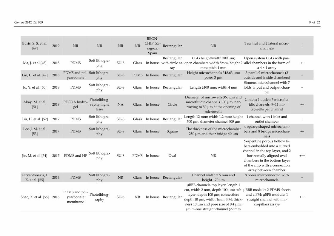
Cancers 2022, 14, 869 8 of 32
Table 1. Microfluidic devices design e fabrication.
Study Year
Manufacturing Geometric Characteristics of Microdevices
Main Material
of Device
Technology
Used Mold Cast Cover
Fabrica-
tion
Culture
Region Shape Device Dimensions Device Structures
Complexity
of Device
Li, Z.; et al. [39] 2021 PDMS Soft lithogra-
phy
SU-8 and
ethyl lactate
No
cover In house Semicircle
Top channel 0.5 × 2 × 11 mm3;
Side channel 0.3 × 2 × 15 mm3;
Center channel 100 × 900 × 11,000
μm3;
Pore size 4 μm
Multi interfaces microdevice
that consists in 3 layers: 1
channel at the top, and center,
2 channels bottom
++
Zhang, Q. et al.
[40] 2020 PDMS
Soft lithogra-
phy SU-8 PDMS In house Circle
Width 60 μm;
Height 100 μm;
Length 150 μm
Inlet for flow injection and
outlet for flow aspiration +
Tricinci, O. et al.
[41] 2020 IP-S photoresist
Two-photon
lithography SU-8 Glass In house
Perimetric cy-
lindrical pil-
lars
Diameter 50 μm;
Thickness 2 μm;
Length 150–800 μm;
Pores 5 μm
Arrangement of 10 micro-
tubes, 2 flat ends, and a cen-
tral cylindrical region with
pores
+++
Samiei, E. et al.
[42] 2020 PDMS
Soft lithogra-
phy SU-8
Co-
verslip In house Rectangular Thickness 200 μm
4 parallel compartments
(posts with gaps separate the
adjacent compartments)
+
Mamani, J.B.
et al. [43] 2020 PDMS
Soft lithogra-
phy SU-8 Glass
SynVivo
Inc., Ala-
bama,
USA
Circle
Outer channel width 200 μm; depth
(height) 100 μm; slit spacing 50 μm;
travel (space between channels) of 50
μm
1 apical chamber; channels (2
external and 1 internal) ++
Yi, H. G. et al.
[44] 2019
GBM-bioink;
HUVEC-bioink;
silicon
3D-printing
system NA Glass In house Circle NR NA +++
Qu, C. et al. [45] 2019 PDMS NR NR Glass In house NR NR CGG unit and an open array
of parallel chambers ++
Pang, L. et al. [46] 2019 PDMS Soft lithogra-
phy
SU-8 and
silicon PDMS In house Squares
Capture channel width 400 μm,
height 25 μm; culture chamber width
2000 μm, height 25 μm, length 4500
μm;
the microwell length 100 μm, width
100 μm; height 75 μm; pore 1 was 2
μm broader than pore 2
Channels (4 output and 1 in-
put), pore and microchannels
arrays
++
Cancers 2022, 14, 869 9 of 32
Burić, S. S. et al.
[47] 2019 NR NR NR NR
BEON-
CHIP, Za-
ragoza,
Spain
Rectangular NR 1 central and 2 lateral micro-
channels +
Ma, J. et al.[48] 2018 PDMS Soft lithogra-
phy SU-8 Glass In house
Rectangular
with circle ar-
ray
CGG height/width 300 μm;
open chambers width 5mm, height 2
mm; pitch 4 mm
Open system CGG with par-
allel chambers in the form of
a 4 × 4 array
++
Lin, C. et al. [49] 2018 PDMS and pol-
ycarbonate
Soft lithogra-
phy SU-8 PDMS In house Rectangular
Height microchannels 318.63 μm;
pores 3 μm
3 parallel microchannels (2
outside and inside chambers) +
Jo, Y. et al. [50] 2018 PDMS Soft lithogra-
phy SU-8 Glass In house Rectangular Length 2400 mm; width 4 mm
Sinuous microchannel with 7
folds; input and output chan-
nel
+
Akay, M. et al.
[51] 2018
PEGDA hydro-
gel
Photolithog-
raphy; light
laser
NA Glass In house Circle
Diameter of microwells 360 μm and
microfluidic channels 100 μm, nar-
rowing to 50 μm at the opening of
microwells
2 inlets; 1 outlet; 7 microflu-
idic channels; 9–11 mi-
crowells per channel
++
Liu, H. et al. [52] 2017 PDMS Soft lithogra-
phy SU-8 Glass In house Rectangular
Length 12 mm; width 1.2 mm; height
700 μm; diameter channel 600 μm
1 channel with 1 inlet and
outlet chamber +
Lee, J. M. et al.
[53] 2017 PDMS
Soft lithogra-
phy SU-8 Glass In house Square
The thickness of the microchamber
250 μm and their bridge 40 μm
4 square-shaped microcham-
bers and 8 bridge microchan-
nels
++
Jie, M. et al. [54] 2017 PDMS and HF Soft lithogra-
phy SU-8 PDMS In house Oval NR
Serpentine porous hollow fi-
bers embedded into a curved
channel in the top layer, and 2
horizontally aligned oval
chambers in the bottom layer
of the chip with a connection
array between chamber
+++
Zervantonakis, I.
K. et al. [55] 2016 PDMS
Soft lithogra-
phy NR Glass In house Rectangular
Channel width 2.5 mm and
height 170 μm
8 pores interconnected with
microchannels +
Shao, X. et al. [56] 2016
PDMS and pol-
ycarbonate
membrane
Photolithog-
raphy SU-8 NR In house Rectangular
μBBB channels-top layer: length 1
cm, width 2 mm, depth 100 μm; sub-
layer: depth 100 μm; connection:
depth 10 μm, width 1mm; PM: thick-
ness 10 μm and pore size of 0.4 μm;
μSPE-one straight channel (22 mm
μBBB module: 2 PDMS sheets
and a PM; μSPE module: 1
straight channel with mi-
cropillars arrays
+++

Cancers 2022, 14, 869 10 of 32
length × 2 mm width × 80 μm depth);
micropillar arrays (30 μm width in-
tervals)
Gallego-Perez, D.
et al. [57] 2016 PDMS
Photolithog-
raphy
SU-8 and
SPR950/SF6
nanowires
NR In house Rectangular 2 μm × 1μm with 2 μm spacing Arrays of parallel ridges ++
Xu, H. et al. [58] 2015 PDMS Soft lithogra-
phy SU-8 Glass In house Rectangular
Upper/lower thickness layer: 190/100
μm
Microstructures with differ-
ent heights +
Yoon, H. et al.
[59] 2014 PDMS
Soft lithogra-
phy SU-8 Glass In house Square
Reservoirs with diameter of inlet 2
mm and outlet 4 mm; channel 170
μm thick
Chambers (4 inlets and 4 out-
lets smaller); single test arena ++
Lou, X. et al. [60] 2014 PDMS Soft lithogra-
phy SU-8 Glass In house Square
Microchannels height 33 μm; culture
channels height 100 μm; filter layer,
3 different channel heights (15, 33,
and 51 μm)
3 layers: glass (top), cell (mid-
dle) and filter (bottom) +++
Abbreviations: PDMS: polydimethylsiloxane; IP-S: polymer photoresist; GBM-bioink: bioink of glioblastoma cells; GBM: glioblastoma multiform; HUVEC-bioink:
bioink of HUVEC cells; HUVEC: human umbilical vein endothelial cells; NR: not reported; PEGDA: poly-(ethylene glycol) diacrylate (MW 700 Da); HF: modified
polyethersulfone (mPES); SU-8: epoxy-based negative photoresist; NA: not applicable; SPR950/SF6 nanowires: nanowires SPR950 (~200 nm); BEONCHIP: biomi-
metic environment on chip (Spain); CGG: concentration gradient generator; μBBB: reconstruction BBB structure and 3D brain microenvironment; BBB: blood-
brain barrier; PM: polycarbonate membrane; μSPE: solid-phase extraction on-chip.
Cancers 2022, 14, 869 11 of 32
3.3. Cells Used in 3D Culture in Microfluidic Devices
We analyzed the cell characteristics (type, origin, and source) and their environment
(medium culture and supplements) for glioblastoma on-a-chip model development in mi-
crofluidic devices. Regarding the model development, tumor and support cells were used
isolated (63.6%) [40,42,43,46–48,50,51,53,57–60] or in co-culture (36.4%)
[39,41,44,45,49,52,54–56] inside of the device (Table 2).
Among the glioblastoma tumor cells, the most reported was U87-MG (40%) [39–
41,44,48,52,53,58], including the use of this cell with modifications (U87-MG-GFP [41] and
U87-MG/KD/SC [42]), then U251-MG (16%) [42,46,54,56], including their modifications
(U251-MG/KD/SC [42] and induced U251-MG [46]), 16% C6 [43,47,59,60] (included the
TMZ and BCNU resistant C6 [47]), 12% of GBM primary [39,44,49,51,55,57], 8% of GSC
[49,57], 4% T98G [50], and F98-GFP [55]. In addition, some studies used different tumor
cells, such as HepG2 (5.6%) [39,54], MCF7 (2.6%) [53], and Caco-2 (2.6%) [54], when com-
pared to all tumor cells, and endothelial cells were used as microenvironment support,
aiming at enriching the ECM, being 20% of HUVEC [44,52], hCMEC/D3 [41,56], and as-
trocyte cells [39,41], and 10% of each of the following cells: HBMEC [49], BMEC [39],
Eahy926 [49], and Bend3 [55]. Of these cells, 80% were of human sources [39–42,44–46,48–
54,56–58], being only 22.85% of primary culture [44,49,51,57], and 20% of animal sources
[39,43,47,55,59,60] (22.22% of primary culture [39,55]). Only two studies that used the pri-
mary culture reported the number of passages that varied from 3 to 10.
Interestingly, the medium of culture used in microfluidic devices varied according
to the type of culture, in co-culture, more than one type of medium culture was reported.
The most used was DMEM (46.2%) [39,41,42,44–48,50,52,53,56,58–60], then DMEM-F12
[46,57], and RMPI-1640 with 15.4% [43,49,54–56]. These types of mediums were the same
as those used in the 2D culture and added in the same proportion of cells seeded. In pri-
mary cell culture, the medium was supplemented with some growth factors (recombinant
human EGF, FGF, LIF, EndoGRO-MV Supplement kit, astrocyte growth supplement,
ECCS, FGFb, B-27, GA-1000, VEGF, hEGF, hFGF-β, R3-IGF-1) due to the complexity of
culturing primary cells.
The complexity evaluation on cell culture—more than half of the studies were classified as level
(++) due to using co-culture, spheroid, or modified cells culture [39,44–47,49,52,54–56], followed
by level (+) classification (36.4%) with isolated culture use [40,42,43,48,50,51,53,57,58,60]. Only the
study by Tricinci (4.5%) used co-culture associated with the spheroid, classified in level (+++) [41].
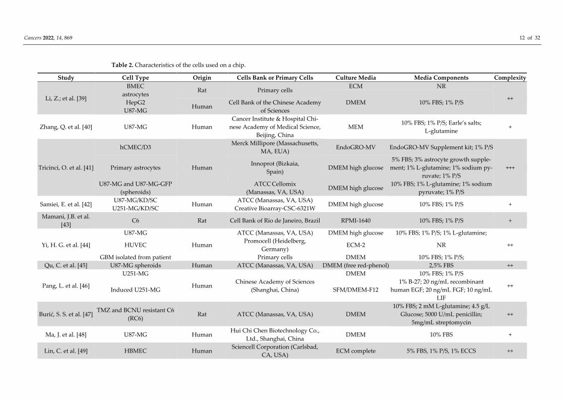
Cancers 2022, 14, 869 12 of 32
Table 2. Characteristics of the cells used on a chip.
Study Cell Type Origin Cells Bank or Primary Cells Culture Media Media Components Complexity
Li, Z.; et al. [39]
BMEC Rat Primary cells
ECM NR
++ astrocytes
DMEM 10% FBS; 1% P/S HepG2 Human
Cell Bank of the Chinese Academy
of Sciences U87-MG
Zhang, Q. et al. [40] U87-MG Human
Cancer Institute & Hospital Chi-
nese Academy of Medical Science,
Beijing, China
MEM 10% FBS; 1% P/S; Earle’s salts;
L-glutamine +
Tricinci, O. et al. [41]
hCMEC/D3
Human
Merck Millipore (Massachusetts,
MA, EUA) EndoGRO-MV EndoGRO-MV Supplement kit; 1% P/S
+++ Primary astrocytes Innoprot (Bizkaia,
Spain) DMEM high glucose
5% FBS; 3% astrocyte growth supple-
ment; 1% L-glutamine; 1% sodium py-
ruvate; 1% P/S
U87-MG and U87-MG-GFP
(spheroids)
ATCC Cellomix
(Manassas, VA, USA) DMEM high glucose
10% FBS; 1% L-glutamine; 1% sodium
pyruvate; 1% P/S
Samiei, E. et al. [42] U87-MG/KD/SC
Human ATCC (Manassas, VA, USA)
DMEM high glucose 10% FBS; 1% P/S + U251-MG/KD/SC Creative Bioarray-CSC-6321W
Mamani, J.B. et al.
[43] C6 Rat Cell Bank of Rio de Janeiro, Brazil RPMI-1640 10% FBS; 1% P/S +
Yi, H. G. et al. [44]
U87-MG
Human
ATCC (Manassas, VA, USA) DMEM high glucose 10% FBS; 1% P/S; 1% L-glutamine;
++ HUVEC Promocell (Heidelberg,
Germany) ECM-2 NR
GBM isolated from patient Primary cells DMEM 10% FBS; 1% P/S;
Qu, C. et al. [45] U87-MG spheroids Human ATCC (Manassas, VA, USA) DMEM (free red-phenol) 2,5% FBS ++
Pang, L. et al. [46]
U251-MG
Human Chinese Academy of Sciences
(Shanghai, China)
DMEM 10% FBS; 1% P/S
++ Induced U251-MG SFM/DMEM-F12
1% B-27; 20 ng/mL recombinant
human EGF; 20 ng/mL FGF; 10 ng/mL
LIF
Burić, S. S. et al. [47] TMZ and BCNU resistant C6
(RC6) Rat ATCC (Manassas, VA, USA) DMEM
10% FBS; 2 mM L-glutamine; 4.5 g/L
Glucose; 5000 U/mL penicillin;
5mg/mL streptomycin
++
Ma, J. et al. [48] U87-MG Human Hui Chi Chen Biotechnology Co.,
Ltd., Shanghai, China DMEM 10% FBS +
Lin, C. et al. [49] HBMEC Human Sciencell Corporation (Carlsbad,
CA, USA) ECM complete 5% FBS, 1% P/S, 1% ECCS ++

Cancers 2022, 14, 869 13 of 32
Eahy926 ATCC (Manassas, VA, USA) RPMI-1640 10% FBS, 1% PS
GSCs from GBM patients The Second Affiliated Hospital of
Soochow University ECM complete
20 ng/mL EGF; 20 ng/mL FGFb;
2% B-27
Jo, Y. et al. [50] T98G Human Korean Cell Line Bank, Seoul, Ko-
rea DMEM 10% FBS, 1% Penicillin +
Akay, M. et al. [51] Patient’s primary
GBM Human
UTHealth and Memorial Hermann,
Texas Medical Center, Houston,
TX, USA
Supplemented EBM
(EGM-2)
FBS; hydrocortisone; GA-1000; VEGF;
hEGF; hFGF-B; R3-IGF-1; acid ascorbic +
Liu, H. et al. [52]
HUVEC
Human
Cancer Institute & Hospital Chi-
nese Academy of Medical Science,
Beijing, China
DMEM 10% FBS; 1% P/S ++ U87-MG
Lee, J. M. et al. [53] MCF7
Human NR DMEM 10% FBS; 1%P/S + U87-MG
Jie, M. et al. [54]
Caco-2
Human
Cancer Institute and Hospital, Chi-
nese Academy of Medical Sciences,
Beijing, China
RPMI-1640 10% FBS; ++ HepG2
U251-MG
Zervantonakis, I. K.
et al. [55]
F98-GFP Rat ATCC (Manassas, VA, USA) RPMI-1640 10% FBS; 1% P/S ++
Bend3 Mice
Shao, X. et al. [56] hCMEC/D3
Human Institute COCHIN, Paris, France RPMI-1640
10% FBS; 100 μg/mL P/S and 1.5 μM
hydrocortisone ++
U251-MG NR DMEM 10% FBS; 100 μg/mL P/S
Gallego-Perez, D. et
al. [57]
GSCs derived tumor: GBM157
and GBM528 Human The Ohio State University DMEM-F12
B27; 2.5 μg/mL heparin; 20 ng/mL
FGFb
and 20 ng/mL EGF
+
Xu, H. et al. [58] U87-MG Human Cell Bank of the Chinese Academy
of Sciences, Shanghai, China DMEM 10% FBS +
Yoon, H. et al. [59] C6 Rat ATCC (Manassas, VA, USA) DMEM
10% FBS, 10,000 units/mL penicillin;
10,000 μg/mL streptomycin, and 25
μg/mL Fungizone
+
Lou, X. et al. [60] C6 Rat ATCC (Manassas, VA, USA) DMEM 10% FBS; 1% P/S +
Abbreviations: BMEC: brain microvascular endothelial cells primary; HepG2: liver hepatocellular carcinoma cell line; U87-MG: glioma cell line; hCMEC/D3: hu-
man cerebral microvascular endothelial cells; U87-MG-GFP: glioma cell line expressing green fluorescent protein; U251-MG: glioma cell line; U87/251-MG/KD/SC:
Atg7 knockdown (KD) and scrambled (SC) U251 and U87 cells; C6: glial tumor of rat; HUVEC: human umbilical vein endothelial cells; GBM: glioblastoma multi-
form; TMZ: temozolomide; BCNU: bis-chloroethyl nitrosourea; RC6: TMZ and BCNU resistant C6; HBMEC: human brain microvascular endothelial cells;
Eahy926: immortalized human vascular endothelial cells; GSCs: glioma stem cells; T98G: glioblastoma cell line; MCF7: breast cancer cell line; Caco-2: colorectal
adenocarcinoma cell line; F98-GFP: glioblastoma cell line expressing green fluorescent protein; Bend3: mouse brain endothelial cell line; GBM157: cell clone iso-
lated of patient; GBM528: cell clone isolated of patient; ATCC: American Type Culture Collection; NR: not reported; ECM: endothelial cell medium; DMEM:
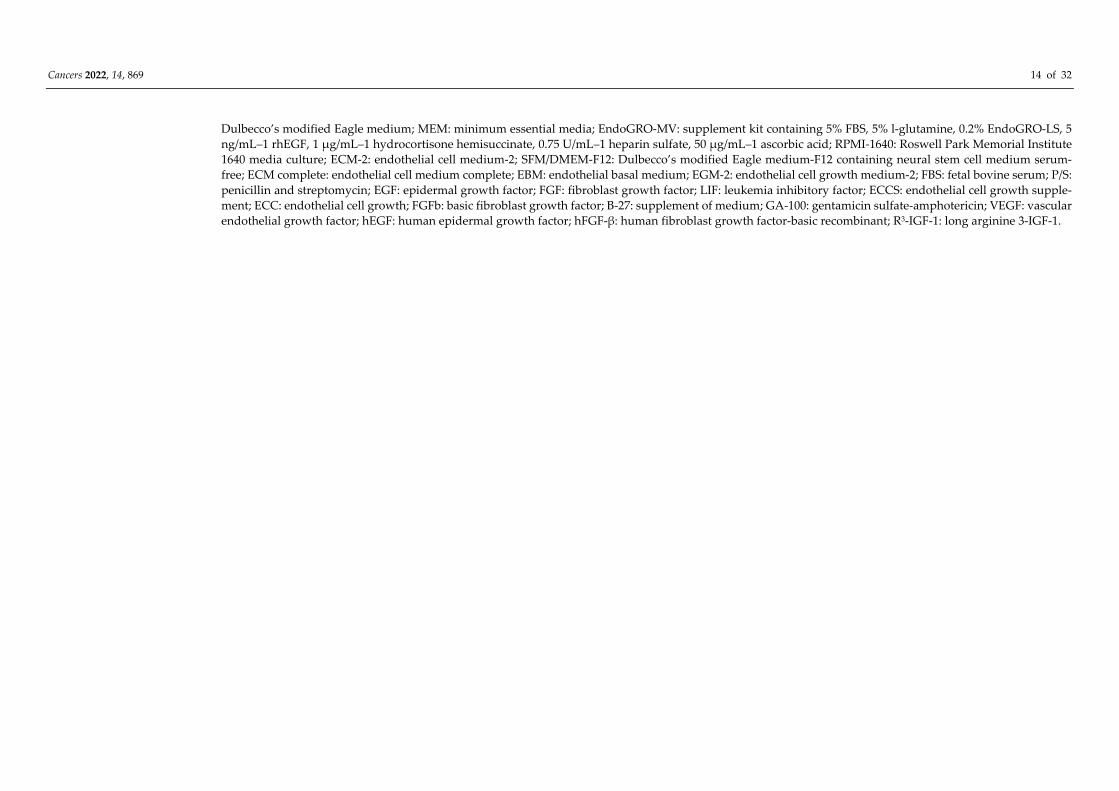
Cancers 2022, 14, 869 14 of 32
Dulbecco’s modified Eagle medium; MEM: minimum essential media; EndoGRO-MV: supplement kit containing 5% FBS, 5% l-glutamine, 0.2% EndoGRO-LS, 5
ng/mL–1 rhEGF, 1 μg/mL–1 hydrocortisone hemisuccinate, 0.75 U/mL–1 heparin sulfate, 50 μg/mL–1 ascorbic acid; RPMI-1640: Roswell Park Memorial Institute
1640 media culture; ECM-2: endothelial cell medium-2; SFM/DMEM-F12: Dulbecco’s modified Eagle medium-F12 containing neural stem cell medium serum-
free; ECM complete: endothelial cell medium complete; EBM: endothelial basal medium; EGM-2: endothelial cell growth medium-2; FBS: fetal bovine serum; P/S:
penicillin and streptomycin; EGF: epidermal growth factor; FGF: fibroblast growth factor; LIF: leukemia inhibitory factor; ECCS: endothelial cell growth supple-
ment; ECC: endothelial cell growth; FGFb: basic fibroblast growth factor; B-27: supplement of medium; GA-100: gentamicin sulfate-amphotericin; VEGF: vascular
endothelial growth factor; hEGF: human epidermal growth factor; hFGF-β: human fibroblast growth factor-basic recombinant; R3-IGF-1: long arginine 3-IGF-1.

Cancers 2022, 14, 869 15 of 32
3.4. Methods Cultivation of Cells Used in the 3D Culture
Regarding the 3D culture of glioblastoma model in a microfluidic device (Table 3),
the culture methodology that involves the ECM components (concentration and volume)
and cell types (concentration, culture time, medium change, and their flow rate) that rep-
resent important aspects for the development of a tumor model biomimetic to evaluate
the different methodologies and therapeutic agents, was analyzed.
Another relevant aspect in 3D co-culture is the ECM addition, which was reported in
73% of the selected studies [39,42–45,47–50,52–56,58,59]. Collagen type I represented 35%,
being the most used in ECM composition [39,42–45,47,48,55,58,59], followed by 15% Mat-
rigel [43,50,54,56], and a smaller proportion (4%) BdECM [44], fibronectin [49], TG-gelatin
[52], GelMA [53], and agarose [56]. Of these studies, 45.8% reported ECM concentration
used ranging from 0.1 to 12 mg/mL [39,42–45,48,50,53–56], and only 16% reported the vol-
ume administration, ranging from 8 to 20 μL [43,47,48,53]. In contrast, 20% did not use
any ECM components [40,41,46,51,60]. The culture methodology in the microfluidic de-
vice focused mainly on the order and position of cell culture, from the treatment of the
device with ECM to maintenance after the culture. Some strategies were used to promote
the formation of a 3D matrix, inverting the device surface [45,48].
For glioblastoma model development, 51.3% of the selected studies used human cells
(25.6% U87 [40,41,46,51,60], 10.3% U251 [42,46,54,56], 7.7% GSC [49,57], 5.1% human GBM
[44,49,51,57], and 2.6% T98G [50]), followed by 12.8% of GBM from rats (10.3% C6
[43,47,59,60] and 2.6% F98-GFP [55]), and in 10.3% of studies different associated tumor
types (carcinoma [39,54] and adenocarcinoma [54]) were used. Furthermore, 31.8% of
studies used some supporting endothelial cells, with one or more of these cells combined
(astrocytes, hCMEC/D3, HUVEC, HBMEC, BMEC, Eahy926, and Bend3), being prevalent
in the co-culture with the first three of these cells (20% each), aiding in chemical commu-
nication and secretion of the ECM, obtaining results closer to those obtained in vivo ex-
periments [61–64]. In relation to the cell type, we also analyzed the concentration used,
which varied between the different types, as well as within the same type, for example,
the U87 number cells ranged from 104 to 107 cells/mL. Cell culture time of 92% of the stud-
ies was from 0.125 to 10 days and medium change during culture was reported in 54% of
the studies, being carried out from 2 to 72 h. The flow rate was reported in only 37.5% of
the studies, ranging from 0.5 to 4.7 × 10³ μL/min, and 8.3% of studies did not apply the
shear rate [56,59], an important factor for tumor growth.
The complexity evaluation of microenvironment construction showed that almost
half (45.5%) of studies used simple ECM level (+) [40,42,43,46,47,51,57–60], followed by
27.3% level (++) [45,48–50,53,55] and (+++) [39,41,44,52,54,56], that match the use of two or
more ECM combined, and ECM primary or synthetic scaffold, respectively.

Cancers 2022, 14, 869 16 of 32
Table 3. 3D culture development of glioblastoma model in microfluidic devices.
Study
Extracellular Matrix
Cells Type (Cells/mL) Culture Time
(d) Cultivation Method on the Device
Medium Change
(h)/Flow (μL/min) Complexity
Type of Matrix Concentration
(mg/mL)
Volume
(μL)
Li, Z.; et al. [39] COL1 6 NR
Astrocytes (5 × 10⁵);
HBMEC (1 × 10⁵); HepG2 (1
× 106); U87 (NR)
2.5
COL1 was perfused into the channels (10
min), following by seeded astrocytes. After
12 h, BMECS were seeded in the same chan-
nels, 24 h later, HepG2 cells were perfused
in the upper chamber and more 24 h, U87-
MG cells were introduced into the lower
right channel
NR +++
Zhang, Q. et al.
[40] NA NA NA U87 (1 × 10⁴ cells/cm2)
0.125, 0.25, 6,
0.5, 0.75
The adherent target single cell in trypsin re-
gion was digested, and the extraction pro-
cess was recorded by microscope camera.
Injection:10
μL/min;
aspiration: 40
μL/min
+
Tricinci, O. et al.
[41] NA NA NA
hCMEC/D3 (3 × 10⁴
cells/cm2); primary astro-
cytes (1 × 10⁴/cells cm2); U87
(2 × 10⁴ cells/30 μL)
5
hCMEC/D3 cells were seeded inside micro-
tubes. After 5 days, the human primary as-
trocytes were seeded on the outside part of
the tubes, and U87-MG cells were seeded in
the MRCSs, after 5 days of cell growth
NR/4.7 × 103 +++
Samiei, E. et al. [42] COL1
3
NR
U251 and U87 (106 viability)
4
COL1/cell suspension was injected into the
channel for 45 min (invasion study) or over-
night (viability study), and the treatment
was started the day after.
NR + 4 U87 (5 × 10⁶ invasion)
Mamani, J.B. et al.
[43] Matrigel 9-12 15
C6
(107) 2
Matrigel was injected into the central chan-
nel for 2 h. Then, C6 cells were injected into
the external channel
4/5 +
Yi, H. G. et al. [44]
BdECM
10 NR
U87, GBM from patients
and HUVEC
(5 × 10⁶)
7
The cell-laden bioinks were encapsulated
with GBM cells or HUVECs into pre-gel so-
lutions of BdECM or collagen.
24/NR +++ COL1
Qu, C. et al. [45] COL1 1.5 NR U87
(3.5 × 10⁵) 2
U87 cells were seeded in the channel. Then,
arrays were generated on the inverted
PDMS surface, using COL1 was used as the
U87 spheroid encapsulating ECM.
24/0.5 ++
Pang, L. et al. [46] NA NA NA U251
(0.25–2.5 × 10⁴) 10
Pluronic F127 was injected from the inlet
into the chambers for 2 h at 20 °C. Cells
Half of the me-
dium 24/5 +

Cancers 2022, 14, 869 17 of 32
were seeded into the chambers for the 20 s
from the inlet (20 μL/min), using different
driving infusion flow rates (25–150 μL/min)
to separate the single-cells, cultured at a
slow perfusion rate (5 μL/min)
Burić, S. S. et al.
[47] COL1 NR 10
RC6
(5 × 10⁶) 3
10 μL of the mixture of RC6 cells with COL1
was injected into the central chamber. After
COL1 polymerization for 15 min, lateral mi-
crochannels were perfused with medium
2/NR +
Ma, J. et al. [48] COL1 1, 5 8 U87
(6 × 10⁵) 3
U87 cells spheroids were formed for 3 days
in chamber lower, in following added COL1
for 45 min and PDMS surface was inverted
to solidify
NA/0.5 ++
Lin, C. et al. [49] Fibronectin NR NR HBMEC, Eahy926 and GSC
(1.26 × 10⁶) 3
HBMECs and Eahy926 cells were seeded in
the upper microchannels, the following day,
the GSCs were cultured in the lower chan-
nels. Fibronectin was placed (12 h) for endo-
thelial cell culture.
12/NR ++
Jo, Y. et al. [50]
Matrigel 0.1 NR
T98G
(105) 4
The microchannel was coated with a PDL or
Matrigel solution for 3 h, and then the T98G
cells were seeded. After cultivate, the flow
was stopped for 24 h. Then shear stress of
(0.1 dyn/cm2) was applied
72/NR ++ PDL 0.1 NR
Akay, M. et al. [51] NA NA NA GBM of patients (5 × 10⁵) 7
The GBM cells were seeded into both inlet
channels simultaneously. The cells were
captured in the microwells and cultured for
7 days
A half medium 48–
72/NR +
Liu, H. et al. [52] TG-gelatin NR NR U87 and HUVEC
(107) 3
TG-gelatin suspension was used for U87
cells culture. Then, these cells were injected
into the channels with the PU fiber. After
the gel polymerized and solidified for 40
min, the PU fiber was pulled out from the
channels. Following, HUVEC cells were
seeded into the lumen (4 h), and the chip
was connected to the peristaltic pump.
NR/(0,5,10,20) +++
Lee, J. M. et al. [53] GelMA 10 20 MCF7 and U87 (2 × 10⁶) 5 MCF7 and U87MG cells were cultured in
square-shaped microchambers 24/NR ++
Cancers 2022, 14, 869 18 of 32
Jie, M. et al. [54] Matrigel 3.86 NR U251, HepG2 and Caco-2
(106) NR
HF was coated with a Matrigel per 1 h at 4
°C. Then, 10 μL of the Caco-2 cells were
seeded into the lumen. HepG2 and U251
cells were injected into chambers b and c
(bottom layer) from the respective inlets.
The inlet and outlet of the HepG2 cell cham-
ber were stoppered. The inlet of the U251
cells chamber (b and c) was stoppered and
the outlet was connected to a waste reser-
voir. After 24 h, the outlet of HF was stop-
pered and the inlet was connected to an in-
fusion pump that continuously infused the
medium (5 μL/h).
12/0.083 +++
Zervantonakis, I.
K. et al. [55] COL1 2 NR
F98-GFP (3 × 10⁵) and Bend3
(2 × 10⁶) 2
F98G cells were seeded in the top layer and
Bend3 cells in the bottom layer, in the de-
vice containing COL1 for 48 h
NR/NR ++
Shao, X. et al. [56]
Matrigel 0.1 NR hCMEC/D3
(5 × 10⁶)
3
μBBB model: hCMEC/D3 cells were seeded
on the upper side of the membrane (to form
cell monolayer) and inferior chambers the
U251 cells were injected and encapsulated
in agarose solution for 24 h.
24/15
+++
Agarose NR NR U251 (5 × 10⁶) 24/NA
Gallegos-Perez, D.
et al. [57] NR NR NR
GSCs derived tumor:
GBM157 and GBM528 NR
GSC, GBM157, and GBM528 clones neuro-
spheres were dissociated and seeded on the
microtextured chip surface (16 h of monitor-
ization)
NR/NR +
Xu, H. et al. [58] COL1 NR NR U87
(5 × 10⁴ cell/cm2) 1
Cells were seeded into the center channel.
The chip was then turned on its side for 5
min. Each chip was then incubated for ei-
ther 24 h, 21% O2 (normoxic condition) or
0.2% O2, 94% N2 (hypoxic conditions). Cells
were allowed to invade for 24 h.
NR/NR +
Yoon, H. et al. [59] COL1 NR NR C6 1
The chip was coated, using a solution of
0.01% COL1, then it was seeded with C6
cells for about 24 h.
NR/NR +
Lou, X. et al. [60] NA NA NA C6
(2 × 10⁶) Overnight
In the gas layer of CGG containing the C6
cells was introduced compressed air and NA +

Cancers 2022, 14, 869 19 of 32
nitrogen, generating an oxygen gradient
from 1.3% (hypoxia range) to 19.1% (ambi-
ent air range).
Abbreviations: COL1: chilled liquid type I collagen ; NA: not applicable; BdECM: brain decellularized ECM; PDL: poly-D-lysine hydrobromide ; TG-gelatin:
gelatin transglutaminase; GelMA: gelatin methacrylate hydrogels ; NR: not reported; HBMEC: human brain microvascular endothelial cells; HepG2: liver hepa-
tocellular carcinoma cell line; U87: glioma cell line; hCMEC/D3: human cerebral microvascular endothelial cells; U251: glioma cell line; C6: glial tumor of rat; GBM:
glioblastoma multiform; HUVEC: human umbilical vein endothelial cells; RC6: C6 resistant to TMZ and BCNU; Eahy926: immortalized human vascular endothe-
lial cells; GSC: glioma stem cells; T98G: glioblastoma cell line; ; MCF7: breast cancer cell line; Caco-2: colorectal adenocarcinoma cell line; F98-GFP: glioblastoma
cell line expressing green fluorescent protein; Bend3: mouse brain endothelial cell line ; GBM157: cell clone isolated of patient; GBM528: cell clone isolated of
patient; BMECS: brain microvascular endothelial cells ; MRCSs: magnetically-responsive cage-like scaffolds ; RPMI: Roswell Park Memorial Institute; PDMS:
polydimethylsiloxane; ECM: endothelial cell medium; PU: polyurethane; HF: hollow fiber; μBBB: reconstruction BBB structure and 3D brain microenvironment;
BBB: brain blood barrier.
Cancers 2022, 14, 869 20 of 32
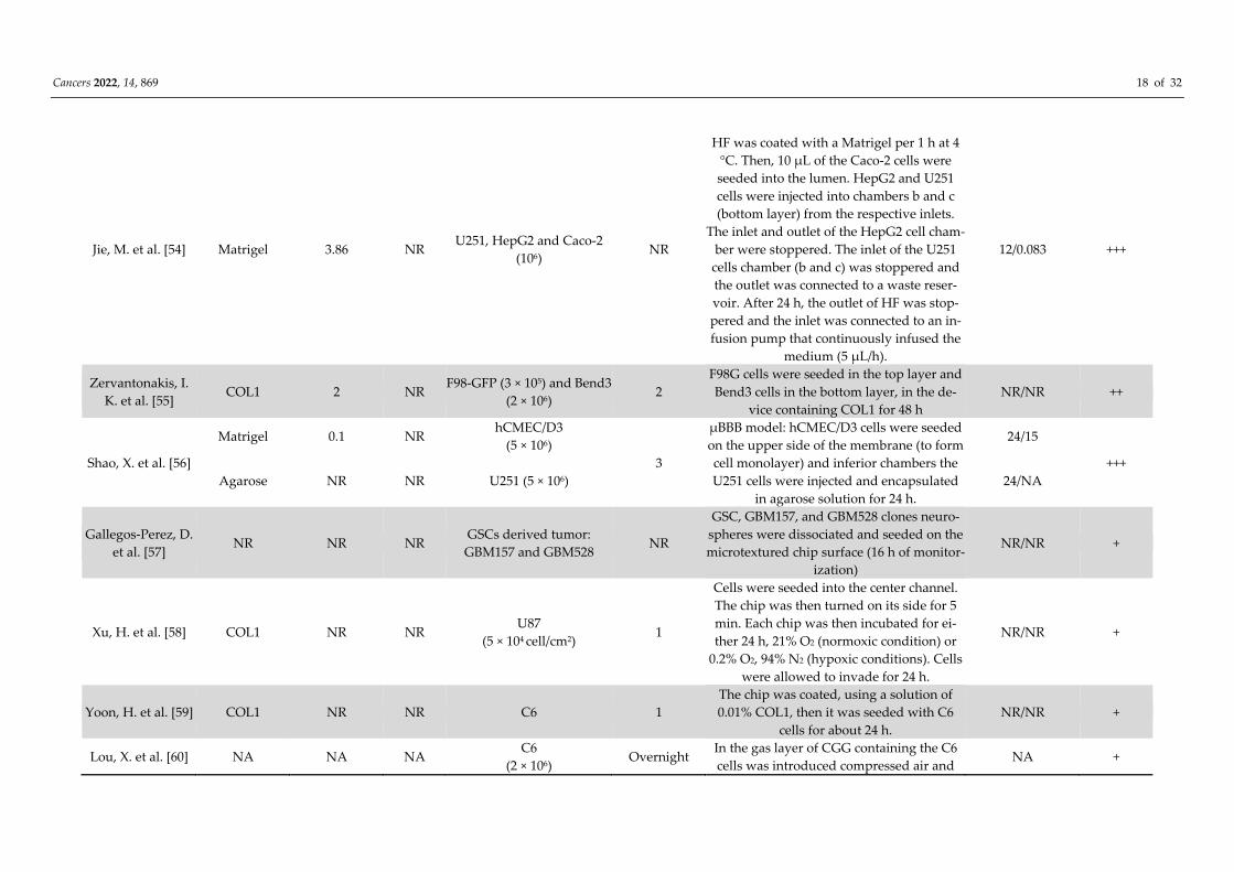
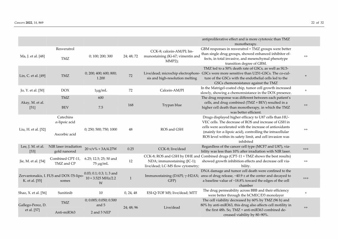
3.5. The Efficiency of Glioblastoma Therapeutic Approach in the Microfluidic Device
Table 4 analyzed the different therapeutic approaches for glioblastoma through the
microfluidic device and allowed it by microenvironment mimicking, combining the ther-
apeutic, can increase the number of conditions to test, besides that the outcome observed
its more similar to in vivo outcomes than in vitro experiments. Regarding the therapeutic
approaches, the use of chemotherapy alone [39–42,44–52,54–57,59] or combined with
other drugs [44,51,54] or conditions were reported in most of the selected studies (77.3%),
followed by different combined therapeutic strategies (13.6%), such as phototherapy
[55,59], and irradiation associated with drug delivery [51], as well as the therapeutic strat-
egy in an isolated way, such as phototherapy (4.5%) and magneto hyperthermia therapy
(4.5%) [43]. The drug most reported in the chemotherapy approach was an alkylating
agent, the temozolomide (45.5%) [40,42,44,47–49,51,54,57] dose ranged from 0.005 to 1200
μM, following by 9.1% for doxorubicin (amphetamine) [50,55] that ranged from 0.03 to 1
μg/mL, and 4.5% of 24 different drugs with varied classes: chemical inhibitors (antibody-
functionalized nutlin-loaded nanostructured lipid carriers, simvastatin, KU60019, meth-
oxyamine, O6-benzylguanine, tamoxifen, irinotecan, and sunitinib), antioxidants (coen-
zyme Q10, resveratrol, catechins, α-lipoic acid, and ascorbic acid), alkylating agent (cis-
platin and cyclophosphamide), antimicrotubular (paclitaxel and vincristine), antimetabo-
lites (capecitabine and 5-fluorouracil), antibiotic (actinomycin D), antifungal (allicin), an-
tibody (bevacizumab), microRNA (Anti-miR363), and siRNA (HIF1α/HIF2α inhibitor).
The time of therapy varied from around 0.16 until 168 h.
The methods used to evaluate the therapeutic approaches include more than one
technique in each study. Cell viability analysis was the most reported in the selected stud-
ies (95.45%), then 27.27% cell proliferation, 18.18% of oxidative stress, and migration/in-
vasion of cells, and a further 4.54% for molecular characterization, DNA methylation, au-
tophagy, metabolites, and permeability. Some techniques were used with more than one
purpose of analysis, such as cell viability and proliferation using live/dead dye (25.71%)
[39,41–43,49,53,54,56,57,59,60], immunostaining (25.71%) [41,42,46,48,54,55,58], CCK-8 kit
(20%) [39,44,48,53,54], and CA/Pi (17.14%) [40,45,47,48,50]. The cell migration and inva-
sion ware also available using 60% CA/PI dye [45,47,48], and 40% immunostaining
(MMP2) [48,58]. The oxidative stress was analyzed by different techniques as CellROX
Orange, DHE and NDA, SOSG, ROS, and GSH [47,52,54,59]. When analyzing the studies’
outcomes, between the chemotherapeutics utilized as well as the different therapeutic ap-
proaches and their impacts on the cellular microenvironment, the recommended therapies
showed efficacy in various therapeutic approaches.
The complexity evaluation of therapeutic approaches in the glioblastoma on-a-chip
model showed that almost half (45.5%) of studies used combinations of drugs (level ++)
[39,40,42,47,48,51,52,54,57,58], following by 31.8% of level (+++) [41,43,44,53,55,59,60], that
reported therapeutic approaches combined and less often (22.7%) the level (+), which used
only one drug for chemotherapy [45,46,49,50,56].

Cancers 2022, 14, 869 21 of 32
Table 4. Glioblastoma therapeutic approach in site of microfluidic device.
Study Therapeutic
Approaches
Therapeutic Dose
(μM)
Time of
Treatment (h) Evaluation Efficacy Treatment Outcomes Complexity
Li, Z.; et al. [39]
PTX 2.3 × 10−3
48 Live/dead; CCK-8 kit; mass spec-
trometry;
In the liver-brain system, the liver had enhanced cyto-
toxicity of CAP on U87 cells by 30% while having no sig-
nificant effect on TMZ. However, the BBB system
showed a 20% decrease in PTX cytotoxicity, already no
significant effect was found on TMZ and CAP
++
CAP 80
TMZ 40
Zhang, Q. et al.
[40]
5-fluorouracil 38.4
3, 6, 12, or 18
Calcein-AM/PI
All drugs no influence under proliferation and viability
into 6 h, already the TMZ showed a reduction cell-ma-
trix adhesion and their effect was less significant with
the increase of the lactic acid.
++
Actinomycin D 10
Allicin 200
TMZ without LA 0; 100; 300; 500
TMZ with LA 500 (TMZ); 0; 104; 2 ×
104 (AL) 6
Tricinci, O. et al.
[41] Ab-Nut-NLCs
400 μg/mL + EMF of
1.31 T 24
Live/dead; immunostaining
(Ki-67);
Ab-Nut-NLCs capacity to cross the BBB and efficacy
about 70% on the treatment +++
Samiei, E. et al.
[42] TMZ and simvastatin
0; 100; 250; 500
(TMZ) and 0; 1; 5; 10
(simvastatin)
72
Live/dead; immunostaining
(cleaved-caspase-3 and PARP,
SQSTM1p62 and LC3);
The viability and invaded cells had a dose-dependent ef-
fect; U251 cells were more sensitive to the treatments
than the U87 cells, showing more effectively TMZ (500
μM) than simvastatin (10 μM).
++
Mamani, J.B.
et al. [43]
MHT and magnetic
nanoparticles
10 mgFe/mL (20 μL)
+ 300 Gauss/305 kHz 0.16; 0.5 Live/dead
After MHT, the cell viability reduced by 20% and 100%
after 10 and 30 min, respectively +++
Yi, H. G. et al.
[44]
CCRT combined with
TMZ, CIS, KU, O6BG
and MX
Different drug com-
binations: 950; TMZ;
950 CIS; 250 KU; 210
O6BG; 150 MX + 15
Gy
24 (1 h gamma
irradiation) CCK-8
The drug combination (TMZ, CIS, KU, O6BG, and MX)
was more effective on the GBM-28-on-a-chip. However,
the GBM-37-on-a-chip showed the highest resistance to
the tested drugs.
+++
Qu, C. et al. [45] TAM 10; 20; 30 24 Cell cycle analysis (acridine or-
ange); calcein-AM/PI;
ER- α36 knockdown increased sensitivity of glioblas-
toma U87 cells to TAM and decreased autophagy in
these cells. However, ER- α36 overexpression decreased
TAM sensitivity and induced autophagy.
+
Pang, L. et al. [46] Vincristine 1.25; 2.5; 5; 10; 20; 40;
80 0, 6, 12, 18, 24 Immunostaining (JC-1; Caspase-3)
Drug resistance in the induced of U251 spheres was
higher than standard U251 cells and dose-dependent. +
Burić, S. S. et al.
[47]
TMZ 250
72
Calcein-AM/PI; ROS (CellROX Or-
ange, Thermofisher, Massachu-
setts, MA, USA)
CoQ10 can suppress invasiveness, the epithelial to mes-
enchymal transition in RC6 cells, as also decrease ROS
and when combined with TMZ, exerted a synergistic
++ CoQ10 10
Cancers 2022, 14, 869 22 of 32
antiproliferative effect and is more cytotoxic than TMZ
monotherapy.
Ma, J. et al. [48]
Resveratrol
0; 100; 200; 300 24; 48; 72
CCK-8; calcein-AM/PI; Im-
munostaining (Ki-67; vimentin and
MMP2);
GBM responses in resveratrol + TMZ groups were better
than single drug groups, showed enhanced inhibitor ef-
fects, in total invasive, and mesenchymal phenotype
transition degree of GBM.
++ TMZ
Lin, C. et al. [49] TMZ 0; 200; 400; 600; 800;
1.200 72
Live/dead; microchip electrophore-
sis and high-resolution melting
TMZ led to a 50% death rate of GSCs, as well as SU3-
GSCs were more sensitive than U251-GSCs. The co-cul-
ture of the GSCs with the endothelial cells led to the
GSCs chemoresistance against the TMZ
+
Jo, Y. et al. [50] DOX 1μg/mL 72 Calcein-AM/PI In the Matrigel-coated chip, tumor cell growth increased
slowly, showing a chemoresistance in the DOX presence. +
Akay, M. et al.
[51]
TMZ 600
168 Trypan blue
The drug response was different between each patient’s
cells, and drug combined (TMZ + BEV) resulted in a
higher cell death than monotherapy, in which the TMZ
was better efficient.
++ BEV 7.5
Liu, H. et al. [52]
Catechins
0; 250; 500; 750; 1000 48 ROS and GSH
Drugs displayed higher efficacy to U87 cells than HU-
VEC cells. The decrease of ROS and increase of GSH in
cells were accelerated with the increase of antioxidants
(mainly for α-lipoic acid), controlling the intracellular
ROS level within its safety limit, and cell invasion was
inhibited
++
α-lipoic acid
Ascorbic acid
Lee, J. M. et al.
[53]
NIR laser irradiation
gold nanorod 20 v/v% + 3A/4.27W 0.25 CCK-8; live/dead
Regardless of the cancer cell type (MCF7 and U87), via-
bility was less than 10% after irradiation with NIR laser. +++
Jie, M. et al. [54] Combined CPT-11,
TMZ and CP
6.25; 12.5; 25; 50 and
75 μg/mL 12
CCK-8; ROS and GSH by DHE and
NDA; immunostaining (JC-1);
live/dead; LC-MS flow cytometry;
Combined drugs (CPT-11 + TMZ shows the best results)
showed growth inhibition effects and decrease cell via-
bility.
++
Zervantonakis, I.
K. et al. [55]
FUS and DOX-TS-lipo-
somes
0.03; 0.1; 0.3; 1; 3 and
10 + 3.525 MHz/2.2
W
1 Immunostaining (DAPI; γ-H2AX;
GFP)
DNA damage and tumor cell death were confined to the
area of drug release, ~40.9 ± at the center and decayed to
a baseline value of ~18.8% toward the edges of the cell
chamber
+++
Shao, X. et al. [56] Sunitinib 10 0, 24, 48 ESI-Q-TOF MS; live/dead; MTT The drug permeability across BBB and their efficiency
were better through the hCMEC/D3 monolayer +
Gallego-Perez, D.
et al. [57]
TMZ 0; 0.005; 0.050; 0.500
and 5 24; 48; 96 Live/dead
The cell viability decreased by 60% by TMZ (96 h) and
80% by anti-miR363, this drug also affects cell motility in
the first 48h. So, TMZ + anti-miR363 combined de-
creased viability by 80–90%.
++
Anti-miR363 2 and 5 NEP

Cancers 2022, 14, 869 23 of 32
Xu, H. et al. [58]
Normoxic and inhib-
ited by siRNA HIF1α
and HIF2α
21%
24 or 48
Immunostaining (Ki-67; MMP2;
Zeb1/2; Snail/Slug; Twist; HIF1/2α;
vimentin); RT-qPCR-RT (GLUT1,
VEGFA, EDN1; EPO; MMP2 and
MMP9); Western blotting (Twist;
MMP2; MMP9);
Hypoxia activates mesenchymal transition and enhances
cell motility in GBM in a HIF-dependent manner, and
this process can be attenuated by pharmacological block-
ade of HIFα. Antiangiogenic therapy associated with
HIFs inhibitors can delay tumor progression
++ Hypoxic and inhibited
by siRNA HIF1α and
HIF2α**
0.2 and 1% (O2)
Yoon, H. et al.
[59]
PDT by MB-PEGDMA
PAA NPs
MB–PEGDMA PAA
NPs, with MB (2.1;
5.5; 12.1 μmol/g) +
(~625 nm/35.2 mW;
LED light doses 0 to
39.2 J/cm2)
0-0.35 Live/dead; singlet oxygen sensor
green (ROS)
C6 cells killing effects of the various MB–PEGDMA PAA
NPs were light-dose-dependent +++
Lou, X. et al. [60]
PDT by MB combined
with hypoxic condi-
tions
0–10 (MB); 0–21%
(O2) + (637 nm; 0–9.5
mW; light dose 42.8
J/cm2)
0.5 Live/dead
Cell viability decreased to around 0% with the increase
of light power until 9.5 mW. Samples with higher drug
concentrations had a viability drop than a lower concen-
tration.
+++
Abbreviations: PTX: paclitaxel ; CAP: capecitabine ; TMZ: temozolomide; LA: lactic acid; Ab-Nut-NLCs: antibody-functionalized nanostructured lipid carriers
loaded with nutlin-3a; MHT: therapy of magnetic hyperthermia; CCRT: concurrent chemoradiation; CIS: cisplatin; KU: improved ATM kinase-specific inhibitor;
O6BG: O6-benzylguanine; MX: methoxyamine; TAM: Tamoxifen; CoQ10: coenzyme Q10; DOX: doxorubicin; BEV: bevacizumab; NIR: near-infrared; CPT-11: iri-
notecan; CP: cisplatin; FUS: focused ultrasound; DOX-TS-liposomes: doxorubicin encapsulated temperature-sensitive liposome formulation; siRNA: small inter-
fering RNA; HIF1-α/HIF2-α: hypoxia-inducible factor 1α/2α; PDT: photodynamic therapy; MB–PEGDMA PAA NPs: MB conjugated polyacrylamide nanoparticles
(PAA NPs), with a polyethylene glycol dimethacrylate (PEGDMA, Mn 550) cross-linker; MB: methylene blue; EMF: external magnetic field; NEP: nanochannel-
based electroporation; LED: light-emitting diode; CCK-8: cell counting kit-8; Calcein-AM: calcein acetoxymethyl ester; PI: propidium iodide; PARP: poly-ADP
ribose polymerase; SQSTM1 p62: sequestosome 1 gene; LC3: microtubule-associated proteins 1A/1B light chain 3B (hereafter referred to as LC3); JC-1: 5,5′,6,6′-
tetrachloro-1,1′,3,3′-tetramethyl benzimidazole-carbocyanine iodide;; ROS: reactive oxygen species; MMP2: matrix metalloproteinase-2; GSH: glutathione; DHE:
dihydroethidium; NDA: 2,3-naphthalenedicarboxaldehyde;; LC-MS: liquid chromatography–mass spectrometry; DAPI: 4′,6-diamidino-2-phenylindole, dihydro-
chloride; GFP: green fluorescent protein; ESI-Q-TOF MS: electrospray ionization quadrupole time-of-flight mass spectrometer; MTT: 3-(4,5-dimethylthiazol-2-yl;
RT-qPCR: quantitative reverse transcription PCR; BBB: blood-brain barrier; GBM-28/37: patient GBM derived cell strains 28 and 37; ER-α36: estrogen receptor
alpha-36; RC6: C6 resistant to TMZ and BCNU; SU3-GSCs: GSCs derived from SU3 of cell line; U251-GSCs: GSCs derived from U251 cell line; GSC: glioma stem
cells; HUVEC: human umbilical vein endothelial cells; MCF7: human breast carcinoma cells; hCMED/D3: human cerebral microvascular endothelial cells;. Note:
** pharmacologic inhibition of HIFs was achieved using an inhibitor of HIF1α-mediated transcription (methyl-3-[[2-[4-(2-adamantyl)phenoxy]acetyl]amino]-4-
hydroxybenzoate) (Santa Cruz Biotechnology, Santa Cruz, CA, USA) or HIF2α translation (methyl-3-(2-(cyano(methylsulfonyl)methylene)hydrazino)thiophene-
2-carboxylate) (Merck Millipore, Darmstadt, Germany) at a concentration of 30 μM in DMSO.

Cancers 2022, 14, 869 24 of 32
We established a global classification of the glioblastoma on-a-chip model for thera-
peutic approaches, at different levels of complexity (I–IV), with level IV being the most
complex, based on all aspects investigated in the present study and the results presented
in the tables. The studies were classified considering their design and fabrication; cell cul-
ture isolated or co-cultures, ECM complexity, besides the therapeutic approaches used.
This way, few studies (4.5%) were classified with low level of complexity due to their used
simple shape, a single-cell type culture, without ECM, and a simple therapeutic approach
[42,52]. Levels II and III often already had the most complexity reported with 36.4%
[39,40,43,49,58] and 40.9%, respectively [45,47,48,50,51,53–55,57,59,60], shown to improve
the design complexity through the use of the concentration gradient, as also parallel cham-
bers with interconnections through pores, or the use of some type of ECM. Level III was
regarded as the use of co-culture, advanced therapeutic approach, or the improvement of
criteria used in level II. Of the studies, 18.2% were classified as level IV due to the use of
intersection between different biological systems (liver–brain or intestine–liver–brain),
BBB model, tri-culture, ECM adaptation, or use the synthetic scaffold [41,44,46,56].
In brief, Figure 3 shows the main aspects found in this systematic review, in which
the development of microfluidic devices was more evident with the use of soft lithogra-
phy technology (68.2%) [39,40,42,43,46,48–50,52–55,58–60] and the PDMS material (72%)
[39,40,42,43,45,46,48–50,52–60]. Regarding the microenvironment, the main ECM used
was collagen type I (35%) [39,42–45,47,48,55,58,59], followed by Matrigel (15%) [43,50,54,56],
and 27% did not report the use of this type of scaffold. The tumor environment was made
up mainly by U87-MG (40%) [39–41,44,48,52,53,58] from human glioblastoma cells and in
31.8% of co-culture, the use of support cells HUVEC [44,52], hCMEC/D3 [41,56], and astro-
cytes [39,41] with 20% each, was reported. The majority of therapeutic approaches evaluated
the efficiency of some type of chemotherapy (77.4%) [39–42,44–52,54–57,59] through the cel-
lular viability and proliferation, as also their migration, invasion, oxidative stress, autoph-
agy, and permeability. Furthermore, some alternative therapies were reported in a few stud-
ies (22.6%) [43,44,53,55,59,60], even in conjunction with chemotherapy.
Figure 3. The systematic review identified three main points in glioblastoma on-a-chip for thera-
peutic application. The development of microfluidic devices was evaluated through the manufac-
turing technology and in the material used; the microenvironment, through the extracellular matrix,

Cancers 2022, 14, 869 25 of 32
type of tumor cell used and support cells; and the therapy applied through different therapeutic
approaches and their evaluation techniques in microfluidic devices.
4. Discussion
In general, the glioblastoma on-a-chip models are developed based on the aims of the
researchers, which reflect the diversity found in this systematic review from the manufac-
ture of microfluidic devices to the reconstitution of the glioblastoma microenvironment in
a 3D model, aiming at therapeutic approaches. Most of the microfluidic devices reported
in the review were fabricated in house (91%) [39–42,44–46,48–60], being little used, the
commercially available devices (9%) [43,47], due to the specificity of the aim to use, that
requires a versatile design technology, capable of providing different combinations of mi-
crosystems for varied therapeutic approaches.
Regarding the device fabrication, the technology most applied for the development
in the studies was lithography (86.4%) [39,40,42,43,46,48–60]; more evident being the use
of soft lithography (68.2%) [39–43,46,48–50,52–55,58–60] and the material often used was
polymers (88%) (PDMS, polycarbonate, PEGDA, and modified polyethersulfone), PDMS
being the the most used (72%) [39,40,42,43,45,46,48–50,52–60]. The soft-lithographic tech-
nique is a simple, inexpensive, high throughput method for fabricating micrometer reso-
lution patterns with good precision. However, for this procedure, another lithography
method is necessary, such as photolithography or e-beam lithography, to fabricate the
mold cast. The material most used for this mold was SU-8, a photoresist (68%) [39–
43,46,48–50,52–54,56–60], that is popular for biological applications due to its high level of
compatibility. However, for submicron resolution across two dimensions, photolithogra-
phy or e-beam lithography is more adequate. Photolithography was the second most used
technique in the review (13.6%) [51,56,57], being considered powerful not only to create a
master mold but also as a stand-alone method that can offer micron-resolution patterns
across a large area of the substrate [65].
In terms of device material, PDMS was the most used (72%) and it has a variety of
advantages, including being durable, inert to most materials (patterned or molded), and
chemically resistant to many solvents. However, this material also suffers from high com-
pressibility, which causes a seal’s shallow relief features to deform, bend, or collapse. The
molding step is facilitated by the elasticity and low surface energy of the PDMS, which
also gives the possibility to replicate the size and shape of the features present in the mold
by mechanical deformation. In addition, PDMS molds can be manufactured from a single
master [66]. Among substrates, glass was most often in the reviews (64%) [41–45,48,50–
53,55,58–60], followed by PDMS (18%) [40,46,49,54], nano/microfluidic glass channels giv-
ing improved control of the chemistry in the microsystem; PDMS or other polymers are
already often used due to their low-cost fabrication process [67], but they are chemically
active and strongly absorb proteins to their surface unlike glass channels, which are inert
to most chemicals. Furthermore, glass channels are easy to clean, maintain, reuse, and
very efficient in microscopic analysis due to optical characteristics [68].
For the glioblastoma model, the most commonly used cells include human-derived
cell lines, such as U87 and U251, and mouse cell lines C6 and F98. U87 human GBM was
the most reported in the selected studies (25.6%) [39–42,44–46,48–54,56–58] as an alterna-
tive preclinical testing model, following by the use of U251 [42,46,54,56] and C6 cells
[43,47,59,60] (10.3% each). All these cell lines exhibit similar morphological characteristics
regarding GBM nuclear pleomorphism and high mitotic index, except F98, which resem-
ble anaplastic glioma. The most aggressive and invasive model is the F98, while the C6
has moderate invasiveness. U87 exhibits profuse neovascularization and has been used to
study angiogenesis. When comparing U87 to U251, it was observed that the U87 cells ex-
hibited a significantly higher rate in relation to their proliferation, invasion, and migration
[69], and this difference was also observed in the 3D model, showing a rapid migration,
and the highest invasion ability (the length of protrusions and the number of cells, invad-
ing into the collagen) [70]. For microenvironment studies, C6 has been well used because

Cancers 2022, 14, 869 26 of 32
it resembles human GBM immune infiltrates, being considered a good model of an im-
munocompetent host for in vivo studies, due to its ability to cause a moderate immune
response, as well as U87 and U251 cells. Other tumor cells, such as HepG2 cells, a liver
hepatocellular carcinoma, were also used in the same device to compare the therapeutic
efficacy and metabolization in different types of drugs and the interaction of the brain–
liver system [39]. Another study also used these cells associated with Caco-2, human col-
orectal adenocarcinoma cells, to evaluate the intestine–liver–glioblastoma biomimetic sys-
tem [54]. Already, MCF7 human breast cancer cells were used only in regard to glioblas-
toma therapeutic efficacy and cell migration [53].
The ECM is another relevant aspect for microenvironment formation inside the mi-
crofluidic device, since helps cells to attach, communicate, and provides physical scaffold-
ing to biochemical and biomechanical processes, necessary for tissue morphogenesis, dif-
ferentiation, homeostasis, and other cell functions [71,72]. Currently, a wide range from
natural proteins to synthetic scaffolds has been used for culturing cells in a 3D environ-
ment. The choice of a suitable matrix depends on the cell type being used and specific
experimental objectives. Materials of natural origin are commonly used, to mimic several
key features of the native ECM, such as type I collagen, hyaluronic acid, laminin, fibron-
ectin, gelatin, alginate, as well as Matrigel (ECM extracts) [73]. Another potential alterna-
tive to Matrigel is GelMA, a natural ECM reported in this review [53].
Of selected studies, 73% used some type of ECM, and collagen type I was the most
reported (35%) [39,42–45,47,48,55,58,59], as well as being the most important ECM com-
ponent with which cancer cells interact during their growth. It is the preferred substrate
for the adhesion and migration of these cells and also stimulates their invasive behavior.
This provides a strong rationale for the use of collagen I matrices in investigations per-
taining to invasive behavior by cancers and metastasis [74]. The second component most
used in the review was Matrigel (15%) [43,50,54,56], this material is chemically similar to
the major components of basement membranes, imparting strength and integrity, but is
unable to mimic the barrier function of intact basement membranes due to lesser re-
sistance to cell penetration [74]. This material showed an influence on the spheroids’ for-
mation due to its composition that contains a high percentage of laminin, collagen IV,
enactin, proteoglycans, and growth factors, associated or not with other natural polymers
(collagen, chitosan, hyaluronic acid) or synthesized (polyethylene glycol). Both materials
can be used in different applications for generation of 3D models (organoids, primary tissue
culture, embryonic stem cells, or induced pluripotent stem cells), tissue explants, and cellu-
lar differentiation, suggesting their importance in the composition and function of the ma-
trix. Furthermore, the main components of the matrices cited above can influence the me-
tabolism of cancer cells, as well as interfere with cell signaling and tumorigenesis [74,75].
In addition to ECM, some studies reported the use together with support cells as the
astrocytes, HUVEC, and hCMEC/D3, for the formation of the 3D model in the microfluidic
device. The supporting cells are able to modulate and produce ECM through secreted fac-
tors [76,77]. HUVEC was also used to evaluate the ability of angiogenesis [78,79]. Only the
study by Tricinci [41] did not report the use of some type of ECM, substituted by the use
of a synthetic scaffold as a similar function.
The HUVEC co-cultures with human glioma cells (U87-MG and T98) resulted in vas-
cular sprout formation. However, no vascular sprout formation was observed in HUVEC
co-cultured with human teratocarcinoma cells (NT2), which do not produce VEGF, sug-
gesting their importance in the angiogenesis role. Hypoxia condition in gliomas is another
way to lead to the upregulation of VEGF expression and angiogenesis, enhancing tumor
growth through neovascularization [80]. EA.hy926 is another endothelial cell used in the
neoangiogenesis model and has the advantage of being reproducible, not depending on
the primary tissue nor having differences in response along with the genetic variation of
each sample as HUVEC [81].
Some microfluidic devices in this review [39,40,56] are aimed at the blood–brain tu-
mor barrier (BBTB) study in the microenvironment, since the molecular selectivity of BBB
Cancers 2022, 14, 869 27 of 32
allow homeostasis in physiological conditions, as also shields the neoplastic cells by block-
ing the delivery of peripherally administered chemotherapies. For this BBTB, different
types of support cells were used, such as hCMEC/D3, when in co-culture with astrocytes
has been reported to restore some of the BBB-differentiated phenotypes of isolated brain
endothelial cells (BECs) by having a particular impact on the expression and maintenance
of tight junction (TJ) proteins [82]. However, the systematic review of human BBTB mod-
els of brain permeability for novel therapeutics [82] showed a great variability of cell
origin (stem cell-derived, primary or immortalized), monoculture versus co-culture, and
other parameters that affected model success. These aspects may cause the under or over-
estimation of drug permeability and therapeutic efficacy. Nevertheless, also highlighted
by some important analyses, as the co-cultures of ECs with astrocytes, and pericytes had
significantly upregulated protein or mRNA expression of tight and adherent proteins, and
transporters, irrespective of whether pluripotent stem cell, primary, or immortalized cells
lines were induced. The murine brain microvascular endothelial cells (bEND3), also used
in this review [55], are known to be successful in forming barriers when co-cultured with
astrocytes [83]. Astrocytes, in turn, have widely distinct morphological, molecular, and
functional properties, suggesting the existence of heterogeneous subpopulations and have
been identified as modulators of the BBB permeability, as well as their impact on TEER,
and gene expression [84].
We also analyzed the methodological steps used in the formation of microenviron-
ments within the device, being relevant to note that 77.3% reported details of the construc-
tion of this microenvironment, in which 31.8% of studies firstly covered the device with
some type of ECM followed by cell culture, in a staggered isolated or in co-culture man-
ner. At the same frequency, the simultaneous infusion of matrix and cells was observed.
Only two studies (9.1%) reported the inversion of the PDMS surface to form the 3D struc-
ture [45,48], and the study by Qu et al.[45] reported the use of type I collagen after the
formation of spheroids. In parallel, the flow of the medium responsible for the shear rate
was described in only eight studies (36.3%), which is a relevant aspect regarding the cell
phenotype, as well as for the renewal of the medium and nutrients.
Regarding the therapy approaches applied in the glioblastoma on-a-chip model,
chemotherapy represented 77.3% in this review, through the main use of alkylating agent
temozolomide (45.5%), alone [40,42,44,47–49,51,54,57] or in combination with other drugs
[44,51,54]. Furthermore, the combined drugs aimed at the same action mechanisms, as the
alkylating agent (CP), or other actions as antimetabolites (CAP, 5-FU, and actinomycin-
D), antimicrotubular (PTX), antifungal (allicin), HMG-CoA reductase inhibitors (simvas-
tatin), antioxidants (CoQ10 and Res), antibody (BEV), topoisomerase I inhibitor (CPT-11),
and microRNA (Anti-miR363), that influence tumor development in different ways.
Therefore, the current gold standard therapy for glioblastoma includes maximal safe sur-
gical resection, followed by TMZ-based chemoradiotherapy [85]. A similar approach was
adopted in the study by Yi et al. [44] that used a concurrent chemoradiation and te-
mozolomide associated with other drugs.
New combined therapeutic approaches that involved using advanced technology
were applied in this review, such as 9.1% of studies that used nanotechnology resources
and focused ultrasound to improve the drug delivery in chemotherapy [41,55]. In addi-
tion, alternative approaches were used as photodynamic therapy (13.6%) [53,59,60], and
4.5% of them use gold and iron nanoparticles associated with near-infrared laser [53], and
alternating magnetic field [43], respectively, that promote the death of tumor cells by hy-
perthermia. Despite the low frequency of studies with these alternative therapies, scien-
tific interest in this area has grown due to technological advances and the development of
multifunctional probes capable of being applied in translational studies, combining more
than one therapy and analysis technology. The 3D model allows the application of these
technological advances in models that use mainly human tumor cells, capable of predict-
ing more mimetic responses than in vitro and in vivo studies.
Cancers 2022, 14, 869 28 of 32
Therefore, considering all aspects involved in this review, we used the classification
system in four levels of glioblastoma on-a-chip fabrication complexity. The increased com-
plexity in the elaboration of the devices verified in this review (18.2% level IV [41,44,46,56],
40.9% level III [45,47,48,50,51,53–55,57,59,60], and 36.4% level II [39,40,43,49,58]) has re-
flected the diversity of components present in the real tumor microenvironment, as well
as the responses obtained from the interaction of the different systems, cells and ECM, as
described in the study by Jo et al. [50] that observed chemoresistance regarding the use of
Matrigel in the treatment with DOX, among other aspects. In this sense, the literature has
proven that the responses of organ-on-a-chip studies have been increasingly closer to in
vivo studies than the results obtained in in vitro [86,87], and represents an excellent plat-
form for validation of therapeutic processes for glioblastoma tumors.
This review showed the current aspects of glioblastoma research through the organ-
on-a-chip device for therapeutic approaches, but the diversity features of device elabora-
tion and approaches did not allow us to conclude which was the most effective therapeu-
tic approach among the studies, being a relevant limitation of this study.
5. Conclusions
This systematic review identified the diversity of glioblastoma on-a-chip to assess
therapeutic approaches, with different levels of complexity. We found that soft lithogra-
phy, a printing process with a high micrometric resolution, and PDMS, a biocompatible
and chemically resistant substance, were found to be the most used in this review. The
tumor microenvironment was mainly composed of ECM rich in collagen type I associated
with human tumor cells, cultivated in a 3D framework. Chemotherapy remains a more
studied approach, alone or in comparison with other therapeutic alternatives, in the
search for more efficient ways of drug delivery, with fewer collateral effects, using
nanocarriers associated with drug activation techniques and hyperthermia promotion for
treatment of glioblastoma.
In terms of experiment complexity, a few researches have shown a low level of com-
plexity by using a simple shape, unicellular culture, without ECM, and a straightforward
treatment strategy. The adoption of a concentration gradient, parallel chambers with in-
terconnections, or some sort of ECM were among the most often reported intermediate
complexity (level II and III) features. The usage of co-culture, an advanced therapeutic
strategy, or an enhancement of the criteria employed in level II, were all explored in the
third level. Interestingly, the most advanced level implemented the intersection of differ-
ent biological systems (liver–brain or intestine–liver–brain), the BBB model, tri-culture,
ECM adaptation, or the use of synthetic scaffolds, allowing for the recognition of advances
in this technology for organ-on-a-chip studies with greater human genetic similarity, re-
producibility, and low cost on a highly customizable platform. Thus, finally, we can con-
clude that, taking into account the studies included in the review, the glioblastoma-on-a-
chip platform is an excellent alternative for evaluating the therapeutic process of this type
of tumor with high reproducibility.
Author Contributions: Conceptualization, A.H.A., M.P.N., J.B.M., E.F.R., and L.F.G.; methodology,
A.H.A., M.P.N., J.B.M., N.M.E.V., and L.F.G.; validation, A.H.A., M.P.N., J.B.M., N.M.E.V., E.F.R.,
and L.F.G.; formal analysis, A.H.A., M.P.N., J.B.M., N.M.E.V., and L.F.G.; investigation, A.H.A.,
M.P.N., J.B.M., G.N.A.R., F.A.O., and L.F.G.; resources, L.F.G.; data curation, M.P.N. and L.F.G.;
writing—original draft preparation, A.H.A., M.P.N., J.B.M., and L.F.G.; writing—review and edit-
ing, A.H.A., M.P.N., J.B.M., N.M.E.V., M.H.T., R.S.S., and L.F.G.; visualization, A.H.A., M.P.N.,
J.B.M., N.M.E.V., E.F.R., G.N.A.R., F.A.O., R.S.S., and L.F.G.; supervision, L.F.G.; project administra-
tion, L.F.G.; funding acquisition, L.F.G. All authors have read and agreed to the published version
of the manuscript.
Funding: This research was funded by “FAPESP (2014/14836-6), CNPq (308901/2020-7) and Sis-
NANO 2.0/MCTIC (442539/2019-3).
Institutional Review Board Statement: Not applicable.
Cancers 2022, 14, 869 29 of 32
Informed Consent Statement: Not applicable.
Data Availability Statement: Not applicable.
Conflicts of Interest: The authors declare no conflict of interest.
References
1. Ostrom, Q.T.; Cioffi, G.; Waite, K.; Kruchko, C.; Barnholtz-Sloan, J.S. CBTRUS Statistical Report: Primary Brain and Other
Central Nervous System Tumors Diagnosed in the United States in 2014–2018. Neuro-Oncology 2021, 23, iii1–iii105,
https://doi.org/10.1093/neuonc/noab200.
2. Yang, F.; He, Z.; Duan, H.; Zhang, D.; Li, J.; Yang, H.; Dorsey, J.F.; Zou, W.; Nabavizadeh, S.A.; Bagley, S.J.; et al. Synergistic
immunotherapy of glioblastoma by dual targeting of IL-6 and CD40. Nat. Commun. 2021, 12, 3424,
https://doi.org/10.1038/s41467-021-23832-3.
3. Weller, M.; van den Bent, M.; Preusser, M.; Le Rhun, E.; Tonn, J.C.; Minniti, G.; Bendszus, M.; Balana, C.; Chinot, O.; Dirven, L.;
et al. EANO guidelines on the diagnosis and treatment of diffuse gliomas of adulthood. Nat. Rev. Clin. Oncol. 2021, 18, 170–186,
https://doi.org/10.1038/s41571-020-00447-z.
4. Lu, Y.; Ng, A.H.C.; Chow, F.E.; Everson, R.G.; Helmink, B.A.; Tetzlaff, M.T.; Thakur, R.; Wargo, J.A.; Cloughesy, T.F.; Prins,
R.M.; et al. Resolution of tissue signatures of therapy response in patients with recurrent GBM treated with neoadjuvant anti-
PD1. Nat. Commun. 2021, 12, 4031, https://doi.org/10.1038/s41467-021-24293-4.
5. Huang, B.; Li, X.; Li, Y.; Zhang, J.; Zong, Z.; Zhang, H. Current Immunotherapies for Glioblastoma Multiforme. Front. Immunol.
2021, 11, https://doi.org/10.3389/fimmu.2020.603911.
6. Banerjee, K.; Núñez, F.J.; Haase, S.; McClellan, B.L.; Faisal, S.M.; Carney, S.V.; Yu, J.; Alghamri, M.S.; Asad, A.S.; Candia, A.J.N.;
et al. Current Approaches for Glioma Gene Therapy and Virotherapy. Front. Mol. Neurosci. 2021, 14,
https://doi.org/10.3389/fnmol.2021.621831.
7. Mahmoudi, K.; Bouras, A.; Bozec, D.; Ivkov, R.; Hadjipanayis, C. Magnetic hyperthermia therapy for the treatment of
glioblastoma: A review of the therapy’s history, efficacy and application in humans. Int. J. Hyperth. 2018, 34, 1316–1328,
https://doi.org/10.1080/02656736.2018.1430867.
8. Bastiancich, C.; Da Silva, A.; Estève, M.-A. Photothermal Therapy for the Treatment of Glioblastoma: Potential and Preclinical
Challenges. Front. Oncol. 2021, 10, 610356, https://doi.org/10.3389/fonc.2020.610356.
9. Skardal, A.; Murphy, S.V.; Devarasetty, M.; Mead, I.; Kang, H.-W.; Seol, Y.-J.; Shrike Zhang, Y.; Shin, S.-R.; Zhao, L.; Aleman, J.;
et al. Multi-tissue interactions in an integrated three-tissue organ-on-a-chip platform. Sci. Rep. 2017, 7, 8837–8837,
https://doi.org/10.1038/s41598-017-08879-x.
10. Seruga, B.; Ocana, A.; Amir, E.; Tannock, I.F. Failures in Phase III: Causes and Consequences. Clin. Cancer Res. 2015, 21, 4552–
4560, https://doi.org/10.1158/1078-0432.Ccr-15-0124.
11. Caballero, D.; Kaushik, S.; Correlo, V.M.; Oliveira, J.M.; Reis, R.L.; Kundu, S.C. Organ-on-chip models of cancer metastasis for
future personalized medicine: From chip to the patient. Biomaterials 2017, 149, 98–115,
https://doi.org/10.1016/j.biomaterials.2017.10.005.
12. Lee, J.; Lilly, G.D.; Doty, R.C.; Podsiadlo, P.; Kotov, N.A. In vitro toxicity testing of nanoparticles in 3D cell culture. Small 2009,
5, 1213–1221, https://doi.org/10.1002/smll.200801788.
13. Miranda, C.C.; Fernandes, T.G.; Diogo, M.M.; Cabral, J.M.S. Towards Multi-Organoid Systems for Drug Screening Applications.
Bioengineering 2018, 5, 49, https://doi.org/10.3390/bioengineering5030049.
14. Zarrintaj, P.; Saeb, M.R.; Stadler, F.J.; Yazdi, M.K.; Nezhad, M.N.; Mohebbi, S.; Seidi, F.; Ganjali, M.R.; Mozafari, M. Human
Organs-on-Chips: A Review of the State-of-the-Art, Current Prospects, and Future Challenges. Adv. Biol. 2021, 6, 2000526,
https://doi.org/10.1002/adbi.202000526.
15. Mak, I.W.; Evaniew, N.; Ghert, M. Lost in translation: Animal models and clinical trials in cancer treatment. Am. J. Transl. Res.
2014, 6, 114–118.
16. Day, C.-P.; Merlino, G.; Van Dyke, T. Preclinical Mouse Cancer Models: A Maze of Opportunities and Challenges. Cell 2015,
163, 39–53, https://doi.org/10.1016/j.cell.2015.08.068.
17. Carvalho, M.R.; Lima, D.; Reis, R.L.; Correlo, V.M.; Oliveira, J.M. Evaluating Biomaterial- and Microfluidic-Based 3D Tumor
Models. Trends Biotechnol. 2015, 33, 667–678, https://doi.org/10.1016/j.tibtech.2015.09.009.
18. Hwang, T.J.; Carpenter, D.; Lauffenburger, J.C.; Wang, B.; Franklin, J.M.; Kesselheim, A.S. Failure of Investigational Drugs in
Late-Stage Clinical Development and Publication of Trial Results. JAMA Intern. Med. 2016, 176, 1826–1833,
https://doi.org/10.1001/jamainternmed.2016.6008.
19. Fang, Y.; Eglen, R.M. Three-Dimensional Cell Cultures in Drug Discovery and Development. SLAS Discov. Adv. Life Sci. R D
2017, 22, 456–472, https://doi.org/10.1177/1087057117696795.
20. Loessner, D.; Stok, K.S.; Lutolf, M.P.; Hutmacher, D.W.; Clements, J.A.; Rizzi, S.C. Bioengineered 3D platform to explore cell-
ECM interactions and drug resistance of epithelial ovarian cancer cells. Biomaterials 2010, 31, 8494–8506,
https://doi.org/10.1016/j.biomaterials.2010.07.064.
21. Somaweera, H.; Ibraguimov, A.; Pappas, D. A review of chemical gradient systems for cell analysis. Anal. Chim. Acta 2016, 907,
7–17.

Cancers 2022, 14, 869 30 of 32
22. Lee, S.H.; Jun, B.-H. Advances in dynamic microphysiological organ-on-a-chip: Design principle and its biomedical application.
J. Ind. Eng. Chem. 2019, 71, 65–77, https://doi.org/10.1016/j.jiec.2018.11.041.
23. Reardon, S. ‘Organs-on-chips’ go mainstream. Nature 2015, 523, 266, https://doi.org/10.1038/523266a.
24. Chen, X.; Zhang, Y.S.; Zhang, X.; Liu, C. Organ-on-a-chip platforms for accelerating the evaluation of nanomedicine. Bioact.
Mater. 2021, 6, 1012–1027, https://doi.org/10.1016/j.bioactmat.2020.09.022.
25. Kim, S.; Lee, H.; Chung, M.; Jeon, N.L. Engineering of functional, perfusable 3D microvascular networks on a chip. Lab Chip
2013, 13, 1489–1500, https://doi.org/10.1039/c3lc41320a.
26. Miller, C.P.; Tsuchida, C.; Zheng, Y.; Himmelfarb, J.; Akilesh, S. A 3D Human Renal Cell Carcinoma-on-a-Chip for the Study of
Tumor Angiogenesis. Neoplasia 2018, 20, 610–620, https://doi.org/10.1016/j.neo.2018.02.011.
27. Hassell, B.A.; Goyal, G.; Lee, E.; Sontheimer-Phelps, A.; Levy, O.; Chen, C.S.; Ingber, D.E. Human Organ Chip Models
Recapitulate Orthotopic Lung Cancer Growth, Therapeutic Responses, and Tumor Dormancy In Vitro. Cell Rep. 2017, 21, 508–
516, https://doi.org/10.1016/j.celrep.2017.09.043.
28. Lee, J.H.; Kim, S.K.; Khawar, I.A.; Jeong, S.Y.; Chung, S.; Kuh, H.J. Microfluidic co-culture of pancreatic tumor spheroids with
stellate cells as a novel 3D model for investigation of stroma-mediated cell motility and drug resistance. J. Exp. Clin. Cancer Res.
2018, 37, 4, https://doi.org/10.1186/s13046-017-0654-6.
29. Jeong, S.Y.; Lee, J.H.; Shin, Y.; Chung, S.; Kuh, H.J. Co-Culture of Tumor Spheroids and Fibroblasts in a Collagen Matrix-
Incorporated Microfluidic Chip Mimics Reciprocal Activation in Solid Tumor Microenvironment. PLoS ONE 2016, 11, e0159013,
https://doi.org/10.1371/journal.pone.0159013.
30. Rizvi, I.; Gurkan, U.A.; Tasoglu, S.; Alagic, N.; Celli, J.P.; Mensah, L.B.; Mai, Z.; Demirci, U.; Hasan, T. Flow induces epithelial-
mesenchymal transition, cellular heterogeneity and biomarker modulation in 3D ovarian cancer nodules. Proc. Natl. Acad. Sci.
USA 2013, 110, E1974–E1983, https://doi.org/10.1073/pnas.1216989110.
31. Li, R.; Hebert, J.D.; Lee, T.A.; Xing, H.; Boussommier-Calleja, A.; Hynes, R.O.; Lauffenburger, D.A.; Kamm, R.D. Macrophage-
Secreted TNFα and TGFβ1 Influence Migration Speed and Persistence of Cancer Cells in 3D Tissue Culture via Independent
Pathways. Cancer Res. 2017, 77, 279–290, https://doi.org/10.1158/0008-5472.can-16-0442.
32. Wang, N.; Wang, J.; Meng, X.; Bao, Y.; Wang, S.; Li, T. 3D microfluidic in vitro model and bioinformatics integration to study
the effects of Spatholobi Caulis tannin in cervical cancer. Sci. Rep. 2018, 8, 12285, https://doi.org/10.1038/s41598-018-29848-y.
33. Guo, Q.-R.; Zhang, L.-L.; Liu, J.-F.; Li, Z.; Li, J.-J.; Zhou, W.-M.; Wang, H.; Li, J.-Q.; Liu, D.-Y.; Yu, X.-Y.; et al. Multifunctional
microfluidic chip for cancer diagnosis and treatment. Nanotheranostics 2021, 5, 73–89, https://doi.org/10.7150/ntno.49614.
34. Peela, N.; Truong, D.; Saini, H.; Chu, H.; Mashaghi, S.; Ham, S.L.; Singh, S.; Tavana, H.; Mosadegh, B.; Nikkhah, M. Advanced
biomaterials and microengineering technologies to recapitulate the stepwise process of cancer metastasis. Biomaterials 2017, 133,
176–207, https://doi.org/10.1016/j.biomaterials.2017.04.017.
35. Yokota, J. Tumor progression and metastasis. Carcinogenesis 2000, 21, 497–503, https://doi.org/10.1093/carcin/21.3.497.
36. Petreus, T.; Cadogan, E.; Hughes, G.; Smith, A.; Pilla Reddy, V.; Lau, A.; O’Connor, M.J.; Critchlow, S.; Ashford, M.; Oplustil
O’Connor, L. Tumour-on-chip microfluidic platform for assessment of drug pharmacokinetics and treatment response. Commun.
Biol. 2021, 4, 1001, https://doi.org/10.1038/s42003-021-02526-y.
37. Low, L.A.; Mummery, C.; Berridge, B.R.; Austin, C.P.; Tagle, D.A. Organs-on-chips: Into the next decade. Nat. Rev. Drug Discov.
2021, 20, 345–361, https://doi.org/10.1038/s41573-020-0079-3.
38. Moher, D.; Liberati, A.; Tetzlaff, J.; Altman, D.G. Preferred reporting items for systematic reviews and meta-analyses: The
PRISMA statement. BMJ 2009, 339, b2535, https://doi.org/10.1136/bmj.b2535.
39. Li, Z.; Li, D.; Guo, Y.; Wang, Y.; Su, W. Evaluation of hepatic drug-metabolism for glioblastoma using liver-brain chip. Biotechnol.
Lett. 2021, 43, 383–392, https://doi.org/10.1007/s10529-020-03043-4.
40. Zhang, Q.; Mao, S.; Li, W.; Huang, Q.; Feng, S.; Hong, Z.; Lin, J.M. Microfluidic adhesion analysis of single glioma cells for
evaluating the effect of drugs. Sci. China Chem. 2020, 63, 865–870, https://doi.org/10.1007/s11426-020-9734-7.
41. Tricinci, O.; De Pasquale, D.; Marino, A.; Battaglini, M.; Pucci, C.; Ciofani, G. A 3D Biohybrid Real-Scale Model of the Brain
Cancer Microenvironment for Advanced In Vitro Testing. Adv. Mater. Technol. 2020, 5, 2000540,
https://doi.org/10.1002/admt.202000540.
42. Samiei, E.; Seyfoori, A.; Toyota, B.; Ghavami, S.; Akbari, M. Investigating Programmed Cell Death and Tumor Invasion in a
Three-Dimensional (3D) Microfluidic Model of Glioblastoma. Int. J. Mol. Sci. 2020, 21, 3162, https://doi.org/10.3390/ijms21093162.
43. Mamani, J.B.; Marinho, B.S.; Rego, G.N.A.; Nucci, M.P.; Alvieri, F.; Santos, R.S.D.; Ferreira, J.V.M.; Oliveira, F.A.; Gamarra, L.F.
Magnetic hyperthermia therapy in glioblastoma tumor on-a-Chip model. Einstein 2020, 18, eAO4954,
https://doi.org/10.31744/einstein_journal/2020AO4954.
44. Yi, H.G.; Jeong, Y.H.; Kim, Y.; Choi, Y.J.; Moon, H.E.; Park, S.H.; Kang, K.S.; Bae, M.; Jang, J.; Youn, H.; et al. A bioprinted
human-glioblastoma-on-a-chip for the identification of patient-specific responses to chemoradiotherapy. Nat. Biomed. Eng. 2019,
3, 509–519, https://doi.org/10.1038/s41551-019-0363-x.
45. Qu, C.; Ma, J.; Zhang, Y.; Han, C.; Huang, L.; Shen, L.; Li, H.; Wang, X.; Liu, J.; Zou, W. Estrogen receptor variant ER-α36
promotes tamoxifen agonist activity in glioblastoma cells. Cancer Sci. 2019, 110, 221–234, https://doi.org/10.1111/cas.13868.
46. Pang, L.; Ding, J.; Ge, Y.; Fan, J.; Fan, S.K. Single-Cell-Derived Tumor-Sphere Formation and Drug-Resistance Assay Using an
Integrated Microfluidics. Anal. Chem. 2019, 91, 8318–8325, https://doi.org/10.1021/acs.analchem.9b01084.
Cancers 2022, 14, 869 31 of 32
47. Burić, S.S.; Podolski-Renić, A.; Dinić, J.; Stanković, T.; Jovanović, M.; Hadžić, S.; Ayuso, J.M.; Virumbrales-Muñoz, M.;
Fernández, L.J.; Ochoa, I.; et al. Modulation of antioxidant potential with coenzyme Q10 suppressed invasion of temozolomide-
resistant rat glioma in vitro and in vivo. Oxidative Med. Cell. Longev. 2019, 2019, 3061607, https://doi.org/10.1155/2019/3061607.
48. Ma, J.; Li, N.; Wang, Y.; Wang, L.; Wei, W.; Shen, L.; Sun, Y.; Jiao, Y.; Chen, W.; Liu, J. Engineered 3D tumour model for study
of glioblastoma aggressiveness and drug evaluation on a detachably assembled microfluidic device. Biomed. Microdevices 2018,
20, 80, https://doi.org/10.1007/s10544-018-0322-4.
49. Lin, C.; Lin, L.; Mao, S.; Yang, L.; Yi, L.; Lin, X.; Wang, J.; Lin, Z.X.; Lin, J.M. Reconstituting Glioma Perivascular Niches on a
Chip for Insights into Chemoresistance of Glioma. Anal. Chem. 2018, 90, 10326–10333,
https://doi.org/10.1021/acs.analchem.8b02133.
50. Jo, Y.; Choi, N.; Kim, H.N.; Choi, J. Probing characteristics of cancer cells cultured on engineered platforms simulating different
microenvironments. Artif. Cells Nanomed. Biotechnol. 2018, 46, 1170–1179, https://doi.org/10.1080/21691401.2018.1446970.
51. Akay, M.; Hite, J.; Avci, N.G.; Fan, Y.; Akay, Y.; Lu, G.; Zhu, J.J. Drug Screening of Human GBM Spheroids in Brain Cancer
Chip. Sci. Rep. 2018, 8, 15423, https://doi.org/10.1038/s41598-018-33641-2.
52. Liu, H.; Jie, M.; He, Z.; Li, H.F.; Lin, J.M. Study of antioxidant effects on malignant glioma cells by constructing a tumor-
microvascular structure on microchip. Anal. Chim. Acta 2017, 978, 1–9, https://doi.org/10.1016/j.aca.2017.05.009.
53. Lee, J.M.; Seo, H.I.; Bae, J.H.; Chung, B.G. Hydrogel microfluidic co-culture device for photothermal therapy and cancer
migration. Electrophoresis 2017, 38, 1318–1324, https://doi.org/10.1002/elps.201600540.
54. Jie, M.; Mao, S.; Liu, H.; He, Z.; Li, H.F.; Lin, J.M. Evaluation of drug combination for glioblastoma based on an intestine-liver
metabolic model on microchip. Analyst 2017, 142, 3629–3638, https://doi.org/10.1039/c7an00453b.
55. Zervantonakis, I.K.; Arvanitis, C.D. Controlled Drug Release and Chemotherapy Response in a Novel Acoustofluidic 3D Tumor
Platform. Small 2016, 12, 2616–2626, https://doi.org/10.1002/smll.201503342.
56. Shao, X.; Gao, D.; Chen, Y.; Jin, F.; Hu, G.; Jiang, Y.; Liu, H. Development of a blood-brain barrier model in a membrane-based
microchip for characterization of drug permeability and cytotoxicity for drug screening. Anal. Chim. Acta 2016, 934, 186–193,
https://doi.org/10.1016/j.aca.2016.06.028.
57. Gallego-Perez, D.; Chang, L.; Shi, J.; Ma, J.; Kim, S.H.; Zhao, X.; Malkoc, V.; Wang, X.; Minata, M.; Kwak, K.J.; et al. On-Chip
Clonal Analysis of Glioma-Stem-Cell Motility and Therapy Resistance. Nano Lett. 2016, 16, 5326–5332,
https://doi.org/10.1021/acs.nanolett.6b00902.
58. Xu, H.; Rahimpour, S.; Nesvick, C.L.; Zhang, X.; Ma, J.; Zhang, M.; Zhang, G.; Wang, L.; Yang, C.; Hong, C.S.; et al. Activation
of hypoxia signaling induces phenotypic transformation of glioma cells: Implications for bevacizumab antiangiogenic therapy.
Oncotarget 2015, 6, 11882–11893, https://doi.org/10.18632/oncotarget.3592.
59. Yoon, H.K.; Lou, X.; Chen, Y.C.; Koo Lee, Y.E.; Yoon, E.; Kopelman, R. Nanophotosensitizers engineered to generate a tunable
mix of reactive oxygen species, for optimizing photodynamic therapy, using a microfluidic device. Chem. Mater. 2014, 26, 1592–
1600, https://doi.org/10.1021/cm403505s.
60. Lou, X.; Kim, G.; Yoon, H.K.; Lee, Y.E.; Kopelman, R.; Yoon, E. A high-throughput photodynamic therapy screening platform
with on-chip control of multiple microenvironmental factors. Lab Chip 2014, 14, 892–901, https://doi.org/10.1039/c3lc51077h.
61. Goers, L.; Freemont, P.; Polizzi, K.M. Co-culture systems and technologies: Taking synthetic biology to the next level. J. R. Soc.
Interface 2014, 11, 20140065, https://doi.org/10.1098/rsif.2014.0065.
62. Rollié, S.; Mangold, M.; Sundmacher, K. Designing biological systems: Systems Engineering meets Synthetic Biology. Chem. Eng.
Sci. 2012, 69, 1–29, https://doi.org/10.1016/j.ces.2011.10.068.
63. van der Meer, A.D.; Orlova, V.V.; ten Dijke, P.; van den Berg, A.; Mummery, C.L. Three-dimensional co-cultures of human
endothelial cells and embryonic stem cell-derived pericytes inside a microfluidic device. Lab Chip 2013, 13, 3562–3568,
https://doi.org/10.1039/c3lc50435b.
64. El-Ali, J.; Sorger, P.K.; Jensen, K.F. Cells on chips. Nature 2006, 442, 403–411, https://doi.org/10.1038/nature05063.
65. Sahin, O.; Ashokkumar, M.; Ajayan, P.M. 3-Micro- and nanopatterning of biomaterial surfaces. In Fundamental Biomaterials:
Metals; Balakrishnan, P., Sreekala, M.S., Thomas, S., Eds.; Woodhead Publishing: Sawston, UK, 2018; pp. 67–78,
https://doi.org/10.1016/B978-0-08-102205-4.00003-9.
66. Acikgoz, C.; Hempenius, M.A.; Huskens, J.; Vancso, G.J. Polymers in conventional and alternative lithography for the
fabrication of nanostructures. Eur. Polym. J. 2011, 47, 2033–2052, https://doi.org/10.1016/j.eurpolymj.2011.07.025.
67. Lee, H.-J.; Yoon, T.-H.; Park, J.-H.; Perumal, J.; Kim, D.-P. Characterization and fabrication of polyvinylsilazane glass
microfluidic channels via soft lithographic technique. J. Ind. Eng. Chem. 2008, 14, 45–51, https://doi.org/10.1016/j.jiec.2007.06.002.
68. Narayan, K. Fabrication and Characterization of Nano/Micrometer Glass Channels with UV Lithography. Master’s Thesis, 2017.
69. Li, H.; Lei, B.; Xiang, W.; Wang, H.; Feng, W.; Liu, Y.; Qi, S. Differences in Protein Expression between the U251 and U87 Cell
Lines. Turk. Neurosurg. 2017, 27, 894–903, https://doi.org/10.5137/1019-5149.jtn.17746-16.1.
70. Diao, W.; Tong, X.; Yang, C.; Zhang, F.; Bao, C.; Chen, H.; Liu, L.; Li, M.; Ye, F.; Fan, Q.; et al. Behaviors of Glioblastoma Cells
in in Vitro Microenvironments. Sci. Rep. 2019, 9, 85, https://doi.org/10.1038/s41598-018-36347-7.
71. Frantz, C.; Stewart, K.M.; Weaver, V.M. The extracellular matrix at a glance. J. Cell Sci. 2010, 123, 4195–4200,
https://doi.org/10.1242/jcs.023820.
72. Yue, B. Biology of the extracellular matrix: An overview. J Glaucoma 2014, 23, S20-S23,,
https://doi.org/10.1097/IJG.0000000000000108.

Cancers 2022, 14, 869 32 of 32
73. Belousov, A.; Titov, S.; Shved, N.; Garbuz, M.; Malykin, G.; Gulaia, V.; Kagansky, A.; Kumeiko, V. The Extracellular Matrix and
Biocompatible Materials in Glioblastoma Treatment. Front. Bioeng. Biotechnol. 2019, 7, 341,
https://doi.org/10.3389/fbioe.2019.00341.
74. Sodek, K.L.; Brown, T.J.; Ringuette, M.J. Collagen I but not Matrigel matrices provide an MMP-dependent barrier to ovarian
cancer cell penetration. BMC Cancer 2008, 8, 223, https://doi.org/10.1186/1471-2407-8-223.
75. Badea, M.A.; Balas, M.; Hermenean, A.; Ciceu, A.; Herman, H.; Ionita, D.; Dinischiotu, A. Influence of Matrigel on Single- and
Multiple-Spheroid Cultures in Breast Cancer Research. SLAS Discov. Adv. Life Sci. R D 2019, 24, 563–578,
https://doi.org/10.1177/2472555219834698.
76. Villanueva, A.G.; Farber, H.W.; Rounds, S.; Goldstein, R.H. Stimulation of fibroblast collagen and total protein formation by an
endothelial cell-derived factor. Circ. Res. 1991, 69, 134–141, https://doi.org/10.1161/01.res.69.1.134.
77. Kuruvilla, L.; Nair, R.R.; Umashankar, P.R.; Lal, A.V.; Kartha, C.C. Endocardial endothelial cells stimulate proliferation and
collagen synthesis of cardiac fibroblasts. Cell Biochem. Biophys. 2007, 47, 65–72, https://doi.org/10.1385/cbb:47:1:65.
78. Rocha, L.A.; Gomes, E.D.; Afonso, J.L.; Granja, S.; Baltazar, F.; Silva, N.A.; Shoichet, M.S.; Sousa, R.A.; Learmonth, D.A.; Salgado,
A.J. In vitro Evaluation of ASCs and HUVECs Co-cultures in 3D Biodegradable Hydrogels on Neurite Outgrowth and Vascular
Organization. Front. Cell Dev. Biol. 2020, 8, 489, https://doi.org/10.3389/fcell.2020.00489.
79. Xiaozhen, D.; Shaoxi, C.; Qunfang, Y.; Jiahuan, J.; Xiaoqing, Y.; Xin, X.; Qifeng, J.; Albert Chih-Lueh, W.; Yi, T. A novel in vitro
angiogenesis model based on a microfluidic device. Chin. Sci. Bull. 2011, 56, 3301–3309, https://doi.org/10.1007/s11434-011-4717-
3.
80. Chen, Z.; Htay, A.; Santos, W.D.; Gillies, G.T.; Fillmore, H.L.; Sholley, M.M.; Broaddus, W.C. In vitro angiogenesis by human
umbilical vein endothelial cells (HUVEC) induced by three-dimensional co-culture with glioblastoma cells. Neuro-Oncology 2009,
92, 121–128, https://doi.org/10.1007/s11060-008-9742-y.
81. ARANDA, E.; OWEN, G.I. A semi-quantitative assay to screen for angiogenic compounds and compounds with angiogenic
potential using the EA.hy926 endothelial cell line. Biol. Res. 2009, 42, 377–389.
82. Urich, E.; Lazic, S.E.; Molnos, J.; Wells, I.; Freskgård, P.-O. Transcriptional Profiling of Human Brain Endothelial Cells Reveals
Key Properties Crucial for Predictive In Vitro Blood-Brain Barrier Models. PLoS ONE 2012, 7, e38149,
https://doi.org/10.1371/journal.pone.0038149.
83. Prashanth, A.; Donaghy, H.; Stoner, S.P.; Hudson, A.L.; Wheeler, H.R.; Diakos, C.I.; Howell, V.M.; Grau, G.E.; McKelvey, K.J.
Are In Vitro Human Blood–Brain–Tumor-Barriers Suitable Replacements for In Vivo Models of Brain Permeability for Novel
Therapeutics? Cancers 2021, 13, 955.
84. Thomsen, L.B.; Burkhart, A.; Moos, T. A Triple Culture Model of the Blood-Brain Barrier Using Porcine Brain Endothelial cells,
Astrocytes and Pericytes. PLoS ONE 2015, 10, e0134765, https://doi.org/10.1371/journal.pone.0134765.
85. Kanderi, T.; Gupta, V. Glioblastoma Multiforme. Available online: https://www.ncbi.nlm.nih.gov/books/NBK558954/ (accessed
on 23 April 2020).
86. Lynch, M.J.; Gobbo, O.L. Advances in Non-Animal Testing Approaches towards Accelerated Clinical Translation of Novel
Nanotheranostic Therapeutics for Central Nervous System Disorders. Nanomaterials 2021, 11, 2632.
87. van der Helm, M.W.; van der Meer, A.D.; Eijkel, J.C.T.; van den Berg, A.; Segerink, L.I. Microfluidic organ-on-chip technology
for blood-brain barrier research. Tissue Barriers 2016, 4, e1142493, https://doi.org/10.1080/21688370.2016.1142493.