Gene Responses in the Central Nervous System of Zebrafish Embryos Exposed to the Neurotoxicant...
10
Gene Responses in the Central Nervous System of Zebra sh Embryos Exposed to the Neurotoxicant Methyl Mercury Nga Yu Ho, Lixin Yang, Jessica Legradi, Olivier Armant, Masanari Takamiya, Sepand Rastegar, and Uwe Strahle* Institute of Toxicology and Genetics, Karlsruhe Institute of Technology, Hermann-von-Helmholtz-Platz 1, 76344 Eggenstein-Leopoldshafen, Germany * S Supporting Information ABSTRACT: Methyl mercury (MeHg) is a neurotoxicant with adverse e ects on the development of the nervous system from sh to man. Despite a detailed understanding of the molecular mechanisms by which MeHg a ects cellular homeostasis, it is still not clear how MeHg causes developmental neurotoxicity. We performed here a genome-wide transcriptional analysis of MeHg- exposed zebra sh embryos and combined this with a whole-mount in situ expression analysis of 88 MeHg-a ected genes. The majority of the analyzed genes showed tissue- and region-restricted responses in various organs and tissues. The genes were linked to gene ontology terms like oxidative stress, transport and cell protection. Areas even within the central nervous system (CNS) are a ected di erently resulting in distinct cellular stress responses. Our study revealed an unexpected heterogeneity in gene responses to MeHg exposure in di erent tissues and neuronal subregions, even though the known molecular action of MeHg would predict a similar burden of exposed cells. The overall structure of the developing brain of MeHg-exposed embryos appeared normal, suggesting that the mechanism leading to di erentiation of the CNS is not overtly a ected by exposure to MeHg. We propose that MeHg disturbs the function of the CNS by disturbing the cellular homeostasis. As these cellular stress responses comprise genes that are also involved in normal neuronal activity and learning, MeHg may a ect the developing CNS in a subtle manner that manifests itself in behavioral de cits. INTRODUCTION Mercury is a well-known teratogenic and neurotoxin in humans. 1-3 Both natural geological sources, industrial activities and enrichment of mercury and its organic derivatives like methylmercury (MeHg) through the food chain can contribute to human exposure. 4-6 While Hg levels in surface waters, groundwater, and oceans are usually low in the range of 20 ng/ L, 7 areas heavily polluted by anthropogenic mercury sources can have MeHg levels of more than 2000 ng/L. 8 The concentration of MeHg can increase by accumulation in the food chain million-fold. 9-11 About 95% of MeHg in the diet is absorbed by the gastrointestinal tract 12 and passes to all main tissues even penetrating the blood-brain barrier. 13,14 MeHg poisoning can result in sensory disturbances, de cits in motor coordination, and somatosensory and psychiatric disorders. The developing human brain is more susceptible to MeHg than the adult brain. Children exposed during pregnancy to subacute doses of MeHg showed impairments in motor coordination, speech and involuntary movement due to damage of the brain, particularly the cerebral and cerebellar cortices. 1-3 After the Minamata poisoning, median hair content of mercury was 30 ppm and the maximum was 920 ppm. 1 The EPA regards 5.8 g Hg/L blood as safe (http://www.epa.gov/hg/exposure.htm). A study of children on the Faroe island suggest that MeHg concentrations below 10 ppm in the hair of the mother correlate with de cient performance in attention, memory and language of her o -spring. 15 MeHg exposure has been shown to result in various cellular changes such as lipid peroxidation, DNA damage, membrane structure alteration, mitochondrial dysfunction, cell cycle alteration, apoptosis, and necrosis. 16 The interactions with sulfhydryl groups, the induction of oxidative stress and the disruption of calcium ion homeostasis have been reported to be the three major and critical mechanisms. 17-24 It is, however, not clear how MeHg acts as a neurotoxicant. Hence vertebrate models are needed to investigate the neurotoxic e ects in detail. The zebra sh embryo is highly sensitive to MeHg. Exposure of zebra sh embryos to 20 and 30 g/L MeHg led to impaired development of the caudal n and caused an abnormal tail exure. 25-27 Doses between 10 and 20 g/L MeHg induced faint heartbeats, severe edema, upward exures of the body axis, reduced swimming activity, impaired prey capture performance, a delayed mortality syndrome, behavioral de cits 28 and impaired tail formation. 27 Global transcriptome study on zebra sh embryos showed that acute exposure to 60 g/L Received: May 30, 2012 Revised: February 28, 2013 Accepted: March 4, 2013 Article pubs.acs.org/est © XXXX American Chemical Society A dx.doi.org/10.1021/es3050967 | Environ. Sci. Technol. XXXX, XXX, XXX-XXX research 1..10 - es3050967 http://pubs.acs.org/doi/pdfplus/10.1021/es3050967 1 of 10 3/21/13 4:27 PM
-
Upload
independent -
Category
Documents
-
view
1 -
download
0
Transcript of Gene Responses in the Central Nervous System of Zebrafish Embryos Exposed to the Neurotoxicant...
Gene Responses in the Central Nervous System of Zebra sh EmbryosExposed to the Neurotoxicant Methyl MercuryNga Yu Ho, Lixin Yang, Jessica Legradi, Olivier Armant, Masanari Takamiya, Sepand Rastegar,and Uwe Strahle*Institute of Toxicology and Genetics, Karlsruhe Institute of Technology, Hermann-von-Helmholtz-Platz 1, 76344Eggenstein-Leopoldshafen, Germany
*S Supporting Information
ABSTRACT: Methyl mercury (MeHg) is a neurotoxicant withadverse e ects on the development of the nervous system from shto man. Despite a detailed understanding of the molecularmechanisms by which MeHg a ects cellular homeostasis, it is stillnot clear how MeHg causes developmental neurotoxicity. Weperformed here a genome-wide transcriptional analysis of MeHg-exposed zebra sh embryos and combined this with a whole-mount insitu expression analysis of 88 MeHg-a ected genes. The majority ofthe analyzed genes showed tissue- and region-restricted responses invarious organs and tissues. The genes were linked to gene ontologyterms like oxidative stress, transport and cell protection. Areas even within the central nervous system (CNS) are a ecteddi erently resulting in distinct cellular stress responses. Our study revealed an unexpected heterogeneity in gene responses toMeHg exposure in di erent tissues and neuronal subregions, even though the known molecular action of MeHg would predict asimilar burden of exposed cells. The overall structure of the developing brain of MeHg-exposed embryos appeared normal,suggesting that the mechanism leading to di erentiation of the CNS is not overtly a ected by exposure to MeHg. We proposethat MeHg disturbs the function of the CNS by disturbing the cellular homeostasis. As these cellular stress responses comprisegenes that are also involved in normal neuronal activity and learning, MeHg may a ect the developing CNS in a subtle mannerthat manifests itself in behavioral de cits.
INTRODUCTIONMercury is a well-known teratogenic and neurotoxin inhumans.1!3 Both natural geological sources, industrial activitiesand enrichment of mercury and its organic derivatives likemethylmercury (MeHg) through the food chain can contributeto human exposure.4!6 While Hg levels in surface waters,groundwater, and oceans are usually low in the range of 20 ng/L,7 areas heavily polluted by anthropogenic mercury sourcescan have MeHg levels of more than 2000 ng/L.8 Theconcentration of MeHg can increase by accumulation in thefood chain million-fold.9!11 About 95% of MeHg in the diet isabsorbed by the gastrointestinal tract12 and passes to all maintissues even penetrating the blood-brain barrier.13,14 MeHgpoisoning can result in sensory disturbances, de cits in motorcoordination, and somatosensory and psychiatric disorders. Thedeveloping human brain is more susceptible to MeHg than theadult brain. Children exposed during pregnancy to subacutedoses of MeHg showed impairments in motor coordination,speech and involuntary movement due to damage of the brain,particularly the cerebral and cerebellar cortices.1!3 After theMinamata poisoning, median hair content of mercury was 30ppm and the maximum was 920 ppm.1 The EPA regards 5.8 !gHg/L blood as safe (http://www.epa.gov/hg/exposure.htm). Astudy of children on the Faroe island suggest that MeHgconcentrations below 10 ppm in the hair of the mother
correlate with de cient performance in attention, memory andlanguage of her o -spring.15
MeHg exposure has been shown to result in various cellularchanges such as lipid peroxidation, DNA damage, membranestructure alteration, mitochondrial dysfunction, cell cyclealteration, apoptosis, and necrosis.16 The interactions withsulfhydryl groups, the induction of oxidative stress and thedisruption of calcium ion homeostasis have been reported to bethe three major and critical mechanisms.17!24 It is, however,not clear how MeHg acts as a neurotoxicant. Hence vertebratemodels are needed to investigate the neurotoxic e ects in detail.The zebra sh embryo is highly sensitive to MeHg. Exposure
of zebra sh embryos to 20 and 30 !g/L MeHg led to impaireddevelopment of the caudal n and caused an abnormal tailexure.25!27 Doses between 10 and 20 !g/L MeHg inducedfaint heartbeats, severe edema, upward exures of the body axis,reduced swimming activity, impaired prey capture performance,a delayed mortality syndrome, behavioral de cits28 andimpaired tail formation.27 Global transcriptome study onzebra sh embryos showed that acute exposure to 60 !g/L
Received: May 30, 2012Revised: February 28, 2013Accepted: March 4, 2013
Article
pubs.acs.org/est
© XXXX American Chemical Society A dx.doi.org/10.1021/es3050967 | Environ. Sci. Technol. XXXX, XXX, XXX!XXX
research 1..10 - es3050967 http://pubs.acs.org/doi/pdfplus/10.1021/es3050967
1 of 10 3/21/13 4:27 PM
MeHg induced genes involved in the acute in ammatoryresponse, amino acid metabolism, transmembrane receptorprotein tyrosine kinase signaling pathways, and the insulinreceptor signaling pathway.26 Similar transcriptome studies inadult zebra sh indicated that sublethal acute or chronicexposure to MeHg or Hg caused gene expression changeslinked to a variety of biological and molecular processes, such asoxidative stress, immune defense, DNA damage, apoptosis, lipidperoxidation, and glycolysis/gluconeogenesis.29!32 Investiga-tion of the localization of MeHg in adult zebra sh after trophicand chronic MeHg exposure revealed that the brainaccumulated high levels of MeHg among the three organsexamined.33 Another study concerning the visual systemshowed that MeHg could pass through the blood-retina barrierand could accumulate in the retina.34
To investigate the mode of action of the neurotoxicantMeHg, we carried out systematic gene expression analysis usingzebra sh embryos as models. By a combination of microarrayand in situ expression studies, we show that sublethalconcentrations of MeHg induce tissue speci c gene expressionchanges in the CNS and in many other organs of the 3-day-oldzebra sh embryo. By gene ontology analysis with functionsinferred from those of mammalian orthologous genes, most ofthese genes were linked to cellular stress responses. Our datasuggest that MeHg a ects neuronal cell homeostasis, therebya ecting the developing brain.
MATERIALS AND METHODSFish Maintenance and MeHg Treatment. Zebra sh were
maintained and treated with MeHg as described.26,27 For detailssee Suporting Information (SI) Material and MethodsDNA Microarray. Two-color DNA microarrays (zebra sh
gene expression microarray chip 4 ! 44K, Agilent Technolo-gies) were hybridized according to the Agilent Low RNA InputLinear Ampli cation Kit protocol (Agilent Technologies).Three independent repeats were performed for both controland MeHg-treated samples. Two !g total RNA from eachsample was reverse transcribed into cDNA (see below). cRNAwas transcribed from the cDNA and labeled with Cyanine3(Cy3) !CTP or Cy5-CTP uorescent dyes (PerkinElmer/NEN Life Science). The cRNA samples were puri ed with theRNeasy Mini Kit (Qiagen, Netherlands). Microarray chips werehandled according to the user guide of the Agilent MicroarrayHybridization Chamber Kit (Agilent Technologies). A dyeswap was performed for each biological repeat.Data Analysis. The software MATLAB (version R2010a;
Math Works) or the MATLAB toolbox Gait-CAD35 wasmodi ed by adding ltering functions, normalization methodsand statistical tests. M-value (Log2 of fold-change) trans-formations were performed to obtain a more symmetricdistribution of the data. The raw microarray data werepreprocessed by background subtraction and normalized bythe LOWESS method.36 Plots of the data distribution of eachmicroarray were examined for control of the quality ofhybridizations. The spike controls contained ten in vitrosynthesized, polyadenylated transcripts in predetermined ratiosfor monitoring linearity, sensitivity, and accuracy. The data ofthe dye swaps were averaged using median. A one-sample t testwas performed over the three biological replicates to identifystatistically signi cant di erences (p < 0.05). The M-values ofthe replicates were averaged (using median). To reduce thefalse positives, we performed besides the t test a M-value cutobased on the variation of the M values. The false positives had
rather small M-values. The variance of M-values over thereplicate was expected to be smaller when arising from a truesignal as compared to being caused by noise. To test this, themean M-values were plotted against the coe cient of variation(CV) over the replicates. A CV value of one was generally usedas cuto between small (CV < 1) and high variance (CV > 1).The M value cuto was determined by the point where the M-values scattered over the CV of one. Di erentially expressedspots (p-value <0.05) with M values greater than 1.4 had asmall variance and therefore represented true biological signal.This double ltering method gave a better validation rate.37
The microarray data were submitted to Gene ExpressionOmnibus (GEO) with the accession number GSE37970.
Gene Ontology Analysis. The regulated genes wereassigned to the closest human homologue using BioMart(http://www.biomart.org/).38 The gene ontology (GO)analyses were then carried out with the Gene Set AnalysisToolkit V2 (http://bioinfo.vanderbilt.edu/wg_gsat/).39,40 Listsof the human homologues were blasted against the humangenome. Hypergeometric test, Benjamini & Hochberg (1995)multiple test adjustment (MTC), and a signi cance level of0.01 were used for the analysis. Three genes was the minimumnumber for a category to be determined as enriched.
Expression Analysis. In situ hybridization and immuno-histochemistry was performed using standard protocols.41,42
(For details see SI Material and Methods). Tissue annotationsof in situ expression data were transformed into a matrix ofgene expression (0 no change, 1 upregulated, !1 down-regulated) and subjected to hierarchical clustering using theuncentered correlation metric and pairwise average linkagemethod.43
Real-time polymerase chain reaction (RT-qPCR) was carriedout following suppliers instructions (For details see SI Materialand Methods).
RESULTSAnalysis of the Transcriptome of MeHg-Exposed
Embryos. We wished to identify genes whose expression wasaltered by exposure to MeHg in the developing zebra shembryo and which might thus serve as reporters ofdevelopmental toxicity. Our previous transcriptome analysis26
of MeHg-treated embryos covered only 10656 probes. Wetherefore repeated the analysis with the more comprehensive 4! 44K chip design (Agilent Technologies), which contains 43803 probes for 13 555 genes (Zebra sh genome assembly, Zv9,v66).Embryos were exposed from 48 to 72 hpf to 60 !g/L MeHg
and changes in gene expression were monitored by comparisonwith the water control using a two color protocol. Statisticalanalysis indicated that a total of 464 genes were up-regulatedwith M-values (log2 of fold-change) greater than or equal to 1.3(p < 0.05, SI Table S3) and 379 genes were down-regulatedwith M-values smaller than or equal to !1.3 (p < 0.05, SI TableS4).We analyzed the gene ontology groups to which the MeHg-
regulated genes belong with the Gene Set Analysis Toolkit V2(http://bioinfo.vanderbilt.edu/wg_gsat/).39,40 One large groupof up-regulated genes is involved in apoptosis, and other geneswere previously implicated in cell redox homeostasis, oxidativestress, and acute in ammatory responses, suggesting that MeHgcaused cell stress and tissue damage. (Table 1, SI Figure 1A,A ). Genes encoding phosphatases and endopeptidases werealso up-regulated. Several genes involved in other cellular
Environmental Science & Technology Article
dx.doi.org/10.1021/es3050967 | Environ. Sci. Technol. XXXX, XXX, XXX!XXXB
research 1..10 - es3050967 http://pubs.acs.org/doi/pdfplus/10.1021/es3050967
2 of 10 3/21/13 4:27 PM

functions were down-regulated by MeHg exposure, such astranscriptional elongation, fatty acid metabolism, DNA repair,and particularly dehydrogenase and isomerase activities (Table2, SI Figure 1B, B ).
In Situ Analysis of Expression. Transcriptome analysis ofwhole zebra sh embryos does not allow examination of tissue-speci c changes in gene expression. In order to verify themicroarray data and to derive at the same time information ontissue speci c expression of the induced genes, we carried outan in situ expression study with 88 signi cantly MeHg-regulated genes. Probes were subcloned (SI Table S1) and 72hpf embryos were subjected to in situ hybridization. Embryoswere treated with 60 !g/L MeHg from 4 to 72 hpf andexpression patterns were compared to those of water controls.Among the 88 genes analyzed by in situ hybridization, thechanges in expression levels of 60 genes were con rmed to becorrelated with the microarray results (Figure 1 see also SIFigure 2). For example, peroxiredoxin1 (prdx1) was up-regulated in the lateral line, the liver and the hypothalamus inresponse to MeHg (Figure 1A, A ). Increased expression ofphosphoenolpyruvate carboxykinase 1 (pck1) was observed in theyolk syncytial layer and the liver (Figure 1B, B ); f ibronectin1b( fn1b) showed a strong up-regulation in the skin throughoutthe whole embryo (Figure 1C, C ). matrix metalloprotease(mmp13) was up-regulated in cells scattered all over the body,the posterior blood island (PBI), where blood cells areproduced, and along the edges of the pectoral and caudal ns(Figure 1D, D ).Clustering of Whole-Mount In Situ Hybridizations.
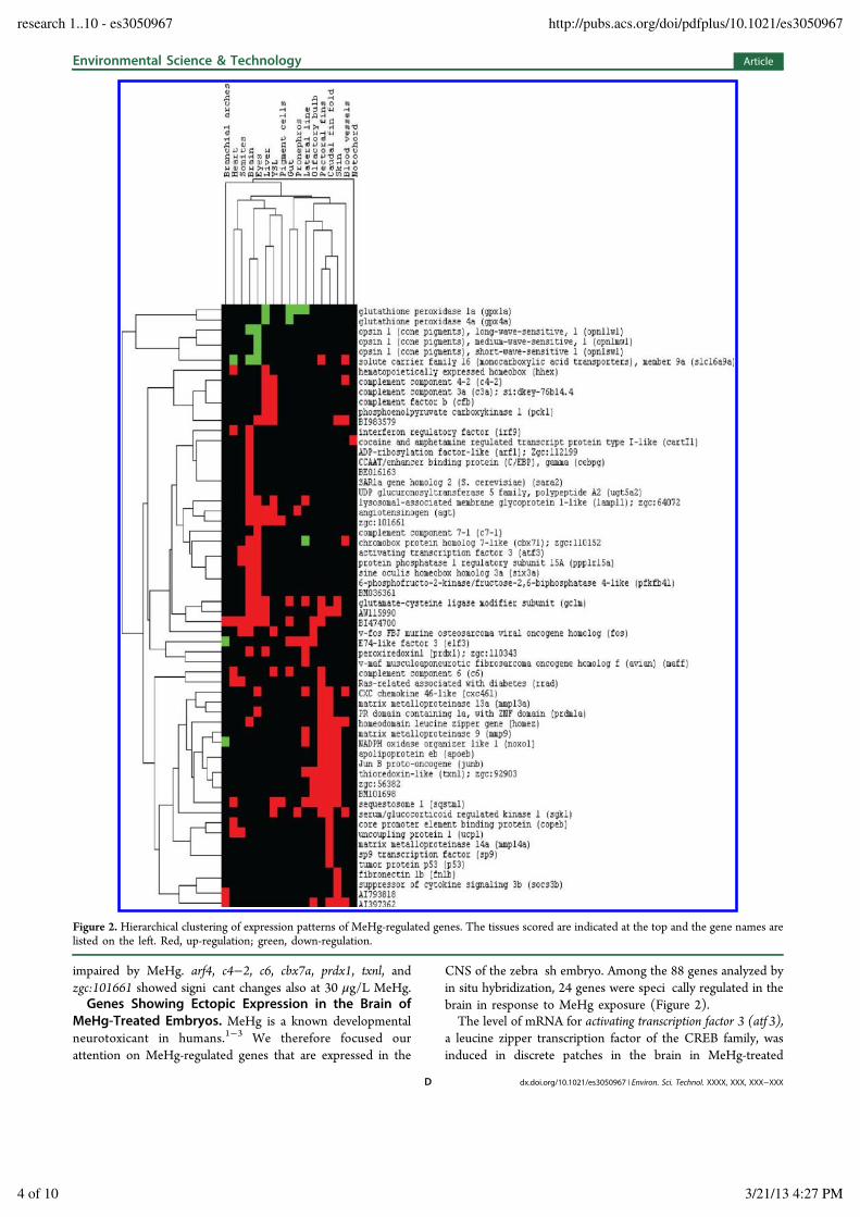
The results derived from the whole-mount in situ hybrid-izations were classi ed using the following 17 anatomical terms:
brain, eyes, olfactory bulb, branchial arches, heart, liver, gut,pronephros, somites, lateral line, pectoral ns, caudal n fold,blood vessels, skin, yolk syncytial layer (YSL), pigment cells,and notochord (Figure 2). Three major clusters of coexpressedgenes were discernible: complement factors b, 3a and 4!2 areexpressed in the YSL and the liver together with pck1 (Figure2). Other genes were coexpressed in the brain and the eyes. Athird prominent cluster of coexpressed genes was found in thecaudal ns and the skin. In 4 cases (elf 3, noxo1l, cbx7a, andslc16a9a), expression was up-regulated in some tissues and wasdown-regulated in others (Figure 2). For example, theexpression of noxo1l was increased in the lateral line, pectoralns, caudal n fold and skin, but decreased in the branchialarches (SI Figure 2A, A ). Taken together, our resultsdemonstrate that most of the MeHg-regulated genes wereexpressed in a tissue restricted manner (Figures 1 and 2, SIFigure 2). Di erent tissues responded with distinct changes ingene expression.To verify the observed changes in gene expression by an
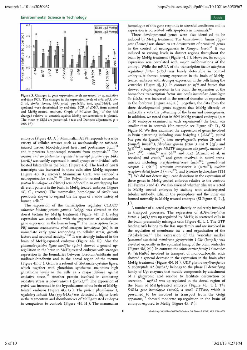
independent method, RT-qPCR was performed with selectedgenes including arf4, atf 3, c4!2, c6, cbx7a, homez, irf 9, prdx1,ppp1r15a, txnl, zgc:101661, and opn1mw1. Zebra sh embryoswere exposed to 30 or 60 !g/L MeHg from 4 to 72 hpf andcDNA samples were subjected to real-time PCR analysis withgene-speci c primers. The expression levels of all the genesexamined, except homez, were signi cantly regulated bytreatment with 60 !g/L MeHg (Figure 3). In situ hybridizationshowed that homez mRNA was up-regulated in embryos withsevere malformations (precardiac edema, reduced n folds,smaller brain, curved body), but not in embryos that weremorphologically not a ected by MeHg treatment. Thiscontributed to variation and thus reduced signi cance of theRT-qPCR results. opn1mw1 showed a repression with an M-value of less than !2 after exposure to 60 !g/L MeHg. Down-regulation of other opsin genes was also noted (Figure 2, SITable S4) suggesting that di erentiation of the retina is
Table 1. Ontology Groups of Genes Up-Regulated by MeHg
gene ontologynumber ofgenes
adjusted p-value
apoptosis 36 3.00 ! 10!4
prostaglandin metabolic process 4 5.50 ! 10!3
calcium independent cell!cell adhesion 5 9.00 ! 10!4
cell redox homeostasis 6 5.50 ! 10!3
response to oxidative stress 12 3.00 ! 10!4
acute in ammatory response 8 2.60 ! 10!3
blood circulation 12 2.00 ! 10!3
MAPKtyrosine/serine/threonin phosphataseactivity
4 4.00 ! 10!4
endopeptidase activty 19 1.00 ! 10!4
serine-type endopeptidase activity 11 5.00 ! 10!4
transcription factor activity 26 6.10 ! 10!3
aryldialkyphosphatase activity 3 2.00 ! 10!4
Table 2. Ontology Groups of Genes down-Regulated byMeHg
gene ontologynumber ofgenes
adjusted p-value
RNA elongation from RNA Pol II promoter 6 3.70 ! 10!3
fatty acid catabolic process 5 6.50 ! 10!3
double strand break repair 6 5.20 ! 10!3
transcription factor activity, nucleic acidbinding
8 1.20 ! 10!3
acyl-CoA binding 4 1.30 ! 10!3
3-hydroxyacyl-CoA dehydrogenase activty 3 1.30 ! 10!3
peptidyl-prolyl cis!trans isomerase activity 5 1.50 ! 10!3
Figure 1. Changes in gene expression patterns after MeHg exposure.Control (A, B, C, D) and MeHg-treated (A , B , C , D ) embryos (72hpf) were hybridized with RNA probes complementary toperoxiredoxin1 (prdx1, A, A ), phosphoenolpyruvate carboxykinase 1(pck1, B, B ), f ibronectin1b (fn1b, C, C ) and matrix metalloprotease13(mmp13, D, D ) mRNA. Abbreviations, c , caudal n fold; hy,hypothalamus; l, liver; ll, lateral line; PBI, posterior blood island; pf,pectoral n; y, yolk; yse, yolk sac extension. Scale bar, A, A ! D, D ,200 !m.
Environmental Science & Technology Article
dx.doi.org/10.1021/es3050967 | Environ. Sci. Technol. XXXX, XXX, XXX!XXXC
research 1..10 - es3050967 http://pubs.acs.org/doi/pdfplus/10.1021/es3050967
3 of 10 3/21/13 4:27 PM
impaired by MeHg. arf4, c4!2, c6, cbx7a, prdx1, txnl, andzgc:101661 showed signi cant changes also at 30 !g/L MeHg.Genes Showing Ectopic Expression in the Brain of
MeHg-Treated Embryos. MeHg is a known developmentalneurotoxicant in humans.1!3 We therefore focused ourattention on MeHg-regulated genes that are expressed in the
CNS of the zebra sh embryo. Among the 88 genes analyzed byin situ hybridization, 24 genes were speci cally regulated in thebrain in response to MeHg exposure (Figure 2).The level of mRNA for activating transcription factor 3 (atf 3),
a leucine zipper transcription factor of the CREB family, wasinduced in discrete patches in the brain in MeHg-treated
Figure 2. Hierarchical clustering of expression patterns of MeHg-regulated genes. The tissues scored are indicated at the top and the gene names arelisted on the left. Red, up-regulation; green, down-regulation.
Environmental Science & Technology Article
dx.doi.org/10.1021/es3050967 | Environ. Sci. Technol. XXXX, XXX, XXX!XXXD
research 1..10 - es3050967 http://pubs.acs.org/doi/pdfplus/10.1021/es3050967
4 of 10 3/21/13 4:27 PM
embryos (Figure 4A, A ). Mammalian ATF3 responds to a widevariety of cellular stresses such as mechanically or toxicant-injured tissues, blood-deprived heart and postseizure brain,44
and it protects hippocampal neurons from apoptosis.45 Thecocaine and amphetamine regulated transcript protein type I-like(cartIl) was weakly expressed in small groups or individual cellslocated bilaterally in the brain (Figure 4B). The level of cart1ltranscripts was increased in these cells after MeHg exposure(Figure 4B, B , arrows). Mammalian Cart1 was ascribed aneuroprotective role.46!48 The Polycomb related chromoboxprotein homologue 7a (cbx7a) was induced in an overlapping butdi erent pattern in the brain in MeHg-treated embryos (Figure4C, C , arrows). The mammalian homologue of cbx7a waspreviously shown to expand the life span of a wide variety ofhuman cells.49
The expression of the transcription regulator CCAAT/enhancer binding protein gamma (cebpg) was elevated in thedorsal tectum by MeHg treatment (Figure 4D, D ). cebpgexpression was correlated with the expression of antioxidantgene expression in the human lung.50 The transcription factorFBJ murine osteosarcoma viral oncogene homologue (fos) is animmediate early gene responding to cellular stress, growthfactors and neuronal activity.51,52 It was strongly induced in thebrain of MeHg-exposed embryos (Figure 4E, E ). Also theglutamate-cysteine ligase modif ier (gclm) showed a general up-regulation in the brain in MeHg-treated embryos with strongerexpression in the boundaries between forebrain/midbrain andmidbrain/hindbrain and in the dorsal region of the tectum(Figure 4F, F ). Gclm is a subunit of Glutamate-cysteine ligase,which together with glutathion synthetase maintains highglutathione levels in the cells as a major defense againstoxidative stress.53 Another protein involved in combatingoxidative stress is peroxiredoxin1 (prdx1).54 The expression ofprdx1 was increased in the hypothalamus of the brain of MeHg-treated embryos (Figure 4G, G ). The protein phosphatase 1,regulatory subunit 15a (ppp1r15a) was detected at higher levelsin the tegmentum and rhombomeres of MeHg-treated embryosin comparison to controls (Figure 4H, H ). The mammalian
homologue of this gene responds to stressful conditions and itsexpression is correlated with apoptosis in mammals.55
Three developmental genes were also identi ed to beinduced by MeHg treatment. The homeodomain leucine zippergene (homez) was shown to act downstream of proneural genesin the control of neurogenesis in Xenopus laevis.56 It wasinduced to varying levels in distinct regions throughout thebrain by MeHg treatment (Figure 4I, I ). However, its ectopicexpression was correlated with major malformations of theembryo. While the mRNA of the transcription factor interferonregulatory factor (irf 9) was barely detectable in controlembryos, it showed strong expression in the brain of MeHg-treated embryos with stronger expression in the cells lining theventricles (Figure 4J, J ). In contrast to irf 9 and homez thatshowed ectopic expression in the brain, the expression of thehomeobox transcription factor sine oculis homeobox homologue3a (six3a) was increased in the normal domains of expressionin the forebrain (Figure 4K, K ). Together, the data from thethree developmental genes suggests that MeHg directly orindirectly a ects the patterning of the brain and neurogenesis.In addition, we noted that in 60% MeHg-treated embryos (n =3, 30 embryos examined in each experiment) the head wassmaller than in controls (for example see Figure 4D, D , SIFigure 6). We thus examined the expression of genes involvedin brain patterning including sonic hedgehog a (shha57), pairedbox gene 6a (pax6a58), bone morphogenetic protein 2b and 4(bmp2b, bmp459), f ibroblast growth factor 3 and 8 ( fgf 3 andfgf 860,61), wingless-type MMTV integration site family, member 1(wnt 162), nestin,63 wnt 5b,64 and sox2 (Armant et al, inrevision) and crestin,65 and genes involved in neural trans-mission including acetylcholinesterase (ache66), cannabinoidreceptor 1 (cb167) serotonin transporter a (serta68), nuclearreceptor-related factor 1 (nurr169), and tyrosine hydroxylase (TH70). We did not detect signi cant deviations in the expression ofthese genes in MeHg-treated embryos relative to the controls(SI Figures 3 and 4). We also assessed whether cilia are a ectedin MeHg treated embryos by staining with antiacetylatedtubulin antibody. Cilia in the pronephros and the inner earformed normally in MeHg-treated embryos (SI Figure 4I, I , J,J ).A number of a ected genes are directly or indirectly involved
in transport processes. The expression of ADP-ribosylationfactor 4 (arf4) was up-regulated by MeHg in scattered cells inthe brain, presumably microglia cells (Figure 4L, L ). The GTPbinding Arfs belong to the Ras superfamily and are involved inthe regulation of membrane tra c and organization of thecytoskeleton.71 The expression of the vesicular markerlysosomal-associated membrane glycoprotein 1-like (lamp1l) waselevated especially in the epithelial lining of the brain ventricles(Figure 4M, M ). In contrast, the solute carrier family 16 member9a (slc16a9a) involved in transport of monocarboxylic acidsshowed a general decrease in the expression in the brain afterMeHg treatment (Figure 4N, N ). UDP glucuronosyltransferase5, polypeptide A2 (ugt5a2) belongs to the phase II detoxifyingfamily of Ugt enzymes that modify compounds by attachmentof a glucuronic acid residue to facilitate destruction orsecretion.72 ugt5a2 was up-regulated in the dorsal region ofthe brain of MeHg-treated embryos (Figure 4O, O ). TheSAR1a gene homologue (sara2), a small GTPase, which ispresumed to be involved in transport from the Golgiapparatus,73 showed moderate up-regulation in the brain ofembryos exposed to MeHg (Figure 4P, P ).
Figure 3. Changes in gene expression levels measured by quantitativereal-time PCR. The changes in the expression levels of arf4, atf 3, c4!2, c6, cbx7a, homez, irf 9, prdx1, ppp1r15a, txnl, zgc:101661, andopn1mwl were determined by real-time PCR of cDNA from controland MeHg-treated embryos. Graph of M-value (log2 of the foldchange) relative to controls against MeHg concentrations is plotted.The mean ± SEM are presented. t test and Dunnett adjustment, p <0.05 (*).
Environmental Science & Technology Article
dx.doi.org/10.1021/es3050967 | Environ. Sci. Technol. XXXX, XXX, XXX!XXXE
research 1..10 - es3050967 http://pubs.acs.org/doi/pdfplus/10.1021/es3050967
5 of 10 3/21/13 4:27 PM
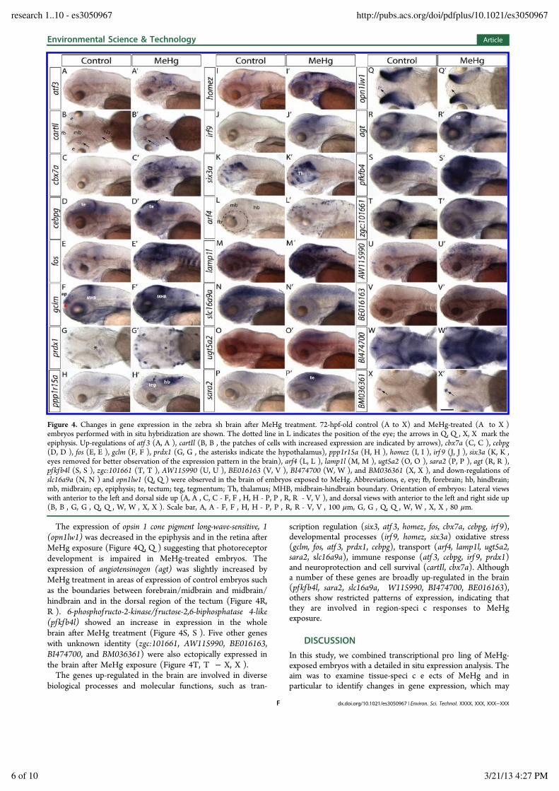
The expression of opsin 1 cone pigment long-wave-sensitive, 1(opn1lw1) was decreased in the epiphysis and in the retina afterMeHg exposure (Figure 4Q, Q ) suggesting that photoreceptordevelopment is impaired in MeHg-treated embryos. Theexpression of angiotensinogen (agt) was slightly increased byMeHg treatment in areas of expression of control embryos suchas the boundaries between forebrain/midbrain and midbrain/hindbrain and in the dorsal region of the tectum (Figure 4R,R ). 6-phosphof ructo-2-kinase/f ructose-2,6-biphosphatase 4-like(pfkfb4l) showed an increase in expression in the wholebrain after MeHg treatment (Figure 4S, S ). Five other geneswith unknown identity (zgc:101661, AW115990, BE016163,BI474700, and BM036361) were also ectopically expressed inthe brain after MeHg exposure (Figure 4T, T ! X, X ).The genes up-regulated in the brain are involved in diverse
biological processes and molecular functions, such as tran-
scription regulation (six3, atf 3, homez, fos, cbx7a, cebpg, irf 9),developmental processes (irf 9, homez, six3a) oxidative stress(gclm, fos, atf 3, prdx1, cebpg), transport (arf4, lamp1l, ugt5a2,sara2, slc16a9a), immune response (atf 3, cebpg, irf 9, prdx1)and neuroprotection and cell survival (cartIl, cbx7a). Althougha number of these genes are broadly up-regulated in the brain(pfkfb4l, sara2, slc16a9a, W115990, BI474700, BE016163),others show restricted patterns of expression, indicating thatthey are involved in region-speci c responses to MeHgexposure.
DISCUSSIONIn this study, we combined transcriptional pro ling of MeHg-exposed embryos with a detailed in situ expression analysis. Theaim was to examine tissue-speci c e ects of MeHg and inparticular to identify changes in gene expression, which may
Figure 4. Changes in gene expression in the zebra sh brain after MeHg treatment. 72-hpf-old control (A to X) and MeHg-treated (A to X )embryos performed with in situ hybridization are shown. The dotted line in L indicates the position of the eye; the arrows in Q, Q , X, X mark theepiphysis. Up-regulations of atf 3 (A, A ), cartIl (B, B , the patches of cells with increased expression are indicated by arrows), cbx7a (C, C ), cebpg(D, D ), fos (E, E ), gclm (F, F ), prdx1 (G, G , the asterisks indicate the hypothalamus), ppp1r15a (H, H ), homez (I, I ), irf 9 (J, J ), six3a (K, K ,eyes removed for better observation of the expression pattern in the brain), arf4 (L, L ), lamp1l (M, M ), ugt5a2 (O, O ), sara2 (P, P ), agt (R, R ),pfkfb4l (S, S ), zgc:101661 (T, T ), AW115990 (U, U ), BE016163 (V, V ), BI474700 (W, W ), and BM036361 (X, X ), and down-regulations ofslc16a9a (N, N ) and opn1lw1 (Q, Q ) were observed in the brain of embryos exposed to MeHg. Abbreviations, e, eye; fb, forebrain; hb, hindbrain;mb, midbrain; ep, epiphysis; te, tectum; teg, tegmentum; Th, thalamus; MHB, midbrain-hindbrain boundary. Orientation of embryos: Lateral viewswith anterior to the left and dorsal side up (A, A , C, C - F, F , H, H - P, P , R, R - V, V ), and dorsal views with anterior to the left and right side up(B, B , G, G , Q, Q , W, W , X, X ). Scale bar, A, A - F, F , H, H - P, P , R, R - V, V , 100 !m, G, G , Q, Q , W, W , X, X , 80 !m.
Environmental Science & Technology Article
dx.doi.org/10.1021/es3050967 | Environ. Sci. Technol. XXXX, XXX, XXX!XXXF
research 1..10 - es3050967 http://pubs.acs.org/doi/pdfplus/10.1021/es3050967
6 of 10 3/21/13 4:27 PM
relate to the neurotoxic e ect of MeHg. The identi ed MeHg-regulated genes are involved in various biological processes andmolecular functions, such as regulation of transcription,antioxidant defense, immune response, and transport. Alteredexpression of genes was noted in various organs including thebrain, eyes, olfactory bulb, branchial arches, heart, liver, gut,pronephros, somites, lateral lines, pectoral ns, caudal n fold,blood vessels, skin, YSL pigment cells, and notochord. Many ofthe examined genes show tissue restricted expression. Thisindicates (1) that MeHg a ects many di erent tissues in theembryo and (2) that tissues respond with speci c generesponses. There were even di erences in the response instructures such as the caudal n and the pectoral n that arecomposed of similar cell types and are equally thin and exposedto the MeHg in the water. The expression of some genes wasaltered in either the pectoral n (c6, gclm, rrad, slc16a9a,BI474700) or the caudal n (copeb, mmp14a, sp9, p53, ucp1,AI397362) in response to MeHg. However, we found alsosimilarities in the gene responses in some tissues. Prominentclustering of coexpression patterns was found in the liver andYSL as well as in the retina and brain. This suggests di erences,but also similarities, in the response to MeHg exposure indi erent tissues. In four cases, we found genes whoseexpression levels were decreased in some tissues and increasedin others. In these cases, the microarray results of wholeembryos scored the net change of expression. This underscoresagain that individual tissues can respond in a very speci cmanner to MeHg exposure.From the 88 genes analyzed by in situ hybridization, 24
genes showed speci c changes of expression in the brain ofzebra sh embryos and are thus prime candidates for genes,which respond to or mediate the neurotoxic e ect of MeHg.These genes cover a wide range of biological and molecularfunctions, including transcription regulation such as six3,74
atf 3,75,76 homez,77 fos,78,79 cbx7a,49,80 cebpg81 and irf 9,82
response to oxidative stress such as gclm,83 fos51,52 andprdx1,84 apoptosis such as agt,85 atf 353 and ppp1r15a,86
immune response such as atf 3,53 cebpg,87 irf 988 and prdx1,54
cytokine production such as cebpg89 and irf 990 and blood vesselregulation such as agt91,92 and gclm.93 Some of the genes(pf kf b4l, sara2, slc16a9a, AW115990, BI474700, andBE016163) are expressed ubiquitously in the brain, otherswere restricted in their pattern of expression. This latterobservation suggests regionally restricted responses in the brainto MeHg exposure. Given the known molecular interaction ofMeHg with cellular processes and molecules such as sulfhydrylgroups, the induction of oxidative stress and the disruption ofcalcium ion homeostasis,17!24 many cell types should bea ected by MeHg. It is thus surprising that quite a number ofthe genes are induced in a regionally restricted manner inMeHg-exposed embryos. Thus, MeHg seems to a ect cellsdi erently leading to cell-speci c responses. In addition toregional di erences in the gene responses to MeHg exposure,unequal accumulation of MeHg94,95 may contribute to thesedi erences.Comparison with E ects Reported on Adult Zebra-sh. MeHg has been demonstrated to be hepatotoxic in adult
zebra sh, rat and mummichog.96,32,97 The increase inexpression of antioxidant genes in the liver in MeHg-treatedzebra sh embryos, such as prx1, gclm, and uricase, indicated thatthe liver su ered oxidative stress, which may be cause orconsequence of hepatocellular damage.98,85 Since the liver is anorgan with a strong innate immunity,99,100 reactive oxygen
species may be also generated as an in ammatory response totissue damage. In this context, it may be of relevance that weobserved up-regulation of complement components c3a, c4!2,and cfb in the liver in response to MeHg exposure.In a study by Gonzalez and co-workers,33 the e ect of
chronic exposure to MeHg was determined after dietary intakeof MeHg for 7, 21, and 63 days. The changes in the expressionlevels of 13 genes known to be involved in antioxidativedefenses, apoptosis, metal chelation, active e ux of organiccompounds, mitochondrial metabolism were determined byreal-time PCR. Although there were changes in the expressionlevels of some of the genes examined in the muscle and liverafter MeHg exposure, no such changes were observed in thebrain. In another study performed by Richter,31 the acute e ectof MeHg exposure on the adult zebra sh brain was investigatedby intraperitoneal injection of 0.5 !g MeHg/g body weight intofemale zebra sh. After 96 h, the sh were sacri ced forexamination of gene expression levels using a 22K-probemicroarray. Seventy-nine genes were shown to be up-regulatedand 76 genes were down-regulated (p > 0.01) in response toMeHg. In contrast to the study performed by Gonzalez et al.,but consistent with our measurements, they detected the up-regulation of oxidative stress and apoptosis genes in the brainafter MeHg exposure.A study of the visual system in adult zebra sh showed that
MeHg could pass through the blood-retina barrier andaccumulate in various regions of the retina, especially in thephotoreceptor layer and in the inner and outer nuclear layer.34
These observations are consistent with our data in the embryothat showed down-regulation of opsin expression in thedeveloping retina. Taken together, our data derived fromzebra sh embryos re ect the MeHg toxicity observed in adultzebra sh.
How Does MeHg Act As a Neurotoxicant? It waspreviously suggested that exposure of embryos to 10, 50, and80 !g/L MeHg decreased cell proliferation in the neuraltube.101 However the authors of this previous study did notobserve a dose-dependence, raising concerns whether theobserved reduction in cell proliferation was indeed caused byMeHg. When exposing zebra sh embryos from 4 to 72 hpf to60 !g/L MeHg, we did not observe changes in the overallpattering of the brain when probing with a number ofdevelopmental regulators (shha, pax6a, crestin, nurr1) andgenes involved in neurotransmission (cb1, serta, TH). Theinduction of the developmental regulators irf 9, homez, andsix3a may thus be related to local repair processes rather thanto a disturbance of the overall pattering of the brain. Thissuggests that the overall patterning of the brain is notsigni cantly a ected. The e ects of MeHg on the developingnervous system may, however, be too subtle to be detected byoverall morphological analysis. Clearly, the heads of MeHg-exposed embryos were smaller. We found increased or ectopicexpression of a number of genes whose function is related tooxidative stress, transport and transcription regulation. It isintriguing that some of these genes (c-fos, cebpg, atf, socs3) areinduced by cAMP in mammalian embryos.102!106 In light of theview that the cAMP pathway and downstream genes like c-fosare involved in learning and memory in mammals,107,108 it istempting to speculate that MeHg-induced cellular stress maya ect the development and function of the nervous system byaberrantly inducing these regulators of brain function. Thisdisturbance may account for the mental de cits seen in humansexposed during the prenatal period to low doses of MeHg.
Environmental Science & Technology Article
dx.doi.org/10.1021/es3050967 | Environ. Sci. Technol. XXXX, XXX, XXX!XXXG
research 1..10 - es3050967 http://pubs.acs.org/doi/pdfplus/10.1021/es3050967
7 of 10 3/21/13 4:27 PM
ASSOCIATED CONTENT*S Supporting InformationAdditional information including tables and gures. Thismaterial is available free of charge via the Internet at http://pubs.acs.org.
AUTHOR INFORMATIONCorresponding Author*E-mail: [email protected] ContributionsThese authors contributed equally to this study.NotesThe authors declare no competing nancial interest.
ACKNOWLEDGMENTSWe thank M. Rastegar for help with the microscopes. We arethankful to N. Borel and her team for zebra sh maintenance.We are grateful to X. Cousin, M. Halpern, V. Laudet, A Fjose,K. Shirabe, and P. Ingham for cDNAs. We acknowledgesupport by EU IP ZF-Models (LSHG-CT-2003 503496),EUTRACC (LSHG-CT-2006-037445), EU IP ZF-HealthGrant (No 242048), BMBF GENDarT2 (AZ:0315190 B)und BMBF DanTox (02WU1053-01).
REFERENCES(1) Ekino, S.; Susa, M.; Ninomiya, T.; Imamura, K.; Kitamura, T.Minamata disease revisited: An update on the acute and chronicmanifestations of methyl mercury poisoning. J. Neurol. Sci. 2007, 262(1!2), 131!44.(2) Eto, K.; Marumoto, M.; Takeya, M. The pathology ofmethylmercury poisoning (Minamata disease). Neuropathology 2010,DOI: 10.1111/j.1440-1789.2010.01119.x.(3) Harada, M. Minamata disease: Methylmercury poisoning in Japancaused by environmental pollution. Crit. Rev. Toxicol. 1995, 25 (1), 1!24.(4) Sams, C. E. Methylmercury Contamination: Impacts on AquaticSystems and Terrestrial Species, and Insights for Abatement; M. Furniss.,C. C., Ronnenberg, K., Ed.; San Diego, CA, 2004.(5) Seigneur, C.; Vijayaraghavan, K.; Lohman, K.; Karamchandani, P.;Scott, C. Global source attribution for mercury deposition in theUnited States. Environ. Sci. Technol. 2004, 38 (2), 555!69.(6) USEPA. National Primary Drinking Water Regulations, Contam-inant Speci c Fact Sheets, Inorganic Chemicals, Technical Version; U.S.Environmental Protection Agency: Washington, DC, 1995.(7) NJDEPE. Final report on Municipal Solid Waste Incineration.; NewJersey Department of Environmental Protection, 1993.(8) DooleyJ. H.Natural Sources of Mercury in the Kirkwood-CohanseyAquifer System of the New Jersey Coastal Plain; New Jersey GeologicalSurvey, 1992(9) Clarkson, T. W. Mercury: Major issues in environmental health.Environ. Health Perspect. 1993, 100, 31!8.(10) Madenjian, C. P.; O'Connor, D. V. Trophic transfer efficiency ofmercury to lake whitefish Coregonus clupeaformis from its prey. Bull.Environ. Contam. Toxicol. 2008, 81 (6), 566!70.(11) USEPA. Water Quality Criterion for the Protection of HumanHealth: Methylmercury; U. S. Environmental Protection Agency, O ceof Science and Technology: Washington, DC, 2001.(12) Clarkson, T. W. The three modern faces of mercury. Environ.Health Perspect. 2002, 110 (Suppl 1), 11!23.(13) Ask, K.; Akesson, A.; Berglund, M.; Vahter, M. Inorganicmercury and methylmercury in placentas of Swedish women. Environ.Health Perspect. 2002, 110 (5), 523!6.(14) Cernichiari, E.; Toribara, T. Y.; Liang, L.; Marsh, D. O.; Berlin,M. W.; Myers, G. J.; Cox, C.; Shamlaye, C. F.; Choisy, O.; Davidson,P.; et al. The biological monitoring of mercury in the Seychelles study.Neurotoxicology 1995, 16 (4), 613!28.
(15) Grandjean, P.; Weihe, P.; White, R. F.; Debes, F.; Araki, S.;Yokoyama, K.; Murata, K.; Sorensen, N.; Dahl, R.; Jorgensen, P. J.Cognitive deficit in 7-year-old children with prenatal exposure tomethylmercury. Neurotoxicol. Teratol. 1997, 19 (6), 417!28.(16) Clarkson, T. W.; Magos, L. The toxicology of mercury and itschemical compounds. Crit. Rev. Toxicol. 2006, 36 (8), 609!62.(17) Ali, S. F.; LeBel, C. P.; Bondy, S. C. Reactive oxygen speciesformation as a biomarker of methylmercury and trimethyltinneurotoxicity. Neurotoxicology 1992, 13 (3), 637!48.(18) Atchison, W. D. Effects of toxic environmental contaminants onvoltage-gated calcium channel function: From past to present. J.Bioenergy Biomembr. 2003, 35 (6), 507!32.(19) Kidd, P. Glutathione: Systemic protectant against oxidative andfree radical damage. Alternate Med. Rev. 1997, 2, 155!176.(20) Quig, D. Cysteine metabolism and metal toxicity. Alternate Med.Rev. 1998, 3 (4), 262!70.(21) Sarafian, T. A. Methylmercury-induced generation of freeradicals: Biological implications. Met. Ions Biol. Syst. 1999, 36, 415!44.(22) Shafer, T. J.; Contreras, M. L.; Atchison, W. D. Characterizationof interactions of methylmercury with Ca2+ channels in synaptosomesand pheochromocytoma cells: Radiotracer flux and binding studies.Mol. Pharmacol. 1990, 38 (1), 102!13.(23) Shanker, G.; Aschner, J. L.; Syversen, T.; Aschner, M. Freeradical formation in cerebral cortical astrocytes in culture induced bymethylmercury. Brain Res. Mol. Brain Res. 2004, 128 (1), 48!57.(24) Vallee, B. L.; Ulmer, D. D. Biochemical effects of mercury,cadmium, and lead. Annu. Rev. Biochem. 1972, 41 (10), 91!128.(25) Samson, J. C.; Shenker, J. The teratogenic effects ofmethylmercury on early development of the zebrafish, Danio rerio.Aquat. Toxicol. 2000, 48 (2!3), 343!354.(26) Yang, L.; Kemadjou, J. R.; Zinsmeister, C.; Bauer, M.; Legradi,J.; Muller, F.; Pankratz, M.; Jakel, J.; Strahle, U. Transcriptionalprofiling reveals barcode-like toxicogenomic responses in the zebrafishembryo. Genome Biol 2007, 8 (10), R227.(27) Yang, L.; Ho, N. Y.; Muller, F.; Strahle, U. Methyl mercurysuppresses the formation of the tail primordium in developingzebrafish embryos. Toxicol. Sci. 2010, 115 (2), 379!90.(28) Samson, J. C.; Goodridge, R.; Olobatuyi, F.; Weis, J. S. Delayedeffects of embryonic exposure of zebrafish (Danio rerio) tomethylmercury (MeHg). Aquat.Toxicol. 2001, 51 (4), 369!76.(29) Cambier, S.; Gonzalez, P.; Durrieu, G.; Maury-Brachet, R.;Boudou, A.; Bourdineaud, J. P. Serial analysis of gene expression in theskeletal muscles of zebrafish fed with a methylmercury-contaminateddiet. Environ. Sci. Technol. 2010, 44 (1), 469!75.(30) Klaper, R.; Carter, B. J.; Richter, C. A.; Drevnick, P. E.;Sandheinrich, M. B.; Tillitt, D. E. use of a 15k gene microarray todetermine gene expression changes in response to acute and chronicmethylmercury exposure in the fathead minnow Pimephales promelaseraginesque. J. Fish Biol. 2008, 72, 2207!2280.(31) Richter, C. A.; Garcia-Reyero, N.; Martyniuk, C.; Knoebl, I.;Pope, M.; Wright-Osment, M. K.; Denslow, N. D.; Tillitt, D. E. Geneexpression changes in female zebrafish (Danio rerio) brain in responseto acute exposure to methylmercury. Environ. Toxicol. Chem. 2011, 30(2), 301!8.(32) Ung, C. Y.; Lam, S. H.; Hlaing, M. M.; Winata, C. L.; Korzh, S.;Mathavan, S.; Gong, Z. Mercury-induced hepatotoxicity in zebrafish:In vivo mechanistic insights from transcriptome analysis, phenotypeanchoring and targeted gene expression validation. BMC Genom. 2010,11, 212.(33) Gonzalez, P.; Dominique, Y.; Massabuau, J. C.; Boudou, A.;Bourdineaud, J. P. Comparative effects of dietary methylmercury ongene expression in liver, skeletal muscle, and brain of the zebrafish(Danio rerio). Environ. Sci. Technol. 2005, 39 (11), 3972!80.(34) Mela, M.; Cambier, S.; Mesmer-Dudons, N.; Legeay, A.;Grotzner, S. R.; de Oliveira Ribeiro, C. A.; Ventura, D. F.; Massabuau,J. C. Methylmercury localization in Danio rerio retina after trophic andsubchronic exposure: a basis for neurotoxicology. Neurotoxicology2010, 31 (5), 448!53.
Environmental Science & Technology Article
dx.doi.org/10.1021/es3050967 | Environ. Sci. Technol. XXXX, XXX, XXX!XXXH
research 1..10 - es3050967 http://pubs.acs.org/doi/pdfplus/10.1021/es3050967
8 of 10 3/21/13 4:27 PM
(35) Mikut, R. B., Braun, O.; S. Reischl, M., The open sourceMATLAB toolbox Gait-CAD and its application to bioelectric signalprocessing. In In Proc., DGBMT-Workshop Biosignalverarbeitung;Potsdam, 2008; pp pp109!111.(36) Simon, R. M. K., McShane, E. L.; Radmacher, L. M.; Wright, M.D.; G. W. Zhao, Y. Design and Analysis of DNA MicroarrayInvestigation; Springer, 2004.(37) Kupershmidt, I.; Su, Q. J.; Grewal, A.; Sundaresh, S.; Halperin,I.; Flynn, J.; Shekar, M.; Wang, H.; Park, J.; Cui, W.; Wall, G. D.;Wisotzkey, R.; Alag, S.; Akhtari, S.; Ronaghi, M., Ontology-basedmeta-analysis of global collections of high-throughput public data.PLoS One 2010, 5 (9).(38) Durinck, S.; Moreau, Y.; Kasprzyk, A.; Davis, S.; De Moor, B.;Brazma, A.; Huber, W. BioMart and bioconductor: A powerful linkbetween biological databases and microarray data analysis. Bio-informatics 2005, 21 (16), 3439!40.(39) Zhang, B.; Kirov, S.; Snoddy, J. WebGestalt: An integratedsystem for exploring gene sets in various biological contexts. NucleicAcids Res. 2005, 33 (Web Server issue), W741!8.(40) Duncan, D. T.; Prodduturi, N.; Zhang, B. WebGestalt2: Anupdated and expanded version of the web-based gene set analysistoolkit. BMC Bioinf. 2010, 11 (supple 4), P10.(41) Oxtoby, E.; Jowett, T. Cloning of the zebrafish krox-20 gene(krx-20) and its expression during hindbrain development. NucleicAcids Res. 1993, 21 (5), 1087!95.(42) Wester eld, M. The Zebra sh Book a Guide for the LaboratoryUse of Zebra sh (Danio rerio); University of Oregon: Eugene, 2000;Vol. 4.(43) de Hoon, M. J.; Imoto, S.; Nolan, J.; Miyano, S. Open sourceclustering software. Bioinformatics 2004, 20 (9), 1453!4.(44) Chen, B. P.; Wolfgang, C. D.; Hai, T. Analysis of ATF3, atranscription factor induced by physiological stresses and modulatedby gadd153/Chop10. Mol. Cell. Biol. 1996, 16 (3), 1157!68.(45) Zhang, S. J.; Buchthal, B.; Lau, D.; Hayer, S.; Dick, O.;Schwaninger, M.; Veltkamp, R.; Zou, M.; Weiss, U.; Bading, H. Asignaling cascade of nuclear calcium-CREB-ATF3 activated by synapticNMDA receptors defines a gene repression module that protectsagainst extrasynaptic NMDA receptor-induced neuronal cell death andischemic brain damage. J. Neurosci. 2011, 31 (13), 4978!90.(46) Jia, J.; Chen, X.; Zhu, W.; Luo, Y.; Hua, Z.; Xu, Y. CARTprotects brain from damage through ERK activation in ischemicstroke. Neuropeptides 2008, 42 (5!6), 653!61.(47) Mao, P.; Ardeshiri, A.; Jacks, R.; Yang, S.; Hurn, P. D.; Alkayed,N. J. Mitochondrial mechanism of neuroprotection by CART. Eur. J.Neurosci. 2007, 26 (3), 624!32.(48) Xu, Y.; Zhang, W.; Klaus, J.; Young, J.; Koerner, I.; Sheldahl, L.C.; Hurn, P. D.; Martinez-Murillo, F.; Alkayed, N. J. Role of cocaine-and amphetamine-regulated transcript in estradiol-mediated neuro-protection. Proc. Natl. Acad. Sci. U. S. A. 2006, 103 (39), 14489!94.(49) Gil, J.; Bernard, D.; Martinez, D.; Beach, D. Polycomb CBX7has a unifying role in cellular lifespan. Nat. Cell Biol. 2004, 6 (1), 67!72.(50) Mullins, D. N.; Crawford, E. L.; Khuder, S. A.; Hernandez, D.A.; Yoon, Y.; Willey, J. C. CEBPG transcription factor correlates withantioxidant and DNA repair genes in normal bronchial epithelial cellsbut not in individuals with bronchogenic carcinoma. BMC Cancer2005, 5, 141.(51) Rao, G. N.; Glasgow, W. C.; Eling, T. E.; Runge, M. S. Role ofhydroperoxyeicosatetraenoic acids in oxidative stress-induced activat-ing protein 1 (AP-1) activity. J. Biol. Chem. 1996, 271 (44), 27760!4.(52) Tormos, C.; Javier Chaves, F.; Garcia, M. J.; Garrido, F.; Jover,R.; O'Connor, J. E.; Iradi, A.; Oltra, A.; Oliva, M. R.; Saez, G. T. Roleof glutathione in the induction of apoptosis and c-fos and c-junmRNAs by oxidative stress in tumor cells. Cancer Lett. 2004, 208 (1),103!13.(53) Thompson, M. R.; Xu, D.; Williams, B. R. ATF3 transcriptionfactor and its emerging roles in immunity and cancer. J. Mol. Med.(Berlin) 2009, 87 (11), 1053!60.
(54) Geiben-Lynn, R.; Kursar, M.; Brown, N. V.; Addo, M. M.; Shau,H.; Lieberman, J.; Luster, A. D.; Walker, B. D. HIV-1 antiviral activityof recombinant natural killer cell enhancing factors, NKEF-A andNKEF-B, members of the peroxiredoxin family. J. Biol. Chem. 2003,278 (3), 1569!74.(55) Scott, D. W.; Mutamba, S.; Hopkins, R. G.; Loo, G. IncreasedGADD gene expression in human colon epithelial cells exposed todeoxycholate. J. Cell Physiol. 2005, 202 (1), 295!303.(56) Ghimouz, R.; Bar, I.; Hanotel, J.; Minela, B.; Keruzore, M.;Thelie, A.; Bellefroid, E. J. The homeobox leucine zipper gene Homezplays a role in Xenopus laevis neurogenesis. Biochem. Biophys. Res.Commun. 2011, 415 (1), 11!6.(57) Krauss, S.; Concordet, J. P.; Ingham, P. W. A functionallyconserved homolog of the Drosophila segment polarity gene hh isexpressed in tissues with polarizing activity in zebrafish embryos. Cell1993, 75 (7), 1431!44.(58) Krauss, S.; Johansen, T.; Korzh, V.; Fjose, A. Expression patternof zebrafish pax genes suggests a role in early brain regionalization.Nature 1991, 353 (6341), 267!70.(59) Nikaido, M.; Tada, M.; Saji, T.; Ueno, N. Conservation of BMPsignaling in zebrafish mesoderm patterning. Mech. Dev. 1997, 61 (1!2), 75!88.(60) Kiefer, P.; Strahle, U.; Dickson, C. The zebrafish Fgf-3 gene:cDNA sequence, transcript structure and genomic organization. Gene1996, 168 (2), 211!5.(61) Maroon, H.; Walshe, J.; Mahmood, R.; Kiefer, P.; Dickson, C.;Mason, I. Fgf3 and Fgf8 are required together for formation of the oticplacode and vesicle. Development 2002, 129 (9), 2099!108.(62) Kelly, G. M.; Moon, R. T. Involvement of wnt1 and pax2 in theformation of the midbrain-hindbrain boundary in the zebrafishgastrula. Dev. Genet. 1995, 17 (2), 129!40.(63) Lam, C. S.; Marz, M.; Strahle, U. gfap and nestin reporter linesreveal characteristics of neural progenitors in the adult zebrafish brain.Dev. Dyn. 2009, 238 (2), 475!86.(64) Blader, P.; Strahle, U.; Ingham, P. W. Three Wnt genesexpressed in a wide variety of tissues during development of thezebrafish, Danio rerio: Developmental and evolutionary perspectives.Dev. Genes Evol. 1996, 206 (1), 3!13.(65) Rubinstein, A. L.; Lee, D.; Luo, R.; Henion, P. D.; Halpern, M.E. Genes dependent on zebrafish cyclops function identified by AFLPdifferential gene expression screen. Genesis 2000, 26 (1), 86!97.(66) Bertrand, C.; Chatonnet, A.; Takke, C.; Yan, Y. L.; Postlethwait,J.; Toutant, J. P.; Cousin, X. Zebrafish acetylcholinesterase is encodedby a single gene localized on linkage group 7. Gene structure andpolymorphism; molecular forms and expression pattern duringdevelopment. J. Biol. Chem. 2001, 276 (1), 464!74.(67) Lam, C. S.; Rastegar, S.; Strahle, U. Distribution of cannabinoidreceptor 1 in the CNS of zebrafish. Neuroscience 2006, 138 (1), 83!95.(68) Wang, Y.; Takai, R.; Yoshioka, H.; Shirabe, K. Characterizationand expression of serotonin transporter genes in zebrafish. Tohoku J.Exp. Med. 2006, 208 (3), 267!74.(69) Escriva, H.; Safi, R.; Hanni, C.; Langlois, M. C.; Saumitou-Laprade, P.; Stehelin, D.; Capron, A.; Pierce, R.; Laudet, V. Ligandbinding was acquired during evolution of nuclear receptors. Proc. Natl.Acad. Sci. U. S. A. 1997, 94 (13), 6803!8.(70) Wullimann, M. F.; Rink, E. Detailed immunohistology of Pax6protein and tyrosine hydroxylase in the early zebrafish brain suggestsrole of Pax6 gene in development of dopaminergic diencephalicneurons. Brain Res. Dev. Brain Res. 2001, 131 (1!2), 173!91.(71) Donaldson, J. G.; Honda, A. Localization and function of Arffamily GTPases. Biochem. Soc. Trans. 2005, 33 (Pt 4), 639!42.(72) King, C. D.; Rios, G. R.; Green, M. D.; Tephly, T. R. UDP-glucuronosyltransferases. Curr. Drug Metab. 2000, 1 (2), 143!61.(73) Jones, B.; Jones, E. L.; Bonney, S. A.; Patel, H. N.; Mensenkamp,A. R.; Eichenbaum-Voline, S.; Rudling, M.; Myrdal, U.; Annesi, G.;Naik, S.; Meadows, N.; Quattrone, A.; Islam, S. A.; Naoumova, R. P.;Angelin, B.; Infante, R.; Levy, E.; Roy, C. C.; Freemont, P. S.; Scott, J.;Shoulders, C. C. Mutations in a Sar1 GTPase of COPII vesicles are
Environmental Science & Technology Article
dx.doi.org/10.1021/es3050967 | Environ. Sci. Technol. XXXX, XXX, XXX!XXXI
research 1..10 - es3050967 http://pubs.acs.org/doi/pdfplus/10.1021/es3050967
9 of 10 3/21/13 4:27 PM
associated with lipid absorption disorders. Nat. Genet. 2003, 34 (1),29!31.(74) Kobayashi, M.; Toyama, R.; Takeda, H.; Dawid, I. B.;Kawakami, K. Overexpression of the forebrain-specific homeoboxgene six3 induces rostral forebrain enlargement in zebrafish.Development 1998, 125 (15), 2973!82.(75) Hsu, J. C.; Laz, T.; Mohn, K. L.; Taub, R. Identification of LRF-1, a leucine-zipper protein that is rapidly and highly induced inregenerating liver. Proc. Natl. Acad. Sci. U. S. A. 1991, 88 (9), 3511!5.(76) Chu, H. M.; Tan, Y.; Kobierski, L. A.; Balsam, L. B.; Comb, M. J.Activating transcription factor-3 stimulates 3 ,5 -cyclic adenosinemonophosphate-dependent gene expression. Mol. Endocrinol. 1994, 8(1), 59!68.(77) Bayarsaihan, D.; Enkhmandakh, B.; Makeyev, A.; Greally, J. M.;Leckman, J. F.; Ruddle, F. H. Homez, a homeobox leucine zipper genespecific to the vertebrate lineage. Proc. Natl. Acad .Sci. U. S. A 2003,100 (18), 10358!63.(78) Curran, T.; Morgan, J. I. Fos: An immediate-early transcriptionfactor in neurons. J. Neurobiol. 1995, 26 (3), 403!12.(79) Kovacs, K. J. c-Fos as a transcription factor: A stressful (re)viewfrom a functional map. Neurochem. Int. 1998, 33 (4), 287!97.(80) Li, Q.; Wang, X.; Lu, Z.; Zhang, B.; Guan, Z.; Liu, Z.; Zhong,Q.; Gu, L.; Zhou, J.; Zhu, B.; Ji, J.; Deng, D. Polycomb CBX7 directlycontrols trimethylation of histone H3 at lysine 9 at the p16 locus. PLoSOne 2010, 5 (10), e13732.(81) Davydov, I. V.; Bohmann, D.; Krammer, P. H.; Li-Weber, M.Cloning of the cDNA encoding human C/EBP gamma, a proteinbinding to the PRE-I enhancer element of the human interleukin-4promoter. Gene 1995, 161 (2), 271!5.(82) Veals, S. A.; Santa Maria, T.; Levy, D. E. Two domains of ISGF3gamma that mediate protein-DNA and protein-protein interactionsduring transcription factor assembly contribute to DNA-bindingspecificity. Mol. Cell. Biol. 1993, 13 (1), 196!206.(83) Lu, S. C. Regulation of glutathione synthesis. Mol. Aspects Med.2009, 30 (1!2), 42!59.(84) Immenschuh, S.; Baumgart-Vogt, E. Peroxiredoxins, oxidativestress, and cell proliferation. Antioxid. Redox Signaling 2005, 7 (5!6),768!77.(85) Liu, Y.; Wang, J.; Wei, Y.; Zhang, H.; Xu, M.; Dai, J. Inductionof time-dependent oxidative stress and related transcriptional effects ofperfluorododecanoic acid in zebrafish liver. Aquat. Toxicol. 2008, 89(4), 242!50.(86) Hollander, M. C.; Poola-Kella, S.; Fornace, A. J., Jr. Gadd34functional domains involved in growth suppression and apoptosis.Oncogene 2003, 22 (25), 3827!32.(87) Hutton, J. J.; Jegga, A. G.; Kong, S.; Gupta, A.; Ebert, C.;Williams, S.; Katz, J. D.; Aronow, B. J. Microarray and comparativegenomics-based identification of genes and gene regulatory regions ofthe mouse immune system. BMC Genom. 2004, 5, 82.(88) Taniguchi, T.; Ogasawara, K.; Takaoka, A.; Tanaka, N. IRFfamily of transcription factors as regulators of host defense. Annu. Rev.Immunol. 2001, 19, 623!55.(89) Gao, H.; Parkin, S.; Johnson, P. F.; Schwartz, R. C. C/EBPgamma has a stimulatory role on the IL-6 and IL-8 promoters. J. Biol.Chem. 2002, 277 (41), 38827!37.(90) Nguyen, H.; Hiscott, J.; Pitha, P. M. The growing family ofinterferon regulatory factors. Cytokine Growth Factor Rev. 1997, 8 (4),293!312.(91) Brand, M.; Lamande, N.; Sigmund, C. D.; Larger, E.; Corvol, P.;Gasc, J. M. Angiotensinogen modulates renal vasculature growth.Hypertension 2006, 47 (6), 1067!74.(92) Nagata, M.; Tanimoto, K.; Fukamizu, A.; Kon, Y.; Sugiyama, F.;Yagami, K.; Murakami, K.; Watanabe, T. Nephrogenesis andrenovascular development in angiotensinogen-deficient mice. Lab.Invest. 1996, 75 (5), 745!53.(93) Nakamura, S.; Sugiyama, S.; Fujioka, D.; Kawabata, K.; Ogawa,H.; Kugiyama, K. Polymorphism in glutamate-cysteine ligase modifiersubunit gene is associated with impairment of nitric oxide-mediatedcoronary vasomotor function. Circulation 2003, 108 (12), 1425!7.
(94) Korbas, M.; Blechinger, S. R.; Krone, P. H.; Pickering, I. J.;George, G. N. Localizing organomercury uptake and accumulation inzebrafish larvae at the tissue and cellular level. Proc. Natl. Acad. Sci. U.S. A. 2008, 105 (34), 12108!12.(95) Korbas, M.; Krone, P. H.; Pickering, I. J.; George, G. N.Dynamic accumulation and redistribution of methylmercury in thelens of developing zebrafish embryos and larvae. J. Biol. Inorg. Chem.2010, 15 (7), 1137!45.(96) Lin, T. H.; Huang, Y. L.; Huang, S. F. Lipid peroxidation in liverof rats administrated with methylmercuric chloride. Biol. Trace Elem.Res. 1996, 54, 33!41.(97) Weis, P.; Bogden, J. D.; Enslee, E. C. Hg-and Cu-inducedhepatocellular changes in the mummichog, Fundulus heteroclitus.Environ. Health Perspect. 1986, 65, 167!173.(98) Jaeschke, H.; Gores, G. J.; Cederbaum, A. I.; Hinson, J. A.;Pessayre, D.; Lemasters, J. J. Mechanisms of hepatotoxicity. Toxicol.Sci. 2002, 65 (2), 166!76.(99) Gao, B.; Jeong, W. I.; Tian, Z. Liver: An organ withpredominant innate immunity. Hepatology 2008, 47 (2), 729!36.(100) Racanelli, V.; Rehermann, B. The liver as an immunologicalorgan. Hepatology 2006, 43 (2 Suppl 1), S54!62.(101) Hassan, S. A.; Moussa, E. A.; Abbott, L. C. The effect ofmethylmercury exposure on early central nervous system developmentin the zebrafish (Danio rerio) embryo. J Appl Toxicol 2011, 32, 707!713.(102) Hart, I. R.; Rao, J.; Wilson, R. E. c-AMP-induced c-fosexpression in cells of melanocyte origin. Biochem. Biophys. Res.Commun. 1989, 159 (2), 408!13.(103) Mehmet, H.; Sinnett-Smith, J.; Moore, J. P.; Evan, G. I.;Rozengurt, E. Differential induction of c-fos and c-myc by cyclic AMPin Swiss 3T3 cells: Significance for the mitogenic response. OncogeneRes. 1988, 3 (3), 281!6.(104) Milne, G. R.; Palmer, T. M.; Yarwood, S. J. Novel control ofcAMP-regulated transcription in vascular endothelial cells. Biochem.Soc. Trans. 2012, 40 (1), 1!5.(105) Sands, W. A.; Woolson, H. D.; Milne, G. R.; Rutherford, C.;Palmer, T. M. Exchange protein activated by cyclic AMP (Epac)-mediated induction of suppressor of cytokine signaling 3 (SOCS-3) invascular endothelial cells. Mol. Cell. Biol. 2006, 26 (17), 6333!46.(106) Yarwood, S. J.; Borland, G.; Sands, W. A.; Palmer, T. M.Identification of CCAAT/enhancer-binding proteins as exchangeprotein activated by cAMP-activated transcription factors that mediatethe induction of the SOCS-3 gene. J. Biol. Chem. 2008, 283 (11),6843!53.(107) Countryman, R. A.; Kaban, N. L.; Colombo, P. J. Hippocampalc-fos is necessary for long-term memory of a socially transmitted foodpreference. Neurobiol. Learn Mem. 2005, 84 (3), 175!83.(108) He, J.; Yamada, K.; Nabeshima, T. A role of Fos expression inthe CA3 region of the hippocampus in spatial memory formation inrats. Neuropsychopharmacology 2002, 26 (2), 259!68.
Environmental Science & Technology Article
dx.doi.org/10.1021/es3050967 | Environ. Sci. Technol. XXXX, XXX, XXX!XXXJ
research 1..10 - es3050967 http://pubs.acs.org/doi/pdfplus/10.1021/es3050967
10 of 10 3/21/13 4:27 PM